
APPROACH TO OBSTRUCTIVE JAUNDICE
Yeo Quan You

OUTLINE
¢ Definition of Jaundice ¢ Anatomy ¢ Causes ¢ Investigations ¢ Management ¢ Q & A

DEFITION OF JAUNDICE ¢ Jaundice is a yellowish pigmentation of the skin,
the conjunctival membranes over the sclerae and other mucous membranes caused by hyperbilirubinemia and subsequently increased levels of bilirubin in extracellular fluids.
¢ Concentration higher than approx. 3 mg/dL (>50µmol/L) leads to jaundice
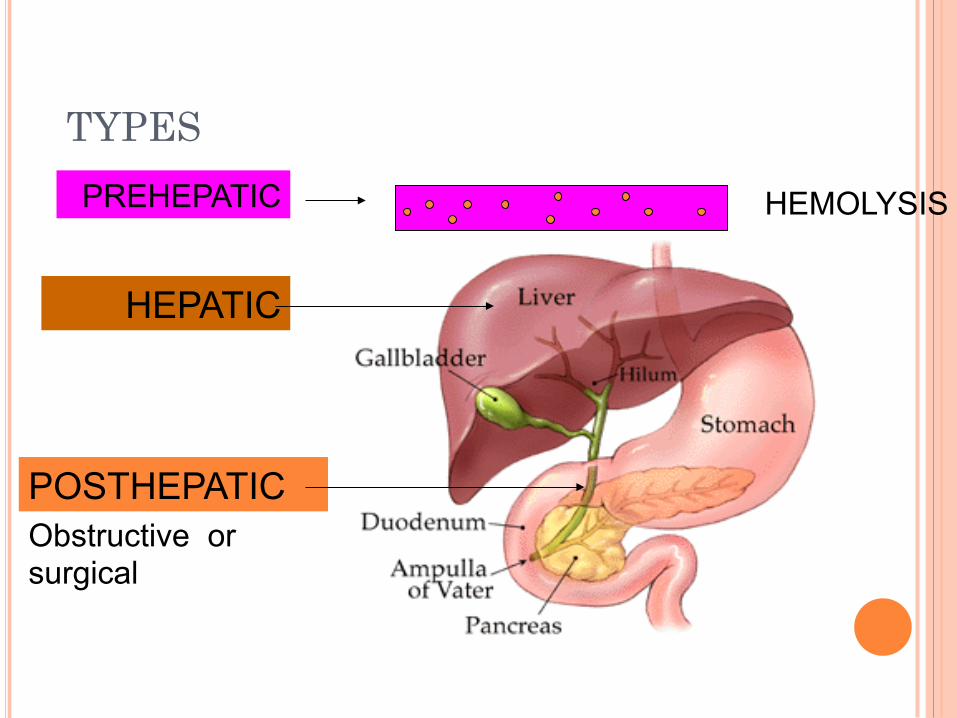
TYPES
PREHEPATIC
HEPATIC
POSTHEPATIC
HEMOLYSIS
Obstructive or surgical

ANATOMY

ANATOMY

BILIRUBIN CYCLE

¢ Heme oxygenase oxidises heme to biliverdin. ¢ Biliverdin is reduced by biliverdin reductase to form
bilirubin. ¢ Bilirubin is conjugated with glucuronic acid to form
bilirubin diglucuronide, or conjugated (direct-reacting) bilirubin. This reaction, catalyzed by the microsomal enzyme glucuronyl transferase, renders the bilirubin water-soluble.
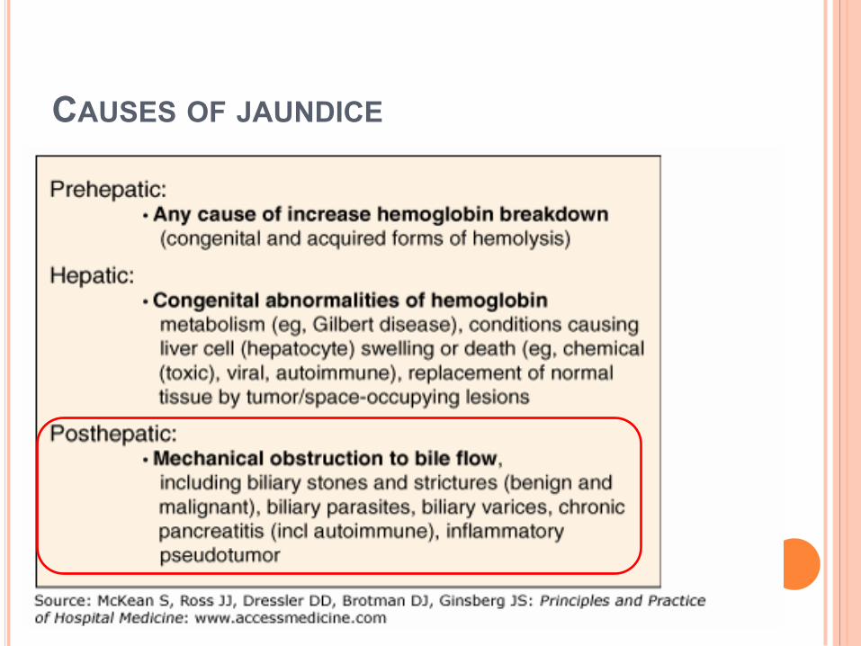
CAUSES OF JAUNDICE

CAUSES IN THE LUNEN
STONE IS THE COMMONEST
HYDATID
ASCARIS
CLONORCHIASIS PARASITES

IN THE WALL:
BENIGN STRICTURES
Post instrumentation
Primary sclerosing cholangitis
Choledochal cyst
MALIGNANCY
Cholangiocarcinoma

OUTSIDE THE WALL
L.N.
Head of the pancrease
Mirizzi syndrome
Any mass outside

EVALUATION
¢ History ¢ Examination ¢ Investigations
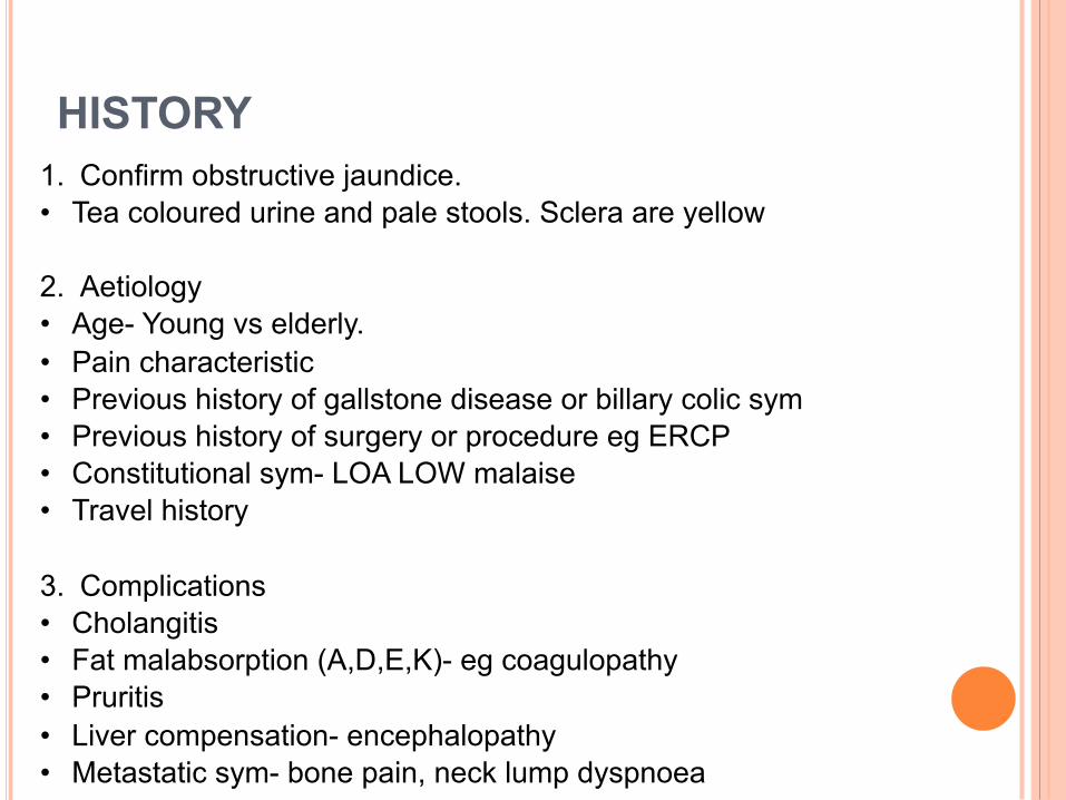
HISTORY 1. Confirm obstructive jaundice. • Tea coloured urine and pale stools. Sclera are yellow
2. Aetiology • Age- Young vs elderly. • Pain characteristic • Previous history of gallstone disease or billary colic sym • Previous history of surgery or procedure eg ERCP • Constitutional sym- LOA LOW malaise • Travel history
3. Complications • Cholangitis • Fat malabsorption (A,D,E,K)- eg coagulopathy • Pruritis • Liver compensation- encephalopathy • Metastatic sym- bone pain, neck lump dyspnoea

CLINICAL EXAMINATION
§ Jaundice
§ Pallor- hemolysis, cancer , cirrhosis
§ Gross weight loss-malignancy
§ Stigmata of chronic liver disease
§ Supraclavicular lymph nodes
§ DRE- pale stools

ABDOMINAL EXAMINATION
§ Abdominal scar
§ Ascitis-Cirrhosis or malignant disease
§ Hepatomegaly
§ Enlarged gall bladder
§ Courvoisier’s Law-palpable non tender gall bladder in a jaundice
patient>malignant biliary obstruction
§ Exceptions
§ Double impaction of stones
§ Impaction of pancreatic calculus at ampulla of vater
§ Mirizzi syndrome
§ Oriental cholangiohepatitis

LABAROTORY STUDIES
¢ FBC: anemia, infection,Hgbpathy ¢ Serum U/E/Cr ¢ LFT ¢ Coagulation profile ¢ Tumor markers: CA 19-9 ¢ Urinalysis : bilirubin, urobilinogen ¢ Stool for ocult blood: ca ampula ¢ Stool microscopy for ova and parasites

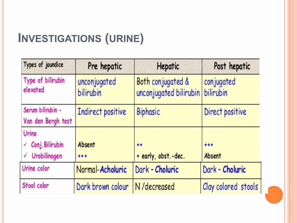
INVESTIGATIONS (URINE)

INVESTIGATION (IMAGING & PROCEDURE) ¢ U/S HBS
� Stones, CBD diameter � More sensitive than CT to pick up stones and gallbladder
pathology ¢ CT abdomen ¢ ERCP/MRCP ¢ EUS ¢ Percutaneous transhepatic cholangiogram

CT scan
¢ Main role in malignant conditions mainly for localization of primary tumors and mets
¢ Best for Pancreatic Carcinoma (Highly sensitive for lesion >1mm)
¢ Identify level and cause of obstruction

MAGNETIC RESONANCE CHOLANGIOPANCREATOGRAPHY (MRCP)
¢ Noninvasive test to visualize the hepatobiliary tree
¢ Entire biliary tree and pancreatic duct can be seen
¢ Best for intra hepatic stones and choledochal cyst
¢ MRCP is better to determine the extent and type of tumor as compared to ERCP

Endoscopic retrograde cholangiogram (ERCP)
¢ Invasive procedure ¢ Direct visualization of biliary tree/
pancreatic duct ¢ ERCP preferred as both therapeutic and
diagnostic ¢ Allows biopsy or brush cytology ¢ Stone extraction, stenting, sphincterotomy

ENDOSCOPIC ULTRASOUND (EUS)
o EUS has been reported to have up to a 98% diagnostic accuracy in patients with obstructive jaundice
o Allows diagnostic tissue sampling via EUS guided fine-needle aspiration (EUS-FNA)
o The sensitivity of EUS for the identification of focal mass lesions in pancreas has been reported to be superior to that of CT scanning
o Compared to MRCP for the diagnosis of biliary stricture, EUS has been reported to be more specific (100% vs 76%)

Percutaneous Transhepatic Cholangiogram (PTC)
¢ Maybe useful for obstruction proximal to CHD
¢ PTC is indicated when percutaneous intervention is needed and ERCP either is inappropriate or has failed.
¢ Can be used to drain biliary obstructions.

Management
Perioperative management of obstructive jaundice ¢ Preoperative biliary decompression improves postoperative morbidity
� usually cause increased hemorrhage & infections � Indicated in severe jaundice or when there are signs of severe
sepsis
¢ Bladder catheterization to monitor output ¢ Broad spectrum antibiotic prophylaxis if cholangitis ¢ Parenteral vitamin K +/- fresh frozen plasma if coagulopathy ¢ Need careful post operative fluid balance to correct dehydration ¢ Antihistamine for symptomatic relief of pruritus

Treatment of Obstructive Jaundice is based on the cause
1) Choledocholithiasis (gallstones) a)Treatment of choice is stone extraction through ERCP b) Mechanical lithotripsy – through modified dormia basket c) Open exploration of common bile duct is indicated in Ø Presence of multiple stones or > 25mm Ø Intra hepatic stones Ø Failure of ERCP Ø Recurrence of CBD stones

2) Ca Head of Pancreas / Periampullary Carcinoma/malignancy of lower 3rd of CBD
a) Whipple resection (pancreaticoduodenectomy) is mainly done which involves removal of head & neck of pancreas, duodenum, distal 40% of stomach, lower CBD, GB, upper 10 cm of jejunum, regional L.Ns and reconstruction through gastrojejunostomy,choledochojejunostmy and pancreaticojejunostomy
b) If not operable then we go for Endoscopic sphincterotomy + stenting or
Percutaneous transhepatic biliary drainage

3) Cholangiocarcinoma
Surgery depends on the stage of tumor and may involve ¢ Removal of the bile ducts
If the tumor is at a very early stage (Stage 1), just the bile ducts containing the cancer are removed. The remaining ducts in the liver are then joined to the small bowel, allowing the bile to flow again.
¢ Whipple procedure If the tumor is larger and has spread into nearby structures, the bile ducts, part of the stomach, part of the small bowel (duodenum), the pancreas, gall bladder and the surrounding lymph nodes are all removed
¢ If surgery to remove the tumour is not possible, it may be possible to relieve the blockage through stents through ERCP or PTC

4) STRICTURES
- usually treated by endoscopic stenting which is comparable to that of surgery, with similar recurrence rates.
- Therefore, surgery should be reserved for those patients with complete ductal obstruction or for those in whom endoscopic therapy has failed.

Questions 1
In obstructive jaundice, urinary examination shows: A) No urobilinogen, no bilirubin B) Increased urobilinogen, increased bilirubin C) Increased urobilinogen, no bilirubin D) No urobilinogen, increased bilirubin

ANSWER 1 (D)

QUESTIONS 2

ANSWER 2

CHOLANGICARCINOMA

QUESTION 3

ANSWER 3 A) ERCP

QUESTION 4

ANSWER 4


