Download - Antigen antibody interaction/Reaction

Antigen- Antibody Reaction
Prabin Shah BScMLT, MSc(Biochemistry)

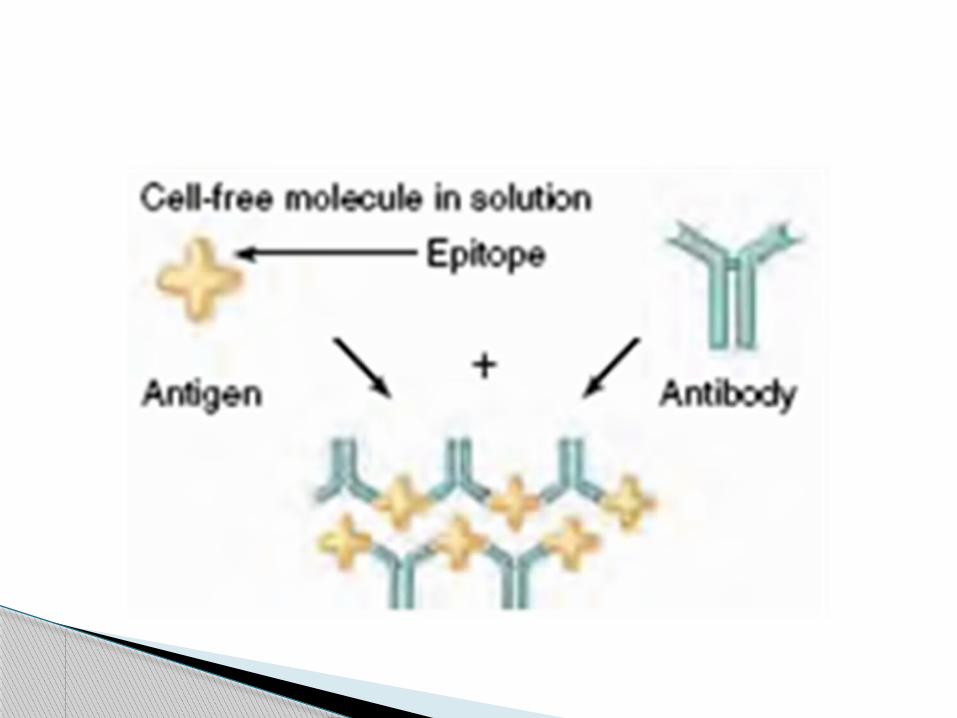
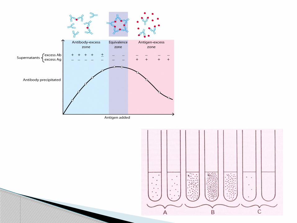
When a soluble Ag combine with its Abs in the presence of electrolytes at a suitable temperature, pH, the Ag- Ab complex forms an insoluble precipitate.
Soluble Ags interact with Abs to form lattice.
Occurs best when Ag and Ab are present in optimal proportions.
Precipitation Tests
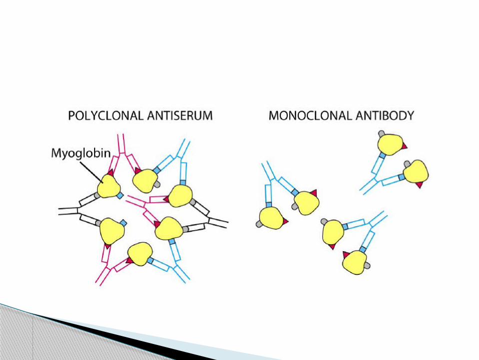
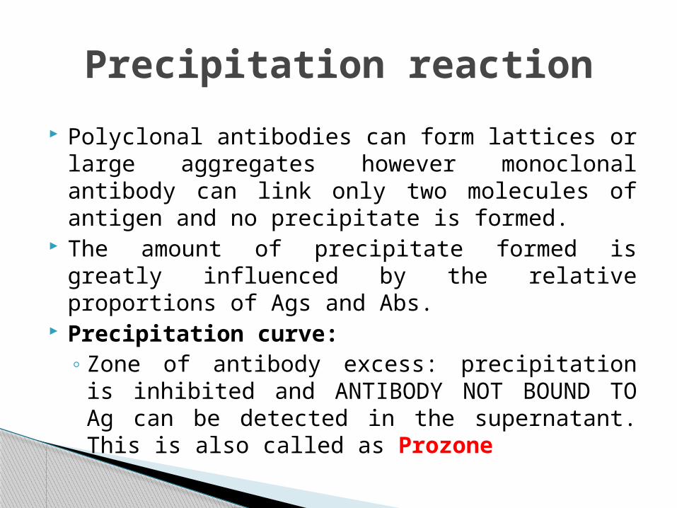
Polyclonal antibodies can form lattices or large aggregates however monoclonal antibody can link only two molecules of antigen and no precipitate is formed.
The amount of precipitate formed is greatly influenced by the relative proportions of Ags and Abs.
Precipitation curve:◦ Zone of antibody excess: precipitation is inhibited and
ANTIBODY NOT BOUND TO Ag can be detected in the supernatant. This is also called as Prozone
Precipitation reaction
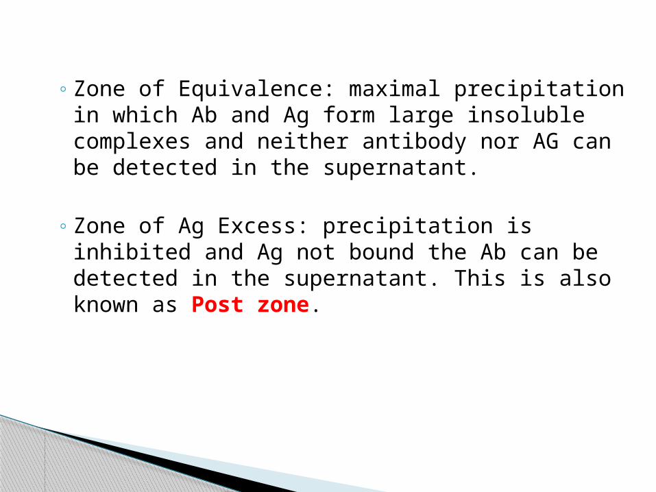
◦ Zone of Equivalence: maximal precipitation in which Ab and Ag form large insoluble complexes and neither antibody nor AG can be detected in the supernatant.
◦ Zone of Ag Excess: precipitation is inhibited and Ag not bound the Ab can be detected in the supernatant. This is also known as Post zone.
Multivalent Ag combines with bivalent Ab in varying proportions, depending on the Ag and Ab ratio in the reacting mixture.
Precipitation results when a large lattice is formed consisting of alternating antigen and Ab molecule. This is possible only in the zone of equivalence.
In the zone of Ag- Ab excess, the lattice does not enlarge as the valencies of the Ab and the Ag are fully satisfied.
MECHANISM
Ring test Slide test Tube test Immunodiffusion Electroimmnunodiffusion
Types of Precipitation Test
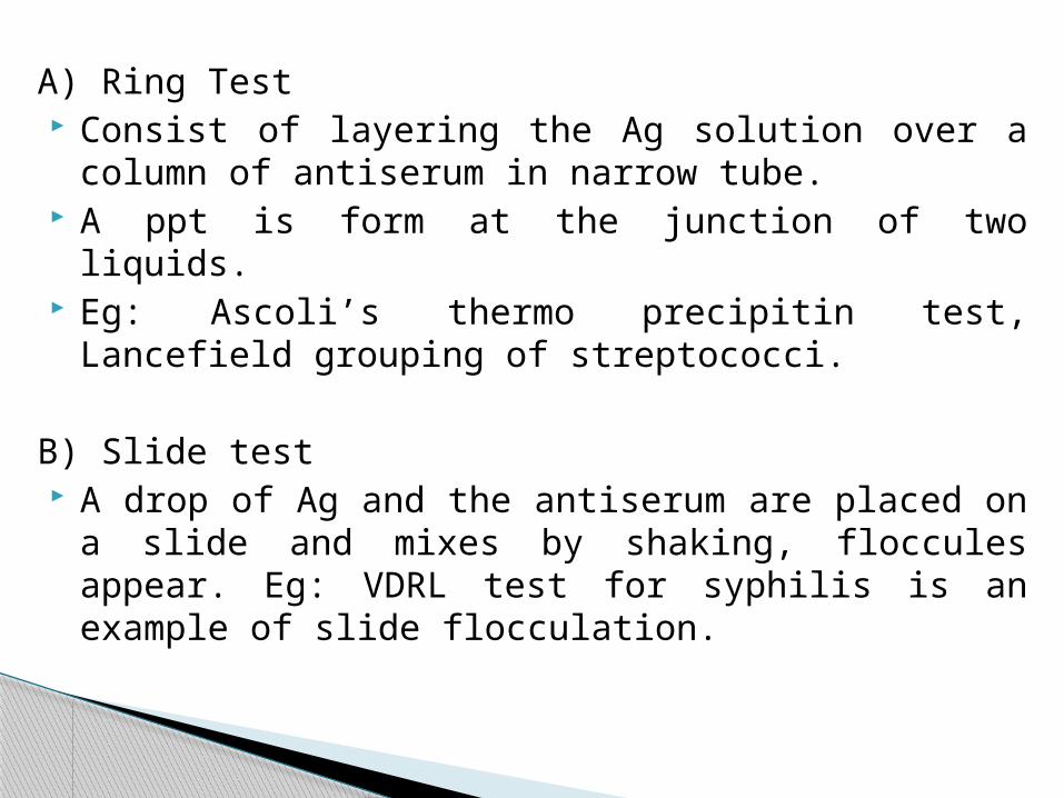
A) Ring Test Consist of layering the Ag solution over a column of antiserum in
narrow tube. A ppt is form at the junction of two liquids. Eg: Ascoli’s thermo precipitin test, Lancefield grouping of
streptococci.
B) Slide test A drop of Ag and the antiserum are placed on a slide and mixes by
shaking, floccules appear. Eg: VDRL test for syphilis is an example of slide flocculation.
C) Tube test A quantitative tube flocculation test is used for the
standardization of toxins and toxoids. Serial dilution of the toxin/toxoid are added to the tubes
containing a fixed quantity of the antitoxin The amount of toxin or toxoid that flocculates optimally with one
unit of the antitoxin is defined as an Lf dose Example: Kahn Test for syphilis is an example of tube
flocculation test
D). Immunodiffusion Test Precipitation test is done in gel. Immunodiffusion procedure are carried out in an agar
or agarose gel The precipitate is easily seen in gel yield precipitin
lines
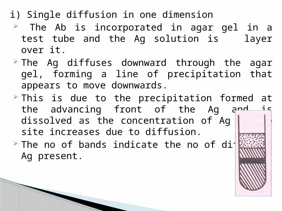
i) Single diffusion in one dimension The Ab is incorporated in agar gel in a test tube and the Ag
solution is layer over it. The Ag diffuses downward through the agar gel, forming a line of
precipitation that appears to move downwards. This is due to the precipitation formed at the advancing front of
the Ag and is dissolved as the concentration of Ag at the site increases due to diffusion.
The no of bands indicate the no of different Ag present.
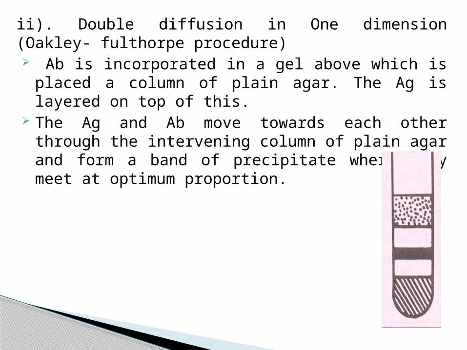
ii). Double diffusion in One dimension (Oakley- fulthorpe procedure) Ab is incorporated in a gel above which is placed a column of
plain agar. The Ag is layered on top of this. The Ag and Ab move towards each other through the intervening
column of plain agar and form a band of precipitate where they meet at optimum proportion.

iii). Single diffusion in two dimension( Radial immunodiffusion) The antiserum is incorporated in agar gel poured on a flat surface. The Ag is added to the wells cut on the surface of the gel. It
diffuses radially from the well and forms ring- shaped bands of precipitation concentrically around the well.
This method has been employed for the estimation of the immunoglobulin classes in sera.
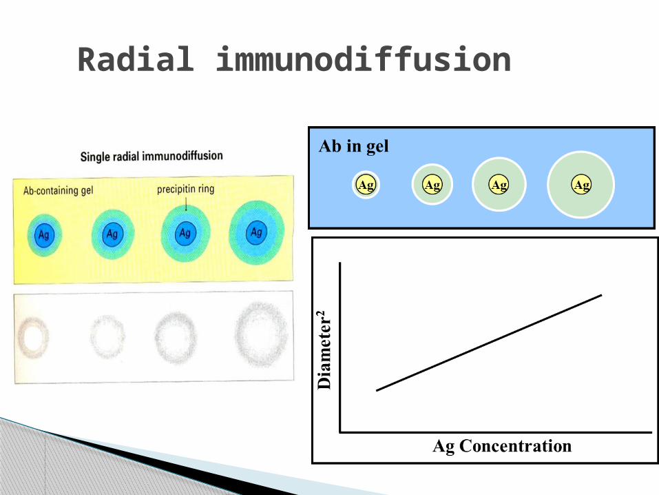
Radial immunodiffusion
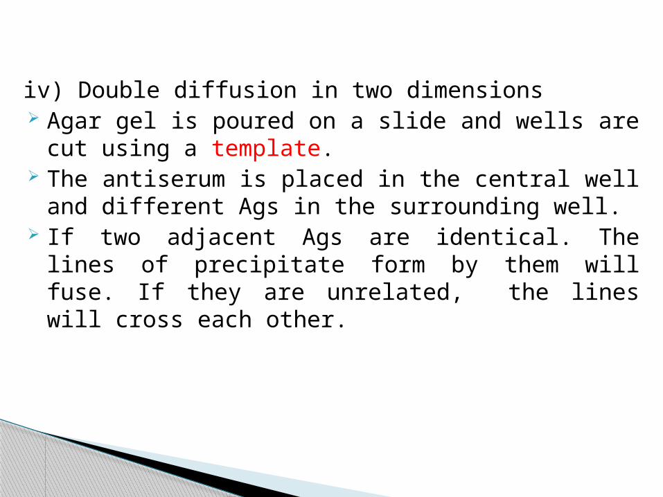
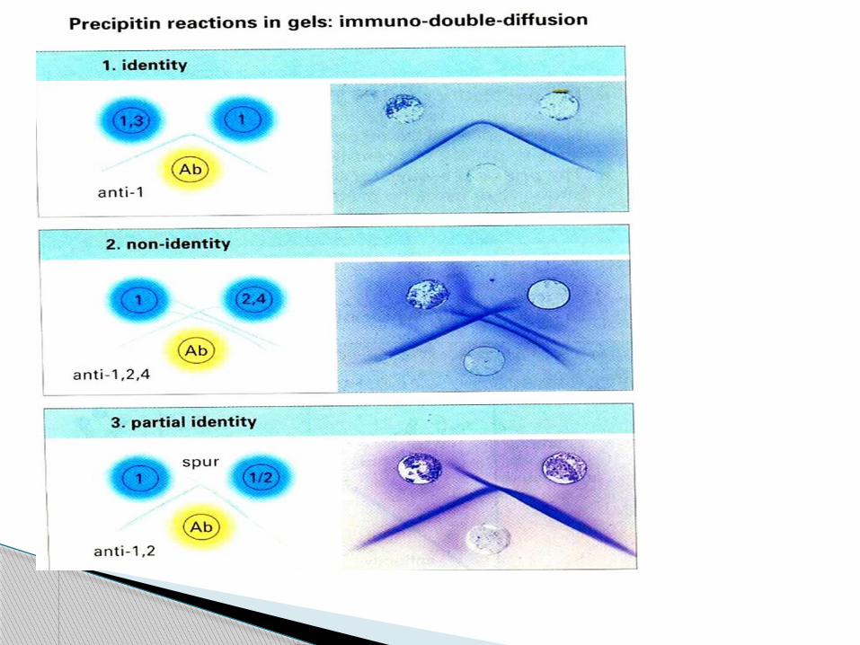
iv) Double diffusion in two dimensions Agar gel is poured on a slide and wells are cut using a
template. The antiserum is placed in the central well and different
Ags in the surrounding well. If two adjacent Ags are identical. The lines of precipitate
form by them will fuse. If they are unrelated, the lines will cross each other.

v) Immunoelectrophoresis: This technique involves the electrophoretic separation of a
composite Ag (such as serum) into its constituent proteins followed by immunodiffusion against its antiserum resulting in separate precipitin lines, indicating reaction between each individual protein with its antibody.
It is performed on agar or agarose gel on a slide with an Ag well and Ab trough cut on it.
The test serum is placed in the antigen well and electrophoresed for about an hour. Ab against human serum is then placed in the trough and diffusion allowed to proceed for 18-24 hrs.
The resulting precipitin lines can be photographed and the slides dried, stained and preserved for record.
This is used for testing normal and abnormal proteins in serum and urine

Plasma (mixture of antigens)
Electrophoresis
Antiserum (mixture of antibodies)
Immunodiffusion
E) Electroimmunodiffusion The development of precipitin can be speeded up by electrically
driving the antigen and Ab. Two most frequently technique used in the lab are:
i) Counterimmunoelectrophoresisii) Rocket electrophoresis
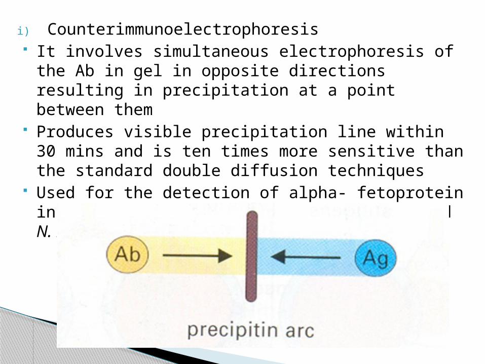
i) Counterimmunoelectrophoresis It involves simultaneous electrophoresis of the Ab in gel in
opposite directions resulting in precipitation at a point between them
Produces visible precipitation line within 30 mins and is ten times more sensitive than the standard double diffusion techniques
Used for the detection of alpha- fetoprotein in serum and specific Ags of Cryptococcus and N. meningitidis in the CSF.
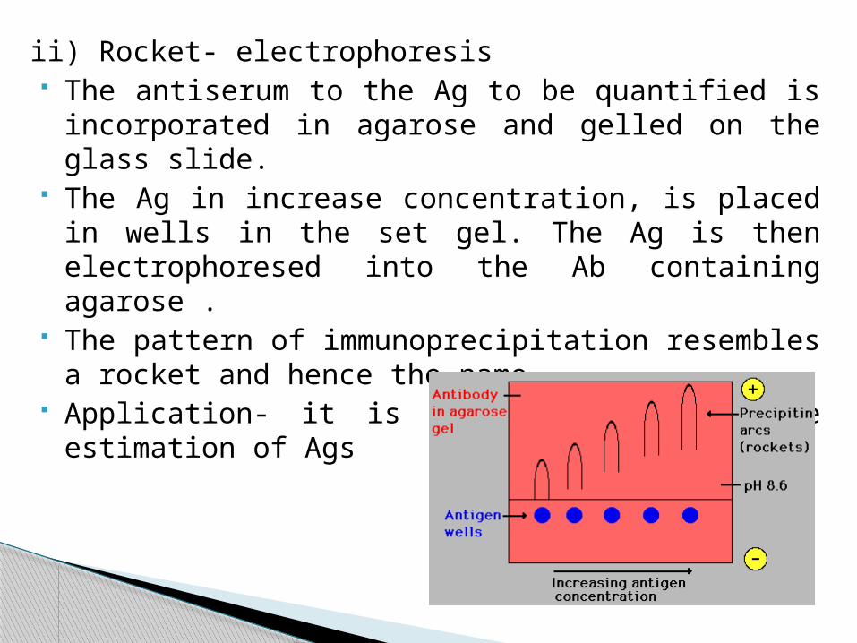
ii) Rocket- electrophoresis The antiserum to the Ag to be quantified is incorporated in
agarose and gelled on the glass slide. The Ag in increase concentration, is placed in wells in the set gel.
The Ag is then electrophoresed into the Ab containing agarose . The pattern of immunoprecipitation resembles a rocket and hence
the name. Application- it is used for quantitative estimation of Ags
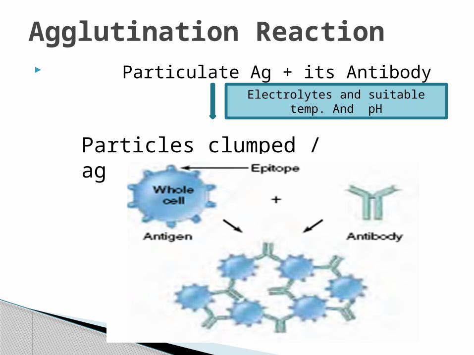
Particulate Ag + its Antibody
Agglutination Reaction
Electrolytes and suitable temp. And pH
Particles clumped / agglutinated
Same principle govern agglutination and precipitation reaction
Zone phenomenon is also seen in agglutination. Incomplete or monovalent Abs do not cause agglutination,
though they combine with Ag. They act as blocking Abs. More sensitive than precipitation for detection of Abs.
A) Slide Agglutination when a drop of the appropriate anti serum is added to a
smooth uniform suspension of a particulate Ag in a drop of saline on a slide, agglutination take place.
Clumping of Ag and clearing of suspension Uses:
◦ Blood grouping and cross matching◦ Confirmation of cultural isolates
Application of Agglutination test
B) Tube Agglutination Standard quantitative method for measuring Abs Titre estimated by:
A fixed volume of a particulate Ag suspension is added to an equal volume of serial dilutions of antiserum in test tubes.Uses: Diagnosis of : Typhoid (Widal test)
Brucellosis
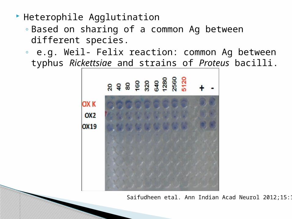
Heterophile Agglutination◦ Based on sharing of a common Ag between different species.◦ e.g. Weil- Felix reaction: common Ag between typhus
Rickettsiae and strains of Proteus bacilli.
Saifudheen etal. Ann Indian Acad Neurol 2012;15:141-4
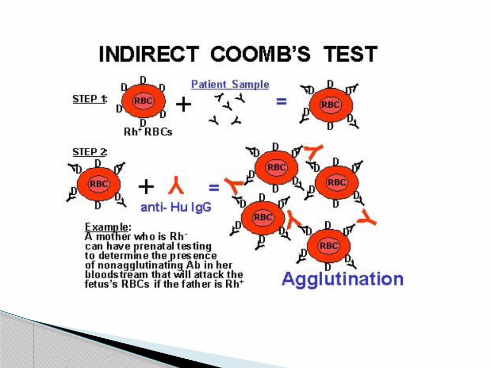
C) The Antiglobulin test It was devised by Coombs, Mourant and Race (1945) Detection of anti- Rh antibodies that do not agglutinate Rh-
positive red cells in saline. Used for the detection of incomplete Abs.
PRINCIPLE: When sera containing incomplete anti- Rh Abs are mixed with
Rh- positive red cells , the Ab globulin coats the surface of the RBCs , though they are not agglutinated.
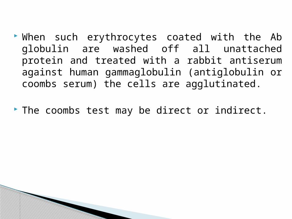
When such erythrocytes coated with the Ab globulin are washed off all unattached protein and treated with a rabbit antiserum against human gammaglobulin (antiglobulin or coombs serum) the cells are agglutinated.
The coombs test may be direct or indirect.

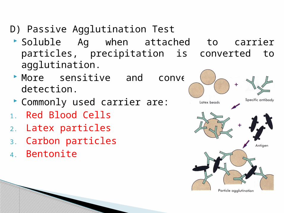
D) Passive Agglutination Test Soluble Ag when attached to carrier particles, precipitation is
converted to agglutination. More sensitive and convenient for Ab detection. Commonly used carrier are:
1. Red Blood Cells2. Latex particles3. Carbon particles4. Bentonite
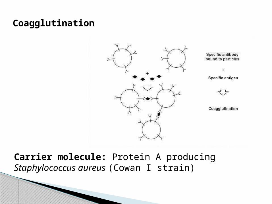
Coagglutination
Carrier molecule: Protein A producing Staphylococcus aureus (Cowan I strain)
In the presence of the appropriate Abs complement◦ Lyses erythrocytes ◦ Kills and in some cases lyses bacteria◦ Immobilises motile organism ◦ Promotes phagocytosis and immune adherence.◦ Contributes to tissue damage in certain types of
hypersensitivity.
The ability of antigen- antibody complexes to fix complement is the basis of CFT.
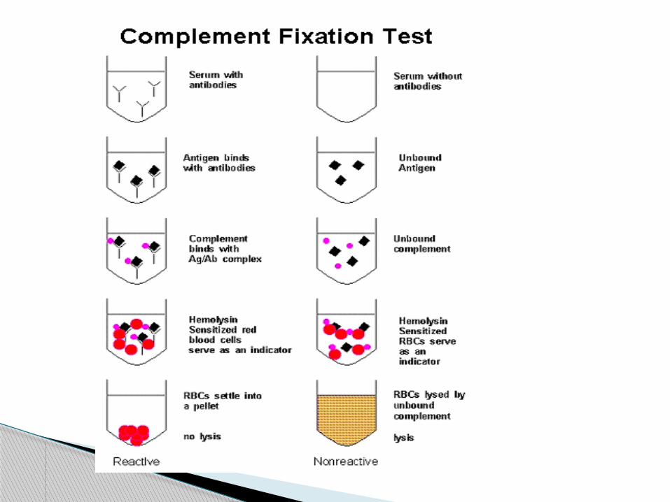
Complement Fixation Test
It is a very versatile and sensitive test. Types of CFT-
◦Wasserman reaction: used for serodiagnosis of syphilis
◦Indirect complement fixation test◦Conglutinating complement adsorption◦Other complement- dependant serological tests.

1) Wasserman ReactionANTIGEN + TEST
SERUM (Contains antibody)
+ Complement
+ Haemolytic system
ANTIGEN + TEST SERUM
(contains no Ab)+ complement
+ Hemolytic system
Complement fixed
Result- no haemolysis
Positive CF test
Complement not fixed
Result- HemolysisNegative of CF test
2) Indirect complement fixation test It is employed in cases of certain avian and
mammalian sera which do not fix guinea pig complement.
The test is set up in duplicate and after the first step the standard serum known to fix the complement is added to one set.
If the test serum contained Ab the Ag would have been used up in the first step and therefore the standard antiserum added would not be fixed to the complement.
Therefore haemolysis indicates a positive result.
ELISAINTRODUCTION:
Enzyme Linked Immuno-Sorbent Assay
Term Was Coined By Engvall and Pearlmann in 1971
Similar To RIA, Except No Radiolabel Can Be Used To Detect Both Antibody and
Antigen
Principle:
Works on the principle of antigen – antibody reaction where an enzyme conjugated with an antibody reacts with a colorless substrate to give a colored reaction product.
Types of ELISA
1. Indirect ELISA
2. Sandwich ELISA
3. Competitive ELISA
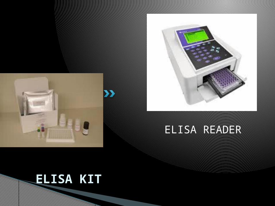
ELISA READER
ELISA KIT

Substrate used is known as chromogenic substrate.
Enzymes used are◦ ALP (p-nitro phenyl phosphate)◦ Horse radish peroxidase (o-phenylene diammine
dihydrochloride)◦ β galactosidase
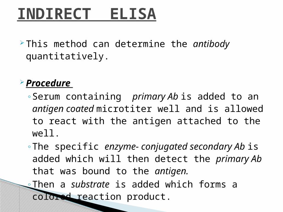
INDIRECT ELISA
This method can determine the antibody quantitatively.
Procedure ◦Serum containing primary Ab is added to an
antigen coated microtiter well and is allowed to react with the antigen attached to the well.
◦The specific enzyme- conjugated secondary Ab is added which will then detect the primary Ab that was bound to the antigen.
◦Then a substrate is added which forms a colored reaction product.
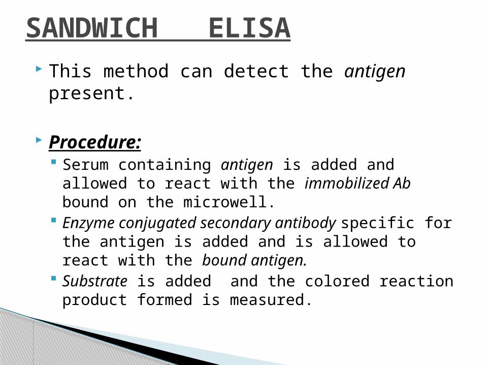
SANDWICH ELISA This method can detect the antigen present.
Procedure: Serum containing antigen is added and allowed to
react with the immobilized Ab bound on the microwell.
Enzyme conjugated secondary antibody specific for the antigen is added and is allowed to react with the bound antigen.
Substrate is added and the colored reaction product formed is measured.
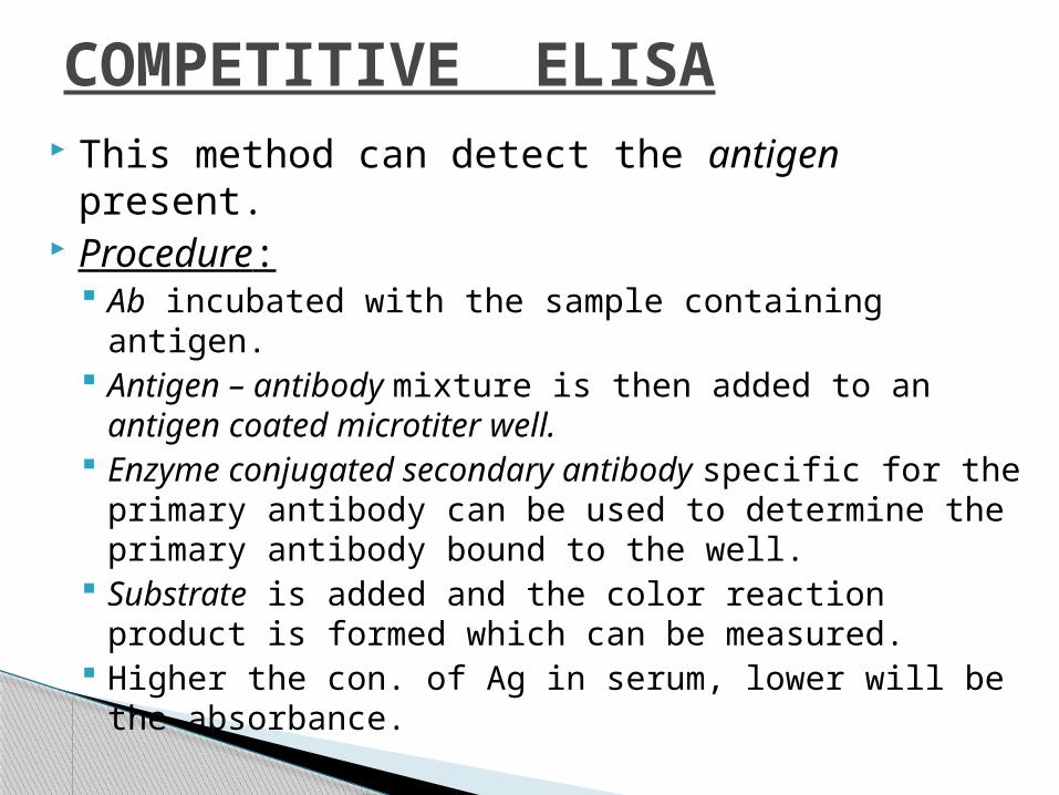
COMPETITIVE ELISA This method can detect the antigen present. Procedure:
Ab incubated with the sample containing antigen. Antigen – antibody mixture is then added to an
antigen coated microtiter well. Enzyme conjugated secondary antibody specific for
the primary antibody can be used to determine the primary antibody bound to the well.
Substrate is added and the color reaction product is formed which can be measured.
Higher the con. of Ag in serum, lower will be the absorbance.

source: kuby 6th edition
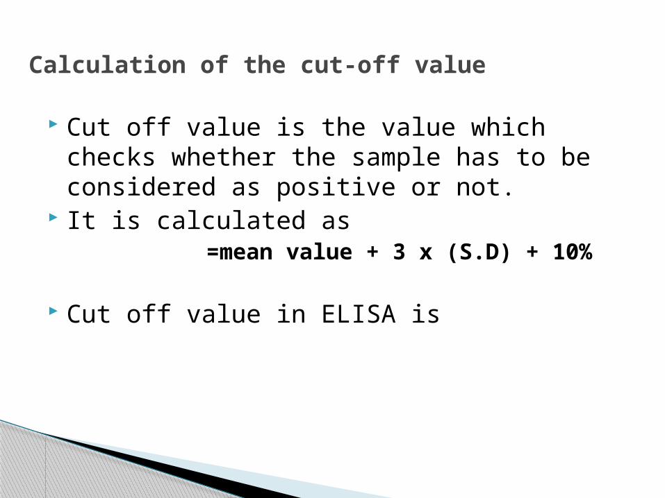
Calculation of the cut-off value
Cut off value is the value which checks whether the sample has to be considered as positive or not.
It is calculated as=mean value + 3 x (S.D) + 10%
Cut off value in ELISA is
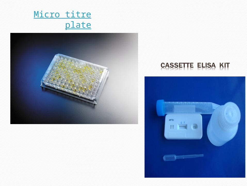
Micro titre plate

Advantages Sensitive Highly specific Readings can be documented Economic Easy to perform

applications Indirect ELISA
◦ HIV antibodies◦ HCV antibodies◦ Leptospira antibodies◦ Dengue antibodies◦ TORCH antibodies
Sandwich ELISA◦ HbsAg antigen◦ P24 antigen

CLIA Chemiluminescence Immuno AssayPrinciple: It is based on the principle by which the light emmission from the excited singlet state occurs when the electron returns to the ground state.It involves the oxidation of an organic compound such as luciferin , acridinium esters, isoluminol etc.
Chemiluminescence is the measurement of this energy generated from the chemical reaction (an oxidation reaction).

Procedure₋ Dispense the stds, samples and controls into the
appropriate wells.₋ Mix for 10 secs₋ Dispense the enzyme conjugate reagent into each
well.₋ Mix gently for 30 secs.₋ Incubate for 1 hour at RT.₋ Remove the incubation mixture.₋ Rinse the microtitre wells 5 times with wash buffer.₋ Add the chemiluminscence substrate solution into
each well.₋ Gently mix and read the wells on the
chemiluminscence microwell reader.

Chem Luminometer
Source: google images

Enzymes used:◦ ALP◦ Horse radish peroxidase
Metal ions ◦ Cu(II)◦ Fe(III)◦ hemin
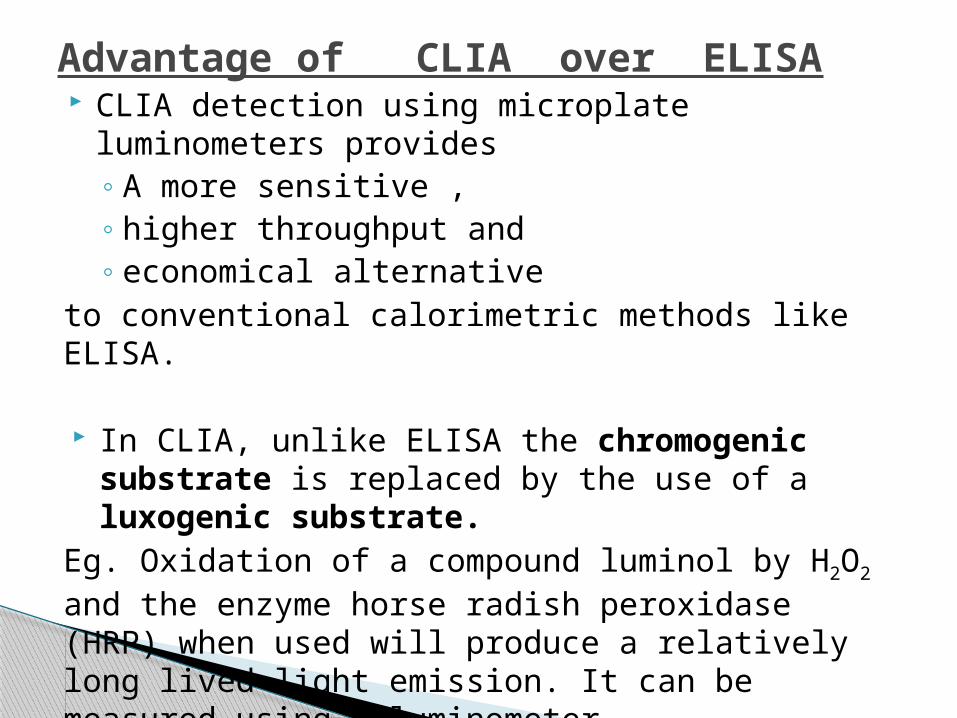
Advantage of CLIA over ELISA CLIA detection using microplate luminometers
provides ◦ A more sensitive , ◦ higher throughput and ◦ economical alternative
to conventional calorimetric methods like ELISA.
In CLIA, unlike ELISA the chromogenic substrate is replaced by the use of a luxogenic substrate.
Eg. Oxidation of a compound luminol by H2O2 and the enzyme horse radish peroxidase (HRP) when used will produce a relatively long lived light emission. It can be measured using a luminometer. Miniminal sensitivity of test is 0.5ng/ml.
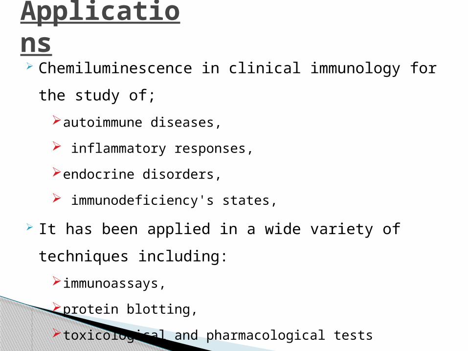
Applications Chemiluminescence in clinical immunology for the
study of;
autoimmune diseases,
inflammatory responses,
endocrine disorders,
immunodeficiency's states,
It has been applied in a wide variety of techniques
including:
immunoassays,
protein blotting,
toxicological and pharmacological tests
IMMUNOFLUoRESCENCE History:This technique was implemented by a scientist by name, Albert Coons in the year 1944. Principle:Based on the principle that fluroscent molecules can absorb light of one wavelength(excitation) and emit light of another wavelength(emission). Fluorescence is a property where light is absorbed and remitted within a few nanoseconds (approx. 10ns) at a lower energy.
It is a type of antigen–antibody reaction
using fluroscent dyes observed using a
fluroscent microscope.
Fluroscent dyes are special dyes that can
absorb the rays of lower wavelength and
emit the rays of higher.
Commonly used dyes are;
Fluroscene isothiocyanate
Lissamine rodamine
Types of immunofluorescencei) Primary (direct) It uses a single antibody that is chemically linked to a
fluorophore. The antibody recognizes the target molecule and binds to
it, and the fluorophore it carries can be detected via microscopy.
This technique has several advantages over the secondary (or indirect) because of the direct conjugation of the antibody to the fluorophore. This reduces the number of steps in the staining procedure making the process faster and can reduce background signal.
However, since the number of fluorescent molecules that can be bound to the primary antibody is limited, direct immunofluorescence is less sensitive than indirect immunofluorescence.

Direct IF
(ii)Secondary( indirect): It uses two antibodies; the unlabeled first (primary)
antibody specifically binds the target molecule, and the secondary antibody, which carries the fluorophore, recognises the primary antibody and binds to it.
Multiple secondary antibodies can bind a single primary
antibody. This provides signal amplification by increasing the number of fluorophore molecules per antigen.
This protocol is more complex and time consuming than the primary (or direct), but it allows more flexibility because a variety of different secondary antibodies and detection techniques can be used for a given primary antibody.

TYPE OF IMMUNOFLUROSCENCE
ADVANTAGES DISADVANTAGES
DIRECT • SHORTER SAMPLE STAINING TIME
• LOWER SIGNAL
• SIMPLER LABELLING PROCEDURE
• HIGHER COST
• LESS FLEXIBILITY
INDIRECT • MORE SENSITIVE • POTENTIAL FOR CROSS REACTIVITY
• AMPLIFICATION OF SIGNAL
• SAMPLE WITH ENDOGENOUS Ig MAY EXIHIBIT HIGH BACKGROUND

Immunofluroscence microscopic view
Source: google images
APPLICATIONS Detection of rabies antigen (direct IF)
Fluroscent Treponema Antibody test for syphilis-
(indirect IF)
Analysis of antigen in fresh, frozen or fixed tissues.
Detection of presence or absence of specific DNA
sequences on chromosomes.
Defines the patterns of gene expression within the
cells/tissues.
LIMITATIONS1. Photo bleaching
2. Autofluroscence
3. Fluroscence overlap

REFERENCES www.wikipedia.com Google images www.pubmed.com Text book on Medical Laboratory Technology
Dr. Praful B Godkar
Annual review of Medicine◦ Introduction to chemiluminscence
Text book of immunology
−Kuby


