
Anatomy learning and retention among students in a
graduate-entry medical course
Michelle Machado
B.Sc (Hons), M.Sc
Thesis submitted in fulfilment of the requirements for the degree of
Doctor of Philosophy (Education)
College of Arts and Education
Victoria University
7 July 2017

Running head: ANATOMY KNOWLEGDE IN MEDICAL STUDENTS i
Abstract
Anatomy forms the basis of clinical examination and is integral to today’s medical
curriculum. Yet, increasingly evident in the literature is feedback from clinicians and
surgeons about the perceived lack of anatomical knowledge among recent medical
graduates. To understand the issues surrounding student learning and retention of
anatomy, a mixed-methods design was utilized to explore medical students’ anatomical
knowledge throughout their Bachelor of Medicine and Bachelor of Surgery (MBBS)
degree. Students enrolled in the four-year graduate-entry MBBS course at Monash
University participated in the study. Participants from the preclinical (Year A) and
clinical years (Years B, C and D) sat an online assessment consisting of 60 clinically
relevant anatomy multiple-choice and extended-matched questions whose objectives
had been previously covered in the preclinical teaching. Altogether, 136 students
participated in the study. The results revealed that knowledge of anatomy declined over
time and this was significant in the final two clinical years. The drop in anatomical
knowledge was not uniform. The regions of anatomy better retained were associated
with frequent exposure and reinforcement in the clinical years. Participants cited an
intense and time-constrained curriculum, poor integration in the clinical years and rare
opportunities for revising and testing anatomy as reasons for the decline of knowledge.
The results of this study highlight the need for conceptual coherence at the time of
teaching; the importance of vertical integration in providing students with frequent
learning opportunities in the clinical years; and, the value of formatively testing
students’ knowledge of anatomy throughout the clinical years.
ANATOMY KNOWLEDGE IN MEDICAL STUDENTS ii
Declaration of Originality
I, Michelle Machado, declare that the PhD thesis entitled “Anatomy learning and
retention in medical students in a graduate-entry MBBS course” is no more than
100,000 words in length including quotes and exclusive of tables, figures, appendices,
bibliography, references and footnotes. The thesis contains no material that has been
submitted previously, in whole or in part, for the aware of any other academic degree or
diploma. Except where otherwise indicated, this thesis is my own work.
Signed: Date: 7 July 2017

ANATOMY KNOWLEDGE IN MEDICAL STUDENTS iii
Acknowledgements
I would like to thank my supervisors Dr. Margaret Malloch and Dr. Catherine
Haigh for their continuous guidance and encouragement throughout this incredible
journey. Their scholarly advice, positive and constructive feedback and unwavering
support helped me to accomplish this enormous task. I would also like to extend my
sincere gratitude to Dr. Bill Haigh, who was a key player in the design and
implementation of the online assessment tool used in this research.
I extend my sincere gratitude to Assoc. Prof Shane Bullock and my colleagues at
Monash University for their moral support and positive reinforcement in helping me
complete my thesis.
To my husband, Gavin Pinto, thank you for your constant support throughout
these years. You always found ways to keep me driven and motivated, even when life
threw challenges our way. Without your love and encouragement, I could not have
accomplished this. To my daughter, Natalie, I’ve spent many days juggling motherhood
and writing a thesis. You are my light and I dedicate this thesis to you in hopes of
inspiring you to always strive for what you want, no matter how difficult the
circumstances. To my family and my in-laws, I am forever grateful for your
encouragement and support you provided me with during those crucial years of
balancing motherhood and completing my thesis. I was able to make progress in my
thesis through all of the various changes in my life because of the love and support you
gave me. So thank you!
Finally, to the medical students in the Monash Graduate-Entry MBBS program.
Thank you for your time and generosity in participating in this study. Without you all,
this thesis would not be possible.

ANATOMY KNOWLEDGE IN MEDICAL STUDENTS iv
Table of Contents
Abstract ............................................................................................................................ i
Declaration of Originality .............................................................................................. ii
Acknowledgements ........................................................................................................ iii
Table of Contents ........................................................................................................... iv
List of Figures ............................................................................................................... vii
List of Tables .................................................................................................................. ix
List of Abbreviations ...................................................................................................... x
List of Appendices ........................................................................................................ xii
Chapter 1: Introduction ................................................................................................. 1 1.1 Foundation of Research .......................................................................................... 1 1.2 Background and Contextual Base for Research ..................................................... 7
1.2.1 Bachelor of Medicine / Bachelor of Surgery Program in Australia ................ 7 1.2.2 Monash Graduate-entry Bachelor of Medicine / Bachelor of Surgery
Program ........................................................................................................... 8 1.3 Purpose, Aims and Rationale for Research .......................................................... 19 1.4 Organisation of Thesis .......................................................................................... 22
Chapter 2: Anatomy: Origins, History and Current Trends ................................... 25 2.1 Anatomy—Importance and Origins ..................................................................... 25
2.1.1 Why is Anatomy Important? ......................................................................... 25 2.1.2 Origins of Anatomy ....................................................................................... 30
2.2 Anatomy in the Twentieth Century—The Early Years ........................................ 35 2.2.1 Traditional Anatomy Curriculum .................................................................. 35 2.2.2 Challenges Associated with the Traditional Curriculum ............................... 38 2.2.3 Early Medical Education Reform .................................................................. 39
2.3 Anatomy in the Twentieth Century—The Past 30 Years and Current Trends ..... 41 2.3.1 Late Medical Education Reform ................................................................... 41 2.3.2 Three Curriculum Formats and Their Instructional Methods ........................ 43 2.3.3 Outcome and Competency-based Education ................................................. 50 2.3.4 Modern Teaching Tools Used in Anatomy ................................................... 55
Chapter 3: Learning and Assessment of Anatomy .................................................... 73 3.1 Learning Approaches and Theories ...................................................................... 73
3.1.1 Theories of Learning ..................................................................................... 73 3.1.2 Adult Learning and Learning in the Workplace ............................................ 77 3.1.3 Approaches to Learning Anatomy ................................................................. 85 3.1.4 Recontextualisation of Knowledge ................................................................ 90
3.2 Assessment ........................................................................................................... 92 3.2.1 Types of Assessment in Anatomy ................................................................. 93 3.2.2 Assessment of Anatomy in the Traditional and Modern Curricula ............... 94
Chapter 4: Anatomy: Current Debates ...................................................................... 98 4.1 Current Debates Surrounding the Modern Anatomy Curriculum ........................ 98
4.1.1 Curriculum Format ........................................................................................ 98 4.1.2 Declining Knowledge with No Vertical Integration ................................... 102 4.1.3 Lack of Core Curriculum and Competency-based Education in Anatomy . 109 4.1.4 Lack of Competency-based Assessments in Anatomy ................................ 112 4.1.5 Learning of Anatomy .................................................................................. 115

ANATOMY KNOWLEDGE IN MEDICAL STUDENTS v
4.1.6 Retention of Anatomy ................................................................................. 118 4.1.7 Anatomy Educators ..................................................................................... 123 4.1.8 Attitudes of Students Towards Anatomy .................................................... 127 4.1.9 Development of Postgraduate Surgical Programs ....................................... 131
4.2 Hypotheses of the Study ..................................................................................... 133
Chapter 5: Methodology ............................................................................................ 134 5.1 Methodology, Research Design and Research Methods .................................... 134
5.1.1 Introduction to Methodology ....................................................................... 134 5.1.2 Research Design .......................................................................................... 137 5.1.3 Rationale for the Research Design .............................................................. 141 5.1.4 Research Methods—What Did They Entail? .............................................. 142
5.2 Ethical Considerations and Approval ................................................................. 144 5.2.1 Recruitment, Conflict of Interest, Power Relations ..................................... 144 5.2.2 Anonymity, Confidentiality and Privacy ..................................................... 145 5.2.3 Informed Consent ........................................................................................ 146 5.2.4 Data Storage and Handling .......................................................................... 147 5.2.5 Ethics Approval ........................................................................................... 150
5.3 Participants ......................................................................................................... 150 5.4 Recruitment ........................................................................................................ 153
5.4.1 Participant Selection for Phase Two ............................................................ 155 5.5 Development and Administration of Test Instrument ........................................ 157
5.5.1 Development of Anatomy Assessment (Phase One) ................................... 157 5.5.2 Test Administration (Phase One) ................................................................ 163
5.6 Development and Administration of Learning Anatomy Survey and Interview
Questions............................................................................................................. 165 5.7 Data Collection ................................................................................................... 166
5.7.1 Phase One .................................................................................................... 166 5.7.2 Phase Two ................................................................................................... 167
5.8 Data Analysis ...................................................................................................... 168 5.8.1 Phase One .................................................................................................... 168 5.8.2 Phase Two ................................................................................................... 169
Chapter 6: Quantitative Results ................................................................................ 176 6.1 Exclusion Criteria ............................................................................................... 177
6.1.1 Identifying and Managing Student Outliers ................................................ 177 6.2 Test Statistics and Question Analysis ................................................................. 181
6.2.1 Reliability, Validity, Borderline Score and Standard Error of
Measurement ............................................................................................... 182 6.2.2 Overall Performance of Participants ........................................................... 184
6.3 Cross-sectional Data and Analysis ..................................................................... 184 6.3.1 Overall Anatomy Scores .............................................................................. 185 6.3.2 Regional Anatomy Scores ........................................................................... 190 6.3.3 Cohort Performance Across Difficulty ........................................................ 194
6.4 Comparison of VIA scores with anatomy assessment scores............................. 196 6.4.1 Year B Results ............................................................................................. 199 6.4.2 Year C Results ............................................................................................. 201 6.4.3 Year D Results ............................................................................................. 204
6.5 Testing Effect ..................................................................................................... 206 6.6 Learning Anatomy Survey Results ..................................................................... 208
6.6.1 Demographics .............................................................................................. 208 6.6.2 Learning Anatomy ....................................................................................... 211

ANATOMY KNOWLEDGE IN MEDICAL STUDENTS vi
6.6.3 Attitudes Towards Anatomy ........................................................................ 215
Chapter 7: Qualitative Results .................................................................................. 221 7.1 Anatomy in the Year A Program ........................................................................ 223
7.1.1 Small-group Teaching ................................................................................. 224 7.1.2 Lectures ....................................................................................................... 228 7.1.3 Cadaveric Teaching ..................................................................................... 229 7.1.4 Body Painting .............................................................................................. 232 7.1.5 Teaching Resources ..................................................................................... 232 7.1.6 Assessment of Anatomy .............................................................................. 234
7.2 Anatomy in the Clinical Years (Years B–D) ...................................................... 236 7.2.1 Importance of Anatomy in Medicine .......................................................... 237 7.2.2 Surface Anatomy and Physical Examination .............................................. 240 7.2.3 Anatomy and Imaging ................................................................................. 241 7.2.4 Anatomy in Surgery and Procedures ........................................................... 242
7.3 Issues Surrounding Learning and Retention of Anatomical Knowledge ........... 244 7.3.1 Learning Anatomy in a Time-constrained Curriculum ............................... 244 7.3.2 Site-dependent Opportunities for Learning and Revising Anatomy ........... 248 7.3.3 Inconsistent Delivery of Curriculum Among Teaching Staff ..................... 251 7.3.4 Attitudes of Teaching Staff Towards Students and the Effect on Learning
and Retention of Knowledge ....................................................................... 252 7.3.5 Lack of Vertical Integration ........................................................................ 257
7.4 Recommendations Offered by Students for Better Learning and Retention of
Anatomy .............................................................................................................. 260 7.4.1 Anatomy Refresher Course ......................................................................... 260 7.4.2 Curriculum Changes and Consistent Delivery ............................................ 262
Chapter 8: Discussion ................................................................................................. 266 8.1 Introduction ........................................................................................................ 266
8.1.1 Overview of the Research ........................................................................... 266 8.1.2 Findings from the Formative Anatomy Assessment ................................... 269
8.2 Anatomy Knowledge and Retention ................................................................... 270 8.2.1 How Does Anatomy Knowledge Differ Among Students Across the Pre-
clinical and Clinical Years? ........................................................................ 270 8.2.2 To What Extent Do Medical Students Retain Anatomy? ............................ 275
8.3 Anatomy-learning Survey................................................................................... 280 8.3.1 Student Approaches Towards Learning of Anatomy .................................. 280 8.3.2 Student Attitudes Towards the Learning and Use of Anatomy ................... 282 8.3.3 Use of Teaching Resources in Learning Anatomy ...................................... 285
8.4 Factors Surrounding Retention of Anatomical Knowledge ............................... 289 8.4.1 Factors Assisting Students in Learning Anatomy ....................................... 289 8.4.2 Factors Affecting Students’ Learning and Retention of Anatomy .............. 299
8.5 Research Limitations .......................................................................................... 324
Chapter 9: Conclusion ............................................................................................... 328 9.1 Recommendations .............................................................................................. 340 9.2 Implications for Future Research ....................................................................... 345
References.................................................................................................................... 347
Appendices .................................................................................................................. 393
ANATOMY KNOWLEDGE IN MEDICAL STUDENTS vii
List of Figures
Figure 5.1: Participant representation relative to each of cohort (Years A–D). ........... 152
Figure 5.2: Bloom’s Taxonomy. .................................................................................. 161
Figure 6.1: Total time to complete anatomy test in each cohort. ................................. 179
Figure 6.2: Completion time on online assessment by cohort. ..................................... 180
Figure 6.3: Overall performance on anatomy assessment by cohort. ........................... 184
Figure 6.4: Anatomy scores on online assessment by cohort. ...................................... 187
Figure 6.5: Anatomy scores on formative assessment. ................................................ 188
Figure 6.7: Mean performance of cohort across six regional anatomy blocks. ............ 192
Figure 6.8: Pairwise comparison of back scores across all cohorts.............................. 193
Figure 6.9: Pairwise comparison of head scores across all cohorts.............................. 194
Figure 6.10: Mean performance of cohort according to taxonomy. ............................. 195
Figure 6.11: Differences in Year B performance between anatomy assessment scores
and anatomy VIA scores (AA–VIA). .................................................................... 200
Figure 6.12: Comparison of participant performance on the VIA assessment in Year A
v. the anatomy assessment in Year B. ................................................................... 201
Figure 6.13: Differences in Year C performance between anatomy assessment scores
and anatomy VIA scores (AA–VIA). .................................................................... 202
Figure 6.14: Comparison of participant performance on the VIA assessment in Year A
v. the anatomy assessment in Year C. ................................................................... 203
Figure 6.15: Comparison of participant performance on the VIA assessment in Year A
v. the anatomy assessment in Year D. ................................................................... 204
Figure 6.16: Difference in scores for Year A cohort between formative and summative
assessments............................................................................................................ 207
Figure 6.17: Comparison of participant performance on the formative assessment in
Year A v. the summative anatomy assessment in Year A. ................................... 208
Figure 6.18: Distribution of participants’ prior degree within each cohort (Years A–D).
............................................................................................................................... 209
Figure 6.19: Effect of participants’ educational background on performance. ............ 210
Figure 6.20: Mean anatomy score on online assessment across the five educational
backgrounds. ......................................................................................................... 211
Figure 6.21: Resources used the most by students for learning anatomy. ................... 212

ANATOMY KNOWLEDGE IN MEDICAL STUDENTS viii
Figure 6.22: Most useful resources for learning anatomy. ........................................... 213
Figure 6.23: Approaches mainly used to learn anatomy. ............................................. 214
Figure 6.24: Performance of high and low scorers by learning approach. ................... 215
Figure 6.26: Participants’ use of anatomy in clinical skills v. their confidence in use of
anatomy knowledge............................................................................................... 218
Figure 6.27: Participants’ perceptions of importance of anatomy. ............................... 219
Figure 7.1: Four themes and subthemes that emerged from participant interviews. .... 222

ANATOMY KNOWLEDGE IN MEDICAL STUDENTS ix
List of Tables
Table 5.1 The criteria for developing a mixed-methods research design .................... 139
Table 5.2 Ethical Issues Prevalent in the Research ..................................................... 149
Table 5.3 Participant Selection for Qualitative Strand ................................................ 157
Table 5.4 Calculation of borderline score using modified Angoff method .................. 160
Table 6.1 Tests of Normality: Skewness and Kurtosis values for each cohort ............. 186
Table 6.2 Tests of Normality: Cohort and Anatomy Score on Online Test .................. 187
Table 6.3 Tukey–Kramer Post Hoc Test: Comparison of Mean Anatomy Scores ....... 190
Table 6.4 Overall Mean Score (%) of Participants by Regions ................................... 191
Table 6.5 Descriptive scores of the clinical cohorts on Anatomy Assessment (AA) and
VIA ......................................................................................................................... 198
Table 7.1 Participant’s progression through the MBBS at the time of Phase I and II of
study ...................................................................................................................... 222

ANATOMY KNOWLEDGE IN MEDICAL STUDENTS x
List of Abbreviations
3D Three-dimensional
AAMC Association of American Medical Colleges
ABMS American Board of Medical Specialties
ACF Australian Curriculum Framework
ACGME Accreditation Council for Graduate Medical Education
AMC Australian Medical Council
AMSA Australian Medical Students Association
ANOVA Analysis of variance
AQF Australian Qualifications Framework
AQFC Australian Qualification Framework Council
ARS Audience response system
ASIS Anterior superior iliac spine
AUQF Australian Quality Forum
BL Borderline
CBME Competency-based medical education
CE Common Era
CT Computed tomography
EMQ Extended-match questions
GI Gastrointestinal
GMC General Medical Council
HA Hand, the alternative
HP High performers
ICU Intensive Care Unit
LP Low performers

ANATOMY KNOWLEDGE IN MEDICAL STUDENTS xi
MANOVA Multivariate-analysis of variance
MBBS Bachelor of Medicine and Bachelor of Surgery
MCQ Multiple-choice questions
MRH Monash Rural Health
MRI Magnetic resonance imaging
NBME National Board of Medical Examiners
NHMRC National Health and Medical Research Council
OBE Outcome-based education
OSCE Objective Structured Clinical Examination
PBL Problem-based learning
RCAS Royal Australasian College of Surgeons
SAQ Short-answer questions
SD Standard deviation
SDL Self-directed learning
SE Standard errors
SEM Standard error of measurement
TBL Team-based learning
UK United Kingdom
US United States
USMLE United States Medical Licensing Examination
VIA Vertically integrated assessment

ANATOMY KNOWLEDGE IN MEDICAL STUDENTS xii
List of Appendices
APPENDIX A Participant Information Form ............................................................. 393
APPENDIX B Consent Form ....................................................................................... 397
APPENDIX C Research Ethics Approval .................................................................... 400
APPENDIX D Research Flyer to Students .................................................................. 403
APPENDIX E List of Anatomy objectives covered in Year A ...................................... 405
APPENDIX F Anatomy Learning Survey & Anatomy Test Questions ........................ 408
APPENDIX G Research Interview Questions ............................................................. 440

ANATOMY KNOWLEDGE IN MEDICAL STUDENTS 1
Chapter 1: Introduction
1.1 Foundation of Research
One of the major aims of an undergraduate medical curriculum is to produce
excellent doctors who are sufficiently equipped with the basic knowledge and skills
required to become competent and safe practitioners (Pabst, 2009). To achieve this solid
foundation of medical knowledge and the necessary procedural skills, the discipline of
anatomy, which offers a unique perspective on the human body, is regarded by many as
one of the most important basic sciences in the curriculum because it “provides the
structure on which other disciplines sit” (Boon, Meiring, & Richards, 2002; Herle &
Saxena, 2011; Marks, Jr, 1996; McKeown et al., 2003, p. 957).
Anatomy is defined as the structure of the body. Gross anatomy, or
topographical anatomy, is the study of the normal macroscopic structure of the human
form and its relationship with other body parts, all of which can be seen with the
unaided eye (Louw, Eizenberg, & Carmichael, 2009). Anatomy is supplemented by
disciplines such as histology (microscopic anatomy), embryology (developmental
anatomy) and evolution (comparative anatomy), and it is complemented by physiology
(functional anatomy), pathology (abnormal anatomy) and radiography (radiological or
sectional anatomy). Further, anatomy plays a key role in clinical practice (Boon et al.,
2002) through its use in physical examination, history-taking and clinical reasoning. It
also underpins clinical disciplines such as surgery and radiology (Dangerfield, Bradley,
& Gibbs, 2000; Louw et al., 2009).
For centuries, anatomy and other related disciplines have been regarded as the
mainstay of medical education (Milgate, 2006). They have played a central role in the
training of medical students, doctors and other healthcare specialists such as nurses,
physiotherapists and radiographers (Abu-Hijleh, 2010; Raftery, 2007; Sugand,

ANATOMY KNOWLEDGE IN MEDICAL STUDENTS 2
Abrahams, & Khurana, 2010). In essence, anatomy forms the language of medicine
(Gogalniceanu et al., 2010). For each generation of doctors trained, knowledge of
anatomy forms the core foundation for clinical examination. When doctors examine
patients, they inspect, palpate (examine by touch) and observe for deviations from the
norm. These behaviours serve as the fundamental framework for identifying an
underlying condition. Core knowledge of what constitutes normal form and function
enables doctors to identify altered states (Banda, 2010; Singh et al., 2015). For doctors to
be proficient, they must have a sound knowledge of anatomy.
The rapid expansion of biotechnology in the twentieth century resulted in a
massive influx of biological science courses such as microbiology, cell biology and
immunology into medical education. Combined with the introduction of social science,
law and ethics courses, anatomy became marginalised and began losing its prominence
within medical education (Kerby, Shukur, & Shalhoub, 2011). A condensed and packed
medical curriculum led to a decline in the amount of time that could be allocated to
teaching anatomy (Ramsey-Stewart, Burgess, & Hill, 2010). This reduction led to an
outcry in the medical community. Milgate (2006) states that “there is a rising chorus of
concern across the medical profession that not so young doctors are being expected to
treat patients to the same standards as their predecessors, without exposure to the
necessary amount of training in anatomy” (p. 6).
To understand the rationale behind such circumstances, it is imperative to
investigate how anatomy was and is delivered to medical students. The literature
suggests that the teaching of anatomy in undergraduate and graduate-entry medical
degrees has undergone considerable changes over the past century. A comparison of
historical data reveals vast reductions globally in anatomy teaching hours over the past
century. In the United States (US), medical schools allocated an average of 147 hours to
ANATOMY KNOWLEDGE IN MEDICAL STUDENTS 3
gross anatomy teaching in 2014 (Drake, McBride, & Pawlina, 2014) compared with 167
hours in 2002 (Drake, Lowrie, & Prewitt, 2002) and 549 hours in 1902. Similar
observations were made for the United Kingdom (UK) and Ireland (Leung, Lu, Huang,
& Hsieh, 2006). Critics in Australian and New Zealand medical schools have opposed
the gradual devaluation of gross anatomy. Studies of 19 Australian and two New
Zealand medical schools revealed significant differences in teaching hours between the
institutions, ranging from approximately 56 to 500 hours, with an average of 171 hours
(Craig, Tait, Boers, & McAndrew, 2010). Given the large reductions in teaching hours,
it is not surprising that medical students today are viewed by medical professionals as
being far less equipped in terms of anatomical knowledge when compared with their
predecessors.
In today’s medical curriculum, all of the basic sciences are covered primarily in
the pre-clinical curriculum, where formal anatomy teaching is confined to the first one
or two years of a four- or five-year medical degree. It is assumed that anatomy learned
during this stage will be retained and retrieved from memory during clinical training in
the later years (Collins, 2009; Sugand et al., 2010). In Australasian medical schools,
anatomy is taught in a series of lectures and practical workshops that largely incorporate
tutorials, prosections (neat dissections of cadavers undertaken by an experienced
professional, with the anatomical specimens used for student learning), dissections
(student-led but supervised explorations of the human body), surface anatomy and
medical imaging workshops. However, not all methods are used by universities in their
teaching practices. Further, different medical schools operate under different curricula.
Some follow an old-school traditional curriculum, which incorporates didactic lectures
and dissections, while others use modern approaches such as problem-based learning
(PBL), which involves small-group teaching sessions that are student-led and facilitator-

ANATOMY KNOWLEDGE IN MEDICAL STUDENTS 4
guided, and some schools use prosections as a replacement for dissections. Assessments
of anatomical knowledge also vary across institutions and can involve a range of text-
and image-based problems in the form of multiple-choice questions (MCQs) or
extended-match questions (EMQs) with or without practical assessments using
cadaveric specimens.
Given that anatomy is central to the practice of medicine (Herle & Saxena,
2011), medical institutions and the wider community expect that medical students will
have attained sufficient knowledge of the discipline during their medical degree to
allow them to practice medicine safely. However, this assumption is questioned when
medical students are placed in the clinical setting. There is increasing feedback and
criticism from clinical practitioners, surgeons and tutors about the perceived lack of
anatomical knowledge among recent medical graduates, as well as a debate about
whether a junior doctor’s knowledge of anatomy is safe enough for clinical practice
(Cottam,1999; Craig et al., 2010; Fitzgerald, White, Tang, James, & Maxwell-
Armstrong, 2008; Leung et al., 2006; Patel & Moxham, 2006; Standring, 2009;
Waterston & Stewart, 2005). It can be argued that claims of students’ inadequate
knowledge of anatomy are unsubstantiated because they are largely based on anecdotal
evidence within the field. According to some authors, the effects of students’ lack of
anatomical knowledge on clinical performance are unclear (Collins, 2008; Mitchell &
Batty, 2009). Further, students are upset about the current state of anatomy education in
medical schools, lamenting its perceived diminished capacity to prepare them for
learning in the clinical workplace and for practice and the lifelong learning that
underpins this (Dawson, Bruce, Heys, & Stewart, 2009; Farey, Sandeford, & Evans-
McKendry, 2014; Gogalniceanu et al., 2010; Linacre, 2005).

ANATOMY KNOWLEDGE IN MEDICAL STUDENTS 5
One could maintain that the unfortunate experiences of some clinicians with
students whose anatomy knowledge does not reflect that of the whole medical
population is being generalised to medical cohorts and subsequently medical courses
everywhere (Linacre, 2005). There is also some evidence to suggest that a clinician or
surgeon’s negative attitude towards a student’s lack of knowledge can dissuade that
student from the particular speciality and alter the student’s own perceptions of their
anatomy knowledge, thereby negatively affecting their confidence in the clinical setting
(Herle & Saxena, 2011; Mitchell & Batty, 2009), resulting in a vicious cycle. Therefore,
although medical students and junior doctors have expressed concerns about their lack
of knowledge in the clinical environment (Ahmed et al., 2010; Mitchell & Batty, 2009;
Smith & Mathias, 2011), these perceptions could be affected by their interactions with
clinicians who think students lack sufficient knowledge.
The few long-term retention studies conducted from the late 1960s to the early
1980s show that there was a modest loss of knowledge in gross anatomy (Blunt &
Blizard, 1975; DuBois, Nemir, Schumacher, & Hubbard, 1969; Kennedy, Kelley, &
Saffran, 1981), although the findings of these studies cannot be applied or generalised to
current times given that curriculum models today differ from those of three to four
decades ago. However, retention studies to date have also shown that knowledge that is
initially learned decays if it is not revisited at a later stage, highlighting the need for
vertical integration (Brunk, Schauber, & Georg, 2017; Jurjus et al., 2014; Custers & Ten
Cate, 2011; Feigin, Magid, Smirniotopoulos, & Carbognin, 2007; Feigin,
Smirniotopoulos, & Neher, 2002; Magid, Hudson, & Feigin, 2009; Rizzolo et al., 2010).
Therefore, the reports of students’ lack of knowledge in the clinical setting could be
linked to learning theories that emphasise the potential positive effects of a spiral
curriculum (Bergman, Verheijen, Scherpbier, Van der Vleuten, & De Bruin, 2014;

ANATOMY KNOWLEDGE IN MEDICAL STUDENTS 6
Bruner 1977; Harden & Stamper, 1999) because knowledge must be revisited, revised
and examined at different stages to ensure competency, retention and progression
towards proficiency (Sugand et al., 2010). However, this assumption is not clear-cut
because the teaching modalities through which knowledge is disseminated and
conveyed to students, the quality of the resources used and the ways in which students
approach learning may play a role in knowledge transfer and whether students retain
information in later years (Fitzgerald et al., 2008; Smith, Martinez-Alvarez, &
McHanwell, 2014). As Rizzolo et al., (2010) demonstrate, by using a curriculum design
that involves a course structure that is relevant to the profession under study, students
can retain anatomy well into and after clerkship training.
Although there are differences in the content and delivery of the curriculum in
universities throughout the world, anatomy, which forms the basis for clinical
examination, is still a core element in the medical curriculum. Therefore, if anatomy is
being taught to medical students, are students really forgetting some or all of their
knowledge as they move towards the clinical setting? Or are the voices presented in the
literature reflecting those of clinicians and surgeons who have had a few unfortunate
interactions with students? Or, do clinicians forget that perhaps their anatomical
knowledge grew and was maintained through years of workplace practice and that
perhaps they, too, had only an adequate knowledge of anatomy when they commenced
their careers? This research will explore some of these questions by first establishing the
extent of anatomy knowledge learned and retained during a medical student’s degree. It
will then explore the situational environments and circumstances that support the
retention of knowledge or the lack thereof. This research will use a cross-sectional
approach to focus on a group of medical students across all year levels in a Bachelor of
Medicine / Bachelor of Surgery (MBBS) course at Monash University.

ANATOMY KNOWLEDGE IN MEDICAL STUDENTS 7
1.2 Background and Contextual Base for Research
1.2.1 Bachelor of Medicine / Bachelor of Surgery Program in Australia
All medical schools and specialty training college programs must undergo an
accreditation and monitoring process governed by the Australian Medical Council
(AMC) (Lawson & Bearman, 2007). There are 21 medical schools in Australia and New
Zealand (Bouwer, Valter, & Webb, 2016), and most of them are hosted by public
institutions. Until about 20 years ago, these medical schools had a traditional six-year
curriculum, with a 50% split between learning in the pre-clinical and clinical
environments. The pre-clinical curriculum followed a traditional format, with each
discipline (e.g., anatomy) taught independently of other disciplines (e.g., physiology,
pharmacology). In the clinical years, students rotated through various departments in
public hospitals (Lawson & Bearman, 2007).
With the development of the PBL curriculum in the 1980s, a large number of
medical institutions in Australia began the transition towards adopting integrated PBL
curricula and developing four-year graduate-entry medicine programs, ultimately
reducing the separation between the pre-clinical and clinical years (Lawson & Bearman,
2007). Australia’s Newcastle University was a pioneer in PBL medical training. The
selection criteria for the direct-entry medical programs include academic performance,
the Undergraduate Medical and Health Sciences Admission Test and an interview
process for all prospective candidates (Lawson & Bearman, 2007). The entry
requirements for most graduate-entry medicine programs in Australia are based on three
components: academic performance in an undergraduate degree, performance on the
Graduate Australian Medical Schools Admissions Test and an objective and structured
set of mini interviews designed to assess potential candidates’ personal attributes and
communication skills (Jones & Harris, 1998).

ANATOMY KNOWLEDGE IN MEDICAL STUDENTS 8
1.2.2 Monash Graduate-entry Bachelor of Medicine / Bachelor of Surgery
Program
Medical Curriculum at the Monash Rural Health, Churchill
In 2008, Monash University opened its doors to a new graduate-entry program
in a newly established medical school in Churchill, Victoria (Solarsh, Lindley, Whyte,
Fahey, & Walker, 2012). The four-year MBBS course that commenced at Monash Rural
Health, Churchill (MRH–Churchill), formerly known as Gippsland Medical School,
was similar to other four-year graduate medical programs in Australia (Bouwer et al.,
2016) in that it placed no restrictions on the type of undergraduate degree that
prospective students had to complete to be considered for admission (Moscova, Bryce,
Sindhusake, & Young, 2015). However, it is the only school to cover all of the pre-
clinical sciences in one year (Bouwer et al., 2016). Candidates from a wide range of
educational backgrounds (such as law, engineering, music, physiotherapy and
biomedical science) were given the opportunity to apply and, if accepted, enrol in a
Monash University graduate medical course.
The features that characterised the four-year graduate-entry course at MRH–
Churchill, and that therefore set it apart from the other eight graduate-entry medical
schools in Australia and New Zealand, was its small cohort of 80–90 students, its rural
setting and the structure of its program, which comprised of one pre-clinical year
followed by three clinical years (other graduate schools offer a two-year pre-clinical and
two-year clinical program structure). The MBBS program at Monash University is
organised according to four longitudinal themes:
Theme I—Personal and Professional Development
Theme II—Population, Society, Health and Illness

ANATOMY KNOWLEDGE IN MEDICAL STUDENTS 9
Theme III—Scientific Basics of Clinical Practice
Theme IV—Clinical Skills
In the clinical years, the relative importance of Themes I, II and III becomes less
evident.
Year A MBBS Program
The pre-clinical year (Year A), which is based at MRH–Churchill, is PBL-
driven in that each PBL case formed the basis of all discipline-specific content delivered
for that week. For example, a case on myocardial infarction (heart attack) paved the
way for students to learn about the anatomy of the heart that week. Year A consisted of
formal teaching delivered to students within the different themes of the course. Year B
also uses PBL as a learning tool for students. No formal instruction is provided post
Year A. In Year A, the four themes were integrated and taught in two 18-week
semesters. In Theme I, students are exposed to law, ethics and professional
development. Theme II covers the topics of evidence-based medicine, population
health, health and society, and community-based practice. Theme III encompasses all of
the basic sciences, including anatomy, histology, physiology, pathology, pharmacology,
immunology, microbiology, biochemistry, nutrition, cell biology, and human and health
behaviour. In Theme IV, students acquire skills for history-taking, explanation (i.e.,
giving information), conducting examinations and procedures. To enhance and further
develop their clinical skills, students are taught to apply all knowledge learned in the
other three themes—especially anatomy. Further, in Year A, all students gain entry into
rural general practice over the course of two semesters under the primary supervision of
general practitioners on site. This provides a platform for learning across all four themes
(Solarsh et al., 2012).

ANATOMY KNOWLEDGE IN MEDICAL STUDENTS 10
Anatomy Classification in the Year A Program
Anatomy in Year A is taught from different perspectives, including systemic and
regional anatomy, surface anatomy, general anatomy and clinical anatomy. As stated by
Louw et al. (2009, p. 13), “the unit or building block of anatomy is an anatomical
structure or organ”. Given that organs form the structural and functional units of a
system and they occupy a specific region in the body, gross anatomy can be classified
into systemic and regional anatomy.
Systemic anatomy is used to study the organisation and intrinsic properties of
organs that have a common function (Louw et al., 2009). It can be categorised into the
integumentary system (skin, hair and nails), skeletal system (bones), articular system
(joints), muscular system (muscles), nervous system (nerves), endocrine system
(hormone-producing glands and tissues), cardiovascular system (heart), respiratory
system (lungs), digestive system (mouth, oesophagus, stomach, intestines), urinary
system (kidneys, bladder and ureters), circulatory system (arteries and veins), lymphatic
system (lymph vessels and lymphoid organs) and reproductive system (male and female
sex organs).
Regional anatomy relates to the study of the extrinsic properties of organs that
share a common location (Louw et al., 2009). This involves not only the position of the
organ within a particular region, but also its relationships to other organs and
surrounding structures (e.g., bones, muscles, nerves, blood vessels and lymphatic
organs). Regional anatomy can be classified into seven components: cephalic or head
region (cranial cavity, i.e., skull and brain), cervical region (neck), thoracic region
(heart and lungs), abdominal region (stomach, liver, pancreas, spleen, kidneys and
intestine), pelvic region (rectum, bladder and reproductive organs), upper limb region
(arm, forearm and hand) and lower limb region (thigh, leg and foot).

ANATOMY KNOWLEDGE IN MEDICAL STUDENTS 11
In addition to systemic and regional anatomy, surface anatomy, which is defined
as the study of the external surface of the body and its anatomical features, plays an
important role in medicine (Azer, 2013). It involves an understanding and appreciation
of not only the internal organisation of the body, but also of the projections that these
underlying organs have on the body’s surface (Louw et al., 2009). As Standring (2012)
states, “surface anatomy was born out of the clinical need to visualise the internal
landscape of the body from the outside” (p. 813). Surface anatomy is therefore explored
through inspection (looking), palpation (touching or feeling), percussion (tapping with
fingers or an instrument) and auscultation (listening through a stethoscope).
While knowledge of systemic and regional anatomy provides specific details
about the structure and organisation of the body, a fundamental understanding of the
conceptual rules surrounding the structural and functional components of organs
(general anatomy) and the clinical relevance of anatomical structures (clinical anatomy)
is crucial for medical students to obtain a good grasp of anatomy and develop
confidence in their ability to apply that knowledge. As a result, new entities of general
and clinical anatomy have emerged in the medical curriculum in recent years.
General anatomy is regarded as an area that provides the foundation for specific
anatomy (i.e., systemic and regional anatomy). It incorporates a set of basic rules and
principles that have been developed over time through observation and exploration of
the human body. Recurring patterns of events linked to various anatomical structures
can then be applied to specific anatomy (Louw et al., 2009). An example of a general
principle relates to pain referral from injury or inflammation to underlying organs.
According to this principle, an unpaired organ (e.g., abdominal organ such as stomach
or intestines) will refer pain to the midline region (in this case, the abdomen) because it
receives bilateral or dual nerve supply from both the right and left sides of the body.

ANATOMY KNOWLEDGE IN MEDICAL STUDENTS 12
The brain, which receives this dual nerve supply, cannot interpret the exact location of
this incoming signal and therefore maps the point of pain referral to the midline.
Conversely, a paired organ (e.g., right kidney) receives unilateral nerve supply from the
right side and hence refers pain to the right side (of the abdomen and back in this case)
(Eizenberg, Briggs, Adams, & Ahern, 2008). Using their knowledge of general
principles, medical students can therefore engage in a process of deductive reasoning
when learning anatomy. It should be noted that general anatomy is not a feature of all
anatomy teaching; rather, this component is unique to the Monash MBBS program
because of the anatomy coordinator on site.
Clinical anatomy can be regarded as the application of general principles and
specific anatomy to a clinical problem. When assessing a patient who has presented
with abdominal pain in the midline, a doctor will engage in a process of inductive
reasoning, which involves acquiring information about the location, site, severity and
type of pain to arrive at a potential diagnosis.
Anatomy in Year A is approached from all perspectives because knowledge of
anatomy and its principles from the macroscopic to the microscopic level is essential to
comprehend the organisation of the body and the implications and manifestations of
disease on both structure and function (Louw et al., 2009; McCuskey, Carmichael, &
Kirch, 2005). Without an understanding of the normal form and function of the human
body and its anatomical variations, a doctor in training will be unable to make the
necessary connections required for a diagnosis resulting from abnormal form and
function (Bergman et al., 2014). Further, clinical anatomy has become an important part
of today’s medical curriculum because it provides a foundation for students to develop
and become competent in the scientific principles of clinical reasoning and problem-

ANATOMY KNOWLEDGE IN MEDICAL STUDENTS 13
solving, which will allow them to diagnose and manage patients in the future (Boon et
al., 2002; Brooks, Woodley, Jackson, & Hoesley, 2015; Collins, 2008; Ramsey, 2005).
Anatomy Curriculum in Year A
The anatomy curriculum in Year A comprised approximately 150 hours of
formal teaching across the year (excluding histology). This is below the average total
hours of gross anatomy teaching (179.7 hours) reported for four-year courses in
Australia and New Zealand (Craig et al., 2010).
The Year A anatomy program primarily used a regional approach. Weekly
content was structured according to regions, and general anatomy principles overarch
each of the topics explored in those regions. In semester one, students covered upper
and lower limb anatomy, as well as back and head anatomy. In semester two, they
learned thorax, abdomen and pelvis. While it appears that a regional approach
dominated the program, students were also exposed to systemic anatomy within each of
the topics explored. For example, within the upper and lower limb regions, the skeletal,
musculoskeletal, integumentary, circulatory, nervous and lymphatic systems are
addressed. As part of the anatomy curriculum, a set of learning objectives was outlined
for each session to notify students of the expectations of the course. This increased the
likelihood that students would develop an understanding of what they must learn, why
they need to learn it and what it means to learn it successfully (Louw et al., 2009; Patel
& Moxham, 2008).
Given that knowledge of anatomy can be enhanced by extending it to include
surface anatomy, clinical skills and imaging (Bergman et al., 2014), different teaching
modalities are employed in the Year A program. Few of these modalities are didactic in
nature, and most of the teaching engages students in active learning. Each semester
comprised a combination of one-hour lectures, tutorials, workshops, two-hour quiz

ANATOMY KNOWLEDGE IN MEDICAL STUDENTS 14
sessions and six-hour specimen days. The lectures took place on Tuesday and
Wednesday mornings, and the tutorials, workshops and quiz sessions took place on
Tuesday afternoons. Student exposure to cadaveric specimens were held on two Fridays
per semester.
Anatomy Lectures and Tutorials
The lectures covered anatomical principles and specific anatomy. Tutorial
sessions were designed to engage students through a set of specific objectives around
clinical anatomy, which are provided in the form of application-style questions. This
ensures that students do not explore tangential issues (Louw et al., 2009). During
tutorial sessions, groups of 10–11 students gathered in a tutorial room with a research-
based scientist, anatomist or medically qualified tutor to discuss each question and
assess how anatomy principles and specific anatomy can be applied to the content.
Three one-hour tutorial sessions were scheduled for upper limb, lower limb and back
anatomy. Four tutorial sessions were devoted to head anatomy, one tutorial to neck
anatomy, three to thoracic anatomy, four to abdominal anatomy and three to pelvic
anatomy.
Anatomy Workshops
The one-hour workshop comprised a 30-minute session of osteology, where
students followed a set of objectives that were constructed to enable them to examine
and study bones that are relevant to the region. Osteology is often combined with
radiology (imaging), where students are predominantly introduced to the basic features
of X-rays, with some computed tomography (CT) and magnetic resonance imaging
(MRI) scans, and are shown how to relate anatomical structures to these images. The
remaining half hour of the one-hour workshop was spent in a simulated ward, where
students explored surface anatomy in groups of five or six. During these sessions,

ANATOMY KNOWLEDGE IN MEDICAL STUDENTS 15
students gather around one of six beds in the ward and were asked to follow a list of
objectives outlined for the session, requiring them to palpate, locate and draw surface
markings of anatomical features on the body using non-permanent markers (e.g., count
and mark the location of the second rib, palpate the meniscus of the knee joint, find the
radial pulse, draw surface markings of the heart). Within each group, one student
volunteers to be drawn upon or examined, while the others gather around the bedside to
discuss each objective using their knowledge acquired from the lectures and tutorials, as
well as the expertise of the tutor overseeing the session. The surface anatomy sessions
preceded the teaching of clinical skills by the clinical skills group (Theme IV), who rely
on the anatomy team to ensure that students have adequate foundational knowledge
against which their material can be delivered.
Anatomy Quiz Sessions
The two-hour formative quiz sessions were overseen by the anatomy coordinator
and were held approximately four times per semester. The quizzes enable students to
test their knowledge of the material covered in lectures and tutorials. During each
session, students were asked to undertake a quiz that consisted of ten MCQs. The quiz
was held under examination conditions, and students were not allowed to discuss the
questions with other students during the first 15 minutes of the session. After this time,
a group discussion session was held for each question, and misconceptions were
clarified and important concepts were highlighted.
Anatomy Specimen Days
At MRH–Churchill, students received minimal exposure to wet specimens and
no exposure to cadavers (human remains) because of a lack of storage facilities and
finances. Resources provided to students at the MRH–Churchill campus included
skeleton models, plastic models, bones, radiological images and potted specimens,
ANATOMY KNOWLEDGE IN MEDICAL STUDENTS 16
which involve a dissected specimen of a particular body region (lower limb) or organ
(lung, kidney) suspended in a medium and stored in a glass jar. The potted specimens
are placed in the Learning Resource Centre, which students can access. However, the
specimens cannot be touched or moved from their original location because of the
weight and delicate nature of the glass jars. Therefore, they are only visible from the
aspect in which they are positioned, and this prevents students from appreciating the
spatial elements of each specimen. To ensure that students have an opportunity to
explore and engage with the human body, four anatomy specimen days (two in semester
one and two in semester two) were scheduled during the year at the main Monash
University Campus, which houses an extensive anatomy museum that consists of
skeletal remains, potted specimens, prosected specimens and fully preserved and
dissected cadavers. The sessions were held upon completion of a regional block (upper
and lower limb, back and head, thorax, abdomen/pelvis block).
During these sessions, students spent six hours rotating in groups of ten across
different activities using the specimens mentioned above. Each activity was overseen
and guided by anatomy tutors and guest tutors such as anaesthesiologists, surgeons and
radiologists. The radiologists guided students through the detailed anatomy of an X-ray
and CT or MRI scan, and they taught students how to detect normal and abnormal
features on the images. These sessions built on the imaging sessions at Churchill and
were mainly designed to revisit important imaging concepts while developing a better
understanding of the spatial relationships between anatomical structures and building
students’ confidence in interpreting medical imaging scans. The surgeons and
anaesthesiologists guided students in performing procedures on a cadaver, such as
intubation (inserting a tube through the oral cavity), tracheostomy (inserting a tube into
the trachea), intercostal nerve block (inserting a needle in the chest to anaesthetise a
ANATOMY KNOWLEDGE IN MEDICAL STUDENTS 17
nerve) and pericardiocentesis (inserting a needle in the layers surrounding the heart to
drain fluid). At the beginning and end of each anatomy day, a briefing session was held
to provide a framework to consolidate the content covered during the day.
Overall Organisation of Anatomy Program in Year A
Operating under the different modalities of teaching described above, 84 hours
were allocated to anatomy in semester one, which represents 15% of the total
curriculum hours for that semester. The hours were distributed into lectures (34 hours),
tutorials and workshops (13 hours each), quiz sessions (12 hours) and specimen days
(12 hours).
In semester two, 64 hours were allocated to anatomy, which represents 12% of
the total curriculum hours for the semester. The hours were distributed into lectures (22
hours), tutorials and workshops (11 hours each), quiz sessions (eight hours) and
specimen days (12 hours).
Anatomy Assessment in Year A
In Year A, students were assessed on anatomy across five written examinations:
two mid-semester examinations, two end-of-semester examinations and a vertically
integrated assessment (VIA) paper at the end of the year.
The VIA is a written examination that covers all of the content delivered
throughout the preclinical year of the graduate-entry MBBS degree. This integrated
paper, which is worth around 120 marks, incorporates questions across the four themes
of the course, with anatomy accounting for approximately 15% of the written paper.
There is no practical (wet specimen) component in the assessment. All questions are
MCQs or EMQs and are primarily based on clinically relevant anatomy (i.e., mainly
taken from the tutorial questions). Some principle-based and image-based questions
occasionally form part of this assessment. The results of the VIA (which students

ANATOMY KNOWLEDGE IN MEDICAL STUDENTS 18
receive) are classed into an overall score and are further broken down into discipline-
specific scores for each of the disciplines incorporated as part of the assessment. The
results also include the overall mean performance for the cohort and for each discipline.
As the semester examinations in Year A only assesses anatomy covered during
the semester, whereas the VIA assesses anatomy throughout the entire year, this
research will use students’ VIA anatomy scores to determine whether students’ current
anatomy knowledge has increased or decreased from when they were in Year A.
Years B, C and D of the MBBS Course
Following formal anatomy teaching during the pre-clinical year, and upon
meeting the progression requirements for the Year A program, students advance to
clinical training in Years B, C and D. At this point, graduate-entry students are
integrated with their peers from the direct-entry (five-year) MBBS program. Therefore,
a Year 3 student (who has finished two years of pre-clinical study) from the direct-entry
program will be at the same level as a Year B student (who has just completed the Year
A program). Following Year A (where students are expected to remain in rural Victoria
to attend pre-clinical teaching), students are dispersed to clinical hospitals and sites
throughout Victoria and Malaysia.
The first clinical year (Year B) is a hospital-based year that provides exposure to
internal medicine, general surgery and pathology, along with their respective sub-
disciplines. The second clinical year (Year C) sees students through four separate block
specialties: children’s health and paediatrics, women’s health (obstetrics–gynaecology),
psychiatry and general practice (Solarsh et al., 2012). In the pre-intern year (Year D),
students rotate through “six-weeks discipline-specific hospital-based blocks in
emergency medicine, aged care (geriatrics), internal medicine and general surgery, plus
one selective and one elective block to prepare for internship” (Solarsh et al., 2012, p.
ANATOMY KNOWLEDGE IN MEDICAL STUDENTS 19
808). During this final year, students also have an opportunity to spend a six-week
block at a recognised and approved hospital-based institution overseas as an elective.
Therefore, upon completion of Year A, students spend the remaining three years
of their degree (Years B, C and D) in a variety of clinical settings, where their exposure
to anatomy is highly varied and dependent on the students’ site of placement.
1.3 Purpose, Aims and Rationale for Research
Students’ exposure to anatomy in the clinical years is wide-ranging and
informal. It mainly occurs through bedside tutorials (with consultants), observations in
theatre and exposure to radiology and pathology, which all build on anatomical
knowledge. Given this variation and the fact that students are formally taught anatomy
in Year A (only), does their exposure to and experience of anatomy in the clinical
setting account for their retention of anatomy knowledge or a lack thereof? Or are
students not learning anatomy thoroughly during the pre-clinical curriculum? If students
are forgetting, are there particular areas of anatomy that are more difficult to learn and
retain than others? What accounts for the complaints of decreased knowledge in the
clinical years, and in what settings do these occur?
To answer these questions, this study sets out to obtain a snapshot of anatomy
knowledge among medical students currently enrolled in the preclinical (Year A) and
clinical years (Year B–D) of the four-year (MBBS) graduate-entry degree at Monash
University. Specifically, the research will explore the subject of anatomy retention
through a three-part question:
How does anatomy knowledge differ among students in the pre-clinical and
clinical years of the MBBS course?
To what extent do medical students retain anatomy knowledge?
ANATOMY KNOWLEDGE IN MEDICAL STUDENTS 20
What factors may account for the loss or retention of anatomy knowledge across
a student’s medical degree?
This research aims to examine the extent of students’ anatomical knowledge
across their degree to investigate how much anatomy knowledge students possess and
what regional areas are better learned, and to determine whether any similarities and
differences exist between students receiving formal anatomy teaching in Year A and
those in the clinical years whose source of learning and revising anatomy is more
informal, opportunistic and applied.
Using the theoretical frameworks of post-positivism and constructivism, the
research employs a mixedmethods approach incorporating a sequential explanatory
design. This design involves a two-phase process with quantitative data collection and
analysis in Phase One to examine how students within and across different year levels
(Years A–D) perform on an anatomy test comprising clinically relevant knowledge
covered during Year A. The data analysed in this phase is compared with the students’
anatomy score in the Year A VIA to ascertain whether performance has changed since
Year A.
To gain insights into the pattern of results obtained in Phase One, qualitative
data collection and analysis in Phase Two is conducted using semistructured interviews
(Creswell & Plano Clark, 2011). This study is unique because it will collect empirical
evidence that will indicate the status of students’ anatomy knowledge beyond the pre-
clinical year. This difference in knowledge will then be explored through student
interviews to develop an understanding of the factors that account for students’
retention of anatomy or lack thereof.
This is the first study of its kind in Australia and New Zealand to investigate
anatomy retention among four cohorts of students in a graduate-entry medical program.

ANATOMY KNOWLEDGE IN MEDICAL STUDENTS 21
Since its inception in 2008, the educational requirements for students to enter the
graduate-entry MBBS program at Monash University have been broad in scope, and
students enrolled in the program from 2008 to 2014 came from a variety of science and
non-science backgrounds. In 2015, changes were made to the intake of students, and
those from non-science or other science backgrounds other than biomedical science and
some health sciences (e.g., physiotherapy) were no longer accepted into the program.
Further, the anatomy curriculum underwent significant modifications in 2015. While the
tutorials and workshops remained the same, all pre-existing lectures were abolished, and
the specimen days increased from four six-hour days per year to 14 two-hour sessions
still completed remotely per year. Therefore, this study presents the final opportunity to
evaluate students across a number of cohorts and explore anatomy knowledge retention
among participants. Although the students have varying educational backgrounds and
therefore possess a different degree of exposure to anatomy prior to starting the course,
they are ultimately instructed using the same curriculum, the same assessments and the
same teaching methods, thereby allowing for a comparison across cohorts.
In light of the fear and concern that resonates among the larger medical
community, the problem of identifying the true nature of anatomy knowledge among
medical students, both in the pre-clinical and clinical years is crucial to the
enhancement of the anatomy curriculum. This involves exploring the topic of retention
through both quantitative and qualitative means to acquire empirical evidence to support
or refute the perceptions underpinning opinions regarding the state of students’ anatomy
knowledge. Of course acquiring new knowledge or reinforcing old knowledge can
occur through clinical experience. However, if such opportunities are not made
available to all students, then the pre-clinical curriculum where formal instruction of
anatomy occurs accounts for the majority of students’ anatomical knowledge and if

ANATOMY KNOWLEDGE IN MEDICAL STUDENTS 22
medical students forget large components of their anatomy knowledge as they transition
from the pre-clinical to the clinical years of their medical course, then the current
constrained anatomy curriculum is hindering our attempts to prepare newly qualified
medical graduates to practice medicine safely and confidently. Although knowledge is
formally instructed in the pre-clinical setting, studies (Kulasegaram et al., 2015; 2013)
have shown that deeper learning and better retention of knowledge occurs through
contextualisation and integration of knowledge, essential components of learning within
the clinical environment. This warrants an integrated approach to learning within the
pre-clinical and clinical years of the MBBS program. Given that these medical students
will become doctors who care for patients on their own, it is imperative to identify gaps
not only in their knowledge, but also in the way anatomy is delivered throughout the
MBBS curriculum and taught in medical schools. This will result in a curriculum that is
well designed, well taught and fit for purpose throughout the four or five years of a
medical degree. In doing so, we will be training doctors to confidently establish
themselves in a growing profession that requires and demands exemplary knowledge of
anatomy.
The results of this research will benefit the entire medical community, including
students, universities and teaching hospitals, because they will lead to a deeper
understanding of medical students’ acquisition and retention of anatomical knowledge
across an MBBS degree, and they will provide insights into appropriate assessment
practices.
1.4 Organisation of Thesis
This thesis has been organised into nine chapters, as outlined below.
Chapters 2, 3 and 4 explore the existing literature to document and analyse the
historical and current state of anatomy in medical education. The chapters present an

ANATOMY KNOWLEDGE IN MEDICAL STUDENTS 23
overview of the importance of anatomy in medical training, and they seek to understand
the challenges associated with both the traditional curriculum, which has dominated the
teaching of medicine for decades, and the modern curriculum, which has made its
appearance in recent years.
Chapter 5 highlights the theoretical framework and assumptions underpinning
this research and provides a detailed explanation of the methodology used, including the
development and administration of the quantitative and qualitative strands. It also
describes the ethical procedures considered prior to the start of the research.
Chapter 6 presents the statistical analysis and the results of the quantitative data.
It is organised according to the questions posed in the research. This chapter highlights
the differences in anatomy knowledge between and among cohorts, and it provides
insights into students’ retention of anatomical knowledge. The results of student’s
learning perspectives and preferences in anatomy via the anatomy-learning survey are
also analysed.
Chapter 7 presents the findings of the qualitative data obtained from the
interviews. They are organised according to six themes that emerged from the
transcripts. The insights revealed in this chapter help to answer the research questions,
and they shed light on questions that arose from the quantitative data analysis.
Chapter 8 combines and discusses the results from the quantitative and
qualitative phases. It sheds light on new findings highlighted by the research, and it uses
the literature review to discuss and debate the current state of anatomy knowledge in
this group of graduate-entry medical students. Further, the chapter explores the potential
of these findings to optimise the anatomy curriculum and assessment processes.

ANATOMY KNOWLEDGE IN MEDICAL STUDENTS 24
Chapter 9 concludes the research by highlighting the purpose of the study, its
findings and its implications for anatomy education in medicine. It also proposes
changes for an optimal curriculum and assessment.

ANATOMY KNOWLEDGE IN MEDICAL STUDENTS 25
Chapter 2: Anatomy: Origins, History and Current Trends
This chapter begins with a discussion of the importance of anatomy—
particularly its significance and use in clinical practice and its subspecialties. It then
explores the origins of anatomy as a way of understanding how anatomy evolved as a
discipline and the role it has played in the field of medicine.
Sections 2.2 and 2.3 focus on the developments and changes that have taken
place in the medical anatomy curriculum over the past century. The traditional way of
teaching anatomy and its challenges are explored and compared with the modern
curriculum and the various instructional formats through which anatomy is taught. This
is contrasted with the graduate-entry MBBS program at Monash University. The
educational reform that took place over the past century is also discussed in relation to
the traditional curriculum and its effect on the development of outcome-based education
and the modern curriculum.
2.1 Anatomy—Importance and Origins
2.1.1 Why is Anatomy Important?
Use of Anatomy in Clinical Practice
The importance of anatomy in clinical practice has been widely acknowledged
in the literature because a sound knowledge of anatomy is imperative for safe clinical
practice, regardless of medical career specialisation following graduation (Berman,
2014; Cottam, 1999; Davis, Bates, Ellis, & Roberts, 2014). In particular, anatomy is
considered extremely important and relevant to the disciplines of diagnostic radiology
(89%) and general surgery (74%) and very important to emergency medicine (65%) and
family practice (60%) (Cottam, 1999). This is not surprising given that the former two
disciplines rely heavily on anatomical knowledge. However, a survey of all residency
program and anatomy course directors (79% response rate) in the US revealed that

ANATOMY KNOWLEDGE IN MEDICAL STUDENTS 26
doctors in the fields of radiology and general surgery rated anatomy as the most
important subject relative to other basic sciences. This is contrary to the latter two
specialties, where anatomical knowledge is primarily used for a physical examination.
In these areas, doctors rated physiology, pharmacology and pathology as the most
important disciplines relative to other basic sciences (Albertine, 2012; Cottam, 1999).
Similar findings were revealed by a study in Spain, where medical professionals (42.9%
response rate) highlighted anatomy as the most relevant discipline for daily clinical
practice and surgery, whereas physiology and pharmacology were regarded as most
relevant to other medical specialties (Arraéz-Aybar, Sanchez-Montesinos, Mirapeix,
Mompeo-Corredera, & Sanudo-Tejero, 2010). These results suggest that anatomy has
multifaceted uses in different areas of medicine, and while it is crucial to the disciplines
of surgery and radiology, the most common application of anatomy that is pertinent to
all medical specialties is during a physical examination of a patient (Lazarus, Chinchili,
Leong, & Kauffman Jr., 2012).
Physical Examination and Patient Care
In any specialty, clinical examination of a patient rests on a solid foundation of
anatomy; thus, knowledge of anatomy plays a very important role in patient care
(Bolander, Lonka, & Josephson, 2008; Boon et al., 2002). Regardless of which branch
of medicine a doctor chooses to further their career (e.g., family medicine, surgery,
obstetrics/gynaecology, orthopaedics, paediatrics), physical examination of patients is
essential to their practice. Knowledge and use of surface anatomy is fundamental when
performing a thorough physical examination to assess a patient. For example, as Day
and Ahn (2010, p. 402) state, “how can we teach the McMurray test for meniscal
injuries when students are unable to palpate for the meniscus at the knee joint?” In the
McMurray test, meniscal tears are evaluated on a patient. Without a sound knowledge

ANATOMY KNOWLEDGE IN MEDICAL STUDENTS 27
of the anatomy of the knee and the ability to palpate the periphery of the knee joint,
signs and symptoms experienced by patients can be dismissed as unimportant, and
patients can remain undiagnosed. Consequently, surface anatomy has gained much
importance in today’s medical curriculum, and its significance in clinical practice is
further emphasised by surface anatomy teaching being reinforced in the pre-clinical
curriculum, not only in anatomy sessions, but also during the teaching of clinical skills
(Sugand et al., 2010). An ability to identify and locate structures under the surface of
the skin by referring to anatomical landmarks and clinically defined borders is a core
clinical skill that will allow doctors in training to detect signs of abnormality in a patient
(Standring, 2012) and potentially diagnose and treat the patient at a much earlier stage.
Surgery and Litigation
Surgery involves exploring anatomical structures to manipulate and repair
damaged structures or remove and/or replace diseased organs. Knowledge of anatomy is
crucial to be successful in the art of surgery. This has become increasingly evident,
especially in the face of surgical malpractice suits and claims (Ellis, 2002; Sugand et al.,
2010). A lack of technical competence or knowledge by the surgeon, often referred to as
“anatomical ignorance” (Sugand et al., 2010, p. 88), results in increased and inadvertent
damage to underlying structures during surgery. For example, a surgeon with
experience in general surgery but not with adrenalectomy (removal of the adrenal
glands that sit on top of the kidneys) mistakenly ligated (tied up) the blood vessel
supplying the kidneys instead of that supplying the adrenal glands. The outcome for the
patient was a nephrectomy (removal of the kidney) resulting from a loss of blood supply
to the organ (Rogers et al., 2006). Another case of surgical error involved damage to the
common bile duct (a tube-like structure responsible for transporting bile from the gall
bladder to the intestines to aid in the digestion process) following a laparoscopic
ANATOMY KNOWLEDGE IN MEDICAL STUDENTS 28
cholecystectomy (removal of the gall bladder). The patient developed severe abdominal
pain and jaundice because of the leakage of bile into the abdomen (Rogers et al., 2006).
Although many factors can be associated with intra-operative errors, such as judgement
errors, communication breakdowns, patient-related factors and memory failures (Rogers
et al., 2006), one author asks, “What is ‘damage to underlying structures’ if not, in the
majority of cases, some anatomical error or even disaster?” (Ellis, 2002, p. i). Given
these avoidable errors, it is not surprising that anatomy plays a critical role in surgery,
since an intimate knowledge of anatomical regions and their surrounding structures is
crucial in maintaining the safety of the patient.
Imaging and Diagnosis
The role of anatomy is most apparent in radiology. With recent advancements in
technology, it is now possible to examine the living body through the use and
application of minimally invasive procedures. New techniques such as endoscopy (a
tube-like instrument that examines the patient’s digestive tract), laparoscopy (tiny
incisions in the skin through which instruments and a camera are inserted during
surgery to guide and perform techniques), CT (X-ray equipment used to produce
detailed pictures of the internal body) and MRI (equipment that applies a magnetic field
and radio frequency pulses to obtain a clearer picture of internal organs and other body
structures) have allowed for three-dimensional (3D) visualisation of the internal body
with improved quality of imaging and better depiction of both spatial anatomy
(relationship of structures to their surroundings) and sectional anatomy (horizontal or
vertical slice cut through clinically important levels) (Louw et al., 2009; Murphy et al.,
2015). Further, knowledge of gross anatomy and surface anatomy is important not only
in allowing a clinician to position devices appropriately on patients for examination

ANATOMY KNOWLEDGE IN MEDICAL STUDENTS 29
purposes, but also in performing interventional procedures such as an angioplasty (a
minimally invasive procedure used to open narrowed coronary arteries) (Azer, 2013).
Given that a strong knowledge of anatomy is required to interpret diagnostic
images produced by these sophisticated techniques (Azer, 2013; McCuskey et al.,
2005), there is greater emphasis on the importance of gross anatomy as a discipline.
Further, diagnostic reasoning, which is obtained through patient interaction (history-
taking and physical examination) and technology (medical imaging scans) has been
shown to result from a five-stage cognitive process involving the use of anatomy in all
its forms. According to Banda (2010), these five stages are: “patient information data
gathering; ascribing the information gathered to anatomical descriptors; interpretation;
anatomical representation of the clinical circumstances; and, diagnosis” (p. 23).
While most medical students may not become radiologists or surgeons, medical
imaging has become an essential tool for modern clinicians and is therefore not unique
to specific disciplines such as radiology and surgery. Interns are now expected to
recognise common imaging abnormalities such as small bowel obstruction (narrowing
or blockage of the small intestines), pneumonia (infection of the lungs), cardiomyopathy
(diseased heart) and intracranial haematoma (bleeding inside the skull) (Saha, Roland,
Hartman, & Daffner, 2013). These warrant a high level of anatomical knowledge so that
diagnosticians can ensure they do not misinterpret medical images, and so that medical
practitioners can ensure they understand the consultation reports received from the
radiologists and accordingly provide appropriate and timely care (Bohl, Francois, &
Gest, 2011; Johnson, Charchanti, & Troupis, 2012; Louw et al., 2009).
Therefore, anatomy has many uses in clinical practice. It underpins physical
examination, diagnosis, interpretation of medical imaging, clinical procedures and
surgery. Further, it aids communication between medical and allied health personnel,

ANATOMY KNOWLEDGE IN MEDICAL STUDENTS 30
and between doctors and patients (Herle & Saxena, 2011; Lazarus et al., 2012; Murphy
et al., 2015; Smith et al., 2014; Standring, 2012).
Given that these findings largely represent the opinions of clinicians and
specialists, it is important to obtain students’ perceptions of the importance of anatomy
in clinical practice because this is most likely to affect their approach to learning
anatomy (Ramsden, 2003). Thus, this research will explore students’ perceptions of
anatomy as it pertains to training in both the pre-clinical and clinical years.
The studies reported in this chapter support anatomy as being important in the
medical curriculum; however, they also suggest that its importance in clinical practice
diminishes if the specialty under concern does not require its daily use. Therefore, while
anatomy is regarded as a foundational and core science in the pre-clinical curriculum,
its importance in the clinical years of the MBBS curriculum must be maintained.
2.1.2 Origins of Anatomy
Historical Perspective
Anatomy as a discipline has undergone several changes during its history, with
the most significant changes taking place over the past century. There is an ongoing
debate in recent literature about the relative merits of the traditional curriculum and how
it was taught versus the contemporary syllabus and its teaching process. Although a
historical treatise is beyond the scope of this research, a brief overview of the
development and teaching of anatomy, as well as its role in medical education, is
necessary to understand the radical differences behind traditional and modern anatomy
curricula (Louw et al., 2009).
Before Common Era (BCE or BC)
The history of anatomy dates back to ancient civilisations (3000 BCE to 250
BCE), when the ancient Egyptians’ knowledge of anatomy enabled them to perfect the

ANATOMY KNOWLEDGE IN MEDICAL STUDENTS 31
art of embalming and mummification—a practice that involved the removal and
preservation of organs from body cavities to preserve corpses following death (Loukas,
Hanna, Alsaiegh, Shoja, & Tubbs, 2011). Egyptians were the first to elucidate the
anatomy of the brain and spinal cord (Fanous & Couldwell, 2012), and the first to
practice the advanced technique of the trans-nasal approach to the brain, which involved
inserting surgical instruments through the nose to gain access to and remove the brain
without disfiguring the face (Loukas et al., 2011, p. 413). In modern-day medicine, this
technique has been modified to a trans-sphenoidal approach (passage through the nose
and the sphenoid bone in the cranial cavity) and is used to treat tumours of the pituitary
gland (a small pea-shaped gland in the brain that controls bodily functions through the
secretion and production of several hormones). The possession of such advanced
knowledge at the time is a testament to the Egyptians’ contributions to the development
of anatomy. In fact, the ancient Egyptians are regarded as the pioneers of modern-day
medicine and surgery (Fanous & Couldwell, 2012).
Common Era (CE or AD)
Exploration of anatomy continued into the second century CE, when the Greek
physician Galen began learning about animals (particularly pigs and primates, whose
form resembles that of humans) through the art of dissection—a process defined as the
peeling back of layers, one by one, from the skin to the skeleton. Galen’s documented
findings and knowledge of anatomy formed the basis of anatomical texts for around
1,300 years (Judson, 2012; Sarkis, Treble, Wing, & Ramsey-Stewart, 2014).
Fifteenth to Eighteenth Centuries
Throughout the fifteenth, sixteenth and seventeenth centuries, anatomy had very
little to do with medicine and was regarded more as a research activity primarily based
on the observation and exploration of the human body. The great artists of this

ANATOMY KNOWLEDGE IN MEDICAL STUDENTS 32
renaissance period, including Raphael and Leonardo da Vinci, were excellent
anatomists and made great contributions to its development through detailed paintings
and sculptures (Keele, 1964).
However, during the sixteenth century, the ardent anatomist and physician
Andreas Vesalius found that Galen’s writings on the function of the human body were
inaccurate because he had primarily deduced his conclusions from the dissection of
animals. After methodical observation and the systematic dissection of human corpses,
Vesalius produced his findings in 1543 in a highly venerated publication titled De
Humani Corporis Fabrica (On the Fabric of the Human Body), which was one of the
most influential anatomy books ever written. It consisted of seven volumes and
contained anatomical diagrams from skin to skeleton (Judson, 2012; Korf et al., 2008).
Vesalius’ anatomical works paved the way for contemporary anatomists and “opened
the floodgates of future anatomical progress” (Louw et al., 2009, p. 374).
By the eighteenth century, dissection emerged as a respectable teaching form
and as a science, taking its rightful place in a society that placed sensualism as its core
philosophy (Judson, 2012). According to sensualism, all wisdom is achieved through
observation of the senses; therefore, anatomical dissection fitted very well with this
model of thinking.
Nineteenth and Twentieth Centuries
As anatomy and surgery evolved together (Judson, 2012), demands to improve
medical care called for increasing and detailed dissection of the human body (Hutton,
2006). Consequently, in the nineteenth century, dissection became integral to the
teaching of anatomy in the medical curriculum. Knowledge of the structure and function
of the body was imparted to students in small-group teaching sessions centred around
an unembalmed cadaver (Judson, 2012). During this period, the bodies of executed
ANATOMY KNOWLEDGE IN MEDICAL STUDENTS 33
murderers were donated for cadaveric dissection. However, an increasing number of
students resulted in a cadaver shortage. Consequently, in Canada, the US, the UK and
other parts of Europe, individuals (called ‘resurrectionists’) engaged in the illegal
activity of bodysnatching, which involved the secret disinterment of bodies from
cemeteries (Hutton, 2006). The public outrage that followed bodysnatching raised
difficult questions regarding how to legalise cadaver donation. This resulted in the
formation of a set of regulations that paved the way for institutions and governments to
legalise the process for obtaining cadavers and engaging in cadaveric dissection, and it
altered the future of body donation programs globally.
In the UK, the Anatomy Act 1832 attempted to alleviate the bodysnatching issue
by creating strict regulations around the way in which bodies could be obtained (Hutton,
2006; McLachlan & Patten, 2006). The Act allowed for doctors, teachers and medical
students to obtain licences to dissect donated cadavers. Hospitals were given permission
to use the bodies of their deceased patients if consent was obtained from relatives, who
could opt to have the patient buried by the institution in exchange for dissection
(Hutton, 2006). In the US, anatomy laws were enacted, but they varied from state to
state, with some prohibiting dissection into the early part of the twentieth century
(Warner & Rizzolo, 2006).
In Australia, the first medical schools were established at the University of
Melbourne and the University of Sydney in 1863 and 1883 respectively (Parker, 2002).
Since formalin, the chemical preservative of bodies, was only introduced in the late
1800s, there was a period during which preservation techniques were absent from these
medical schools. Consequently, all bodies had to be dissected within a six-week period
of their arrival at the medical schools (Parker, 2002). Thus, Australian medical schools
were faced with the same shortage of cadavers that was prevalent in Europe, and

ANATOMY KNOWLEDGE IN MEDICAL STUDENTS 34
bodysnatching starting to become prevalent. As a result, an Anatomy Act was passed by
the Victorian Parliament in 1862 to help legalise the process of cadaver acquisition and
dissection (Jones, 2012). However, the Act led to many complaints from the public,
who were concerned that there was no mention of where the cadavers would be
acquired. Relatives of the deceased also protested about the 24 hours’ notice they were
given to reclaim the bodies of their loved ones, after which the bodies were volunteered
for dissection. Similar to the issues faced in the UK, the inmates in a North Melbourne
benevolent asylum were concerned that, following their death, their bodies would be
used for dissection purposes without consent. A petition they signed against the Act
went unheeded, and unclaimed bodies and those of inmates remained the main source
for cadaveric dissection in Australian medical education until the late 1900s, when body
donation programs were put into place (Jones, 2012). Today, each state has its own
anatomy act. In Victoria, the Human Tissue Act 1982 outlines the legislation for
individuals who are considering body donation for research and teaching purposes, and
for institutions involved in the body donation program (Victorian Government Health
Information, 2010).
Thus, anatomy in medical education has a history that is well rooted in the art of
dissection. What began as research-based activity thousands of years ago expanded into
a science—that is, a body of knowledge that has warranted further study not for the sole
purpose of exploring the dead, but for its usefulness in treating the living. Consequently,
in the early twentieth century, the discipline of anatomy remained the focal point for
students entering medicine, and it occupied a large proportion of time and space in the
medical curriculum.

ANATOMY KNOWLEDGE IN MEDICAL STUDENTS 35
2.2 Anatomy in the Twentieth Century—The Early Years
2.2.1 Traditional Anatomy Curriculum
Anatomy and dissection were considered the mainstay and the starting point of
medical education well into the first half of the twentieth century (Brooks et al., 2015).
In 1902, a survey of existing medical schools with a four-year curriculum revealed that
students spent an average of 549 hours in gross anatomy classes (Leung et al., 2006).
However, in 1909, as part of a request by the Council on Medical Education to
survey medical education in North America, high school principal Abraham Flexner
visited all medical schools in the US and Canada to determine the probability of these
schools producing high-calibre physicians (Mitka, 2010). His resulting report on
Medical Education in the United States and Canada was published in 1910. It brought
about radical change and reform in medical education and had two main outcomes: the
formation of accredited medical schools that became part of larger academic
institutions, and the closure of multiple unregulated medical schools (Mitka, 2010).
Flexner stated that medical training should involve a 2 + 2 curriculum that comprised
two years of basic science coursework followed by two years of clinical training
(Brooks et al., 2015; Kulasegaram, Martimianakis, Mylopoulos, Whitehead, & Woods,
2013). This practice is still followed in many medical schools around the world.
As part of this two-year basic science training, anatomy was to occupy a large
part of the curriculum, remaining as a stand-alone course alongside other major
disciplines (physiology, histology, pathology), which were considered fundamental to
medical education at the time (Australian Doctors Fund, 2006; Drake, McBride,
Lachman, & Pawlina, 2009). The remaining two clinical years were thought to stand on
the pillars of the foundational basic science knowledge acquired during the pre-clinical
years.

ANATOMY KNOWLEDGE IN MEDICAL STUDENTS 36
Flexner’s philosophy on education revolved around the idea that students must
be actively involved in their learning. They must be stimulated to learn not only through
observation, but also through practice, which combines action with critical thinking to
enable students to understand the significance of what it is they have learned. He
believed that most learning should occur in the laboratory, with lectures supplementing
the practical lab work—that is, dissection (Ebert, 1992). Therefore, in the traditional
curriculum, numerous hours were devoted to lectures, with the teacher conveying large
volumes of fact-based information to students. In this didactic form of teaching, lectures
contain an overwhelming amount of detail with little clinical relevance; thus, medical
students are passive recipients of the information (Heylings, 2002; Older, 2004; Turney,
2007).
In contrast, dissection was thought to significantly contribute to students’
learning of anatomy because it involved active participation on their part (Older, 2004).
The value-added benefits arising from dissection (as outlined below) fell into three main
categories: knowledge acquisition, integration of knowledge, and development of skills
and attitudes (McLachlan & Patten, 2006). Consequently, medical students spent many
hours exploring the human body through dissection.
Where dissection is concerned, anatomical variation must be considered because
individual differences in anatomical structures exists in all forms, both externally and
internally (Older, 2004). Anatomists, who were the prime teachers of dissection at the
time, believed that students would learn how to distinguish normal anatomical variants
from pathological anatomical variants by exploring the human body and appreciating its
3D orientation. Dissection was therefore viewed as an extremely valuable and enriching
experience that formed a critical component of students’ medical training (Collins,
2008; Korf et al., 2008; Lachman & Pawlina, 2006; Leung et al., 2006). Further,
ANATOMY KNOWLEDGE IN MEDICAL STUDENTS 37
anatomists argued that the field of medicine, which involves complex problem-solving
skills through the application of scientific knowledge, was best learned and developed
by examining the internal components of the body and their spatial relationships with
other bodily organs—a skill best achieved through dissection (Older, 2004; Pabst, 2002;
Sugand et al., 2010).
Additionally, dissection was seen to have other benefits, such as: fostering the
development of teamwork and communication skills through student group interactions;
allowing students to develop manual dexterity and analytical skills through practice; and
acclimatising students to the notions and reality of death, as well as the accompanying
moral and ethical issues (Korf et al., 2008; McLachlan & Patten, 2006; Older, 2004;
Pratten, Merrick, & Burr, 2014; Raftery, 2007; Warner & Rizzolo, 2006).
Many anatomists argue that the ideal time for educating students about death is
at the start of medical school—that is, during the pre-clinical years (Marks, Bertman, &
Penney, 1997). For many students, their first exposure to death will be an encounter
with a cadaver in the anatomy laboratory. While this can be a confronting experience, it
enables the student to transform into a future physician, and it fosters the development
of respect, empathy, compassion and professionalism, which cannot be taught or
learned in a classroom setting (Swick, 2006). Additionally, a student’s first experience
with a cadaver instils a certain level of “compassionate detachment” (Older, 2004, p.
81). This is essential for physicians, who will have to cope with the issues surrounding
death and bereavement at some point in their career.
The value-added benefits of dissection meant that it remained a large and
compulsory component of pre-clinical anatomy teaching in all nine medical schools in
Australia until the 1970s. Students were expected to dissect almost every day, with
some schools spending up to 700 hours on dissection (Parker, 2002).

ANATOMY KNOWLEDGE IN MEDICAL STUDENTS 38
2.2.2 Challenges Associated with the Traditional Curriculum
One of the major issues with institutions adopting Flexner’s 2 + 2 curriculum
was the failure of the format to integrate basic science and clinical knowledge
(Kulasegaram et al., 2013). Under the traditional curriculum, students were exposed to,
and expected to learn, a significant amount of detail. Clinical relevance was not
necessarily emphasised with anatomical knowledge; therefore, successfully passing an
anatomy course “became a rite of passage rather than an educationally valid process”
(Abu-Hijleh, 2010, p. 601). Further, non-medically trained anatomists were criticised
for imparting too much anatomy knowledge and overwhelming students with details
that were irrelevant for general clinical practice (Orsbon, Kaiser, & Ross, 2014). In
addition, the didactic passive learning paradigm encompassing the traditional style of
teaching anatomy had been negatively associated with student learning outcomes. Many
studies had reported poor knowledge of anatomy and increased reliance on
memorisation or rote learning styles, which decreased students’ ability to apply,
synthesise and integrate anatomy to its clinical aspect (Johnson et al., 2012; Kerby et
al., 2011; Sugand et al., 2010; Turney, 2007).
Some authors have criticised the extensive hours of dissection associated with
the traditional curriculum as an ineffective pedagogical learning tool, with some
believing that dissection is essential for future surgeons but not physicians (Collins,
2008). This argument stems from the observation that students must have a good
knowledge of anatomy when determining what structures they are examining and when
dissecting. Therefore, it is believed that anatomy should not form the primary tool of
instruction for a program in which the majority of students will be trained as general
practitioners.

ANATOMY KNOWLEDGE IN MEDICAL STUDENTS 39
Further, by the mid-twentieth century, there was an explosion of knowledge in
the field of biotechnology and scientific medicine, with advances made in the genetic
and molecular basis of health and disease (McCuskey et al., 2005). To better equip
students with the foundational but current knowledge necessary to practice medicine,
there was a huge influx of biological science courses such as microbiology, cell biology
and immunology, which all gained increasing importance within medical education.
Further, the new biological science courses were coupled with the introduction of social
science, law and ethics courses, which were seen to give students a holistic view of
what it meant to practice as a doctor and the challenges of applying their knowledge in
the clinical arena. These new developments, combined with a concomitant increase of
clinical diagnostic techniques and interventions, resulted in an overcrowded, condensed
and packed medical curriculum (McCuskey et al., 2005; Ramsey-Stewart et al., 2010;
Sugand et al., 2010), and medical students had to learn twice as much as their
predecessors in the same amount of time (Leung et al., 2006).
2.2.3 Early Medical Education Reform
Given the expanding body of knowledge and the condensed curriculum, medical
education was on the verge of being reformed. This reform, which involved culling the
major science disciplines, began to take place in North America and Europe in the late
1950s and early 1960s, with Asia–Pacific joining the trend in the 1990s (Leung et al.,
2006). The rationale for the reform was that a densely packed medical curriculum
placed high demand on students’ cognitive load; thus, existing disciplines had to be
reduced, cut down or abolished. The anatomy curriculum was one of the major
disciplines whose content within the curriculum was reduced to accommodate the
expanding body of knowledge (Ramsey-Stewart et al., 2010). Anatomy was an easy
target because many of the challenges associated with the traditional curriculum had

ANATOMY KNOWLEDGE IN MEDICAL STUDENTS 40
been based on the pedagogical principle of comprehensive coverage (Rizzolo et al.,
2006).
Consequently, the average number of teaching hours in gross anatomy in North
American medical schools fell to 330 in 1955 and 290 in 1966 (Blevins & Cahill, 1973;
Leung et al., 2006), 50% less than what it was half a century earlier. According to
Warner and Rizzolo (2006), teachers of anatomy were left “scrambling to salvage the
prestige and salience of their field” (p. 407). Following these cuts, it was suggested that
there would be few additional changes in the anatomy curriculum given the reductions
that had already taken place (Eldred & Eldred, 1961). However, medical education
reform did not end there. In 1968, the Royal Commission of Medical Education was
chaired by Lord Alexander Robertus Todd to review medical education in Great Britain.
This led to the Todd Report, which further transformed medical education by calling
upon universities to increase their intake of medical students and to develop a five-year
undergraduate medical curriculum—three years of which would be spent in basic
clinical training. This was similar to the Flexner Report, which recommended a two-
year period of pre-clinical training for all medical students. The Todd Report also
emphasised further widening of the undergraduate curriculum to incorporate other
disciplines (Olowo-Ofayoku & Moxham, 2014). The graduate-entry MBBS Monash
curriculum follows the Flexner and Todd Reports in that it consists of a period of pre-
clinical and clinical training and incorporates numerous disciplines such as population
health, health and society, law, and ethics. However, it is also unique because it consists
of a 1 + 3 curriculum rather than a 2 + 2 curriculum. Therefore, students in this program
have less time to learn the pre-clinical sciences, but they receive more clinical
experience than their counterparts elsewhere in Australia, highlighting the importance

ANATOMY KNOWLEDGE IN MEDICAL STUDENTS 41
of understanding students’ learning of anatomy and their retention of anatomical
knowledge in this study.
In 1973, following the implementation of recommendations from the Todd
Report, the average time spent on the anatomy curriculum in medical schools in the US
and Canada further declined to 197 hours (Blevins & Cahill, 1973). Concerns were then
raised about the level of anatomy knowledge among medical graduates declining below
a recognised safety level (Cottam, 1999). Further to the decreasing number of teaching
hours, the period between the 1930s and the 1980s saw the sciences being taught in
detail, but without clinical relevance to or integration with the clinical sciences,
resulting in both “dissonance and dissatisfaction among preclinical teachers and
students alike” (Drake et al., 2009, p. 253). These challenges and criticisms began to
dominate medical education forums and resulted in a second wave of medical education
reform.
2.3 Anatomy in the Twentieth Century—The Past 30 Years and
Current Trends
2.3.1 Late Medical Education Reform
The 1980s marked the beginning of the next great reform in medical education,
which was initiated by McMaster University in Canada and supported by the General
Medical Council (GMC) in England. Some criticisms that drove the reform included the
overcrowded medical curriculum, which consisted of didactic teaching and passive
learning, a lack of patient communication and the imparting of unrelated facts to
students, which resulted in focused memorisation (Older, 2004; Olowo-Ofayoku &
Moxham, 2014). As Prideaux (2003) states in his article on curriculum design, "in
contemporary medical education…the curriculum should achieve a “symbiosis” with
the health services and communities in which the students will serve. The values that

ANATOMY KNOWLEDGE IN MEDICAL STUDENTS 42
underlie the curriculum should enhance health service provision. The curriculum must
be responsive to changing values and expectations in education if it is to remain useful”
(p.326).
Consequently, anatomy began to be appreciated less as a science and more for
its application to clinical practice (Warner & Rizzolo, 2006). Subsequently, in the
1990s, the GMC published Tomorrow’s Doctors (GMC, 1993) and called for a
reduction in the amount of information presented within medical curricula, stating that
the large volume of unnecessary detail was not required for a medical course (Johnson
et al., 2012; Patel & Moxham, 2006). The report recommended that basic biomedical
sciences be taught alongside their clinical applications, and that traditional didactic
teaching be replaced by integrated and interdisciplinary curricula in some schools
(Brooks et al., 2015; Olowo-Ofayoku & Moxham, 2014). The Monash MBBS
curriculum was also established around four longitudinal themes that put emphasis not
just on the sciences but also integrated the basic biomedical sciences with those of
population health, social sciences and clinical practice (Solarsh et al., 2012) and the
PBL curriculum employed by Monash was selected for the purpose of supporting the
student’s ability to synthesize, understand and integrate difficult concepts – skills which
are developed via the process of applying basic science knowledge to clinical scenarios
(Chang, 2016).The aim was to reduce the focus on independent disciplines and increase
horizontal and vertical integration (Klement, Paulsen, & Wineski, 2011). Horizontal
integration occurs when concepts across different disciplines (e.g., anatomy,
physiology, pathology) are related, to provide an understanding of how they connect
and build upon one another as learning progresses. Vertical integration relates to the
process of combining knowledge across different disciplines and year levels—that is,

ANATOMY KNOWLEDGE IN MEDICAL STUDENTS 43
connecting the basic and clinical sciences to foster an understanding and help increase
the performance of the professional activities of medicine (Kulasegaram et al., 2013).
These recommendations for change had a significant effect on the anatomy
curriculum because “gross anatomy came under siege, eclipsed in curricula by the
newer experimental sciences. It was the most antiquated of the basic medical sciences”
(Warner & Rizzolo, 2006, p. 408). Around this time, there was a gradual shift in the
medical curriculum towards outcome-based education (Harden, Crosby & Davis, 1999)
and different curriculum formats. The anatomy curriculum in the Monash MBBS
graduate-entry course followed suit, limiting the amount of fact-based information that
was disseminated to students, and instead focusing heavily on clinically related anatomy
and its implications for patient care.
2.3.2 Three Curriculum Formats and Their Instructional Methods
The educational reform that began in the 1980s has had a profound effect on the
current medical curriculum, as medical schools have reduced the number of hours
allotted to teaching basic biomedical sciences. Although the change was across all
science disciplines (Cottam, 1999), anatomy was affected the most, with an almost 50%
reduction in the time and content of anatomy teaching compared with 25 years ago
(Older, 2004). Similar reductions in gross anatomy teaching occurred worldwide, as
reported in the literature (Azer & Eizenberg, 2007; Collins, Given, Hulsebosch, &
Miller, 1994; Cottam, 1999; Dangerfield et al., 2000; Fasel, Bader, & Gailloud, 1999;
Leung et al., 2006).
Compared with the traditional model, which views the teacher as the ‘sage on
the stage’, the modern medical curriculum is based on constructivist approaches to
learning which views the teacher as the ‘guide on the side’. The theoretical bases of
pragmatism, meaningful learning, cognitive psychology and social constructivism have

ANATOMY KNOWLEDGE IN MEDICAL STUDENTS 44
realigned the focus of learning from teacher-centred education to learner-centred
education (Silen, Wirell, Kvist, Nylander, & Smedby, 2008). Consequently, anatomy
education has been incorporated into two other forms in the modern medical
curriculum: the PBL curriculum and the organ/systems-based curriculum (Bergman et
al., 2013; Brooks et al., 2015; Heylings, 2002).
Problem-based Curriculum
PBL is an instructional method that drives all learning and is based on the notion
that learning occurs when we solve everyday problems (Barrows & Tamblyn, 1980;
Marra, Jonassen, Palmer, & Luft, 2014). It is similar to inquiry-based learning, which is
grounded in the philosophy established by John Dewey and states that students learn
through active inquiry, investigation, discoveries and reflection. Although both are
student-centred, the major difference is that the tutor takes on a facilitator role in PBL
and supports students through their inquiry rather than acting as a primary source of
information (Savery, 2006). PBL was first introduced by McMaster University in 1969
as an alternative to the 2 + 2 curriculum, which involved traditional lectures and a
practical-based science curriculum followed by clinical sciences. Although it underwent
revisions and modifications in subsequent decades, the pedagogical philosophy of the
original PBL curriculum was based on the notion of a spiral curriculum (Bruner, 1977),
wherein basic sciences and clinical sciences could be integrated and, under the guidance
of a facilitator, students could engage in self-directed study and peer teaching while
working towards identifying essential learning outcomes (Barrows & Tamblyn, 1980;
Neville & Norman, 2007).
The prevailing force behind PBL in the modern medical curriculum is that of
integration between disciplines (Abu-Hijleh, 2010). During a PBL session, students are
presented with a real-world problem in the context of a clinical case and must work in

ANATOMY KNOWLEDGE IN MEDICAL STUDENTS 45
small groups to learn the basic science and clinical information surrounding the case,
while also incorporating concepts surrounding the legal, ethical and social aspects of
health and disease that are pertinent to the case (Association of American Medical
Colleges [AAMC], 2001). Historically, the theoretical underpinnings of PBL have been
based on constructivism and situated learning theory (Hung, 2002; Jonassen, 1991).
Constructivism follows the principle that individuals’ knowledge and understanding of
the world is constructed through their interaction with the environment and their
reflection of that interaction. In the PBL setting, this translates to students using
problem-solving techniques such as information gathering, discussion and reflection to
create more knowledge and develop a better understanding of the clinical case (Marra et
al., 2014). Situated-learning theory is based on the principle that students learn best in
the environment in which they will apply that learning. According to this theory,
students form part of a community. At the beginning of their medical career, they
occupy the periphery of that community (pre-clinical year[s]) as passive members.
Through the learning process they move towards the centre of the community (clinical
years) and become active and recognised members (Bolander et al., 2008; Lave &
Wenger, 1991).
The PBL concept did not become popular until Harvard Medical School adopted
a hybrid curriculum combining PBL with a few lectures and practical sessions to
supplement students’ learning. (Moore, Block, Style, & Mitchell, 1994). The authors
demonstrated that with a PBL curriculum, there was no reduction in basic science
knowledge. Instead, students acquired better pre-clinical learning experiences because
the clinical scenarios challenged them to develop active learning strategies, memorise
less and apply basic science knowledge to clinical medicine, while also acquiring a
distinct set of skills and attitudes that would see them through the rest of their career.

ANATOMY KNOWLEDGE IN MEDICAL STUDENTS 46
PBL is based on the pedagogical principle that learning takes place in the
context in which it will be used. Therefore, creating such learning environments fosters
better learning outcomes for students by increasing their understanding and facilitating
the storage, retrieval and retention of relevant knowledge (Mann, 2002; Prince et al.,
2003; Rizzolo et al., 2006). Moreover, the focal point of the PBL curriculum model is to
prepare students for professional practice because medical students will engage in an
interdisciplinary environment upon entering clinical training, and thereafter as clinical
practitioners.
To date, medical schools that have adopted a PBL curriculum (e.g., Harvard
Medical School in the US, Maastricht Medical School in Amsterdam and the Monash
graduate-entry MBBS program in Australia) use an interdisciplinary approach to
teaching. In this curriculum, problem-based cases are constructed based on real-world
clinical situations (Yiou & Goodenough, 2006; Prince et al., 2003), and they form the
basis of the content to be covered for the week or block. By integrating the basic and
clinical sciences with scientific reasoning, clinical reasoning and decision-making skills
(Chakravarty et al., 2005; Herle & Saxena, 2011), students develop an understanding of
the relevance of each discipline and its contribution to medicine. With respect to
anatomy, students are directed to ask thoughtful questions about the importance and
relevance of anatomical structures in the context of the clinical problem, and this
process is thought to evoke interest beyond that of the learning objectives (Sugand et
al., 2010). The PBL approach minimises didactic teaching, although some medical
schools that use a PBL approach have reintroduced didactic anatomy lectures as part of
their modified PBL program (Johnson et al., 2012). Strong emphasis is placed on active
learning and self-directed learning, and academic staff mainly serve as facilitators to
guide students in their learning (Brooks et al., 2015; Drake, 2007; Johnson et al., 2012;

ANATOMY KNOWLEDGE IN MEDICAL STUDENTS 47
Yiou & Goodenough, 2006). Small-group work is the main mode through which PBL
cases are disseminated, and visual aids, clinical images and technology are the main
tools employed in this form of teaching. No cadavers are used in the PBL setting
(McLachlan, Bligh, Bradley, & Searle, 2004), but schools using PBL might still use
dissection. Although students value the contextualised learning experience in this
setting, PBL and self-directed learning have been regarded as both the best and worst
features of medical education—primarily because some institutions lack a structural
framework for the basic sciences (Lawson & Bearman, 2007). This is not to say that
traditional learning of anatomy is better and that PBL does not have a positive outcome
on the student learning experience. After all, no amount of time spent of teaching
anatomy is useful unless the quality of the resources underpinning it is high (Fitzgerald
et al., 2008).
Organ/Systems-based Curriculum
In the systems-based curriculum, schools organise their content according to
system blocks, with each block extending over a few weeks. All of the basic science
courses that were delivered separately in the traditional curriculum are now integrated
into the organ-system course. The curriculum is structured over one to two years using
traditional style lectures, with an equal distribution in terms of emphasising normal
structure and function and abnormal structure and function of organ systems (Brooks et
al., 2015; Johnson et al., 2012).
There is still some debate on the usefulness of this type of curriculum. Some
authors have reported that the systems-based course has a positive effect on students’
knowledge of anatomy (Klement et al., 2011), whereas another study reported a
negative effect on students’ knowledge of surface anatomy in particular (McKeown et
al., 2003). It is unclear whether this can be applied to knowledge of gross anatomy
ANATOMY KNOWLEDGE IN MEDICAL STUDENTS 48
because many factors (e.g., teaching hours, teaching staff and mode of delivery) must be
considered when determining whether a particular curriculum aids in students’ learning.
The graduate-entry MBBS program at Monash uses the systems-based curriculum
combined with a PBL curriculum so that for each system block (e.g., musculoskeletal),
which runs for approximately four weeks, there are four PBL cases. Each case occurs at
the beginning of a week and sets the tone for the topics to be covered during that period.
Given the combined PBL system-based curriculum and the lack of literature findings on
the usefulness and effectiveness of such a curriculum, this study provides a unique
opportunity to explore the learning and retention of anatomy among medical students
immersed in such a curriculum and subsequently identify the ‘gaps’ in their current
knowledge base.
Modified Traditional Curriculum
The modified traditional curriculum follows that of its predecessors, and some
medical schools have resisted the urge to integrate anatomy with other basic science
disciplines; instead, they have opted to teach anatomy as a stand-alone course (Eisentein
et al., 2014; Vasan, DeFouw, & Holland, 2008). The pedagogical delivery of this
modified curriculum follows a regional approach and ranges from faculty-centred
didactic lectures (Sugand et al., 2010) to a combination of didactic lectures and student-
centred team-based learning (TBL) sessions (Brooks et al., 2015; Johnson et al., 2012).
Didactic teaching has always been regarded as a passive pedagogical approach
to teaching. However, the introduction of technology in the classroom has altered this
perception. Today, technological tools such as audience response systems (ARSs) are
used to engage students in the classroom. ARSs allow students to respond to clinically
relevant MCQs posed in the classroom setting and receive immediate feedback. This
system is believed to enhance students’ learning because they become actively involved

ANATOMY KNOWLEDGE IN MEDICAL STUDENTS 49
in the delivery of content in the classroom. Therefore, lectures no longer have to be
perceived as a passive process of conveying information to students. In fact, research
conducted on the effectiveness of ARSs in all didactic teaching across
anatomy/radiology and histology/genetics sessions over three years found a significant
positive correlation between the use of ARSs and students’ performance in summative
assessments (Alexander, Crescini, Juskewitch, Lachman, & Pawlina, 2009).
Further, there is a positive association between student learning outcomes and
the TBL approach, which involves a combination of individual pre-class preparation, in-
class work and group-based in-class activity. In-class sessions begin with a quiz (first
attempted individually and then as a team) to assess students’ knowledge of the pre-
class content. This is followed by a set of clinical application exercises to help students
apply their content knowledge and develop their analytical and communication skills.
The TBL approach to teaching gross anatomy has been shown to have a positive effect
on developing students’ critical thinking skills, as well as their long-term retention of
knowledge (Vasan et al., 2008; Johnson et al., 2012). This approach was also viewed
favourably by all medical students in a TBL anatomy course, irrespective of their
performance in the examination (Vasan, DeFouw, & Compton, 2009). Group
assessments associated with TBL activities have been shown to increase cooperative
learning and summative performance in examinations (Pratten et al., 2014).
In relation to learning using cadavers, some schools that use the modified
traditional curriculum have continued to use dissection supplemented by demonstrations
of prosected material as their primary teaching method (Older, 2004). Studies
comparing students’ knowledge of anatomy within the traditional and PBL curricula
have found that those in the traditional curriculum tend to have a stronger grounding in
anatomy (Hinduja, Samuel, & Mitchell, 2005).
ANATOMY KNOWLEDGE IN MEDICAL STUDENTS 50
Thus, there is substantial variation in the organisation of the anatomy curriculum
throughout institutions worldwide. Subsequently, there is a lack of consensus on what
constitutes core knowledge of anatomy for junior doctors (Older, 2004; Orbson et al.,
2014). Further, there is conflicting evidence in the literature regarding the best
curriculum model. One study reports no significant difference in students’ anatomy
knowledge between schools that offer a PBL curriculum and those that offer a
traditional curriculum (Prince et al., 2003). Another study indicates that students who
are actively involved in peer-to-peer learning through problem-based and team-based
approaches are more likely to perform well on examinations than those who learn
primarily and passively through didactic lectures (Vasan et al., 2008).
According to Bergman, Prince, Drukker, van der Vleuten and Scherpbier (2008),
irrespective of the curriculum, there is a correlation between students’ performance in
anatomy and three other factors: the amount of time spent on anatomy, the clinical
relevance of anatomy being taught, and the revision and integration of that clinical
knowledge in later years.
Regardless of the curriculum model used, the current pedagogical goal in
medical education is focused on outcome-based education and developing or adopting a
multimodal approach to teaching and learning—that is, an approach that involves active
and contextualised learning (to increase students’ engagement and critical thinking
skills), as well as horizontal and vertical integration (to increase students’ retention and
consolidation of knowledge) (Drake, 2014; Drake et al., 2014).
2.3.3 Outcome and Competency-based Education
Outcome-based education (OBE) is rooted in the theoretical assumptions of
behaviourism, which states that learners should be guided by an explicit set of outcomes
to achieve a change in behaviour (Morcke, Dornan, & Eika, 2013). It primarily aims to

ANATOMY KNOWLEDGE IN MEDICAL STUDENTS 51
establish learner and program outcomes, and the processes used to attain these outcomes
are regarded as secondary. OBE specifies what a learner should be able ‘to do’, and it
emphasises the type of doctor produced rather than the process of education (Harden,
Crosby & Davis, 1999), thereby contributing a “visionary new approach to medical
education”—that is, a paradigm shift (Morcke et al., 2013, p. 853). In essence, the end
goal of education is to ensure competency so that individuals can adequately perform
their role in professional practice (Basu, Roberts, Newble, & Snaith, 2004; Miller,
1990).
In medical education, the term competency is defined as “the habitual and
judicious use of communication, knowledge, technical skills, clinical reasoning,
emotions, values, and reflection in daily practice for the benefit of the individuals and
communities being served” (Epstein & Hundert, 2002, p. 226).
Competency-based medical education (CBME) can be considered a component
of OBE. CBME involves a transparent and structured organisation of the curriculum in
the form of a competency framework such that the educational model that is developed
specifies a set of competencies or pre-defined abilities expected of the learner at the end
of a program. These competencies are listed as learning outcomes within the
curriculum, and students are expected to acquire the necessary knowledge and skills
associated with these outcomes by seeking out experiential activities in the workplace
and actively engaging in the learning process (Frank et al., 2010). A curriculum or
competency framework is viewed as necessary for sustaining professional competence
(Frank et al., 2010). Further, it specifies the need to ensure that every medical graduate
attains and demonstrates a certain level of competence in all domains (e.g., knowledge,
clinical skills, communication, professionalism) required of practicing doctors.

ANATOMY KNOWLEDGE IN MEDICAL STUDENTS 52
Competency-based education has only recently come to the forefront of medical
education (Leung, 2002), despite having been proposed decades earlier (McGaghie,
Miller, Sajid, & Tedler, 1978) and having been used in professions such as pharmacy
(Marshall, Adams & Janich, 1997) and chiropractic (Wangler, 2009). CBME has four
main goals through which it aims to achieve its place in medical education (Frank et al.,
2010). First, it focuses on developing curricular outcomes that are intrinsically related to
the needs of those served by medical graduates. Second, it uses a constructivist
approach to design curricular outcomes that are similar to a spiral curriculum in that
each learning element builds upon previous components and continually incorporates
prior elements while emphasising observable abilities (Frank et al., 2010; Phillips,
1995). Third, it emphasises developing learners’ abilities in a time-flexible curriculum
to sufficiently and efficiently engage students in mastering the skills required for their
practice, given that all individuals are different and will achieve outcomes at a different
pace. Finally, it epitomises learner-centredness, so that learners are encouraged to take
responsibility for their learning by mapping the progress of each milestone required to
become competent in a particular skill at a pace that reflects their learning style (Frank
et al., 2010).
In Australia, national reform of competency-based education began in the early
1990s with the introduction of the Australian Qualifications Framework (AQF)—a
national competency-based framework designed to ensure alignment and quality of
Australian qualifications across all accredited education and training programs in the
country (Australian Qualification Framework Council [AQFC], 2016). The framework
consists of a taxonomic structure of ten levels—each with its own set of learning
outcomes that describe the complexity and level of achievement and autonomy required
of graduates. Within this organising framework, one represents the lowest level of

ANATOMY KNOWLEDGE IN MEDICAL STUDENTS 53
complexity and ten represents the highest (AQFC, 2016). Currently, most medical
schools producing MBBS or MBBS (Hons) graduates operate at an AQF level of seven
or eight respectively. However, most medical schools are now moving towards a new
Doctor of Medicine (MD) program, which has an AQF of nine (Australian Medical
Students Association [AMSA], 2015).
Upon completion of a medical degree, students enter the prevocational phase—
that is, the period between graduation and vocational training (AMC, 2016). A number
of prevocational competency frameworks have been established in Australia and New
Zealand, including: the National Intern Training Accreditation Framework (AMC,
2014), which provides a national standard for intern training in Australia; the general
registration standards for new medical graduates (Medical Board of Australia [MBA],
2014) in Australia and New Zealand following internship; and the continuing
professional development registration standard (MBA, 2016), which requires medical
practitioners to continually maintain and enhance their knowledge, skills and
performance through participation in professional development programs. In addition,
there is the Australian Curriculum Framework (ACF) for Junior Doctors, established by
the Confederation of Postgraduate Medical Education Councils (CPMEC, 2012), which
consists of five domains—clinical management, professionalism, communication,
clinical problems and conditions, and skills and procedures—each with its own set of
categories and learning outcomes. The ACF builds on the principle of self-directed
learning, workplace-based learning and vertical integration, and it specifies to
prevocational doctors the outcomes they are expected to achieve during this phase
(CPMEC, 2012).
In the US, similar competency frameworks in medical education were
established by the AAMC with the introduction of six major domains of competence—

ANATOMY KNOWLEDGE IN MEDICAL STUDENTS 54
“patient care, medical knowledge, interpersonal and communication skills,
professionalism, practice-based learning and improvement and systems-based practice”
(Englander et al., 2013, p. 1089)—each with its own set of competencies regarded as
necessary for certification by the Accreditation Council for Graduate Medical Education
(ACGME) and the American Board of Medical Specialties (ABMS) (Morcke et al.,
2013). The ACGME/ABMS framework provides medical students with the expectations
for each competency, and it “currently forms the basis of all accredited residency and
fellowship training programs in the US” (Englander et al., 2013, p. 1089).
While there are professional competency frameworks for medical graduates in
Australia and New Zealand, there is no core competency-based framework for the
discipline of anatomy. In fact, medical education programs in the region are built on the
principles of integration. Over the past 15 years, nine new medical schools have been
established in Australia—twice as many as there were prior to the year 2000 (Medical
Deans Australia and New Zealand, 2011). These medical curricula house the five- or
six-year direct-entry MBBS programs, where students can enter medicine directly from
high school if all entrance requirements are met, as well as the four-year graduate-entry
curriculum, which students enter after having obtained a previous degree. In both
curricula, disciplines such as epidemiology, society, law and ethics are integrated with
the basic sciences and clinical skills and are underpinned by an overarching curriculum
delivery model, which is the PBL systems-based curriculum model at Monash
University. Therefore, students must successfully pass integrated examinations as a
whole and not worry about meeting competencies in any particular discipline. The
assessment of the curricula in this manner has led to many debates in the literature,
which will be discussed further in Chapter 4.

ANATOMY KNOWLEDGE IN MEDICAL STUDENTS 55
2.3.4 Modern Teaching Tools Used in Anatomy
In the modern curriculum, where time for learning has been contracted, the
focus on what must be learned has shifted, and there is a need to ensure that an adequate
amount of anatomy is taught to prepare medical students for safe clinical practice. With
declining resources and reduced teaching hours, a multimodal approach to teaching
anatomy has been implemented (Johnson et al., 2012), and a number of methods may be
employed by medical schools to facilitate the delivery of anatomy. These include
teaching mainly through formal lectures; small-group teaching through PBL, TBL and
tutorial-based sessions; dissection of cadavers; prosections with tutor demonstrations;
models (both plastic and plastinated); e-learning; and living and radiological approaches
(Olowo-Ofayoku & Moxham, 2014; Patel & Moxham, 2006; Sugand et al., 2010). Not
all methods are used by all institutions; however, modern teaching of anatomy involves
an integrated and multimodal approach, and several methods are used in the delivery of
the course (Sugand et al., 2010).
Didactic and Small-group Teaching
Institutions around the world have employed, either in combination or as a sole
pedagogy, the use of lectures, tutorials and/or problem-based sessions to teach anatomy
to students (Olowo-Ofayoku & Moxham, 2014). In the US, the reported average
number of lecture hours for anatomy is 41 (standard deviation [SD]+/- 24), with a range
of 0–110 hours (Drake et al., 2014). Given that the average number of course hours for
anatomy is around 147 (SD+/-41), with a range of 65–249, this number represents
almost 50% of the anatomy teaching time. The difficulty with the ‘sage on the stage’
style of lecturing is that students’ attention starts to diminish after 15 minutes in a one-
hour lecture. If the content of the lecture is overly fact-based and dry, and if students are
not engaged during the session, it could overwhelm their cognitive load capacity,

ANATOMY KNOWLEDGE IN MEDICAL STUDENTS 56
leading to limited attention. Perhaps using ARS to pose clinically-relevant questions to
students can be a way of increasing student engagement during lectures (Alexander et
al., 2009).
To engage students in their learning, small-group teaching has emerged as a
teaching modality, with students at one end immersed in discussion of the allocated
topics and a tutor at the other end serving as the facilitator for the session. Small-group
teaching that fosters active participation and learning can result in increased student
engagement, attention and motivation (Moscova et al., 2015). However, research has
shown that many students still prefer didactic teaching, dissection and prosection when
they are first exposed to anatomy in a medical course (Azer & Eizenberg, 2007;
Snelling et al., 2003; Lempp, 2005). This is not surprising, as one would expect that
when exposed to the large volumes of information that encompass the discipline of
anatomy, novice medical students would likely find comfort in being directed by an
experienced teacher or faculty member than by peers and their own learning (Fitzgerald
et al., 2008). Further, educational theory postulates that self-directed learning is
enhanced by giving students a firm foundation of basic principles (Davis et al., 2014).
In the modern curriculum, new methods of delivering lectures have been introduced as
part of the blended learning model. Within this model, the flipped classroom technique
involves individual self-study and preparation for class, which is achieved by reading
the prescribed resources and/or listening to an online recorded lecture so that classroom
time is devoted to discussion, problem-solving and other team-based activities that
promote a more active and engaging learning environment (Moscova et al., 2015). In
this way, students are actively involved in their learning instead of passively receiving
information. The Monash curriculum offers a combination of didactic lectures and
small-group teaching in anatomy and consequently, this study explores students’

ANATOMY KNOWLEDGE IN MEDICAL STUDENTS 57
perceptions about the usefulness of these two types of teaching modalities to obtain
insights into how their learning of anatomy takes place in a time-pressured and
condensed medical curriculum.
Dissection
The exploration of the body through dissection has been a focal point of
traditional anatomy teaching since the beginning of ancient civilisations, and it is
dissection that separates historical and traditional medicine from modern-day medicine.
Questions surrounding the importance and use of dissection have been debated for
decades. Given that anatomy is a constant science, there is always a question of whether
we have learned all that we can from the human body. Further, should medical students
with no foundation or knowledge of anatomy be given an opportunity to dissect at the
start of medical school?
As the evidence below will illustrate, there were and still are many opponents of
dissection who believe that its presence in the medical curriculum should be removed,
much like the older forms of the traditional curriculum. At a time when anatomy was
being squeezed out, ‘traditionalists’ (anatomy teachers favouring dissection) struggled
to retain dissection as a teaching form, claiming that it had practical value as an applied
science, and therefore its place in the curriculum was warranted (Turney, 2007; Warner
& Rizzolo, 2006). Further, the increasing complexity of biomedical knowledge meant
that collaboration among healthcare professionals was essential in providing optimal
patient care. Therefore, the dissection laboratory was viewed as the most appropriate
venue to instil the values of teamwork, collaboration, professionalism and leadership
(Swick, 2006). However, the modernists’ approach was to phase out dissection-based
teaching in favour of newer teaching modalities (Turney, 2007).

ANATOMY KNOWLEDGE IN MEDICAL STUDENTS 58
The formation of new anatomy laws to govern the acquisition of bodies for
teaching and research purposes, as well as the introduction of body donation programs
and an increase in the number of medical schools worldwide, led to a lack of cadavers
available for medical institutions (Leung et al., 2006). Fewer people were donating their
bodies to science, and the governing laws meant that unclaimed bodies could no longer
be sourced to meet the increased demand. Consequently, dissection was phased out as a
teaching aid to learning anatomy in many, but clearly, not all, institutions.
Critics believe there is insufficient evidence in the literature to suggest that
dissection is essential in producing good doctors with long-term retention of anatomical
knowledge (McLachlan & Patten, 2006; Pawlina & Lachman, 2004; Sugand et al.,
2010). Dissection is also seen to have health and safety risks, including exposure to
embalming fluids and infectious diseases within cadaveric tissue such as human
immunodeficiency virus, tuberculosis and other pathogens (McLachlan & Patten, 2006;
Pratten et al., 2014). However, the Human Tissue Act 1982 in Victoria, Australia
prevents the donation of bodies plagued with infectious diseases.
Other critics have opposed the idea of the students’ first patient encounter being
with a cadaver because it could result in a level of desensitisation and detachment and
lead to physicians treating patients—particularly dying patients—with a lack of
empathy (Marks et al., 1997; McLachlan & Patten, 2006). However, students in today’s
integrated medical curriculum often encounter patients within the first few weeks of
medical school, both in the anatomy laboratory and on clinical placements, so this view
is no longer warranted. Further, some studies suggest that the confrontation with death
experienced through exposure to a cadaver causes some students to undergo severe
anxiety, stress and depression (Davis et al., 2014; Druce & Johnson, 1994; Nnodim,
1996; Quince, Barclay, Spear, Parker, & Wood, 2011). While there are psychological

ANATOMY KNOWLEDGE IN MEDICAL STUDENTS 59
and emotional effects associated with cadaveric exposure, medical schools are now well
equipped to offer support to students who have difficulty coping with these encounters.
A student who has recently lost a loved one and struggles with participating in the
dissection room is encouraged to seek support or is offered counselling through the
university. In addition, a plan is formulated between the student and the teacher to assist
them in their learning. For example, the student may be asked to initially attend a one-
on-one session with the tutor using prosected or potted specimens rather than a full
cadaver. This allows students to slowly transition into the anatomy laboratory, which is
equipped with large numbers of fully dissected bodies.
Although these factors may affect an institution’s decision to retain or abandon
dissection as a teaching tool, the issue of finance has had the greatest influence.
Dissection is an expensive resource to maintain; it not only requires an extensive
storage facility with proper ventilation and drainage, but the cost of transporting
cadavers and disposing of the bodies following their use either via burial or cremation
must be considered (Leung et al., 2006; Older, 2004). In addition, the issue of staffing
resources, and of having appropriate and qualified teaching staff to prepare and
maintain the cadavers, is further associated with increased costs, which puts a strain on
an already-constrained university budget. Consequently, institutions are often faced
with the task of measuring the pros and cons associated with maintaining such a facility.
In the face of economic stringency, dissection rarely weighs up (Older, 2004) and is
often sidelined in favour of newer cost-effective visualisation software such as Visible
Body—3D Human Anatomy (www.visiblebody.com), virtual dissection software such
as Anatomage (www.anatomage.com), 3D printing (http://www.3danatomyseries.com)
and multimedia learning software such as Ana@tomedia (www.anatomedia.com).
Additionally, to accommodate dissection as a teaching resource, a significant portion of
ANATOMY KNOWLEDGE IN MEDICAL STUDENTS 60
time needs to be invested in a medical curriculum that is already time-constrained and
heavily scheduled (Leung et al., 2006). Therefore, as a cost-saving measure, most
medical institutions worldwide abandoned dissection as a form of teaching to support
learning in anatomy (Guttmann, Drake, & Trelease, 2004; McLachlan, 2004;
McLachlan et al., 2004), thereby instituting what is known as “the reformed
curriculum” (Pabst, 2009, p. 543). In light of this reformed curriculum, and “in hope of
coping with modern practice, the conventional pedagogy of dissection is in the process
of being revolutionised and enhanced if not replaced by more innovative modalities”
(Sugand et al., 2010, p. 83). The “cadaverless gross anatomy lab” then remains (Korf et
al., 2008, p. 18).
Australia has not followed the trend for all schools to abandon dissection.
Currently, there are 19 medical schools in Australian and two in New Zealand. Of these,
15 universities give students an opportunity to experience dissection, with six schools
offering it as an optional elective (Craig et al., 2010) and three making it a compulsory
experience (Parker, 2002). Another recent study indicates that 17 of the 21 medical
schools give students an opportunity to dissect during their medical degree. However,
the opportunity varies, with some schools making it optional and others having
compulsory dissection (Bouwer et al., 2016). Given the financial considerations
associated with housing such facilities, institutions that offer dissection as part of their
curricula must have well-established body donation programs and in-house facilities for
storage to maintain a supply of cadavers for teaching purposes.
It has been speculated that anatomists who believe dissection is important to
medicine would be reluctant to accept a move away from dissection as a teaching tool
for anatomy. However, a survey measuring the attitudes of anatomists towards
curricular change appears to be positive, with 92% of respondents being receptive to
ANATOMY KNOWLEDGE IN MEDICAL STUDENTS 61
such modifications in the curriculum (Patel & Moxham, 2006). However, some studies
ascertain that anatomy teaching using cadaveric dissection is recognised by clinicians,
anatomists and students to be a superior tool and the gold standard for learning anatomy
and achieving course aims (Davis et al., 2014; Patel & Moxham, 2006; Raftery, 2007).
An in-depth analysis of the outcomes of cadaveric dissection suggest a slight in-favour
vote for dissection over prosection (Winkelmann, 2007). Some critics feel that in the
absence of justifiable evidence for modern teaching methods, medical students should
be given an opportunity to engage in dissection, which is the time-intensive traditional
instructional tool for anatomy (Farey et al., 2014). Other studies investigating students’
attitudes towards anatomy teaching have favoured dissection as the most preferred
approach for both teaching and learning (Olowo-Ofayoku & Moxham, 2014).
Unfortunately, this theory cannot be tested in this study because dissection is not a
component of the graduate-entry anatomy curriculum. Given the learning theories
discussed in the next section, it can be argued that students’ preference for dissection
(Azer and Eizenberg, 2007; Lempp, 2005). can be attributed to the constructivist
approach that this teaching tool offers, because students are actively engaged in the
process of learning and consolidating knowledge from lectures in the dissection room,
which subsequently helps them learn and retain anatomy better. However, studies have
also shown that students learn through a variety of different ways (Davis et al., 2014;
Smith et al., 2014) and knowledge of anatomy (as demonstrated by examination scores)
is therefore not dependent on whether students learnt from a prosection-based or a
dissection-based curriculum (Cuddy et al., 2013; Nnodim, 1990). Ultimately, whether
medical schools choose to offer dissection as part of their curriculum largely depends on
institutional costs, facilities and staffing support. For those that do not or cannot offer
dissection, prosection is an alternative solution.

ANATOMY KNOWLEDGE IN MEDICAL STUDENTS 62
Prosection
Soon after dissection was abandoned in institutions, many medical schools
worldwide began investing in prosections—that is, a cadaver that has been carefully
dissected into regional or systemic components by a professional anatomist (Pabst,
2009; Parker, 2002). These pre-dissected wet specimens are subsequently used to teach
gross anatomy, thereby giving students an opportunity to explore, visualise, understand
and appreciate the 3D and spatial relationships between the different anatomical parts.
The debate between dissection and prosection has been ongoing, and it appears
that the medical anatomy curriculum has undergone repeated transformations, with
some institutions reverting to their original mode of teaching. This occurred as a result
of complaints from the medical student body about the lack of opportunities to engage
with and learn from specimens, with some schools reverting to dissections following
complaints from clinicians about students’ lack of anatomy knowledge (Pabst, 2009;
Rizzolo & Stewart, 2006). In a resource-poor environment—both in terms of teaching
staff and sourcing cadavers—prosections offer all the benefits of dissection, but they do
not allow medical students to develop technical skills in the art of dissecting. Hence,
prosected specimens occupy a large part in the modern curriculum in Australian and
New Zealand medical schools (Bouwer et al., 2016).
Comparison Between Dissection and Prosection
In a survey conducted by Azer and Eizenberg (2007), first-year medical students
indicated a strong preference for dissection as a preferred tool for learning anatomy,
indicating that it is important for developing a deep understanding of anatomy (Smith &
Mathias, 2007) while supporting them in better recall of knowledge. In contrast, second-
year students preferred textbooks to dissection, indicating that learning anatomy is a
dynamic process. That is, as students acquire infrastructure knowledge and experience,

ANATOMY KNOWLEDGE IN MEDICAL STUDENTS 63
the process of teaching and the resources required to support them in their learning also
evolve.
Similar views have been observed elsewhere, with most students favouring
dissection over alternatives because it builds on their anatomy knowledge and helps
develop their confidence (Johnson, 2002; Kerby et al., 2011). At a Singaporean medical
school, 90% of students agreed that dissection was more valuable and should not be
replaced by prosected specimens (Leong, 1999). However, prosections accompanied by
tutor demonstrations of the prosected cadavers was viewed as the second-best option for
learning anatomy (Kerby et al., 2011). This suggests that guided exposure to anatomical
specimens can provide similar benefits to dissection. This theory was corroborated by a
study that showed little difference between the prosection and dissection groups when
students’ retention of anatomy knowledge was compared five years after anatomy
teaching (Nnodim, Ohanaka, & Osuji, 1996). One could speculate that students’
approaches to learning styles affect their preference for dissection over prosection and
vice versa. Given that there are fewer course hours in a prosection class relative to
dissection, this suggests that prosection is equally effective and is probably the most
efficient tool for achieving course outcomes in anatomy (McLachlan & Patten, 2006).
Further, prosection is considered a close second to dissection (Davis et al., 2014)
because it offers a practical learning experience without the need to carry out dissection.
Once again, the evidence for dissection versus prosection is conflicting. Studies
that compare the anatomy performance of students who have undertaken dissection with
those who have undertaken prosection have found educational outcomes to be similar,
with no significant difference in performance (Nnodim et al., 1996; Snelling, Sahai, &
Ellis, 2003). Conversely, some studies have found a small but statistically significant
difference favouring students who perform dissections (Dinsmore, Daughtery, & Zeitz,
ANATOMY KNOWLEDGE IN MEDICAL STUDENTS 64
1999; Winkelmann, 2007; Yaeger, 1996). Regardless of whether the teaching modality
is dissection or prosection, the Anatomy Museum, which houses a large collection of
wet specimens, cadavers, skeletons, electronic teaching platforms, and plastic and
plastinated models, remains an important teaching resource in many institutions
worldwide (Jones & Harris, 1998).
Teaching Models—Plastination
Plastination is a relatively new and advanced science that enables the
preservation of bodies in their entirety, as individual body parts or as cross-sectional
slices of individual body parts (Dhingra, Taranikanti, & Kumar, 2006). Since its
development in 1977 by the German anatomist Gunther von Hagens, the technique of
plastination has revolutionised the way anatomy is taught in medical schools. It
involves replacing bodily fluids such as water and fat with polymers such as resin,
silicone and polyester, which possess different mechanical properties (von Hagens,
Tiedemann, & Kriz, 1987). The result of the plastination process is that the tissues are
dry and mimic life-like specimens, while their robustness and lack of odour enable their
use in educational settings (Reidenberg, & Laitman, 2002; von Hagens et al., 1987).
As is the case with any instructional tool, proponents and opponents have voted
for and against its presence in an educational setting. The proponents of plastination
highlight the importance of these specimens in teaching, stating that the extremely high
quality of plastinated material (which is produced from bodies obtained through the
body donation program) allows students to fully appreciate the human form and to
study it repeatedly, with minimal wear and tear occurring. Plastinated specimens are
considered more valuable than plastic models because they retain anatomical variations
and pathology. As demonstrated by Latoree et al. (2007), plastinated specimens are
highly used and appreciated by veterinary students in learning anatomy, and statistically

ANATOMY KNOWLEDGE IN MEDICAL STUDENTS 65
significant differences are recorded in post-test anatomy scores between the control
(without use of plastinates) and experimental groups (use of plastinates).
Opponents of plastination acknowledge that plastinates are useable and durable,
but also that they are rigid in nature, so the haptic and tactile experience of manipulating
actual specimens is lost. Therefore, plastinate models are limited in their ability to offer
students a truly valuable learning experience. In essence, these models lack a vital
component of the human form, leading to a loss of authenticity in the final product.
Water and fat, which largely form the human body, are replaced by synthetic material,
thereby restricting mobility and subsequently an understanding of the relationships
between the underlying and surrounding structures (Korf et al., 2008). In addition, while
a series of plastinated models depicting a list of anatomical variations may be
interesting to a medical student who first encounters them, they soon lose their novelty
value as the student begins to memorise the list of variations (Korf et al., 2008). When
compared with the process of dissection—where every cadaver has unique anatomical
variations—dissection provides a greater opportunity to strengthen students’ cognitive
and analytical skills (Slotnick & Hilton, 2006).
Thus, several authors believe that, while plastination provides many advantages
to students, it should be used in conjunction with dissection (Older, 2004; Sugand et al.,
2010) or prosection. In Australia and New Zealand, plastinated models are used as part
of the anatomy teaching program in nine of the 19 medical schools surveyed (Craig et
al., 2010).
Plastic Teaching Models
Plastic models can be found in every medical school. These specimens, which
are modelled on prototypical representations of anatomical structures and are devoid of
variations or pathology, are often found in tutorial rooms, anatomy museums and

ANATOMY KNOWLEDGE IN MEDICAL STUDENTS 66
laboratory rooms. They are considered an alternative to dissection because they give
students a 3D perspective of an anatomical organ or structure. However, in reality,
structures within a plastic model are colour-coded to be distinct from surrounding
organs and moulded with such perfection that they lack authenticity and fail to give
students an understanding of variation. This leads to a false sense of, and a superficial
orientation to, the human body, which can result in misdiagnosis, especially if this is the
only resource used by the student when learning anatomy (Pawlina & Lachman, 2004;
Sugand et al., 2010) and a review of the literature has found no significant benefits of
the use of plastic models against traditional teaching using dissection, textbooks and
lectures (Azer & Azer, 2016).
E-learning and Technology
Anatomy as a discipline is concerned with studying the different parts that make
up the body and the spatial relationships between these parts. Therefore, possessing
good spatial ability is the key to improving one’s understanding of the 3D relationships
between various anatomical structures (Moscova et al., 2015). Spatial ability can be
defined as “the ability to rotate images mentally or the ability to mentally interpret the
spatial relationships between parts of an object or between different objects in space”
(Vorstenbosch et al., 2014, p. 109). Students who have well-developed spatial abilities
have been found to be more successful in practical anatomy examinations than those
who have poor spatial skills (Rochford, 1985) because 3D visualisation stimulates deep
learning within students and increases their motivation to develop further insights into
the topic area (Silen et al., 2008).
Developing spatial ability also requires students to be exposed to the presence of
visual images. This is seen to influence both the learning processes and learning
outcomes and is therefore fundamental to learning anatomy (Vorstenbosch et al., 2014).

ANATOMY KNOWLEDGE IN MEDICAL STUDENTS 67
In the absence of dissection, many interactive multimedia tools have surfaced in
the education market to give so-called ‘digital natives’ a variety of resources with which
they can learn anatomy and subsequently better equip themselves to develop their
spatial skills. The term digital natives is used to describe the new generation of learners
who have grown up in the digital age, who are proficient in accessing technological
resources and who are “no longer the people our educational system was designed to
teach” (Prensky, 2001, p. 1). Award-winning resources mainly used by this generation
of learners include Acland’s DVD of Anatomy (Acland, 2003), which contains
exquisitely dissected specimens as they appear in the body; Anatomy.tv (2006), an
interactive online 3D anatomy database; and An@tomedia online (Eizenberg, Briggs,
Barker, & Grkovic, 2000), a comprehensive online learning package that gives students
an opportunity to explore and learn anatomy from general, systemic, regional and
radiological perspectives.
Further, students can rely on these online tools and access them from any device,
such as a desktop, laptop, tablet or phone (Srinivasan, Keenan, & Yager, 2006), thereby
increasing their usability outside of the classroom setting. Studies on computer-assisted
learning have found that not all students use such tools in their learning, and usage is
predominantly associated with gender and learning style. Also, students who accessed
online content scored significantly higher on examinations than their counterparts (Kish,
Cook, & Kis, 2013; McNulty, Sonntag, & Sinacore, 2009). Another study that examines
the applicability of mobile technology and its efficacy in increasing learning in medical
students finds that students access learning resources provided through mobile
technology on a daily basis and often use such resources while travelling to placements
(Fuller & Joynes, 2015). This not only allows them to better prepare for patients they
ANATOMY KNOWLEDGE IN MEDICAL STUDENTS 68
are about to see, but it also makes their workplace learning experience more enjoyable
and meaningful.
Given that the anatomy curriculum in many medical schools moves students
away from the dissection room, there is often a struggle to retain what has been learned
in anatomy without the visual reinforcement obtained through prosected or dissected
specimens. Therefore, the importance of web- and mobile-based instruction tools is
crucial to the future of anatomy teaching and learning because poor 3D visualisation of
anatomical structures can lead to inefficient use and interpretation of 3D imaging
technology and subsequent errors during surgery and treatment (Marks, 2000, 2001).
Errors and incompetencies in reading and interpreting anatomical images could have
disastrous consequences. Therefore, the use of e-learning and technology in medical
education is highly warranted.
Imaging
Imaging is used in clinical practice as outlined earlier in this chapter. However,
given its significance in patient diagnosis and patient care, medical imaging and its
innovations in technology have produced powerful new tools for studying anatomy. As
a result, it is being used as a teaching tool for anatomy in many medical schools around
the world. CT, MRI and ultrasound are among the modern techniques that have
revolutionised medicine and highlighted the importance of cross-sectional anatomy by
enabling detailed observations of the internal anatomy of the body—especially that of
spatial and sectional anatomy (Murphy et al., 2015; Older, 2004). These instruments are
viewed as adjuncts to dissection, which is regarded as a destructive process because the
body is taken apart layer by layer to appreciate and understand the organisation and
spatial relationships of the different body parts. With imaging tools—for example, an
MRI of the brain—a detailed internal visualisation of the intracranial structures is
ANATOMY KNOWLEDGE IN MEDICAL STUDENTS 69
provided without the need to deconstruct the body. Additionally, these technological
tools enable the study of brain function and relevant physiology, as well as depictions of
muscles, ligaments, tendons, cartilages and nerves, which are usually difficult to
achieve through other means (Collins, 2008). The increased use of imaging technology
in medicine has placed a greater demand on medical education to incorporate medical
imaging in the anatomy curriculum (Davis et al., 2014). Many studies have called for
imaging to be introduced in the pre-clinical years because it is one of the most basic and
important aspects encountered in everyday clinical practice (Ahmed et al., 2011;
McLachlan et al., 2004). However, there are variations within different schools
regarding the amount of teaching that students receive in this area, if any at all.
Further, the field of radiology integrates the disciplines of anatomy, physiology
and pathology to interpret signs that may be visible on these images (Gunderman &
Wilson, 2005). Therefore, many institutions are now implementing radiology and cross-
sectional anatomy sessions into pre-clinical teaching to prepare students for the clinical
years (Chowdhury, Wilson, & Oeppen, 2008). In doing so, students are more likely to
develop key skills in interpreting anatomical structures on medical images, and
therefore recognise the importance of medical imaging in clinical practice (Moscova et
al., 2015). Radiological images used in teaching can also be stored electronically and
made available for students to access at a later stage, thereby increasing their usability
outside of the classroom.
Surface Anatomy
As previously outlined, surface anatomy is the interpretation of anatomical and
clinically relevant surface landmarks in patient care (Boon et al., 2002; Sugand et al.,
2010). Given the demographic variations in age, gender, ethnicity, body weight and
height, textbook descriptions of surface anatomy cannot be directly translated into a

ANATOMY KNOWLEDGE IN MEDICAL STUDENTS 70
one-size-fits-all category (Standring, 2012). Its importance in clinical practice is widely
recognised because it primarily applies to the act of physical examination, radiological
interpretation and diagnosis. Many institutions now implement surface anatomy
workshops as part of their teaching curriculum to foster an environment where students
can not only apply anatomy knowledge to the body surface, but also develop confidence
in examining peers and being examined by peers (Aggarwal, Brough, & Ellis, 2006).
Students in a traditional curriculum have better knowledge of surface anatomy than
those in an integrated curriculum (McKeown et al., 2003) for reasons that are yet to be
determined. It may be because of the amount of curriculum time devoted to learning
anatomy, as well as the opportunities students receive to practice what is being learned
in class. Nonetheless, surface anatomy is a relatively important skill for medical
students because it forms the basis of physical examination in any medical speciality
and paves the way for other anatomy-related learning activities, such as body painting.
Body Painting
Body painting involves the application of non-toxic paint to the body surface in
an attempt to appreciate surface and clinical anatomy. A good knowledge of surface
anatomy is required so that students can paint the organ onto the body surface at the site
of its projection (Op Den Akker, Bohnen, Oudegeest, & Hillen, 2002). In essence, body
painting combines art and science and allows students to draw anatomical structures
such as muscles, tendons, nerves, blood vessels and organs on other students. It enables
students to transfer knowledge obtained from a textbook onto a 3D figure, resulting in
consolidation of that knowledge. Body painting is seen to enhance students’ learning of
anatomy while promoting retention and recall of knowledge (Finn & McLachlan, 2009;
McMenamin, 2008). However, this may not apply to students who do not participate in
such activities, and who are passive members within the group, because

ANATOMY KNOWLEDGE IN MEDICAL STUDENTS 71
contextualisation and recontexutualisation of knowledge is more likely to occur when
students are actively engaged in the learning process (Evans, Guile, Harris & Allan.,
2010).
Further, while surface anatomy and body painting require participation from all
students and a level of camaraderie among peers, research has shown that physically
examining a peer creates issues of embarrassment for some students—particularly
females and ethnic minority groups—because it requires exposure of certain body parts
that are being examined or drawn upon (Aggarwal et al., 2006). Therefore, during
surface anatomy, clinical anatomy and body-painting sessions, male students often
volunteer themselves to be examined and drawn upon. This type of gender segregation
is considered a hindrance because it creates a barrier to clinical examination (Aggarwal
et al., 2006). Given that the clinical environment does not discriminate against patients,
it is important for medical students to become familiar and comfortable with the
activities surrounding physical examination.
Textbooks
Irrespective of other teaching methodologies, medical students deem textbooks
to be the primary and most valuable resource for learning anatomy—particularly
because they provide a strong foundation in understanding anatomical content and
clinical applications (Azer & Eizenberg, 2007). Further, most anatomy textbooks
recommended by institutions offer surface anatomy, radiological anatomy and practice
questions for testing and consolidating learning (Azer, 2013). This gives students a one-
stop resource for acquiring and evaluating their anatomy knowledge. Two of the most
common textbooks used in the anatomy curriculum today are Clinically Oriented
Anatomy (Moore, Agur, & Dalley, 2013) and Gray’s Anatomy for Students (Drake,
Vogl, Mitchell, & Gray, 2010). Students rely on textbooks as a guiding reference tool

ANATOMY KNOWLEDGE IN MEDICAL STUDENTS 72
through which they approach anatomy learning while appreciating the relevance and
significance of anatomical structures in medicine (Smith & Mathias, 2007).
To conclude, there is no single best method for teaching anatomy because all
pedagogical tools have advantages. Instead, modern anatomy teaching involves both
integrated and multimodal approaches, with several of the methods described above
being used in the curriculum (Estai & Bunt, 2016; Davis et al., 2014). By providing
students with multiple modalities for learning and a variety of experiential learning
opportunities, those with different learning styles can engage with and meet their
learning goals (Johnson et al., 2012). Visualisation of the 3D relationships of
anatomical structures helps to reinforce anatomy knowledge and makes content more
meaningful, thereby decreasing cognitive load (Pratten et al., 2014). Students who learn
anatomy through a condensed and integrated curriculum that combines lectures (to
provide the framework), case-based active learning (to facilitate clinical integration),
tactile hands-on practical applications such as dissections, prosections, interpretation of
imaging (to facilitate 3D spatial anatomy learning), and frequent assessments (to test
and consolidate knowledge) are more likely to have better anatomy knowledge and
scores (Halliday, O’Donoghue, Klump, & Thompson, 2015). In fact, similar to a spiral
curriculum (Bruner, 1977), increased horizontal and vertical integration of anatomy
throughout the MBBS curriculum, combined with increased exposure to clinically
relevant concepts results in better student performance, regardless of the teaching
approach utilised (Bergman et al., 2008). However, in a modern medical curriculum that
is driven by OBE, it is important to explore the theories behind learning, the processes
surrounding assessment of anatomy and how the curriculum influences students’
approaches and outcomes because these can affect the learning activities developed
within the different arenas of medical education.

ANATOMY KNOWLEDGE IN MEDICAL STUDENTS 73
Chapter 3: Learning and Assessment of Anatomy
In this chapter, Section 3.1 describes some of the different learning theories and
examines how learning occurs not just in a formal educational setting, but also in the
workplace, where the physical environment plays a role. It discusses three different
approaches to learning as they relate to the discipline of anatomy, as well as how these
approaches can affect learning and knowledge retention.
Section 3.2 outlines the different modalities through which anatomy used to be
assessed in the traditional curriculum, how it is assessed in the modern curriculum and
the similarities and differences in how it is assessed in the graduate-entry MBBS
program at Monash University.
3.1 Learning Approaches and Theories
Given the nature of this research, the theoretical aspects of learning must be
considered and explored to develop a better understanding of how students acquire and
retain knowledge when this is presented in a variety of settings.
3.1.1 Theories of Learning
Learning is a process of change that occurs when individuals acquire knowledge
about the world. By understanding the process involved in absorbing information and
the factors that influence learning, theorists have described learning from two different
perspectives: objectivist and constructivist. Historically, the teaching and learning of
anatomy draws upon a range of these theories.
Both behaviourists and cognitivists are regarded as objectivists; that is, they
view knowledge as objective—as existing outside the mind of an individual.
Behaviourists
Behaviourists such as Skinner, Pavlov, Watson and Thorndike view learning as
a process that is determined by the external environment rather than the learner, which

ANATOMY KNOWLEDGE IN MEDICAL STUDENTS 74
results in an observable and quantifiable change in behaviour (Merriam & Caffarella,
1991). They view the world as fundamental to transmitting knowledge to the learner in
the absence of any contextualisation or interpretation, and repetition and reinforcement
are the key components of the behaviourist approach to learning (Hartley, 1998). The
traditional form of teaching anatomy, which centred around numerous hours of lectures,
could be regarded as operating under the behaviourist approach, wherein the teacher is
the active provider of information, or the “sage on the stage” (Collins, 2009, p. 19), and
the learner is the passive recipient. This didactic approach to teaching results in students
adopting memorisation techniques and learning by rote (Collins, 2009) to assimilate
information presented by the teacher, and there is evidence to suggest that such
approaches to learning make it more difficult to retain knowledge (Vasan et al., 2008).
Cognitivists
Cognitivists such as Lewin and Gagne focus on the learner’s internal (mental)
processes (e.g., information processing, storage and retrieval) that are involved in the
act of learning (Ertmer & Newby, 2013; Merriam & Caffarella, 1991). According to the
cognitive approach, conditions for learning must be optimal in that instruction must be
clearly structured, well organised, easy to understand and contain feedback so that
learners can begin to appreciate the importance of prior knowledge to the learning
process (Hartley, 1998).
The cognitive model has the student as the learner at its centre. It emphasises
developing or constructing tools that allow the student to learn and apply that
knowledge in a variety of settings—a process that involves deep learning (Collins,
2009). For this to occur, new knowledge and concepts must be presented considering
what is already known, as this has been shown to facilitate learning (Norman, 2009).
Theoretically, it can be argued that dissection follows the cognitive model of learning

ANATOMY KNOWLEDGE IN MEDICAL STUDENTS 75
because students apply knowledge from lectures to the dissection table, where they
actively engage in the process of dissecting (Older, 2004; McLachlan & Patten, 2006).
However, given that anatomy is a fact-based discipline, students must remember to
apply a vast amount of information in the practical setting. For example, for every
structure, students need to know its anatomical name, its origin and course through the
body, and where it can be damaged during dissection. In the traditional curriculum,
anatomists were criticised for their role in providing such detail because it not only
overwhelmed students and subsequently affected their learning, but it was also
considered irrelevant to clinical practice (Orbson et al., 2014).
Therefore, it is not enough to create activities that one hopes will allow students
to make inferences. It is equally important to understand how students acquire and
retain knowledge, and how the transfer of knowledge from short-term to long-term
memory occurs (Magid et al., 2009) because it is important to manage students’
cognitive load to maximise learning.
Cognitive load is defined as “the amount of mental activity that a particular task
imposes on a working memory” (Moscova et al., 2015, p. 216). For students to learn,
information must be processed first within their working memory (i.e., short-term
memory) before it can be transferred into long-term memory (which has an unlimited
capacity to store information). Since working memory has limited capacity, the transfer
of knowledge becomes difficult if the cognitive load is too high. Total cognitive load is
based on a combination of intrinsic load (the inherent level of difficulty of the content to
be learned) and extraneous load (the way in which instructional material is conveyed to
students). If students are presented with too much extraneous load through large
amounts of information at any given time, they are more likely to experience cognitive
overload—particularly if their intrinsic load is already high (Kirsh, 2000). This is most

ANATOMY KNOWLEDGE IN MEDICAL STUDENTS 76
likely to occur in a condensed and integrated curriculum where time is constrained but
the volume of information to be covered increases. Consequently, students become
unable to process the information, let alone store it in their long-term memory, and this
is viewed as detrimental to their learning (Moscova et al., 2015; Mousavi, Low, &
Sweller, 1995). To combat the overload of content, students adopt surface approaches to
learning (Pandey & Zimitat, 2007). Therefore, to maximise students’ learning and
cognitive capacity, content must be delivered in a simple way. For example, the use of
images accompanied by an audio explanation has been found to be more beneficial in
learning than the use of both audio and text in addition to an image (Kalyuga, Chandler,
& Sweller, 1999). This finding can be further extrapolated to combine other approaches
to provide information to students and help them with the transfer of knowledge from
short-term to long-term memory.
Constructivists
Constructivists are contemporary cognitive theorists who question the
philosophical nature of objectivity and ascertain that although an external reality exists,
individuals construct reality differently depending on their beliefs and interpretations of
their experiences with the external world (Jonassen, 1991). Piaget, Bruner and Dewey
are among the theorists who represent the constructivist perspective in which learners
are considered central to the learning process because they use their existing knowledge
and personal experiences to construct and adapt to new knowledge (Ertmer & Newby,
2013; Phillips, 1995). Therefore, with the paradigm shift along the behaviourist,
cognitivist and constructivist continuum, the focus of instruction moves from passive
learning, where the teacher is the ‘sage on the stage’, to active learning, where the
student is a key participant in problem-solving and reasoning to develop and extend
knowledge structures.

ANATOMY KNOWLEDGE IN MEDICAL STUDENTS 77
Thus, learning involves a combination of processes that are both external and
internal to the learner. External processes involve memorisation, extensive knowledge
of a topic and acquiring facts and skills that can be used when required. Internal
processes involve abstract reasoning, making sense of the world from the information
acquired and comprehending the world by interpreting and reinterpreting knowledge
(Ramsden, 2003). The discipline of anatomy involves a certain level of memorisation
and comprehension to apply factual knowledge to a variety of clinical settings. Thus,
learning cannot be simplified into an ‘either–or’ approach. Possessing knowledge of a
particular task does not necessarily translate into being able to perform that task
(Lazarus, Chinchilli, Leong, & Kauffman, 2012).
While it appears that the constructivist approach to learning dominates modern
education, Alexander, Schallert and Reynolds (2009) remind us that there is no grand
theory of learning. Further, the philosophical assumptions of learning that highlight the
developmental processes of cognition and behaviour cannot be fully applied to the
processes used in adult learning. Therefore, a new set of principles and theories have
been developed to generate a better understanding of how adults learn—particularly in
the workforce.
3.1.2 Adult Learning and Learning in the Workplace
Adult learning is defined as “the process of adults gaining knowledge and
expertise” (Knowles, Holton, & Swanson, 2015, p. 157). All decisions regarding the
curriculum, teaching and assessments are significantly influenced by learning
perspectives (Mann, 2002), and curriculum experts in tertiary institutions will often
examine how adults learn when creating or developing learning outcomes for a course.
There is no single theory that can be applied to adult learning. Instead, multiple
learning models have been discussed in the literature to identify how adults acquire

ANATOMY KNOWLEDGE IN MEDICAL STUDENTS 78
knowledge and how educators can help adult learners achieve the best outcomes from
their practice. According to Bennett, Blanchard and Hinchey (2012), “adult learning
theory is a complex phenomenon” (p. 129) comprising an interconnected set of learning
models to address how adult learners gain knowledge. Although several learning
models are discussed in the literature, those of andragogy, self-directed learning and
workplace-based learning will be discussed in the corresponding sections.
Andragogy
Leading theorist Malcolm Knowles (1980) is renowned for distinguishing the
adult learner from the child learner. His andragogy theory, which is defined as the “art
and science of helping adults learn” (Knowles et al., 2015, p. 40), outlines six
foundational principles through which adults learn. This theory is differentiated from
that of pedagogy, which is defined as “the art and science of teaching children”
(Knowles et al., 2015, p. 19). Although pedagogical assumptions are teacher-directed
and based on the learner being dependent on the teacher—that is, learning only what the
teacher teaches—assumptions about adult learners include need to know (learners must
understand why they need to learn something to invest full energy into learning); self-
concept (learners become increasingly self-directed and take responsibility and control
for their life and learning); life experience (learners use their life experiences to aid their
learning); willingness to learn (upon assuming a new life or social role, learners are
ready to learn from real-world problems); orientation to learn (learners are problem-
centred and contextualise their learning using real-world or simulated problems to apply
that learning effectively and immediately); and motivation to learn (learners are
motivated by internal factors such as increased job satisfaction, self-esteem and quality
of life) (Knowles et al., 2015). The authors further lament that theories of learning are
only useful if they are applied in some way to facilitate learning.

ANATOMY KNOWLEDGE IN MEDICAL STUDENTS 79
According to the principles of andragogy, for educators to instil effective
learning, effective teaching practices should be developed and enforced. Therefore, as
the facilitators of learning, instructors and teachers should inform and clarify for adult
learners why the content taught is necessary, and how the skills obtained through such
learning can benefit learners in the workplace. The pre-clinical anatomy curriculum
seeks to do this by planning and implementing clinically relevant teaching sessions such
as tutorials, surface anatomy and imaging. Additionally, if learning outcomes and
learning tasks are consolidated through practice while incorporating real-world
problems (such as in PBL, case-based teaching, clinical settings and even clinically
relevant assessments) rather than through memorisation, it would enrich students’
learning experiences and increase their participation, willingness and motivation to
learn. This might in turn affect the learning approach used by adults, and subsequently
their knowledge retention.
Self-directed Learning
As described by Knowles (1975), self-directed learning is a process:
…in which individuals take the initiative, with or without the help of others, in
diagnosing their learning needs, formulating learning goals, identifying human
and material resources for learning, choosing and implementing appropriate
learning strategies, and evaluating learning outcomes. (p. 18)
Self-directed learners engage in self-teaching, which is an informal process that
takes place outside the classroom setting and involves “taking control of the mechanics
and techniques of teaching themselves” (Knowles et al., 2015, p. 171). In addition, a
level of personal autonomy develops as a result of self-directed learning (SDL). This
involves learners taking control of their own learning, including establishing the
purpose and goals of learning (Knowles et al., 2015). It is important to note that not all

ANATOMY KNOWLEDGE IN MEDICAL STUDENTS 80
learners possess good SDL skills; in such individuals, learning in an environment that is
highly dependent on SDL can make them feel frustrated and intimidated. Similarly, not
all self-directed learners can be autonomous, as is the case when teaching and the
curriculum are highly structured and controlled. Therefore, self-teaching and autonomy
are independent of each other, and possessing one form does not necessarily translate
into possession of the other.
SDL has been widely incorporated into medical education—particularly with the
introduction of blended learning models such as the flipped classroom and TBL. This is
considered beneficial to helping learners recognise the importance of self-study—
particularly as they move from the classroom setting to the real world, where formal
teaching is absent or not as prevalent, and where lifelong learning is considered an
essential skill. Adult learners must learn to develop such skills by incorporating SDL
into their daily regime using their own learning preferences. SDL forms a large part of
the PBL-style curriculum in the graduate-entry Monash program, and this study will
explore whether students are successful in learning independently of formal instruction.
The reason for incorporating SDL into the program is to prepare students for the clinical
years. SDL is a large component of adult learning in the workplace, as formal modes of
instruction are replaced by adults assuming responsibility for their own learning. This is
both an intrinsic and extrinsic process because learners develop a professional identity
and workplaces impose criteria that require initiative and autonomous effort.
Workplace-based Learning Theories
All individuals are in a phase of constant learning that is neither restricted nor
enhanced by intentional or educational means, and their everyday thoughts, actions and
interactions with their environment shape what they learn. Therefore, workplace-based
learning theories help educators to understand how and where learning takes place.

ANATOMY KNOWLEDGE IN MEDICAL STUDENTS 81
Although many people associate learning with knowledge that has been gained mainly
through formal education and training, and they regard working and learning as two
separate and non-intersecting activities, many leading theorists in this area, including
Stephen Billet and Michael Eraut, have observed the opposite—that is, on-the-job
learning is an essential component of adults’ overall learning experience (Eraut, 2004).
Learning occurs through active participation in the workplace, where learning
and participation are inseparable (Billet, 2001). This is similar to situated learning
theory. Students occupy the periphery of a community in the pre-clinical years and then
move towards the centre of the community in the clinical years as they acquire and
develop more knowledge and experience and contribute as active and useful members
of society (Lave & Wagner, 1991). As students become engaged in communities of
practice, the workplace and the learner play an equal role in enhancing learning (Billett,
2001). Learning takes place when learners actively seek and engage with opportunities
that are available in the workplace. Further, workplaces (e.g., clinical settings) that have
a particular physical and social environment enhance adult learning by offering a
supportive atmosphere that not only engages learners, but also encourages participation.
According to Billett (2016):
their occupationally authentic goal-directed activities and interactions grant
access to and assist in securing the kinds of knowledge required for effective
health care work, in ways that classroom-based experiences alone cannot. (p.
125)
A unique feature of the Monash graduate 1 + 3 curriculum is that students have
three years of clinical ‘on-the-job’ training through which they can acquire, build and
perfect their skills. Therefore, in the absence of any formal anatomy teaching during this
period, it can be hypothesised that students can learn and revise anatomy if the clinical

ANATOMY KNOWLEDGE IN MEDICAL STUDENTS 82
environment is conducive to allowing them to gain exposure and experience through
supportive means.
Learning at work is viewed as a process that involves individual and social
aspects (Eraut, 2004). Learning in the workplace occurs through informal means,
through the learner’s experience and through interactions with other individuals and
colleagues in their environment in the absence of any formal structured or organised
event. Informal learning can be implicit, deliberate or reactive. Learning that takes place
without the learner being consciously aware of what has been learned is known as
implicit or tacit learning (Eraut, 2000; Reber, 1993). Such reflective learning can occur
during the clinical years, when students who are immersed in their clinical environment
assimilate knowledge through observation and practical experience. This is different to
tacit knowledge, which arises from implicit processing of knowledge and is described as
personal knowledge used to generate a hypothesis or other source of action, such as in
making a medical diagnosis (Eraut, 2004). Deliberative learning occurs during a time
specifically set aside for it and includes deliberate engagement in activities and definite
learning goals. Students often recognise and acknowledge learning something when
they have been engaged in deliberative learning that takes place within formal teaching
sessions. In contrast, reactive learning is “near spontaneous and unplanned” (Eraut,
2000, p. 115) because the learner’s level of intentionality is variable (as it occurs during
the middle of action), although they are aware of what has been learned (Eraut, 2000,
2004).
Considering the ways in which learning takes place in the workplace, Dreyfus
and Dreyfus (1986) developed and described a five-stage progression model of learning
through which adults acquire skills in the workplace. The model begins with the learner
as a novice member of their workplace setting (Year B student) and can be contrasted to

ANATOMY KNOWLEDGE IN MEDICAL STUDENTS 83
the novice learner in an educational setting (Year A student), where they are part of a
larger cohort in a similar state (Eraut, 2008). The Dreyfus model, which is similar to
situated learning theory, documents the process of informal learning and tacit
knowledge that develops as an individual acquires experience in the workplace setting.
They view the learner as progressing from a novice to an advanced beginner, wherein
they begin to follow a set of explicit rules and guidelines to develop an understanding of
their day-to-day environment. Individuals become competent when they progress to the
third level (final-year medical student), and it becomes easier to complete explicit tasks
that previously required much effort. Upon mastering the skills in this stage, the learner
progresses towards becoming a proficient worker (intern/resident) who can view and
tackle situations using a holistic approach. As Eraut (2008) states:
The difference between being competent and being proficient is neatly captured
by the old training distinction between a trained worker and an experienced
worker. The experienced worker will normally be more productive, need less
supervision, be more aware of contextual variations and be competent in a wider
range of situations. (p. 4)
The final stage in this five-step model is the expert (consultant/specialist), who
no longer relies on rules or guidelines because they have a deep and tacit understanding
of the situation at hand. This understanding usually comes about because of experience,
which encapsulates basic science concepts and constructs more progressive and
sophisticated schemas around clinical activity. While basic science information
becomes implicit as the learner progresses further away from being a novice (i.e., as
pre-clinical students progress through the clinical years), it forms a framework around
which clinical knowledge is built (Kulasegaram et al., 2013). Therefore, medical
curricula must inculcate such knowledge to provide the best learning opportunities and

ANATOMY KNOWLEDGE IN MEDICAL STUDENTS 84
environments for students. Of course, one must also account for the fact that learning
does not always progress in such a linear fashion as each individual develops at a
different rate and possesses a range of prior experiences which impact on their learning.
Therefore, a spiral approach to expertise must also be considered – one in which an
individual experiences phases of progression, regression and pauses in their learning
process.
When examining factors that affect learning in the workplace, two major issues
are confidence and commitment to learn, and the performance and progress of the
individual’s role. When students are proactive in seeking opportunities to learn in the
clinical environment, and when they become engaged in the act of doing—that is, when
they successfully and progressively meet new challenges and receive feedback and
support from their clinicians and supervisors—they not only develop confidence, but
also a motivation to learn (Eraut, 2004, 2007). However, if the learning environment
does not foster good professional relationships and encouragement and trust between
students and their colleagues, then learners (students) will be uncomfortable in the
environment. As a result, they will be less likely to seek opportunities and take the
initiative to enhance their learning; hence, they are less likely to be confident. In this
context, ‘confidence’ refers to the concept of self-efficacy, or students’ ability to
successfully execute or perform a task or role (Bandura, 1982).
Further, for students to progress in their learning, they must be aware of the
learning outcomes. If learning goals are not made explicit or are too broad, students
may find it difficult to understand what needs to be accomplished and how to go about
achieving these goals (Billett, 2016). In addition, tasks set for students who have just
entered the clinical years (i.e., novice learners) in the workplace must be gauged at an
appropriate level so students can approach the tasks in a positive way. The work should

ANATOMY KNOWLEDGE IN MEDICAL STUDENTS 85
be varied enough that it challenges students but does not daunt them, otherwise students
may develop ineffective coping mechanisms and be less confident because “learning at
work is either facilitated or constrained by (1) the organization and allocation of work
and (2) relationships and the social climate of the workplace” (Eraut, 2004, p. 270).
Although workplace environments such as clinical settings can vary in structure,
they are rarely structured with learning in mind (Eraut, 2004). Therefore, environments
that lack sufficient access to activities that develop learners’ skills and provide guidance
for performing such activities in clinical settings could be considered detrimental to
effective learning because students are deprived of opportunities to gain appropriate
clinical knowledge. These issues will be explored in this study from a constructivist
perspective that considers the views and experiences of participants in the clinical
setting and how they affect students’ learning of anatomy. However, regardless of the
stage of learning, the task of acquiring knowledge is ultimately based on learners’
actions and intentions. The physical and social environments of the workplace
significantly contribute to learners effectively acquiring the skills they need for their
occupation.
3.1.3 Approaches to Learning Anatomy
The process of learning has several meanings. In one aspect, it can be regarded
as an outcome (a process that demonstrates what learning has occurred) or as a means to
an end (Louw et al., 2009); in another aspect, learning is seen as “a process whereby
knowledge is created through the transformation of experience” (Kolb, 1984, p. 38).
This section explores students’ approaches to learning anatomy.
Much of the research in educational psychology over the past four decades has
provided an understanding of how students learn. Marton and Saljo (1976) have made
significant contributions in this area. They introduced the term approach to learning to

ANATOMY KNOWLEDGE IN MEDICAL STUDENTS 86
characterise how students approach a learning task. In a study of students learning
anatomy, Eizenberg (1988) identifies five approaches to learning: avoid the task,
memorise factual information, memorise chunks or sections of information, understand
some aspects of anatomy, and comprehend the anatomical structure/site and its
components as a whole—that is, grasp the common functions shared by organs and how
they can be grouped into systems. These approaches suggest that the process of
memorising and understanding might not be mutually exclusive after all, and that some
memorisation is essential to scaffold later understanding. Studies by other authors
characterise three distinctive approaches that students take to learning—surface
approach, deep approach and strategic approach (Biggs, 2003; Louw et al., 2009;
Newble & Entwistle, 1986; Ramsden, 2003)—which are all related to different learning
outcomes (Ramsden, 2003). The learning approach adopted by a student depends on a
variety of factors (none of which are fixed): their conception of learning (recognising
what they are learning and why), their perception of the context in which learning takes
place (Ramsden, 2003), the influence of their past learning experiences, their intentions
for learning (to pass examinations or to understand) and their preferences for learning
(Smith et al., 2014; Smith & Mathias, 2007). That is, depending on the topic, content,
delivery style and assessment methods, students can adopt one learning style for one
activity and a different learning style for another activity. Learning styles can also be
based on students’ personal attributes, goals and cultural differences (Mitchell, Xu, Jin,
Patten, & Gouldsborough, 2009). The next section will closely examine the different
learning approaches and their outcomes.
Surface Approaches
The surface approach is referred to as superficial learning and is characterised
by rote memorisation of facts and regurgitation of material in assessment tasks (Smith et
ANATOMY KNOWLEDGE IN MEDICAL STUDENTS 87
al., 2014), often without links to or connections made with other content. Students who
adopt this approach often aim to achieve only the basic requirements for a task (Smith
& Mathias, 2007), which in most cases involves an intention to memorise information
and recite it back for assessment purposes (Louw et al., 2009; Pandey & Zimitat, 2007;
Ramsden, 2003). As part of this approach, students often develop mnemonic devices,
which are memory tools or devices that assist with remembering large pieces of
information through the formation of a set of lists, phrases, words or steps. Mnemonics
are used by most medical students in anatomy, and they usually form the foundation
upon which students build their knowledge. For example, to remember the cervical
nerve root supply of the phrenic nerve, students often remember the phrase ‘C 3, 4, 5
keeps the diaphragm alive’.
It has been assumed that in the early stages of the pre-clinical curriculum, when
students are novices in the field of medicine and are exposed to a vast array of science
and non-science disciplines, they are more inclined to adopt surface approaches—
particularly for content-rich subjects, such as anatomy, as a way of coping with the
large volumes of information they must grapple with (Smith et al., 2014). Studies that
investigate how undergraduate medical students learn anatomy show that if students are
not motivated to learn and do not appreciate the importance and relevance of the basic
science disciplines for their course of study (similar to the principles of andragogy),
they fail to make the connection between the two. Consequently, these students adopt
surface (superficial) approaches to cope with large volumes of information (Smith &
Mathias, 2007, 2011), and they fail to associate what they memorise with its application
in clinical practice (Louw et al., 2009). Further, students who learn by rote report that
learning is a difficult task and that their primary aim for adopting this approach is to
pass their examinations (Smith & Mathias, 2007). This may be linked to a lack of

ANATOMY KNOWLEDGE IN MEDICAL STUDENTS 88
sophistication rather than a lack of motivation. Subsequently, these students perform
worse in anatomy than those who aim to understand content through a deep-learning
approach (Louw et al., 2009; Ward, 2011).
Deep Approaches
The deep approach involves an active search for meaning through learners’
willingness and motivation to understand the content being taught and to apply this
material to a variety of settings. By engaging in deep learning, students focus on
significant concepts related to problem-solving while using prior knowledge and
knowledge from different courses upon which to build new and existing information
(Louw et al., 2009; Ramsden, 2003). This model of learning is one that educators aim to
have students adopt when approaching the learning of a subject. Studies have proven
that medical students prefer to use a deep approach to learning (Smith & Mathias, 2007;
Ward, 2011) so they can experience high-quality learning and the subsequent
application of knowledge (Smith & Mathias, 2007). In the context of anatomy, deep
learning occurs when students can relate the importance of anatomical structures to their
clinical significance, thereby finding meaning in clinical problems. This integration of
the basic science with the clinical science leads to conceptual coherence (Woods, 2007).
That is, it allows learners to create a conceptually coherent mental map in which basic
science concepts are organised and subsequently form the framework for developing
clinical reasoning (Kulasegaram et al., 2015).
As a discipline, anatomy has its own language to describe the organisation of the
body and to distinguish between different structures in the body. The language requires
students to make a considerable effort to learn or memorise anatomical terms, identify
structures and apply knowledge to clinical practice. Therefore, it is “one discipline area
where the distinctions between deep- and surface-learning strategies are blurred”
ANATOMY KNOWLEDGE IN MEDICAL STUDENTS 89
(Pandey & Zimitat, 2007, p. 8) because students often employ a combination of the two
approaches to achieve the course outcomes (Pandey & Zimitat, 2007; Smith et al.,
2014). That is, both approaches are often considered a necessary part of developing and
consolidating knowledge. Therefore, a surface approach should not be considered a bad
one because it provides an infrastructure for constructing meaning and understanding
(Hattie, 2009).
Strategic Approaches
Another form of learning adopted by students is the strategic approach, which is
based on or driven primarily by assessments. Students choose a mixture of surface and
deep approaches when preparing for examinations. Those who adopt a strategic
approach base their decision on their perception of which learning method will generate
the best outcome in the assessment (Smith et al., 2014; Ward, 2011). Strategic learners
will at times be able to distinguish between factual knowledge, which warrants a surface
approach, and knowledge that is essential for clinical understanding, which warrants a
deep approach (Smith & Mathias, 2007). Given that students are under immense
pressure to pass examinations to progress further in their course, more students are
using the strategic approach. Research shows that the mean grades of students who use
the strategic approach are significantly higher than the grades of those who primarily
use a deep or surface approach (Ward, 2011). Such research needs to be explored
further to determine the long-term consequences of students who employ the strategic
approach because one could hypothesise that a superficial understanding of anatomy
and learning for assessment purposes only could result in information not being
transferred to long-term memory.

ANATOMY KNOWLEDGE IN MEDICAL STUDENTS 90
3.1.4 Recontextualisation of Knowledge
The modern curriculum has been designed to facilitate two main types of
knowledge transfer: ‘forward reaching’ and ‘backward reaching’. Forward-reaching
transfer refers to the process of learning basic foundational knowledge so it can be
applied to future clinical situations—for example, during the physical examination of a
patient. Backward-reaching transfer refers to the process of searching one’s memory to
retrieve foundational knowledge to solve a present clinical problem (Bolander et al.,
2008). Given that a surface approach precedes a deep approach, with the latter more
usually applied in recall, the use of both approaches can assist with the transfer of
knowledge from one setting to another. To facilitate this contextualisation of knowledge
in medical students, it is best to use a backward-reaching approach by presenting a
clinical problem first and then following through with the relevant basic science
information to help students integrate their knowledge (Bolander et al., 2008; Evans et
al., 2010). This approach underpins the philosophy of the PBL curriculum.
To conclude, modern medical education is based on the principle of active
learning, which seeks to engage students in the acquisition and application of
knowledge. Consequently, passive approaches to teaching and learning are outdated,
and surface approaches to learning that involve rote memorisation have been
deemphasised and downgraded in the curriculum. Instead, teaching styles such as small-
group work, which involves application activities, SDL, PBL and TBL to encourage
student interaction and embrace the principles of active learning, have been adopted and
are at the forefront in medical instruction (Magid et al., 2009; Smith et al., 2014). This
style of teaching has been largely adopted in the graduate-entry medical curriculum;
consequently, this study will explore its effect on students’ learning and retention of
anatomy.

ANATOMY KNOWLEDGE IN MEDICAL STUDENTS 91
This is significant because instructors’ approaches to teaching are equally
important in fostering good learning practices in students. A teacher-centred approach,
or information transfer paradigm, results in students adopting surface approaches,
whereas a learner-centred paradigm is considered more likely to foster deep-learning
approaches in students (Trigwell, Prosser, & Waterhouse, 1999). Therefore, for
effective learning to take place, the design of the curriculum must be underpinned by
theories of adult learning and instructional learning (Moseley et al., 2005) so that
educators can facilitate the kind of learning they want students to adopt, and to support
students’ gradual transition from novice to expert learners. According to Ward (2011):
There is no such thing as a deep student, surface student or strategic student
because approaches to study are not ingrained personality traits but context-
dependent ways in which students respond to their learning environments. In
fact, the same group of students can manifest different approaches to study in
separate classes, depending upon the course design. (p. 126)
The importance of understanding how students learn anatomy in the context of a
PBL systems-based curriculum is crucial to understand the claims surrounding medical
students’ inability to retain and apply anatomy knowledge. Better knowledge transfer
can be achieved by presenting clinical problems, which provides a structural framework
to which basic science disciplines such as anatomy can be anchored (Patel & Kaufman,
2002). However, reinforcement, revisitation and re-exposure to anatomy in later clinical
years are equally important. Therefore, in addition to the student variables isolated
above, the way in which educators design the curriculum and the mode that teachers use
to teach can influence the learning approaches adopted by students.
ANATOMY KNOWLEDGE IN MEDICAL STUDENTS 92
3.2 Assessment
Assessment is an essential tool for learning. As Boud (2016) suggests,
assessment must be sustainable in that it must provide students with opportunities and
experiences for developing lifelong learning skills. In medical education, assessment is
used as a competence instrument to ensure that good-quality doctors are produced upon
completion of a medical degree (Vorstenbosch et al., 2014). Assessment has many
benefits, and students that have frequent and repeated testing followed by focused
feedback, which consolidates understanding and addresses deficits, develop long-term
retention of that knowledge—a phenomenon known as the testing effect (Larsen, Butler,
& Roediger, 2009).
Assessment also drives students’ behaviour towards learning (Newble & Jaeger,
1983); that is, it exerts a strong influence on how students approach learning (Smith &
Mathias, 2007). It has been shown that students are quite strategic in their approach to
learning. This strategy is primarily determined by assessment, and probably more so in
a time-constrained and condensed curriculum. Depending on the content and format of
an assessment, students will mould their study approaches to adopt superficial learning
strategies, a deep and meaningful understanding of the content (Larsen et al., 2009;
Smith & Mathias, 2007) or a combination of the two. Therefore, it is important to
consider the content, format and frequency of assessment during the planning and
blueprinting phase of the curriculum and curriculum assessment. The blueprinting
process, which involves mapping examination questions to learning outcomes for a
course, has been developed to enhance and align assessment processes with curriculum
content (Crossley, Humphris, & Jolly, 2002). In the design of an assessment, it is
equally important to consider the type of feedback that will be provided to students
ANATOMY KNOWLEDGE IN MEDICAL STUDENTS 93
following their performance evaluation; this will help to improve their learning
experience (Halliday et al., 2015).
3.2.1 Types of Assessment in Anatomy
There are different modes of assessment in the anatomy curriculum. They range
from written examinations to practical evaluations such as spotter tests and oral-based
assessments such as viva voce (Vorstenbosch et al., 2014).
The written component of an anatomy examination usually involves MCQs
(consisting of one stem and up to five distractors with one best answer), EMQs
(consisting of short cases called vignettes [Wood, 2003], a number of distractors and
one best answer for each stem) or short-answer questions (SAQs). The advantages of
MCQs include objectivity and ease of grading; thus, they are favoured for use in
education today (Wood, 2003). Testing students’ ability to recall information from long-
term memory is often dependent on retrieval cues, and it has been found that
recognition tests such as MCQs are better placed to use retrieval cues than recall tests
such as EMQs and SAQs (Hall & Durward, 2009).
Practical-based assessments usually include spotter tests that contain a number
of wet specimens, such as cadavers or prosections, as well as radiological images, and
students are asked to identify the pinned anatomical structures in relation to that
particular specimen (Vorstenbosch et al., 2014). A variant of the spotter test is the
“steeplechase method” (Smith & McManus, 2015, p. 63), where students are asked to
identify not only the pinned structure, but also the function and/or relevance of that
structure. Given that spotter assessments use cadaveric specimens, they are considered
valuable tools for testing students’ understanding of the 3D spatial elements of
anatomical structures. They are also useful for assessing students’ ability to differentiate
between blood vessels, nerves and other structures (which are usually depicted as

ANATOMY KNOWLEDGE IN MEDICAL STUDENTS 94
distinct colours—e.g., red arteries, blue veins and yellow nerves in anatomical
textbooks, atlases and plastinated models), and for understanding and appreciating
anatomical variation—all of which are important in clinical practice, especially in
diagnosis and surgery (Smith & McManus, 2015).
Finally, the oral-based assessment for anatomy can form part of an Objective
Structured Clinical Examination (OSCE), which tests the application of anatomical
knowledge to patients (Yaquinuddin, Zafar, Ikramm, & Ganguly, 2013), an Objective
Structured Practical Examination, where a number of clinical cases in the form of
vignettes are presented to students along with a problem that requires the use of
anatomy (Yaquinuddin, 2013), or an oral assessment (viva voce) in which examiners
pose anatomy questions that students must verbally answer on the spot (Smith &
McManus, 2015).
3.2.2 Assessment of Anatomy in the Traditional and Modern Curricula
In the traditional curriculum, anatomy had its own place in assessment, and
students had to sit through all three forms of assessment (written, practical and oral). In
fact, prior to 2005, medical students were assessed more often by practical (94.2% v.
33.3%) and oral (84.5% v. 13.1%) methods and less often by written methods (25.2% v.
68.7%) when compared with graduates post-2005 (Rowland et al., 2011). However,
despite being used for a considerable amount of time in medical education, the practical
spotter tests have been criticised for testing low levels of knowledge in students
(Yaqinuddin et al., 2013). Thus, in the modern curriculum, spotter examinations have
been eliminated in most institutions (Smith & McManus, 2015). These types of
examinations also require more labour to set up stations and invigilate the session, and
they place an increased workload on staff to mark the results. Oral forms of assessment
have also been abandoned because of examiner bias and reliability issues (Smith &

ANATOMY KNOWLEDGE IN MEDICAL STUDENTS 95
McManus, 2015). There are no practical-based assessments or vivas for anatomy in the
graduate-entry program at Monash University. The demise of practical forms of
assessment, which are considered the better option for testing anatomy knowledge
(Rowland et al., 2011), as well as oral forms of assessment, has led to an emphasis on
written examinations that test students’ ability to recall, apply and critically analyse
information (Rowland et al., 2011; Vahalia, Subramaniam, Marks Jr, & De Souza,
1995; Vorstenbosch et al., 2014). As the most common form of assessment, some
authors have argued that written examinations primarily rely on “pattern recognition”
(Herle & Saxena, 2011, p. 662). However, the quality of the written assessment (i.e.,
how each MCQ is constructed) determines whether an assessment functions purely as a
fact-based testing tool or as a critically analytical and synthesis tool. In Australia and
New Zealand, 16 of the 19 medical schools surveyed used MCQs as the most common
examination form for assessing anatomy, while 12 schools used SAQs, six used EMQs
and nine used practical-based spotter identification tests (Craig et al., 2010).
In the modern curriculum, the inclusion of newer biomedical sciences (e.g.,
immunology, biochemistry, microbiology and genetics), other disciplines (e.g.,
population health, social sciences and clinical skills) and older and foundational
sciences (e.g., anatomy, histology, physiology and pharmacology) has led to the
development of integrated assessments, as is the case with the Monash graduate-entry
MBBS curriculum. With these forms of testing, students in the pre-clinical years are
assessed through a number of integrated written papers. That is, rather than a separate
anatomy or physiology examination, the integrated written paper contains questions
from all disciplines taught during the pre-clinical year. Following an assessment,
students in the pre-clinical year are given combined and discipline-specific feedback. In
the Monash curriculum, anatomy is assessed directly (as a component of integrated

ANATOMY KNOWLEDGE IN MEDICAL STUDENTS 96
written examinations) and indirectly (as part of OSCEs). In the written component,
MCQs and/or EMQs are linked to clinical scenarios, and students are expected to use
their anatomical knowledge to answer questions posed in the clinical context. For
OSCEs, students’ knowledge of anatomy is primarily assessed during examination and
procedural skills; however, during OSCE feedback, they do not receive a specific
anatomy score. This is similar to students in the clinical years, who receive combined
feedback. Therefore, students do not discover how they perform on components of an
examination that may have emphasised anatomical knowledge (e.g., an EMQ or OSCE
station). There is a practice of yellow carding students who make critical errors, but this
has not been found to identify high-risk trainees (Schoenmakers & Ryssaert, 2013), and
it often relates to higher-order skills incorporating anatomical knowledge such as
diagnosis, differential diagnosis and management.
Although anatomy assessment in the modern curriculum is integrated, it can still
function as a reliable tool for demonstrating students’ anatomy knowledge if the
assessment tasks are designed to foster a deep approach to learning (Logan & Marskak,
2011). Hence, if questions are primarily aimed at knowledge regurgitation, students are
more likely to employ surface approaches to learning. Conversely, if questions are
constructed in a manner that assists learners to bring together chunks of factual
knowledge and then clarify and organise them in the first instance (relational thinking),
before applying and synthesising that knowledge into context (elaborated thinking),
students preparing for assessments are more likely to adopt deep approaches to learning
(Smith et al., 2014; Vorstenbosch et al., 2014). Therefore, although MCQs form the
main components of written examinations, the process of blueprinting ensures that the
assessment items selected are constructed in a way that “rewards a deep approach to
learning and is reflective of the purpose of learning, that is, clinical practice” (Smith &

ANATOMY KNOWLEDGE IN MEDICAL STUDENTS 97
McManus, 2015, p. 64). Additionally, incorporating repeated testing across a particular
period and providing feedback after every assessment can assist students in long-term
and better retention of anatomical knowledge (Larsen et al., 2009).
Given that students are directly assessed on anatomy in written examinations
only, this study seeks to replicate this through the development of a similar type of
assessment. It aims to determine whether students who have been taught and assessed in
a certain way retain their anatomical knowledge when assessed at a later stage and, if
not, it explores the factors affecting the retention of anatomy.

ANATOMY KNOWLEDGE IN MEDICAL STUDENTS 98
Chapter 4: Anatomy: Current Debates
In this chapter, the numerous issues and existing debates surrounding the
modern anatomy curriculum will be explored, including the lack of vertical integration
and core competencies for anatomy. The modern trend for teaching and assessing
anatomy combined with the condensed medical curriculum is explored in relation to its
effect on medical students’ learning of anatomy, their retention of anatomical
knowledge, their performance in clinical practice and the subsequent development of
postgraduate surgical programs. These issues are also discussed in relation to the
graduate-entry MBBS curriculum. Finally, a brief overview of the methodology of the
research study is described, and the hypotheses of the study are formulated.
4.1 Current Debates Surrounding the Modern Anatomy Curriculum
4.1.1 Curriculum Format
In a constrained and condensed curriculum, anatomy is afforded a small amount
of time relative to its significance in medicine; therefore, it is under-valued, under-
assessed, under-resourced and under-appreciated (Cottam, 2012). Confined primarily to
the pre-clinical years in accordance with the traditional Flexner model, one of the
biggest challenges in the modern medical anatomy curriculum is that information about
normal form and function that is taught in the pre-clinical years is not easily transferred
into pathological form and function in the clinical years (Bolander et al., 2008). At one
end of the spectrum, the modern medical curriculum has resulted in fewer dissection
opportunities, less teaching time and fewer lectures when compared with the traditional
curriculum. At the other end, anatomy instruction involves a greater number of
problem-based and tutorial-based sessions involving SDL, as well as increased use of
prosected specimens, technology and web-based resources (Pandey & Zimitat, 2007).

ANATOMY KNOWLEDGE IN MEDICAL STUDENTS 99
Knowledge of anatomy is a crucial requirement when performing a physical
examination or interventional procedure, or when viewing and/or interpreting medical
imaging (Standring, 2009). However, representatives from the Australian Students
Medical Association (AMSA), have called for a core anatomy curriculum (Farey et al.,
2014), noting that many factors, such as the PBL curriculum, the reduction in time
devoted to anatomy and the lack of dissection opportunities, have made them feel
inadequate and insufficiently equipped with their anatomy knowledge (Mitchell &
Batty, 2009; Smith, 2005; Linacre, 2005; McHanwell et al., 2007; Older, 2004; Prince
et al., 2003; Prince, van der Wiel, Scherpbier, van der Vleuten, & Boshuizen, 2000;
Watmough, O’Sullivan, & Taylor, 2010).
There have been many debates in medical education regarding the efficacy of
learning through PBL. In one sense, the trend to incorporate PBL and reduce didactic
lectures in anatomy is seen as a way of enhancing students’ knowledge of anatomy by
allowing for horizontal integration across all of the other disciplines. However, one of
the main issues surrounding PBL is that students can only employ reflective and critical
thinking skills once they are familiar with all the facts behind the problem in question.
Therefore, some authors argue that PBL is not the best way for students to acquire new
knowledge (Gogalniceanu et al., 2010). The literature suggests that while graduates
from PBL programs become confident in problem-solving, communication and
examination skills, many feel that their knowledge of anatomy is inadequate, uncertain
and deficient (Prince et al., 2003), and they prefer a more structured didactic teaching
approach to gain more knowledge (Watmough et al., 2010). Given that graduates in the
Monash program were immersed in a PBL-driven curriculum, it would be interesting to
note whether they felt that the anatomy knowledge taught in Year A was adequate.

ANATOMY KNOWLEDGE IN MEDICAL STUDENTS 100
In a study comparing students experiencing two different curricula, second-year
students who were enrolled in the traditional curriculum scored significantly higher on a
standardised anatomy test consisting of 50 true/false anatomy questions than those
enrolled in an integrated PBL curriculum (Hinduja et al., 2005). Similar results were
found when comparing knowledge of surface anatomy between students enrolled in the
traditional curriculum versus a system-based integrated curriculum (which incorporates
the teaching of basic sciences by systems alongside clinical training), with the former
group scoring higher than the latter (McKeown et al., 2003). Case and Swanson (2001
argue that in attempting to phrase true/false stems that are unambiguous, the questions
that result are more likely to assess students’ recall of an isolated fact rather than
application or analysis of content. Therefore, the results of these studies could be seen
to favour students from a traditional curriculum because they are more likely to have
received formal didactic instruction, and thus spend more time learning fact-based
information. However, other studies have contradicted these findings. When anatomy
performance is compared across three different schools with three different curricula (a
full PBL curriculum; a mixed PBL and a traditional curriculum, or a full traditional
curriculum), performance in anatomy between the PBL students and other non-PBL
students was found to be similar (Prince et al., 2003). Once again, the results of this
study are based on an assessment tool that entirely consists of true/false questions,
which raises the issue of reliability and validity. Also, this is a superficial recognition
test, not a sophisticated test of the application of knowledge. As Case and Swanson
(2001) state, true/false questions require additional judgement on the part of the
examinees to determine the extent to which a response may be true. To ensure the
validity of such questions, certain guidelines for constructing true/false questions are
required, including having clear and unambiguous stems and options that are absolutely

ANATOMY KNOWLEDGE IN MEDICAL STUDENTS 101
true or false. Another study reveals that over a period of five years, medical students
who were taught anatomy through a problem-based format, in which clinical cases were
used to highlight important knowledge and were tested in a similar format using a
problem-focused assessment, were more likely to perform well on department anatomy
examinations and the national board anatomy examinations (Vasan & Holland, 2003).
Given these conflicting findings, it has been hypothesised that:
…students’ perceived and actual basic science (anatomical) knowledge is less
determined by the general educational approach of the curriculum than by
educational strategies such as time on task, repetition, and teaching in context.
This shows the importance of a vertically integrated and spiral curriculum
design. (Bergman et al., 2013, p. 2)
Vertical integration can be considered the integration of basic sciences and
clinical sciences in one setting, whereas a spiral curriculum is one where basic ideas and
concepts are revisited repeatedly, “building upon them” each time “until the student has
grasped the full formal apparatus that goes with them” (Bruner, 1977, p. 13). In most
cases, vertical integration tends to be unidirectional and mainly occurs in pre-clinical
years during the teaching of basic sciences (Bergman et al., 2014). However,
theoretically, it should be emphasised during the clinical years to revisit the basic
sciences. The trend to incorporate vertical integration in a curriculum has already
occurred in some schools in the UK (Evans & Watt, 2005) and is now in even greater
demand in medical institutions globally. This concept will be explored in this study by
asking participants their views on anatomy learning in the clinical years and how it can
be improved upon.

ANATOMY KNOWLEDGE IN MEDICAL STUDENTS 102
4.1.2 Declining Knowledge with No Vertical Integration
It is widely accepted that with scientific knowledge constantly growing in the
face of technological advances, medical knowledge is doubling every 2–5 years
(Johnson et al. 2012). Thus, there are immense pressures and challenges to reform the
medical curriculum to accommodate this growing body of knowledge so that students
will have the necessary information to practice medicine safely as junior doctors, albeit
acknowledging the need to embrace lifelong learning behaviours.
The discipline of anatomy has remained constant for decades because it
comprises foundational knowledge that is essential for medical students in training.
However, as a result of large reductions in teaching hours, the anatomy curriculum has
been drastically altered over the past century, and some critics have labelled this
transformation a downward spiral (Brooks et al., 2015; Older, 2004; Pratten et al., 2014;
Rainsbury, Barbour, & Mahadevan, 2007). To illustrate the significance of this change,
a comparison of historical data indicated that, in the US alone, medical schools allocated
an average of 190 hours to gross anatomy teaching in 1990 compared with 549 hours in
1902 and 330 hours in 1955. This further decreased to 167 hours in 2002 with a range of
55–252 hours (Drake et al., 2002), 149 hours in 2009 with a range of 56–231 hours (Drake
et al., 2009) and 147 hours in 2014 with a range of 65–249 hours (Drake et al., 2014).
Similar data were found in the UK and Ireland, with an average decline in anatomy of
around 100 hours (Leung et al., 2006; Older, 2004).
Further, research conducted in 19 Australian and two New Zealand medical schools
shows a large difference in teaching hours between these institutions. The time allocated
to teaching anatomy ranged from approximately 56–500 hours across a student’s medical
training, with an average of 171 hours across all 4–6-year courses. The five-year MBBS
courses had the lowest teaching time, with students spending an average of 96.2 hours
ANATOMY KNOWLEDGE IN MEDICAL STUDENTS 103
on anatomy throughout their course. This was compared with the six-year courses
(221.7 hours) and the four-year courses (179.7 hours) (Craig et al., 2010). This large
variation creates an area of immediate concern because there is no magic number for the
total number of hours of anatomy teaching that a medical student must undertake prior to
graduation. Each institution designs its own curriculum; there is no set standard for
anatomy or a national curriculum, and such diversity is regarded as a highly valued
feature of Australian universities (Booth, Melano, Sainsbury, & Woodley, 2011).
However, given the large variation in teaching hours, is there a large gap in anatomy
knowledge between graduates from one university and those from other universities?
According to the Australian Medical School Assessment Collaboration, which benchmarks
the performance of medical students in the pre-clinical years, standards in pre-clinical
teaching are similar across universities in Australia (O’Mara et al., 2015). However, the
article does not provide details across disciplines; hence, it would be important for future
research to examine the differences in anatomy knowledge across universities.
It has been estimated that since the introduction of graduate medical programs
and problem-based curricula, anatomy teaching time has been reduced by as much as
80% (Craig et al., 2010), causing fears and concerns about students’ anatomical
knowledge or lack thereof upon graduation. For example, the University of Sydney’s
four-year graduate medical program came under intense scrutiny for its lack of
dissection, its emphasis on PBL and the significant decline of anatomy teaching from
253 hours in 1996 to 50.5 hours in 1997 (Ramsey-Stewart et al., 2010). The negative
press surrounding this drastic change in the curriculum was further compounded in
2006, when a review by the Royal Australasian College of Surgeons revealed that the
University of Sydney’s graduates had the lowest pass rate in the Basic Sciences
Examinations for surgical trainees among all Australasian universities (Ramsey-Stewart

ANATOMY KNOWLEDGE IN MEDICAL STUDENTS 104
et al., 2010). Following the bad press and a subsequent curriculum review, the
University of Sydney tripled its anatomy teaching time from 50.5 hours to 170.5 hours
in 2007, which is the mean in Australia and New Zealand medical anatomy programs.
This example demonstrates that a significant decline in anatomy knowledge
leads to dissatisfaction among the medical community, in particular the surgical
community where knowledge of anatomy is paramount in terms of safe surgical
interventions. “The falling standard of anatomy has been inferred as a major factor in
the rising failure rate in surgery” (Singh & Tubbs, 2015, p.56) and studies have shown
that surgeons believe that anatomy in the modern curriculum is taught to very poor
standards (Lawson & Bearman, 2007) and consequently surgeons often faced
difficulties when attempting to teach clinical medicine and surgery to a group of
students lacking essential anatomy knowledge (Staskiewicz et al., 2007). Ellis (2002),
who was an examiner in Part II of the Membership of the Royal College of Surgeon’s
examination, which allows students to apply for specialist registrar training posts,
expresses amazement regarding students’ lack of anatomy knowledge:
I have sat in on this examination on a number of occasions and have been
amazed at candidates who do not know how to find the ureter, or who have such
little idea of surface anatomy that they would put a chest drain into the liver if
they carried out in practice their description of this procedure. (p. ii)
Ellis (2002) further elaborates that when students are faced with a ten-minute
examination in practical surgical anatomy, it becomes evident that some of the
candidates are encountering a real anatomical specimen for the first time. Oliver Beahrs,
an internationally acclaimed surgeon and the first President of the American
Association of Clinical Anatomists, states that “today’s residents in surgery are learning
their anatomy on sick patients for the first time in the middle of the night: operating

ANATOMY KNOWLEDGE IN MEDICAL STUDENTS 105
without a firm knowledge of anatomy leads to increased morbidity and mortality”
(Green, 1998, p. 69). As Singh and Tubbs (2015) state, “A skilled and successful
surgeon has to be a perfect anatomist first, by not only mastering anatomy at the start of
medical education but also by commanding/refreshing it during surgical practice”
(p.56).
Critics of the modern curriculum have expressed concern that the new methods
of teaching, which involve problem-based sessions, e-learning, computer-based
technology, and plastinated and plastic models, and that have replaced the centuries-old
practice of dissection and lecture-based teaching, have been adopted without an ‘external
audit or validation’ (Judson, 2012). Some critics have argued that the cessation of
dissection in most medical schools has resulted in graduates leaving medical school
with far less anatomical knowledge than ever before (Ellis, 2002; Older, 2004;
Standring, 2009). Others feel that without a good understanding of the workings of the
human body obtained through a sound knowledge of anatomy and dissection, doctors
are more likely to order unnecessary tests and imaging studies, which not only increase
costs for patients, but also create undue stress on the healthcare system (Kanzaria et al,
2015; Leung et al., 2006). Further, the lack of dissection opportunities provided to
students affects their ability to develop key critical thinking skills required during
emergency circumstances (Sugand et al., 2010). Having access to a cadaver or a
simulated surgical resource enables students to practice procedures in the presence of a
trained clinician to improve their technique, skills and confidence on the ward.
Examples of such procedures include cricothyroidotomy (incision in the neck between
the thyroid and cricoid cartilage to establish an airway in a patient in a life-threatening
situation), lumbar puncture (insertion of a needle in the back to assess for meningitis or
a haemorrhage in the brain) and intercostal chest drains (insertion of a tube in the chest
ANATOMY KNOWLEDGE IN MEDICAL STUDENTS 106
in the event of air or blood or a combination of the two accumulating within the chest
wall and compressing the lungs).
For years, cognitive psychologists have emphasised the importance of
integrating knowledge in the learning process because it facilitates the storage and
retrieval of information. Therefore, modern curriculum design has been based on this
theory of learning, with the aim that integration of the different sciences will enable
students to be better prepared for clinical practice. However, this integration has mostly
taken place on a horizontal basis, and it has only been covered in the pre-clinical years,
with all disciplines incorporated into a number of clinical cases and assessed as part of
an integrated examination. There also appears to be a lack of vertical integration of
anatomy throughout students’ clinical years. As Craig et al. (2010) state:
A key tenet of problem-based courses has been to strongly integrate clinical
subject matter into the earlier stages of courses, often at the expense of basic
sciences. However, it appears that as yet, basic science has not been integrated
into the later stages of courses to compensate. (p. 215)
This is an indictment of the so-called spiral curriculum because, there is often
not enough time or willingness to revisit material taught earlier. In the absence of a
vertically integrated and core curriculum, institutions have been free to develop their
own curriculum and teaching style. Further, there is a lack of sufficient evidence on the
effect of reduced anatomy teaching in various institutions (Judson, 2012).
Consequently, upon graduation, many undergraduate students from universities around
the world have felt inadequately prepared for clinical practice because of their self-
appraised poor anatomy knowledge (Farey et al., 2014; Fitzgerald et al., 2008; Linacre;
2005). Some graduates—particularly those who are interested in a surgical career—who
want to develop and expand on their anatomical knowledge enrol in postgraduate

ANATOMY KNOWLEDGE IN MEDICAL STUDENTS 107
surgical training programs to further their professional development and increase their
confidence in the workplace (Insull, Kejriwal, & Blyth, 2006; Raftery, 2007).
According to clinicians from a variety of specialities, anatomy is viewed as one
of the most important basic sciences in the medical curriculum (Arraéz-Aybar et al.,
2010; Cottam, 1999; Orsbon et al., 2014) because it enables anatomical reasoning to
occur through integration of the three-dimensional aspects of the body with an
understanding of the various structural, functional and pathological processes (Hall &
Durward, 2009). Therefore, “inadequate retention of anatomical detail may adversely
affect efficiency and effectiveness of clinical practice” (Hall & Durward, 2009. p. e27).
Given this view, it is not surprising that the overall effect of undervaluing anatomy in
the medical curriculum has led to an outcry in the medical community, with clinicians
(especially surgeons) complaining about the low levels of anatomical knowledge among
medical students and new medical graduates (Cottam, 1999; Craig et al., 2010;
Gogalniceanu et al., 2010; Herle & Saxena, 2011; Lawson & Bearman, 2007; Waterston
& Stewart, 2005). It has been suggested (by many clinicians) that the current standards
of anatomy education in medical schools are inadequate for preparing students to
practice medicine safely (Staskiewicz et al., 2007; Lawson & Bearman, 2007; Craig et
al., 2010; Warner & Rizzolo, 2006; Waterston & Stewart, 2005) and the main factors
associated with this perceived decline in students’ knowledge are reduced curriculum
and teaching time, loss of experienced anatomy teachers and integrated curricula
(Standring, 2009).
The attitudes of clinicians towards medical students’ knowledge of anatomy are
not unwarranted. A study that examines the retention of anatomy by third-year medical
students commencing their clinical rotations in general surgery and obstetrics and
gynaecology shows that students’ anatomical knowledge declined by 30% from the first

ANATOMY KNOWLEDGE IN MEDICAL STUDENTS 108
year, as measured by 20- and 25-question anatomy tests assessing students’ knowledge
of the abdomen and pelvis respectively (Jurjus et al., 2014). Given that students receive
formal anatomy teaching and basic science teaching during their pre-clinical years
before entering clinical rotations, these results suggest that knowledge retention and
transfer into long-term memory cannot take place without reinforcement and relearning.
Hence, a spiral curriculum needs to be implemented in medical education.
Further, results from Cottam’s (1999) survey provide strong evidence to support
clinicians regarding the importance of anatomy in clinical practice. A retrospective
study by Pabst and Rothkotter (1997) reveals that physicians who were seven years out
of medical school considered anatomy the most relevant discipline to clinical practice.
Similar findings are evident in a larger study of 1,000 specialist doctors, who rated
anatomy as the most relevant discipline by surgeons, and anatomy and physiology
together were rated as very relevant by non-operating doctors, although anatomical
knowledge was applied differently depending on their speciality (Pabst, 2009).
As medicine heads towards technological advances and innovations relating to
super specialised and minimally invasive interventions, the importance of anatomy and
its finer details are critical to a doctor’s ability to investigate, diagnose and treat future
patients (Sugand et al., 2010). Thus, for the modern curriculum to be most effective in
optimising learning and retention of learning, there must be vertical integration of
anatomy across the pre-clinical and clinical years in addition to horizontal integration.
In this way, students can be trained to further develop their knowledge of anatomy in
clinical practice, and therefore become better equipped as future physicians. As far as
the Monash graduate-entry curriculum is concerned, no formal teaching of anatomy is
implemented in the clinical years; thus, the concept of vertical integration will be
explored in this study as a measure of how it affects the learning of anatomy.

ANATOMY KNOWLEDGE IN MEDICAL STUDENTS 109
4.1.3 Lack of Core Curriculum and Competency-based Education in Anatomy
The accrediting body for all medical education programs and providers in
Australia and New Zealand is the AMC (2017). The AMC assesses all institutions based
on context, outcomes, curriculum, learning, teaching and assessment of the medical
program to ensure that graduates are sufficiently competent to practice safely as interns
under supervision (AMC, 2017). Although all institutions must meet the necessary
standards to be approved for accreditation, the AMC does not direct the specific
structure and processes employed to meet these standards. Therefore, each institution is
free to develop and implement different curricula and different modes of instruction and
assessment. Schiller, Lucewicz and Yang (2011) have criticised the AMC’s guidelines,
stating that:
…the guideline for curriculum content of the basic biomedical sciences, which
occupies one line of the document, does not even mention the names of the
various biomedical disciplines….Either the AMC is not prepared to put more
specific guidelines in the public domain, or little guidance exists to direct
curriculum development. The open-ended regulatory framework has seemingly
acted for more than a decade to feed a process of medical schools constantly
reinventing the wheel with ‘revolutionary’ medical programs. (p. 10)
This heterogeneity in teaching is evident when Craig et al. (2010) compare
anatomy teaching across universities in Australia and New Zealand and find differences
in curriculum content, teaching hours, delivery and assessment. As many studies have
shown, the absence of a core anatomy curriculum is considered a major contributing
factor to students’ apparent difficulty in retaining anatomical knowledge (Judson, 2012;
Raftery, 2007; Sugand et al., 2010). According to Olowo-Ofayoku & Moxham (2014),
ANATOMY KNOWLEDGE IN MEDICAL STUDENTS 110
“if there is inadequate anatomical knowledge, the reasons are structural within the
curriculum and do not relate to student attitudes or motivation” (p. 985).
Both clinicians and students are in favour of instituting a core curriculum (Craig
et al., 2010; Drake et al., 2009; Heylings, 2002; Older, 2004) that involves more
clinically relevant anatomy teaching (Fitzgerald et al., 2008; Gupta, Morgan, Singh, &
Ellis, 2008), more involvement of clinical skills in anatomy teaching (Davis et al.,
2014) and more vertical integration of anatomy across the undergraduate medical
curriculum (Bhangu, Boutefnouchet, Yong, Abrahams, & Joplin, 2010; Jurjus et al.,
2014; Waterston & Stewart, 2005).
Rafferty (2007) argues that, at the bare minimum, there should be a national core
curriculum for anatomy when it comes to three main components of clinical practice:
physical examination, interpretation of medical images, and knowledge and practice of
basic procedures, which would facilitate safe practice in emergency situations. Others
emphasise the importance of a core anatomy curriculum in providing the foundational
principles, as well as its relevance to the various branches of clinical medicine, and not
just to the specialty of surgery (Abu-Hijleh, 2010). Similarly, another study suggests
that culling the amount of anatomy within the medical curriculum prevents future
doctors from receiving the fundamental anatomy education necessary for safe practice
(Fasel, 1996). Therefore, a core anatomy curriculum could be implemented for the
general practice of medicine, and that this core anatomy content could be modelled to fit
with existing curricula (Fasel, 1996; Fasel et al., 1999). While all physicians deem
anatomy to be important to the study of medicine (Fasel, Bader, & Gailloud, 1998;
Orbson et al., 2014), there are differing views regarding which regions/topics are most
important. Except for medical imaging, which is rated as essential for medical practice,
there is a lack of consensus among physicians regarding what constitutes core anatomy
ANATOMY KNOWLEDGE IN MEDICAL STUDENTS 111
knowledge for medical students (Orbson et al., 2014). This can be a confounding issue
when attempting to consider or specify a set of core anatomical competencies.
Current standardisation policies for the anatomy curriculum have been
recognised by the American Association of Clinical Anatomists (Leonard et al., 1996),
the Netherlands Association of Anatomists (Van Engelshoven & Wilmink, 2001) and
the Anatomical Society of Great Britain and Ireland (McHanwell et al., 2007), who
have set out to publish documents that aim to “define the indispensable anatomical
knowledge for a competent physician and thereby ensure that all medical students
receive thorough anatomical training, regardless of the institution attended” (Gupta et
al., 2008, p. 336). However, no such policy or curriculum exists in Australia and New
Zealand. The AMC and other organisational bodies with a vested interest in anatomy
(e.g., the Royal Australasian College of Surgeons) have not produced a set of guidelines
or instructions regarding a core anatomy curriculum (Craig et al., 2010) and have not
mandated a specific number of teaching hours in anatomy for accreditation purposes
(Lawson & Bearman, 2007). However, there are recent trends towards this with the
International Association of Federation of Anatomists (IFAA) moving towards
publishing core syllabi (ones which would indicate the bare minimum needed for
students to practice medicine safely) for all areas of anatomy (Moxham, McHanwell,
Plaisant & Dais, 2015; Moxham, Plaisant, Smith, Pawlina & McHanwell, 2014). Of
course, the directive offered by the IFAA is merely a guiding tool and not a prescriptive
one. This offers the flexibility of how and when schools can adopt the syllabus. Given
the vast array of disciplines that students have to cover in medical school, the benefit of
a core anatomy curriculum would help to resolve the debate in medical anatomy
education.

ANATOMY KNOWLEDGE IN MEDICAL STUDENTS 112
4.1.4 Lack of Competency-based Assessments in Anatomy
The structure of assessments in the modern medical curriculum also poses many
challenges for medical students in training. Using a sample of medical graduates, Smith
and Mathias (2011) find that the main motivation for learning anatomy in medical
school is for assessment purposes. This indicates that students’ motivation for learning
is influenced by their need to pass the course and progress to the next year, without
necessarily appreciating or acknowledging the connection and importance between
anatomy and clinical practice. This problem is further compounded by the presence of
integrated assessments.
Students, trainees and specialists recommend practical assessment as the most
meaningful method for assessing anatomy because this approach supports students to
revise and retain their knowledge of anatomical structures for a longer period (Rowland
et al., 2011). Anatomy is mainly assessed in the modern curriculum as a small part of a
largely integrated paper consisting of written content using MCQs, EMQs and, in some
cases, short-answer questions (SAQs).
In 2010, more than 53% of Australian and New Zealand medical schools used
integrated assessments as part of their pre-clinical curriculum and did not have a
separate gross anatomy examination. Instead, all basic science disciplines were
integrated into “applied, case-based assessments” (Craig et al., 2010, p. 215).
Given that the medical curriculum is now examined through integrated means,
there is no minimum level of achievement for the anatomy component of an
examination, and this is the case for 11 of the 19 medical schools surveyed in Australia
and New Zealand (Craig et al., 2010). Students are only required to pass the integrated
examination as a whole, which means that they can perform poorly on, or even fail,
anatomy, yet score high on other components of the paper and still pass the assessment

ANATOMY KNOWLEDGE IN MEDICAL STUDENTS 113
through compensation (Gogalniceanu, Fitzgerald O’Connor, & Raftery, 2009; Older,
2004).
With anatomical knowledge being downscaled in the modern curriculum,
integrated assessments give students a choice when it comes to passing examinations.
That is, they can devote more time to other disciplines within the medical curriculum,
and they can avoid learning anatomy (which can seem like a daunting and endless task)
because they can compensate for their lack of anatomy knowledge by performing well
in other components of the paper. This becomes a problem upon graduation, when
students are required to sit postgraduate surgical examinations or, even more
concerning, practice on patients. During such assessments and situations, students’ poor
knowledge of anatomy becomes so evident that it requires recall that may be impossible
and/or new learning (Raftery, 2007). This can be dangerous—particularly when it
comes to treating patients.
Adding to this dilemma is the absence of a national standardised or medical
licensing examination in Australia and New Zealand (Craig et al., 2010) to assess the
anatomy knowledge of medical students upon graduation. Without this qualifying step,
students can graduate and practice medicine without an explicit standard of anatomy
knowledge (Moscova et al., 2015). It is therefore not surprising that clinicians and
surgeons are concerned about the perceived lack of anatomical knowledge among
medical students in the clinical years and among medical graduates because it is then
difficult to teach clinical medicine and surgery (McKeown et.al., 2003; Ramsey-Stewart
et.al., 2010). Many senior doctors feel that medical graduates need an anatomy refresher
course (Cottam, 1999) because the consequences of poor anatomy knowledge include an
increase in surgical errors and malpractice suits (Judson, 2012; Rogers et al., 2006).

ANATOMY KNOWLEDGE IN MEDICAL STUDENTS 114
Although a national core curriculum is needed in anatomy (Farey et al., 2014), it
would be difficult to argue the case for a standard curriculum in one discipline and not
others. It would be better to implement a national examination similar to that of the US
Medical Licensing Examination (USMLE) to create a benchmark to ensure that
graduates have achieved the necessary standards to practice medicine in Australia and
New Zealand (Schiller et al., 2011).
The USMLE is an example of a standardised assessment that encompasses three
different stages taken at different phases within medical students’ training. Students in
the US must sit this licensing examination to progress through the medical program and
become certified medical practitioners. The USMLE is considered a high-stakes
examination and is well known for being challenging because it seeks to ensure an
“excellent benchmark standard for graduates” (Schiller et al., 2011, p. 10).
No such structure exists in Australia and Zealand. Instead, the AMC mandates
the requalification of overseas-trained doctors through a set of assessments that test
medical knowledge and clinical skills. These assessments are constructed at a level that
would be attainable and achievable by local medical graduates just before internship.
However, it is odd that international doctors sit a seven-hour licensing examination that
incorporates MCQs and clinical reasoning, whereas “newly qualified local graduates
whose skills supposedly provide the benchmark of this examination are not themselves
made to sit it” (Schiller et al., 2011, p. 10).
Additionally, although existing competency frameworks, such as the ACF
(CPMEC, 2012) for junior doctors, seek to guide educators, employers and
prevocational doctors regarding the expectations of doctors at different stages of their
career, there are no specific competencies associated with anatomy. Although
knowledge of anatomy is important for meeting some of the learning outcomes within

ANATOMY KNOWLEDGE IN MEDICAL STUDENTS 115
the ‘clinical problems and conditions’ domain of the ACF, this knowledge is inferred
and is not explicitly mentioned.
4.1.5 Learning of Anatomy
In a tightly packed and condensed pre-clinical curriculum, medical students
must learn a substantial amount of detailed anatomy and try to connect it to an
integrated whole within a short amount of time, which is perceived to be challenging for
students (Smith et al., 2014; Wilhelmsson et al., 2010). A surface approach to learning
is said to result in heavy workloads, a shortage of time in the curriculum and
questionable assessment methods that foster superficial learning (Smith & Mathias,
2007). Consequently, many medical students who adopt these approaches to learning
anatomy (Moscova et al., 2015) develop mnemonics to memorise branches of arteries,
nerves and other structures. However, it has been shown that if students do not enhance
their use of mnemonics through a deeper understanding of the content, their long-term
retention of knowledge will be affected (Meyer, Armson, Losco, Losco, & Walker,
2015). Therefore, each institution should ask the following question: If students adopt a
surface approach to learning depending on their perception of the workload, are we
overloading the curriculum too much and not providing a clear framework outlining
what material is to be learned and to what depth and breadth?
In research on what improves learning in elementary, junior high and high
school students, Hattie (2009) indicates that surface learning must precede deep
learning. The problem with this in a time-constrained medical curriculum is that a
considerable amount of anatomical facts must be memorised before they can be put into
a context in which they can be understood and applied. Subsequently, given the lack of
time, students do not develop more sophisticated learning strategies, and they never
reach the deep-learning stage because their perception of the large volume of anatomy

ANATOMY KNOWLEDGE IN MEDICAL STUDENTS 116
facts that need to be learned or memorised may be considered a barrier to their learning
(Smith et al., 2014). Further, adopting surface approaches is associated with poor
retention. Therefore, when students move into the clinical years, the pure recall of
information they learned superficially in the pre-clinical years is challenging (Bolander
et al., 2008). This could be the driving force behind clinicians’ disappointment and
students’ frustration that they are underprepared for the anatomy that is required during
the clinical years. Another challenge is that students who attempt to use both surface
and deep approaches quickly run out of time because the fast-paced curriculum of the
pre-clinical years moves from one region or system of the body to another within a
matter of weeks, giving students insufficient time to master the content and test
themselves on it, as is the case with the anatomy curriculum in the MBBS graduate-
entry program. An initiative like the testing effect would be useful in this case. This
effect is based on the principle that regular testing that is grounded in retrieval-related
processes enhances retention more than traditional learning that includes an equivalent
amount of additional studying opportunities (Toppino & Cohen, 2009). Participants’
views and experiences with learning anatomy will be explored in this study, along with
the effect of the anatomy curriculum and the medical curriculum on students’
approaches to learning.
Depending on the type of curriculum that a medical school adopts, some
students have found the lack of clinical relevance to be a hindrance to their learning
because they fail to understand or make the connection between the anatomical
structure and the patient. As Ramsden (2003) states, the context in which learning takes
place influences the learning approach adopted, and subsequently affects performance
in examinations (Smith & Mathias, 2007). If teachers are to impart knowledge to
students in the context in which they will apply it (e.g., dissecting in a dissection room),

ANATOMY KNOWLEDGE IN MEDICAL STUDENTS 117
they may motivate students to not only enjoy learning, but also to become better at it
(Kerby et al., 2011). Thus, if students understand the relevance of what they are
learning, they will become more motivated to learn (Smith et al., 2014). The pre-clinical
anatomy curriculum in the graduate-entry program employs this type of approach in that
the focus and learning outcomes of the anatomy sessions are clinically oriented. As
Ramsey-Stewart (2014) states, emphasizing the teaching of essential clinical anatomical
facts is paramount to ensuring safe clinical practice in a time-poor modern medical
curriculum. The importance of clinical relevance should be emphasised in the
curriculum because it has been shown to positively influence and increase retention of
knowledge by increasing students’ interest and enjoyment of the course, thereby
transitioning students from a surface-learning approach to a deep-learning approach
embodied by internal motivation and personal interest (Dahle, Brynhildsen, Behrbohm
Fallsberg, Rundquist, & Hammar, 2002; Meyer et al., 2015). This further translates into
the clinical environment, where learning occurs through active participation (Billet,
2001). By developing an interest in the topic, students become adept at understanding
how to recontextualise basic anatomical facts acquired in the pre-clinical context into
real-life situations during their clinical years (Bolander et al., 2008). This perceived
need-to-know attitude could affect subsequent efforts to learn in the clinical years.
Moreover, students who heavily rely on 3D and visual images to learn and
understand the spatial relationships of the body find it difficult to learn in an
environment where access to such specimens is limited or unavailable (Smith et al.,
2014). Studies have found that, regardless of the variety of e-learning resources made
available to students, many prefer and value access to real cadaveric specimens and
textbooks (Davis et al., 2014; McLachlan et al., 2004; Moxham & Moxham, 2007;
Raftery, 2007; Ramsey-Stewart et al., 2010). These findings show students’ preferences

ANATOMY KNOWLEDGE IN MEDICAL STUDENTS 118
for learning rather than an issue with e-learning resources. This could also be attributed
to the quality of e-learning resources and their inability to provide students with
anatomical representations that are as realistic as a dissection.
Although dissection has been replaced in most anatomy departments by
prosections, plastic models, plastinations, skeletons and computer-generated images
(Older, 2004; Sugand et al., 2010), most students believe that dissection fosters deep
learning because the act of doing, discussing, deconstructing and reconstructing the
body requires students to develop and use active learning strategies (Korf et al., 2008).
The lack of such opportunities in the modern medical curriculum poses an additional
challenge for students who are trying to learn anatomy within a time-constrained
curriculum.
4.1.6 Retention of Anatomy
Contrary to popular belief, there is little evidence in the literature to suggest that
all knowledge obtained during pre-clinical years is lost within a short period (Custers,
2010; Custers et al., 2011). In fact, it is estimated that one-third of the knowledge that
has been gained but has not been used, revisited or relearned since it was first learned is
likely to be forgotten during the first year, followed by 50%–60% loss within two years
(Custers, 2010). The few studies that have focused on retention of knowledge past a
two-year period suggest that retention declines over time, but at a decelerating pace
(Custers et al., 2011; Ellis, Semb, & Cole, 1998; Rico, Galindo & Marset, 1981). A
cross-sectional study by Custers et al. (2011) involves three groups of participants:
medical students in their final year, medical students who recently graduated and
practicing doctors who in some instances had graduated 55 years ago from the time of
the study. The participants were examined for their knowledge of basic sciences
involving anatomy, physiology, biochemistry and pathophysiology. The study finds a

ANATOMY KNOWLEDGE IN MEDICAL STUDENTS 119
modest decline in basic science knowledge, including anatomy, with students in medical
school and recent graduates performing better than doctors who had been out of medical
school for many years. The test questions in the study were mailed to participating
doctors, who were advised to refrain from using books and computers, and from
discussing the questions with others, when completing the test. Since it was impossible
to control for cheating on such a test, one could question whether the results of the study
represent an honest reflection of the participants’ knowledge in the general basic
sciences. However, the trend is in the expected direction and is therefore intuitively
plausible.
To date, few studies have specifically assessed students’ knowledge of anatomy.
Rizzolo et al. (2010) highlight the importance of clinical anatomy and its significance in
maintaining long-term retention by redesigning a classical anatomy course at Yale
University so that their new and shortened version placed a relatively large emphasis on
clinical anatomy. They then compared the performance of students from both the
classical and clinical courses one and a half years and three years after completing the
anatomy course (i.e., before entering clerkship and after completing clerkship
respectively) using a computer-graded assessment with 49 MCQs. The results show
that, compared with students who undertook the original course, those who were part of
the shorter, more clinically focused anatomy course performed as well, or better, on
every section of the anatomy examination. Therefore, retention was better in the clinical
anatomy group. This supports the evidence that if a course is designed and structured in
a way that highlights its relevance to the profession at hand, it can result in high levels
of satisfaction and better learning outcomes for students.
In another retention study, Spielmann and Oliver (2005) concurrently examine
medical students in the final two years of their five-year degree, as well as junior and

ANATOMY KNOWLEDGE IN MEDICAL STUDENTS 120
senior doctors (including surgical trainees), regarding their knowledge of carpal bone
anatomy (eight bones of the hand). They find that junior medical students performed
worse on specific knowledge-based assessments than senior clinicians, and 30% of all
respondents (the majority of them senior doctors) scored 100%, with medical students
displaying the poorest retention. Similar findings are made in a study that examines
carpal bone anatomy among second-year (pre-clinical) and fifth-year (clinical) students
in a chiropractic course (Meyer et al., 2015). These two studies have quite a few
limitations. First, the results are not surprising because one can hypothesise that senior
physicians and surgeons, who have had more clinical, focused and repeated exposure
and experience, are more likely to perform better on an anatomy test than medical
students, who have probably not spent sufficient time learning the material. Similarly,
clinical students are more likely to perform better than pre-clinical students. Therefore,
exposure is a confounding variable. Further, the carpal bone test administered in this
study is a crude measure of assessment. It does not provide a clear indication of
students’ anatomical knowledge because it only assesses a small region of anatomy, and
it does not provide insights into the level of anatomy knowledge present during
different stages of students’ medical training (Spielmann & Oliver, 2005). In addition,
the main clinical significance of the eight carpal bones lies with that of the scaphoid,
which is the most commonly fractured bone in a fall or accident (Meyer et al., 2015).
Coincidently, this bone was correctly identified by 92% of participants in the study, this
was followed by the lunate and hamate as the most identifiable carpal bones, and the
remaining five were more difficult for medical students than for the senior house
officers. Another limitation of this study is that carpal bone anatomy assesses specific
knowledge in students—most of which is not clinically relevant. Therefore, medical
students who are faced with the challenge of memorising and assimilating large

ANATOMY KNOWLEDGE IN MEDICAL STUDENTS 121
amounts of information often remember content that has clinical significance rather than
content that is purely fact-based and is probably required for specialities such as surgery
and radiology.
A longitudinal study that examines retention of anatomy demonstrates that the
identification of normal and abnormal radiographic anatomy, which was learned and
assessed in the first two pre-clinical years, was extremely poor when medical students
were assessed in the fourth year, with more than 90% failing to identify key features
(Feigin et al., 2002). Similarly, recall of specific chest radiological landmarks by
second-year medical students was excellent at the time and immediately after the
teaching was delivered, but the knowledge dissipated two years later, as demonstrated
by students’ scores, which were assessed before commencing fourth year. A revision
course in the area was then delivered and, not surprisingly, the results show that
knowledge that is initially learned and forgotten quickly rebounds in post-test scores
following the revision course, and the scores are higher than both the pre-test scores (in
fourth year) and the original scores obtained during the second year (Feigin et al.,
2007). This supports the rationale that if students are prone to adopting a surface
approach the first time around (particularly with specific knowledge), either because of
time constraints or a lack of engagement with the material, then the knowledge acquired
during that period is quickly forgotten because there has not been sufficient time for
consolidation and long-term retention to occur. However, the study also reveals the
significance of the testing effect in aiding retention and the importance of vertical
integration in the curriculum, suggesting that knowledge that is initially learned and
forgotten can be relearned with greater ease if the opportunity and the environment for
refreshing the knowledge are appropriately structured—that is, relearning takes place
within or alongside the clinical environment.

ANATOMY KNOWLEDGE IN MEDICAL STUDENTS 122
A similar study investigates the processes of knowledge retention and retrieval
in chest radiographic anatomy (Magid et al., 2009). Students who had previously
received instruction in radiology during their second year had their pre-test and post-test
anatomy scores in the fourth year compared with a group of fourth-year students from
another institution. The latter students had not received a formal radiology instruction
course in their second year, but they were due to complete a one-month elective in
radiology during the clinical years. The students from the second institution were
assessed at the beginning (pre-test scores) and end (post-test scores) of the elective
course. The results reveal a statistically significant and large difference in post-test
scores between students with no formal radiology teaching in the second year (i.e.,
students scored lower) and students enrolled in the institution that mandated the course
(i.e., students scored higher) (Magid et al., 2009). The results emphasise the importance
of introducing clinical concepts such as radiology teaching during the pre-clinical years.
Although learning new content for the first time might be easily forgotten if the content
is not revised or used consistently, the results demonstrate that revision and repetition of
prior knowledge enhances the acquisition of knowledge and leads to rapid relearning
(Magid et al., 2009), thereby justifying the implementation of a spiral curriculum. It also
suggests that curriculum experts should not assume that casual exposure to imaging
during clinical years will be sufficient in conveying radiological anatomy to students
who have not been exposed to these concepts in the pre-clinical environment.
According to Magid et al. (2009), retention of knowledge for initially learned material is
33% at one week, decreasing to 14% at nine weeks. However, if students are
encouraged to revise and rehearse existing material, knowledge retention increases to
70%–83%.

ANATOMY KNOWLEDGE IN MEDICAL STUDENTS 123
Lastly, a pilot study of newly qualified doctors at the University of Aberdeen
finds that only 17% had attained the anatomical knowledge required to pass a clinically
relevant anatomy assessment (Waterston, Keenan, & Stewart, 2004). The assessment is
based on material covered in the first-year anatomy course at the university, and the
results challenge the notion that clinically relevant anatomy introduced in the pre-
clinical curriculum is sufficient to provide students with the foundations for clinical
practice. However, one can argue that the method of delivery during the first-year
course needs to be explored further before the results can be generalised. Nonetheless,
the study suggests that more vertical integration of anatomy is warranted throughout the
medical curriculum to aid students and future doctors in retaining this knowledge. All of
the retention studies indicate that knowledge declines over time—particularly when
there is no exposure to the discipline during the clinical years. The concept of retention
of anatomical knowledge will be explored in this study as it relates to pre-clinical
students who are immersed in formal anatomy teaching, as well as clinical-year students
whose exposure to formal teaching may be highly varied. The results of this study will
provide insights into how much anatomy students learn, when and why they forget
anatomy and how the curriculum can be improved to foster better learning and
retention.
4.1.7 Anatomy Educators
Trigwell et al. (1999) identify five approaches to teaching that are often used by
educators. The first two are teacher-focused and aim to transmit information to students
so that they acquire concepts of the discipline. The third approach uses teacher–student
interactions to help students grasp concepts. The final two approaches are student-
focused and are aimed at students constructing their own knowledge to develop and
change their conceptions of the topics under study. The authors find that the strategies

ANATOMY KNOWLEDGE IN MEDICAL STUDENTS 124
used by teachers in their teaching affect how students approach their learning. Teachers
no longer stand in front of a class and talk ‘at’ students by reciting ubiquitous volumes
of anatomical facts without paying much attention to whether the students assimilate the
knowledge. Instead, teachers in the modern curriculum, in addition to being content
experts and having a firm grasp of anatomy, must also: have a passion for teaching and
learning; develop skills and knowledge regarding the relevant educational theories of
how students learn; appreciate strategies to foster understanding; and create or design
teaching tools to facilitate students’ learning and adoption of deep approaches to
learning (Smith et al., 2014). Additionally, students must be viewed by modern
educators as emerging professionals or doctors in the making, and as junior colleagues
that require guidance, nurturing and inspiration, but not as empty vessels that need to be
filled with large volumes of information (Cottam, 2012). Therefore, it is essential that
educators engage with students in the learning process.
Most, if not all, universities and medical faculties aim to produce good-quality
research output, since this generates funding and prestige for them. As indicated by
several authors, universities must publish or perish (Berman et al., 2014; Monkhouse &
Farrell, 1999). There are fewer staff to teach this basic foundational discipline of
medicine because anatomy departments have expanded to incorporate research-based
staff, faculties have enveloped departments, there are reduced hours in the anatomy
curriculum and a diminished cadaver supply, and there has been an increase in student
numbers. Qualified anatomists of the past have been sidelined; some face retirement,
others have lost their jobs and a few have been moved to less prestigious teaching-only
roles (Jones & Harris, 1998).
Non-medical but qualified anatomy teachers are sometimes dismissed in the
literature because history indicates that anatomists are keen to impart anatomical

ANATOMY KNOWLEDGE IN MEDICAL STUDENTS 125
knowledge to students in an in-depth fashion, regardless of whether the structure has
clinical significance. As one author puts it, “non-medical anatomy teachers are said to
have no notion why the subject is taught or why it is relevant from a clinical
perspective” (Bergman et al., 2014, p.299), and that students’ anatomical knowledge is
negatively influenced by teachers who fail to provide a clinical perspective. This
statement is derogatory because it implies that anatomists have no regard for their
discipline. However, research has shown that anatomists have positive attitudes towards
the changing curricula, and that integrated curricula have allowed them to re-evaluate
the anatomical knowledge necessary to practice medicine (Brooks et al., 2015; Chapuis,
Fahrer, Eizenberg, Fahrer, & Bokey, 2010; Louw et al., 2009; Patel & Moxham, 2006).
In the planning of the modern curriculum, the learning outcomes are the first to be
constructed, usually by the curriculum experts or heads of anatomy departments. These
outcomes provide a roadmap for teachers or tutors who, regardless of whether they are
medically qualified, can use it as a guiding tool to determine the content that needs to be
covered and what learners should be able to do upon completion of that content
(Berman, 2014; Louw et al., 2009).
In most institutions today, research-based staff—such as cellular and molecular
biologists who have little or no experience in anatomy, let alone in teaching the
subject—have replaced the experienced anatomists of the past (McCuskey et al., 2005;
Older, 2004). While this has been a bold move, it has raised the issue of hiring clinically
qualified staff in anatomy departments because the research scientists and the few non-
medically qualified anatomists who teach anatomy are unable to offer students a clinical
perspective during their anatomy sessions. Moreover, a research-based academic
member must prioritise research, with teaching as a secondary responsibility because
the latter is valued less than the research outcomes that are generated. Therefore,

ANATOMY KNOWLEDGE IN MEDICAL STUDENTS 126
research-based academic staff often learn to teach on the job (McCuskey et al., 2005).
In a condensed curriculum where the extraneous knowledge is being replaced by
clinically relevant anatomy, some authors argue that it is time to hire medically
qualified anatomists (Older, 2004). Thus, to combat this problem, clinicians and/or
surgeons have been recruited on a part-time basis to teach and impart their expert
knowledge to medical students in many institutions. However, clinicians who are
employed part-time as an educator, and whose genuine interest may be teaching, will
likely downgrade instruction as a secondary responsibility in some instances because
patient care and administrative duties at their practice must take priority (Bergman et
al., 2014). Moreover, the sessional (part-time) tutors who are often employed in
anatomy departments are not aware of what has been delivered in other courses, so they
cannot allow for integration in the curriculum. For example, clinical skills teaching on
the respiratory system often relies on students’ knowledge of regional and surface
anatomy of the thoracic organs. If anatomy teachers are made aware of the content of
clinical skills teaching or the problem-based case for that particular week, they may be
able to add clinically relevant anatomy content to assist students in forward-reaching
transfer (Bolander et al., 2008), thereby engaging students in a level of integration that
is essential for their professional development as doctors.
Another issue related to hiring anatomy educators is the pressure on staff to
generate research output. Given that the primary aim of all tertiary institutions is to
achieve research targets, with funding geared more towards research development and
outcomes (Older, 2004), the few qualified staff who are committed to teaching and who
are keen to create useful learning resources for students increasingly must compete for
funds to develop these resources. Their plans and creative ideas tend to be
overshadowed by other proposals that demonstrate increased potential for generating

ANATOMY KNOWLEDGE IN MEDICAL STUDENTS 127
good-quality research and publications (Bergman et al., 2014; Granger & Calleson,
2005). Combined with a lack of full-time clinically qualified staff and an increase in
research-based staff, this is a contributing factor to the decline in anatomy knowledge
(Cahill et al., 2000; Fraser, 1991; Monkhouse & Farrell, 1999).
As outlined by Hattie (2009), the best teaching practices are those that strike to
the heart of constructive alignment. That is, students must be aware of what is expected
of them (through explicit use of learning outcomes), what learning activities will be
used to meet these outcomes and how they will be examined (what methods will be
used to assess the learning outcomes). Given the many roles required of teachers in
medical education, including facilitating student learning, it is important that teachers
are supported and guided in developing good teaching skills. While this has been a
difficult task in the past, especially in institutions where priorities are ‘to publish’ rather
than ‘to teach’, universities worldwide have created initiatives for such individuals by
developing and implementing education-focused and teaching-focused academic
positions.
4.1.8 Attitudes of Students Towards Anatomy
Medical students around the world are taking part in the debate about anatomy
education in the current medical curriculum. When considering the attitudes of students
towards this topic, many studies have shown that most students favour clinically
relevant anatomy teaching and rate anatomy as highly relevant to clinical practice
(Moxham & Plaisant, 2007; Olowo-Ofayoku & Moxham, 2014; Pabst & Rothkotter,
1996; Pabst, Nave, Rothkotter, & Tschernig, 2001; Smith et al., 2014). In one study,
junior doctors indicated that 70% of the anatomy they learned in medical school was
used during one year in clinical practice (Smith & Mathias, 2007). However, studies
assessing students’ and junior doctors’ perceptions of anatomy knowledge during and
ANATOMY KNOWLEDGE IN MEDICAL STUDENTS 128
after medical school have also found that students perceive their level of anatomy to be
inadequate and of immediate concern because of insufficient teaching, the quality of the
teaching, the lack of summative assessments and the overall design of the curriculum
(Farey et al., 2014; Lawson & Bearman, 2007). In fact, most have expressed a need for
further anatomy training during the clinical years (Prince et al., 2003), although time for
self-directed study is also at a premium. Further, decreased exposure to anatomy has
resulted in students feeling less confident in practicing medicine on the wards, and
many students have subsequently experienced decreased respect from clinical
supervisors as a result of this perceived shortfall (Linacre, 2005; Mitchell & Batty,
2009; Ramsey-Stewart et al., 2010).
In the UK, a study of recent graduates from the University of Nottingham finds
that those who were enrolled in the undergraduate medical course (usually a five- or
six-year course) perceived their anatomy knowledge to be insufficient compared with
those who had enrolled in the four-year graduate-entry medical course (Fitzgerald et al.,
2008). The factors believed to account for this difference in opinion were pre-existing
anatomy knowledge in graduate-entry students, better teacher–student ratios for the
graduate prosection-based anatomy course compared with the undergraduate dissection
course and the lower rate of graduate students contemplating surgery as a career. Many
students enrolling in a graduate-entry course come from diverse backgrounds, and some
possess Masters and Doctoral degrees. When these students decide to enrol in medicine,
most, if not all, students steer away from surgery as their field of specialisation because
of the extensive training involved following graduation. Given that most of the students
who believed their anatomy knowledge was insufficient were those contemplating a
surgical career (Fitzgerald et al., 2008), it is not surprising that graduate students
perceive their anatomy knowledge to be superior or sufficient when compared with

ANATOMY KNOWLEDGE IN MEDICAL STUDENTS 129
undergraduate students, thereby reflecting a lack of understanding about the importance
of anatomy in other areas, as outlined above.
At the University of Birmingham in the UK, only 14% of final-year medical
students felt confident in their knowledge of anatomy (Bhangu et al., 2010), while a
study conducted at the Penn State College of Medicine in Pennsylvania in the US shows
that medical students perceived the transfer of anatomy knowledge from the classroom
to the clinical environment to be challenging (Lazarus, Chinchilli, Leong, & Kauffman,
2012). Similar concerns stemmed from a cohort of medical students at Monash
University in Australia, who were dissatisfied with the amount of anatomy teaching
received. They stated that their limited exposure to dissection and reduced teaching time
would impair their ability to practice medicine safely during their intern years and
beyond (Smith, 2005).
When considering the importance of anatomy, a retrospective evaluation of the
medical undergraduate curriculum was conducted in Germany. It found that 86% of
doctors at the end of their residency training from all specialties (general medicine,
paediatrics, surgery and radiology) said that anatomy was fundamental to practicing
medicine, and 13% said that anatomy was necessary (Pabst & Rothkotter, 1997). These
findings were similar to those made regarding students in a Singaporean medical school,
where 77% of students in all five years of the medical course indicated that their gross
anatomy course was very relevant to clinical practice (Leong, 1999).
The debate about the reduced anatomy curriculum and its effects on students and
clinical practice has led to an outcry within the medical community. In Australia, this
outcry has led to a national call for a core curriculum in anatomy, not only by clinicians,
but also by medical students. A 2006 survey by the AMSA revealed that 72.6% of
responding students (610 students) felt that the number of hours devoted to anatomy

ANATOMY KNOWLEDGE IN MEDICAL STUDENTS 130
teaching in the medical curriculum was insufficient, whereas 53.7% felt that anatomy
was inadequately represented in assessment (Mitchell & Batty, 2009) and less than 40%
believed that their anatomy knowledge was sufficient to work safely as a junior doctor
(Craig et al., 2010; Mitchell & Batty, 2009). These results suggest an emerging
perception among medical students regarding a feeling of incompetence because of their
anatomy training. In 2014, the AMSA called for explicit guidelines to be made available
on the core anatomical knowledge expected of graduates for safe clinical practice given
the lack of established curriculum guidelines by the AMC (Farey et al., 2014). In the
UK, the GMC does not specify detailed curriculum content for anatomy; thus, the
Anatomical Society of Great Britain and Ireland has attempted to resolve the issue of
core knowledge by publishing a core syllabus in anatomy. As Hanwell et al. (2007)
elaborate, this document:
…starts with a broad general statement which includes surface anatomy, the
interpretation of standard clinical images, and the importance of the knowledge
for understanding of common pathologies. This is followed by a more detailed
specification of the topographical knowledge required to meet these general
aims. (pp. 6–7)
The primary aim behind this document is to ensure that, in the face of a reduced
curriculum, the most relevant anatomy concepts required for newly qualified doctors to
practice safely are identified (McHanwell et al., 2007). However, to date, no such
curriculum exists in Australia and New Zealand.
Following AMSA’s report in 2006, the University of Sydney introduced a
seven-week elective dissection course to students at the end of their third year to give
them an opportunity to engage in dissection and improve their knowledge of anatomy
(Ramsey-Stewart et al., 2010). Many proponents of dissection state that the process of

ANATOMY KNOWLEDGE IN MEDICAL STUDENTS 131
dissecting rewards students with the ability to create a 3D mind map of the human body,
which ultimately results in an understanding of the relationships between regional
anatomical structures—a concept that is difficult to grasp without the tool of dissection.
The findings from this seven-week elective course revealed that there was a significant
increase in anatomical knowledge and retention, and a general positive attitude towards
dissection, with all participants recommending that the course be made available to all
students (Ramsey-Stewart et al., 2010; Sarkis et al., 2014). This suggests that students
regard dissection as a valuable resource for increasing their knowledge of anatomy, and
they consider it a skill that is so fundamental to the practice of medicine that it should at
least be reintroduced into the medical curriculum as an elective, if not as a mandatory
component.
The literature reviewed above paints a clear picture of a disgruntled student
body who have called upon medical institutions to give them sufficient training in
anatomy during medical school so they feel competent and confident, and subsequently
better trained to practice medicine in the clinical environment. These issues, along with
students’ attitudes towards learning anatomy, will be explored in this study. Participants
will be asked to comment on their perceptions of the adequacy of the anatomy
curriculum in preparing them for the clinical years and for medicine, as well as the
importance of anatomy in the clinical years and in medicine. Given that students learn
through different modalities, educational institutions must provide a variety of learning
opportunities to a wide and diverse student body.
4.1.9 Development of Postgraduate Surgical Programs
In the face of today’s anatomy teaching and reported declining knowledge,
medical graduates are forced to seek further training to advance into specialties and
independent practice (Collins, 2008). This is considered necessary for those wanting to

ANATOMY KNOWLEDGE IN MEDICAL STUDENTS 132
undertake surgery as a speciality. Consequently, many graduates enrol themselves in
postgraduate surgical training courses to facilitate their knowledge and enhance their
skills in the art of dissection (Older, 2004; Ramsey-Stewart, 2010; Smith, 2005). In
Australia and New Zealand, 14 accredited postgraduate anatomy courses are currently
offered (AOA, 2015). It is evident that the knowledge of graduates entering these
training programs is lacking and that, for some, this postgraduate course presents the
first opportunity for a student to be in the dissecting room (Briggs, 2014; Standring,
2009).
As Raftery (2007) states, “when and how will candidates gain their knowledge if
anatomy is considered largely as a postgraduate subject?” (p. 2). It has been argued that
detailed anatomy should only be taught to those who want to specialise in surgery,
because students need to have detailed knowledge of anatomy to know what to look for
and how to achieve the greatest benefit (Collins, 2008). Therefore, the postgraduate
medical education bodies provide a second opportunity for students to learn anatomy
and are responsible for ensuring that their graduates have mastered the detailed anatomy
required for a particular specialty (Collins, 2008). However, anatomy cannot be taught
through the window of surgery because a good basis of anatomy knowledge is needed
by all students before they get to the operating table (Raftery, 2007) and medical
students must be equipped with sufficient foundational anatomy knowledge to allow
them to practice medicine safely (Ramsey-Stewart, 2014; Collins, 2008). As such, this
study explores the delivery of the anatomy curriculum in the Monash graduate-entry
program and seeks to understand the extent to which the curriculum impacts students’
learning of anatomy. The results of this study will provide insights into how teachers
can help students to learn and retain anatomy better, and how the anatomy curriculum in
a medical program might be improved.

ANATOMY KNOWLEDGE IN MEDICAL STUDENTS 133
4.2 Hypotheses of the Study
Given the questions raised in the existing literature on anatomy learning and
retention, this study will examine and explore the retention of anatomy knowledge and
the delivery of the anatomy curriculum among medical students across all four years of
the MBBS course at Monash University, with a focus on the graduate-entry medical
program at MRH–Churchill.
In Phase One, participants undertook a test with 60 MCQs and/or EMQs
encompassing all regions and systems of anatomy. Since students in the clinical years
have varied exposure to anatomy and many, if not all, do not receive formal anatomy
teaching, the hypothesis is that anatomy knowledge will decline as students progress
further away from their pre-clinical year (when anatomy is formally taught). That is,
students in the clinical years will have a poorer knowledge of anatomy when compared
with students in the pre-clinical year (Year A). This hypothesis is similar to that of
previous retention studies (Feigin et al., 2002, 2007), which stated that students who are
currently immersed in the learning process of a particular task perform better on
assessments administered at the same time compared with a similar assessment
administered years later, thereby suggesting that knowledge declines over time.
However, the reasons for the decline will be explored through qualitative means (Phase
Two). The hypothesised contributors to the loss of retention in anatomy knowledge are
a lack of formal teaching and anatomy assessments, a lack of opportunities for vertical
integration of learning and a subsequent lack of students’ motivation to study or revise
anatomy.

ANATOMY KNOWLEDGE IN MEDICAL STUDENTS 134
Chapter 5: Methodology
5.1 Methodology, Research Design and Research Methods
5.1.1 Introduction to Methodology
The term methodology can be defined as “the philosophical framework and the
fundamental assumptions” that underpin all research (Creswell & Plano Clark, 2007, p.
4). All research studies contain assumptions about the world (i.e., a particular
worldview) and about how to capture the knowledge that is sought. Possessing a
worldview or a paradigm is central to the research process because it incorporates a
belief system (i.e., how we view the world) and a framework that guides all research
(i.e., how we undertake research) (Creswell & Plano Clark, 2007).
Crotty (1998) identified four key elements for consideration when designing a
study: the philosophical assumption (epistemology, ontology) of how knowledge is
acquired; the theoretical stance (e.g., social science, feminist) that a researcher may or
may not use, which is informed by the former; the methodological approach or research
design (experiment, mixed methods, ethnography); and the methods (interviews,
surveys, instruments) used to gather, analyse and interpret data from the study (Creswell
& Plano Clarke, 2011).
Given that the purpose of this study is to explore anatomy retention in students
enrolled in a graduate-entry MBBS course, two worldviews or assumptions about
anatomy retention were made, which ultimately formed the philosophical foundations
for this research.
The first follows the epistemological view of post-positivism, which states that
there is an objective reality that can be measured, controlled and predicted according to
patterns of observations. Therefore, the phenomenon observed is “hard, real and
external to the individual” (Cohen, Manion, & Morrison, 2007, p. 8). The post-positivist

ANATOMY KNOWLEDGE IN MEDICAL STUDENTS 135
view is primarily based on the assumption of determinism, which states that “events
have causes and events are determined by other circumstances” (Cohen et al., 2007, p.
11). Therefore, a cause-and-effect relationship takes place in the world, and “these
causal links can be uncovered and understood” (Cohen et al., 2007, p. 11). The term
event refers to anatomy knowledge, causes refers to anatomy teaching in the medical
curriculum (e.g., time, duration, type), and effect refers to retention of anatomy
knowledge in medical students. Therefore, under this view, the teaching of anatomy
(which is largely determined by circumstances such as program duration, money, time,
resources and university initiatives) in the medical curriculum could be seen to affect
students’ knowledge and retention of anatomy in the later clinical years; thus, this
potential causal link is explored further through appropriate research methods. As
Creswell and Plano Clarke (2011) state, “postpositivism is often associated with
quantitative approaches” (p. 40), and its aim is that of theory verification through
deductive reasoning. The hypothesis tested in this study is that the more anatomy
teaching a student is exposed to over the course of their MBBS degree, or the more
recent their formal anatomy instruction, the better their retention of anatomy
knowledge.
The second epistemological view associated with this study is that of
constructivism, which is defined by Spratt et al. (2004) as:
a theory which holds that social phenomena and their meanings are constructed
by the people involved in using them, rather than being external objects existing
independently of them, in contrast to positivism. (p. 7)
That is, participants’ views and experiences in a situation are important for
exploring the interactions and outcomes that these individuals have with the world
(Cohen et al., 2007). Thus, the assumption here is that any phenomenon observed can

ANATOMY KNOWLEDGE IN MEDICAL STUDENTS 136
only be understood through the subjective reality of participants and their views. These
views are usually shaped by participants’ social interactions with the world and their
own past experiences (Cohen et al., 2007; Creswell & Plano Clarke, 2011). Therefore,
good or poor retention of anatomy knowledge may depend on students’ experiences in
the pre-clinical and clinical years, as well as their motivation and interest in anatomy
and their priorities at the time. Thus, using qualitative methods, an in-depth description
and exploration of these issues can be observed from multiple perspectives, and a
bottom–up, inductive reasoning approach can be applied to generate broad patterns and
understandings of anatomy retention among participants (Creswell & Plano Clarke,
2011).
Using the two philosophical assumptions as the basis for this research, and
considering the research questions posed, a mixed-methods research approach is
employed to answer the three research questions:
How does anatomy knowledge differ among students in the pre-clinical and
clinical years of the MBBS course?
To what extent do medical students retain anatomy knowledge?
What factors may account for the loss or retention of anatomy knowledge across
a student’s medical degree?
Mixed-methods research dates to the late 1980s and early 1990s, when several
authors documented their approach to using both quantitative and qualitative strands in
a single study (Brewer & Hunter, 1989; Bryman, 1988; Creswell, 1994; Fielding &
Fielding, 1986; Greene, Caracelli, & Graham, 1989) to provide a comprehensive picture
of complex research problems (Creswell & Plano Clark, 2011). Despite the
incompatibility thesis controversy, which suggests that researchers are doomed to fail
when they employ two research methods together because of the incongruity of the

ANATOMY KNOWLEDGE IN MEDICAL STUDENTS 137
principal paradigms underlying each of the methods (Tashakkori, 2003), many
researchers have demonstrated that a “peaceful coexistence of multiple methodologies
is possible” (Venkatesh et al., 2013, p. 22).
Today, mixed-methods research is known as the “third methodological
movement or paradigm” (Venkatesh et al., 2013, p. 21) because more than one
worldview can be used in a mixed-methods study. Further, the adoption of multiple
paradigms often relates to the type of design chosen in a mixed-methods study
(Creswell & Plano Clark, 2011).
5.1.2 Research Design
With the guiding philosophical framework of post-positivism and constructivism
established for this study, a mixed-methods research design was selected as the principal
model for collecting, analysing and interpreting data from a group of graduate-entry
MBBS students, since this represented an opportunity to “move beyond the ideological
clashes between the quantitative and qualitative purists” (Trahan & Stewart, 2013, p.
60)—a label assigned to individuals who refuse to mix paradigms (Rossman & Wilson,
1985)—“and instead focus on the pragmatic value of each approach" (Trahan &
Stewart, 2013, p. 60).
A mixed-methods research design is defined as “the combination of two or more
methods in a single study, which produces both quantitative and qualitative data” (Hall,
2008, p. 119). The reasons underpinning the adoption of a mixed-methods design are
numerous and stem from the fact that, in isolation, quantitative or qualitative strands
would be insufficient to answer the research question, but by combining the two strands,
a more complete picture of the problem at hand can be obtained. That is, a mixed-
methods design strengthens the research process by offsetting the weaknesses of either
the quantitative or qualitative method alone, and it supplements the information

ANATOMY KNOWLEDGE IN MEDICAL STUDENTS 138
provided by the two strands (Creswell & Plano Clark, 2007, p. 9; Feilzer, 2010). Further,
mixed-methods research offers five main advantages: a) Triangulation: it allows for
corroboration and completeness of results through multiple methods and data sources;
b) Complementarity: it uses the results from one method to enhance and clarify results
from the other method; c) Development: it uses the results from one method to develop
(the instrument) and inform (select cases) in the other method; d) Initiation: it uses the
results from one method to question and identify concepts that would have gone
unnoticed from a single-method study; and e) Expansion: it uses different methods to
answer different components of a research question (Creswell & Plano Clark, 2011;
Green, Caracelli, & Graham, 1989). Although other authors have documented further
reasons for using mixed-methods research, the five advantages listed above provide a
general framework for its use today.
Mixed-methods research can be carried out in multiple ways, as outlined in Table
5.1 (Creswell & Plano Clark, 2011, pp. 63–68). However, the epistemological views and
research questions are central to the adaptation of the type of mixed-methods design
adopted for this study. For the purpose of choosing an appropriate design for this
research, four criteria had to be addressed: the interaction of data obtained from
quantitative and qualitative strands; the importance given to each strand in answering
the research questions; when the qualitative and quantitative phases would be
implemented; and how data from both strands would be combined to answer the
research questions.

ANATOMY KNOWLEDGE IN MEDICAL STUDENTS 139
Table 5.1
The criteria for developing a mixed-methods research design
Source: Adapted from Creswell and Plano Clark (2011)
Following careful consideration of the aims of this research, and given the four
criteria above, an independent sequential explanatory mixed-methods design was
selected as the most appropriate methodology to address the research questions. The
purpose of this design was twofold: to use quantitative data analysis in the first phase (a test
of anatomical knowledge, demographic and background questionnaires) to identify and
guide participant selection for the second qualitative phase (interviews); and to use data from
the qualitative analysis to explain the initial results obtained from the quantitative analysis
(performance on anatomy test). As Creswell and Plano Clarke (2011) state, the sequential
explanatory design is “most useful when the researcher wants to assess trends and
relationships with quantitative data but also to be able to explain the mechanism or reasons
behind the resultant trends” (p. 82).

ANATOMY KNOWLEDGE IN MEDICAL STUDENTS 140
Under this design, the research began with a Phase One component involving
quantitative data collection and analysis to determine the extent of medical students’
anatomy knowledge and the difference in this anatomy knowledge between students in
the pre-clinical and clinical years. Demographic and background information on how
students learn anatomy was collected during this initial phase. Once again,
epistemological views formed the basis for each approach. The underlying paradigm for
the quantitative phase, as discussed above, was that of the post-positivist. The data
collected from participants, although based on an objective reality, would be interpreted
with caution, especially regarding one-sided interpretations. Using this perspective,
appropriate quantitative instruments were developed for this phase of the research, and
they are discussed in the following section. The quantitative phase was later followed
up by a Phase Two component involving qualitative data collection and analysis to
explore the factors responsible for students’ retention of anatomy or lack thereof. The
underlying paradigm in this instance was that of the constructivist.
In this way, the integration of both quantitative and qualitative methodologies
was considered paramount to exploring the issue of anatomy retention (and
subsequently answering the three research questions) because this provided an
opportunity to quantitatively collect evidence of anatomy knowledge and retention as it
currently stood in a group of graduate-entry medical students. The results from the
quantitative strand would be used to develop and select the participants for the
qualitative strand, and to identify concepts that would be explained by the qualitative
strand through the process of initiation. Using qualitative measures, the study can
develop a deeper understanding of the observed phenomenon—that is, good or poor
retention of anatomy through complementarity (results of the qualitative phase help to
explain quantitative findings), expansion (explanation of observations from the
ANATOMY KNOWLEDGE IN MEDICAL STUDENTS 141
quantitative strand), corroboration (assessing inferences made from the quantitative
strand) and compensation (offsetting weaknesses of the quantitative strand) (Venkatesh
et al., 2013).
5.1.3 Rationale for the Research Design
A body of knowledge in the literature has highlighted the fact that, over the
years, medical students’ knowledge of anatomy has declined as perceived by clinicians,
surgeons and students themselves (Cottam, 1999; Dawson, Bruce, Heys, & Stewart,
2009; Farey et al., 2014; Fitzgerald et al., 2008; Gogalniceanu et al., 2010; Linacre,
2005; Standring, 2009; Waterson & Stewart, 2005). Consequently, medical students
perceive a need for an anatomy refresher course, and universities require a national
anatomy curriculum (Cottam, 1999; Craig et al., 2010; Golganiceanu et al., 2010; Herle
and Saxena, 2011; Leung et al., 1996; McKeown et al., 2006; Ramsey-Stewart et al.,
2010). While a number of related studies have explored anatomy performance by
students in the pre-clinical year, and subsequently in the clinical year (Meyer et al.,
2015; Rizzolo et al., 2006, 2010), a topic that is yet to be explored relates to how much
students have retained and/or forgotten and why. For changes to the curriculum to take
place in any institution, a detailed understanding of the phenomenon (retention of
anatomy knowledge or lack thereof) must be observed, measured and documented not
just through objective reality, but also through the eyes of the subjects under
investigation. This requires data to be obtained using both quantitative and qualitative
methods. The strength of the research question(s) will dictate which strand takes
priority.
This study places a greater emphasis on the quantitative strand; hence, the
sequential explanatory design was the most appropriate method to facilitate this research
because it provided the best and most practical means to address two of the three
ANATOMY KNOWLEDGE IN MEDICAL STUDENTS 142
research questions. If data were to be obtained only through quantitative methods, the
results would only identify the extent to which students retained anatomy and how the
pre-clinical students differed from clinical students. There would be no further
understanding or insights into the third research question (what factors accounted for
students’ retention and/or loss of anatomy knowledge), because this could only be
achieved by implementing a follow-up qualitative phase. This particular research design
therefore helps to “plug the gap”—that is, to explain the results obtained from the
quantitative analysis (Creswell & Plano Clark, 2007, p. 30; Feilzer, 2010), thereby
providing an overall richness to the entire study (Venkatesh et al., 2013) and a deeper
understanding of the research problem(s).
5.1.4 Research Methods—What Did They Entail?
The chosen research methods offered the best potential for answering the
research questions. This study consists of two phases: a quantitative phase (Phase One),
which was delivered online, and a qualitative phase (Phase Two), which was conducted
face-to-face or via telephone.
The quantitative phase (Phase One) was delivered in two parts. Part one
consisted of a demographic and anatomy-learning survey that incorporated 13 questions
pertaining to the participants’ previous background, resources used in learning anatomy,
motivation for learning, challenges perceived with studying anatomy, participants’
perceptions of the importance of anatomy, usefulness of anatomy to participants in the
pre-clinical and clinical settings, and learning approaches to anatomy (see Appendix F).
This questionnaire was developed for this study, and no time limit was allotted to this
component of the quantitative data-collection phase. Upon completing the
questionnaire, participants were directed to part two of this phase—the anatomy
assessment task. This assessment consisted of 60 anatomical questions constructed in

ANATOMY KNOWLEDGE IN MEDICAL STUDENTS 143
multiple-choice and extended-matching format. The questions were considered relevant
to clinical practice, and the assessment covered content from all regions of the body
(i.e., head and neck, back, upper limb, lower limb, thorax, abdomen and pelvis) (see
Appendix F). The development and refinement of these questions will be discussed in
Section 5.5.
Upon completion of the online questionnaire and assessment, participants
received immediate feedback in the form of their overall performance on the test, along
with their scores within each of the six regions represented on the assessment task. Once
the quantitative data were analysed (using SPSS Statistics version 22), a comparison
was made of participants’ performance on the anatomy assessment task across the years
(cross-sectional approach). This provided an explanation of how the pre-clinical-year
students differed from the clinical students in terms of their anatomy knowledge. It also
provided comparisons across the different anatomical regions and across the different
clinical years.
In addition, participants in the clinical years had their overall score in this
anatomy assessment compared with their anatomy score in the Year A VIA paper.
Comparing the clinical-year students’ anatomy scores on the VIA to their scores on the
anatomy assessment task provided insights regarding the extent to which medical
students retained anatomy knowledge.
During the recruitment process, participants were advised that, following Phase
One, they might be invited to participate in the qualitative phase (Phase Two) of the
research. This consisted of a semi-structured interview conducted either face-to-face or
via telephone to uncover participants’ experiences with anatomy in the pre-clinical and
clinical setting, and to obtain insights that could explain participants’ loss or retention of

ANATOMY KNOWLEDGE IN MEDICAL STUDENTS 144
anatomy knowledge. The development of the interview questions is discussed in
Section 5.6.
5.2 Ethical Considerations and Approval
Research ethics is the practice and monitoring of ethical conduct in research. It
ensures that the integrity of the researcher and the university as a whole is preserved
and, more importantly, it serves to protect the subjects who are approached to
participate in the research. Therefore, it is crucial to ensure that ethical obligations are
met before conducting the research.
Prior to commencement, the ethical issues most pertinent to this research had to
be considered and addressed in terms of how they could be minimised. Some of the
major issues explored are detailed below, and the remaining ones are listed in Table 5.2.
5.2.1 Recruitment, Conflict of Interest, Power Relations
Research participants are an integral component of any mixed-methods study.
For this study, the research centred on the recruitment of Monash University graduate-
entry medical students from all four year levels of the MBBS course. This presented one
of the first issues that had to be addressed, because there was a direct conflict of interest
between the student researcher who, as an employee of Monash University, was also
involved in teaching the pre-clinical students, some of whom would later form part of
the research sample. Close involvement with the teaching and assessment of the first-
year students meant that the recruitment of these participants could be construed as
coercive, which could ultimately lead to research misconduct. The issue of power
relations was also an area that had to be considered because it could result in students
feeling pressured or obliged to participate because of their existing or previous
relationship with the student researcher (Allmark et al., 2009). Further, the researcher
ANATOMY KNOWLEDGE IN MEDICAL STUDENTS 145
had to account for the fact that the participants might feel that their grades could be
affected, or that they would be treated differently if they did not choose to participate.
As Section 4.6 of Victoria University’s Research Integrity policy (2012) states:
“All persons involved in research must recognise and manage actual, potential and
perceived conflicts”. Therefore, to minimise this risk, the research team decided to
engage a third-party individual—in this case, a professional staff member with no direct
involvement in the teaching or assessment of the students—to assist in the recruitment
process of all participants in the research.
5.2.2 Anonymity, Confidentiality and Privacy
Other major issues facing the team were that of anonymity, confidentiality and
privacy. Anonymity in research means that the identity of participants is not known.
Confidentiality in research refers to not revealing the identity of participants and
avoiding the disclosure of such information to the public (Johnson & Christensen, 2004,
p. 112).
An ethics procedure guarantees participants’ privacy and confidentiality during
the research study, and it functions to protect subjects against “unjustifiable deception
as well as physical or psychological harm, including loss of dignity, loss of autonomy,
and loss of self-esteem” (King & Horrocks, 2010, p. 155). Anonymity, confidentiality
and privacy are also extremely important during the interview phase because
participants might not provide consent or reveal information if they feel threatened by
exposure. Further, they might conceal embarrassing or conflicting statements that they
feel could be associated with them out of fear of being identified.
Protecting the participants’ rights and maintaining confidentiality is central to
Victoria University’s Research Integrity policy (2012) and the National Health and
Medical Research Council’s (NHMRC) Code of Research Conduct (2007); thus, it was

ANATOMY KNOWLEDGE IN MEDICAL STUDENTS 146
paramount to this research. To minimise this risk, research participants were reassured
that all information obtained during the research would remain confidential. Although
confidentiality would be preserved, the research design required that some participants
be identified following the quantitative study to allow the research team to make
judgements and decisions about selection for the qualitative phase. Therefore, it was not
possible to promise participants that their identity would not be known to the team.
However, they were assured that no one outside of the research team would be privy to
the information. In the participant information form (see Appendix A), students were
advised that their identity would be coded and that all data collected and stored would
use only their student ID.
5.2.3 Informed Consent
All research involving human participants must proceed only when the
participants have provided informed consent (see Appendix B). Further, in a mixed-
methods study, the researcher must obtain consent to use and publish data collected
from both quantitative and qualitative research. Brewer and Hunter (1989) state that:
Researchers…need to make sure that their respondents are aware that the data
they provide, say on a survey, may in fact be used in yet a second study and that
in some cases, they may be re-contacted for a follow-up interview. (p. 151)
To obtain informed consent for this research, it was imperative that students
were given all necessary information to make an informed decision, because the facts
could influence a subject’s willingness to participate (Johnson & Christensen, 2004, p.
102). To that effect, a participant information form that documented the purpose of the
study, what it entailed, the methods employed and the risks and benefits associated with
participation were emailed to those who expressed initial interest in the research. This
included advising students that all information obtained from the research would be

ANATOMY KNOWLEDGE IN MEDICAL STUDENTS 147
presented at conferences, as well as published in journals after all identifiers had been
removed. The information form also emphasised that all individual data would remain
confidential.
Participants were also advised that they were free to withdraw up until a pre-
specified stage of the research process. For the quantitative phase, withdrawal had to
take place via email or telephone within one week of participation, whereas in the
qualitative phase, participants were given the option to withdraw four weeks following
participation. To further ensure accuracy of the transcribed conversation, and to prevent
misinterpretation by the researcher (Allmark et al., 2009), participants were informed
that they would be given a transcript of their interview (Creswell, 2008b, p. 267) to
allow them to check the transcribed comments for accuracy.
5.2.4 Data Storage and Handling
Data management and protection approaches are essential for appropriate
research conduct (NHMRC, 2007). As this study engages human participants, all
information acquired during the research process had to be handled in a secure way to
protect participants’ rights and confidentiality. Thus, all data collected during the
research (consent forms, questionnaires, test results, recordings, transcripts) were
catalogued and stored securely on Victoria University’s R: drive and on an external hard
drive with encryption. Further, they were protected from damage so that materials could
be accessed in a retrievable form at a later period (NHMRC, 2007).
As part of Victoria University’s Research Integrity policy (2012), the university
provides its students with a repository where data can be stored. To manage the data
collection and storage process, a ‘Research Data and Materials Plan’ was drawn up and
recorded with the Office of Research at Victoria University during the research. The
document detailed how and where the data were to be stored, who the research team

ANATOMY KNOWLEDGE IN MEDICAL STUDENTS 148
members were and which individuals could manage and access the information. Also
included were procedures on when the data should be destroyed and whether the data
could be shared with any external parties. As indicated by the NHMRC (2007) and
Victoria University’s Integrity policy (2012), the standard retention rate is five years. At
this point, the data will be maintained for this period unless specified otherwise by the
research team.
Table 5.2 lists the remainder of the ethical issues that were of concern in this
research, along with the steps taken to minimise them.

ANATOMY KNOWLEDGE IN MEDICAL STUDENTS
149
Table 5.2
Ethical Issues Prevalent in the Research

ANATOMY KNOWLEDGE IN MEDICAL STUDENTS 150
5.2.5 Ethics Approval
Given that the research focused solely on Monash University medical students
and involved the use of human participants and the acquisition of private and
confidential information from these participants, it was important to ensure that ethical
and professional standards were met prior to the commencement of the study. Further,
as this research formed the basis of a thesis for a Victoria University PhD student, ethics
approval was sought from two professional bodies (see Appendix C).
The research was considered low risk and was first approved by the Victoria
University Human Research Ethics Committee in 2014 and subsequently by the
Monash University Human Research Ethics Committee in the same year. The approval
process at Monash University had to be sanctioned by the Dean of the Faculty of
Medicine, Nursing and Health Sciences in the first instance, prior to approval by the
Monash University Ethics Committee.
5.3 Participants
The participants of interest in the research were first-year (Year A), second-year
(Year B), third-year (Year C) and fourth-year (Year D) medical students who were
enrolled in the four-year graduate-entry MBBS program at Monash University. These
students had commenced their MBBS degree between 2011 and 2014. Regardless of
which year they commenced, all cohorts went through Year A studying the same
anatomy curriculum. That is, every participant was exposed to the same number of
teaching hours, teaching methods, lectures, practicals, workshops, tutorials and
specimen days. Except for two tutors, who left mid-way through this period and were
replaced, all anatomy personnel remained the same across the four years. In a research
study, many of these external factors (curriculum, teaching methods, teaching time,
personnel and available resources) have the potential to alter the findings of a study

ANATOMY KNOWLEDGE IN MEDICAL STUDENTS 151
such as this. To minimise these issues, one medical school was selected as the site of
interest for this research.
The maximum number of students that could be sampled from all four cohorts
of the graduate-entry course was 300. However, only 144 students (48%) expressed an
interest in participation (78 males and 66 females). Of these, 140 participants (97%)
returned signed consent forms, and 138 students (96%) progressed to complete Phase
One—the anatomy-learning questionnaire and assessment. Two participants did not log
on to complete the assessment on the allocated dates because of personal commitments
and hence were excluded from the research. Two participants in the sample were
repeating students (having re-attempted one of their clinical years)—that is, they were
enrolled in the fourth year of the graduate-entry program (Year D) and hence were
accepted into the research. However, they had commenced the MBBS program in 2010,
and their experience of Year A was similar to that of subsequent cohorts.
Figure 5.1 shows the representation of participants in each cohort compared with
those who did not participate.

ANATOMY KNOWLEDGE IN MEDICAL STUDENTS 152
Figure 5.1: Participant representation relative to each of cohort (Years A–D).
Approximately 50% of Year A and Year C students were represented in the
sample, followed by 40% of Year B and 23% of Year D students.
As evident from the participant distribution across the four cohorts of the MBBS
graduate-entry degree in Figure 5.2, the Year A and Year C cohorts were highly
represented within the sample of 138 participants, with 40 and 45 students recruited
respectively. This was because students wanted an opportunity to practice their anatomy
knowledge prior to their upcoming summative assessments. Recruitment was slightly
lacking for the Year B cohort, with 32 students volunteering their participation, and the
numbers were highly deficient for the final Year D cohort, with only 21 students
recruited despite many efforts to encourage participation.
0%
10%
20%
30%
40%
50%
60%
Year A Year B Year C Year D
Pa
rtic
ipa
nt
Re
pre
se
nta
tio
n in
eac
h c
oh
ort
Cohort
ANATOMY KNOWLEDGE IN MEDICAL STUDENTS 153
5.4 Recruitment
Convenience sampling was used to recruit participants. This type of sampling,
which involved choosing participants who were available and accessible at the time
(Cohen et al., 2007), was selected because students in the clinical years were scattered
across various clinical sites and, depending on their current workload and priorities,
gaining access to them would have been problematic. The recruitment process took
place via an email invitation sent by a professional staff member at Monash University
to students in all four year levels of the graduate-entry MBBS program. The email was
drafted by the researcher and was entitled ‘Test your anatomy knowledge and help
contribute to new research’. In the email, students were advised of the title of the
research and the makeup of the research team, and they were notified that the study
consisted of two phases: a learning anatomy survey and a formative anatomy
assessment task to be delivered online in late 2014, with a potential invitation for an
interview in early 2015. Further, a flyer was embedded in the email (see Appendix D)
that gave students non-financial incentives to participate, such as an opportunity to
revise for examinations and to test their knowledge, and an opportunity to understand
the strengths and weaknesses in their anatomy knowledge.
Students who wanted to participate in the research were asked to contact the
main researcher for further information via a separate email address
([email protected]) that was dedicated specifically to the project.
The recruitment process commenced in September 2014, approximately eight
weeks prior to the scheduled Phase One data collection. The assessment task in Phase
One was due to take place a few weeks prior to the students’ summative written
examinations; therefore, the reason for recruiting later in the year was to keep students
motivated and committed to participation. Consequently, three emails were sent to all

ANATOMY KNOWLEDGE IN MEDICAL STUDENTS 154
participants over a span of four weeks to maximise recruitment. During recruitment,
there was an influx in participant numbers in the Year A and Year C categories, with
approximately 50% of each cohort expressing an interest in the research. This was
expected, because the students in these cohorts were to be engaged shortly thereafter in
high-stakes summative written assessments—part of which would test the anatomy
knowledge covered in Year A. Unfortunately, only 17% of the Year B cohorts and 8%
of the Year D cohorts registered their interest. Although there are integrated
assessments in Year B and Year D that would require anatomical knowledge, these
students do not sit a formal anatomy assessment or receive an anatomy score from the
integrated assessments during this period; hence, it could be argued that there was little
incentive for them to participate. Therefore, a separate email highlighting the
opportunity to participate in the research was sent to each of the Year B and Year D
students by the clinical site administrators to attempt to increase recruitment. The
resulting influx of 18 additional students from the Year B cohort (22%) and 14
additional students from Year D cohort (16%) was added to the pool of participants for
this study.
Overall, 144 students responded to the call for participants. After the initial
expressions of interest had been received, the researcher using the research email
address ([email protected]) provided students with further information on
the research via a participant information form and a participant consent form that were
sent via email. Students were advised that if they wanted to participate after having read
the information form, they should email their signed consent form and student ID to be
formally accepted into the research study. The method of receiving consent forms via
email was selected to facilitate the ease with which consent could be obtained from
students scattered across Victoria and overseas.
ANATOMY KNOWLEDGE IN MEDICAL STUDENTS 155
There were 140 students who provided a signed consent form to participate in
both phases of the research. Four students did not consent or respond to further emails;
hence, they were omitted from the study. Two of these students were overseas at the
time. Although consent was obtained for participation in Phases One and Two of the
research, participants were made aware that only a selected few would be invited to
participate in Phase Two.
5.4.1 Participant Selection for Phase Two
The purpose of Phase Two was to allow the research team to gain further
insights into the findings obtained through the quantitative data analysis. The selection
process for the qualitative phase could only take place once all participants had
completed the online anatomy questionnaire and assessment.
Once the quantitative data were analysed, the research team employed a
purposive sampling technique to guide the development and refinement of Phase Two—
the qualitative strand (Creswell & Plano Clark, 2011, p. 84.).
To explore the learning, forgetting and retention of anatomy knowledge by
participants, the research team identified the top and bottom performers from each year
level, and from as many different backgrounds as possible—i.e., previous learning in
anatomy (from a prior degree) in a professional course or a biomedical science course,
little to no anatomy in a biological science or other science course and no anatomy
exposure at all in a non-science course (Cohen et al., 2007). Given the aim of the
qualitative phase, it was imperative to select participants who engaged sufficiently with
the online learning and assessment process. Consequently, a criterion for exclusion of
participants in Phase Two was included, with the ‘time taken to complete the test’ as the
key variable. The rationale for this was that a short response time could be associated
with a lack of effort or engagement on the participant’s behalf (although this could also

ANATOMY KNOWLEDGE IN MEDICAL STUDENTS 156
be seen as a surrogate for poor performance). Two students were excluded on the basis
of this criteria. Detailed information regarding this has been presented in section 6.1.
The analysis of the quantitative data guided the recruitment of the participants
for Phase Two. To fulfil the final selection process, the research team decided on three
criteria that participants had to meet to be invited for an interview: performance,
educational background and student engagement on the anatomy assessment task.
Student performance was judged using overall scores on the anatomy assessment, while
background information was obtained through the survey in part one of the quantitative
phase—the learning questionnaire. Student engagement was factored in using student
response time (i.e., the time taken to complete the test), which was recorded as part of
the online tool. The chosen participants had to have spent an average of 25–30 minutes
or more on the anatomy assessment (the mean time for completion, as calculated for
their cohort).
Subsequently, four participants from each year level (two top performers and
two bottom performers) were chosen for this phase, with a total of 16 participants across
the four cohorts. An email invitation was sent out to these 16 individuals to invite them
to participate in Phase Two of the research, which was to consist of one 60–90-minute
semi-structured interview.
Of the 16 invitations that were emailed, 12 participants accepted, and a private
face-to-face or phone conversation was scheduled. The top performers in all four
cohorts accepted the invitation. While they represented the five different educational
backgrounds, it was not possible to meet this criterion with the bottom performers, as
both students in this category in Year D declined to participate as well as one student in
Year A and one in Year C failed to respond, despite many efforts to contact them. In
this instance, the next students identified as meeting the three criteria were selected and

ANATOMY KNOWLEDGE IN MEDICAL STUDENTS 157
invited to participate in an interview. However, it became very challenging to source
sufficient participants; many were unable to take part because of personal and
professional commitments at the time. Therefore, the prior educational background of
the bottom performers represented those who came from a biological science, other
science and no science degree. Ultimately, 13 interviews were scheduled, as outlined in
Table 5.3.
Table 5.3
Participant Selection for Qualitative Strand
5.5 Development and Administration of Test Instrument
5.5.1 Development of Anatomy Assessment (Phase One)
It is widely accepted that assessments drive student learning (Newble & Jaeger,
1983). Hence, it is important to assess concepts relevant to the student’s profession to
ensure their learning progresses in the right direction (Vorstenbosch et al., 2014).
Using this concept, the anatomy questions constructed for this task were derived
from clinically relevant anatomy objectives that were formally taught to all students in
Year A and that were similar to what students would have been exposed to in the Year
A VIA. Clinical anatomy is defined as “anatomy that is indispensable to a good
understanding of the medical physical examination, modern imaging techniques,
diagnosis and many invasive and non-invasive procedures” (Prince et al., 2003, p. 16).
CohortNo. of Top Performers
interviewed
No. of Bottom
Performers interviewed
Year A 2 1
Year B 2 2
Year C 2 1
Year D 2 1
Total Participants 8 5

ANATOMY KNOWLEDGE IN MEDICAL STUDENTS 158
Therefore, clinically relevant objectives are those that emphasise the importance of
anatomy as it pertains to a clinical condition—whether it is diagnosis or management of
a presenting problem.
Given that “valid assessments that approximate real-world situations result in
valid and meaningful behaviour” (Van der Vleuten, 1996, p. 108), real-world clinical
vignettes in the form of MCQs and EMQs were drafted according to Year A objectives
and reviewed by expert consultants in the area, thereby ensuring content validity (Aday
& Cornelius, 2006; Schuwirth, van der Vleuten, & Donkers, 1996). Further, validity
was also ensured through blueprinting and sampling widely across the course content
and learning objectives. All students had access to the objectives because they formed
the basis of their weekly tutorials in Year A. Questions previously administered on
summative assessments in Year A were not used for this task to preserve the security
and integrity of examination material. As Vorstenbosch, Klassen, Koolos, Bolhuis and
Laan (2013) indicate in their research, image-based questions test different skills; thus,
the validity of the test items was affected. Therefore, these questions were not used for
this assessment because the skills required to answer correctly on these questions
require students to extract information from the image first, which is different to the
skills required to extract information from a provided list of options.
While numerous objectives were covered in the first pre-clinical year, 70
random objectives were selected for this task, and questions around these objectives
were constructed. Following the general principles of the scientific method (objectivity,
reliability and validity) to make an assessment ethical, a standard set process was
implemented, whereby the 70 anatomy questions constructed for the research were
reviewed by six individuals: two colleagues in the field of medical education (both
anatomy lecturers) and four clinicians who also teach the Year A program as anatomy

ANATOMY KNOWLEDGE IN MEDICAL STUDENTS 159
and clinical skills tutors. The purpose of this was to minimise the subjective nature of
‘blooming’ examination questions and to ensure that the questions were testing the
learning objectives and targeting the cognitive level appropriate for the learner
(National Research Council [NRC], 2001; Thompson & O’Loughlin, 2015). Using two
reviewers or less when assessing examination questions for taxonomy leads to disparate
results (Thompson & O’Loughlin, 2013). Therefore, by using more than two observers
and reaching a consensus for the taxonomy bracket of each question, this issue is
resolved. The standard setting procedure “is a judgemental process involving arbitrary,
but not capricious decisions of what is considered good enough” (Pell & Roberts, 2006,
p. 92). It has a significant place in education and is most commonly used in the
assessment of professional competence by assigning a score to each assessment item.
As Nedelsky (1954) states:
The passing score is to be based on the instructor’s judgement of what
constitutes an adequate achievement on the part of the student and not on the
performance of the student relative to his/her class or to any particular group of
students. In that sense, the standard to be used for determining the score is
absolute. (p. 3)
Among the many options available within the standard setting, the method used
by our group of experts was the modified Angoff approach. During this process, each
judge was called to assess each question according to the proportion of borderline
students who would answer that question correctly (Friedman, 2000). That is, each
judge assigned a borderline score that ranged from 0 to 1. A question that was assigned
a borderline score of 0.3 indicated that the item tested a difficult concept; hence, only
30% of the struggling students would answer that item correctly. In contrast, a high
borderline score of 0.8 represented an easy question, which 80% of struggling students
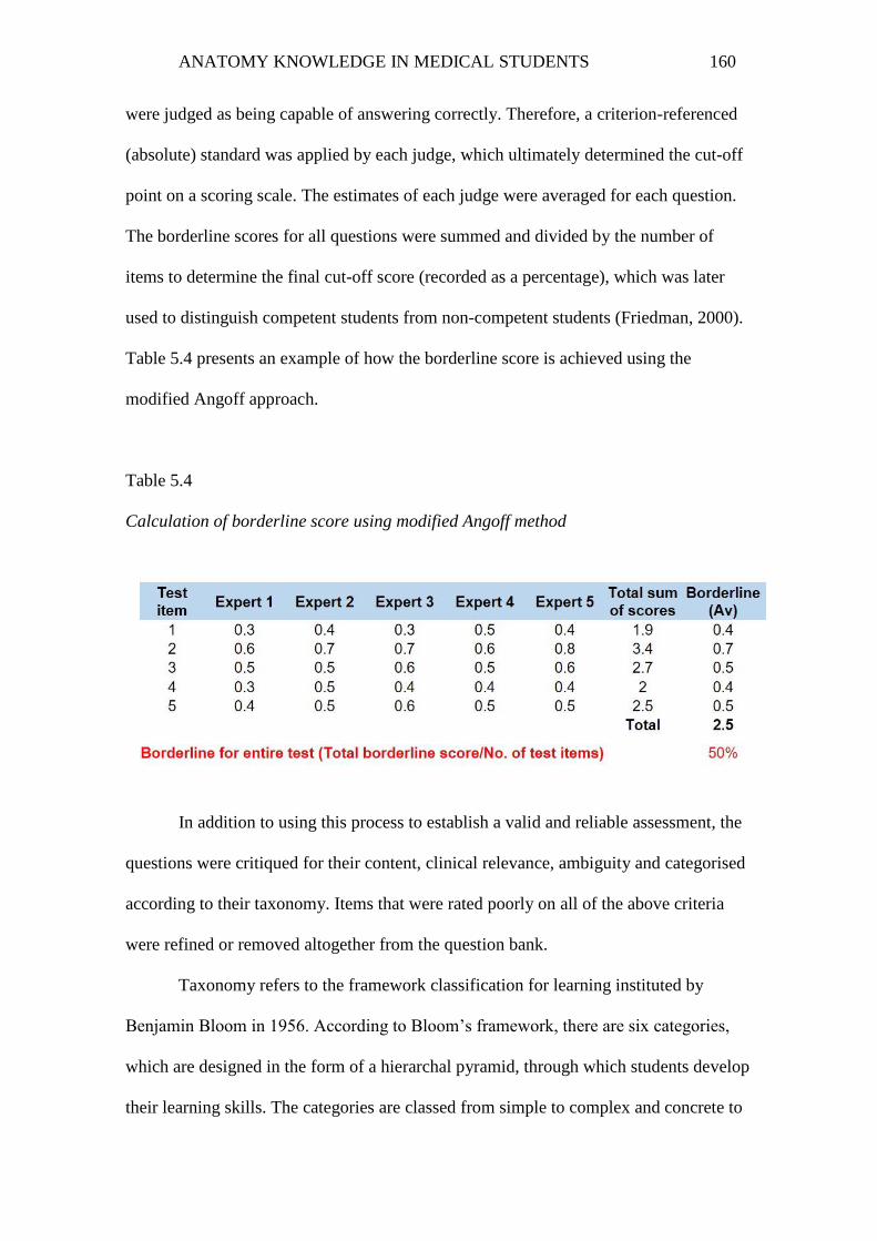
ANATOMY KNOWLEDGE IN MEDICAL STUDENTS 160
were judged as being capable of answering correctly. Therefore, a criterion-referenced
(absolute) standard was applied by each judge, which ultimately determined the cut-off
point on a scoring scale. The estimates of each judge were averaged for each question.
The borderline scores for all questions were summed and divided by the number of
items to determine the final cut-off score (recorded as a percentage), which was later
used to distinguish competent students from non-competent students (Friedman, 2000).
Table 5.4 presents an example of how the borderline score is achieved using the
modified Angoff approach.
Table 5.4
Calculation of borderline score using modified Angoff method
In addition to using this process to establish a valid and reliable assessment, the
questions were critiqued for their content, clinical relevance, ambiguity and categorised
according to their taxonomy. Items that were rated poorly on all of the above criteria
were refined or removed altogether from the question bank.
Taxonomy refers to the framework classification for learning instituted by
Benjamin Bloom in 1956. According to Bloom’s framework, there are six categories,
which are designed in the form of a hierarchal pyramid, through which students develop
their learning skills. The categories are classed from simple to complex and concrete to
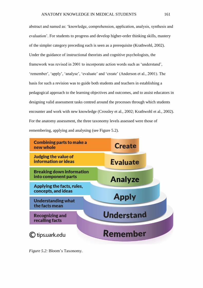
ANATOMY KNOWLEDGE IN MEDICAL STUDENTS 161
abstract and named as: ‘knowledge, comprehension, application, analysis, synthesis and
evaluation’. For students to progress and develop higher-order thinking skills, mastery
of the simpler category preceding each is seen as a prerequisite (Krathwohl, 2002).
Under the guidance of instructional theorists and cognitive psychologists, the
framework was revised in 2001 to incorporate action words such as ‘understand’,
‘remember’, ‘apply’, ‘analyse’, ‘evaluate’ and ‘create’ (Anderson et al., 2001). The
basis for such a revision was to guide both students and teachers in establishing a
pedagogical approach to the learning objectives and outcomes, and to assist educators in
designing valid assessment tasks centred around the processes through which students
encounter and work with new knowledge (Crossley et al., 2002; Krathwohl et al., 2002).
For the anatomy assessment, the three taxonomy levels assessed were those of
remembering, applying and analysing (see Figure 5.2).
Figure 5.2: Bloom’s Taxonomy.
ANATOMY KNOWLEDGE IN MEDICAL STUDENTS 162
Source: http://tips.uark.edu/using-blooms-taxonomy/
Following the standard setting process with the clinicians and anatomy tutors, 60
anatomy questions were selected for the assessment task using the relevant objectives to
which each question was matched. Ten questions that were initially constructed were
deleted from the final question bank because of a lack of clinical relevance and
importance, as identified by the clinicians. To illustrate the purpose of clinical relevance
in the assessment, these 60 topic areas/objectives are outlined in Appendix E. The
questions used in the online assessment are listed in Appendix F. Ultimately, a common
borderline score was attributed by all reviewers to each of the 60 anatomy questions,
bringing the overall cut-off point for the assessment to 50.2%. This value was used in
conjunction with the standard error of measurement to establish clear pass scores for the
assessment.
As stated by Brown (1999), “the standard error of measurement is related to test
reliability in that it provides an indication of the dispersion of the measurement errors
when you are trying to estimate students’ true scores from their observed test scores” (p.
21). A student’s observed score is the raw score obtained by that student on a test,
whereas a true score is part of the student’s observed score that is uninfluenced by
random events such as fatigue. To determine the effect of measurement errors on an
observed score, the standard error of measurement (SEM) must be calculated (Tavakol
& Dennick, 2011). This is done using the SD of the test scores and the reliability (r) of
the test. The calculation of these values can only be determined following data
collection (see Chapter 6. However, the formula used for the calculation is
SEM = SD (1− r).
When determining a cut-off score for a test, as has been established with our
standard setting process and borderline score of 50.2%, one must be 95% confident that
ANATOMY KNOWLEDGE IN MEDICAL STUDENTS 163
students on one side of the cut-off point might fluctuate to the other side if they have an
opportunity to re-take the test (Brown, 1999). To do so, the SEM must be considered.
Using this value, the clear pass score is calculated as the borderline score plus the SEM,
and the clear fail score is calculated as the borderline score minus 2*(SEM). The
decision to utilise BL – 2(SEM) was to maintain consistency with the approach utilised
by Monash University in their analysis of assessment data so as to allow a fair
comparison of assessment results when obtaining the clear pass and clear fail scores.
Students scoring in between these two points are referred to as borderline students.
5.5.2 Test Administration (Phase One)
Given that participants were scattered across many regions of Victoria, it was
impossible to ask students to attend the assessment in person, which would have been
the case in a summative written examination. Therefore, the administration of the
anatomy-learning questionnaire and assessment was delivered online to facilitate the
ease with which students could enrol and participate in the research. This gave students
the opportunity and flexibility to access the assessment in their own time on a given
day, either from a computer, laptop or mobile device.
Development of Online Site
The site was designed with the assistance of a site developer. The questionnaire
and anatomy questions were securely emailed to the site developer, who then began the
process of constructing the web page. The limitations and flaws associated with
delivering an online tool had to be considered. These included opportunities for students
to ‘cheat’ by accessing other resources, to sit the assessments with other students or
friends, or to exit the system and make multiple attempts. Given the aims of this
research, it was imperative for the team to ensure, as best as possible, that honest data
could be obtained that were reflective of participants’ current anatomical knowledge.

ANATOMY KNOWLEDGE IN MEDICAL STUDENTS 164
Thus, the online tool was developed with these considerations in mind. Other than the
anatomy-learning questionnaire, the assessment component of Phase One was set up as
a timed system. Participants were instructed that they had a maximum of one minute to
respond to each question, with a maximum total time of 60 minutes to answer 60
questions. The strict time limit of one minute per question was added as a feature of the
online tool to prevent participants from using online or other resources to answer
questions. It was also used as a surrogate measure to gauge students’ engagement.
When assessments are designed to require students to tap into their higher-order
cognitive skills, it allows them to develop a deeper understanding of the content and to
increase their engagement in learning (Vogler, 2002). Given that the assessments were
constructed around real-life clinical vignettes that require students to think deeply, if
students answered each question within a few seconds, their engagement during the
assessment could be in question. Students were also informed not to press the ‘back’
button on their web browser or mobile device, because this would prevent them from
continuing with the assessment task.
The assessment was clinically relevant and incorporated content from all regions
of the body. The 60 anatomical questions were grouped into six categories pertaining to:
back (5), head and neck (10), upper limb (10), lower limb (10), thorax (10), and
abdomen and pelvis (15). The site was constructed so that participants would view the
questions within a block system. That is, all questions pertaining to one region (e.g.,
head and neck) would be followed by all questions within another region (e.g., thorax).
The order in which the block of questions was presented varied for each participant.
Each participant would have a different set of regional questions appear as the first
block on the assessment task; thus, if two participants were to sit the test together, there
was an 83% chance that each student would have a different regional block of anatomy

ANATOMY KNOWLEDGE IN MEDICAL STUDENTS 165
questions appear first. The aim of designing the online tool in this fashion was to
prevent collusion when attempting the test, and to limit the opportunities for
participants to be dishonest in their reflections.
5.6 Development and Administration of Learning Anatomy Survey
and Interview Questions
The learning anatomy questionnaire was developed from a similar tool that was
pilot tested on a previous cohort, from which findings were presented at conferences.
The qualitative approach to Phase Two of the research involved conducting an
interview session with participants to explore their experiences with learning, forgetting
and retention of anatomy knowledge. The interview questions utilised for this research
were developed as part of an existing research project and modified for the purposes of
this study.
The purpose of the tool was to explore the research questions and to gain a better
understanding of the data obtained through the quantitative analysis. The interview
aimed to focus on topics relating to how students learned anatomy; what approaches and
teaching methods they preferred for mastering anatomical knowledge (particularly in
Year A), how much anatomy they felt they had retained, how much anatomy they had
used in the clinical setting and how important they perceived anatomy to be in the
clinical setting.
Using Seidman (2013) and King and Horrocks’ (2010) guide to interviewing and
qualitative research, a set of questions was formulated for use during the interviews, as
listed in Appendix G.

ANATOMY KNOWLEDGE IN MEDICAL STUDENTS 166
5.7 Data Collection
5.7.1 Phase One
Once consent had been obtained, participants were given an approximate
timeframe and advised that, one week prior to commencement of the data collection for
Phase One, they would receive an email containing a link to the secure online anatomy
assessment site, along with their user ID and password. The user ID for accessing the
online tool was the participant’s student ID. Each student was also assigned a randomly
generated password. Participants were instructed that the link could only be accessed on
the specified day for a period of 24 hours, and if they tried to access it before then, the
site would display ‘This page is not available’.
The clinical-year students (Years B–D) had upcoming summative assessments
in early November, and the Year A students were due to complete their final week of
classes in early November. Therefore, two separate dates were nominated for the data
collection for this quantitative phase. The data collection for participants in Years B–D
took place in the last week of October, whereas the data collection for participants in
Year A took place in the first week of November.
On the date prior to that specified for Phase One, participants were emailed a
reminder advising them that the site would be open in less than 24 hours. A similar
email was sent on the ‘go live’ date, with participants able to access the online site by
entering their user ID and password. Once logged in, participants were introduced to the
research tasks and the research team via a welcome message. The purpose of the study
was highlighted once again, and participants were informed about the two parts
associated with this phase. Thereafter, participants commenced the anatomy-learning
questionnaire. Upon completion, participants were directed to part two—the anatomy
assessment task. An example screen displayed the style in which the questions would
ANATOMY KNOWLEDGE IN MEDICAL STUDENTS 167
appear, and the location of the countdown timer enabled students to visualise the
appearance of the site, the placement of the questions and their options, and how to
monitor their time for each question. Subsequent to this, students began the online
assessment.
During the ‘go live’ dates, the student researcher and the site developer
monitored students’ participation on the site. Those who had not yet logged on were
sent a further email reminder to do so before the assessment task and site closed.
As with any online system, there were implementation issues. Participants
would be unable to complete the task if they clicked on the ‘back’ button on their web
browser; therefore, the research team implemented rules in case a student encountered
this issue. It was decided that if the student had completed fewer than ten questions,
they would be allowed to re-enter the site and complete the task. As was expected, four
participants were accidently locked out of the site and were unable to progress further
on the assessment task. The participants immediately notified the research team and,
because they had not advanced very far on the task, their access to the site was
reinstated and they were able to log on and complete the assessment. When analysing
the data for these students, no changes were noted in their responses from their first
attempt to answer the questions.
5.7.2 Phase Two
In a sequential explanatory mixed-methods study, the key purpose of the
quantitative data collection, in addition to answering part of the research question, is to
guide the development and selection of participants for the second follow-up phase,
which involves the collection of qualitative data. Participants selected for Phase Two
were emailed an invitation to meet for a face-to-face or phone interview. Ultimately, 13
interviews were conducted with participants from the four cohorts: three from Year A,

ANATOMY KNOWLEDGE IN MEDICAL STUDENTS 168
four from Year B, and three each from Years C and D. During the interviews,
participants were advised that all information would remain confidential. All interviews
were recorded and later transcribed verbatim. Each interview lasted approximately
45 minutes to one hour. The shortest interview was 32 minutes and the longest was two
hours.
5.8 Data Analysis
5.8.1 Phase One
All Phase One data (i.e., the learning anatomy questionnaire and the anatomy
assessment scores) were downloaded as two separate Excel files by the site developer.
These files contained raw data, so a clean-up of the data was carried out using SPSS
Statistics version 22. To collate the responses from both the learning anatomy
questionnaire and the anatomy assessment, the information from both files was
transferred and merged into one SPSS document. This ensured that subsequent analysis
could be carried out using a number of key variables, and that comparisons could be
inferred between the variables. The main areas of focus within the analysis included the
following:
examination of original data to determine extreme outliers and the subsequent
exclusion of participants’ data
statistical analysis of anatomy questions, including the calculation of reliability,
SD and SEM
overall performance of participants on the anatomy test and comparisons
between the four cohorts
regional performance of participants on the anatomy test and comparisons
between the four cohorts

ANATOMY KNOWLEDGE IN MEDICAL STUDENTS 169
effect of the three different types of questions (taxonomies) on participants’
performance on the anatomy test
comparison of participants’ original Year A VIA scores and their performance
on the anatomy test
comparison of Year A participants’ performance on the anatomy test and the
effect this had on their subsequent summative assessment (VIA)
comparison of participants’ prior educational background and performance on
the anatomy test
teaching resources mostly used by participants in learning anatomy
best approaches used by participants in learning anatomy and their relationship
to performance on the anatomy test
participants’ perceptions of anatomy.
The statistical tests utilised in the analysis of data involved a range of parametric tests
such as ANOVA and paired t-tests where the data met the assumptions for normality (as
assessed by skewness, kurtosis, Shapiro-Wilk, Q-Q plot and boxplot). This is followed
by post-hoc tests for pairwise comparisons (Tukey-Kramer) and Pearson correlations
for statistical significance. Where data did not meet normal assumptions, the analyses
were undertaken using a series of non-parametric tests involving the Chi-square test of
independence, Spearman’s rank correlation, Mann-Whitney test and Kruskal-Wallis H
test followed by appropriate post-hoc tests for pairwise comparisons (Dunn’s procedure
using Bonferroni correction). The results of the above analysis are presented in Chapter
6.
5.8.2 Phase Two
The qualitative data were analysed using thematic analysis (King & Horrocks,
2010) to determine which aspects provided insights into the quantitative data. Thematic

ANATOMY KNOWLEDGE IN MEDICAL STUDENTS 170
analysis is the process of identifying, analysing, interpreting and reporting patterns
(themes) within qualitative data (Boyatzis, 1998; Braun & Clarke, 2006). This method
allows for flexibility because it can be applied to a variety of theoretical frameworks and
shaped or constructed in a way that is useful and that suits sequential designs (Braun &
Clarke, 2006). Four decisions had to be made in relation to approaching the data to
determine the form that the thematic analysis could take (Braun & Clarke, 2006).
The first criterion necessary to shape the analysis was to determine the content
from the data set that could be categorised as a theme. A theme captures data that are
essential in answering the research question. It contains prevalent and meaningful patterns
of responses that can be found within a particular data item or across the whole data set
(Trahan & Stewart, 2013). A data set refers to all individual data items that are collected
within a data corpus (i.e., all data collected in a research project, such as quantitative and
qualitative data) and that are important and relevant to the topic at hand. Therefore, the
data set for Phase Two referred to all 13 interviews that were recorded, and the data item
referred to each individual interview recording and transcript (Braun & Clarke, 2006). It is
unclear how prevalent a concept must be to be classified as a theme. In the recent body of
literature on thematic analysis, a void still exists around establishing universally accepted
standards for recognising the relevance of a pattern. Instead, researchers are advised to use
their own judgement in this process, and to pay particular attention to issues that are
relevant to the research question and/or issues that help explain the quantitative results and,
in both cases, to ensure that a consistent style is applied to the entire process (Braun &
Clarke, 2006; Trahan & Stewart, 2013). Using this approach, the data set was explored for
repeated patterns of meaning, and a set of themes was identified and established. These
themes are discussed in Chapter 7.

ANATOMY KNOWLEDGE IN MEDICAL STUDENTS 171
The second criterion involved the description of the data and whether it would
consist of detailed accounts of the entire data set or only certain aspects of it. As Trahan
and Stewart (2013) state, “In mixed methods research, the scope of thematic analysis
should be guided primarily by the research model” (p. 66). Given the nature of the
sequential explanatory model, wherein qualitative data were used to explore and explain
the quantitative results, the data were examined for themes that related to the research
questions and for themes that helped elaborate the specific findings of the quantitative
data. That is, the description of the data as documented in the subsequent chapter does
not contain a detailed account of each participant’s journey; rather, it highlights specific
issues as they relate to the study.
The third criterion involved determining whether the themes identified should be
determined via an inductive or deductive approach. An inductive or bottom–up
approach ensures that the themes identified are linked directly to the data; therefore,
they may have little significance to the research questions. In contrast, a deductive or
theoretical approach is explicitly driven by the research questions and the researcher’s
interest in the area, and it often calls for engagement with the literature prior to thematic
analysis (Braun & Clarke, 2006). Once again, the research model for the study assisted
in this decision; accordingly, the sequential explanatory design was best served by a
deductive approach to thematic analysis. This is because “the quantitative antecedent
can be used to identify the most salient concepts and test relationships between them.
The subsequent qualitative study can then be used to identify these concepts in the
qualitative data and elaborate on their interactions” (Trahan & Stewart, 2013, p. 67).
The final decision to be addressed involved determining the ‘level’ of thematic
analysis (i.e., sematic or latent analysis). A semantic approach involves identifying
themes within the explicit and surface-level meanings of the data. These themes can

ANATOMY KNOWLEDGE IN MEDICAL STUDENTS 172
then be organised, described or summarised and interpreted to generate an explanation
of a particular concept or pattern and its broader implications. However, a latent
approach involves looking beyond the semantics to examine, analyse and theorise the
themes identified within the data (Braun & Clarke, 2006; Trahan & Stewart, 2013). For
this study, a semantic approach was used to generate themes across the entire data set.
Following the decisions made on the above four criteria, a six-step procedure to
thematic analysis was followed, as outlined by Braun and Clarke (2006):
1. Familiarisation with the data: This involved engaging in a process of
active reading and re-reading of the data to allow repeated patterns of
meaning to be identified. The process began at the outset of data
collection because the primary researcher was involved in conducting
interviews with the participants. All 13 interviews were transcribed
verbatim and then printed and compiled into one data set. During the
reading phase, notes and coding ideas were listed against the transcripts
for future use and analysis.
2. Generation of preliminary codes: Once familiar with the data, a set of
initial codes was listed. These codes (semantic in nature) alert the
researcher to data that are of interest to the study. As Boyatzis (1998)
describes, codes offer “the most basic segment or element of raw data or
information that can be assessed in a meaningful way regarding the
phenomenon” (p. 63). Each data item (i.e., interview transcript) was
read, and code words were generated manually using a theory-driven
approach—that is, considering the research questions and the
quantitative results. The key issues identified within each data item were
then reviewed, and further codes for the entire data set were developed.
ANATOMY KNOWLEDGE IN MEDICAL STUDENTS 173
Data extracts (individual chunks of information from a data item) were
coded from each data item, and all data extracts for a particular code
across the data set were collated. Some data extracts were coded more
than once.
3. Creation of themes: Following a review of all codes generated in the
previous step, a search was conducted for repeated patterns or themes.
This involved analysing the codes from a broader perspective to identify
general patterns (themes) and subthemes within those patterns across the
data set. It also involved organising the data extracts under such themes.
4. Revision of themes: During this phase, the reliability and accuracy of the
themes identified in the previous step were examined (Trahan & Stewart,
2013) to ensure that the apparent patterns could indeed be classified as
themes. Two criteria (internal homogeneity and external heterogeneity)
had to be verified to ensure that the patterns constituted themes (Patton,
1990). Internal homogeneity refers to data within a theme that coheres in
a meaningful way, whereas external heterogeneity refers to clear and
identifiable differences that exist between the different themes. To assess
these criteria, the data were analysed at two levels, as recommended by
Braun and Clarke (2006). The first involved re-reading all of the data
extracts for each theme to determine whether a meaningful pattern
existed between them. If this was the case, then internal homogeneity
was maintained. However, if the data did not fit into the general pattern
of the theme, the data were either reassigned to another existing theme, a
new theme was created to accommodate the data, the existing theme
itself was reworked or the data were discarded from the analysis. The

ANATOMY KNOWLEDGE IN MEDICAL STUDENTS 174
final step in this stage of the analysis was the creation of a thematic map
to capture the contours of the coded data. The second level of analysis
involved re-examining (i.e., re-reading) the entire data set to ensure the
validity of each theme in relation to the data, and to code any additional
data that might have been missed the first time around. As Trahan and
Stewart (2013) write, “the themes are considered heterogeneous if,
without intersecting, they precisely reflect the content of the data without
missing any important concepts” (p. 70).
5. Defining of themes: This stage involved further refinement of the themes
that would later be presented for analysis by creating definitions that
captured the essence of each meaning. The purpose of this was to create
a detailed analysis of each theme to build a broader picture of its
significance to the research questions, and to ensure that there was no
overlap with other themes. Braun and Clarke (2006) suggest that if the
analysis has been undertaken correctly, it should be possible for the
researcher to describe the content of each theme and its implications in
only a few sentences.
6. Write-up of analysis: This phase, which is presented in Chapter 7
conveys the results of the analysis as clearly and concisely as possible.
Evidence of the story obtained through the data is organised in a logical
and non-repetitive way. Data extracts and vivid examples are used to
illustrate the essence of the story being told. The chapter not only
describes the qualitative results, but also provides an analytic narrative
for the data to demonstrate how the results answer the research questions
(Braun & Clarke, 2006).

ANATOMY KNOWLEDGE IN MEDICAL STUDENTS 175
In order to ensure triangulation data obtained through thematic analysis
(Creswell & Plano Clark, 2011), all researchers in the study were consulted in the data
analysis of the interview transcripts and agreed on the themes identified. In employing
these stages of thematic analysis—namely, descriptive coding, which involves
highlighting relevant material and assigning descriptive codes; integrative coding,
whereby one groups and makes sense of the descriptive codes and their significance in
relation to the research question; and overarching themes, which identify key themes
across the entire data set (King & Horrocks, 2010)—a relationship was found between
the main themes, which provided insights into the importance of anatomy teaching,
retention and knowledge in clinical performance. The results of the quantitative and
qualitative strands were explored together to determine the outcomes of the research
questions.

ANATOMY KNOWLEDGE IN MEDICAL STUDENTS 176
Chapter 6: Quantitative Results
This chapter presents the results of the data analysis from Phase One, whereby
data were collected and analysed in response to the three research questions posed in
this study. This research aims to explore the retention of anatomy knowledge across all
four cohorts of the graduate-entry MBBS program, as examined cross-sectionally, with
the ultimate aim of understanding what causes students to retain or lose their anatomical
knowledge as they transition from student to doctor.
Accordingly, the quantitative data analysis presented in this chapter is separated
into five major sections, as outlined below.
Section 6.1 elaborates on the exclusion criteria of student scores undertaken
following the preliminary data analysis. The ‘exclusion criteria’ were set as a priori.
This was a process of identifying and dealing with outliers and with implementing the
rationale underlying the decision to exclude the results of some students from the
analysis.
Section 6.2 discusses the approach taken to analyse the psychometric properties
of the questions developed as the assessment task for the study (i.e., to test students’
knowledge of clinically focused anatomy). It presents statistical analysis, including the
reliability of the anatomy questions, SD of the test scores and SEM. This allows for the
borderline scores to be determined, as well as the clear pass and clear fail scores. In
addition, this section includes the overall participant results and a comparison between
high performers (HPs) and low performers (LPs).
Section 6.3 uses data from the formative anatomy assessment to run a
comparison across the four cohorts and determine whether there are any differences in
anatomy retention, to assess where these differences (if any) lie and to explore patterns

ANATOMY KNOWLEDGE IN MEDICAL STUDENTS 177
of responses across all cohorts on the framework classifications (recall, application and
analysis) for learning.
Section 6.4 compares the anatomy scores of students in the clinical years from
two assessments: the formative anatomy assessment administered in 2014 and the
summative VIA examination from their pre-clinical year. The purpose here is to
compare students’ knowledge of anatomy from when anatomy was formally taught to
them as pre-clinical students to their current position as a clinical student or intern,
where formal teaching in anatomy is highly variable.
Section 6.5 analyses the pre-clinical-year participants’ performance on the
formative and subsequent summative assessment, highlighting the importance of the
testing effect.
Section 6.6 analyses students’ responses to the questions on the learning
anatomy survey.
The findings presented in this chapter demonstrate certain trends relating to
students’ retention of anatomy knowledge, which could have implications for the future
of anatomy education. The implications are explored in Chapter 7.
6.1 Exclusion Criteria
6.1.1 Identifying and Managing Student Outliers
All data collected through the online site from the 138 participants were
downloaded in Excel format and organised according to cohort, overall scores, regional
scores and response times. Prior to conducting any further analysis, the first task was to
scan the data for evidence of incomplete results, duplicate results, data-entry errors and
measurement errors.
There were four duplicate entries corresponding to the four participants who
were accidently locked out of the system. This occurred either because the participants

ANATOMY KNOWLEDGE IN MEDICAL STUDENTS 178
clicked the ‘back’ button on their web browser or they exited the browser completely,
thereby locking themselves out of the system. The four participants contacted the
researcher on the same day to gain access to the site again. When examining the data
from these participants, it was evident that their responses to the few questions they had
answered on the first try did not change during their second attempt. As a result, the
completed data from the second attempt were retained, and the incomplete entries from
the first attempt for all four participants were deleted during analysis. No other data-
entry errors were observed or recorded.
Given that student engagement is strongly associated with performance on
assessment tasks (Bae & Kokka, 2016), it was imperative to ensure that students were
engaged throughout the test, because this would potentially allow for the collection of
information that was objective, useful and reflective of students’ capacity to recall,
recognise or reason out their knowledge of anatomy. Therefore, the response time for
each question, which was recorded as part of the online tool facility, was incorporated
into an overall completion time for each participant and was used as a surrogate
measure of participants’ engagement with the anatomy assessment. The time taken to
complete the assessment was then analysed across all four cohorts.
The preliminary analysis of participants’ engagement revealed two outliers. To
investigate these further, a normal Q-Q plot (see Figure 6.1) and a boxplot analysis (see
Figure 6.2) were constructed using completion time and cohort as the two comparison
variables. The purpose of this was to explore how participants’ data would appear
compared with the rest of the sample.
The Q-Q plot forms a dark straight (diagonal) line when the observed values
conform to the hypothetical distribution (Garson, 2012). If the data are normally
distributed, all cases for that variable will align closely with or on the diagonal line.

ANATOMY KNOWLEDGE IN MEDICAL STUDENTS 179
Figure 6.1: Total time to complete anatomy test in each cohort.
The boxplot shows the median of each cohort as a dark horizontal line. From
there, the interquartile range (25th–75th percentile) is depicted as the length of the box.
The line extending from either end of the box (known as whiskers) represents the
minimum and maximum values for that cohort when they are within 1.5 times of the
interquartile range. Any values greater than 1.5 times this range are depicted as circles
(o) and are considered outliers. Any values that are three times greater than the
interquartile range are depicted as asterisks (*) and are considered extreme outliers
(Ghasemi & Zahediasl, 2012).

ANATOMY KNOWLEDGE IN MEDICAL STUDENTS 180
Figure 6.2: Completion time on online assessment by cohort.
A visual inspection of Figures 6.1 and 6.2 confirms the presence of two outliers
on the Q-Q plot (indicated by the red arrow), whereas the boxplot identifies one
participant (119) in the Year D cohort as an extreme outlier and the other participant
(76) in the Year C cohort as an outlier. Although the Year C participant was not a
distant outlier, the research team decided to look into these participants’ data.
Further investigation of their values showed that both participants completed the
assessment in a very short time—less than three minutes for the Year D student and
nine minutes for the Year C student. Looking through the regional performance, the
Year D participant spent approximately 30 seconds on each of the six regional blocks of
questions, which comprised a minimum of ten questions, whereas the Year C
participant spent as little as one second on numerous questions to a maximum of 20
seconds on others. The overall scores for these two participants were 25% and 28%

ANATOMY KNOWLEDGE IN MEDICAL STUDENTS 181
respectively. The short amount of time devoted to completing this task is clear evidence
of a lack of engagement with the assessment. Further, including these participants’ data
in the analysis would compromise the results because it could lead to a false judgement
of these participants’ knowledge. Given that the mean time for completion of the
assessment was approximately 30 minutes across all four cohorts, and the fact that far
outliers have a significant effect on small sample sizes (Ghosh & Vogt, 2012), the
research team decided to exclude these individuals’ results from the analysis.
Therefore, all results presented henceforth relate to a sample of 136 participants
(97% of the sample initially recruited).
While two other outliers (participants 109 and 138) were also detected,
inspection of their values did not reveal them to be extreme. These two participants
spent a significant amount of time (approximately 48 minutes each) on the assessment
relative to other participants, indicating that they were engaged with the tool in a
maximum capacity, and perhaps spending more time considering their choice of
answers. Their overall performance was 48% and 58% respectively, suggesting that
they were engaged with the assessment task and were attempting to recall and apply
their anatomy knowledge. They did not do significantly well or poorly compared with
others; therefore, their data were retained in the analysis.
6.2 Test Statistics and Question Analysis
As documented in Chapter 5, a standard set process was implemented following
construction of the anatomy questions, where each question was carefully reviewed by a
team of experts for content, question ambiguity, clarity and checking of answers to
ensure there was only one correct response. The overall process resulted in 60 out of the
70 questions initially written being selected for the formative anatomy assessment. The
borderline (BL) score for this test was 50.2%.
ANATOMY KNOWLEDGE IN MEDICAL STUDENTS 182
Following data collection, each question was initially subjected to item analysis
according to the percentage of correct versus incorrect responses. The process set in
place for any item that performed poorly (when compared with the borderline score for
that question) was to have the question reviewed by the anatomy tutors and/or clinicians
for ambiguity. Accordingly, the question was eliminated if there was a level of
uncertainty and retained if it was unambiguous. Of the 60 questions on the test, four
(Upper limb – Q.4 and Q.8; Head, Q.1; Thorax, Q.9) were reviewed because of poor
performance (<20% of participants answered the questions correctly). These questions
were on a higher level of Bloom’s taxonomy (application or analysis); therefore, they
challenged all participants. No ambiguity was found with the content of the question
and its options; hence, all questions were retained in the analysis.
6.2.1 Reliability, Validity, Borderline Score and Standard Error of Measurement
Reliability and validity are fundamental components of a test instrument and
often go hand in hand. The extent to which an instrument measures what it is supposed
to is known as validity, whereas reliability is a measure of internal consistency that
“describes the extent to which all items in a test measure the same concept or construct”
(Tavakol & Dennick, 2011, p. 53). An instrument must be reliable before it can be valid
(Venkatesh, Brown, & Bala, 2013). Therefore, the question bank was tested for internal
consistency using the responses of all 136 participants to the 60 questions. According to
Downing (2004), for assessment data to have a meaningful interpretation, they must be
reproducible (i.e., good internal consistency or reliability). Without this, generalisations
made from assessment data are limited. Cronbach’s alpha () coefficient was used to
assess internal consistency within written examinations. The resulting product of the
test is a value that ranges from 0 to 1. A value of 0.70 or greater indicates that the
written test has good internal consistency and reliability (Cronbach, 1951; Tavakol &

ANATOMY KNOWLEDGE IN MEDICAL STUDENTS 183
Dennick, 2011). However, when the analysis involves a comparison between groups, as
is the case with this study, the minimum standard of reliability that is acceptable can be
lower (i.e., 0.50–0.70) (Aday & Cornelius, 2006). Using SPSS Statistics version 22, the
alpha coefficient for the four cohorts of participants across all questions was calculated
as 0.75, indicating a good level of internal consistency for the formative anatomy
assessment. When examining the alpha for the scale as a whole against each individual
item, there was no change when an item was removed. This suggests that its correlation
with other items was unaffected. Therefore, all items were retained in the sample (Aday
& Cornelius, 2006). Calculations were also performed for each individual cohort. Thus,
Cronbach’s alpha for the Year A (n = 40), Year B (n = 32), Year C (n = 44) and Year D
(n = 20) cohorts were 0.73, 0.76, 0.71 and 0.68 respectively.
Using participants’ overall anatomy scores on the assessment, the SD for the
sample was calculated at 11.1%. With a Cronbach’s alpha of 0.75, the SEM was
calculated using the formula SD*(√1 ). The resulting SEM score was 5.6%. As
Tighe, McManus, Dewhurst, Chis and Mucklow (2010) state, “the greater the
reliability, the smaller the SEM and hence the more accurate the examination, which is a
desirable outcome” (p. 2). The SEM scores and the borderline score of 50.2% were then
used to identify the clear pass mark for the test, which was 55.8% (BL+SEM).
Similarly, the same two scores were used to calculate the clear fail mark for the test,
which was 38.9% (BL 2*SEM).
Therefore, participants who scored at or above 55.8% obtained a clear pass in
the assessment, whereas participants who obtained a clear fail scored below 38.9%, and
the remaining participants, who scored between 39% and 55.7%, were classified as
borderline students.

ANATOMY KNOWLEDGE IN MEDICAL STUDENTS 184
6.2.2 Overall Performance of Participants
Overall, 52% of students obtained a clear pass, 41% were in the borderline
category and 7% failed the assessment. These data were further segregated into
performance by each cohort within the three categories.
Figure 6.3: Overall performance on anatomy assessment by cohort.
As shown in Figure 6.3, most Year A students passed the assessment. There was
an equal spread of Year B participants across the clear pass and borderline categories.
Across the 3 categories of performance, a higher proportion of Year C students had
borderline scores, and a larger number of Year D students passed the formative anatomy
assessment than were classified as borderline or failed.
6.3 Cross-sectional Data and Analysis
Following preliminary analysis of the data, it was necessary to examine the
research questions to determine the statistical methods that could be used to answer
them. One of the questions posed was: How does anatomy retention differ among
0
10
20
30
40
50
60
70
Clear Pass Borderline Clear Fail
Pe
rce
nta
ge
(%
) o
f s
tud
en
ts r
ep
res
en
ted
wit
hin
e
ac
h c
oh
ort
Student performance on anatomy assessment
Year A
Year B
Year C
Year D
ANATOMY KNOWLEDGE IN MEDICAL STUDENTS 185
students in the pre-clinical and clinical years of their MBBS course? To answer this, the
formative anatomy assessment scores obtained from all cohorts during the Phase One
data collection allowed for a cross-sectional comparison of knowledge to be made
across the board using one test instrument, thereby providing evidence for how students
in all cohorts differed in their anatomy knowledge. Therefore, in answering the research
question, the data were explored from multiple perspectives. In the first instance, the
overall scores across all cohorts were analysed while looking for statistically significant
differences between the cohorts. The regional anatomy scores across all cohorts were
then analysed to determine the same, and finally, the performance of cohorts as per the
classification system of questions (i.e., difficulty of questions posed) was explored to
identify whether the pre-clinical and clinical students performed better in one category
of question than another.
6.3.1 Overall Anatomy Scores
To answer the research question, a one-way analysis of variance (ANOVA) was
conducted using the participants’ overall anatomy scores on the formative anatomy
assessment to determine whether there were any statistically significant differences
between the means of students enrolled in different year levels of the graduate-entry
MBBS degree (Schmider, Ziegler, Danay, Beyer & Bühner, 2010). This test was
selected over independent t-tests for each cohort because multiple t-tests increase the
Type I error rate (rejecting the null hypothesis when it is actually true; i.e., false
positive) by 5% each time, whereas an ANOVA controls for this so that the Type I error
rate remains at 5% (Laerd Statistics, 2015).
Testing for Assumptions of Normality, Outliers and Equal Variance
Since the output of most statistical tests requires that data be normally
distributed (Field, 2009), the data was checked for normality and significant outliers
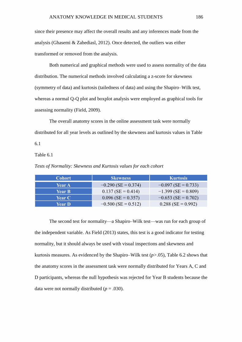
ANATOMY KNOWLEDGE IN MEDICAL STUDENTS 186
since their presence may affect the overall results and any inferences made from the
analysis (Ghasemi & Zahediasl, 2012). Once detected, the outliers was either
transformed or removed from the analysis.
Both numerical and graphical methods were used to assess normality of the data
distribution. The numerical methods involved calculating a z-score for skewness
(symmetry of data) and kurtosis (tailedness of data) and using the Shapiro–Wilk test,
whereas a normal Q-Q plot and boxplot analysis were employed as graphical tools for
assessing normality (Field, 2009).
The overall anatomy scores in the online assessment task were normally
distributed for all year levels as outlined by the skewness and kurtosis values in Table
6.1
Table 6.1
Tests of Normality: Skewness and Kurtosis values for each cohort
Cohort Skewness Kurtosis Year A −0.290 (SE = 0.374) −0.097 (SE = 0.733) Year B 0.137 (SE = 0.414) −1.399 (SE = 0.809) Year C 0.096 (SE = 0.357) −0.653 (SE = 0.702) Year D −0.500 (SE = 0.512) 0.288 (SE = 0.992)
The second test for normality—a Shapiro–Wilk test—was run for each group of
the independent variable. As Field (2013) states, this test is a good indicator for testing
normality, but it should always be used with visual inspections and skewness and
kurtosis measures. As evidenced by the Shapiro–Wilk test (p>.05), Table 6.2 shows that
the anatomy scores in the assessment task were normally distributed for Years A, C and
D participants, whereas the null hypothesis was rejected for Year B students because the
data were not normally distributed (p = .030).

ANATOMY KNOWLEDGE IN MEDICAL STUDENTS 187
Table 6.2
Tests of Normality: Cohort and Anatomy Score on Online Test
However, as indicated above, visual inspection through a normal Q-Q plot (see
Figure 6.4) and a box plot (see Figure 6.5) were explored as additional methods of
confirmation to determine whether there was normal distribution within each of the
cohorts.
Figure 6.4: Anatomy scores on online assessment by cohort.
CohortShapiro-Wilk
Statistic df Sig
Year A .985 40 .854
Year B .926 32 .030
Year C .972 44 .346
Year D .942 20 .262
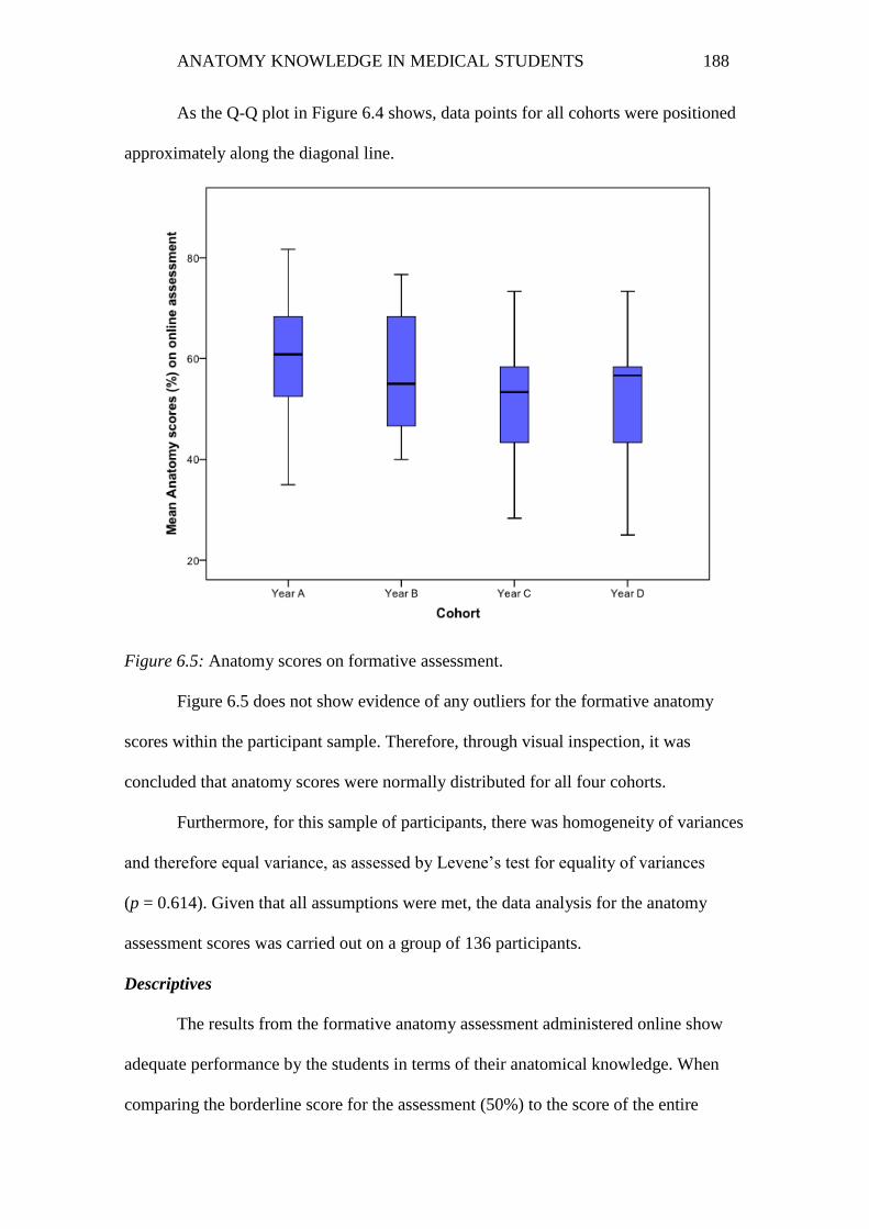
ANATOMY KNOWLEDGE IN MEDICAL STUDENTS 188
As the Q-Q plot in Figure 6.4 shows, data points for all cohorts were positioned
approximately along the diagonal line.
Figure 6.5: Anatomy scores on formative assessment.
Figure 6.5 does not show evidence of any outliers for the formative anatomy
scores within the participant sample. Therefore, through visual inspection, it was
concluded that anatomy scores were normally distributed for all four cohorts.
Furthermore, for this sample of participants, there was homogeneity of variances
and therefore equal variance, as assessed by Levene’s test for equality of variances
(p = 0.614). Given that all assumptions were met, the data analysis for the anatomy
assessment scores was carried out on a group of 136 participants.
Descriptives
The results from the formative anatomy assessment administered online show
adequate performance by the students in terms of their anatomical knowledge. When
comparing the borderline score for the assessment (50%) to the score of the entire
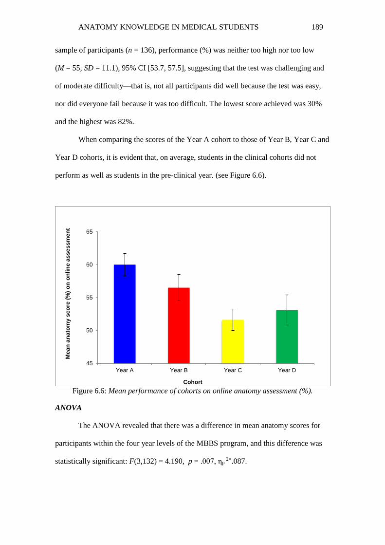
ANATOMY KNOWLEDGE IN MEDICAL STUDENTS 189
sample of participants (n = 136), performance (%) was neither too high nor too low
(M = 55, SD = 11.1), 95% CI [53.7, 57.5], suggesting that the test was challenging and
of moderate difficulty—that is, not all participants did well because the test was easy,
nor did everyone fail because it was too difficult. The lowest score achieved was 30%
and the highest was 82%.
When comparing the scores of the Year A cohort to those of Year B, Year C and
Year D cohorts, it is evident that, on average, students in the clinical cohorts did not
perform as well as students in the pre-clinical year. (see Figure 6.6).
Figure 6.6: Mean performance of cohorts on online anatomy assessment (%).
ANOVA
The ANOVA revealed that there was a difference in mean anatomy scores for
participants within the four year levels of the MBBS program, and this difference was
statistically significant: F(3,132) = 4.190, p = .007, ηp 2=.087.
45
50
55
60
65
Year A Year B Year C Year D
Me
an
an
ato
my s
co
re (
%)
on
on
lin
e a
ss
es
sm
en
t
Cohort

ANATOMY KNOWLEDGE IN MEDICAL STUDENTS 190
A Tukey–Kramer post hoc test (see Table 6.3)—was automatically run using
SPSS Statistics version 22 (Hayter, 1984).
Table 6.3
Tukey–Kramer Post Hoc Test: Comparison of Mean Anatomy Scores
The results show that the pre-clinical students performed the best when
compared with their clinical counterparts. As shown in Table 6.3, there was a
statistically significant decrease of 7.84%, p = .006, 95% CI [1.74, 13.9] in the mean
anatomy scores from the Year A to the Year C group of participants. All other
comparisons were non-significant (p > .05). Therefore, knowledge appears to decline in
the latter half of the clinical years and this difference is most evident and statistically
significant in the group that is highly represented in the sample (Year C).
6.3.2 Regional Anatomy Scores
The next component in answering the research question was to analyse cohort
performance according to the different regional anatomy blocks to determine whether
any statistically significant differences could be reported.
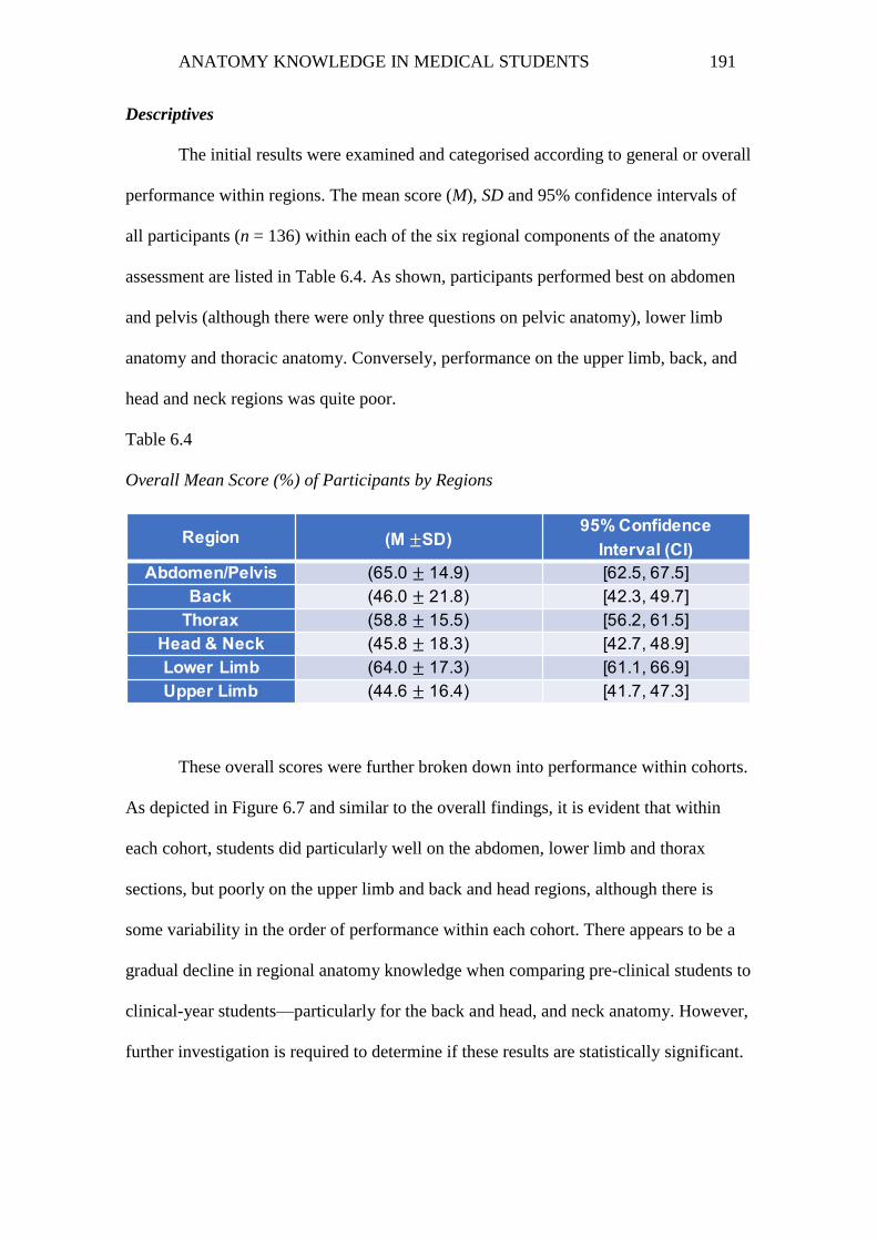
ANATOMY KNOWLEDGE IN MEDICAL STUDENTS 191
Descriptives
The initial results were examined and categorised according to general or overall
performance within regions. The mean score (M), SD and 95% confidence intervals of
all participants (n = 136) within each of the six regional components of the anatomy
assessment are listed in Table 6.4. As shown, participants performed best on abdomen
and pelvis (although there were only three questions on pelvic anatomy), lower limb
anatomy and thoracic anatomy. Conversely, performance on the upper limb, back, and
head and neck regions was quite poor.
Table 6.4
Overall Mean Score (%) of Participants by Regions
These overall scores were further broken down into performance within cohorts.
As depicted in Figure 6.7 and similar to the overall findings, it is evident that within
each cohort, students did particularly well on the abdomen, lower limb and thorax
sections, but poorly on the upper limb and back and head regions, although there is
some variability in the order of performance within each cohort. There appears to be a
gradual decline in regional anatomy knowledge when comparing pre-clinical students to
clinical-year students—particularly for the back and head, and neck anatomy. However,
further investigation is required to determine if these results are statistically significant.

ANATOMY KNOWLEDGE IN MEDICAL STUDENTS 192
Figure 6.7: Mean performance of cohort across six regional anatomy blocks.
Analysis
A non-parametric test—a Kruskall–Wallis H-test was run to explore the
differences in the medians on each of the regional scores between the four cohorts: Year
A (n = 40), Year B (n = 32), Year C (n = 44) and Year D (n = 20). The results, which
are presented below, indicate that the cohort scores for the four anatomical regions
(thorax, abdomen, upper limb and lower limb) were not significantly different.
The two regions that produced a statistically significant difference (p < .05)
were the back and head and the neck components of the formative anatomy assessment,
with the difference in median scores being χ2 (3) = 13.374, p = .004 and χ2
(3) = 18.188,
p = .000 respectively.
A post hoc analysis (see Figure 6.8) using Dunn’s procedure with Bonferroni
correction for multiple comparisons revealed a statistically significant difference in
0
10
20
30
40
50
60
70
80
Abdomen Back Thorax Head Lower Limb Upper Limb
Me
an
sc
ore
(%
) o
f C
oh
ort
Anatomy Regions
Year A
Year B
Year C
Year D

ANATOMY KNOWLEDGE IN MEDICAL STUDENTS 193
back scores between the Year A (Mdn = 60) and Year C (Mdn = 40) cohorts, but not for
the Year B (Mdn = 40, p = .447) or Year D (Mdn = 50, p = .593) cohorts or any other
combination.
Figure 6.8: Pairwise comparison of back scores across all cohorts.
Similarly, pairwise comparisons for head anatomy (see Figure 6.9) scores using
Dunn’s procedure and Bonferroni correction for multiple comparisons revealed
statistically significant (p < .05) differences in performance between the Year A
(Mdn = 50) and Year C (Mdn = 40) (p = .001) cohorts and between the Year A and Year
D (Mdn = 40) (p = .024) cohorts, but not for any other combination.

ANATOMY KNOWLEDGE IN MEDICAL STUDENTS 194
Figure 6.9: Pairwise comparison of head scores across all cohorts.
Therefore, there appears to be a statistically significant difference in head, neck
and back anatomy knowledge between the pre-clinical and later-year clinical students.
6.3.3 Cohort Performance Across Difficulty
During the standard setting process, each question was assigned to one of three
taxonomic classifications levels—remembering, applying or analysing—which are
categorised according to the cognitive skills involved in responding to a question. These
three categories of questions were similar to what students would have been exposed to
in Year A examinations, with each classification increasing in the level of difficulty and
the effort required by the student to generate the right answer. Therefore, the final

ANATOMY KNOWLEDGE IN MEDICAL STUDENTS 195
component of assessing the differences between pre-clinical and clinical students was to
detect whether there were any significant distinctions in how the students responded to
the three different types of questions that comprised the anatomy assessment.
Descriptives
Of the 60 questions, there were 23 recall questions (38%), 29 application
questions (48%) and eight analytical questions (14%). The overall mean performance
(n = 136) for questions involving remembering knowledge was (M = 61.2, SD = 14.6).
Performance decreased by approximately 6% for application-type questions (M = 54.8,
SD = 12.8) and by 18.5% for analysis-type questions (M = 42.7, SD = 17.6). These data
were further broken down, and the mean scores from each cohort were clustered
according to the framework classification (see Figure 6.10).
Figure 6.10: Mean performance of cohort according to taxonomy.
0
10
20
30
40
50
60
70
80
Recall Application Analysis
Me
an
pe
rfo
rma
nc
e o
f c
orr
ec
t re
sp
on
se
s (
%)
by
co
ho
rt
Taxonomy
Year A
Year B
Year C
Year D

ANATOMY KNOWLEDGE IN MEDICAL STUDENTS 196
Analysis
A non-parametric test—Kruskal–Wallis H-test—was run to determine whether
the median scores of the four cohorts for responses across the three different types of
questions were equal (HO) or different (HA).
All four assumptions for this test were met, and the shapes of the distribution for
each group of the independent variable (cohort) on the dependent variable (scores by
taxonomy) were similar, as confirmed through visual inspection of a boxplot.
Initial analysis revealed that there was a statistically significant difference on the
recall questions, with the difference in median scores among the cohorts of χ2 (3) =
14.630, p = .002.
Pairwise comparisons for each cohort on the recall scores were run using Dunn’s
procedure with Bonferroni correction for multiple comparisons. The findings revealed
statistically significant differences in performance between the Year A (Mdn = 69.6)
and Year C (Mdn = 56.5) (p = .001) cohorts. The differences between the medians of all
other combinations of variables were not significant.
Therefore, the null hypothesis for these groups was retained—that is, the
distribution of performance on the application and analysis questions was the same
across all cohorts.
6.4 Comparison of VIA scores with anatomy assessment scores
The other research question posed in this study was: To what extent do medical
students retain anatomy? The purpose here was to identify differences in the retention of
anatomy knowledge from when students were formally taught in Year A to when they
were retested in their later clinical years on the content covered during their first pre-
clinical year.

ANATOMY KNOWLEDGE IN MEDICAL STUDENTS 197
In an ideal world, best-practice measures for analysing the retention of
knowledge would be to have participants assessed on the same test every year until they
graduated, similar to a study by the research team at the Yale University of Medicine
(Rizzolo et al., 2010). However, the longitudinal approach to this design would create
delays in collecting the required data, and this was not the primary goal of the research,
which, in addition to exploring anatomy retention, was to obtain a snapshot of anatomy
knowledge across medical students in the pre-clinical and clinical settings.
One of the options initially considered to assess retention was to have the
clinical cohorts (Years B–D) sit the same anatomy paper from their Year A VIA
examination. This examination, which takes place at the end of Year A, assesses the
anatomy objectives formally taught in Year A. Although the objectives remain the same
every year, the questions assessed on the VIA were slightly modified from year to year
to maintain examination security. Hence, to preserve the integrity of the examination
material, it was not possible for the research team to acquire and administer these
questions.
Consequently, the option selected was to construct anatomy questions at a
similar level of difficulty which students would have been exposed to in Year A, using
objectives that all cohorts would have covered in Year A, and to then compare the
scores with participants’ anatomy scores in the summative Year A VIA paper.
Given that the aim of this research question was to test how much anatomy
knowledge participants had retained from Year A, participant data from the 2014 Year
A cohort was excluded from the analysis because these individuals had not sat the VIA
examination prior to the anatomy assessment. Therefore, only data from the three
clinical cohorts (Years B–D) were used to explore anatomy retention among
participants.

ANATOMY KNOWLEDGE IN MEDICAL STUDENTS 198
Descriptives
Table 6.5 depicts the anatomy assessment scores for all clinical cohorts, as well
as the VIA scores for these participants, the VIA scores for the non-participants, the
cohort mean on the VIA and the borderline of the VIA paper for each cohort.
Table 6.5
Descriptive scores of the clinical cohorts on Anatomy Assessment (AA) and VIA
By comparing the VIA scores of participants in each cohort to that of the non-
participants and the cohort mean, it is evident that the stronger students in anatomy
participated in the research. This does represent as an apparent bias within the study.
However, if the results show that the stronger students in anatomy perform less well
than their pre-clinical counterparts, then it could be surmised that a similar pattern
would exist for the poorly performing individuals.
Furthermore, the cohort mean scores for the VIA reveal large differences between the
three cohorts. While this could be attributed to the level of anatomy knowledge varying
significantly between cohorts at the Year A level, examination of the VIA borderline
score for each cohort provides evidence of the inconsistency in difficulty of the
assessment from year to year, therefore resulting in lower cohort means when the
examination was more difficult (low borderline) and higher cohort means when the
examination was less difficult (high borderline). Given that the overall borderline for

ANATOMY KNOWLEDGE IN MEDICAL STUDENTS 199
the anatomy assessment (50.2%) was similar to or easier than what other cohorts would
have sat in Year A, the VIA score could arguably be used as a baseline comparison
point for each cohort.
Analysis
To answer the research question, separate paired t-tests were conducted for each
cohort using participants’ anatomy scores on the VIA and on the formative assessment.
To detect the presence of outliers, a normal Q-Q plot and boxplot analysis of the
differences between the anatomy assessment and VIA scores was plotted for each
cohort of participants. This was further compared with Shapiro–Wilk’s test of normality
to identify whether the differences were normally distributed. The results and data
analysis for each are presented for each cohort.
6.4.1 Year B Results
For the Year B cohort, one outlier was detected on the Q-Q plot. However, when
comparing the differences in scores between the formative anatomy assessment and the
summative anatomy VIA in Year A, two outliers (participants 41 and 72) were detected
more than 1.5 box-lengths from the edge of the box in a boxplot (see Figure 6.11), with
values at two ends. Inspection of their values revealed that one of them (participant 72)
was extreme, with a difference of −39%. The other participant had a difference
of + 33%, which was considered small relative to the other scores. The differences in
the scores for the anatomy assessment and VIA were normally distributed, as assessed
by the Shapiro–Wilk’s test (p = .332). This test is usually recommended for small
sample sizes (< 50), and when the researcher is not confident with visual interpretations
of Q-Q plots (Laerd Statistics, 2015).

ANATOMY KNOWLEDGE IN MEDICAL STUDENTS 200
Figure 6.11: Differences in Year B performance between anatomy assessment scores
and anatomy VIA scores (AA–VIA).
Given the normal distribution, a decision had to be made regarding the far
outlier (participant 72). To assess whether the presence of this outlier would have a
substantial effect on the overall results, the analysis was run with and without these
outliers.
In excluding the far outlier from the data (participant 72), the mean for the
formative anatomy assessment was relatively the same (M = 56.5, SD = 11.5), whereas
the mean for the summative anatomy VIA score decreased by 1% (M = 52.9,
SD = 16.4). Shapiro–Wilk’s test of normality was also met, as indicated by p = .179
Further analysis through the paired-sample t-tests did not reveal any statistically
significant difference between the two scores in the absence of the outlier.
As part of the analysis, a correlation output was generated between the two
scores, which revealed a statistically significant moderate correlation (see Figure 6.12)

ANATOMY KNOWLEDGE IN MEDICAL STUDENTS 201
between anatomy scores on the summative Year A VIA and scores on the formative
assessment sat in Year B, as indicated by r(32) = .541, p = .001. This increased to a
larger correlation when the far outlier was removed, r(31) = .615, p = .000.
Figure 6.12: Comparison of participant performance on the VIA assessment in Year A
v. the anatomy assessment in Year B.
Although the outlier elicited an increase in variability, it did not unduly
influence the mean difference or alter the conclusion of the paired-samples t-test;
therefore, its value was retained in the analysis.
6.4.2 Year C Results
Similar findings were evident for the Year C cohort, with the Q-Q plot and
boxplot analysis detecting one outlier (participant 95) more than 1.5 box-lengths from
the edge of the box in a boxplot. The difference in scores between the anatomy
assessment in Year A and the anatomy VIA in Year A for this outlier lay at one extreme
(-49%), as shown in Figure 6.13.
0
10
20
30
40
50
60
70
80
90
100
0 10 20 30 40 50 60 70 80 90 100
An
ato
my A
ss
es
sm
en
t s
co
re in
Ye
ar
B (
%)
VIA anatomy score (%)

ANATOMY KNOWLEDGE IN MEDICAL STUDENTS 202
Figure 6.13: Differences in Year C performance between anatomy assessment scores
and anatomy VIA scores (AA–VIA).
Additionally, the assumption of normality was violated; hence, the differences in
the scores were not normally distributed, as assessed by the Shapiro–Wilk’s test
(p = .023). Given that the paired t-test is quite robust to deviations from normality
(Laerd Statistics, 2015), a paired-samples t-test was run with and without the outlier to
determine whether its presence altered the outcome of the test.
As is evident from the initial analysis of all Year C participants, there was a
statistically significant mean decrease in performance of 11.5% (SD = 10.5), 95% CI
[14.6, -8.3], t(43) = 7.310, p = .000 in the anatomy assessment sat in Year C
compared with the anatomy scores in Year A VIA. There was also a strong positive
correlation between the two scores, which was statistically significant, r(44) = .666,
p = .000.

ANATOMY KNOWLEDGE IN MEDICAL STUDENTS 203
When the outlier was excluded from the analysis, the assumption of normality
was no longer violated, and the data appeared to be normally distributed, as assessed by
the Shapiro–Wilk’s test (p = .721). The mean anatomy score for the formative
assessment for the Year C participants increased by 0.4% (n = 43, M = 52.6, SD = 10.2),
whereas that of their summative Year A VIA decreased by 0.4% (M = 63.2, SD = 13.7).
The correlation between the two scores remained strong, positive and statistically
significant, r(43) = .770, p = .000 (see Figure 6.14).
Figure 6.14: Comparison of participant performance on the VIA assessment in Year A
v. the anatomy assessment in Year C.
With the exclusion of the outlier, the resulting mean difference between the
formative and summative anatomy scores was -10.6% (SD = 8.8), 95% CI [13.3, -7.9],
t(42) = −7.939, p = .000, which was statistically significant.
Therefore, it was questionable whether the data for participant 95 should be
excluded from the analysis. One could argue that outliers contain useful data with vital
information about a student’s knowledge of anatomy as they progress from the pre-
0
10
20
30
40
50
60
70
80
90
100
0 10 20 30 40 50 60 70 80 90 100
An
ato
my a
ss
es
sm
en
t s
co
re in
Ye
ar
C (
%)
VIA anatomy score (%)

ANATOMY KNOWLEDGE IN MEDICAL STUDENTS 204
clinical years to the clinical years. Given that the conclusion was not altered with the
exclusion of this participant’s results, it was decided that the data should be retained.
6.4.3 Year D Results
No outliers were identified through the Q-Q plot or boxplot analysis for the Year
D participants. In addition, the assumption of normality was not violated; hence, the
data of the differences between the formative assessment and summative anatomy VIA
scores were normally distributed, as assessed by the Shapiro–Wilk’s test (p = .265). The
paired-samples correlation between the two scores also indicated a strong positive
correlation, which was statistically significant, r(20) = .709, p = .000 (see Figure 6.15).
Figure 6.15: Comparison of participant performance on the VIA assessment in Year A
v. the anatomy assessment in Year D.
When examining the mean differences between performance on the anatomy
assessment in Year D versus performance in the anatomy VIA paper in Year A, there
was a mean decrease of −19.7% (SD = 10.0), 95% CI [24.3, 15.1], which was
statistically significant, t(19) = −8.840, p = .000.
0
10
20
30
40
50
60
70
80
90
100
0 10 20 30 40 50 60 70 80 90 100
An
ato
my a
ss
es
sm
en
t s
co
re in
Ye
ar
D (
%)
VIA anatomy score (%)
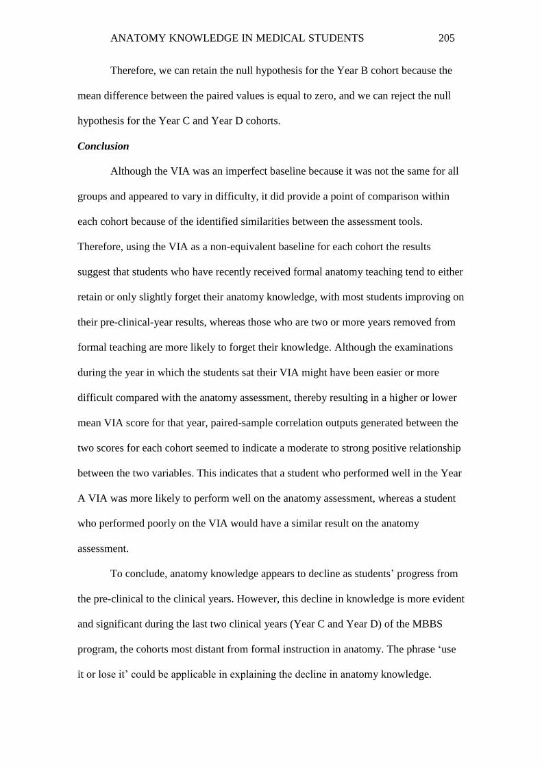
ANATOMY KNOWLEDGE IN MEDICAL STUDENTS 205
Therefore, we can retain the null hypothesis for the Year B cohort because the
mean difference between the paired values is equal to zero, and we can reject the null
hypothesis for the Year C and Year D cohorts.
Conclusion
Although the VIA was an imperfect baseline because it was not the same for all
groups and appeared to vary in difficulty, it did provide a point of comparison within
each cohort because of the identified similarities between the assessment tools.
Therefore, using the VIA as a non-equivalent baseline for each cohort the results
suggest that students who have recently received formal anatomy teaching tend to either
retain or only slightly forget their anatomy knowledge, with most students improving on
their pre-clinical-year results, whereas those who are two or more years removed from
formal teaching are more likely to forget their knowledge. Although the examinations
during the year in which the students sat their VIA might have been easier or more
difficult compared with the anatomy assessment, thereby resulting in a higher or lower
mean VIA score for that year, paired-sample correlation outputs generated between the
two scores for each cohort seemed to indicate a moderate to strong positive relationship
between the two variables. This indicates that a student who performed well in the Year
A VIA was more likely to perform well on the anatomy assessment, whereas a student
who performed poorly on the VIA would have a similar result on the anatomy
assessment.
To conclude, anatomy knowledge appears to decline as students’ progress from
the pre-clinical to the clinical years. However, this decline in knowledge is more evident
and significant during the last two clinical years (Year C and Year D) of the MBBS
program, the cohorts most distant from formal instruction in anatomy. The phrase ‘use
it or lose it’ could be applicable in explaining the decline in anatomy knowledge.

ANATOMY KNOWLEDGE IN MEDICAL STUDENTS 206
However, this retention or lack thereof will be explored further in the qualitative phase
of this research.
6.5 Testing Effect
Prior to the summative VIA examination in 2014, the Year A (pre-clinical)
students sat the formative anatomy assessment, for which their mean performance was
(M = 60.0%, SD = 10.8). When these students undertook their VIA examination a
month later (BL =52%), their mean anatomy performance was much higher
(M = 75.2%, SD = 13.7) relative to that of the entire cohort (M = 71.2%) and non-
participants (M = 67.1%).
Analysis
To further investigate this, a paired-sample t-test was performed to compare the
difference between the means of two paired observations and determine whether the
difference is statistically significant from zero.
For this test, the null hypothesis (H0) was that the mean difference of the
population between the paired values is equal to zero (diff = 0). The alternative
hypothesis (HA) was that the mean difference is not equal to zero (diff ≠ 0).
Assumptions one and two have already been met, as illustrated by the dependent
(anatomy performance) and independent variables (formative anatomy assessment v.
VIA anatomy score). Assumptions three (no significant outliers) and four (distribution
of the differences between the two related groups on the dependent variable should be
approximately normally distributed) were tested using a boxplot analysis and Shapiro–
Wilk’s test of normality respectively.
To detect the presence of outliers, a normal Q-Q plot and boxplot analysis of the
differences between the anatomy assessment and VIA scores were plotted for each
cohort. This was further compared with Shapiro–Wilk’s test of normality to identify

ANATOMY KNOWLEDGE IN MEDICAL STUDENTS 207
whether the differences were normally distributed. For the Year A cohort, there were no
outliers in the data, as assessed by the inspection of the boxplot for values greater than
1.5 box-lengths from the edge of the box.
Figure 6.16: Difference in scores for Year A cohort between formative and summative
assessments.
The differences in the scores for the anatomy assessment and VIA were
normally distributed, as assessed by the visual inspection of a normal Q-Q plot (see
Figure 6.16) and the Shapiro–Wilk’s test (p = .901).
The paired t-test showed that the VIA assessment elicited a statistically
significant mean increase of 15.21%, 95% CI [11.93, 18.48], p = .000 in the summative
anatomy score compared with the formative anatomy assessment. As part of the
analysis, a correlation output was generated between the two scores, which revealed a
statistically significant large correlation (see Figure 6.17) between the two scores, as
indicated by r(40) = .672, p = .000.

ANATOMY KNOWLEDGE IN MEDICAL STUDENTS 208
Figure 6.17: Comparison of participant performance on the formative assessment in
Year A v. the summative anatomy assessment in Year A.
6.6 Learning Anatomy Survey Results
This section outlines the participants’ prior educational background and the
results of the learning anatomy questionnaire. Some aspects are compared with the
anatomy scores in the formative assessment in 2014 and with differences of scores from
the VIA to determine significance between variables.
6.6.1 Demographics
For this research, students’ prior undergraduate degrees were classed into five
categories depending on the amount of anatomy exposure they had: anatomy in a
professional course (physiotherapy, occupational therapy), anatomy in a biomedical
science course, biological science (botany, zoology, genetics, forensic science), other
science (chemistry, physics, engineering) and no science (law, philosophy, music).
0
10
20
30
40
50
60
70
80
90
100
0 10 20 30 40 50 60 70 80 90
VIA
Sc
ore
(%
)
Anatomy assessment score in Year A (%)

ANATOMY KNOWLEDGE IN MEDICAL STUDENTS 209
Of the 136 students, 12% had studied anatomy in a professional course, 30%
had studied anatomy in a biomedical science course, 18% had a biological science
degree and very little anatomy or no anatomy at all, 25% had an ‘other’ science degree
but no anatomy and 15% had a prior degree that was not science-based. Figure 6.18
shows the participants’ educational background within each cohort.
Figure 6.18: Distribution of participants’ prior degree within each cohort (Years A–D).
There is high variability in the prior educational background of participants
within each cohort. Although most participants had a biomedical science degree, which
is largely represented in each cohort (except for Year D), Figure 6.19 also shows that a
large proportion of students came from a background with no anatomy exposure at all
(as represented by other science and no science degrees), and this is most evident in the
Year D cohort. Therefore, it would be interesting to measure any trends in anatomy
performance across the educational backgrounds. To do so, the participants were split
into high and low scorers. Participants who scored at or above 55.8% and obtained a
0
5
10
15
20
25
30
35
40
45
50
Year A Year B Year C Year D
Pa
rtic
ipa
nt
dis
trib
uti
on
%
Anatomy in professional course
Anatomy in biomedical course
Biological Science
Other science
No science

ANATOMY KNOWLEDGE IN MEDICAL STUDENTS 210
clear pass were categorised as high scorers, and the remaining participants (i.e., those
who scored below 55.8% according to the test features outlined at the outset) were
categorised as low scorers. Although the low scorers could be broken down further into
a borderline and clear fail category, for the purpose of comparing performance, the
participants were divided into two groups. Overall, 52% were high scorers (M = 64.3,
SD = 6.35) and 48% were low scorers (M=46.1, SD = 6.31).
Participant backgrounds were analysed against these two groups to analyse
performance on the anatomy assessment.
Figure 6.19: Effect of participants’ educational background on performance.
Of the 12% and 30% of participants who studied anatomy in a professional
course and biomedical science respectively, most obtained a clear pass in the anatomy
assessment, with the remaining individuals scoring below 55.8%. Participants in the
‘biological science’ and ‘other science’ categories were more likely to perform worse
on the anatomy assessment than any other group, with 60% of participants in each
0
10
20
30
40
50
60
70
80
Anatomy inprofessional
course
Anatomy inbiomedical
course
BiologicalScience
Other science No science
Pe
rce
nta
ge
of
hig
h a
nd
lo
w s
co
rers
(%
)
Prior educational background
High Scorers
Low Scorers

ANATOMY KNOWLEDGE IN MEDICAL STUDENTS 211
group classified as low scorers. Finally, students who had no anatomy or science
exposure whatsoever were almost equivalent in their spread across the high scorer and
low scorer categories.
The mean scores on the anatomy assessment across the five groups (see Figure
6.20) shows no statistical significance (p >.05) as examined by the Kruskal-Wallis H
test, although the trend was for participants with prior anatomy knowledge to perform
better. This suggests that prior educational background does not necessarily play a role
in how much knowledge is retained by students.
Figure 6.20: Mean anatomy score on online assessment across the five educational
backgrounds.
6.6.2 Learning Anatomy
In relation to the resources used the most by participants for learning anatomy,
40% chose textbooks, 26% selected anatomy tutorials and 21% used online anatomy
software An@tomedia. A few students selected lectures (4%), while 2% opted for study
groups and 2% for specimen days as their main learning resource. Those who selected
0
10
20
30
40
50
60
70
80
Anatomy inprofessional
course
Anatomy inbiomedical
course
BiologicalScience
Other science No scienceMe
an a
nat
om
y sc
ore
(%
) o
n o
nlin
e a
sse
ssm
en
t
Educational Background

ANATOMY KNOWLEDGE IN MEDICAL STUDENTS 212
the ‘other’ category (8%) indicated that resources used for learning anatomy involved
the internet, Acland’s DVD and anatomy apps, and one student used a combination of
all resources, as shown in Figure 6.21.
Figure 6.21: Resources used the most by students for learning anatomy.
Students were also asked to select the one resource they found the most useful
for learning anatomy. It was expected that the results would mirror those of the previous
question regarding which resource they used the most for learning anatomy. However,
this was not the case. As shown in Figure 6.22, the anatomy tutorials were reported to
be the most useful resource, as selected by 29% of participants. This was followed by
textbooks (21%), anatomy specimen days (16%) and study groups (10%).
0
5
10
15
20
25
30
35
40
45
Textbooks Anatomedia SpecimenAnatomy
days
Tutorials Lectures StudyGroups
Other
Pa
rtic
ipa
nt
us
sa
ge
(%
)
Anatomy Resources

ANATOMY KNOWLEDGE IN MEDICAL STUDENTS 213
Figure 6.22: Most useful resources for learning anatomy.
A chi-square test of independence was run to determine if there was any
significant association between high and low scorers and the resources identified as the
most useful for learning anatomy. While all resources were selected in higher
proportion by the high scorers, there was no statistically significant association between
the two variables. However, given that tutorials appeared to stand out when compared to
low scorers and hence was explored further in the qualitative analysis.
Students were also asked to indicate their approach to learning anatomy to
determine whether they preferred one learning approach to another and whether the
learning approach adopted affected their overall performance on the anatomy
assessment.
Although students can adopt either deep (searching for meaning) or surface
(memorisation) approaches when engaging in a learning task, 70% of participants
0
5
10
15
20
25
30
35
Textbooks Anatomedia Specimenanatomy
days
Tutorials Lectures StudyGroups
Other
Pa
rtic
ipa
nt
us
ag
e (
%)
Anatomy Resources

ANATOMY KNOWLEDGE IN MEDICAL STUDENTS 214
adopted both approaches extensively, with most (50%) searching for meaning first and
then memorising (see Figure 6.23).
Figure 6.23: Approaches mainly used to learn anatomy.
Students who approached learning through pure memorisation of content
(n = 17) had a mean score of 50.3% (SD = 3.2), whereas those employing a search for
meaning approach (n = 20) or search for meaning followed by memorisation (n = 68)
had a mean score of 56.1% (SD = 2.2 and 1.4 respectively). Students who memorised
anatomical content first, followed by an attempt to search for meaning (n = 29),
appeared to perform slightly better, with a mean of 56.7% (SD = 1.9). Two students
selected the ‘other’ approach to learning but did not specify what this was. Their mean
score was 64%, but because they did not meet the criteria for the qualitative phase, it is
not clear what they meant by ‘other’. There does not appear to be any significant
relationship between the learning approach adopted and students’ mean scores as
demonstrated by the Kruskal-Wallis H Test. This contrasts with other studies (Pandey
0
10
20
30
40
50
60
Search formeaning in
content
Memorise content Memorise, thensearch formeaning
Search formeaning, then
memorise
Other
Pe
rce
nta
ge
(%
) o
f p
art
icip
an
ts a
do
pti
ng
ap
pro
ac
h
Learning approach to anatomy

ANATOMY KNOWLEDGE IN MEDICAL STUDENTS 215
& Zimitat, 2007), as evident by the 40% of high scorers and 32% of low scorers who
adopted a combination of surface and deep-learning approaches (see Figure 6.24).
Figure 6.24: Performance of high and low scorers by learning approach.
6.6.3 Attitudes Towards Anatomy
Figure 6.25 depicts students’ attitudes towards their anatomy experience through
a stacked bar chart. The percentage of respondents who agreed with the statement are
shown to the left, whereas the percentage who disagreed are shown to the right and the
percentage who neither agreed nor disagreed are shown in the midline in grey. Overall,
most students agreed that the volume of anatomy in Year A was daunting. When asked
what they perceived to be the challenges to learning anatomy, 60% indicated that the
amount needed to be learned was the biggest hurdle, and 24% stated that visualisation
of structures was challenging. Given that the emphasis on anatomy in Year A is on
clinical relevance, most students considered the volume necessary for clinical practice,
while 14% were unsure. When comparing students’ attitudes towards learning anatomy,
almost half of the participants disagreed that their motivation for learning anatomy was
0
5
10
15
20
25
30
Search for meaning incontent
Memorise content Memorise, then searchfor meaning
Search for meaning,then memorise
Pa
rtic
ipa
nt
us
ag
e (
%)
Learning Approach primarily adopted
High Scorers
Low Scorers

ANATOMY KNOWLEDGE IN MEDICAL STUDENTS 216
to pass examinations; however, 40% indicated that this was their intent. Cross-
tabulation with chi-square statistics did not reveal any statistically significant results
between anatomy performance and the above mentioned variables.

ANATOMY KNOWLEDGE IN MEDICAL STUDENTS
217
Had solid foundation in anatomy prior to Year A
Anatomy perceived as an important part of becoming a doctor
Frequently use anatomy knowledge during clinical skills
Difficult using anatomy knowledge due to lack of confidence in it
Don't see anatomy as the most important subject in my medical career
Main motivation for learning anatomy is to pass exams
Volume of Anatomy in Year A was necessary
Volume of Anatomy in Year A was daunting
Figure 6.25: Anatomy perceptions by participants

ANATOMY KNOWLEDGE IN MEDICAL STUDENTS 218
The relevance of anatomy to clinical skills is apparent when viewing
participants’ responses, with 62% indicating that anatomy knowledge is frequently used
during clinical skills sessions (see Figure 6.26). However, 22% found it difficult to use
their anatomy knowledge because of a lack of confidence. A Spearman’s rank-order
correlation was run to assess the relationship between frequency of use in clinical skills
and difficulty using anatomy because of a lack of confidence in anatomy knowledge.
The preliminary analysis showed the relationship to be monotonic, as indicated by the
visual inspection of a scatterplot. There was a weak negative association between
participants’ frequent use of anatomy in clinical skills and their confidence in using
anatomy, and this was statistically significant, rs(134) = −.298, p < 0.01.
Figure 6.26: Participants’ use of anatomy in clinical skills v. their confidence in use of
anatomy knowledge.
When examining participants’ perceptions of anatomy in medicine, 26% felt that
anatomy was not the most important subject in their medical career, with 49%
0
5
10
15
20
25
30
35
40
45
Agree Disagree
Pe
rce
nta
ge
(%
) o
f p
art
icip
an
ts
Difficult using anatomy due to lack of confidence in anatomy knowledge
Frequently use anatomy in clinical skills
Agree
Disagree

ANATOMY KNOWLEDGE IN MEDICAL STUDENTS 219
disagreeing and the rest unsure (see Figure 6.27). However, a large proportion of
students (95%) viewed anatomy as an important part of being a doctor. Statistical
analysis (Spearman’s rank-order correlation coefficient test rs), showed that there was a
moderately negative association (rs = −0.331) between viewing anatomy as important
for becoming a doctor, but not as an important subject in their medical career (p < 0.01).
These findings will be explored further in the qualitative interviews.
Figure 6.27: Participants’ perceptions of importance of anatomy.
Conclusion
The quantitative phase of this research addresses two research questions: How
do medical students from all four cohorts of an MBBS program differ in their anatomy
knowledge, and how much knowledge is retained by medical students as they progress
0
10
20
30
40
50
60
Agree Unsure Disagree
Pe
rce
nta
ge
(%
) o
f p
art
icip
an
ts
Anatomy is not the most important subject in my medical career
Anatomy is an important part of becoming a doctor
Agree
Unsure
Disagree

ANATOMY KNOWLEDGE IN MEDICAL STUDENTS 220
from the pre-clinical to the clinical years? In exploring the data further, some
unanswered questions have arisen:
Why did the pre-clinical (Year A) students perform better than the clinical
students (Year B–D) in terms of anatomical knowledge?
Why is knowledge of some regional sections of anatomy, such as abdomen,
thorax and lower limb, generally better retained than knowledge of other
regions, such as upper limb, back, and head and neck?
Why were tutorials seen as the most useful resource for learning anatomy, and
how does this resource affect retention?
Why do students perceive anatomy as being important for a doctor, but not as an
the important subject in their medical career?
These issues and others surrounding the learning of anatomy will be explored
further in the sequential qualitative phase of this research.

ANATOMY KNOWLEDGE IN MEDICAL STUDENTS 221
Chapter 7: Qualitative Results
This chapter documents the analysis arising from Phase Two of the research
study—the interviews. The findings demonstrate students’ perceptions of the anatomy
program both in the pre-clinical and clinical years. They provide insights into the
retention and learning of anatomy knowledge by students and outline implications and
students’ suggestions for the future of anatomy education.
Data analysis from the 13 interviews was undertaken using a deductive approach
driven primarily by the research questions and the analysis of quantitative data guided
by the existing literature. The overarching research question guiding the qualitative
phase was: What factors may account for the loss or retention of anatomy knowledge
across a student’s medical degree?
Issues that arose from the quantitative research, and some of the issues posed by
the literature, are addressed by the findings from this phase:
horizontal and vertical integration of anatomy teaching and learning
teaching via dissection or prosection
teaching via body painting
attitudes of clinicians towards medical students’ knowledge of anatomy
assessment of anatomical knowledge.
To understand the issues associated with the loss or retention of anatomical
knowledge, it was necessary to explore the teaching and learning of anatomy as it
occurred in the pre-clinical years, as well as subsequent exposure to anatomy in the
clinical years. After analysing all 13 interviews, four major themes and multiple
subthemes were highlighted as being pertinent to providing insights into the questions
posed (see Figure 7.1). Each of these themes is discussed in the subsequent sections.

ANATOMY KNOWLEDGE IN MEDICAL STUDENTS 222
Figure 7.1: Four themes and subthemes that emerged from participant interviews.
Since all participants had progressed into the next year or graduated from the
MBBS program during the collection of the qualitative data, all excerpts listed in this
chapter cite participants in the year they were in when the interviews were conducted
(see Table 7.1). Therefore, although the interviews are referenced according to the year
in which they were conducted, they were partly retrospective because they related to the
students’ previous experiences in addition to their current experiences in their existing
clinical year.
Table 7.1
Participant’s progression through the MBBS at the time of Phase I and II of study
Phase of Study Cohort
I: Anatomy Assessment Year A Year B Year C Year D
II: Interview Year B Year C Year D Intern
. In addition, all excerpts from interviews are listed as that from a high
performer (HP) or low performer (LP). During the thematic analysis, there was no

ANATOMY KNOWLEDGE IN MEDICAL STUDENTS 223
apparent pattern of difference between responses from high and low performers.
Consequently, information presented in this chapter has been pooled together under the
various themes.
7.1 Anatomy in the Year A Program
This section highlights participants’ views about the pre-clinical (Year A)
anatomy program, including teaching, resources available to students and the
assessment of anatomy. The theme provides insights into why participants in the pre-
clinical year generally performed better on assessments than their clinical counterparts.
It also provides a deeper understanding of the different ways in which anatomy is taught
and learned, as well as its effect on retention of anatomical knowledge in the subsequent
clinical years.
Most participants (69%) were very satisfied with the teaching of the Year A
program. They acknowledged that the amount of time devoted to anatomy teaching
within the whole Year A curriculum was quite extensive (and therefore beneficial for
participants without an anatomy background), especially when compared with other
disciplines, and that not much more could be added into an already-packed program.
The clinical focus of the anatomy curriculum was also considered valuable in helping
students to learn the important concepts in a condensed curriculum. As one participant
in the final clinical year stated:
I liked the fact that the coordinator of the anatomy teaching program, given his
medical and anatomy background was able to blend the two quite nicely and
come up with these clinical applications of anatomy. And I’ve come across quite
a few of these as well during my placements. (HP, Year D)
The remaining 31% of participants who were moderately or not very satisfied
were the LPs of the group. These participants found the Year A anatomy program

ANATOMY KNOWLEDGE IN MEDICAL STUDENTS 224
intense and overwhelming, given the “sheer amount of content that was being
covered…in a very short period of time”. One student indicated that he had to “learn
how to learn anatomy” (LP, Year B); that is, he had to learn how to manage his time
better between the different regions that were covered. Nonetheless, most participants
(85%) agreed that anatomy in Year A provided a good grounding for the first clinical
year (Year B), with the remaining 15% stating that anatomy was not sufficient and that
they would have liked more of it. From the interviews, six major subthemes emerged
regarding the Year A anatomy program, and these are presented below.
7.1.1 Small-group Teaching
Students spent a large proportion of the year learning anatomy in small-group
sessions. As such, the feedback from participants on the different small-group learning
opportunities provides an insight into what worked well and what improvements could
be made for the future.
The small-group teaching sessions usually involved students participating in a
variety of anatomy-related activities in groups of 10–12 individuals guided and/or led
by the tutor as a facilitator. In the Year A anatomy program, small-group teaching
occurred as part of tutorial sessions, surface anatomy classes and imaging/osteology
workshops. During the interviews, participants highlighted certain aspects of these
workshops that were either beneficial or problematic.
Tutorials
The tutorial sessions in anatomy followed a flipped classroom approach wherein
students were responsible for learning information prior to the session through assigned
pre-reading material, and the in-class time was devoted to developing critical thinking
and reasoning skills through the discussion and application of anatomical concepts and
principles to a set of clinically oriented anatomy questions. Given that the tutorial

ANATOMY KNOWLEDGE IN MEDICAL STUDENTS 225
setting was used as a platform for learning clinically relevant anatomy, all participants
valued small-group teaching within tutorials: “Clinical relevance is the most important
thing when studying anatomy because it’s what cements it. You have to have a reason
why you need to know it” (HP, Year C). However, some participants argued that
clinical anatomy is just one aspect of anatomy, and although tutorials are good for
studying clinically relevant material, they did not agree that they had a sound grasp of
anatomy:
The anatomy is very clinically oriented so it is very relevant. But it’s good to
have a bit of the wet specimens as well otherwise you’d end up having holes in
your knowledge. If the only thing you know about the perineal nerve is that it
can be impinged at that spot, then you don’t know the rest of it or what else is
going to happen. So you’re very well prepared for a common scenario and have
absolutely no knowledge about anything else. That’s the problem with clinically
oriented anatomy—It’s very helpful for most situations but it leaves you having
no idea about large sections of anatomy which you will face at some point and
you’ll have no clue. (HP, Intern).
Since the tutorials involved answering a set of questions, this was considered
different to exploring the anatomy around those questions. As a high-performing
participant in the final year stated: “Because it’s very directed and there’s lot of work to
do, more people will google a quick answer but what you need is time to explore the
body” (HP, Intern).
Additionally, the outcome of the tutorial depended on the tutor. Participants
stated that some tutors would take over a session and showcase their knowledge with
long PowerPoint presentations on the detailed anatomy of the region rather than allow

ANATOMY KNOWLEDGE IN MEDICAL STUDENTS 226
students to discuss the questions and state the clinical significance and importance of
the topic at hand. Tutors that had a clinical background were valued more highly than
those who were pure scientists or anatomists.
Surface Anatomy
In the surface anatomy workshops, each group of students and their tutor
gathered around the bedside in a simulated ward. One student volunteered to be drawn
upon, examined and palpated for surface landmarks, and the others engaged in carrying
out the tasks set out for that session. All participants indicated that surface anatomy
teaching in Year A was valuable—especially when clinical tutors were involved. A
high-performing participant in the final clinical year stated:
I felt my Year A anatomy knowledge helped me in the clinical years…Felt
confident with most things. Doctors were impressed when they asked me about
landmarks. They love landmarks you know and rightly so, because it’s how you
can tell where you are without using imaging modalities. (HP, Year D)
Overall, the surface anatomy workshops were perceived as being very useful to
learning anatomy. However, one of the pitfalls of these sessions was the issue of
students volunteering as participants. A student in their intern year elaborated on this
issue:
The issue I had, that is, that it seemed that the same people would have to
volunteer and everyone else would back off…You have to have a degree of trust
and everyone has to just strip down to their undies and just do it...just get over
that fear. I think that would have made everything a bit more worthwhile.
Because, I think it makes it more interactive and then people are more likely to
actually look at things...I think if you’re expecting your patients to have that

ANATOMY KNOWLEDGE IN MEDICAL STUDENTS 227
trust in you, then you should have that trust in your colleagues as well. I know
it’s very confronting but the more comfortable people are at doing it, the less the
stigma and barrier is. (LP, Intern)
Osteology
These workshops involved students examining a set of bones as they relate to
the region being covered that week (e.g., forearm, arm and hand bones during upper
limb sessions) and identifying the different features of those bones. Most participants
found the osteology workshops to be beneficial, but they also stated that too much time
was allocated to these during the year, which resulted in students becoming disengaged
with the tasks that subsequently seemed to be repetitive:
We’d be looking at a bone from one limb and then next week, we’d be looking
at a bone from a different limb. That’s important because you have to know
some of the anatomical landmarks but there’s not that much difference between
bones and it would have been better to learn more specific things that could be
used clinically such as focusing a bit more on imaging. (HP, Year C)
Imaging
Imaging sessions in the Year A anatomy program were twofold. Students spent
half an hour every week in imaging workshops led by tutors, where X-rays, CTs and
MRI scans of different body regions were presented via an online anatomy program
called An@tomedia. Occasionally, there would also be hard-copy X-ray images of real
patients for students to mull over and discuss with a tutor. In addition, twice per
semester, students were taught by a radiologist how to interpret imaging on the anatomy
specimen days. There was a consensus among the participants that introducing imaging
in Year A and learning how to take a systematic approach to interpretation was
important and vital to providing students with a foundation for the later clinical years:

ANATOMY KNOWLEDGE IN MEDICAL STUDENTS 228
I think a little bit of imaging is good. The earlier you see X-rays, CTs, even if
not at an in-depth physician level, but looking at it, knowing how to take a
systematic approach to it is good in Year A. So that when you are in the clinical
years and you start seeing more general medicine, it allows you to recognise
normal structures. (HP, Year D)
It was felt that these sessions were more beneficial when led by clinical tutors as
opposed to non-clinical tutors. Three participants (two HPs and one LP) indicated a
preference for more imaging sessions during Year A, with greater use of scans and X-
rays of real patients as opposed to screenshots of images on An@tomedia. In contrast,
one low-performing participant in the second clinical year indicated a dislike of the
radiology sessions on the anatomy specimen days because the organisation of those
sessions (one tutor for 20 students gathered around a small area in a large, noisy room)
was not conducive to the overall learning experience.
7.1.2 Lectures
The importance of lectures emerged as another theme and an area in which
improvements could be made within the Year A program. In general, there was a mixed
reaction to participants’ perceptions of the usefulness of lectures. Participants liked
lectures when they were delivered by some individuals, but reported that they did not
benefit much from lectures that were side-tracked, disorganised and/or haphazard in
presentation. However, most participants attended lectures for the extra bit of clinical
information they would receive from the lecturer, regardless of whether the presenter
managed to deliver the entire scheduled lecture topic. In particular, lectures on the
principles of anatomy were deemed to be the most useful, with one participant
highlighting their significance in learning anatomy: “Principle lectures are quite good. If

ANATOMY KNOWLEDGE IN MEDICAL STUDENTS 229
you know those, then without even having a really good understanding of everything in
the body, you can go back to basic principles from the principle lectures. They’re really
good!” (HP, Year D)
7.1.3 Cadaveric Teaching
Cadaveric teaching forms an essential component of learning anatomy. All
participants highlighted the importance of such a resource in the MBBS curriculum.
However, similar to the arguments covered in the literature review, there was no clear
response to the debate of dissection v. prosection, with participants noting advantages
and disadvantages of both instructional pedagogies.
There were four anatomy specimen days in the Year A program during which
students had exposure to cadavers. These consisted of six-hour-long days with a one-
hour break. Although all participants found these sessions to be extremely useful to
learning and remembering anatomy in Year A, the sessions were also considered long
and overwhelming because of the sheer volume of information presented to students
within a short timeframe. Nonetheless, being able to access human cadavers, appreciate
the 3D anatomical structures and talk through the anatomy with visual reinforcement
and with guidance from tutors and specialists was seen as a valuable opportunity and an
excellent use of participants’ time. Below are some excerpts from participants who
highlighted the benefits of these sessions:
Anatomy days were not useful for me in terms of learning all the facts but they
were useful in getting an idea of the three-dimensional aspect of anatomy. (HP,
Year B)
Wet specimens make a huge difference to learning anatomy. Pictures don’t
really do it justice, you know. Being able to see where things go was really
helpful. (LP, Year C)

ANATOMY KNOWLEDGE IN MEDICAL STUDENTS 230
It’s all really neat and lovely in textbooks and things are exposed the way you
want them to be exposed and they are colourful. But cadavers really make you
think and allow you to explore. I find it really useful. (HP, Intern)
Dissection v. Prosection Debate
Participants were asked whether they had any prior dissection experience and
about their thoughts regarding dissection versus prosection for learning anatomy.
Although all participants knew the difference between the two forms of learning, only
participants from a biomedical science background had performed cadaveric dissection
during their undergraduate degree. Those from science backgrounds (e.g., zoology,
pharmacy) had carried out dissection with animals (e.g., mice, rats), and the remaining
participants had no dissection experience whatsoever.
The feedback from participants regarding the dissection v. prosection debate was
mixed. Three participants wanted the opportunity to dissect and have access to
prosected specimens. Four were in favour of dissection and five were in favour of
prosection. The latter group felt that having a set of neatly prosected specimens and a
tutor who could guide the student through each of the parts provided a better process for
learning anatomy. Conversely, participants who preferred dissection as a teaching
opportunity were in their second clinical year and stated that “the more you know about
anatomy, the better you are at med” (LP, Year C). These participants also highlighted
the difficulty associated with having such a resource made available to students and, in
doing so, suggested that prosection was the next best teaching tool for learning
anatomy:
The first pre-clinical year is so heavy in content that I don’t think I’d be able to
focus on dissections and really get into it. I don’t think the curriculum allows for

ANATOMY KNOWLEDGE IN MEDICAL STUDENTS 231
dissection in the first year. If we had it over 2 years, then dissection would be
helpful. (LP, Year C)
Honestly, I would like to get my hands on a specimen to dissect but I think on a
more pragmatic level involving the whole cohort, I think specimens would get
butchered and it would be a real shame. I myself would be meticulous and
careful with mine but I can’t say the same for everyone. I think people would
enjoy it but I think if they were to go without dissecting their own specimens, it
wouldn’t be a huge loss for them. I think prosection is great. Next to dissection,
especially for a substantial sized cohort, prosection is more than sufficient. As a
learning tool for a cohort, I think prosection is better. (HP, Year D)
The trouble is to do dissection you have to come so prepared. You have to really
know what you’re going to do and the reality is, no one does. And if you do a
dissection when people aren’t prepared, it’s a waste of a cadaver. And then
people don’t get anything out of it…I don’t think it’d be a bad thing. If it could
happen, that’d be great. But my understanding is, it’s a limited resource and…I
don’t know if there’s going to be that much benefit gained by doing it. (HP,
Year C)
In the first year, you need more time with the tutor using specimens
[prosections]. Guided instructions from a tutor using wet specimens would be
optimal. It’s that discussion with the person with decades of experience—that’s
the important part. Because, in that, they cull out the extraneous parts that you
just don’t need to know. (LP, Year C)
One participant indicated that it was good to have either resource as opposed to
none, but was unsure as to which would have provided a better learning experience,
stating that “Prosections are good. Dissection is hard especially if you’re doing it alone.
ANATOMY KNOWLEDGE IN MEDICAL STUDENTS 232
Not sure if one would be better than the other since we did not do dissection but having
one option is better than having none at all” (LP, Year D).
7.1.4 Body Painting
Body painting occurred as a once-off activity during the anatomy specimen
days. Given the rare occurrence of this teaching methodology, participants were asked
about the usefulness of such an activity in the learning of anatomy.
During the body-painting sessions, students were tasked with working in groups
of five or six to carry out different activities, which often consisted of mapping out the
dermatomal patterns of innervation (i.e., painting the area of skin innervated by a
particular spinal nerve). Within each group, one student had to volunteer to be painted
on, one or two students were to take on the role as the ‘painter’ and the remaining
students were to assist their group members to ensure accuracy of the tasks being
carried out. Six of the 13 participants found body painting to be a fun exercise, two
participants did not comment on body painting because they could not remember the
activity and five found it not to be helpful in learning:
Helped at the time with the dermatomes and painting and trying to figure out
where it actually fits. But the trouble with that stuff is it’s so hard to stick in
your brain…the peripheral nerves and dermatomes are difficult topics to
remember. At the time I did know it but at the moment I’d struggle…With
body-painting, two people are doing it whereas the rest in the group are talking
about what we’re going to do after. (HP, Year C)
7.1.5 Teaching Resources
E-learning
Although there are many online tools for learning anatomy, such as Acland’s
DVD, Visible Body and Netter’s images, An@tomedia was embedded within the Year
ANATOMY KNOWLEDGE IN MEDICAL STUDENTS 233
A anatomy program as a prescribed resource and made available to students at no
additional cost. Therefore, it was a resource used by many, if not all, medical students.
An@tomedia is a “comprehensive, self-paced learning program that explores
anatomy from four different perspectives. These perspectives teach you how the body is
constructed (from regions and systems) and how you can deconstruct the body (with
dissection and imaging techniques)” (An@tomedia, 9 March 2016).
All participants liked and used An@tomedia on a regular basis. It was found to
be particularly useful for assisting students with the tutorial questions because all of the
answers to the clinically oriented questions were contained within the program.
Participants liked the illustrations and specimen pictures and the facility to obtain
further information on a particular structure by clicking on various tabs within the
program. One participant described his use of An@tomedia as:
An@tomedia was very good when I was struggling to study. I would just go on
and work through the modules and click on things. I liked An@tomedia because
it would have little things…you click on it and ask ‘what’s this?’ and another
box would open and it would explain something else in detail. So you could
remind yourself about things and then go back to principles. (HP, Year C)
Textbooks
Textbooks were the main resource used to provide students—especially those
without an anatomy background—with a basic framework of anatomical regions. The
three most popular ones used among participants were Clinically Oriented Anatomy
(Moore, Dalley, & Agur, 2009, 2013), Gray’s Anatomy for Students (Drake, Vogl, &
Mitchell, 2010) and Netter’s Clinical Anatomy (Hansen, 2009). The first two were
prescribed texts, whereas the latter was adopted for use by students. Clinically Oriented
ANATOMY KNOWLEDGE IN MEDICAL STUDENTS 234
Anatomy was used by half of the participants and was especially favoured over the rest
because of its clinical focus. Netter’s was used mainly for visual imagery.
Other resources made available to the students, such as the Anatomy Guide and
Dissector manuals (created by the coordinator of the program), were not used as often in
learning anatomy because the information contained in them was found to be
overwhelming and simultaneously dry.
7.1.6 Assessment of Anatomy
Anatomy assessment in Year A involved students answering a set of MCQs and
EMQs on five written examinations throughout the year. While participants found
anatomy MCQs to be a good way of assessing knowledge, a recurrent pattern that
emerged across participants’ responses was that the anatomy questions should have
included some sort of visual assessment, preferably along the lines of a spotter test with
cadaveric specimens or a flag race to get students to learn the anatomical structure
rather than just focusing on the clinical material:
Anatomy questions were good. A good way to assess. It would be good to have
a flag race type of exam. It would actually be quite a good way to assess it. It’s
much harder to identify. It’ll be more challenging, plus you’d learn the anatomy
better. (HP, Year B)
Exam questions were mainly clinically relevant questions. Having progressed
into the clinical years, those questions are sort of relevant as well. There’s
always the situation where you fall over and break your hand and it’s your
scaphoid bone but doctors will still sort of ask that stuff. So some of the
anatomy has stuck. (LP, Year C)
We just had written exams in Year A. Apart from that, you could have pins in
cadavers which wouldn’t be a bad thing to get you to learn that actual structure.

ANATOMY KNOWLEDGE IN MEDICAL STUDENTS 235
It would force you to learn it rather than just learn the clinical stuff. There was a
lot of anatomy in the exams. (HP, Year D)
Having spotter tests where you would label a specimen or answer a question
about this kind of area is the best way to assess anatomy. It would probably flesh
out some people’s skills a bit more than the written questions…You can get
some people who are a bit more visual who are suddenly be able [sic] to do all
this stuff and who would do well in a surgical or clinical situation but who might
not be able to do well in written question. So it might be a useful way to
examine. (HP, Intern)
In addition, one participant stated that if a written question was not worded
correctly, it could alter the response of the participant to that particular question, and
subsequently the purpose of the assessment: “MCQ or EMQ doesn’t necessarily assess
people’s knowledge. It does assess recall but also, someone can get tripped up just by a
play on words and not reading the question properly” (LP, Intern). It should be
acknowledged that the converse could also be true—where the answer is obvious but
the student does not know why it is correct.
Overall, participants found that the clinical content assessed in Year A anatomy
examinations helped with their acquisition of clinical knowledge, and subsequently with
the transition into the clinical years, where this material was constantly being assessed
on the wards, thereby demonstrating the testing effect phenomenon (Larsen & Butler,
2013; Roediger, 2006).

ANATOMY KNOWLEDGE IN MEDICAL STUDENTS 236
7.2 Anatomy in the Clinical Years (Years B–D)
In this theme, participants’ views and experiences of anatomy in the clinical
setting are highlighted and presented along with their perceptions of the significance of
anatomy within the different specialties.
Overall, anatomy was considered the framework that underpinned medicine in
the clinical years, and participants felt that the anatomy covered in Year A—especially
the clinical material from tutorials—was relevant because knowledge of such content
was frequently encountered in hospitals: “More anatomy and clinical anatomy are really
what you’re tested on in the wards” (HP, Year C). Students acknowledged that their
knowledge of anatomy from the pre-clinical year was not as strong in the clinical years.
The primary reason for this decline in knowledge was the demands of the curriculum,
time constraints and a lack of opportunities to formally revise anatomical knowledge.
In Year B, the main area of clinical exposure for most participants was around
gastrointestinal, cardiovascular and respiratory systems, thereby requiring students to
link their knowledge of anatomy with the relevant regions. Some participants
encountered clinical situations in neurology, whereas others did not. During the year,
students felt overwhelmed learning clinical conditions, their various presentations and
how to interpret results to the point that revising or relearning anatomy in addition to
this was considered too much. In fact, all participants indicated that the first clinical
year (Year B), which was based primarily around medicine and surgery, involved
learning about pathophysiology (i.e., diseases and management of conditions), and
anatomy took a back seat within the large arena of information that students had to
master, with students neglecting to revise their anatomy:
You’re looking more at disciplines in Year A [e.g., anatomy/physiology]. In
Year B, the focus is more on diseases and management. That’s probably why
ANATOMY KNOWLEDGE IN MEDICAL STUDENTS 237
anatomy has been left behind. That being said, you still have to think about
what’s going on in a particular area and how that will affect nearby
structures…So if you have pathology in the heart, if it’s tamponade, what’s that
going to do to the surrounding structures? (HP, Year C)
In contrast, in the second clinical year (Year C), students rotated through four
major disciplines: paediatrics, general practice, obstetrics and gynaecology, and
psychiatry. During the year, participants stated that anatomy resurfaced primarily during
general practice and obstetrics/gynaecology rotations, where knowledge of
musculoskeletal and pelvic anatomy respectively were accorded much importance:
I can’t remember that we had to do much anatomy in Year B but in Year C, it
came back to the pelvis with obstetrics/gynaecology and I just went back and
had a look at it again. There’s a lot of SDL especially for women’s health
because of the whole pelvic area. I remember going back to try and learn that
(HP, Intern).
Little anatomy was needed during psychiatric rotations, whereas the paediatrics
rotation required good knowledge of developmental anatomy, and this was seen to be
lacking in the Year A anatomy curriculum. As one participant indicated, “For paediatric
rotations in Year C, developmental anatomy was very important, which again was
something I felt was a bit poor in terms of we didn’t do it” (LP, Intern).
7.2.1 Importance of Anatomy in Medicine
Although anatomy was not specifically called to mind during most of the
clinical years, as indicated by participants, it was still perceived by all participants as
being important for medical practice. Participants further elaborated that medical

ANATOMY KNOWLEDGE IN MEDICAL STUDENTS 238
students should have a good base of anatomical knowledge to be able to communicate
appropriately with colleagues in the medical and health allied field, and to treat patients
safely. As outlined below in excerpts of participants’ comments in the clinical years,
this view was held regardless of whether the participants were interested in surgery or
radiology—two specialties that are highly dependent on excellent anatomical
knowledge:
Until you work in the Emergency Department and you see how fast they work,
and you think, there’s no time for stuffing around here. You’ve actually got to
know what you’re doing. So I now see the relevance and that’s where your
knowledge of anatomy is very relevant…Even if you’re not a surgeon, you’re a
physician or a resident and if you are going to communicate…I need to be able
to communicate to the person doing the surgery in a manner using their
knowledge and using terms they understand and I know what they want to
know. (LP, Year C)
With any kind of emergency situation, any kind of injury or procedure, there’s
anatomy. In a physician setting, if you’re such and such ‘ologist’, you probably
not going to need it that much but that’s so far down the track and there’s so
much of time where you’re doing everything else and doing general jobs and
being a GP or being in emergency. If you can’t find a fracture, if you don’t
know what’s endangered in a fracture, then… (HP, Year C)
I think for certain specialties like surgery, anatomy is extremely important and in
others it has little importance (for example, psychiatry). That being said, I think
everyone still needs to have a good level of anatomy knowledge going through
medical school and through the early years of internship and first couple of years
of residency because everyone will be doing similar jobs…It’s important,

ANATOMY KNOWLEDGE IN MEDICAL STUDENTS 239
particularly as the intern, stuck there at night, by yourself, trying to call the
surgeon about a patient…you need to be able to talk about the anatomy and to
communicate and understand what might be underlying the presentation. So I
think, at an early stage, everyone should be putting in a big emphasis on having
at least the good basic level of anatomy. (HP, Year D)
In addition, participants indicated that the disciplines of physiology and
pharmacology were equally important—particularly in the clinical setting, where
interpretation of results involved possessing knowledge of these two discipline areas.
“As a discipline, anatomy is more important to surgery, and physiology is more
important to medicine” (HP, Year C). Some participants elaborated further on this:
I think anatomy is very important for surgery or radiology. Some specialties
really require good anatomy knowledge, but from my experience in the general
medicine ward, anatomy is not really talked about. So in that case, it’s
physiology and pharmacology which are really important. It sort of depends
where you are. (HP, Year B)
In Year B, it’s a lot of learning diseases and pathology, so physiology is getting
more emphasised so there’s more medicine and surgery this year. In fact, I’m
revising physiology a lot more, and anatomy is not getting much coverage in my
revision. (HP, Year B)
If I was going to become a surgeon, I would think that anatomy would be the
most important thing…I do think it’s important that we have a basic
understanding because we’re treating the body but for me, something like
physiology would be more important and pharmacology. That’s because I’m
interested in internal medicine. Anatomy is important if you’re a surgeon, in

ANATOMY KNOWLEDGE IN MEDICAL STUDENTS 240
ICU [intensive care unit], emergency. It’s really important you have a basic
knowledge of it. (HP, Year D)
When asked about career prospects, only three participants (two HPs and one
LP) expressed an interest in the field of radiology or surgery, with the rest stating that
the training, hours and lifestyle involved with the surgical and radiology specialties
were deterrents. The other six high-performing participants opted for general practice or
internal medicine as their first choice of specialisation because they were more
interested in problem-solving, diagnoses, treatment and management of different
medical issues within patients.
7.2.2 Surface Anatomy and Physical Examination
One area where all participants highlighted the use of anatomy in clinical
practice was physical examination. Here, surface anatomy was regarded as paramount
to performing clinical examinations because knowledge of this led to an appreciation of
“what’s under the surface” (HP, Year C) and an understanding of disease presentation:
“If you’re hearing this murmur here and if you can’t think what valve it is, it doesn’t
really help…You need the background knowledge particularly when something’s not
right” (HP, Year D). Another participant referred to surface anatomy and physical
examination as “bread and butter” (HP, Year D). As the participant further elaborated:
I was used to looking at the human body in a book, all colour-coded. But to
know where to palpate, it becomes really important when you’re doing a lumbar
puncture for example. Being able to clarify where ASIS [anterior superior iliac
spine] is. How do I define this spot? How do I find it on an obese person? We
need to know where it is, what it feels like and to be able to trust my assessment
of where the landmark is. I think it’s the most important part. (HP, Year D)

ANATOMY KNOWLEDGE IN MEDICAL STUDENTS 241
There is no doubt that surface anatomy and physical examination go hand in
hand, and good knowledge of surface anatomy is extremely important in diagnosing and
treating patients with confidence:
Look at epigastric and upper abdominal pain. It’s not just abdominal organs that
could cause that pain. It could be vascular…could be the lungs. So if you don’t
have that underlying knowledge of how things are related to each other and
nerve supply, and that kind of thing, then you won’t think about the diagnoses
that are possible. So I think it is important. (LP, Year D)
A patient comes in with an undifferentiated illness and you’re trying to figure
out what could be going on—I think if you don’t understand what’s going on
underneath and the relationship between organs, then you could potentially miss
something. Or do unnecessary tests. (LP, Intern)
7.2.3 Anatomy and Imaging
Imaging was highlighted as being crucial in the day-to-day operation of
medicine because it surfaced regularly throughout the clinical years and required very
good knowledge of anatomy: “You’ve always got an X-ray, MRI or CT to look at and
so you need to know what you’re looking at” (LP, Year C).
All participants indicated that a good knowledge of anatomy was essential to
interpret X-rays, CT and MRI scans, with most agreeing that imaging taught in Year A
gave them a basic understanding and a systematic approach to understanding
radiological scans:
As Year B student, we’re getting all the X-rays and CTs shoved in front of us
and we’re being asked about them. I think it’s a perfect way to learn and

ANATOMY KNOWLEDGE IN MEDICAL STUDENTS 242
reinforce and have a read. It’s an easy opportunity to look at normal anatomy as
well as pathology. (LP, Year B)
Year A radiology was good in terms of getting basics done. How to read a chest
X-ray, abdominal X-ray? How to present or systematically work through an X-
ray and what are the things you’re looking for? So you turn up to Year B and
you don’t feel like an idiot. (LP, Year C)
The earlier you see X-rays, CTs, even if not at an in-depth physician level, but
looking at it, knowing how to take a systematic approach to it is good in Year A.
So that when you are in the clinical years and you start seeing more general
medicine, it allows you to recognise normal structures. (HP, Year D)
Radiology extends on anatomy. Depends on whether you had registrars or
consultants who went through things with you with the scan as part of the
teaching. But you have to know the basics before, otherwise you’d be lost. (LP,
Intern)
Some participants indicated that the sessions could have been better organised,
and more in-depth teaching on complex imaging such as CT scans that related to the
clinical teaching for the week would have been more beneficial.
7.2.4 Anatomy in Surgery and Procedures
Another specialty where anatomical knowledge was listed as crucial was
surgery: “Surgery reminds me a lot of the dissections. It doesn’t look like it does in a
textbook so you have to really think about anatomical landmarks and know where
things run to get you back in perspective” (HP, Year C). Students spent a portion of
their first clinical year (Year B) being exposed to surgery, and all participants indicated
that this was when their anatomy knowledge was tested. Consequently, students gave

ANATOMY KNOWLEDGE IN MEDICAL STUDENTS 243
anatomy much more importance during surgical rotations: “Only if I was in theatre
when I’m being quizzed that I thought anatomy was important. It wasn’t too important
on the wards. Looking back, it would have been useful for me and I should have done
more” (HP, Year C). Some participants attempted to revise specific anatomy knowledge
(dependent on the surgical procedure) ahead of time to avoid embarrassment in theatre:
I would try to prepare before going in for surgery because you know you’d have
to sit in a corner and the surgeon would ask you questions and you don’t want to
look foolish. I learned that…to try and prepare. And it’s the only way you
remember it—if you answer a question wrong, and you look like a fool, you’ll
always remember it and if you see it, you’ll remember it as well. (HP, Year D)
In addition, the range of surgical experience that medical students had was
highly varied. Across the board, students spent the most amount of time in theatre,
mainly observing a range of general gastrointestinal (GI) surgeries such as hernias,
colonoscopies, appendicitis and cholecystitis. Others had the opportunity to be involved
with plastics procedures (e.g., hand injury, lacerations and skin repairs), orthopaedic
surgeries (e.g., hip and knee replacements) and thyroid surgeries, although opportunities
to go into theatre were site-dependent, with some students not as fortunate as others.
Further, many of the GI surgeries that were observed took place under
laparoscopic conditions, and one participant highlighted this as a problem for students
whose knowledge of anatomy had not previously been learned, let alone tested under
such conditions:
In Year B, we had a reasonable amount of general surgery. A lot of it was
laparoscopic and again you’re just watching the screen and it’s just like…‘that’s
the cystic artery’…and I’m like, ‘is it’? It’s really, really difficult on the screens

ANATOMY KNOWLEDGE IN MEDICAL STUDENTS 244
to pick up your anatomy. And it’s the same with colonoscopies and you’re not
picking up anything and most people aren’t engaged there. (HP, Year C)
If I didn’t know an answer and I was put on the spot, I’ll try and stall by talking
through it to try and get the answer. This happened not uncommonly in obs and
gynae when they were doing laparoscopic procedures. Everything is blown up
with carbon dioxide, you’re not sure what you’re looking at inside, the camera is
turned upside down and you don’t know what’s going on and they ask what’s
this here and there. So I’d talk through it—saying its medial to the ovary, round
ligament etc. So sometimes I’d get it, and sometimes not. (LP, Year C)
7.3 Issues Surrounding Learning and Retention of Anatomical
Knowledge
This section presents participants’ perceptions of the challenges faced during the
Year A program and in the clinical years. Some of the major subthemes emerging from
the interviews concerned the structure of the pre-clinical and clinical MBBS program
and its effect on students’ experiences within the wards and on their retention of
anatomy knowledge.
7.3.1 Learning Anatomy in a Time-constrained Curriculum
Students can employ many different approaches towards learning, such as: the
surface approach, which involves rote learning and memorisation; the deep approach,
which involves developing an understanding of the content to apply that information in
a variety of different situations; and the strategic approach, which involves adopting a
combination of the two approaches as per the goals of the student at the time (i.e., to
pass an examination or to learn and remember the content for future use).

ANATOMY KNOWLEDGE IN MEDICAL STUDENTS 245
Year A Program
One of the biggest challenges faced in Year A by participants was learning
anatomy within the time-constrained and condensed curriculum of the Year A program.
Given that students covered all of the basic science disciplines (e.g., anatomy,
physiology, pharmacology, immunology), along with law, ethics, population health,
health and society, and clinical skills in 36 weeks, many found the pace of the
curriculum to be extremely overwhelming. As some participants stated:
Found the whole year quite challenging and confronting academically because
it’s just a completely different learning style…So Year A as a whole was just a
shock to the system in the change of learning style and having so many subjects
to learn simultaneously. (LP, Year B)
One of the reasons I haven’t retained as much was because anatomy in Year A
was a lot and was very fast-paced. So I’ve learnt it maybe a bit superficially to
be on top of the next tutorial and learning it for exams. (LP, Year C)
The volume of information was overwhelming that I said it’s too much, I’ll look
at it another time and that time never came… (LP, Year D)
What needed to be covered and what ended up getting remembered is different.
There’s a lot to learn in a year and it technically was covered but whether
enough time was spent for it to sink it, I’m not sure. I think most people ended
up cramming for exams or just quickly doing their homework before a class.
Yes I’d done the work but it hadn’t actually sunk in. All my friends struggled in
Year B when we started our pathology classes because not all of the lessons had
sunk in just due to limitation of time. (HP, Intern)

ANATOMY KNOWLEDGE IN MEDICAL STUDENTS 246
When interviewing the HPs and LPs across the four cohorts, there was no
distinct pattern of learning between the two groups. Most participants stated that the key
to learning anatomy was to understand the content. Although students had good
intentions for wanting to adopt deeper approaches to learning, the sheer volume of
information and time pressures meant that the process of learning and retaining
information became one that was primarily geared towards meeting assessment goals
(i.e., to pass examinations). For some students, this translated to devoting more time to
memorisation. As one participant stated, “I’m not good at memorising stuff. When I
study, I need to understand the content otherwise I’m never going to remember it.
Closer to exam time, when you’re trying to shove stuff in, I was memorising” (LP, Year
C). For others, the focus shifted towards the tutorial questions, which were found to be
highly assessed during examinations:
Tutorials were very good for learning clinical anatomy. Studied for exams based
on tutorials but don’t feel my grasp of anatomy is fantastic based on that. (LP,
Intern)
Tutorials were good, very clinical and relevant but was too quick so I started
forgetting stuff I’d done in previous weeks. So much to cover that you go
through it quickly and fingers crossed, you get through the exams… (LP, Year
D)
Therefore, participants—especially those without a science background—
became strategic in how they approached learning; that is, they primarily focused on the
relevant content that was assessed during examinations. In Year A, students isolate what
is assessed quite quickly following the first mid-semester examination because they
identify the majority of Year A examination questions to be based on clinically oriented
anatomy as presented in the tutorial questions. Consequently, the approach that most

ANATOMY KNOWLEDGE IN MEDICAL STUDENTS 247
students adopt in preparation for assessments involves a combination of both surface
and deep approaches: “Had to work really hard because I didn’t have a science
background. Focused on clinical material given the volume of information that was
there and let other things fall to the side” (HP, Year D).
Clinical Years
In the clinical years, the emphasis of the curriculum on disease processes and
management within the different specialties forced students to prioritise other aspects of
medicine over learning or revising discipline-specific subjects such as anatomy:
Year B is all about diseases and you need to have a basic framework—is it
common? Not common? What’s the pathophysiology? How do you treat it? And
you open up so much that you just don’t have time. I know anatomy is not that
useful to me right now in Year B that I’m going to forego it to get a basic
clinical perspective. (HP, Year C)
Another participant stated that “Year B is more about pathophysiology. That’s
by far the thing we get questioned on the most—about disease states and management
for those disease states” (LP, Year B). Consequently, priorities over what areas were
focused on shifted from learning about normal form and function to abnormal form and
function.
To be able to retain anatomy, there should be an opportunity to learn or revise it.
When asked if students had revised anatomy learned in Year A during the clinical years,
most participants said they did not because Year B was overwhelming and there was so
much to learn that there was not enough time to do so. Some participants described
revising specific anatomy while on a specific rotation, and particularly during Year C in
preparation for the VIA examination. One high-performing participant in her intern year

ANATOMY KNOWLEDGE IN MEDICAL STUDENTS 248
talked about her experience during the clinical years: “For my ENT [Ear, Nose and
Throat] rotation, they were doing mastoidectomies so that’s when I looked at the facial
nerves and its roots but prior to that, I didn’t really care”. Similarly, another LP who
was in their intern year said that he had only revised during surgical rotations, but that
there was “nothing regular and formal enough”.
7.3.2 Site-dependent Opportunities for Learning and Revising Anatomy
Participants stated that there was no formal teaching of anatomy in the clinical
years. Instead, the opportunity for revising anatomy usually arose during surgical
rotations. Depending on the site and the tutor, some students had informal anatomy
teaching; that is, anatomy was briefly covered in relation to the topic under discussion
(e.g., cardiovascular case) as part of bedside or surgical tutorials:
There’s been a bit of radiology that’s specific and there’s been surgical, more
condition-based integrated anatomy, not pure anatomy. For example, there’ll be
a condition and they’ll talk about appendicitis. There’ll be lots of surgical tutes.
Mostly done GI. It’s not pure anatomy teaching…It’s more passive physiology
with a necessary underpinning of some anatomy. (HP, Year B)
The surgical tutes run in such a way that we present a case to the general
surgeon and then he sort of picks it apart, then questions us. It’s really good but
you know there might only be a couple of questions on anatomy. We might
present a case on a partial thyroidectomy and there might only be one question
about the nerve passing by near the thyroid and maybe a quick question on
arterial supply and that might be it…That could be the only thing on anatomy
we are asked for the whole week. (LP, Year B)
In Year B, received some surgical tutes from a retired surgeon and from
practicing surgeons. They would go over some anatomy with us. The tutes were

ANATOMY KNOWLEDGE IN MEDICAL STUDENTS 249
on topics like hernias or something like that. That was probably the last time I
had actual teaching. No lecture theatre sort of tutes. (HP, Intern)
Further, the opportunity to observe or be involved in surgery or any surgical
procedure was also highly site-dependent. Participants based in rural locations had more
prospects of being involved and engaged in a variety of surgical procedures because
there were fewer medical students at these sites. Conversely, students in metropolitan
hospitals were often compelled to compete with their peers and with students from other
universities; thus, because of the high student numbers at these sites, there were limited
opportunities to participate in surgical procedures:
GP placements in rural areas were good because I got to do things like suturing
and joint aspirations, steroid injections (things metro students don’t get). That
prompted me to revise a lot of anatomy as well…Even things like wedge
resections which metro hospitals did in theatre under general anaesthesia, we
would do in the treatment rooms with a ring block. (HP, Year D)
Did anatomy in country so had a lot more clinical exposure to metro students.
Metro students did two cannulas whereas we’d do countless and we’d suture in
third year itself. (HP, Year D)
At the site we were at, students that went through there didn’t get a rotation in
every area…Because of where I was, I didn’t get into respiratory and other
cardiovascular areas. (HP, Year C)
Did a lot of GI, colonoscopies, laparoscopies, hernias. Some skin stuff. There
wasn’t any orthopaedics…I saw one carpal tunnel release and I didn’t get
thoracic surgery opportunities in the country. (LP, Year C)

ANATOMY KNOWLEDGE IN MEDICAL STUDENTS 250
One Year D participant who was in their intern year also discussed the lack of
opportunities available to learn procedures in hospital: “The only procedures were in the
GP office when I had the opportunity for someone to teach me corticosteroid injection,
joint aspiration, sutures. But that was someone taking the time to do that with me” (HP,
Intern).
As a result of the lack of formal anatomy teaching and site-dependent
opportunities to learn and revise anatomy, most participants felt that their retention of
anatomical knowledge in the clinical years had declined compared with when they were
Year A students. Below are some of the statements from participants expressing their
views on their retention of anatomy in the clinical years:
I think it’s definitely decreased overall. That’s just because I haven’t really used
a lot of it so far this year. But yeah, again I’ve learnt a few new things, but
overall I think I’ve probably forgotten more than I remembered, (HP, Year B)
Nowhere near as good as last year, which is quite concerning because last year it
wasn’t that good. (LP, Year B)
Nowhere near as good as it was. Different things I know better—the more
clinical stuff you’re seeing all the time. If someone was to give me a quiz on
anatomy, I don’t think I’d remember half as much. It’s mainly because you’re
not using it. (HP, Year D)
I think I have a better understanding but I don’t think I’ve progressed really
well. I could get by but I don’t think it’s good enough. (LP, Year D)
One high-performing participant in the clinical year said his anatomy knowledge
had improved because of his interest in anatomy and surgery as a specialty:

ANATOMY KNOWLEDGE IN MEDICAL STUDENTS 251
Gotten better. Although the intricacies of upper, lower limb and pelvis, all the
nitty gritty stuff might have escaped me by now, they’re not out of my reach. A
glance back at my notes and I’d have it in the front of my brain again. I still
remember a lot of it and I’ve got knowledge from things that I’ve learnt along
the way. (HP, Year D)
7.3.3 Inconsistent Delivery of Curriculum Among Teaching Staff
In relation to the clinical educators, the lack of consistency in curriculum
delivery between different small-group sessions and across the different clinical sites
caused frustration among students. Participants said that educators who were tasked
with the responsibility of imparting knowledge to students taught and emphasised what
they thought was important as opposed to what was mandated or specified by the
curriculum. In addition, their expectations of students were exceedingly high given the
time-constrained and condensed curriculum. This led to confusion among students
regarding what knowledge they needed to learn and retain. Many stated that the
outcome of the tutorials depended on the tutor:
In Year A, found tutorials difficult. Not because of the questions but because of
the ego. When students were presenting there was a lot of ego. The questions
were set up around the clinical framework but then a tutor would do this
PowerPoint and it would have so much extraneous detail in it and I just didn’t
know what was important and what was relevant from the 60-page slide. Some
tutors say you need to know it all, others say focus on relevant topics only. In
Year A, you have no idea what’s relevant or not. I was frustrated trying to sort
the important from the non-important. (LP, Year C)
At one hospital, you get to see good things but it’s at the highest level - the
teaching. Sometimes we don’t know what they’re talking about (this was in

ANATOMY KNOWLEDGE IN MEDICAL STUDENTS 252
Year B), whereas at another site, Year B was really good. They were more
specific to exam-oriented questions. The topics they covered were specific areas
that popped up on exams…Problem with clinical years and sites is that the stuff
you learn depends on the doctors at the sites. They wouldn’t necessarily know
what should be taught or what sort of questions should be asked. Some assume
you know these things so they just teach you what things are appropriate on the
day. (LP, Year D)
People at different clinical sites got different amounts of teaching. Bedside tutes
were very variable depending on the site but also formal teaching on the
teaching days was very variable depending on the site. (LP, Intern)
7.3.4 Attitudes of Teaching Staff Towards Students and the Effect on Learning
and Retention of Knowledge
Although participants highly valued sessions with staff who were enthusiastic
and passionate about teaching, it quickly became evident that tutors, clinicians and
consultants both in the pre-clinical and clinical years played a significant role in
influencing students’ perceptions, interest in and learning of anatomy:
If you’ve got someone who is an enthusiastic lecturer or teacher, you know it’s
going to be much more efficient use of your time and you’re going to get much
more out of the experience with the lecturer rather than doing it all yourself.
(HP, Year B)
Surgeon was great and inclusive. Gets you to scrub in, see thymic tissue, touch
the heart, lung resections, mediastinal tumours. That was the best anatomy. That
was more of a reflection of him as a person wanting to teach and get involved.
(HP, Year C)

ANATOMY KNOWLEDGE IN MEDICAL STUDENTS 253
Last year, I did oncology stuff at two hospitals and they were both very good at
teaching. Lots of time, seemed sort of relaxed. So that made a lot of difference.
And that was mainly based on the people I was working with. It’s not because I
had a particular interest. (HP, Year C)
Tutors, clinicians and consultants who taught via a process of exclusion—that is,
those who barraged students with specific and detailed questions until they got one
wrong—left students feeling demoralised and demotivated to learn, as highlighted by a
participant in the second clinical year:
When you’re in the wards and in surgery, having someone put you down can be
a de-motivational thing. Yes, you should be criticised if you don’t know but
some doctors put it well than others. When some people say you need to know
it, it motivates you to go and look at it. But when some people say something in
the way they do, it is demoralising. (LP, Year D)
In the clinical years, participants indicated that this type of teaching mostly
occurred during surgical rotations. One participant stated that “surgeons do ask
questions but you’re never going to know everything and literally they will keep asking
you questions until they get to the one you don’t know” (LP, Year C). Another declared
that “surgeons are strange in that they ask you questions that I would say are not
relevant” (HP, Intern).
One of the high-performing participants was emphatic in describing his Year B
surgical experience as one that was dreadful and that put him off learning during that
year:

ANATOMY KNOWLEDGE IN MEDICAL STUDENTS 254
My first surgery rotation—‘describe to me the brachial plexus and draw it for
me, trace out the radial artery’. The surgeon was horrendous. He just wanted to
show that he was top dog. They also want you to have it at such a level that you
can call on it in an emergency. Some surgeons do that embarrassing thing to you
so it cements it I think. There is a good intention behind their questions. But
some surgeons would keep asking questions until I got one wrong and then
went. Ha!, whereas the physicians would be like…‘Do you know this?...No?
Okay, I’ll teach you that’. (HP, Year C)
The participant further stated that:
In Year B, the surgery there was old school with the embarrassments and those
sorts of things. You get a bit defensive. So you kind of just avoid going to
theatre and things like that…It’s the intense level of grilling and interrogating
your knowledge of it in a setting where there’s heaps of other people and
everyone’s quiet…and surgical registrars have just had anatomy exams and then
they go like…‘oh really, you don’t know this?’…They ask questions in very odd
language. Like the other day, someone asked me: How many components is
there of the rectus abdominus…and I was like…Does he mean between the
tendinous intersections or does he mean the two muscles? So I think, I said ten
and then they’re like…‘Oh my god, oh my god…Monash, Typical
Monash…No…no…there’s 2!’ and I’m like well, if you asked it properly…And
you’re a bit off, not feeling too confident then…And some of them do it quite
nastily…Someone would ask me to describe this fracture…and as I’m doing
it…they’d go…‘oh you’re killing us’…And then the surgeon would just
disregard me for the rest of the day…and the whole rotation, they wouldn’t take
me seriously after that…if people are exclusive and embarrassing you, then it

ANATOMY KNOWLEDGE IN MEDICAL STUDENTS 255
puts you off learning. But the physicians are nice and I’m getting things right
and they’re more inclusive. They’ll say…‘Oh come and have a look at
this’…and you’re like, that’s really interesting…I’m going to go read about that
because I want to know more next time. It’s the approach that people take that
sometimes puts the students off. I think the surgeons - their theory is, weed the
strong from the weak. Some have good intentions and want you to be strong in
your anatomy, some are just insecure and want to belittle others…I don’t think
it’s just to surgery…I think there are certain surgeons and certain people that
like to do that…And it absolutely puts you off from learning. It did for me for a
year. (HP, Year C)
While some of the accounts of interactions with the teaching staff were negative,
some participants described having good experiences while on the wards:
Some surgeons can be tough but it’s probably coz they’re busy doing what
they’re doing and there’s time constraints. But usually if there’s a bit of time in
between patients and if you show a bit of interest, most people are generally
nice. (HP, Year C)
I don’t feel like I ever got drilled in a way medical students supposedly get
drilled. I would ask a registrar if I had any questions and it was more of a
discussion around it. (HP, Year D)
Most participants indicated that surgeons in general always felt that students’
knowledge of anatomy was lacking, although participants disagreed with this notion,
stating that their knowledge, while not the best, was sufficient to get around the wards:

ANATOMY KNOWLEDGE IN MEDICAL STUDENTS 256
When compared with the undergraduate members of the cohort, I think our
anatomy knowledge is more clinically orientated, so that’s very beneficial. But
then when we do have a surgeon asking us some anatomy questions that aren’t
necessarily clinically oriented…like ‘can you tell me about this structure’ or
things like that, I would say my knowledge is probably lacking still…We have a
relatively good grasp of anatomy if it’s directly related to some sort of pathology
but if it’s more pure anatomy, we probably struggle. (LP, Year B)
You always come across a situation when people tell you you don’t know
enough anatomy. In surgery…ahhh…They’ve said…You don’t know anything
these days…we used to know every single branch of the external carotid
artery…and it’s just old blokes talking crap…It’s a comparison of what they
learnt v. what we learnt…It’s more in surgery that we get these comments…But
I think for the most part, all the physician stuff and that side of things, Monash
students are pretty well fared I think…So their assumptions that we don’t know
anything is unrealistic…Most of the time you get these comments you don’t
know any anatomy…is when you’re looking at the screen and they’ve got a
mask on so you can’t hear them half of the time and they’re just like…‘what’s
this?’…We’ve not studied it through a camera. (HP, Year C)
Although you don’t know much at a Year B level, basic things like ‘what’s this
nerve?’ or ‘how can you tell large from small bowel’—the things you learnt in
Year A allow you to answer questions at a level of a third-year student quite
sufficiently. (LP, Year C)
I think sometimes junior doctors in medical schools are sometimes ridiculed for
their lack of anatomy knowledge or teaching. I’ve seen certain things in the
paper and online that young doctors don’t know this and that about anatomy. I
ANATOMY KNOWLEDGE IN MEDICAL STUDENTS 257
think that’s not really representative of at least what’s being taught at Monash.
(HP, Year D)
One high-performing participant in the clinical year from a non-science
background acknowledged the limitations of knowledge as a medical student and noted
that true learning comes with time and experience:
I feel like I’m still learning. I don’t think you really hit your stride until you’re
in it all day, everyday and you’re actually working within the field. But
definitely, if I was to see myself from Year A as I am now, I’d be very surprised,
I never thought I’d get to here. I never thought I’d be able to understand all the
different words. I couldn’t understand Year A, why they use different
terminology and now I understand it. Because you can get things done quicker.
(HP, Year D)
7.3.5 Lack of Vertical Integration
Participants recognised the importance of a spiral curriculum in helping them to
retain anatomical knowledge. When asked about the areas of anatomy students were
lacking in and the areas where they were more confident, a pattern seemed to emerge
from participants’ responses. Overall, most, though not all, participants felt that their
knowledge of musculoskeletal (limbs), neuroanatomy (head and neck) and pelvic
anatomy was not as great as in Year A, whereas knowledge of abdomen and thoracic
anatomy was much better. When probed further, they claimed that their retention of
knowledge was much better in areas where they had frequent clinical exposure: “the
knowledge that I’ve used has remained in my brain and whatever I’ve not used has been
left behind” (LP, Year C), as this presented an opportunity to revise their anatomy and
refresh their memory, as indicated below by participants in the clinical years:
ANATOMY KNOWLEDGE IN MEDICAL STUDENTS 258
The things I’ve done more often over the past three years, they come to my mind
easier…Spent a lot more time on Abdo and thorax in Year A and it got
reinforced in clinical years doing GI and Cardio rotations. Especially in Year B.
(HP, Year D)
Neuroanatomy stuck pretty well but that was probably because it was such a
good clinical correlation to it that you can map and think of territories and
symptoms to match it and we had a lot of stroke cases last year [Year B] and
really good teaching. (HP, Year C)
In contrast, participants attributed their lack of knowledge in some regional
anatomy areas to not learning it properly in Year A: “Probably because it’s been two
years since we learnt the pelvis and I hadn’t thought about it. Also I didn’t really
understand the pelvis in Year A” (HP, Year D). Lack of vertical integration (sufficient
exposure to region-specific cases) in the clinical years was also cited as a reason for loss
of retention. As one participant stated, “there’s so much to know and there’s no
reinforcing in Year B and C. You’re so busy with other stuff so you forget it. I know
that I have to revise my anatomy” (LP, Year D).
Below are excerpts from participants regarding the issue of vertical integration
and retention:
I feel like I’m already forgetting a lot of it, but you don’t get as much exposure.
Forgotten a lot of details like all the nerves in the brachial plexus…I think I
learnt it. Well, musculoskeletal was difficult to learn I think in the first place.
Pelvis I know I learnt, but both of these this year, I’ve sort of forgotten because I
haven’t been exposed to them that much this year…The things I do remember

ANATOMY KNOWLEDGE IN MEDICAL STUDENTS 259
are thorax and abdomen because you get a lot of exposure to that this year [B] in
medicine. (HP, Year B)
Anatomy is a use it or lose it sort of science. You don’t see a lot of patients. If I
was on ortho rotation for 6 months and saw lots of fractures every day, then
yeah I would remember radial nerve and that it’s endangered in the spiral
groove. (HP, Year C)
Didn’t learn it properly the first time. And it’s because the Year A teaching was
overwhelming and the quality of neuroanatomy was so detailed that it put me
off. In reality, the only time you use anatomy is when you’re looking at it. In
Year B, I didn’t have much experience or exposure to musculoskeletal…The
thing is, it’s not hard. It’s just the teaching and of course I let myself get scared
with the sheer volume of information. (LP, Year C)
I never learnt it properly the first time. It’s hard when you have such a short
amount of time. I’m much more comfortable with abdomen, head and limbs.
And the pelvis, I got very comfortable during my women’s rotations. Also, my
big problem with neck and pelvis would have been overcome had I been able to
explore the anatomy. I don’t have very good spatial imagination. So when
someone’s telling you in a lecture, the trigone of the bladder, I can’t imagine it
and you can’t even work it out from a 2D picture. So when you’re trying to gain
an understanding of a very three-dimensional structure, you struggle if those are
the only tools you’d be given. Exposure facilitates the learning. (HP, Intern)
To summary, “if you don’t use it, you don’t remember, regardless of whether
it’s anatomy, pharmacology etc. You need to go over it” (HP, Year D). Therefore,

ANATOMY KNOWLEDGE IN MEDICAL STUDENTS 260
according to the participants, constant reinforcement and revision of content helps with
retention of knowledge.
7.4 Recommendations Offered by Students for Better Learning and
Retention of Anatomy
This section presents participants’ views on how anatomy teaching and learning
can be improved. The students were enthusiastic about the idea of an anatomy refresher
course and discussed the possibility of implementing a standardised curriculum with
some form of consistent teaching during both the pre-clinical and clinical years.
7.4.1 Anatomy Refresher Course
When asked if there should be some form of anatomy revision, all participants
were in favour of having an anatomy refresher course introduced at some point during
the clinical years. One participant in the final clinical year stated that “I think it’s a
must, especially for graduate-entry students who haven’t had the background in
anatomy” (LP, Year D).
Although opinions differed among participants as to when the refresher course
could be offered, most thought that it would be beneficial if it was integrated into the
clinical years and specific to the rotation that the student was in at the time. Participants
also suggested that the course be conducted online and consist of a clinical framework:
I would have found that useful. Like when we did your quiz, I was
like…Damn…there’s all this stuff I’ve forgotten…It’s kind of prompted me just
to flick through and bring it back. Little prompts. That’s the key with anatomy.
It just prompts to study it again because the prompts you get in the clinical years
is when you’re caught out. (HP, Year C)

ANATOMY KNOWLEDGE IN MEDICAL STUDENTS 261
I think it would be excellent. As for Year C, the anatomy knowledge starts
sliding away and if I don’t do it now, it’s fading because I don’t use it everyday,
it’s very easy to forget. (LP, Year C)
Maybe we do need a bit more formal teaching as we go through the clinical
years because that’s when it’s useful and that’s when I’ve forgotten it. (LP,
Year C)
One participant did not think an anatomy refresher course was necessary, but
acknowledged that it would be beneficial for students who do not have the opportunity
to revise anatomy at clinical sites:
Perhaps…Don’t feel it needs to be retaught because the opportunities are there
to learn it again. It’s based on what opportunities you have and what clinical
environment you’re at and who your supervisors are. So it’s a hit and miss and
some people might miss out on it. (HP, Intern)
Although it would be logistically difficult to run a face-to-face revision day, two
participants in their intern year suggested that having anatomy specimen days that
incorporated procedures would be extremely valuable because it would be an
opportunity for them to refresh their memory of anatomical structures and to practice
clinical procedures on cadavers in a supervised setting, since learning through such
opportunities in the clinical environment is rare:
Some revision on procedures…like chest tubes, would be good…I think
practicing on cadavers would be great because if an opportunity arises, you can
feel confident that you’ll know what you want to do…And it’ll be formalised

ANATOMY KNOWLEDGE IN MEDICAL STUDENTS 262
teaching rather than your being someone who’s lucky enough that someone’s
actually taught you in a sensible manner about the procedure. (HP, Intern)
Procedural days would be useful as you don’t do pleural taps or lumbar
punctures unless you’re really really lucky and someone’s got the time to sit
with you and do it. Some people graduate with having one or two of those
experiences or none at all. There were plenty of opportunities for me to put in a
central venous line in final year but I never did it because the patients were sick
and it wasn’t a good time to learn. But if I had done it before and had basic
amount of confidence that someone could supervise me doing it, I would have
said yes and would have been able to do it. (HP, Intern)
7.4.2 Curriculum Changes and Consistent Delivery
Participants also suggested that Year A anatomy teaching could be better
improved by having fewer didactic lectures, more clinically relevant teaching, more
imaging sessions using real patient scans and more exposure to anatomy wet specimens
and cadavers.
One low-performing participant in the first clinical year who did not have an
anatomy background stated that a peer-assisted study session could be beneficial for
students who were struggling in anatomy because “it’s not a great idea to teach
someone anatomy in one year”.
Participants also stated that there should be more stability in teaching in relation
to the delivery of the curriculum, and that sessions should be structured so there is
consistency among tutors in what is being delivered. This reduces confusion among
students across the various groups and different clinical sites:
In the clinical years, I don’t think there was a particular curriculum that all
schools had to meet. I think that’s important because if you’re at one school and

ANATOMY KNOWLEDGE IN MEDICAL STUDENTS 263
they’re not teaching things properly or teaching what’s meant to be covered…I
know at the end of Year B, everyone has to pass the same test, so you know,
that’s the measuring bar. I still feel like the radiology thing is a hit and miss.
And it depends on…we had quite good bedside tutors who would sit down and
show us what you see on this X-rays but that was not really up to the school but
if you were fortunate enough to be there…So it’s highly variable. (HP, Year C)
It would be good to incorporate some anatomy as part of the clinical curriculum.
I don’t know if doctors get a template of the things that need to be taught but it
would be good to have some consistency across the sites. I did find that it
depends on which hospital you are, you get taught differently. (LP, Year D)
However, the onus also falls on the student to take responsibility for their
learning. One participant in the second clinical year stated that:
Any deficiencies you see with the anatomy, they weren’t all curriculum based. I
take responsibility for 60% of it because learning is a two-way street. Yes, you
can provide all the water for the horse but you can’t make the horse drink. If I
was using what I use now in anatomy, developing a skeleton, a framework, I’d
be good. (LP, Year C)
I think students need to be made aware that it (anatomy) is more important for
everyone and that everyone should be putting in a bigger effort to learn things
because everyone’s going to need to apply it during intern and residency years.
Everyone will need to know it for everything, whether you’re going to be on the
medical rotation, surgical rotation, ED or GP rotation. (LP, Intern)

ANATOMY KNOWLEDGE IN MEDICAL STUDENTS 264
The qualitative findings presented in this chapter provide insights into the
unanswered questions from the quantitative data analysis, and they shed light on the key
issues identified at the beginning of this chapter.
The majority of the participants were satisfied with the teaching of the Year A
Anatomy program, and given the condensed nature of the curriculum, recognised
that not much else could have been compressed into the anatomy curriculum.
Tutorials were recognised as the most useful for learning given their clear clinical
relevance. This aided in students’ learning of anatomy and contextualisation of
knowledge both within the pre-clinical and clinical years.
Participants would have liked to have had more formal exposure to imaging
teaching sessions in the pre-clinical and clinical years as this was often encountered
on the wards.
The use of cadaveric prosected specimens was considered useful for participants in
helping them visualise the anatomical structures.
Textbooks were also valued for their clinical relevance and for providing a basic
framework to approach the learning of anatomy.
The medical curriculum in the clinical years put greater emphasis on learning
disease processes, their management and treatment and these detracted students
from devoting specific time to learning and/or revising anatomy.
Anatomy was considered important for being a doctor but participants stated that
depending on the speciality, anatomy was more or less important. That is, detailed
anatomical knowledge was more relevant to surgery and Emergency Medicine,
whereas physiology and pharmacology were more relevant to internal medicine.
Participants agreed that knowledge of anatomy was not as good as it was in the
preclinical year. For some, it was better due to a vested interest in a specialty field
ANATOMY KNOWLEDGE IN MEDICAL STUDENTS 265
requiring anatomical knowledge. For others, it was worse due to lack of time in
revising anatomy.
Knowledge of anatomy was better in areas that afforded the greatest clinical
exposure (gastrointestinal, cardiovascular, respiratory etc.) as compared to areas that
afforded the least clinical exposure (back and neurological cases)
In the clinical years, participants had differing amounts of exposure to anatomy with
some fortunate enough to experience formal teaching sessions and others not having
any exposure to certain regional anatomy. This led to participants’ view that some
sites had better teaching opportunities than others and the perception that there was
inconsistency in the delivery of the curriculum and a lack of vertical integration of
content across the four years of the MBBS.
Most participants agreed that an anatomy refresher course offered at some point
within the clinical years or prior to the start of every rotation would be beneficial to
having participants revise and/or build upon their knowledge of anatomy.
The results from both phases of this research are combined and discussed in
detail in the next chapter.

ANATOMY KNOWLEDGE IN MEDICAL STUDENTS 266
Chapter 8: Discussion
This chapter begins with an overview of the research questions and methodology
and is followed by the presentation of the overall findings from the formative anatomy
assessment, which provides the framework for the discussion that ensues. The results of
the three research questions are presented and discussed as they pertain to the existing
literature. The following questions are addressed using a cross-sectional approach: How
does anatomy knowledge differ among students in the pre-clinical and clinical years of
the MBBS course? To what extent do medical students retain anatomy knowledge?
What factors may account for the loss or retention of anatomy knowledge across a
student’s medical degree?
The discussion continues by exploring the factors that participants believe help
them learn and retain anatomy, and the factors that have affected retention—particularly
in the clinical years. The significance of this study and its relevance to anatomy
education and assessment is outlined, and potential limitations are acknowledged.
8.1 Introduction
8.1.1 Overview of the Research
Changes in curriculum models, teaching methodologies and time allocation over
the past few decades have highlighted concerns regarding the retention of anatomy
teaching. Most recent literature findings have reported students to be deficient in their
knowledge of anatomy (Brunk et al., 2017; Estai & Bunt, 2016; Jurjus et al., 2014;
Bergman et al, 2011; Custers et al., 2011; Cottam,1999; Feigin et al., 2002; Speilmann
& Oliver, 2005). It is not clear whether anatomy is being taught inappropriately,
whether sufficient time is being committed to teaching, whether students fail to learn or
whether they forget what they have previously learned. In response to these concerns,
this study explored anatomy learning and retention among graduate-entry medical
ANATOMY KNOWLEDGE IN MEDICAL STUDENTS 267
students, as well as the strengths and weaknesses of the anatomy curriculum and the
delivery model as it existed in the Monash MBBS program between 2011 and 2014,
since this offered a unique snapshot of the implementation of a curriculum model across
an entire cohort. In particular, this study aims to gain insights into the level of anatomy
knowledge that medical students possessed during their training to become doctors.
Four cohorts of students from the pre-clinical to the clinical years of a four-year
graduate-entry MBBS program were approached for voluntary participation in the
study. All participants from the four cohorts were exposed to the same anatomy
curriculum and teaching methods in the pre-clinical year, with no formal anatomy
teaching scheduled in the subsequent years, thereby allowing for a comparison across
cohorts.
To answer the research questions, a mixed-methods design was selected for the
study because the combination of both quantitative and qualitative methods allowed the
research team to use both post-positivist and constructivist views to develop deep
insights into the phenomenon of anatomy knowledge acquisition and retention
(Venkatesh et al., 2013). Through principles of triangulation, complementarity,
development, initiation and expansion, data obtained through quantitative analysis
provided answers to the first two research questions in terms of students’ anatomical
knowledge and retention, while also helping to shape the formulation of the second
phase. Data from the qualitative analysis provided many insights into the third research
question because they helped to illuminate the findings from the quantitative data
analysis and offered possible explanations for students’ retention of anatomy.
Additionally, participants were asked to give their views on anatomy teaching,
including their experiences of teaching in the pre-clinical year and clinical years; how
adequate the anatomy teaching hours and methods were in the pre-clinical year; and

ANATOMY KNOWLEDGE IN MEDICAL STUDENTS 268
what areas of the program were successful (or not) and where improvements could be
made in the training of future doctors. This provided useful insights into how and why
students learn anatomy, why they have difficulty retaining knowledge and how they
think learning could be optimised, retained and expanded. All results obtained from the
mixed-methods design will be combined and explored in this chapter.
A total of 138 students participated out of 341 total students from four cohorts.
Although this response rate (41%) is reasonable, a variety of factors, such as
motivation, increasing demands of the curriculum on students’ time, and perhaps self-
perceived competency of students (i.e., students either thinking they knew enough to not
participate in an assessment, or they knew very little and did not want to partake for fear
of embarrassment) could have resulted in the lower number of volunteers.
Although the sample for the study was small, the population sampled for the
interviews (high and low scorers for each cohort) was selected to obtain a broad view of
students’ experiences in the MBBS program. At the time of the interviews, all students
were in their clinical years or had graduated the MBBS program and were interns. The
opinions of the clinical-year students and interns were highly valuable because, having
settled into the MBBS program and acquired more experience, they were better suited
to provide commentary on key components of their medical school experience.
Therefore, the rich qualitative data obtained from the participants provide many insights
into the wide-ranging phenomenon observed within the quantitative analysis, as well as
the issues currently debated in the literature.
As stated by Lawson and Bearman (2007), who also conducted a mixed-
methods study on medical students, interns, junior doctors and consultants:
The quantitative methodology was designed to engage the consumers of medical
education in a meaningful way to enable their responses, their experiences and
ANATOMY KNOWLEDGE IN MEDICAL STUDENTS 269
aspirations to be reflected in the qualitative components of the study.
Importantly, the qualitative components of the methodology encouraged
unanticipated outcomes of the educational process to emerge. (p. 10)
8.1.2 Findings from the Formative Anatomy Assessment
Of the 138 students who participated in the study, data from 136 participants
were analysed. Using the SEM, SD and reliability score, the clear pass mark for the
formative anatomy assessment administered online was 55.8%, with 52% of
participants obtaining a clear pass, 41% falling in the borderline category and 7%
failing the assessment (with a score below 38.9%). These results suggest that the
formative assessment was challenging for students, which supports the belief that
difficult examinations in anatomy are necessary after undergraduate entry to ensure that
candidates have adequate anatomy knowledge before they can start practicing on
patients (Raftery, 2007). The difficulty of this test suggests that it is appropriate for
measuring students’ knowledge. If everyone obtained a clear pass, it could be argued
that the test did not appropriately assess students’ anatomical knowledge.
The highest number of participants in the clear pass category belonged to the
Year A cohort, followed by the Year B and Year D cohorts. Less than 50% of Year C
students passed the assessment. The fact that two-thirds of the Year A students obtained
a clear pass relative to the other cohorts, where there is no formal anatomy instruction,
is not surprising. In fact, these results suggest that pre-clinical students who have
recently received formal anatomy teaching and are subjected to more summative
assessments on anatomy during the year are more likely to retain the knowledge
because it has been recently acquired and reinforced according to test-enhanced learning
principles. According to these principles, the process of remembering and retrieving
information from memory can assist in the long-term retention of that knowledge

ANATOMY KNOWLEDGE IN MEDICAL STUDENTS 270
(Larsen et al., 2009, 2013, 2015). However, 30% of Year A students were in the
borderline category, and 5% failed the assessment, suggesting that students are not
learning anatomy thoroughly when it is formally taught. This was confirmed in the
qualitative phase, where participants who had recently completed Year A stated that the
sheer volume of anatomy, the fast-paced curriculum and the volume of information that
had to be learned in a very time-constrained pre-clinical curriculum made it difficult for
them to acquire and retain that knowledge in the long term.
8.2 Anatomy Knowledge and Retention
8.2.1 How Does Anatomy Knowledge Differ Among Students Across the Pre-
clinical and Clinical Years?
Cross-sectional Findings: Mean Anatomy Score
When comparing the overall means on the anatomy assessment across all four
cohorts, a gradual decline is observed in anatomy knowledge. Students in the pre-
clinical years (Year A) performed better on average than those in the clinical years.
Further, this difference in means was statistically significant between the Year A and
Year C cohorts. This result is not surprising given that the pre-clinical (Year A) students
had multiple opportunities in which to learn and revise their anatomy through lectures,
tutorials, small-group workshops (osteology, surface anatomy, imaging) and cadaveric
teaching. Additionally, these students are formally absorbed in a learning and testing
process of their anatomical knowledge because they are assessed numerous times during
the pre-clinical year—both in formative and summative contexts. Therefore, knowledge
that had just been learned by the Year A students tended to remain fresh in their minds
relative to the clinical-year students, whose application of anatomy is indirectly
assessed through clinical EMQs, OSCEs and in formal or opportunistic workplace-
based assessments. However, the low average of 60% by Year A students was

ANATOMY KNOWLEDGE IN MEDICAL STUDENTS 271
unexpected, suggesting that students cannot learn and retain anatomy properly in one
pre-clinical year given the time-constrained and fast-paced anatomy curriculum.
No statistical significance was found between the Year A and Year D cohort.
The fact that the population represented by the Year D cohort (23%) was less than half
of that represented by the Year C cohort (52%) could have been a contributing factor in
relation to the difference in scores observed between the cohorts. Therefore, the
observed results could be attributed to a Type II error, wherein there is a failure to reject
the null hypothesis because of insufficient evidence (i.e., fewer participants in Year D).
Some studies have found that prior knowledge of anatomy has a positive effect
on students’ performance compared with the mode of delivery (Attardi & Rogers,
2015). In this study, students were categorised into five educational backgrounds based
on prior exposure to anatomy (if any) in their undergraduate degree. These included:
anatomy in a professional course, anatomy in a biomedical science course, biological
science (little to no anatomy), other science (no anatomy), and no science. In comparing
the mean scores on the anatomy assessment across the five groups, to determine
whether the scores affected students’ performance, there was no statistically significant
difference between the two variables as demonstrated by the Kruskal-Wallis H test,
suggesting that educational background does not necessarily play a role in how much
knowledge is retained by students. However, those with prior anatomical knowledge
tended to perform better, suggesting that prior knowledge helps to increase students’
understanding of content that needs to be learned (Tedman, Alexander, Massa, &
Moses, 2011), thereby resulting in better outcomes. However, the lack of statistical
significance in this regard, and the fact that many participants from other science and
non-science backgrounds also performed well on the anatomy assessment and obtained
a clear pass, suggests that prior anatomy exposure is not a definite predictor of retention

ANATOMY KNOWLEDGE IN MEDICAL STUDENTS 272
of anatomical knowledge. As highlighted by Mitchell et al. (2009), personal attributes,
goals and cultural differences could be contributing factors that affect students’ ability
to achieve good knowledge of anatomy, which might ultimately translate to a useful
infrastructure for clinical reasoning and management of patients in the clinical
workplace. Of course, all students need a drive to succeed in medicine, but studies have
shown that the lack of understanding of science by students who do not possess the
prerequisite background results in this group of individuals developing a higher drive
and motivation to succeed in medicine (Liddell & Davidson, 2004; Tedman et al.,
2011).
Cross-sectional Findings: Regional Anatomy Score
A subsequent comparison of cohort performance across the six regional anatomy
blocks indicated that across all four years, participants performed best on abdomen and
pelvis, lower limb and thoracic anatomy. It should be noted that there were far more
abdomen questions than pelvis questions given that the pre-clinical students were still in
the process of receiving formal teaching sessions on the pelvis. Participants noted that
in the clinical years, performance was better on abdominal, thoracic and lower limb
anatomy compared with other regions (head and neck, pelvis, and upper limb) since
these areas afforded the greatest clinical exposure. That is, frequent exposure to cases
requiring the application of gastrointestinal, lower limb, cardiovascular and respiratory
anatomy were regarded by participants as having a positive effect in helping them to
revise, refresh and build upon existing anatomy knowledge. From the constructivist
perspective, this supports the notion of workplace-based learning theory in that an
environment that provides adequate support, resources and opportunities or activities for
students to develop their knowledge base and skills can enhance students’ learning
(Billet, 2001; Eraut, 2004). These findings also support the notion of a spiral

ANATOMY KNOWLEDGE IN MEDICAL STUDENTS 273
curriculum, because frequent revision and repetition of existing knowledge enhances
retention of knowledge and can lead to relearning in cases where knowledge has been
forgotten (Magid et al., 2009).
There was a statistically significant decline in anatomy scores between the pre-
clinical Year A and clinical Year C cohorts in relation to performance on back anatomy,
and between the pre-clinical and final two clinical cohorts (Year C and Year D) in
relation to performance on head and neck anatomy. This poor knowledge of back and
head and neck anatomy is a cause for concern because it has implications for safe
medical practice as junior doctors. The statistically significant difference in head, neck
and back anatomy scores suggests that the pre-clinical students, having just spent the
year learning this material, were more likely to retain it, whereas in the clinical years,
the amount of exposure to cases involving knowledge of back and head and neck
anatomy is minimal, as noted by the participants. This was highlighted by participants
as a major factor influencing their performance in anatomy during the clinical years,
along with a strong emphasis on medicine (diseases, treatment and management), a lack
of formal anatomy teaching, site-dependent learning opportunities and a lack of vertical
integration in the clinical years. Given that learning occurs through active participation
in the workplace (Billet, 2001), the opportunities afforded by the workplace (through a
supportive environment with proper guidance and access to activities that help students
develop their skills) are crucial to enhancing effective learning in students. As one
participant said, ‘Anatomy is a use it or lose it sort of science’ (HP, Year C). Therefore,
curriculum experts need to ensure that students in the clinical years obtain sufficient
exposure to all regions of clinical anatomy to maintain their knowledge and optimise
their learning and revision during this period.

ANATOMY KNOWLEDGE IN MEDICAL STUDENTS 274
Cross-sectional Findings: Taxonomy
The questions were categorised according to complexity (by taxonomy), and the
results were analysed to identify any patterns between the level of difficulty and
students’ performance. Not surprisingly, questions on the lowest domain of Bloom’s
taxonomy—recall—revealed higher scores when compared with the questions listed in
the higher domains of Bloom’s taxonomy—that is, application and analysis. This is not
surprising considering that the questions that require students to analyse information
can be much more challenging for students than questions that require recall or
regurgitation of factual knowledge. In examining the results for each of the three
classifications across the four cohorts, performance was quite similar and differences
were non-significant, except for recall-type questions, where a statistically significant
difference was found between the Year A and Year C participants. The results suggest
that knowledge on the lower classification scale of Bloom’s taxonomy, which often
requires low-level understanding and more memorisation, was best recalled by the pre-
clinical-year students, whose exposure to formal anatomy teaching assisted them to
recall the information. Given that students in the pre-clinical curriculum are more likely
to adopt surface approaches to learning as a way of coping with large amounts of
information (Smith et al., 2014), this finding was expected. In contrast, questions that
require not only recollection, but also the application and analysis of that information,
were poorly answered by all cohorts when compared with the recall-based questions.
This could be because the pre-clinical students are yet to master the skill of employing a
deep approach to learning, which occurs when students make connections between
anatomical structures and their clinical significance. Without this, it is difficult to obtain
conceptual coherence (Woods, 2007); that is, students struggle to create a mental map
wherein basic anatomical facts can be organised and applied in the context of clinical
ANATOMY KNOWLEDGE IN MEDICAL STUDENTS 275
cases, which ultimately forms the foundation for developing clinical reasoning
(Kulasegaram et al., 2015). Although a fair amount of superficial learning must occur in
anatomy given the numerous anatomical terms and structures (Pandey & Zimitat, 2007),
unless a deep approach to learning is also used and a connection is made between the
anatomical structure and its clinical significance, retention of knowledge remains a
challenge. Therefore, perhaps the clinical students have difficulty remembering the
factual knowledge that is necessary to make the clinical application or analysis.
Unfortunately, this issue was not explored as part of the qualitative analysis; hence, one
can only speculate as to the findings in this regard. Other studies have also been unable
to find a direct link between taxonomic classification and performance levels
(Cunnington et al.,1996; Kibble and Johnson, 2011).
8.2.2 To What Extent Do Medical Students Retain Anatomy?
Findings from comparison of VIA with anatomy assessment scores
So far in the literature, reported retention averages of anatomy knowledge range
from 75% to 93% for retention intervals of approximately 18–30 months (Blunt &
Blizard, 1975; Custers, 2010; Kennedy et al., 1981). A study that examined knowledge
of abdominal and pelvic anatomy among third-year medical students at the start of two
rotations—general surgery and obstetrics/gynaecology—found that retention of
anatomy was poor (approximately 50%), especially when matched with test items from
the first year (Jurjus et al., 2014). However, this study only examined students over one
year of an entire MBBS program and measured knowledge of two anatomical regions.
To explore retention of anatomical knowledge formally taught in Year A, the
formative anatomy scores of participants from the three clinical years were compared
with their summative anatomy score from Year A. While the formative assessment was
constructed in such a way that the type of questions and level of difficulty would be

ANATOMY KNOWLEDGE IN MEDICAL STUDENTS 276
similar to that of the VIA, there are limitations to the inferences made from this analysis
because the same assessment tool was not utilised in obtaining the VIA scores for each
cohort although there were similarities with the assessment tools utilised. Additionally,
stronger anatomy students self-selected to participate in this study and hence this can be
viewed as a bias.
Nonetheless, the descriptive findings of these results indicate that the final-year
students (Year D) showed lower levels of anatomy knowledge of almost 20% from the
pre-clinical year VIA. This was followed by the second clinical cohort (Year C)
showing lower levels of knowledge by 11% and finally the first clinical cohort (Year
B), which showed a slightly higher increase in anatomy knowledge of 2%. These
findings support those of Feigin et al. (2007), who found that recall of specific anatomy
was superior in students at the time and immediately following the delivery of teaching
(i.e., Year B), but it was poor when examined two years later (i.e., Year C and Year D).
Although all participants in this study indicated that anatomy was not prioritised
in Year B because of the emphasis on pathophysiology, there was a positive and
statistically significant moderate correlation between scores on the formative
assessment and that on the VIA, perhaps indicating that some form of tacit learning may
have occurred (Eraut, 2000). Perhaps the clinical experience afforded by rotations in
surgery and medicine in Year B can be partly credited with the preservation of
anatomical knowledge among this group of students.
The results from the final two clinical cohorts were statistically significant,
suggesting and supporting the fact that retention is regarded as a “negatively accelerated
logarithmic forgetting curve” (Meyer et al., 2015; p. 133). Further, more time spent
away from formal anatomy teaching and fewer opportunities for vertical integration in
the clinical years will result in a higher likelihood of knowledge being forgotten (Feigin

ANATOMY KNOWLEDGE IN MEDICAL STUDENTS 277
et al., 2007). Although these students had acquired more training and experience in the
clinical years compared with the first-year clinical students, their results demonstrate
poor retention of anatomical knowledge compared with Year A. Participants indicated
that the blocked nature of the clinical rotations in Year C, which were based around
paediatrics, general practice, obstetrics and gynaecology, and psychiatry, called for less
anatomy revision; thus, the focus on clinical medicine resulted in a shift away from
anatomy. For students in their final clinical year (Year D), there was no formal
assessment of anatomy during the year, although they had to undergo a clinical
knowledge test and hurdle modules for each rotation, suggesting that they are less likely
to consider anatomical revision until there is internal motivation to do so.
The findings from this study is suggestive of those of other studies in that
knowledge declines over time, especially if it is not revisited. The results of the
quantitative data analysis were further supported by students’ perceptions from the
interviews, with most stating that their retention of anatomical knowledge was nowhere
near what it used to be when they were in the pre-clinical year. They cited a lack of
formal anatomy teaching and curricular demands in the clinical years as reasons for this
decline. Participants also stated that knowledge not used was forgotten, which is similar
to the concept “out of sight, out of mind” (Sugand et al., 2010, p. 84). A study that
examined chest radiographic anatomy at two different points in the medical curriculum
showed that content learned initially is forgotten if it is not used. This particularly
applies to content that involves surface rather than deep learning (Magid et al., 2009).
Given that memorisation is an essential component in framing the fundamentals of a
discipline such as anatomy, such information can remain as superficial knowledge when
efforts of retrieval practice and understanding the significance of content are not made
to transfer the information to long-term memory (Brame & Biel, 2015).
ANATOMY KNOWLEDGE IN MEDICAL STUDENTS 278
The statistically significant and strong Pearson correlations obtained between the
summative and formative assessment scores from the three clinical cohorts also
suggests that students who performed well in the Year A summative examination were
more likely to perform well on the formative anatomy assessment in the clinical year,
and students who know how to learn effectively retain this knowledge more effectively
than their peers. This was evident in the findings from the handful of participants in the
final two clinical years, who performed quite well on the VIA in Year A, but who also
recorded slightly better results on the formative assessment. One of these participants,
who was interviewed, acknowledged the result and suggested personal goals (i.e.,
interest in anatomy and surgery as a future career) as motivating factors to keep up to
date with anatomy knowledge. However, some of the high-performing participants from
the pre-clinical year who were still high-performing in the clinical year believed that
their anatomy was not as good as it had been in Year A. This perception of declining
knowledge could be a “discrepancy in what students think they know and what they
think they need to know” (Older, 2004, p. 87).
Nonetheless, given that preclinical learning of anatomy (which occurs through
didactic and integrated means) accounts for the majority of students’ knowledge base in
the area, and provides the framework upon which further knowledge and clinical
knowledge is built, our results highlight the importance of two concepts—a spiral
curriculum and frequent assessment and remediation—in helping students retain their
knowledge. If students struggle with acquiring the necessary knowledge during this
period, they are more likely to encounter problems with consolidation and clinical
reasoning in the clinical years, given that the foundational knowledge upon which
clinical information is built remains weak (Kulasegaram et al., 2015). Although the re-
contextualisation of knowledge obtained within workplace based settings can assist with

ANATOMY KNOWLEDGE IN MEDICAL STUDENTS 279
this consolidation (Evans et al., 2010), our results show that not all students are afforded
this opportunity. Therefore, early intervention and careful monitoring of learning is just
as important as early introduction to content (Madig et al., 2009). Material that is
introduced in the pre-clinical year must be reintroduced again formally within the
clinical years to allow all students access to the same curriculum and to foster long-term
retention of knowledge, because there is a certain level of unreliability associated with
opportunistic presentations requiring anatomy knowledge in the clinical rotations,
which might not necessarily revisit the relevant anatomy. The belief or assumption that
students are absorbing information or learning from clinical exposure is not as reliable
as one might hope (Magid et al., 2009). As such, intervention strategies must be put in
place to assist students in need of further support.
Validity of the Formative Assessment
When designing and administering the assessment tool for this study, an
important issue that had to be accounted for was that of validity. Does the test measure
what it intends to measure? In this study, a set of new MCQs and EMQs that
approximated real-world situations (Van der Vleuten, 1996) was drafted according to
Year A anatomy objectives (see Appendix E), thereby ensuring content validity. If the
same test questions that students had been exposed to in the pre-clinical summative VIA
examination (the results of which were used as the pre-test score for the Year B–D
cohorts) were used in the online formative anatomy assessment (to obtain the post-test
scores), then one could argue that students’ (particularly those in the pre-clinical and
first clinical year) response times to the questions could have been altered because of
recognition of context within the questions. Research has shown that if students are
exposed to the same question over a brief period, they are most likely to use recall
rather than using the higher-order thought processes expected for the cognitive level of
ANATOMY KNOWLEDGE IN MEDICAL STUDENTS 280
that question (McGuire, 1963; Miller, 1990). For the later clinical-year students, there
would have been a time lapse of 1–3 years from when they had seen the pre-clinical
Year A anatomy questions. However, the results obtained from the formative anatomy
assessment can be said to be an objective measurement of a student’s anatomy
knowledge because administering the formative assessment so close to the Year A and
Year C summative VIA examination (the two cohorts that produced the most
statistically significant differences in performance) indicates that students most likely
used the tool as a means of checking and assessing their own knowledge of anatomy
rather than as a means for learning anatomy (McNulty et al., 2015). Further, the
significant increase in performance on the summative VIA by the Year A students
(15.2%) highlights the effect of test-enhanced learning and positive outcomes of
repeated retrieval in promoting learning (Brame & Biel, 2015). Additionally, the benefit
of participating in formative assessments gives students an indication of their
knowledge, and perhaps motivates them to revisit the content and consolidate their
knowledge further.
8.3 Anatomy-learning Survey
8.3.1 Student Approaches Towards Learning of Anatomy
As medical educators, one of our goals is to create an optimal environment for
students to learn anatomy. Thus, it is imperative to understand how students approach
learning anatomy and why they perform poorly or fail to understand anatomy. In 1976,
Marton and Saljo (1976) introduced the term ‘approach to learning’ to characterise the
different ways in which students tackled the learning of a concept or skill. The literature
has identified three major approaches to learning in the literature—surface approach
(memorisation), deep approach (understanding) and strategic approach (combination
of both)—all of which relate to different learning outcomes (Biggs, 2003; Louw et al.,

ANATOMY KNOWLEDGE IN MEDICAL STUDENTS 281
2009; Newbie & Entwistle, 1986; Ramsden, 2003). According to Ramsden (2003), the
learning approach that is adopted influences the outcomes.
The strategic approach was highlighted during the interviews as a learning
approach frequently used by students. Although participants indicated their intentions to
adopt deep approaches to learning anatomy, with an emphasis on understanding content,
they admitted that closer to assessment time, a strategic approach was instead
employed, with some participants focusing on memorisation of facts and others
focusing heavily on tutorial questions that they had deduced from previous
examinations to be highly assessed during examinations. This supports Smith’s (2007)
assumption that students consciously and subconsciously mould their strategies for
learning based on content and format of examinations.
In this study, there was no clear pattern of learning evident among the HPs and
LPs with the student groups. Majority of students reported that they adopted a
combination of deep and surface approaches, with half of the participants placing more
emphasis on the search for meaning prior to memorisation. Only a handful of
participants (13%) employed a deep approach, and 15% employed a surface approach in
isolation. In a discipline such as anatomy, there is much surface learning that must
occur, especially in the initial stages of exposure to the subject. Without surface
learning, deep learning cannot take place (Smith et al., 2014). When the learning
approach adopted was compared across the HPs and LPs, there was no relationship or
statistical significance between the two, which suggests and supports the findings in the
literature that other factors may influence students’ learning and retention of anatomy.
These include the context in which learning takes place, past learning experiences,
intentions and preferences for learning, personal attributes, goals and cultural
differences (Mitchell et al., 2009; Ramsden, 2003; Smith & Mathias, 2007; Smith et al.,
ANATOMY KNOWLEDGE IN MEDICAL STUDENTS 282
2014). The study did not delve into details of the abovementioned factors; hence, no
conclusive statement can be made regarding student approaches to learning and their
effect on assessment. However, it brought to light some additional issues, such as time
pressure, teaching methods and curriculum structure, that may affect performance.
8.3.2 Student Attitudes Towards the Learning and Use of Anatomy
Many factors can influence students’ attitudes towards learning. These include
learning styles adopted, teaching methodologies, student motivation for learning, past
experiences and perceptions (Gogolin & Swartz, 1992; Tedman et al., 2011). In our
study, most participants felt satisfied with the teaching of anatomy in the pre-clinical
year, especially when it came to content, stating that not much more could have been
incorporated into such a packed program. More than 80% of participants indicated that
the anatomy covered in Year A, which was highly clinically oriented, was considered
necessary for clinical practice, suggesting that students recognise the importance of the
discipline in the field of medicine. Moreover, this way of teaching anatomy paved the
way for participants to appreciate its significance, with almost all students stating that it
was the framework that underpinned medicine in the clinical years, and that it was an
important part of being a doctor—a finding that has been echoed in other studies
(Fitzgerald et al., 2008; Moxham & Plaisant, 2007; Prince et al., 2003). Participants also
highlighted that regardless of what specialty they would choose down the road, it was
important for all medical students to have a good knowledge of anatomy because this
would not only assist in communication with other healthcare professionals, but it
would essentially be needed and called upon during rotations in general practice, on the
wards and particularly in emergency rooms, where time is crucial and a doctor’s
knowledge of anatomy could mean the difference between life and death for a patient.
Such knowledge is also required following graduation, when students are interns and

ANATOMY KNOWLEDGE IN MEDICAL STUDENTS 283
are potentially placed in a hospital doing the night shift alone—especially in regional,
rural and remote sites.
More than half of the participants indicated that anatomy was used frequently
during clinical skill sessions. This is not surprising given that many patient physical
examinations require sound knowledge of surface anatomy (Standring, 2009) and the
underlying structures. Participants generally felt confident about their surface anatomy
knowledge because this was covered extensively in the Year A anatomy sessions. This
was further supported by the fact that students’ confidence in anatomy helped them to
use their knowledge during clinical skills, especially when performing a physical
examination. A weak negative association was found between students’ use of anatomy
in clinical skills and their confidence when using anatomy, suggesting that students are
less likely to apply their anatomy knowledge in clinical skills if they lack confidence in
their knowledge. This provides a good indication of insights and reflections by student
of their skills and knowledge. However, perhaps the lack of confidence that participants
had in their anatomical knowledge could be partly attributed to their inability to assess
their own anatomical competence. As stated by Lazarus et al. (2012), the transfer of
anatomical knowledge from the classroom to the clinic can be a challenging task for
students.
Slightly more than half of the participants disagreed or were unsure if anatomy
was an important subject in their medical career. This faulty perception could be
attributed to the fact that students, being novices in the field and yet to be immersed in
their profession, are oblivious to the level of knowledge that is required of them in the
clinical setting. On the path to becoming an expert in a particular field, students begin as
novice learners who, with the guidance of instructors, develop their skill set and
gradually transition to be expert learners. For some, this process can take longer than

ANATOMY KNOWLEDGE IN MEDICAL STUDENTS 284
others (Horii, 2007). There was also a moderately negative but statistically significant
association between students’ perception of anatomy being important for becoming a
doctor, but not as an important subject for their medical career. It could be that students
view basic anatomy as an important part of medical practice, but that the specific
anatomy required for specialisation in a discipline would only be important when the
student branches out into that specialist field. This was confirmed during the interview
phase, where participants placed varying degrees of importance on anatomy relative to
the different specialities that students could embark upon as a career choice. Students
perceived that anatomy was most important in emergency rooms and in surgical and
radiological specialties. In contrast, the disciplines of physiology and pharmacology
were considered most important to the fields of internal medicine and paediatrics.
Similar findings have been reported by Lawson and Bearman (2007), wherein junior
doctors and interns were more likely to understand the importance of anatomy in
clinical practice, and were hence more concerned about their lack of knowledge in the
area and its relation to surgery, whereas medical students were more concerned about
their lack of knowledge in pharmacology and physiology and “the integral application
of this skill in medication prescribing” (p. 28). In another study, medical students highly
valued the importance of physiology as they did anatomy, noting that students
appreciate the clinical significance of the basic biomedical sciences during pre-clinical
training (Olowo-Ofayoku & Moxham, 2014). This reason could be because, in the
clinical years, a strong emphasis is placed on disease processes, treatment and
management, which all require a great deal of knowledge in physiology, pathology and
pharmacology, in addition to anatomy, which provides the structural framework for the
other two disciplines.
ANATOMY KNOWLEDGE IN MEDICAL STUDENTS 285
8.3.3 Use of Teaching Resources in Learning Anatomy
Aside from tutorials, the most-utilised resources for learning anatomy were
textbooks and An@tomedia. Similarly, after tutorials, which were rated the highest,
textbooks, cadaveric exposure and An@tomedia were also extremely valued for being
useful in helping students learn anatomy. The difference in responses between which
resource was most used and which was most useful was explored further during the
interviews, where participants indicated that while students preferred textbooks and
tutorials when initially learning anatomy, the most useful resource for remembering
anatomical knowledge was tutorials, because they emphasised the clinical relevance of
anatomical structures. In effect, participants were describing the concept of
contextualisation as helping them to learn anatomy. This concept is an essential
component of integration at the course level, and its importance is based on its capacity
to demonstrate the applicability of principles of basic science to clinical settings
(Kulasegaram et al., 2013). In addition, most textbooks contain clinically relevant
sections, and this component is frequently deemed by students to be the most useful
resource. Further, the specimen days involving cadaveric exposure allowed students to
transfer the 2D knowledge obtained from textbooks into a 3D understanding, thereby
consolidating their learning and transferring concepts into long-term memory.
Lectures
Lectures were regarded as one of the least useful resources for learning anatomy.
Again, during the qualitative phase, participants emphasised that lectures that were
delivered in didactic form were only considered beneficial when all of the content for
the session was covered during the scheduled lecture (i.e., when the lecturer did not get
side-tracked) and the lecture contained clinical information that signified the importance
of anatomical structures. This supports findings from the literature that dry lecture

ANATOMY KNOWLEDGE IN MEDICAL STUDENTS 286
content fails to engage students, with lectures being far superior when basic science
information is integrated in a casual manner (Kulasegaram et al., 2013; Stuart &
Rutherford, 1978). Additionally, students found that the principles of anatomy
lectures—those that provided a guiding framework for the organisation of the body—
were the most useful for learning anatomy. During the interviews, a few participants
highlighted the importance of the principle lectures in anatomy, in allowing students to
work through anatomical problems. This is important because students often encounter
the problem of knowledge transfer—failure to apply what has been learned in one
context (e.g., classroom) to a different context (e.g., clinic) (Spencer & Weisberg,
1986). According to Norman (2009), this transfer only occurs about 10%–30% of the
time. By possessing knowledge of the principles of anatomy—for example, how pain
referral works with respect to superficial pain, deep pain and pain from paired and
unpaired organs—students are better able to understand and apply those principles to
different situations (e.g., patient presenting with abdominal pain v. pain in lower leg v.
chest pain).
Much has been said in the literature about the didactic approach to teaching. The
behaviourist approach to learning, which views the teacher as the sage on the stage, has
been associated with memorisation of fragmented facts, leading to superficial learning
(Collins, 2009). Thus, to enhance learning, we need to actively engage students (Magid
et al., 2009). If lectures continue to be adopted in the modern curriculum, there should
be modifications to how this didactic mode of instruction is delivered to increase
students’ engagement and allow students to achieve the learning outcomes. That is, the
process of delivery of instruction needs to move towards the cognitive model, which
views the learner as central to the process of learning (Collins, 2009).
ANATOMY KNOWLEDGE IN MEDICAL STUDENTS 287
Surface Anatomy
While the surface anatomy classes were reported to be very useful for examining
and palpating surface landmarks, a common criticism highlighted by participants was
the lack of student volunteers during these sessions. Most often, it was the same group
of students offering to be palpated, drawn upon or examined, and this was seen to be
associated with a different learning experience for these students compared with their
peers. These findings are similar to those reported by Aggarwal et al. (2006), with
gender segregation highlighted as a barrier to effective learning of surface anatomy, and
with males comprising the majority of volunteers.
Through personal experience of teaching surface anatomy and body painting,
various factors such as issues of confidence, body image, the notion of feeling
disadvantaged in learning the material as a volunteer, and the underlying cultural
sensitivities associated with some students play a major role in whether a student agrees
to participate as a volunteer during practical classes. In fact, what commonly happens is
that each week, the same students volunteer for the examination. If the same individual
is forced to volunteer each time because no one else wants to, that equates to passive
learning for that volunteer because they are unable to actively be involved in examining
the body for its surface anatomical projections and landmarks.
In a profession that is based on patient care regardless of gender, shape and size,
body image issues must be addressed early in medical school to develop a sense of
confidence in students’ ability to examine their peers and, subsequently, patients (Finn
& McLachlan, 2009; Sugand et al., 2010). It is therefore the responsibility of tutors and
academic staff to monitor and gently break these barriers by encouraging other
participants in the group to take turns volunteering for the examination.

ANATOMY KNOWLEDGE IN MEDICAL STUDENTS 288
Thus, while surface anatomy is essential for providing students with a strong
foundation in the clinical years, students benefit the most from these sessions when they
are led by clinical teaching staff, and when all students participate and take turns to
volunteer as the examinee for the sessions. After all, there is no substitute for learning
surface anatomy.
Osteology and Body Painting
Osteology and body painting were learning resources that participants felt were
useful to a certain extent. Given that all bones share a number of similar features, most
felt that numerous sessions of osteology were not required, and more time could have
been spent on medical imaging instead. Similarly, body painting, which occurred once a
year after an anatomy specimen day, was viewed by participants as a fun activity that
helped with learning some of the surface anatomy at the time. However, most did not
find it helpful to their learning process, contradicting the study by McMenamin et al.
(2008), which emphasised the usefulness of body painting in prompting retention and
recall of knowledge. There are limitations to the inferences made here given that
participants in our study had a “one-off” experience with the body-painting activity.
However, the participants’ opinions do reinforce Standring’s (2012) view that body-
painting activities “can appear to engage students in the process of knowledge
acquisition but do not necessarily aid the subsequent implementation of that knowledge
in the clinic” (p. 814).
An@atomedia
An@tomedia online, the comprehensive online anatomy package used by
Monash University students, allowed them to explore anatomy from both systemic and
regional perspectives, while also allowing for examination of the body through
dissection and medical imaging. All participants accessed the online tool—in particular,

ANATOMY KNOWLEDGE IN MEDICAL STUDENTS 289
before tutorials—because the clinical references to anatomical structures embedded
within the site were regarded as very useful for students’ preparation for tutorials, their
learning and their alignment with assessment tasks.
8.4 Factors Surrounding Retention of Anatomical Knowledge
Overall, students acknowledged that factors that assisted their learning of
anatomy included active and collaborative learning achieved through small-group
teaching in Year A, contextualisation of concepts achieved through medical imaging
and clinical applications of anatomical knowledge, and experiential learning acquired
through exposure to cadaveric specimens. However, there was room for improvement
within these arenas.
The main factors negatively influencing retention of anatomical knowledge
stemmed from cognitive overload resulting from a heavy workload in a packed
curriculum, a lack of vertical integration, few workplace-based opportunities for
learning stemming from a lack of anatomy exposure during clinical placement (i.e., site-
dependent opportunities), negative attitudes of clinicians and educators to supporting
learning, and assessments that had very little focus on anatomy in the clinical years.
8.4.1 Factors Assisting Students in Learning Anatomy
Active and Collaborative Learning
Small-group teaching involving a small number (8–10) of students and a tutor
serving as the facilitator has been shown to increase students’ engagement and
motivation, provided that the session allows students to actively participate in their
learning (Moscova et al., 2015) and collaborate with others. In the modern curriculum,
small-group teaching occurs in different settings through different modes of delivery:
PBL, TBL and tutorial-based sessions.
ANATOMY KNOWLEDGE IN MEDICAL STUDENTS 290
Participants in this study rated tutorials, which occurred as part of small-group
teaching, as the most useful resource for learning anatomy. During the interviews, they
explained that tutorials in the pre-clinical year were perceived as being mostly valued
for their clinical content because this provided students with the framework for why
anatomy is important, where it was used and how it could be applied in the clinical
context, thereby reinforcing the importance of contextualisation as helping students to
learn and retain such clinically relevant knowledge (Kulasegaram et al., 2013). In
medical education, contextualisation can be achieved in many ways—either through
integration of basic science and clinical concepts in the context of a patient or through
the integration of basic science information during clinical sessions and vice versa.
Participants also indicated that the small-group learning and clinical application
questions that comprised the tutorials helped create an appropriate environment for
students to take an active part in their learning by engaging in discussion. Similar
findings were reported by the Australian Medical Education Study (Lawson &
Bearman, 2007), wherein students highly valued teaching methods associated with
small-group learning, teaching in the presence of the patient, clinical skills and
simulation experiences. This supports the notion that students are more motivated to
learn anatomy when they can perceive the importance and relevance of the material, and
this motivation is linked to students adopting deep-learning approaches and
subsequently increased retention (Dahle et al., 2002; Meyer et al., 2015).
The concept of small-group teaching and PBL follows the principles of adult
learning—that is, by calling on learners to draw upon past experiences and by fostering
internal motivation through a focus on clinical relevance of concepts, which aim to
foster students’ SDL. SDL is an important skill, particularly because its usefulness in
promoting lifelong learning practices manifests in the clinical years, where there is a

ANATOMY KNOWLEDGE IN MEDICAL STUDENTS 291
clear need to develop and maintain SDL in a dynamic field like medicine. However, this
approach has critics who argue that students are limited in their capacity to self-assess
their strengths and weaknesses (Norman, 1999). Thus, while they may learn a skill
through the process of SDL, it does not necessarily manifest into possessing
competence in that skill in the short or long term. However, given a structured approach
to the organisation of SDL, a student’s self-assessment of their strengths and
weaknesses can be quite good. In a study by Lawson and Bearman (2007), students
rated PBL to be both the best and worst features of medical education. It is easy to
understand this notion when considering the prior learning experiences of students.
Many students enter medical school having had a didactic approach to teaching and
learning from their previous degree/school. This leads students to expect a similar style
of teaching to ensue in medical school (Moscova et al., 2015). However, when faced
with the reality, most struggle with the novelty of SDL and integrated curricula to the
point that there is an adjustment period where students must come to terms with a new
form of curriculum and teaching and develop ways that can help them adapt to the
learning styles that are expected of them. Given that the medical curriculum in the
graduate-entry program is PBL-based, participants expressed similar issues regarding
SDL and how they had to adapt to this new style of learning. This adjustment period
can be rapid for some, but much longer for others, making the transition time from
novice to a trained professional variable for each person (Horii, 2007).
Thus, there is a difference between small-group sessions that foster active
participation and learning and those that promote SDL. As demonstrated by Whelan et
al. (2016), if small-group teaching is designed in such a way that there is more emphasis
on SDL and less support from the tutor, students feel frustrated and disadvantaged in
their learning because of a lack of direction and feedback from facilitators. This leads to

ANATOMY KNOWLEDGE IN MEDICAL STUDENTS 292
incomplete progression through learning objectives and poor scores on an anatomy
examination. This is particularly prominent in learners who lack good SDL skills. In
contrast, when small-group teaching fosters active learning and is accompanied by tutor
demonstrations, guidance, discussion and engagement, students can progress through
the learning objectives for the session and subsequently achieve higher scores on an
anatomy examination than the comparison group—especially when dealing with
questions that involve application of knowledge. Given that a firm foundation of basic
principles is necessary to enhance students’ SDL (Davis et al., 2014), these findings are
not surprising.
Therefore, the clinical context and experience afforded in a properly designed
anatomy curriculum that embodies small-group teaching and fosters active and
collaborative learning can help to increase students’ buy-in and engagement, deepen
and strengthen their anatomy knowledge and give students a rationale for learning
anatomy.
Experiential Learning
Cadaveric exposure achieved through the anatomy specimen days, wherein
students could observe prosected cadavers and be guided by a tutor demonstration, was
viewed as a useful methodological tool for learning anatomy. This is similar to studies
wherein students perceived small-group demonstrations around prosections to be the
most beneficial tool in learning anatomy (Chapman et al., 2013; Murphy et al., 2015). It
could be that the hands-on experiential learning approach offered through cadaveric
exposure and tutor guidance (Kolb, 1984; Mathiowetz, Yu, & Quake-Rapp, 2016)
affects students’ perceptions of the effectiveness of such teaching days.
In this study, the use of cadavers was highly valued by participants because it
allowed them to appreciate the visual and spatial representation of anatomical

ANATOMY KNOWLEDGE IN MEDICAL STUDENTS 293
structures. Knowledge of anatomy and its spatial relationships becomes important in
routine clinical practice—especially during a physical examination—because both
students and doctors rely heavily on such knowledge to understand the internal
structures that lie beneath the body’s surface (Smith & Mathias, 2011; Vorstenbosch et
al., 2016).
However, the quantitative analysis also showed that the specimen days were not
the most-used resource, with only 2% of participants selecting this option. The reasons
for this could be twofold. First, students had limited opportunity to engage with
cadaveric specimens in the pre-clinical year because there were only four anatomy
specimen days scheduled throughout the year, which, combined with an overloaded
curriculum, can affect students’ perceptions of the usability of this resource
(Dissabandara, Nirthanan, Khoo, & Tedman, 2015). Second, as each cadaveric session
occurs after a big block (musculoskeletal, neuroanatomy, thorax, and abdomen and
pelvis), students would have had limited time in which to consolidate their theoretical
knowledge with exposure to the cadaver. Given that most of the pre-clinical students
did not have prior exposure to cadavers from their undergraduate degree, these sessions,
while offering great value, could be construed as being taxing, because students had to
absorb a lot of information in a very short period (i.e., six hours) (Dissabandara, et al.,
2015). This would be even more challenging for beginner students who came into the
program with little to no knowledge of anatomy (non-science students) because they
would first need to comprehend the anatomical terminology before attempting to
identify all of the important structures within a cadaver, and to understand their context,
complexity and clinical significance (Dissabandara et al., 2015). Studies have shown
that when students from non-science backgrounds are first confronted with science-
related material, they are more anxious and stressed than the science students, although

ANATOMY KNOWLEDGE IN MEDICAL STUDENTS 294
this anxiety can reduce over time with good-quality exposure to the material (Ellaway et
al., 2014; Gogolin & Swartz, 1992). Therefore, while cadaveric exposure was
considered a valuable tool in learning anatomy, participants indicated that they had to
possess a framework of basic knowledge and require constant exposure to anatomical
specimens before the usefulness of these specimens could be fully appreciated and used
in the learning of anatomy. As stated by Dissabandara et al. (2015), adequate
preparation is necessary (e.g., through lectures, prosections, models) to prepare students
for the active learning that occurs during the dissection process. Seeing as learning
anatomy through cadaveric specimens also engages students in an active process,
because students are required to explore the cadaver to identify and understand the
significance of anatomical structures, the same could be argued for prosected
specimens.
In the literature, the time-intensive teaching tool of dissection has been
extensively debated, with predictions that this pedagogical approach to learning
anatomy will be gradually erased and replaced by modern approaches such as prosected
specimens, computer-assisted learning, 3D anatomy software and plastinated specimens
(Craig et al., 2010; Eizenberg et al., 2000; Latoree et al., 2007). Currently, with the
decline of anatomy hours over the past few decades, the teaching modality that has
suffered the most in terms of decreased hours is that of dissection. Although this decline
has reached a plateau, one could question that any further reductions in anatomy course
hours is likely to be at the expense of the students’ learning experience (Drake et al.,
2009) and is even more likely to result in further dissatisfaction among the wider
medical community. However, a survey by Bouwer et al. (2016) provides evidence that
dissection is still an important part of medical education in Australia and New Zealand,
with 17 out of 21 medical schools in the region using this resource. The literature has
ANATOMY KNOWLEDGE IN MEDICAL STUDENTS 295
also shown that many medical students regard dissection-based learning to be central to
their understanding of anatomy (Kerby et al., 2011; Snelling et al., 2003) and to
achieving the learning outcomes of an anatomy undergraduate course (Chapman et al.,
2013). Students in the graduate-entry MBBS course at Monash University had no
opportunities for dissection given the nature of the MBBS curriculum. However, some
participants had prior dissection experience (from their undergraduate degree) with
cadavers and animals. Therefore, participants were asked whether they would have
liked to have had some dissection during the year. Similar to the dissection versus
prosection debate observed in the literature (Azer & Eizenberg, 2007; Davis et al.,
2014; Johnson, 2002; Kerby et al., 2011; McLachlan & Patten, 2006; Pabst, 2009;
Rizzolo & Stewart, 2006; Smith & Mathias, 2007), the students in our study recounted
mixed feelings about the preference for one over the other. While there was some
predilection for wanting to dissect, all participants agreed that having some form of
cadaveric exposure was better than none. Studies have shown that there is little to no
significant difference in educational outcomes between students that undertake
dissection versus prosection (Dinsmore et al., 1999; Nnodim et al., 1996), and that
prosection is more efficient at achieving course outcomes (McLachlen & Patten, 2006).
In addition, all participants stated that learning anatomy using cadaveric specimens was
an essential and important resource that institutions should retain through whatever
means possible, whether it be through dissection or prosection. While participants
acknowledged the packed curriculum in Year A, stating that there would have been no
time or space to include dissection in the pre-clinical year, some liked the idea of having
dissection incorporated in the later clinical years. Perhaps having accumulated some
years of experience in the clinical setting, they felt that dissection would have been
helpful to learning anatomy as this would make them better doctors.

ANATOMY KNOWLEDGE IN MEDICAL STUDENTS 296
In schools where dissection is introduced as an elective in the later clinical years
(Ramsey-Stewart et al., 2010), participants’ experience of it is rated positively, with
some stating that the course should be made available to all medical students. Having an
optional dissection course is one way of ensuring that students who have a genuine
interest in advancing their knowledge of anatomy can experience exposure to a cadaver
and acquire key professional and technical skills, as well as reinforce and extend their
anatomy learning. Most schools that are unable to offer the opportunity for dissection
have limited resources, finances and/or lack the infrastructure necessary to house a
dissection-based facility (Korf et al., 2008; Older, 2004). As such, most turn to
prosection-based, plastinated specimens and computer-assisted software to help students
learn anatomy (Latoree et al., 2007; Leung et al., 2006; Sugand et al., 2010). Given that
we are teaching a generation that embraces technology, there is no shortage of choices
for students to learn anatomy through computer simulation technology, especially with
the development of numerous anatomy software packages such as Netter’s 3D Anatomy
and Visible Body. However, one study has reported that students in a computer-based
laboratory are at a disadvantage when asked to identify anatomical structures on a
cadaver because they have not had the opportunity to transfer their learning from one
context to another (Saltarelli et al., 2014). Therefore, in the absence of dissection
opportunities, institutions must retain some form of cadaveric exposure using human
specimens so that students can develop a deep approach to learning anatomy (Smith &
Mathias, 2010). The usefulness of cadaveric specimens is further supported by surgeons
who underwent extensive training two to three decades ago. According to their views,
cadaveric exposure and prosections represent the best modalities through which
anatomy can be taught (Sheikh et al., 2016). This finding is also corroborated by

ANATOMY KNOWLEDGE IN MEDICAL STUDENTS 297
another study, where students felt that their prosection-based learning experience was
sufficient for them to learn anatomy (Whelan et al., 2016).
Therefore, although dissection was not an option for participants in the Monash
graduate-entry medical curriculum, students valued the 3D anatomical knowledge
acquired through their limited exposure to prosected cadavers, and studies have shown
that students learning anatomy in a prosection-based curriculum result in similar (Cuddy
et al., 2013) or improved (Nnodim, 1990) examination scores compared with those in a
dissection-based curriculum; thus, performance is not hindered using this methodology.
Contextualisation Through Medical Imaging
All participants viewed the discipline of anatomy as important to medicine, but
what students particularly highlighted was the role that anatomy played in interpreting
medical imaging. This notion supports what has already been reported in the literature
in that anatomical knowledge is heavily relied on in areas of physical examination,
medical imaging, diagnostics and patient diagnosis (Lazarus et al., 2012; Leveritt et al.,
2016; Orbson et al., 2014). Similar findings have been reported by consultants, junior
doctors and medical students in the UK (Leveritt et al., 2016), who state that medical
imaging is a field in which all medical students should be adept—especially prior to
entering clinical placements or specialties—because of its frequent use in clinical
placements.
Participants appreciated the introduction of medical imaging in Year A anatomy
and the systematic approach to interpretation, particularly when they were led by
medical tutors as opposed to non-medically qualified tutors. However, they also
suggested that medical imaging needed to be incorporated in more depth in the pre-
clinical curriculum (i.e., more time allocated to learning how to approach CT scans and
MRIs) to prepare them better for the clinical years because living anatomy has become

ANATOMY KNOWLEDGE IN MEDICAL STUDENTS 298
one of the primary means through which patients are evaluated and examined. As a
group of surgeons indicated, exposure to imaging—especially integrating CT scans with
anatomy—is essential in today’s medical world (Sheikh, Barry, Gutierrez, Cryan, &
O’Keeffe, 2016). Therefore, medical schools should consider integrating anatomy with
radiology because these are positively associated with helping students to learn, as well
as applying anatomical knowledge and interpreting images from different medical
imaging tools (Murphy et al., 2015).
Given the increasing shift towards minimally invasive surgical procedures, it is
important for medical students to be sufficiently equipped with the skills to interpret
anatomical structures as depicted through the lens of a camera. A number of participants
highlighted their lack of knowledge in anatomy when asked questions during
laparoscopic surgery. The fact that medical students today struggle with the
interpretation of medical images (Moscova et al., 2015) warrants its introduction into
basic science teaching because it forms a critical component for developing the skills of
medical students and future doctors. Therefore, designing a course that incorporates
medical imaging techniques such as radiology, endoscopy, and ultrasound, together
with the teaching of anatomy, will help to generate interest among students and increase
students’ engagement, subsequently improving their learning experience (Moscova et
al., 2015). Equally important when considering the incorporation of radiology and
anatomy is the mode of delivery. Given that students learn best when engaged in an
active process of thinking and reasoning, imaging classes must be designed with a
stronger practical component that is supplemented through imaging lectures, because
students rate the former much higher than the latter since it increases spatial awareness
and understanding of anatomy (Machado et al., 2013; Moscova et al., 2015).

ANATOMY KNOWLEDGE IN MEDICAL STUDENTS 299
8.4.2 Factors Affecting Students’ Learning and Retention of Anatomy
Curricular Design and Cognitive Overload
Two major factors that were highlighted consistently during the qualitative
phase as issues affecting students’ learning and retention of anatomy were that of a
condensed curriculum eventually leading to cognitive overload. Jurjus et al. (2014)
claims that knowledge of basic sciences including anatomy does not depend on the type
of curriculum during the pre-clinical years. This is contrary to another study, which
claims that any deficiencies in anatomical knowledge that are present are related to
structural issues surrounding the curriculum and not associated with student intent and
motivation regarding learning anatomy (Olowo-Ofayoku & Moxham, 2014). Whether a
curriculum is PBL-based or traditionally-based, it has been argued that schools can
teach anatomy effectively if it is taught in a clinically relevant manner (Older, 2004).
However, what students are taught and what is learned and retained is ultimately
dependent on the cognitive activity of the learner. To achieve cognitive integration,
learners must be able to explicitly view and develop an understanding of the causal
relationships between basic science mechanisms and clinical features (Kulasegaram et
al., 2015; Lisk et al., 2016). In medicine, the field of pathology is used as a bridge
between the basic sciences like anatomy and clinical presentations of injury, illness and
disease.
The results of our study, as made clear by the students’ performance on the
formative anatomy assessment and their scores on the summative VIA, provide
evidence supporting both viewpoints—that is, that both curriculum design and student
intent/motivation plays a role in how students learn and retain knowledge, although
these are not the only contributing factors. Some retention studies (Custers et al., 2011;
Feigin et al., 2002, 2007) have supported the notion that students immersed in the
ANATOMY KNOWLEDGE IN MEDICAL STUDENTS 300
learning process of a particular task are more likely to perform better on assessments
administered at the same time. Therefore, when comparing the anatomy assessment
scores across all four cohorts, it was expected that the mean Year A scores would be
significantly higher than all the clinical cohorts. However, the overall mean for Year A
participants was only 4% higher than Year B suggesting that anatomy is not being
learned effectively by the Year A students, even though it is formally taught in a
clinically relevant way. When probed further about this issue during the interviews,
cognitive overload emerged as one of the major issues facing students in today’s pre-
clinical curriculum. In fact, students are continuously faced with challenges of how to
absorb not just all of the different sciences, but also the social, ethical, legal and
epidemiological aspects of health within a time-constrained and condensed curriculum
that spans one pre-clinical year in this case. This finding and frustration was echoed in
both phases of the study, as most participants agreed that the volume of anatomy in
Year A was daunting and that the amount of anatomy students needed to learn in such a
time-constrained curriculum was the biggest hurdle for them in Year A. Given that
anatomy occupies a large part of the MBBS graduate-entry curriculum, with all body
regions covered within a period of 36 weeks in the pre-clinical year, these findings are
not surprising. Further, almost half of the participants stated that their immediate
motivation for learning anatomy was to pass examinations, indicating that a prime
motivator for learning anatomy in Year A is to successfully pass the assessment
components. It has been established that if the demands of the curriculum are high, the
drive for learning becomes strategic and students learn not with passion or interest, but
mainly to meet assessment goals, thereby adopting more of a surface approach to
learning (Pandey & Zimitat, 2007), which results in students possessing superficial
knowledge about a topic. Although participants recognised the importance of anatomy

ANATOMY KNOWLEDGE IN MEDICAL STUDENTS 301
in medicine and valued the clinical relevance associated with anatomy teaching, they
stated that the fast-paced curriculum and the amount of information they had to learn,
alongside all the other disciplines, was so overwhelming that, closer to assessment time,
participants directed their learning of anatomy primarily towards meeting assessment
goals.
When students are expected to learn a large volume of information in a short
amount of time, their cognitive capacity is overloaded, thereby hindering the learning
process. Consequently, the student becomes unable to process information, let alone
transfer it to long-term memory (Moscova et al., 2015; Mousavi et al., 1995), and this
ultimately results in students adopting surface approaches to learning or not learning a
concept at all. This lack of knowledge then transfers over into the clinical years, when
there is greater demand on students to learn about various disease forms with little or no
time for revision or relearning of anatomy. The lack of workplace-based learning
opportunities for anatomy in the clinical years, combined with a shortage of formal
teaching at most clinical sites, results in a vicious cycle, with students who began with a
perfunctory knowledge of anatomy in the pre-clinical year never having the opportunity
to develop this further. This was evidenced by the moderate to high positive correlation
between students’ scores in the pre-clinical summative assessment (VIA) and scores on
the formative assessment in the clinical years. However, these correlation scores also
suggest that if high-performing students from the pre-clinical year can maintain high
levels of anatomical knowledge in the clinical years, when there is not much formal
anatomy teaching, then it can be argued that there are other factors such as intrinsic
motivation, cultural differences such as learning styles and approaches and attitudes
towards learning that affect students’ knowledge; hence, the onus on revising anatomy
does not solely depend on curriculum content and vertical integration, but also on the
ANATOMY KNOWLEDGE IN MEDICAL STUDENTS 302
student who should evaluate their own knowledge and accordingly fill in the gaps
through SDL.
Comprehensive Coverage
Anatomy is a fact-based discipline filled with volumes of anatomical terms and
structures that comprise the human form. Traditional anatomy is built on the
pedagogical principle of comprehensive coverage (Rizzolo et al., 2006), with
traditionalists insisting that students must learn every single term, structure and branch
of every vessel because this is crucial to making them a good doctor. In contrast,
modern anatomy follows a different approach, with modernists stating that anatomy
serves the foundation upon which clinical knowledge is built. To accommodate the
growing body of knowledge in the field of medicine, gross anatomy teaching has been
substantially reduced and undergone a downward spiral, having declined by 55% over
the past six decades, with the biggest decline occurring between the years 1955 and
1973 (Drake et al., 2009; Pabst, 2009). Subsequently, in the modern curriculum, a large
proportion of anatomy courses are part of an integrated curriculum because of the
assumption that integration of basic science, clinical and social aspects “provides
contextual learning, thereby increasing deep understanding” (Wilkerson et al., 2009, p.
816).
Although some authors state that anatomy is in such a state of decline that it
may soon disappear from the medical curriculum (Raftery, 2007), this can be
considered an over-exaggeration. Over the past few years, anatomy teaching hours in
the US have plateaued, reducing only slightly and reaching a stable level in 2014, with
an average of 147 hours (Drake et al., 2014). Nonetheless, this reduction is viewed as
detrimental to learning and retention because students are expected to master a large
volume of anatomical information in a much shorter period compared with their

ANATOMY KNOWLEDGE IN MEDICAL STUDENTS 303
predecessors. Within this shortened curriculum is the expectation that students should
retain all of their pre-clinical knowledge in the clinical years—a notion that is
unrealistic given the design of today’s medical curriculum. Participants in our study
recognised that their anatomy knowledge from the pre-clinical years was not as strong
as it used to be, primarily because of the curricular demands placed upon them to learn
medicine in the clinical years. The lack of formal anatomy teaching, combined with an
intensive focus on mastering different diseases, their treatment, and formulating
management plans and other medically-related issues, were factors that students
highlighted as negatively affecting their ability to retain or extend their knowledge of
anatomy past the pre-clinical year.
Clinicians complain about students’ lack of anatomical knowledge and their
inability to recall knowledge learned in the pre-clinical years on the wards. However,
with a condensed pre-clinical curriculum and a segregated clinical curriculum, how can
students be expected to retain knowledge that has been taught in one or two years of an
MBBS program without providing them with the opportunity to revisit and consolidate
this knowledge through vertical integration and application of content in the clinical
years?
In a time-intensive curriculum, we must equip students during the pre-clinical
years not only with sufficient knowledge, but also with the necessary skills required of a
junior doctor. When it comes to the discipline of anatomy, the components that have
clinical significance, and that will be used frequently in clinical practice, are regarded as
most important. Any knowledge that is recognised as a prerequisite for a specialty can
then be further developed during postgraduate training and residency programs (Brooks
et al., 2015; Craig et al., 2010; Heylings, 2002; McKeown et al., 2003; Older, 2004).

ANATOMY KNOWLEDGE IN MEDICAL STUDENTS 304
To understand the specific knowledge required within the different specialties,
the importance and use of anatomy within these areas must be examined. While
literature findings (Arraéz-Aybar et al., 2010; Cottam, 1999; Orbson et al., 2014) and
this study (as indicated by participants) have supported the notion that anatomy remains
one of the most important basic sciences in the study of medicine, a hierarchical system
of importance is accorded to the different anatomical topics within different specialties
(Anaesthesiology, Medicine, Neurology, Ob/Gyn, Pathology, Paediatrics, Radiology
and Surgery). Achieving consensus among the clinicians within varying specialties on
the ranking order of the anatomical topics (embryology, function, innervation, lymph,
medical imaging, structure and vasculature) that is important for medical students to
know can be a challenging task, although all agreed that medical imaging was an
essential requirement in every specialty as this was and is the primary mode through
which anatomical knowledge is used in clinical practice (Orbson et al., 2014). This
finding is similar to that reported by Leveritt, McKnight, Edwards, Pratten and Merrick
(2016), wherein medical students, junior doctors and consultants stated that medical
students must be sufficiently adept at interpreting the different imaging modalities prior
to entering a clinical placement or a clinical specialty. Students in this study have also
reiterated the importance of medical imaging in the clinical years and have expressed a
greater need to incorporate more medical imaging within the pre-clinical years. Topics
relating to lymph and embryology received the lowest rating in terms of importance
(Orbson et al., 2014).
In terms of physician opinions regarding the perceived importance of anatomy
topics by course module (abdomen, head and neck, thorax, upper limb, lower limb,
pelvis), no particular region was deemed more important than any others. Once again,
the importance of a module depended on the specialty, with those that have the most

ANATOMY KNOWLEDGE IN MEDICAL STUDENTS 305
obvious anatomical specialisation understandably ranking the importance of that
particular region higher relative to others. For example, specialists within obstetrics and
gynaecology ranked pelvis and abdomen higher relative to other regions, neurologists
ranked head and neck, upper limb and lower limb higher than others, and surgeons gave
the most importance to abdomen, possibly because of the numerous responses from
general surgeons in the survey (Orbson et al., 2014). These findings are consistent with
the experiences of our participants in that frequent exposure to clinical cases requiring
specific knowledge of abdomen, thorax and lower limb allowed students to better retain
that knowledge.
Given these findings, the question that arises is: What constitutes core
anatomical knowledge that would be important for a junior doctor? Currently, there is
no ‘one-size-fits-all’ answer to the development of a core anatomy curriculum. In
Australia, there is no national curriculum, although some have called for it primarily
because of the major differences in anatomy teaching hours that exist among the
universities in Australia (Craig et al., 2010; Herle & Saxena, 2011), possibly resulting
in questions of what content is taught and emphasised in these institutions. Although the
American Association of Clinical Anatomists (Leonard et al., 1996) and the British
Anatomical Society (McHanwell et al., 2007) have proposed a core syllabus for
anatomy through consultations with physicians of a particular specialty and/or group of
expert anatomists (Fasel et al., 1998, 1999), it can be argued that a non-medically
trained anatomist would have differing views as to what constitutes core curriculum
compared with a clinically trained physician. These differences can be observed in the
guidelines proposed by the two organisations, with the former emphasising detailed
anatomical knowledge and the latter emphasising competence-based objectives (e.g.,
able to describe, predict functional effects of damage) linked with clinical relevance

ANATOMY KNOWLEDGE IN MEDICAL STUDENTS 306
(Vorstenbosch et al., 2016). Even with the development of an anatomy curriculum, the
teaching of anatomy should involve placing the relevance and significance of
anatomical structures into a clinical context. Teaching should be relevant to the
profession (Leveritt et al., 2016), and any specific anatomical knowledge (e.g., muscle
attachments) pertaining to a specialty (orthopaedics or plastics) should be dealt with in
advanced anatomy courses specialised for that field. Thus, medical educators must be
directed as to what to teach to maintain consistency across all groups, both in the pre-
clinical and clinical years.
Variability in Teaching
A recurring theme across participant interviews was that of inconsistent delivery
of curriculum and the role that medical educators played in this. Although active
learning enhances the student’s learning experience, it is highly dependent on the
teaching style of the tutor (Whelan et al., 2016). Not all participants in this study
perceived tutorials in the pre-clinical year to be the most useful way for learning
anatomy stating that the success of a tutorial was seen to be highly dependent on the
teaching staff. There were many inconsistencies reported between the various tutorial
groups, and participants stated that tutors who used a more teacher-centred rather than
student-centred approach limited student engagement. Our findings highlighted a
teacher-focused approach, wherein the tutor is fixated on transmitting information to
students rather than allowing students to construct their knowledge (Trigwell et al.,
2009). Many participants in this study expressed dissatisfaction with teaching in the
clinical years as well, citing their frustration with the inconsistent curriculum delivery
across rural and metropolitan clinical sites, leaving some feeling disadvantaged in their
learning. Some participants stated that some tutors and educators had extremely high
expectations of students. Students were often confused as to what was expected of them

ANATOMY KNOWLEDGE IN MEDICAL STUDENTS 307
and how much knowledge they needed to acquire, not just at clinical placements, but
also for assessment purposes, which goes against the principles of constructive
alignment (Biggs & Tang, 2011). This disconnect between the different tutors and the
different clinical sites, defined as “a lack of coordination and communication,
particularly from the central administration to the clinical environment”, was also
reported by students and doctors in the Australian Medical Education Study conducted
by Lawson and Bearman (2007, p. 30).
The role of an educator, whose passion and interest lies in medical education, is
viewed as enhancing students’ learning experience (Lawson & Bearman, 2007).
Clinical educators occupy a pivotal role in medical education, acting not only as
educators, but also as mentors, supervisors and role models for students. However,
many are hired from different departments, making it difficult to have all educators on
the same page. Some are full-time academics devoted to teaching and research, while
others are practicing physicians, surgeons and specialists whose part-time and
potentially primary focus is their patients and their practice, and others are retired and
expected to volunteer their time. Further, many are perceived by students to be
insufficiently trained for their role as clinical educators or supervisors (Lawson &
Bearman, 2007). Even if a training session was to be organised, studies have reported
that some educators are uncomfortable with the idea of continuing to educate for free.
Others worry that clinicians and consultants who are busy juggling their own practice
(competing priorities) and their teaching demands are limited in their time and hence
would probably not attend training sessions on how to teach. These are some of the
limitations associated with employing clinical educators, and that pose challenges when
attempting to ensure that all educators are on board to understand the curriculum they
have been charged to teach.

ANATOMY KNOWLEDGE IN MEDICAL STUDENTS 308
However, if clinicians and teaching staff are not made familiar with the
curriculum and what is expected from them in their role as clinical educators, what
results is high variability in teaching quality, with very little control for how teaching is
delivered and, more importantly, what content is delivered. Consequently, these factors
become barriers to effective teaching, with students the unfortunate recipients. To allow
students to progress efficiently in their learning, the principles of constructive alignment
should be utilised and implemented (Biggs, 2014; Biggs & Tang, 2008). That is, the
learning outcomes should be explicit to all parties involved in teaching and learning
(i.e., educators and students respectively) and students must be aware of the
expectations required from them to accomplish these learning goals (Billett, 2016). In
addition, such outcomes should be focused rather than broad-based (Billet, 2016)
because this can lead to variability in what needs to be covered or learned, as
encountered by our participants. This stance is further complemented by Eraut (2004),
who states that the level at which knowledge is pitched should be appropriate for
students to grasp. If novice students are taught at an expert level when they have yet to
attain that status, the knowledge facing them appears daunting and, subsequently, they
are more likely to develop ineffective learning strategies and coping mechanisms, which
will ultimately lead to decreased knowledge and confidence in the workplace (Eraut,
2004, 2007). Students must also be given assurances that no matter which clinical site at
which they are based, they will have “equivalent educational experiences and learning
outcomes to those in central program” (Solarsh et al., 2012, p. 809). More importantly,
a clearly standardised clinical curriculum needs to be developed in the clinical years to
ensure that no student is disadvantaged in their learning experience.

ANATOMY KNOWLEDGE IN MEDICAL STUDENTS 309
Assessment of Anatomy
The purpose of assessment is to ensure competence—that is, to validate our
belief that, upon completion of a medical degree, good-quality doctors have been
produced (Vorstenbosh et al., 2014). To that effect, competence-based medical
education is generalised to indicate a specific set of curriculum outcomes that must be
met for a student to graduate and practice medicine (Frank et al., 2010). The term
‘anatomical competence’, which Schoeman and Chandratilake (2012) define as
possessing appropriate anatomical knowledge along with the practical and clinical
applications of that knowledge, is regarded as not being “properly operationalized nor
incorporated into a published evidence base” (Vorstenbosch, Kooloos, Bolhuis, & Laan,
2016, p. 9).
In today’s pre-clinical curriculum, anatomy is usually assessed through larger
integrated examinations incorporating numerous other disciplines. In the graduate-entry
MBBS program at Monash University, anatomy is assessed five times throughout the
pre-clinical year as part of integrated written examinations. As Wilkerson et al. (2009)
state, “application of knowledge requires both mastery of facts and deep understanding
acquired through deliberate practice and the use of multi-modal learning methods” (p.
813). Therefore, anatomy questions are framed as a series of MCQs and EMQs, linked
to clinical scenarios and categorised according to the three levels of Bloom’s
taxonomy—know, apply and analyse—thereby testing students on these facets of
anatomical knowledge. By assessing students not just on factual recall of information,
but also on how such information relates to and can be applied to clinical practice, the
faculty can determine whether students have developed a sophisticated understanding of
content. In addition, this mode of assessment conveys to students the importance and
value of integration within the curriculum (Kulasegaram et al., 2013).

ANATOMY KNOWLEDGE IN MEDICAL STUDENTS 310
When probed about the examination process in Year A, participants viewed the
assessment of anatomical knowledge in the pre-clinical years quite positively indicating
that such clinically relevant knowledge was frequently tested on the wards and hence,
the Year A anatomy examinations helped prepare them for the transition to the clinical
years and to be ward ready. However, some participants suggested that there should
have been an opportunity to assess topographical anatomy through cadaveric specimens,
particularly as students found the visualisation of structures they encountered during the
anatomy specimen days to be quite challenging. This is not surprising given that
students are immersed in a curriculum where they have limited access and exposure to
wet specimens and cadavers, with only four specimen days allocated throughout the
year. There were no spotter tests associated with anatomy examinations in the pre-
clinical graduate-entry MBBS program. This can be viewed as detrimental to students’
retention of anatomy knowledge because students have different learning styles and
doctors—particularly new graduates—frequently use spatial knowledge of anatomical
structures to assist them in their recall of anatomical knowledge during clinical practice
(Vorstenbosch et al., 2016). Therefore, the ability to retain sufficient knowledge of 3D
anatomy is important for developing anatomical competence; hence, testing visual
representations of anatomy should form an essential part of its various assessments.
Further, it has been shown that students create and manipulate mental images to arrive
at answers to questions that provide no visual cues (Vorstenbosch et al., 2014). Given
that this falls into most questions assessed in anatomy, it provides a greater reason to
implement a spotter examination that seeks to assess students’ visualisation skills and
knowledge of 3D anatomical structures. This would enhance students’ repository of
visual imagery and subsequently assist them in later clinical years and in clinical

ANATOMY KNOWLEDGE IN MEDICAL STUDENTS 311
practice as they seek to generate mental images of anatomical structures when faced
with patients (Vorstenbosch, 2016).
In the past, practical anatomical (spotter) tests as well as MCQs—the most
favoured and highly used tool of assessment in medical education—were criticised for
being based largely on pattern recognition (Herle & Saxena, 2011), and for testing low
levels of knowledge or rather, testing recall of isolated facts (Wood, 2003). However,
Smith and McManus (2015) suggest that with the right design—that is, questions that
assess students’ higher cognitive thinking, this problem can be overcome. Therefore, by
asking questions that not only require fact-based recall but also integration and
application of factual knowledge, deep learning can be fostered—an approach that is
very beneficial to the learner in the long term. Similarly, many studies have indicated
that if MCQs are well constructed, they can allow for the assessment of students’
higher-order cognitive (thinking) skills (Clifton & Schriner, 2010; Epstein, 2007;
Palmer & Devitt, 2007). Thus, it is important for question developers to be skilled in
constructing effective test items so as “to ensure that the materials used to evaluate
learners are valid assessments of a learner’s knowledge” (Collins, 2006, p. 551). The
National Board of Medical Examiners (NBME) has developed an item-writing manual
resource (Case & Swanson, 2001) entitled ‘Constructing Written Test Questions for the
Basic and Clinical Sciences’. This manual is used extensively in the pre-clinical
graduate-entry assessment and is aimed at providing test writers with best-practice
guidelines for constructing good and effective MCQs and EMQs.
Following the pre-clinical year, there is no formal teaching nor assessment of
anatomy in the clinical years, except for a small anatomical component in the second
clinical year (Year C) VIA examination. The combination of integrated examinations in
the pre-clinical year and a lack of anatomy assessments in the clinical years is a
ANATOMY KNOWLEDGE IN MEDICAL STUDENTS 312
significant flaw in the medical curriculum and poses two problems. First, students’
perception of the reduced weight given to anatomy assessment within integrated
examinations, and the lack of assessment in anatomy during medical training reinforces
the notion that anatomy is not deemed important to pass pre-clinical and clinical-year
examinations (Whelan et al., 2016). This sends the wrong message to students that they
need not invest too much time in studying anatomy because a poor performance in this
area can still result in the student passing the overall integrated examination (Craig et
al., 2010). Second, how does one determine, let alone measure, anatomical competence
in the clinical years given that it is an attribute that an individual must possess
(Vorstenbosch et al., 2016)? As stated by Pabst (2009), “transferring a teaching concept
without also transferring the assessment in detail might result in a major
disappointment” (p. 545). Further, the term competency is not static—that is, a recent
graduate who is certified to practice after having met the necessary milestones required
for mastering a particular competency will not maintain competence indefinitely
because the opportunities afforded by the workplace setting are an important component
for maintaining those skills. For example, “a physician may find that some aspects of
her abilities atrophy during the course of her career, while others develop to the mastery
level” (Frank et al., 2010, p. 641). Therefore, given that competencies can develop or
recede over time, it should be regarded as a dynamic ever-changing concept—one that
is “grounded in the environment of practice or learning” (Frank et al., 2010, p. 641).
Additionally, the concept of competency currently operates on “time-based
credentialing” (Frank et al., 2010, p. 642)—the point at which a physician is capable of
performing as a medical practitioner independently. Frank et al. (2010) suggest that “the
term ‘competent’ be used instead with modifiers that specify which domains of ability,
which context, and what stage of medical education or practice it refers to” (Frank et al.,

ANATOMY KNOWLEDGE IN MEDICAL STUDENTS 313
2010, p. 642). For example, at end of the pre-clinical year, the student, having obtained
the necessary requirements for progression, can be deemed ‘ward ready’—that is,
competent to enter a supervised clinical rotation in a teaching hospital. This same
student would then have to meet a different set of requirements to be considered
competent to progress to the next clinical year, and so on. Therefore, to demonstrate
competency in anatomy, it not only needs to be revised in the clinical years, but also
examined especially when integrated into the clinical setting (Sugand et al., 2010).
While it may seem that “medical school basic curricula ultimately succeed or
fail at the bedside when students need to draw upon their entire fund of knowledge as
they learn to form nuanced clinical decisions” (Wilkerson, Stevens, & Krasne, 2009, p.
812), one needs to identify how students call upon their knowledge in a clinical setting.
As demonstrated by Vorstenbosch et al. (2016), using the think-aloud process, junior
doctors are more explicit in their use of anatomy, frequently using spatial knowledge of
anatomical structures to assist their recall of anatomical facts during clinical practice as
compared with more experienced doctors whose use of basic science knowledge
(anatomy, physiology, biochemistry) becomes encapsulated and more implicit
(Boshuizen & Schmidt, 1992; Schmidt & Rikers, 2007). This encapsulation theory is
what separates the novices from the experts who, through experience, have enfolded
basic science knowledge into clinical knowledge. Therefore, although junior doctors do
not verbalise their thought process during a clinical consultation with a patient, they rely
heavily on anatomical information stored in memory and often use basic science
knowledge when solving clinical problems (Lisk et al., 2016), with the clinical
reasoning that takes place being strongly associated with the use of anatomical terms
(Vorstenbosch et al., 2016). Although most participants in this study indicated that they
did not use their anatomical knowledge in the clinical setting, it can be argued that is a

ANATOMY KNOWLEDGE IN MEDICAL STUDENTS 314
case of failure to acknowledge that the application of learning is implicit (Eraut, 2004).
Students in training, unaccustomed to talking about ways in which they learn may not
be able to consciously report what has been learned unless it has occurred in a formal
setting, thereby represent deliberative learning. Hence, anatomical competence in this
regard is difficult to measure, and whether students are learning, revisiting or building
upon their existing knowledge base is a metacognitive issue.
Until curricular reform can take place again and more summative assessments
can be incorporated into the medical curriculum, formative assessments are one way of
assessing anatomical competence because they offer students insights into their own
level of mastery of the discipline and provide the faculty with a necessary tool for
effective remediation. As studies have shown, participation in formative assessments on
a voluntary basis is associated with a better performance on summative assessments
(Hattie, 2009; Kibble, 2011; McNulty et al., 2015) in comparison to non-participants.
This theory is supported by our findings from the pre-clinical year cohort. When scores
from the 2014 Year A participants’ formative anatomy assessment were compared with
the 2014 summative VIA anatomy scores which (in this group only) occurred a month
following and was of a similar difficulty to that of the formative test, the results yielded
statistically significant increased scores of 15% on average, supporting test-enhanced
learning theory. Although the same tests were not used to obtain these scores, the strong
positive and significant correlation between the two variables support the view that the
testing effect may have played a role (Larsen & Butler, 2013; Larsen et al., 2009).
Further, the participants who undertook the formative assessment performed much
better on average than those who did not by (non-participants). Therefore, students’
participation in the formative assessment helped them to better prepare for the
summative examination (Brame & Biel, 2015; Larsen et al., 2009, 2013). One could

ANATOMY KNOWLEDGE IN MEDICAL STUDENTS 315
argue that the self-selection bias of participants cannot be excluded; hence, the
interpretation of this result should be approached cautiously. However, other studies
support the notion that formative assessments provide learners with appropriate
resources to assist them in their learning and to help monitor their progress (McNulty et
al., 2015). Our results also support the importance of the testing effect in that active
retrieval opportunities and written testing can enhance retention in learners (Carpenter
& Kelly, 2012; Larsen et al., 2009, 2013; Toppino & Cohen, 2009; Wissman, Rawson,
& Pyc, 2011). Of course, the positive effects of this on student learning are only
applicable when formative assessments are well designed in that they provide an
appropriate environment and authentic context for learning through the development of
clinically relevant scenarios while offering students the opportunity to integrate their
basic science knowledge with the clinical sciences (Velan et al., 2008).
In our study, a web-based online assessment was used as the mode of delivery.
This had clear advantages because it provided students with a safe environment in
which they could participate, while giving them the flexibility of time and place where
they could undertake the assessment. The online delivery mode also limited the need for
gathering many students in one place at a specific time to undertake a paper-based
formative assessment (Velan, Kumar, Dziegielewski, & Wakefield, 2002), all of which
could have further limited participation and hence resulted in decreased number of
participants. A comparative study of both online and paper-based assessments has found
that the former provides more opportunity for practice and learning outside of the
classroom and is thus more beneficial than the latter (DeSouza & Fleming, 2003).
Similar findings were reported by Coulby, Hennessey, Davies and Fuller (2009) in that
students undertaking a competency-based assessment through personal digital assistants

ANATOMY KNOWLEDGE IN MEDICAL STUDENTS 316
resulted in immediate feedback and positive learning outcomes for students during
clinical placements.
While participants in our study did not emphasise that anatomy be assessed in
the clinical years, they did indicate a preference for revising anatomical knowledge
during this period through whatever means possible (e.g., online refresher courses,
quizzes, face-to-face teaching, cadaveric exposure). Studies have shown that online self-
assessment tools are well-favoured by students and the best predictors of performance
(Hattie, 2009; Rizzolo et al., 2002). Therefore, it would be useful for students to have
ample opportunities to assess their strengths and weaknesses in anatomy in the clinical
years using online assessments. Formative assessment that is planned and deliberate
allows students the opportunity to revisit, revise, consolidate and retain knowledge
(Bahrick & Hall, 2005) while also providing them with immediate and regular
feedback.
Attitudes of Clinicians and Surgeons Towards Students’ Knowledge of Anatomy
There were two perspectives to the attitudes of clinical teaching staff towards
students’ learning and retention of knowledge. On one hand, students would look to
clinicians and surgeons to help them assimilate and build upon their medical
knowledge. This finding is supported by studies that show medical students look
favourably upon teaching staff and value the support they offer in learning anatomy
(Johnson et al., 2013; Mathiowetz et al., 2016). Experiential learning (hands-on
experience—i.e., learning by ‘doing’) can be affected not only by the learner’s
motivation, confidence and commitment to learn but also by the learning environment
itself (Eraut, 2004). Participants in our study felt that learning in the clinical years was
most useful when the clinical staff took the time to teach and create a supportive
environment from which they could learn. This is similar to findings from the

ANATOMY KNOWLEDGE IN MEDICAL STUDENTS 317
Australian Medical Education Study (Lawson & Bearman, 2007), where students found
an active clinical environment that incorporated appropriate learning opportunities for
students, with a certain degree of responsibility, as well as some degree of supervision,
was highly conducive to enhancing the students’ learning experience. Such results
reinforce learning in the workplace theories which state that a supportive environment
which provides proactive learners with plenty of opportunities to seek out and meet new
challenges, while providing feedback help to develop a learner’s confidence in the
workplace and their motivation to learn (Billet, 2001; Eraut, 2007, 2008).
In contrast, when the workplace environment is unfavourable towards learning,
and is accompanied by poor working relationships, lack of encouragement and trust
among colleagues, students become demotivated, less confident and less likely to seek
out opportunities to enhance their learning (Billet, 2016; Eraut, 2007). This was evident
during the interview with participants who claimed to be discouraged from learning
anatomy when faced with teaching staff who demeaned and embarrassed students by
barraging and intimidating them with questions until one was not answered correctly.
One participant, reporting on experiences from both pre-clinical and clinical years, felt
completely demoralised and demotivated to learn. Once again, similar findings were
echoed by Lawson and Bearman’s (2007) study, wherein students and junior doctors
found intimidation, humiliation and ridicule of students by clinicians and senior staff to
be the least appropriate and efficient way of teaching and learning.
The anatomy curriculum of today is far different and much less intensive as
compared with that of the student’s predecessors so perhaps a surgeon’s criticism of
students’ knowledge of anatomy may reflect their predilection to being hard taskmasters
(Herle & Saxena, 2011). If a surgeon compares their training in anatomy to that of the
current student, they would probably feel that the knowledge medical students possess

ANATOMY KNOWLEDGE IN MEDICAL STUDENTS 318
today is far less than what is required for a surgeon and hence there are bound to be
negative perceptions and attitudes towards students. However, not all medical students
move on to specialise in surgery. For the majority, their main exposure to surgery will
be when they encounter it as part of their clinical rotations. When students encounter
negative experiences during their surgical rotation, it most likely puts them off
specialising in such a field—something that surgeons should consider before they move
to detract from the appeal of this specialty for students.
As voiced by one participant, knowledge accumulates over time and with
experience. If clinicians, surgeons and consultants at clinical sites were to acknowledge
this, it would make the learning experience of medical students much more fruitful and
enjoyable on the wards. Studies (Lisk, Agur, & Woods, 2016; Woods, Brooks, &
Norman, 2005; Woods et al., 2006) have shown that when students are exposed to the
causal relationships between basic science and disease processes during teaching of
clinical medicine, retention of knowledge and diagnosis of clinical presentations is
improved. This is where anatomy revision becomes important in the clinical years as
“basic science instruction allows students to develop a coherent framework for the
understanding of clinical knowledge, which, in turn, prepares students for future
learning” (Mylopoulos & Woods, 2014, p. 671) and this is where both students and
surgeons can help one another. If students can refresh their knowledge prior to surgical
rotations (through revision and formative assessment), then surgeons might feel more
motivated to teach and present the relationships between anatomical structures and their
implications for clinical procedures.
Much of the literature findings reported clinicians’ and surgeons’ views of
medical students’ anatomical knowledge to be negative with many stating that they had
insufficient knowledge necessary to practice medicine safely (Staskiewicz et al., 2007;

ANATOMY KNOWLEDGE IN MEDICAL STUDENTS 319
Warner & Rizzolo, 2006; Waterson & Stewart, 2005). The results of our assessment
clearly show a decrease in knowledge from the pre-clinical years. However, the
majority of our participants from the interviews perceived the anatomy covered in the
pre-clinical year did prepare them for the clinical years and the knowledge they had,
while not the best, was sufficient to get around the wards, a finding similar to other
studies which show that medical students in the clinical years and the majority of junior
doctors in the UK rated their anatomy course as sufficiently preparing them for clinical
placement (Lawson & Bearman, 2007; Leveritt et al., 2016). As some authors have
suggested, the negative perceptions of students’ anatomical knowledge are perhaps a
manifestation of the unrealistic expectations of their predecessors (Chuang, Canny, &
McMenamin, 2011).
Integration of Knowledge
Integration of basic science knowledge with clinical skills has been a major
challenge in medical education especially since the Flexner Report of 1910 included
basic science as a major component of medical education (Kulasegaram et al., 2013).
Many studies (Bhangu et al., 2010; Feigin et al., 2007; Jurjus et al., 2014;
Waterston & Stewart, 2005) have called for the incorporation of vertical integration in
the curriculum, viewing its principles of repetition and reinforcement in the clinical
years as a necessity to help students retain knowledge well into clinical training. The
whole concept of integration is associated with the cognitive activity that occurs in the
mind of the learner when learning takes place. That is, the focus of integration should be
learner-centred, focusing on changes that occur as students explore the basic and
clinical sciences together. It is important to note that simply creating curricula that are
integrated may not necessarily lead to cognitive integration (Kulasegaram et al., 2013).
For integration to be successful, it must be intentional—that is, it must be teacher-led
ANATOMY KNOWLEDGE IN MEDICAL STUDENTS 320
and driven by a need for both basic scientists and clinicians to demonstrate the
importance and effect of science and medicine working together. Therefore, the content,
not the delivery method is important when considering the design and implementation
of integrated curricula.
The findings from our study that knowledge declines significantly over time
supports this notion of integration and provides evidence that knowledge must be
revisited during the clinical years to be retained. During the interviews, students
identified the lack of vertical integration in the Monash medical curriculum and site-
dependent opportunities as some of the reasons for their downfall in retaining
anatomical knowledge. Although participants felt that the anatomy taught during the
pre-clinical years was of a sufficient level to provide them with a foundation to enter
clinical placement and participate in the day-to-day activities expected from a medical
student, studies have shown that learning can only be consolidated when it is revisited
and relearned over time (Feigin et al., 2002, 2007; Magid et al., 2009). And the medical
curriculum in which these students are immersed, appeared to fail to integrate anatomy
through all four years. Subsequently, students are left to their own devices to self-direct
their learning which amid learning clinical medicine and building upon clinical
knowledge was considered a fraught task. If there was a specific need for anatomy on a
clinical rotation, participants would consider undertaking some sort of revision pertinent
to that rotation. Although some participants had opportunities at their clinical
placements for revision and informal teaching that occurred through bedside teaching,
not all were fortunate in this regard. Students who were placed in rural hospitals valued
the experiences gathered through working in those environments as there was less
competition for clinical experience because of smaller numbers of students at these sites
when compared with metropolitan hospitals. Consequently, students at rural placements

ANATOMY KNOWLEDGE IN MEDICAL STUDENTS 321
were afforded more opportunities to learn and practice their skills, subsequently taking
on greater responsibilities. This is similar to a study by Lawson and Bearman (2007),
students rated rural rotations as the best in terms of the experiences offered by such
placements. This further supports the notion that on-the-job learning is an essential
component of the learning experience (Eraut, 2007) and that learning and active
participation in the workplace go hand in hand in terms of enhancing the student’s
learning experience (Billet, 2001). Therefore, the type of activities that we offer
students in the clinical environment can affect how much knowledge they build upon
and retain.
The consensus from participants in our study was to integrate some sort of
anatomy revision through the clinical years to reinforce and build upon their anatomical
knowledge, a finding similar to other studies wherein 73% of junior doctors called for
vertical integration in the clinical years (Leveritt et al., 2016) and medical students
called for anatomy revision sessions, especially during surgical placements (Insull et al.,
2006). Students are more likely to request formal teaching in the clinical years as this
leads to deliberative learning and it is here that students recognise that the activities
with which they have been formally engaged have contributed to their learning (Eraut,
2000). However, acquisition of knowledge obtained through simple exposure to clinical
anatomy in the clinical settings (example – time spent in operating room) is not
sufficient to increase recall or maintain knowledge (Jurjus et al., 2016). What facilitates
better retention and deeper understanding is the application of content to a variety of
different settings and contexts (Pandey & Zimitat, 2007).
Thus, although studies have shown that the design of a curriculum is essential to
providing students with appropriate learning environments in which they can assimilate
and build upon knowledge (Sugand et al., 2010), what curriculum experts need to bear
ANATOMY KNOWLEDGE IN MEDICAL STUDENTS 322
in mind is how to prepare medical students to meet the basic demands of a variety of
different specialties in medicine, all of which require a foundational knowledge of
anatomy. This can only be achieved by creating space within the curriculum for
horizontal and vertical integration to occur. In providing students with foundational
anatomy knowledge that is clinically relevant in the pre-clinical year, in allowing them
the opportunity to revise basic knowledge in the clinical years and in ensuring that
content is revisited in different settings pertinent to their clinical rotations, medical
educators become engaged in acts that not only foster integrated teaching but also
encourage integrated learning (Kulasegaram et al., 2013).
Therefore, to produce such a change within the curriculum and incorporate
vertical integration, multiple factors must align. These include willingness by staff to
integrate knowledge; acceptance that students cannot learn every single detail and
therefore, careful selection of what should be taught must be decided and agreed upon
by the various curriculum experts; collaboration between basic scientists and clinicians
to develop learning sessions that emphasise the importance and value of the topic under
discussion; collaboration between academic teaching staff and curriculum experts to
incorporate vertical integration into the existing framework; sufficient resources
allocated to staff to allow for development and implementation of key learning
activities, and finally; design of appropriate assessments to ensure competence.
Of course, in ensuring that we have integration within our curricula, we must
ascertain that “the science is not diluted to the point where there is a lack of
understanding of the scientific process and a lack of integration within each biomedical
discipline that reduces anatomy and physiology to mere instruments of present day
clinical thinking” (Olowo-Ofayoku & Moxham, 2014, p. 986). This issue was raised by
a high-performing participant who stated that while tutorials were clinically relevant

ANATOMY KNOWLEDGE IN MEDICAL STUDENTS 323
and good for making connections between the anatomical structures and their clinical
implications and significance, the focused nature of the clinical questions acted as a
hindrance because students quickly went in search of the ‘right answer’ rather than
exploring the anatomy around the region and building their knowledge to incorporate
more than just the clinical facts.
To help bridge the gap in students’ knowledge of anatomy in the later clinical
years and reduce the significant number of students who feel they have lost out on their
anatomy knowledge, institutions could provide additional electives (Brooks et al.,
2015). These additional and optional courses are viewed quite positively by students
(Ramsey-Stewart et al., 2010) and would provide them with an opportunity to revisit
their anatomy should they want to pursue a specialty that requires a much more detailed
knowledge of anatomy (e.g., surgery, radiology, or emergency medicine). Thereafter,
advanced anatomy courses can be developed and offered that are specific to the
practical needs of each specialty to help students meet the demands of residency and
specialist training (Orbson et al., 2014).
It is impracticable to expect students to remember content that has been taught in
one or two pre-clinical years, but that has not been formally institutionalised in the
clinical curriculum. However, what is also important to remember is that while content
initially learned is forgotten when not used, the fact of having learned it once before
leads to rapid relearning the next time that it is introduced and much better scores when
assessed (Magid et al., 2009). Therefore, curriculum experts, medical educators and
clinicians must be reminded that long-term learning and retention requires constant
revisitation, repetition and assessment to allow integration of old facts with new
material. Only then does it lead to long-term consolidation and intellectual maturation
of the student (Gagne, 2004; Magid et al., 2009).
ANATOMY KNOWLEDGE IN MEDICAL STUDENTS 324
8.5 Research Limitations
There are a number of limitations associated with this study. First, students were
given notice of the formative assessment and therefore could have prepared for the test,
resulting in a possible bias of results. However, the difficulty of the assessment and low
scores from participants makes this unlikely as the results suggest that participants
engaged in this assessment task to measure their own base level of knowledge.
Given that approximately 50% of the cohort population offered their time for
this study (which is to be expected when using volunteers in a study), the issue of
selection bias must be considered. It can be argued that it is usually the better students
who participate in a study. Therefore, these results may not be applicable to the general
population of medical students. To assess this, the summative VIA scores of
participants were compared with those of non-participants. As expected, the mean
performance of the participant group on the VIA was higher than the non-participants
by approximately 4% in the Year B and Year C clinical cohorts and by approximately
8% in the Year D cohort. Lack of access to non-participant data meant that statistical
analysis could not be conducted. However, if our results show that knowledge declines
in a group of students who appear to be the better participants from their cohort, one
could perhaps argue that that performance from the non-participants would be similar or
lower?
The anatomy questions chosen for assessment were limited and not fully
representative of all topics covered in curriculum. Additionally, only one assessment
method was utilised in the testing process (i.e., MCQs/EMQs) with the questions
consisting of the theoretical aspects of anatomical knowledge and not the practical
component (e.g., there were no image-based questions or practical test). Therefore,
while students may have performed at a low level in this area, it cannot be concluded

ANATOMY KNOWLEDGE IN MEDICAL STUDENTS 325
that their knowledge in other aspects of anatomy is poor because students have different
learning styles and consequently some students do better with some forms of
assessment than others. However, the assessment tool utilised in this study emulated
that of the anatomy component on the VIA and consisted of MCQs and EMQs framed
around real-world problems. Therefore, it can be argued that the results are “adequately
reliable and valid with respect to the cognitive processes” (Vorstenbosch et al., 2014, p.
114) required in using anatomical knowledge.
While the formative assessment may be regarded as a crude measure of
students’ knowledge, it can be considered an objective test that reveals level of
knowledge of individuals at different levels of training, similar to a study conducted by
Spielmann and Oliver (2005), who examined only carpal bone anatomy. Further, a
larger number of questions on the assessment would have called for a higher demand on
students for their time and consequently, could have possibly dissuaded students from
participation. Therefore, because of time constraints and gaining student participation, a
limited range of questions was used. Nonetheless, efforts were made to select high-yield
clinically oriented anatomy questions and experienced assessors contributed to their
development and standardisation.
There is also the issue of unequal representation of cohorts in our sample which
may have skewed overall results in favour of those who were better represented.
However, the Year A (pre-clinical) and Year C (second clinical year) cohorts were two
groups of participants where there was approximately 50% recruitment and the results
from these two cohorts on the anatomy assessment also revealed the most statistically
significant differences, thereby indicating that some loss of anatomy knowledge occurs
in the clinical years.

ANATOMY KNOWLEDGE IN MEDICAL STUDENTS 326
Further, the timed response of this assessment task (i.e. the timer counting down
on screen) required that participants think critically and quickly similar to the setting of
a medical practice, particularly emergency rooms, and surgery where decision-making
must occur rapidly and where there is little room for error. However, this exact feature
could have also limited students’ performance by shifting their focus on the ticking time
clock rather than the question itself, hindering students from reading the question
properly and answering it correctly (Attardi et al., 2016). Perhaps in allowing students
more time to answer questions, it would have yielded different outcomes. Ideally
administering a face-to-face quiz would be better, but constraints of time, location,
travelling and commitment by students to all attend a quiz at a given time and location
reduces the likelihood of participation and as shown, majority of students prefer online
quizzes as opposed to face-to face delivery (Attardi et al., 2016).
Finally, our findings were based on data collected from one site and revealed
anatomy knowledge among a group of graduate-entry students, who came from varying
educational backgrounds and had one year in which to learn anatomy. As far as the
literature has highlighted, the standard curriculum format in all other institutions within
Australia consists of a two-year pre-clinical and a two-year clinical curriculum.
Therefore, there might be limited generalizability to the population because of the
unique curriculum and also the differences in student populations. However, one of the
strengths of this assessment was that all four cohorts were subjected to the same entry
requirements and experienced the same anatomy curriculum and teaching methods,
thereby allowing for a comparison across cohorts. The results therefore provide
evidence that the medical curriculum plays a key role in how students learn anatomy
and this can be generalised to other institutions. Although knowledge does decline over
time and this concept of decreased anatomical retention can be applied in other contexts

ANATOMY KNOWLEDGE IN MEDICAL STUDENTS 327
(Jurjus et al., 2014), our results also show that in a condensed fast-paced curriculum that
is the medical curriculum of today, knowledge acquisition in the pre-clinical years is
perhaps the beginning point for what transpires to a loss of retention in the clinical
years.
ANATOMY KNOWLEDGE IN MEDICAL STUDENTS 328
Chapter 9: Conclusion
Anatomy, which is the language of medicine (Gogalniceanu et al., 2010), forms
the cornerstone of clinical practice. The discipline of anatomy has been central to the
education of medical and healthcare professions around the world (Abu-Hijleh, 2010;
Raftery, 2006; Sugand et al., 2010). Thus, such training—particularly in medical
schools—is seen to provide the foundation upon which specialty training can be
developed (Lawson & Bearman, 2007).
A review of the history of anatomy education shows that the amount of time
devoted to the teaching of anatomy has declined considerably over the past century, and
the anatomy curriculum has subsequently undergone a range of curriculum and delivery
changes (Craig et al., 2010; Drake et al., 2002, 2009, 2014; Leung et al., 2006). Even in
modern medicine, teaching hours have declined since the medical education reform in
the 1980s. These changes have been attributed to three main factors. The first is
pedagogical, in which integrated, PBL and systems-based curricula have replaced the
traditional curriculum in most schools (Drake et al., 2014; Klement et al., 2011; Louw
et al., 2009; Muller et al., 2008). The second is economic/financial, whereby dissection
facilities are scarce among institutions worldwide because they require a good source of
cadavers. Body donation programs are required, which necessitate time, money and
resources that institutions are unable to provide (Leung et al., 2006; Older, 2004;
Sugand et al., 2010). The third is temporal, because the addition of other course content
to the curriculum has resulted in a condensed anatomy curriculum. Further, there is
pressure to reduce pre-clinical curricular time to free up more time for the teaching of
clinical medicine (Drake et al., 2014; Wilkerson et al., 2009). As Cottam (2012) states,
‘the educational landscape has changed. The students, the integrated and clinical nature
of the curriculum, the changing role of the educator, the skills that are assessed, and the
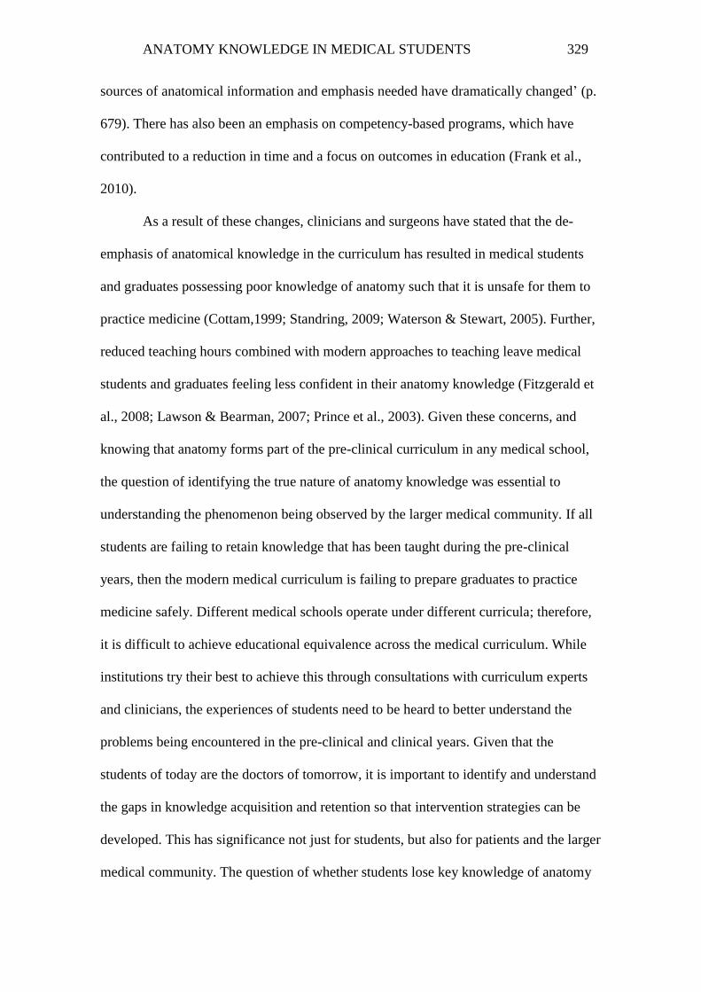
ANATOMY KNOWLEDGE IN MEDICAL STUDENTS 329
sources of anatomical information and emphasis needed have dramatically changed’ (p.
679). There has also been an emphasis on competency-based programs, which have
contributed to a reduction in time and a focus on outcomes in education (Frank et al.,
2010).
As a result of these changes, clinicians and surgeons have stated that the de-
emphasis of anatomical knowledge in the curriculum has resulted in medical students
and graduates possessing poor knowledge of anatomy such that it is unsafe for them to
practice medicine (Cottam,1999; Standring, 2009; Waterson & Stewart, 2005). Further,
reduced teaching hours combined with modern approaches to teaching leave medical
students and graduates feeling less confident in their anatomy knowledge (Fitzgerald et
al., 2008; Lawson & Bearman, 2007; Prince et al., 2003). Given these concerns, and
knowing that anatomy forms part of the pre-clinical curriculum in any medical school,
the question of identifying the true nature of anatomy knowledge was essential to
understanding the phenomenon being observed by the larger medical community. If all
students are failing to retain knowledge that has been taught during the pre-clinical
years, then the modern medical curriculum is failing to prepare graduates to practice
medicine safely. Different medical schools operate under different curricula; therefore,
it is difficult to achieve educational equivalence across the medical curriculum. While
institutions try their best to achieve this through consultations with curriculum experts
and clinicians, the experiences of students need to be heard to better understand the
problems being encountered in the pre-clinical and clinical years. Given that the
students of today are the doctors of tomorrow, it is important to identify and understand
the gaps in knowledge acquisition and retention so that intervention strategies can be
developed. This has significance not just for students, but also for patients and the larger
medical community. The question of whether students lose key knowledge of anatomy

ANATOMY KNOWLEDGE IN MEDICAL STUDENTS 330
in the clinical years and whether they are poorly prepared for clinical training, as
indicated in the literature, or whether they are not learning anatomy properly the first
time around had to be investigated, along with what regions of anatomical knowledge
were most easily retained or lost and why. Therefore, this study set out to explore
anatomy learning and retention in one Australian graduate-entry medical school. This is
the first study of its kind in Australia and New Zealand. Although all students entered
the graduate-entry program from a variety of prior educational backgrounds, and hence
had varying degrees of anatomy knowledge before starting medical school, they were
instructed in the pre-clinical year according to the same anatomy curriculum and used
similar teaching and assessment methods, thereby allowing for a comparison across
cohorts.
Students in this study had received one pre-clinical year of formal anatomy
teaching, after which time their exposure to anatomy and formal teaching of anatomy in
the clinical years was highly varied and dependent upon clinical sites. Therefore, the
topic of learning and retention was explored through both quantitative and qualitative
means using the theoretical frameworks of post-positivism and constructivism to answer
the three research questions:
How does anatomy knowledge differ among students in the pre-clinical and
clinical years of the MBBS course?
To what extent do medical students retain anatomy knowledge?
What factors may account for the loss or retention of anatomy knowledge across
a student’s medical degree?
The hypothesis for this study was similar to that of past retention studies (Feigin
et al., 2002, 2007) in that students’ knowledge of anatomy would decline over time as
they progressed from the pre-clinical year (where anatomy was formally taught and

ANATOMY KNOWLEDGE IN MEDICAL STUDENTS 331
assessed) to the clinical years, where such activities were highly varied. However, this
study is unique because it explored knowledge of all seven anatomical regions and did
not focus on only one topic, as many other studies have done (Feigin et al., 2002, 2007;
Meyer et al., 2015; Speilmann & Oliver, 2005). Further, the factors accounting for the
retention or loss of anatomy knowledge were also explored through qualitative
mechanisms (a feature not addressed in other studies), with the potential hypothesised
contributors to such loss of knowledge being lack of formal teaching and anatomy
assessments in the clinical years, lack of opportunities for vertical integration of
learning and subsequent lack of student motivation to specifically study and/or revise
anatomy. Hypothesised contributors to the retention of anatomical knowledge were
learning styles, with deeper understanding facilitating better retention, interest in
anatomy and perhaps motivation through future career aspirations such as surgery,
which would require extensive knowledge of anatomy.
To summarise the findings of this study, participants in the pre-clinical years
performed better on the formative anatomy assessment task than participants in the
clinical years, with the most statistically significant difference in knowledge evident
between that of the pre-clinical students and Year C students (i.e., clinical students in
the second-last year of the MBBS program). Additionally, one of the strengths of the
study was that it not only measured factual knowledge, but it also provided insights into
learners’ capacity to apply anatomical concepts to clinical problems (Kulasegaram et
al., 2013). We could not assess clinical reasoning directly because students could have
arrived at the answer by choice; however, our findings indicate that overall, all students
were better at recall but poorer on application and analytical-type questions:
Given this evidence, integration of curricula should focus on efficiently and
effectively promoting the cognitive meshing of content knowledge from basic

ANATOMY KNOWLEDGE IN MEDICAL STUDENTS 332
and clinical science. This linking is likely achieved most effectively at the level
at which students make direct contact with the content of the formal curriculum:
the teaching sessions. (Kulasegaram et al., 2013, p. 1581)
When comparing retention of anatomy from when participants were in the pre-
clinical year, those in the final two clinical years (Year C and Year D) demonstrated a
statistically significant lower level of knowledge. Although the limitations of the study,
in particular the use of the VIA scores as a non-equivalent baseline, has been
acknowledged in in the thesis, the similarities of the assessment tools for the VIA and
the anatomy assessment, followed by the similarity of difficulty of both assessments (as
indicated by the borderline) suggests that there is some loss of knowledge from the pre-
clinical to clinical years. Further, the findings of this study, obtained through a mixed-
methods approach suggests that this decline of knowledge over time occurs particularly
when students are not exposed to the content on a regular basis either via formal
instruction of anatomy in the clinical years or frequent clinical exposure to different
anatomical regions. Across all cohorts, knowledge of anatomical regions (thorax,
abdomen and lower limb) that was either most recently covered in pre-clinical teaching
or was most often encountered during training in the clinical years (through
cardiovascular respiratory, gastrointestinal and lower limb cases) was better retained
than their counterparts. These findings support those of other retention studies (Custers
et al., 2011; Feigin et al., 2002, 2007), which found that knowledge of anatomy is much
better at the time when content is delivered and assessed (as it is in the pre-clinical
year), and it is most likely forgotten and subsequently declines over time when it has
not been relearned or revisited (which occurs during the clinical years). However, the
results of this study highlight a significant problem that has not been discussed much in
the literature before—that is, the problem of anatomy knowledge acquisition. A mean

ANATOMY KNOWLEDGE IN MEDICAL STUDENTS 333
performance of only 60% among pre-clinical participants, students who had received
frequent formal teaching in anatomy during the year and multiple summative
assessments suggests that students are not learning anatomy to a high standard the first
time around. In fact, performance in the pre-clinical years was mainly affected by a
condensed medical curriculum and fast-paced anatomy curriculum, resulting in a
cognitive overload (Kirsh, 2000). Participants said that this resulted in them using
predominantly surface or strategic approaches to learn anatomy (Eizenberg, 1988).
For effective learning to take place:
the design of courses needs to be underpinned, either explicitly or implicitly,
both by theories of student learning but also by the more practical theories of
instructional learning and course design that seek to give practical guidance to
the designing of courses. (Smith et al., 2014, p. 270)
Perhaps what then needs to occur is a modification of the existing curriculum to
match Flexner’s philosophy of education. Curriculum experts need to examine the
different disciplines that are compressed into the pre-clinical teaching years and
examine the necessity and timing of each. To allow students the opportunity to learn
and develop their critical thinking skills, a balance must be struck between the
extraneous and intrinsic loads (Moscova et al., 2015). The effectiveness of a curriculum
that is primarily constructed around the basic sciences while also incorporating social
sciences and clinical skills was evident in a study by Wilkerson et al. (2009), whereby
the authors implemented the principles of cognition and learning to deliver a curriculum
using the AIDERS mnemonic (attention, interpretation, deep understanding, exposition,
reinforcement/restructuring and social construction). This led to a PBL curriculum
consisting of system-based blocks with disciplines of anatomy/pathophysiology,
genetics, pharmacology, clinical skills and clinical reasoning integrated within each

ANATOMY KNOWLEDGE IN MEDICAL STUDENTS 334
block. This is supplemented by traditional lectures, active and collaborative learning
(i.e., small-group work) and experiential learning (i.e., hands-on laboratory sessions),
and it concludes with a formative assessment at the end of each week. This block
system was effective because it allowed students to direct their focus and attention
towards a smaller set of topics, thereby creating a manageable cognitive load. Having
less class time for students and limiting the number of other courses they needed to
focus on enhanced students’ self-directed study, gave them time to process content
learned during the week and enabled the integration of different disciplines within the
system.
In the clinical environment, workplace-based environments that were not
supportive of participants’ learning (such as clinical educators who were unaware of
what needed to be taught, who taught to an extremely high level or who barraged
students about their knowledge) affected participants’ ability to use the opportunities
and experiences available to learn, with some stating that it even demotivated them
(Billet, 2001; Eraut, 2007). Further, the lack of opportunities for anatomy revision,
which mainly occurred informally through bedside tutorials and was site-specific,
combined with increased pressures to learn about diseases, diagnosis, treatment and
management in the clinical years, meant that participants had limited time in which to
review previous anatomy knowledge. This was compounded by the lack of formal
teaching of anatomy via vertical integration and a lack of formal assessments of
anatomy in the clinical years (although anatomy was indirectly assessed during clinical
written examinations and OSCEs).
This study shows that the integration of basic science and clinical knowledge is
highly valued and used, especially in the teaching of anatomy during the pre-clinical
years. However, vertical integration of basic science and clinical medicine is not
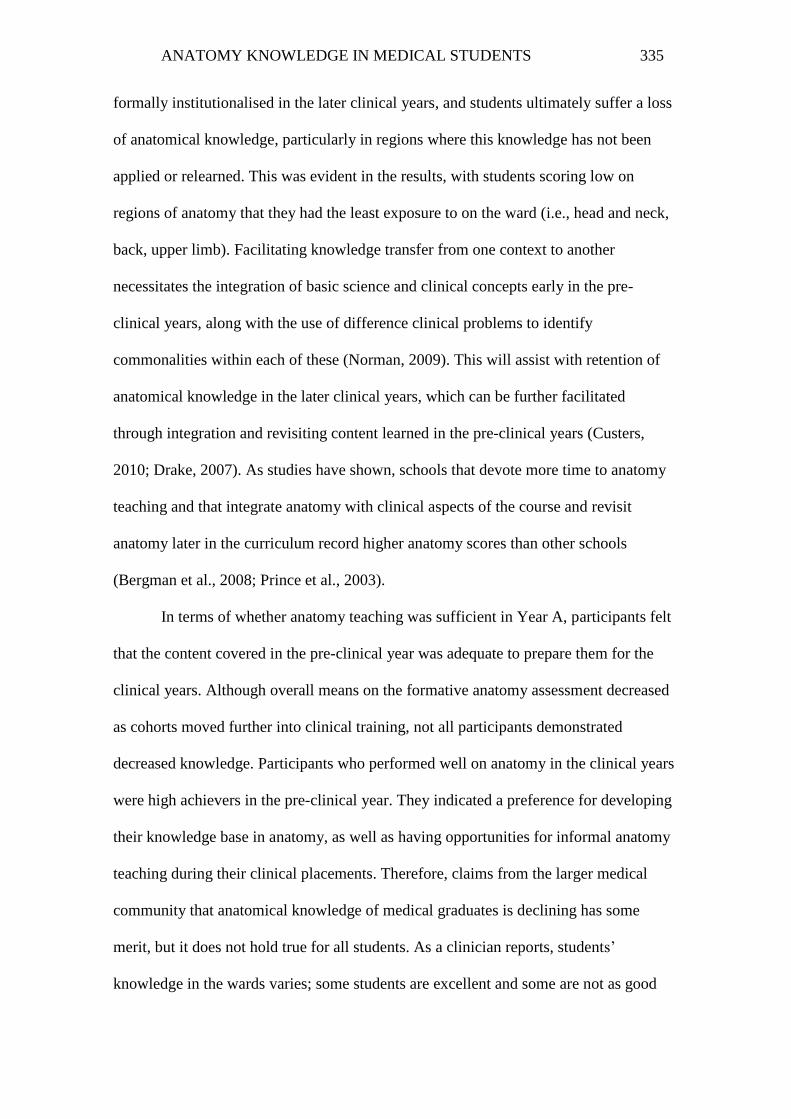
ANATOMY KNOWLEDGE IN MEDICAL STUDENTS 335
formally institutionalised in the later clinical years, and students ultimately suffer a loss
of anatomical knowledge, particularly in regions where this knowledge has not been
applied or relearned. This was evident in the results, with students scoring low on
regions of anatomy that they had the least exposure to on the ward (i.e., head and neck,
back, upper limb). Facilitating knowledge transfer from one context to another
necessitates the integration of basic science and clinical concepts early in the pre-
clinical years, along with the use of difference clinical problems to identify
commonalities within each of these (Norman, 2009). This will assist with retention of
anatomical knowledge in the later clinical years, which can be further facilitated
through integration and revisiting content learned in the pre-clinical years (Custers,
2010; Drake, 2007). As studies have shown, schools that devote more time to anatomy
teaching and that integrate anatomy with clinical aspects of the course and revisit
anatomy later in the curriculum record higher anatomy scores than other schools
(Bergman et al., 2008; Prince et al., 2003).
In terms of whether anatomy teaching was sufficient in Year A, participants felt
that the content covered in the pre-clinical year was adequate to prepare them for the
clinical years. Although overall means on the formative anatomy assessment decreased
as cohorts moved further into clinical training, not all participants demonstrated
decreased knowledge. Participants who performed well on anatomy in the clinical years
were high achievers in the pre-clinical year. They indicated a preference for developing
their knowledge base in anatomy, as well as having opportunities for informal anatomy
teaching during their clinical placements. Therefore, claims from the larger medical
community that anatomical knowledge of medical graduates is declining has some
merit, but it does not hold true for all students. As a clinician reports, students’
knowledge in the wards varies; some students are excellent and some are not as good

ANATOMY KNOWLEDGE IN MEDICAL STUDENTS 336
(Leveritt et al., 2016). To understand the reports from clinicians and surgeons on
students’ poor anatomy knowledge in the wards, one must consider clinicians’
expectations of students’ knowledge. From their perspective, the training that clinicians
received decades ago must be considered because it varies substantially from the
anatomical training that medical students receive in the modern curriculum. With
advancements in medicine and technology, modern clinicians have different
expectations of medical students and graduates. They must understand the body from a
clinical and disease-based perspective, and they must be able to use medical imaging
tools to interpret and diagnose illnesses—all in a shorter period than their predecessors
would have had. Additionally, they must possess sufficient knowledge of anatomy and
continue to enhance that knowledge throughout their career (Rizollo et al., 2006).
Consequently, the basic knowledge that a graduate of today is expected to possess
differs from and outweighs that of a medical graduate 20 or 30 years ago. Given these
expectations, the literature findings are not surprising, although one can question
whether they are warranted. However, this study reveals that, in addition to condensed
curricular time, lack of integration of knowledge and fewer assessments on anatomy in
the clinical years, factors such as career aspirations/motivation, interest in anatomy, and
approach and attitude towards teaching and learning also influence knowledge retention.
Currently, there is an international debate on the different modalities of
instruction and which one offers the best solution to increasing anatomical knowledge
(Pabst, 2009). However, to date, there is no definitive answer. With globalisation,
institutions comprise students and educators from different educational and cultural
backgrounds, and as research has shown, there are obvious learning differences among
different cultures (Mitchell et al., 2009). Therefore, to assist students in this regard, a
multimodal approach to teaching anatomy should continue to be employed, and

ANATOMY KNOWLEDGE IN MEDICAL STUDENTS 337
adequate guidance must be provided to students within each of the teaching modalities,
because SDL can falter without proper guidance. Overall, participants in this study were
positive in their learning experiences when these involved small-group teaching that
actively engaged them in their learning, and when content was clinically related.
However, given that anatomy teaching and its resources have dwindled over the past
few decades, the question to ask relates to how to maximise the teaching of content in
this discipline without compromising quality. This can best be achieved through vertical
integration—that is, subsequent revisitation of pre-clinical anatomy in the clinical years,
but with progressively more clinical content to showcase the clinical relevance of the
topic (Smith et al., 2014).
The shift in pedagogical instruction of anatomy from the traditional discipline-
based curriculum to an integrated and interdisciplinary curriculum has been gradually
increasing over the past two decades. While many reasons have been cited for this
phenomenon, one of the most important reasons is that clinicians must integrate basic
science knowledge and clinical knowledge when treating a patient. Therefore, it is
imperative to frame the curriculum in a similar manner (Brooks et al., 2015) while
training students to become doctors. However, the constant call for integration within
the literature and the urge to view this as a priority in medical education suggests that
“integration is not a solved problem” (Kulasegaram et al., 2013, p. 1578). Through the
reintroduction of content and clinical association, students will be able to increase their
understanding and subsequent retention of that knowledge (Mann, 2002). According to
adult learning theories, when students use prior learning experiences, their application
of newly acquired knowledge is enhanced (Lazarus et al., 2014; Phillips, 1995); thus,
our curricula must seek to improve this outcome.

ANATOMY KNOWLEDGE IN MEDICAL STUDENTS 338
Our findings also reveal that medical educators play a big role in influencing
how students approach the learning of anatomy. The Monash medical curriculum has
undergone significant modifications, from a six-year curriculum in its inception in 1961
to a five-year curriculum adopted post-2000, to the addition of a four-year graduate-
entry medicine program that commenced in 2008 (Solarsh et al., 2012). This graduate
program is unique in that it consists of a 1:3 split across pre-clinical to clinical
experience, as opposed to a 50:50 split between pre-clinical and clinical training offered
by other graduate-entry programs in the country. While the curriculum is based around
four longitudinal themes that “vertically integrate the basic biological, population and
social sciences with clinical practice” (Solarsh et al., 2012, p. 807) across the four years
of study, this study has shown that this is far from what occurs. The difficulties in
communication and monitoring across a distributed program have resulted in wide-
ranging experiences for students. While this is not necessarily a bad thing, and one
cannot maintain absolute consistency in clinical environments, it should be reasonable
to assure students across all sites that their experience in the clinical setting will evolve
around clearly prescribed and common curriculum content. Yet, excerpts from
participants reveal that many clinicians on site are oblivious to what needs to be taught
and to what level of detail. This is contrary to the usual procedures outlined by
institutions, which state that courses are delivered with the same content and in the same
way across sites. Often, students do not acquire learning experiences that their peers at
other sites have obtained (Solarsh et al., 2012). This study’s results not only emphasise
the continued need for constructive alignment (Biggs & Tang, 2008) and for experts to
design a curriculum that is context-appropriate and specific to the profession, but also to
develop tutor training programs that highlight the pedagogical aspects of teaching and
the roles that medical educators play in today’s medical curriculum (Trigwell et al.,

ANATOMY KNOWLEDGE IN MEDICAL STUDENTS 339
1999). Although compliance from educators is an issue, this could be achieved by
incorporating students’ evaluations of the tutors, which would allow tutors to attain
meaningful feedback on their performance and identify areas for further improvement
(Whelan et al., 2016). Participants highly valued tutors who demonstrated a passion for
teaching while keeping content clinically relevant compared with those who were
unaware of the curriculum or who wanted to showcase their knowledge. As Pawlina
(2010, p. 1) states, “great teachers are valuable commodities now more than ever”. In
the face of increasing pressure on academic institutions to generate economic growth
and the subsequent increase in recruiting research-based academics and clinicians, there
is an increasing need to introduce innovation in education. Therefore, non-medically
qualified or teaching-focused individuals are now being recognised for their high-
quality teaching, and many institutions are working towards retaining and advancing the
careers of such individuals by promoting teaching and learning (Pawlina, 2010).
Given the importance of anatomy in clinical practice (Berman, 2014; Cottam,
1999; Davis, et al., 2014; Herle & Saxena, 2011; Kerby et al., 2011; Standring, 2012;
Sugand et al., 2010), there has been an outcry at the declining knowledge perceived to
be present among medical students and new graduates (Cottam, 1999; Craig et al., 2010;
Gogalniceanu et al., 2010; Herle and Saxena, 2011; Singh et al., 2015; Waterston &
Stewart, 2005), and the fact that assessments drive students’ behaviour towards learning
(Newble & Jaeger, 1983). Therefore, institutions need to revise their curricula to reduce
cognitive overload, introduce active forms of teaching and learning to achieve better
knowledge of anatomy, incorporate more assessments into anatomy and design effective
tools to test anatomical knowledge and administer them frequently—especially during
clinical training. Only then can we be confident that medical students are attaining
anatomical competence. The findings of this study show that students do not lose all

ANATOMY KNOWLEDGE IN MEDICAL STUDENTS 340
anatomical knowledge, and knowledge that is retained is related to frequent clinical
exposure to those anatomical regions on the wards, thereby emphasising the value of the
workplace environment and workplace-based learning (Billett, 2001, 2004; Eraut, 2004,
2007, 2008). Therefore, our results support the conclusion that no matter what the
curriculum model, students’ knowledge of anatomy is affected by the amount of time
spent learning anatomy, the clinical context associated with anatomy teaching, and the
revision and integration of clinical anatomy knowledge in medical training (Bergman et
al., 2008). Students are more likely to thrive when there is consistency in delivery, more
exposure to anatomy in clinical sites, and more time to learn, assimilate and test their
knowledge. These factors can all be generalised to the larger population of medical
programs globally.
9.1 Recommendations
This study set out to explore anatomy learning and retention among medical
students in a graduate-entry MBBS program. The findings presented in this study have
highlighted issues surrounding the medical curriculum in one graduate-entry institution,
and they have provided insights into gaps in knowledge among students and why these
gaps occur. This section outlines how we can address these gaps.
The literature views the threat to anatomy in the modern curriculum as being
manifold, including increased workloads and pressure on students and clinical services,
a lack of qualified teachers, a lack of opportunities for learning anatomy in the clinical
environment, and increased pressure to research in academic departments (Raftery,
2007). The findings in this study suggest that there are areas for optimisation within the
MBBS program and areas where we can facilitate improvement going forward.

ANATOMY KNOWLEDGE IN MEDICAL STUDENTS 341
Recommendation 1: The pre-clinical curriculum needs to be reviewed and
revised in the first instance to reduce cognitive overload, because overcrowding and the
amount of information that students are expected to learn can be overwhelming.
Given that basic science knowledge is viewed as the “cognitive framework for
anchoring clinical knowledge” (Kulasegaram et al., 2013, p.1581), medical education
experts need to ensure that their curriculum allows students to achieve cognitive
integration. In a compressed curriculum, students are overloaded with so much
information that the learning process is hindered and they are unable to process
knowledge in a deep manner. Both horizontal and vertical integration need to be
thoughtfully designed and implemented in the curriculum. Of course, “simply
presenting basic science in close proximity to clinical knowledge may not guarantee the
putative benefits of integration” (Kulasegaram et al., 2015, p. s67). That is, scheduling a
basic science lecture and clinical lecture on the same day might not necessarily lead to
integration. There must be faculty buy-in from all interested parties (Kulasegaram et al.,
2015). To facilitate better learning and the subsequent retention of learned knowledge,
the teaching of basic sciences must be embedded and contextualised so that learners can
appreciate the true significance of content and its relation to the clinical sciences and
medicine. Subsequently, this leads to conceptual coherence and better diagnostic
abilities and patient management (Kulasegaram et al., 2013; Woods, 2007). Such
integration can be achieved not only through PBL and small-group teaching sessions in
the pre-clinical years, but also through back-to-base science days during clinical
rotations and shared teaching models that seek to engage students in a truly meaningful
learning session.
Recommendation 2: The opportunities afforded by the workplace should be
widespread in that they should provide students with a variety of experiences to advance

ANATOMY KNOWLEDGE IN MEDICAL STUDENTS 342
their learning. Further, the core curriculum should be communicated clearly and simply
to clinical educators so that the communities of practice come together to support
students appropriately.
The findings in this study suggest that although there is a decline in students’
anatomical knowledge, all is not lost; knowledge of regional areas that have the most
clinical exposure and those that are relevant to current learning are retained; thus,
efforts must be made by curriculum experts and clinicians in the workplace
environment to increase exposure to all regions of anatomy in the clinical years to
optimise learning during this period. This can be achieved by establishing core
competencies for anatomy. Developing a core national curriculum for anatomy that is
similar to what exists in the UK would be the first step towards resolving the anatomy
debate on a national scale. However, on a smaller scale, it would be useful for each
institution to have its own set of core anatomical competencies that are made explicit to
all parties and that must be followed by all sites in the clinical years. This would ensure
that students receive similar learning experiences regardless of where they are placed
during clinical training. Specifying the curriculum in this way allows instruction to
occur in a formal setting as opposed to a setting in which the student may or may not be
fortunate enough to have someone teach a particular concept.
Recommendation 3: Enhance both horizontal and vertical integration (Bhangu
et al., 2010; Fitzgerald et al., 2008; Waterston & Stewart, 2005) to link relevant
teaching of anatomy, pathology and clinical exposure to key conditions in the clinical
years.
Although participants valued the importance of anatomy in medicine, this
realisation tended to occur during the clinical years, when the lack of formal anatomy
teaching placed additional pressure on students to undertake more SDL. This requires a

ANATOMY KNOWLEDGE IN MEDICAL STUDENTS 343
significant effort on the part of students to schedule revision time for anatomy in an
already-condensed curriculum. In light of student concerns over their anatomical
knowledge (Ahmed et al., 2010; Mitchell & Batty, 2009; Smith & Mathias, 2011), an
anatomy refresher course, could be introduced to help students revise their knowledge
in the clinical years, as suggested by participants. As demonstrated by Jurjus et al.
(2016), an anatomy refresher course that is incorporated during the clinical years as a
sort of a flipped classroom approach can assist in addressing students’ deficit in
anatomy knowledge while subsequently improving clinically relevant knowledge of
medical students on a clinical placement. Therefore, in designing an anatomy
curriculum and creating pathways for vertical integration, significant cross-links must
be made between anatomy and medicine with ties to clinical applications to facilitate
better knowledge transfer (contextualisation) and recall in the wards and thereafter.
Recommendation 4: Students’ performance and progress in anatomy knowledge
should be monitored with the aim of providing remediation.
As stated by Larsen et al. (2009), retrieval practice is essential to boosting long-
term retention. Therefore, to increase retention, there must be repetition of content in a
variety of different forms and, to offer remediation to students in need, any knowledge
gaps that exist need to be identified. Only then can appropriate tools of remediation be
developed. To ensure equity of access, a standardised formative assessment tool for
competence could be implemented via e-modules at the beginning and/or end of a
refresher course to allow students to gauge whether their knowledge is at an acceptable
level (Jurjus et al., 2014). This could consist of a pre-test, interactive learning activity
and post-test structure that is further supplemented with hands-on learning experience
(Ramsey-Stewart et al., 2010) perhaps offered through cadaveric or virtual exposure in
the first instance and opportunities for practicing procedures later in the clinical years.

ANATOMY KNOWLEDGE IN MEDICAL STUDENTS 344
The results of students’ performance on formative tasks associated with the refresher
modules could be used by faculty as a guiding tool to offer further support and
remediation for students who appear to be struggling with the discipline. Of course, care
must be taken when deciding how often to administer/offer formative assessments
because participation and compliance declines if these are administered too often
(McNulty et al., 2015). However, the useful predictive validity of the Year A VIA
might also allow us to identify students who need further teaching, revision and
monitoring within the spiral curriculum.
Recommendation 5: Prior to graduation and during residency, all students
should undertake a licensing examination that assesses various components of their
anatomical knowledge, similar to step 1, 2 and 3 of the licensing examination (USMLE)
that medical students and graduates undertake in the US.
If a national examination existed, it would ensure some form of quality control
because all medical graduates in Australia and New Zealand would be required to sit the
assessment, regardless of the curriculum, teaching hours, mode of delivery and internal
assessment they were exposed to during their medical program. In essence, it would
“act as a de facto core curriculum, encouraging universities to prepare their students in
the stated competencies of the examination” (Schiller et al., 2011, p. 10).
According to Korf et al. (2008), knowledge resides not only in books, but also in
the mind. It survives through both production and reproduction, and it remains useless if
it is not applied and practiced. Therefore, a standardised assessment would ensure that
all students and graduates meet the necessary requirements for medical practice;
consequently, this would assure the larger medical community that graduates are
competent to practice medicine safely.

ANATOMY KNOWLEDGE IN MEDICAL STUDENTS 345
9.2 Implications for Future Research
Leveritt et al. (2016) find that graduate medical students are more confident in
their anatomy knowledge at the beginning of a placement compared with the
undergraduate cohort. Given that the direct-entry medical curriculum at Monash
University consists of two pre-clinical years and three clinical years, and medical
students have two years in which to learn anatomy, it would be interesting to examine
the anatomy performance of students in the undergraduate medical program and
compare it with students in the graduate-entry program. This could then be expanded to
investigate students in other medical schools across different states and countries.
Assessing a student’s knowledge can be a difficult task because the type of
assessment modality used is MCQs, which relies on a combination of recall, recognition
and analysis. As Jurjuis et al. (2016) states, “there is not a single agreed-upon measure
of knowledge retention, and the length of time between knowledge acquisition and
measuring recall or recognition to determine retention is fraught with ambiguity” (p.
342). Further, assessment scores provide little information regarding how knowledge is
applied at the bedside and on the wards. If the goal of integration is to observe how
cognitive changes occur within the minds of learners, then future research will need to
focus not just on retention of basic science knowledge, but also on how that knowledge
is applied in the clinical setting. Examining students’ use of anatomical knowledge
during clinical reasoning or in the performance of a clinical skill would provide
valuable evidence of the effectiveness of integration.
This study did not explore visualisations and cross-sectional radiological
anatomy in the formative assessment. Thus, while retention may have been lost over
time, areas that are frequently encountered in clinical practice might have been retained.
Further research is needed in this area to identify gaps in students’ knowledge.

ANATOMY KNOWLEDGE IN MEDICAL STUDENTS 346
A final perspective that should be considered is that of medical educators. This
could be explored in tandem with medical students to provide insights into the
challenges faced by educators in academic institutions and in the workplace.
Overall, the results of this study show that in a compressed curriculum, students
are forced to make decisions about their learning and how they approach it, perhaps not
by choice but by the need to keep pace with the delivery of the curriculum which is
relentless and to progress through each year of their medical degree. With the design of
such a curriculum, the goal of medical education, which is to produce medical graduates
who are capable and confident in their capacity to practice medicine safely, remains
unaccomplished. By training students with a mindset to pass examinations, medical
institutions are doing a disservice to themselves and the community. We need to instil
confidence in our students and graduates, as well as in society and communities, that the
doctors produced in medical schools are of the highest calibre. This can be achieved by
reassessing and modifying the existing curriculum to reduce cognitive overload,
creating frequent opportunities to formatively assess students’ anatomical knowledge
and subsequently making more room for students to learn efficiently. After all:
a pre-clerkship medical curriculum succeeds to the extent that its students arrive
on the wards with an organized approach for analysing their patient’s problems
and an ability to selectively draw on, interpret and apply information from the
vast reservoir of material covered in the pre-clerkship years. (Wilkerson et al.,
2009, p. 820)

ANATOMY KNOWLEDGE IN MEDICAL STUDENTS 347
References
Abu-Hijleh, M. F. (2010). The place of anatomy in medical education: Guide
Supplement 41.1—Viewpoint. Medical Teacher, 32(7), 601–603.
Acland, R. D. (2003). Acland’s atlas of human anatomy [DVD version 1.0]. Baltimore,
MD: Lippincott William & Wilkins.
Aday, L. A., & Cornelius, L. J. (2006). Designing and conducting health surveys: A
comprehensive guide (3rd ed.). San Francisco, CA: Jossey-Bass. Retrieved from
http://citeseerx.ist.psu.edu/viewdoc/download?doi=10.1.1.476.5561&rep=rep1&
type=pdf
Aggarwal, R., Brough, H., & Ellis, H. (2006). Medical student participation in surface
anatomy classes. Clinical Anatomy, 19(7), 627-631.
Ahmed, K., Rowland, S., Patel, V. M., Ashrafian, H., Davies, D. C., Darzi, A., . . .
Paraskeva, P. A. (2011). Specialist anatomy: Is the structure of teaching
adequate? Surgeon, 9, 312–317.
Ahmed, K., Rowland, S., Patel, V. M., Khan, R. S., Ashrafian, H., Davies, D. C., . . .
Paraskeva, P. A. (2010). Is the structure of anatomy curriculum adequate for
safe medical practice? Surgeon, 8(6), 318–324.
Albertine, K. H. (2012). Cottam’s landmark paper for clinical anatomy. Clinical
Anatomy, 25(6), 681–682.
Alexander, C. J., Crescini, W. M., Juskewitch, J. E., Lachman, N., & Pawlina, W.
(2009). Assessing the integration of audience response system technology in
teaching of anatomical sciences. Anatomical Sciences Education, 2, 160–166.
Alexander, P. A., Schallert, D. L., & Reynolds, R. E. (2009). What is learning anyway?
A topological perspective considered. Educational Psychologist, 44, 176–192.

ANATOMY KNOWLEDGE IN MEDICAL STUDENTS 348
Allmark, P. J., Boote, J., Chambers, E., Clark, A., McDonnell, A., Thompson, A., &
Tod, A. (2009). Ethical issues in the use of in-depth interviews: Literature
review and discussion. Research Ethics Review, 5(2), 48–54.
American Association of Clinical Anatomists. (1996). A clinical anatomy curriculum
for the medical student of the 21st century: Gross anatomy. Clinical Anatomy,
9(2), 71–99.
Association of American Medical Colleges. (2001). Report IV contemporary issues in
medicine: Basic science and clinical research (Medical school objective
project). Washington, DC: Association of American Medical Colleges.
Retrieved from
https://members.aamc.org/eweb/upload/Contemporary%20Issues%20in%20Med
%20Basic%20Science%20Report%20IV.pdf
Anatomy TV. (2006). Interactive anatomy. London, United Kingdom: Primal Pictures
Ltd. Available from www.anatomy.tv
Anderson, L. W., Krathwohl, D. R., Airasian, P. W., Cruikshank, K. A., Mayer, R. E.,
Pintrich, P. R., . . . Wittrock, M. C. (2001). A taxonomy for learning, teaching
and assessing: A revision of Bloom’s taxonomy of educational objectives. New
York, NY: Longman.
Arraéz-Aybar, L. A., Sanchez-Montesinos, I., Mirapeix, R. M., Mompeo-Corredera, B.,
& Sanudo-Tejero, J. R. 2010. Relevance of human anatomy in daily clinical
practice. Annals of Anatomy, 192(6), 341–348.
Attardi, S. M., & Rogers, K. A. (2015). Design and implementation of an online
systemic human anatomy course with laboratory. Anatomical Sciences
Education, 8, 53–62.

ANATOMY KNOWLEDGE IN MEDICAL STUDENTS 349
Auer, R. N., & McDonald, D. S. (2003). Anatomy is still essential. Canadian Medical
Association Journal, 168(7), 829.
Australian Medical Council. (2012). Standards for assessment and accreditation of
primary medical programs schools. Retrieved from
http://www.amc.org.au/accreditation/primary-medical-education
Australian Medical Council. (2014). National internship framework. Retrieved from
http://www.amc.org.au/accreditation/prevoc-standards/national-internship-
framework
Australian Medical Council. (2016). Prevocational standards education. Retrieved
from http://www.amc.org.au/accreditation/prevoc-standards
Australian Medical Students Association. (2015). Classification of medical programs
policy: Medical school programs and admissions. Retrieved from
https://www.amsa.org.au/sites/amsa.org.au/files/Classification%20of%20Medic
al%20Programs%20(2015).pdf
Australian Orthopaedic Association. (2015). AOA Anatomy Courses. Sydney, NSW:
Author. Available from https://www.aoa.org.au/docs/default-
source/training/2014jul_approved-anatomy-courses-for-2015_final.pdf?sfvrsn=2
Australian Qualifications Framework Council. (2013). Australian qualifications
framework (2nd ed.). Retrieved from
http://www.aqf.edu.au/wpcontent/uploads/2013/05/AQF-2nd-Edition-January-
2013.pdf
Azer, S. A. (2013). The place of surface anatomy in the medical literature and
undergraduate anatomy textbooks. Anatomical Sciences Education, 6(6), 415–
432.

ANATOMY KNOWLEDGE IN MEDICAL STUDENTS 350
Azer, S.A., & Azer, S. (2016). 3D Anatomy models and impact on learning: A review
of the quality of the literature. Health Professions Education, 2(2), 80-98.
Azer, S. A., & Eizenberg, N. (2007). Do we need dissection in an integrated problem-
based learning medical course? Perceptions of first and second-year students.
Surgical and Radiologic Anatomy, 29(2), 173–180.
Bae, S., & Kokka, K. (2016). Student engagement in assessments: What students and
teachers find engaging. Stanford, CA: Stanford Center for Opportunity Policy in
Education and Stanford Center for Assessment, Learning and Equity.
Bahrick, H. P., & Hall, L. K. (2005). The importance of retrieval failures to long-term
retention: A metacognitive explanation of the spacing effect. Journal of Memory
and Language, 52, 566–577.
Banda, S. S. (2010). An investigation of how clinicians use anatomical knowledge in
diagnostic reasoning: A grounded theory study of clinicians in Zambia. Medical
Journal of Zambia, 37(1), 23–30.
Bandura, A. (1982). Self-efficacy mechanism in human agency. American Psychologist,
37(2), 122–147.
Barrows, H. S., & Tamblyn, R. M. (1980). Problem-based learning: An approach to
medical education. New York, NY: Springer.
Basu, S., Roberts, C., Newble, D. I., & Snatih, M. (2004). Competence in the
musculoskeletal system: Assessing the progression of knowledge through an
undergraduate medical course. Medical Education, 38, 1253–1260.
Bennett, E. E., Blanchard, R. D., & Hinchey, K. T. (2012). AM last page: Applying
Knowles’ andragogy to resident teaching. Academic Medicine, 87(1), 129.
Bergman, E. M., De Bruin, A. B., Herrler, A., Verheijen, I. W., Scherpbier, A. J., van
der Vleuten, C. P. (2013). Students’ perceptions of anatomy across the

ANATOMY KNOWLEDGE IN MEDICAL STUDENTS 351
undergraduate problem-based learning medical curriculum: A
phenomenographical study. BMC Medical Education, 13(1), 1–11.
Bergman, E. M., Prince, K. J., Drukker, J., van der Vleuten, C. P., & Scherpbier, A. J.
(2008). How much anatomy is enough? Anatomical Sciences Education, 1(4),
184–188.
Bergman, E. M., Verheijen, I. W., Scherpbier, A. J., van der Vleuten, C. P., & De
Bruin, A. B. (2014). Influences on anatomical knowledge: The complete
arguments. Clinical Anatomy, 27(3), 296–303
Berman, A. C. (2014). Anatomy of curriculum: Digging to the core. Anatomical
Sciences Education, 7(4), 326–328.
Bhangu, A., Boutefnouchet, T., Yong, X., Abrahams, P., & Joplin, R. (2010). A three-
year prospective longitudinal cohort study of medical students’ attitudes towards
anatomy teaching and their career aspirations. Anatomical Sciences Education,
3(4), 184–190.
Biggs, J. (2003). Teaching for quality learning at university. Buckingham, United
Kingdom: Open University Press.
Biggs, J. (2014). Constructive alignment in university teaching. HERDSA Review of
Higher Education, 1, 5–22. Retrieved from
https://www.tru.ca/__shared/assets/Constructive_Alignment36087.pdf
Biggs, J., & Tang, C. (2011). Teaching for quality learning at university. Maidenhead,
United Kingdom: Open University Press/McGraw Hill.
Billett, S. (2001). Learning through work: Workplace affordances and individual
engagement. Journal of Workplace Learning, 13, 209–214.
Billett, S. (2004). Workplace participatory practices: Conceptualising workplaces as
learning environments. Journal of Workplace Learning, 16, 312–324.

ANATOMY KNOWLEDGE IN MEDICAL STUDENTS 352
Billett, S. (2016). Learning through health care work: Premises, contributions and
practices. Medical Education, 50, 124–131.
Blevins, C. E., & Cahill, D. R. (1973). Gross anatomy: Current courses, training
programs and perspective needs. Journal of Medical Education, 48(3), 264–270.
Blunt, M. J., & Blizard, P. J. (1975). Recall and retrieval of anatomical knowledge.
British Journal of Medical Education, 9, 255–263.
Bohl, M., Francois, W., & Gest, T. (2011). Self-guided clinical cases for medical
students based on postmortem CT scans of cadavers. Clinical Anatomy, 24(5),
655–663.
Bolander, L. K., Lonka, K., & Josephson, A. (2008). How do medical teachers address
the problem of transfer? Advances in Health Sciences Education: Theory and
Practice, 13(3), 345–360.
Boon, J. M., Meiring, J. H., & Richards, P. A. (2002). Clinical anatomy as the basis for
clinical examination: Development and evaluation of an introduction to clinical
examination in a problem-oriented medical curriculum. Clinical Anatomy, 15(1),
45–50.
Booth, S., Melano, A., Sainsbury, H., & Woodley, L. (2011). Articulating and
comparing standards through benchmarking of assessment. Paper presented at
the Australian Universities Quality Forum: Demonstrating quality, Melbourne,
VIC.
Boshuizen, H. P., & Schmidt, H. G. (1992). On the role of biomedical knowledge in
clinical reasoning by experts, intermediates and novices. Cognitive Sciences, 16,
153–184.
Boud, D., & Soler, R. (2016). Sustainable assessment revised. Assessment & Evaluation
in Higher Education, 41(3), 400-413.
ANATOMY KNOWLEDGE IN MEDICAL STUDENTS 353
Bouwer, H. E., Valter, K., & Webb, A. L. (2016). Current integration of dissection in
medical education in Australia and New Zealand: Challenges and successes.
Anatomical Sciences Education, 9, 161–170.
Boyatzis, R. E. (1998). Transforming qualitative information: Thematic analysis and
code development. Thousand Oaks, CA: Sage.
Brame, C. J., & Biel, R. (2015). Test-enhanced learning: The potential for testing to
promote greater learning in undergraduate science courses. CBE—Life Sciences
Education, 14(2, es4), 1–12. Retrieved from
http://www.lifescied.org/content/14/2/es4.full.pdf+html
Braun, V., & Clarke, V. (2006). Using thematic analysis in psychology. Qualitative
Research in Psychology, 3(2), 77–101.
Brewer, J., & Hunter, A. (1989). Multimethod research: A synthesis of styles. Newbury
Park, CA: Sage.
Briggs, C. (2014). Postgraduate diploma in surgical anatomy: The University of
Melbourne experience. ANZ Journal of Surgery, 84(11), 808-809.
Brooks, W. S., Woodley, K. T., Jackson, J. R., & Hoesley, C. J. (2015). Integration of
gross anatomy in an organ system-based medical curriculum: Strategies and
challenges. Anatomical Sciences Education, 8(3), 266–274.
Brown, J. D. (1999, April). Standard error vs. standard error of measurement. Shiken:
JALT Testing & Evaluation SIG Newsletter, 3(1), 20–25. Retrieved from
http://jalt.org/test/PDF/Brown4.pdf
Bruner, J.S. (1977). The Process of education. Cambridge, Mass.: Harvard University
Press. (Original work published in 1960).

ANATOMY KNOWLEDGE IN MEDICAL STUDENTS 354
Brunk, I., Schauber, S., & Georg, W. (2017). Do they know too little? An inter-
institutional study on the anatomical knowledge of upper-year medical students
based on multiple choice questions of a progress test. Annals of Anatomy, 209,
93-100.
Bryman, A. (1988). Quantity and quality in social research. London, United Kingdom:
Routledge.
Buchanan, E. A., & Hvizdal, E. E. (2009). Online survey tools: Ethical and
methodological concerns of human ethics research. Journal of Empirical
Research on Human Research Ethics, 4(2), 37–48.
Cahill, D. R., Leonard, R. J., Marks, S. C. Jr. (2000). A comment on recent teaching of
human anatomy in the United States. Surgical and Radiologic Anatomy, 22(2),
69–71.
Carpenter, S. K., & Kelly, J. W. (2012). Tests enhance retention and transfer of spatial
learning. Psychonomic Bulletin and Review, 19, 443–448.
Case, S. M., & Swanson, D. B. (2001). Constructing written test questions for the basic
and clinical sciences (3rd ed.). Retrieved from
http://www.nbme.org/PDF/ItemWriting_2003/2003IWGwhole.pdf
Chakravarty, M., Latif, N. A., Abu-Hijleh, M. F., Osman, M., Dharap, A. S., &
Ganguly, P. K. (2005). Assessment of anatomy in a problem-based medical
curriculum. Clinical Anatomy, 18(2), 131–136.
Chang, B.J. (2016). Problem-based learning in medical school: A student’s perspective.
Annals of Medicine & Surgery, 12, 88-89.
Chapman, S. J., Hakeem, A. R., Marangoni, G., & Prasad, K. R. (2013). Anatomy in
medical education: Perceptions of undergraduate students. Annals of Anatomy,
195(5), 409–414.

ANATOMY KNOWLEDGE IN MEDICAL STUDENTS 355
Chapuis, P., Fahrer, M., Eizenberg, N., Fahrer, C., & Bokey, L. (2010). Should there be
a national core curriculum for anatomy? ANZ Journal of Surgery, 80(7–8), 475–
477.
Chowdhury, R., Wilson, I. D., & Oeppen, R. S. (2008). The departments of radiology
and anatomy: New symbiotic relations? Clinical Radiology, 63(8), 918–920.
Chuang, M. K., Canny, B. J., & McMenamin, P. G. (2011). Should there be a national
core curriculum for anatomy? ANZ Journal of Surgery, 81(12), 950.
Clifton, S. L., & Schriner, C. L. (2010). Assessing the quality of multiple choice test
items. Nurse Educator, 35(1), 12–16.
Cohen, L., Manion, L., & Morrison, K. (2007). Research methods in education. New
York, NY: Routledge.
Collins, J. (2006). Writing multiple-choice questions for continuing medical education
activities and self-assessment modules. Radio Graphics, 26(2), 543–551.
Collins, J. (2009). Are the changes in anatomy teaching compromising patient care? The
Clinical Teacher, 6(1), 18–21.
Collins, J. P. (2008). Modern approaches to teaching and learning anatomy. British
Medical Journal, 337(7671), 665–667.
Collins, T. J., Given, R. J., Hulsebosch, C. E., & Miller, B. T. (1994). The status of
gross anatomy in the U.S. and Canada—Dilemma for the 21st century. Clinical
Anatomy, 7, 275–296.
Confederation of Postgraduate Medical Education Councils. (2012). Australian
Curriculum Framework for Junior Doctors (v. 3.1). Retrieved from
http://curriculum.cpmec.org.au/introduction.cfm
Cook, M., Irby D. M., & O’Brien, B. C. (2010). Educating physicians: A call for reform
of medical school and residency. San Francisco, CA: Jossey-Bass.
ANATOMY KNOWLEDGE IN MEDICAL STUDENTS 356
Cottam, W. W. (1999). Adequacy of medical school gross anatomy education as
perceived by certain postgraduate residency programs and anatomy course
directors. Clinical Anatomy, 12(1), 55–65.
Cottam, W. W. (2012). Comparing the landscape—1999 and now. Clinical Anatomy,
25(6), 679–680.
Craig, S. J., Tait, N., Boers, D., & McAndrew, D. J. (2010). Review of anatomy
education in Australian and New Zealand medical schools. ANZ Journal of
Surgery, 80(4), 212–216.
Creswell, J. W. (1994). Research design: Qualitative and quantitative approaches.
Thousand Oaks, CA: Sage.
Creswell, J. W. (2011). Controversies in mixed methods research. In N. K. Denzin & Y.
S. Lincoln (Eds.), The SAGE handbook of qualitative research (4th ed.).
Thousand Oaks, CA: Sage.
Creswell, J. W., & Plano Clark, V. L. (2007). Designing and conducting mixed methods
research. Thousand Oaks, CA: Sage.
Creswell, J. W., & Plano Clark, V. L. (2011). Designing and conducting mixed methods
research (2nd ed.). Thousand Oaks, CA: Sage.
Cronbach, L. J. (1951). Coefficient alpha and the internal structure of the tests.
Psychometrika, 16(3), 297–334. Retrieved from
http://kttm.hoasen.edu.vn/sites/default/files/2011/12/22/cronbach_1951_coeffici
ent_alpha.pdf
Crossley, J., Humphris, G., & Jolly, B. (2002). Assessing health professionals. Medical
Education, 36(9), 800–804.
Custers, E. J. (2010). Long-term retention of basic science knowledge: A review study.
Advances in Health Science Education: Theory & Practice, 15(1), 109–128.

ANATOMY KNOWLEDGE IN MEDICAL STUDENTS 357
Custers, E. J., & Ten Cate, O. T. (2011). Very long-term retention of basic science
knowledge in doctors after graduation. Medical Education, 45(4), 422–430.
Dahle, L.O., Brynhildsen, J., Behrbohm Fallsberg, M., Rundquist, I., & Hammar M.
(2002). Pros and cons of vertical integration between clinical medicine and basic
science within a problem-based undergraduate medical curriculum: Examples
and experiences from Linkoping, Sweden. Medical Teacher, 24(3), 280–285.
Dangerfield, P., Bradley, P., & Gibbs, T. (2000). Learning gross anatomy in a clinical
skills course. Clinical Anatomy,13, 444–447.
Datta, L. 1994. Paradigm wars: A basis for peaceful coexistence and beyond. In C. S.
Reichardt & S. F. Rallis (Eds.), The qualitative-quantitative debate: New
perspectives (pp. 53–70). San Francisco, CA: Jossey-Bass.
Davis, C. R., Bates, A. S., Ellis, H., & Roberts, A. M. (2014). Human anatomy: Let the
students tell us how to teach. Anatomical Sciences Education, 7(4), 262–272.
Dawson, A. G., Bruce, S. A. M., Heys, S. D., & Stewart, I. J. (2009). Student views on
the introduction of anatomy teaching packages into clinical attachments.
Clinical Anatomy, 22(2), 267–272.
Day, C. S., & Ahn, C. S. (2010). The importance of musculoskeletal medicine and
anatomy in medical education. Academic Medicine, 85(3), 401–402.
DeSouza, E., & Fleming, M. (2003). A comparison of in-class quizzes vs. online
quizzes on student exam performance. Journal of Computing in Higher
Education, 14, 121–134.
Dhingra, R., Taranikanti, V., & Kumar, R. (2006). Plastination: Teaching aids in
anatomy revisited. National Medical Journal of India, 19(3), 171.
ANATOMY KNOWLEDGE IN MEDICAL STUDENTS 358
Dinsmore, C. E., Daughtery, S., & Zeitz, H. J. (1999). Teaching and learning gross
anatomy: Dissection, prosection or ‘both of the above’. Clinical Anatomy, 12(2),
110–114.
Dissabandara, L. O., Nirthanan, S. N., Khoo, T. K., & Tedman, R. (2015). Role of
cadaveric dissections in modern medical curricula: A study on student
perceptions. Anatomy & Cell Biology, 48(3), 205–212.
Downing, S. M. (2004). Reliability: On the reproducibility of assessment data. Medical
Education, 38(9), 1006–1012.
Drake, R. L. (2007). A unique, innovative, and clinically oriented approach to anatomy
education. Academic Medicine, 82(5), 475–478.
Drake, R. L. (2014). A retrospective and prospective look at medical education in the
United States: Trends shaping anatomical sciences education. Journal of
Anatomy, 224, 256–260.
Drake, R. L., Lowrie, D. J., & Prewitt, C. M. (2002). Survey of gross anatomy,
microscopic anatomy, neuroscience, and embryology courses in medical school
curricula in the United States. The Anatomical Record, 269, 118–122.
Drake, R. L., McBride, J. M., Lachman, N., & Pawlina, W. (2009). Medical education
in the anatomical sciences: The winds of change continue to blow. Anatomical
Sciences Education, 2(6), 253–259.
Drake, R. L., McBride, J. M., & Pawlina, W. (2014). An update on the status of
anatomical sciences education in United States medical schools. Anatomical
Sciences Education, 7, 321–325.
Drake, R. L., Vogl, W., Mitchell, A. W., Gray, H. (2010). Gray’s anatomy for students
(2nd ed.). Philadelphia, PA: Churchill Livingstone/Elsevier.

ANATOMY KNOWLEDGE IN MEDICAL STUDENTS 359
Dreyfus, H. L., & Dreyfus, S. E. (1986). Mind over machine: The power of human
intuition and expertise in the era of the computer. New York, NY: Free Press.
Druce, M., & Johnson, M. H. (1994). Human dissection and attitudes of pre-clinical
students to death and bereavement. Clinical Anatomy, 7(1), 42–49.
DuBois, A. B., Nemir, P. Jr., Schumacher, C. F., & Hubbard, J. P. (1969). Graduate
medical education in basic sciences. Journal of Medical Education, 44(11),
1035–1043.
Dunn, O. J. (1964). Multiple comparisons using rank sums. Technometrics, 6, 241–252.
Ebert, E. H. (1992). Flexner’s model and the future of medical education. Academic
Medicine, 67(11),737–742.
Eisentein, A., Vaisman, L., Johnston-Cox, H., Gallan, A., Shaffer, K., Vaughn, D., . . .
Joseph, L. (2014). Integration of basic science and clinical medicine: The
innovative approach of the cadaver biopsy project at the Boston University
School of Medicine. Academic Medicine, 89(1), 50–53.
Eisenberg, N. (1988). Approaches to learning anatomy: Developing a program for pre-
clinical medical students. In P. Ramsden (Ed.), Improving learning: New
perspectives (pp. 178–198). London, United Kingdom: Kogan Page.
Eizenberg, N., Briggs, C., Adams, C., & Ahern, G. (2008). General anatomy:
Principles & applications. New York, NY: McGraw-Hill.
Eizenberg, N., Briggs, C., Barker, P., & Grkovic, I. (2000). Anatomedia Online.
Retrieved from http://www.djrinteractive.com.au/anatonline/Anatmain.html
Ellaway, R. H., Bates, A., Girard, S., Buitenhuis, D., Lee, K., Warton, A., . . . Graves,
L. (2014). Exploring the consequences of combining medical students with and
without a background in biomedical sciences. Medical Education, 48(7), 674–
686.

ANATOMY KNOWLEDGE IN MEDICAL STUDENTS 360
Eldred, E., & Eldred, B. (1961). Supply and demand for faculty in anatomy. Journal of
Medical Education, 36(2), 134–147.
Ellis, H. (2002). Medico-legal litigation and its links with surgical anatomy. Surgery,
20(8), i–ii.
Ellis, J. A., Semb, G. B., & Cole, B. (1998). Very long-term memory for information
taught in school. Contemporary Educational Psychology, 23, 419–433.
Englander, R., Cameron, T., Ballard, A. J., Dodge, J., Bull, J., & Aschenbrener, C. A.
(2013). Toward a common taxonomy of competency domains for the health
professions and competencies for physicians. Academic Medicine, 88(8), 1088–
1094.
Epstein R. (2007). Assessment in medical education. New England Journal of
Medicine, 356, 387–396.
Epstein, R. M., & Hundert, E. M. (2002). Defining and assessing professional
competence. Journal of the American Medical Association (JAMA), 287(2),
226–235.
Eraut, M. (2000). Non-formal learning and tacit knowledge in professional work.
British Journal of Educational Psychology, 70,113–136.
Eraut, M. (2004). Informal learning in the workplace. Studies in Continuing Education,
26(2), 246–273.
Eraut, M. (2007). Learning from other people in the workplace. Oxford Review of
Education, 33, 403–422.
Eraut, M. (2008). Chapter A2: How professionals learn through work. Learning to be
professional through a higher education e-book. Retrieved from
http://learningtobeprofessional.pbworks.com/w/page/15914952/How%20profess
ionals%20learn%20through%20work

ANATOMY KNOWLEDGE IN MEDICAL STUDENTS 361
Ertmer, P. A., & Newby, T. J. (2013). Behaviourism, cognitivism, constructivism:
Comparing critical features from an instructional design perspective.
Performance Improvement Quarterly, 26, 43–71.
Estai, M., & Bunt, S. (2016). Best teaching practices in anatomy education: A critical
review. Annals of Anatomy, 208, 151-157.
Evans, D. J., & Watt, D. J. (2005). Provision of anatomical teaching in a new British
medical school: Getting the right mix. Anatomical Record. Part B: The New
Anatomist, 284(1), 22–27.
Evans, K., Guile, D., Harris, J., & Allan, H. (2010). Putting knowledge to work: A new
approach. Nurse Education Today, 30(3), 245-251.
Fanous, A. A., & Couldwell, W. T. (2012). Transnasal excerebration surgery in ancient
Egypt. Journal of Neurosurgery, 116(4), 743–748.
Farey, J. E., Sandeford, J. C., & Evans-McKendry, G. D. (2014). Medical students call
for national standards in anatomical education. ANZ Journal of Surgery, 84(11),
813–815.
Fasel, J. H. (1996). General medical practice as a guideline for preclinical anatomy
teaching? The veins. Surgical and Radiologic Anatomy, 18(2), 143–146.
Fasel, J. H., Bader, C., & Gailloud, P. (1998). Teaching of gross anatomy to medical
undergraduates: General practice as a guideline? A synopsis. Journal of
Anatomy, 192(2), 305–306.
Fasel, J. H., Bader, C., & Gailloud, P. (1999). Anatomy teaching for medical
undergraduates: General practice as a guideline? The brain. Clinical Anatomy,
12(2), 115–119.

ANATOMY KNOWLEDGE IN MEDICAL STUDENTS 362
Feigin, D. S., Magid, D., Smirniotopoulos, J. G., & Carbognin, S. G. (2007). Learning
and retaining normal radiographic chest anatomy: Does preclinical exposure
improve student performance? Academic Radiology, 14(9), 1137–1142.
Feigin, D. S., Smirniotopoulos, J. G., & Neher, T. J. (2002). Retention of radiographic
anatomy of the chest by 4th year medical students. Academic Radiology, 9(1),
82–88.
Feilzer, M. Y. (2010). Doing mixed methods research pragmatically: Implications for
the rediscovery of pragmatism as a research paradigm. Journal of Mixed
Methods Research, 4(1), 6–16.
Field, A. (2009). Discovering Statistics using SPSS (3rd ed). London, United Kingdom:
Sage.
Fielding, N. G., & Fielding, J. (1986). Linking data: The articulation of qualitative and
quantitative methods in social research. Beverly Hills, CA: Sage.
Finn, G. M., & McLachlan, J. C. (2009). A qualitative study of student responses to
body painting. Anatomical Sciences Education, 3(1), 33–38.
Fitzgerald, J. E. F., White, M. J., Tang, S. W., James, D. K., & Maxwell-Armstrong, C.
A. (2008). Are we teaching sufficient anatomy in medical school? The opinions
of newly qualified doctors. Clinical Anatomy, 21, 718–724.
Frank, J. R., Snell, L. S., Cate, O. T., Holmboe, E. S., Carraccio, C., Swing, S. R., . . .
Harris, K. A. (2010). Competency-based medical education: Theory to practice.
Medical Teacher, 32(8), 638–645.
Fraser, R. C. (1991). Undergraduate medical education: Present state and future needs.
British Medical Journal, 303(6793), 41–43.
Friedman, B. D. M. (2000). AMEE Guide No. 18: Standard setting in student
assessment. Medical Teacher, 22(2), 120–130.
ANATOMY KNOWLEDGE IN MEDICAL STUDENTS 363
Fuller, R., & Joynes, V. C. T. (2015). Should mobile learning be compulsory for
preparing students for learning in the workplace? British Journal of Educational
Technology, 46(1), 153–158.
García-Sánchez, S., & Santos-Espino, J. M. (2017). Empowering pre-service teachers to
produce ubiquitous flipped classes. PROFILE Issues in Teachers’ Professional
Development, 19(1), 169–185. http://dx.doi.org/10.15446/profile.v19n1.53857
Garson, G. D. (2012). Testing statistical assumption. In Statistical Associates Blue Book
Series (pp. 17–18). Retrieved from
http://www.statisticalassociates.com/assumptions.pdf
General Medical Council. (1993). Tomorrow’s doctors: Recommendations on
undergraduate medical education. London, United Kingdom: General Medical
Council. Retrieved from
http://www.gmcuk.org/Tomorrows_Doctors_1993.pdf_25397206.pdf
Ghasemi, A., & Zahediasl, S. (2012). Normality tests for statistical analysis: A guide for
non-statisticians. International Journal of Endocrinology and Metabolism,
10(2), 486–489.
Ghosh, D., & Vogt, A. (2012). Outliers: An evaluation of methodologies. Section on
Survey Research Methods—JSM 2012, 3455–3460. Retrieved from
http://ww2.amstat.org/sections/srms/Proceedings/y2012/files/304068_72402.pdf
Gogalniceanu, P., Fitzgerald O’Connor, E., & Raftery, A. T. (2009). Undergraduate
anatomy teaching in the UK. Annals of the Royal College of Surgeons, 91, 102–
106.
Gogalniceanu, P., Palman, J., Madani, H., Sheena, Y., Birch, W., Paraskeva, P., &
Douek, M. (2010). Traditional undergraduate anatomy education: A
contemporary taboo. ANZ Journal of Surgery, 80(1–2), 6–7.

ANATOMY KNOWLEDGE IN MEDICAL STUDENTS 364
Gogolin, L., & Swartz, F. (1992). A quantitative and qualitative inquiry into the
attitudes toward science of nonscience college students. Journal of Research in
Science Teaching, 29(5), 487–504.
Granger, N. A., & Calleson, D. (2005). Finding support for educational projects in
anatomy: Tips, tools, and lessons learned. Anatomical Record. Part B: The New
Anatomist, 282(1), 13–17.
Green, N. A. (1998). Anatomy training for surgeons. Letter. Journal of the Royal
College of Surgeons Edinburgh, 43(1), 69–70.
Greene, J. C., Caracelli, V. J., & Graham, W. F. (1989). Toward a conceptual
framework for mixed-method evaluation designs. Educational Evaluation and
Policy Analysis, 11(3), 255–274.
Gunderman, R. B., & Wilson, P. K. (2005). Exploring the human interior: The roles of
cadaver dissection and radiologic imaging in teaching anatomy. Academic
Medicine, 80(8), 745–749.
Gupta, Y., Morgan, M., Singh, A., & Ellis, H. (2008). Junior doctors’ knowledge of
applied clinical anatomy. Clinical Anatomy, 21(4), 334–338.
Guttmann, G. D., Drake, R. L., & Trelease, R. B. (2004). To what extent is cadaver
dissection necessary to learn medical gross anatomy? A debate forum.
Anatomical Record. Part B: The New Anatomist, 281(1), 2–3.
Hale, L. S., Mirakian, E. A., & Day, D. B. (2009). Online vs. classroom instruction:
Student satisfaction and learning outcomes in an undergraduate allied health
pharmacology course. Journal of Allied Health, 38, e36–e42.
Hall, A. S., & Durward, B. R. (2009). Retention of anatomy knowledge by student
radiographers. Radiography, 15, e22–e28.

ANATOMY KNOWLEDGE IN MEDICAL STUDENTS 365
Hall, R. (2008). Applied social research: Planning, designing and conducting real-
world research. South Yarra, VIC: Macmillan Education.
Halliday, N., O’Donoghue, D., Klump, K. E., & Thompson, B. (2015). Human structure
in six and one-half weeks: One approach to providing foundational competency
in an era of compressed medical school anatomy curricula. Anatomical Sciences
Education, 8(2), 149–157.
Harden, R.M., Crosby, J.R., & Davis, M.H. (1999). An introduction to outcome-based
education, Medical Teacher, 21(1), 7-14.
Harden, R.M., & Stamper, N. (1999). What is a spiral curriculum? Medical Teacher,
21(2), 141-143.
Hartley, J. (1998). Learning and studying. A research perspective. London, United
Kingdom: Routledge.
Hattie, J. (2009). Visible learning: A synthesis of over 800 meta-analyses relating to
achievement. Abingdon, United Kingdom: Routledge.
Hayter, A. J. (1984). A proof of the conjecture that the Tukey-Kramer multiple
comparisons procedure is conservative. Annals of Statistics, 12(1), 61–75.
Herle, P., & Saxena, A. (2011). Contemporary teaching of anatomy in Australian
medical schools: Are we doing enough? ANZ Journal of Surgery, 81(10), 662–
663.
Heylings, D. J. (2002). Anatomy 1999–2000: The curriculum, who teaches it, and how?
Medical Education, 36(8), 702–710.
Hinduja, K., Samuel, R., & Mitchell, S. (2005). Problem-based learning: Is anatomy a
casualty? Surgeon, 3(2), 84–87.
Horii, C. V. (2007). Teaching insights from adult learning theory. Journal of Veterinary
Medical Education, 34, 369–376.

ANATOMY KNOWLEDGE IN MEDICAL STUDENTS 366
Howe, K. R. (1988). Against the quantitative-qualitative incompatibility thesis or,
dogmas die hard. Educational Researcher, 17(1), 10–16.
Hung, D. (2002). Situated cognition and problem-based learning: Implications for
learning and instruction with technology. Journal of Interactive Learning
Research, 13(4), 393–414.
Hutton, F. (2006). The working of the 1832 Anatomy Act in Oxford and Manchester.
Family and Community History, 9(2), 125–139.
Insull, P. J., Kejriwal, R., & Blyth. P. (2006). Surgical inclination and anatomy teaching
at the University of Auckland. ANZ Journal of Surgery, 76(12), 1056–1059.
Johnson, B., & Christensen, L. B. (2004). Educational research: Quantitative,
qualitative and mixed approaches (2nd ed.). Thousand Oaks, CA: Sage.
Johnson, E. O., Charchanti, A. V., & Troupis, T. G. (2012). Modernization of an
anatomy class: From conceptualization to implementation. A case for integrated
multimodal-multidisciplinary teaching. Anatomical Sciences Education, 5(6),
354–366.
Johnson, I. P., Palmer, E., Burton, J., & Brockhouse, M. (2013). Online learning
resources in anatomy: What do students think? Clinical Anatomy, 26, 556–563.
Johnson, J. H. (2002). Importance of dissection in learning anatomy: Personal versus
peer teaching. Clinical Anatomy, 15(1), 38–44.
Jonassen, D. H. (1991). Objectivism versus constructivism: Do we need a new
philosophical paradigm? Educational Technology Research and Development,
39(3), 5–14.
Jones, D. G., & Harris, R. J. (1998). Curriculum developments in Australasian Anatomy
Departments. Clinical Anatomy, 11(6), 401–409.

ANATOMY KNOWLEDGE IN MEDICAL STUDENTS 367
Jones, R. (2012, April 17). Donating your body to science? Don’t worry, it’s not what it
used to be. The Conversation. Retrieved from
http://theconversation.com/donating-your-body-to-science-dont-worry-its-not-
what-it-used-to-be-5567
Judson, J. P. (2012). The anatomy of anatomy. International e-Journal of Science,
Medicine & Education, 6(S1), S48–S52.
Jurjus, R. A., Brown, K., Goldman, E., Galoosian, A., Butera, G., & Krapf, J. M.
(2016). Curricular response to increase recall and transfer of anatomical
knowledge into the obstetrics/gynecology clerkship. Anatomical Sciences
Education, 9, 337–343.
Jurjus, R. A., Lee, J., Ahle, S., Brown, K. M., Butera, G., Goldman, E. F., & Krapf, J.
M. (2014). Anatomical knowledge retention in third-year medical students prior
to obstetrics and gynecology and surgery rotations. Anatomical Sciences
Education, 7(6), 461–468.
Kalyuga, S., Chandler, P., & Sweller, J. (1999). Managing split attention and
redundancy in multimedia instruction. Applied Cognitive Psychology, 13, 351–
371.
Kanzaria, H.K., Hoffman, J.R., Probst, M.A., Caloveras, J.P., Berry, S.H. & Brook,
R.H. (2015). Emergency physician perceptions of medically unnecessary
advanced diagnostic imaging. Academic Emergency Medicine, 22(4), 390-398.
Keele, K. (1964). Leonardo da Vinci’s influence on renaissance anatomy. Medical
History, 8, 360.
Kennedy, W. B., Kelley, P. R., & Saffran, M. (1981). Use of NBME examinations to
assess retention of basic science knowledge. Journal of Medical Education, 56,
167–173.

ANATOMY KNOWLEDGE IN MEDICAL STUDENTS 368
Kerby, J., Shukur, Z. N., & Shalhoub, J. (2011). The relationships between learning
outcomes and methods of teaching anatomy as perceived by medical students.
Clinical Anatomy, 24(4), 489–497.
Kibble, J. D. (2011). Voluntary participation in online formative quizzes is a sensitive
predictor of student success. Advances in Physiology Education, 35, 95–96.
King, N., & Horrocks, C. (2010). Interviews in qualitative research. London, United
Kingdom: Sage.
Kirsh, D. (2000). A few thoughts on cognitive overload. Intellectica, 30, 19–51.
Kish, G., Cook, S. A., & Kis, G. (2013). Computer-assisted learning in anatomy at the
International Medical School in Debrecen. Hungary: A preliminary report.
Anatomical Sciences Education, 6(1), 42–47.
Klement, B. J., Paulsen, D. F., & Wineski, L. E. (2011). Anatomy as the backbone of an
integrated first year medical curriculum: Design and implementation.
Anatomical Sciences Education, 4(3), 157–169.
Knowles, M. (1975). Self-directed learning: A guide for learners and teachers.
Chicago, IL: Follett.
Knowles, M. (1980). The modern practice of adult education: Andragogy versus
pedagogy (Rev. and updated ed.). Englewood Cliffs, NJ: Cambridge Adult
Education.
Knowles, M. S. (1984). Andragogy in action: Applying modern principles of adult
education. San Francisco, CA: Jossey Bass.
Knowles, M. S., Holton, E. F., & Swanson, R. A. (2015). The adult learner: The
definitive classic in adult education and human resource development (8th ed.).
New York, NY: Routledge.

ANATOMY KNOWLEDGE IN MEDICAL STUDENTS 369
Kolb, D. A. (1984). Experiential learning: Experience as the source of learning and
development (Vol. 1). Englewood Cliffs, NJ: Prentice-Hall.
Korf, H. W., Wicht, H., Snipes, R. L., Timmermans, J. P., Paulsen, F., Rune, G., &
Baumgart-Vogt, E. (2008). The dissection course: Necessary and indispensable
for teaching anatomy to medical students. Annals of Anatomy, 190(1), 16–22.
Krathwohl, D. R. (2002). A revision of Bloom’s taxonomy: An overview. Theory into
Practice, 41(4), 212–218.
Kulasegaram, K., Manzone, J. C., Ku, C., Skye, A., Wadey, V., & Woods, N. N.
(2015). Cause and effect: Testing a mechanism and method for cognitive
integration of basic science. Academic Medicine, 90(11), s63–s69.
Kulasegaram, K. M., Martimianakis, M. A., Mylopoulos, M., Whitehead, C. R., &
Woods, N. N. (2013). Cognition before curriculum: Rethinking the integration
of basic science and clinical learning. Academic Medicine, 88(10), 1578–1585.
Lachman, N., & Pawlina, W. (2006). Integrating professionalism in early medical
education: The theory and application of reflective practice in the anatomy
curriculum. Clinical Anatomy, 19(5), 456–460.
Laerd Statistics. (2015). Statistical tutorials and software guides. Retrieved from
https://statistics.laerd.com/
Larsen, D. P., Butler, A. C., Aung, W. Y., Corboy, J. R., Friedman, D. I., & Sperling,
M. R. (2015). The effects of test-enhanced learning on long-term retention in
AAN annual meeting courses. Neurology, 84(7), 748–754.
Larsen, D. P., Butler, A. C., Lawson, A. L., & Roediger, H. L. III. (2013). The
importance of seeing the patient: Test-enhanced learning with standardized
patients and written tests improves clinical application of knowledge. Advances
in Health Sciences Education: Theory and Practice, 18(3), 409–425.

ANATOMY KNOWLEDGE IN MEDICAL STUDENTS 370
Larsen, D. P., Butler, A. C., & Roediger, H. L. III. (2009). Repeated testing improves
long-term retention relative to repeated study: A randomised controlled trial.
Medical Education, 43(12), 1174–1181.
Latoree, R. M., Garcıa-Sanz, M. P., Moreno, M., Hernandez, F., Gil, F., Lopez, O., . . .
Henry, R. W. (2007). How useful is plastination in learning anatomy? Journal of
Veterinary Medical Education, 34(2), 172–176.
Lave, J., & Wenger, E. (1991). Situated learning: Legitimate peripheral participation.
Cambridge University Press: Cambridge, UK.
Lawson, M., & Bearman, M. (2007). Medical education in Australia. What makes for
success? (Summary Report on PRN9183 & PRN9255 for the Department of
Education, Science and Training). Australian Medical Education Study.
Retrieved from
https://www.researchgate.net/publication/242616961_Medical_Education_in_A
ustralia_What_makes_for_success
Lazarus, M. D., Chinchili, V. M., Leong, S. L., & Kauffman, G. L., Jr. (2012).
Perceptions of anatomy: Critical components in the clinical setting. Anatomical
Sciences Education, 5(4), 187–199.
Lazarus, M. D., Kauffman, G. L., Kothari, M. J., Mosher, T. J., Silvis, M. L.,
Wawrzyniak, J. R., . . . Black, K. P. (2014). Anatomy integration blueprint: A
fourth-year musculoskeletal anatomy elective model. Anatomical Science
Education, 7, 379–388.
Leonard, R. J., Acland, R. D., Agur, A., Blevins, C. E., Cahill, D. R., Collins, J. D., . . .
Younoszai, R. (1996). A clinical anatomy curriculum for the medical student of
the 21st century: Gross anatomy. Clinical Anatomy, 9, 71–99.
Leong, S. K. (1999). Back to basics. Clinical Anatomy, 12(6), 422–426.

ANATOMY KNOWLEDGE IN MEDICAL STUDENTS 371
Lempp, H.K. (2005). Perceptions of dissection by students in one medical school:
beyond learning about anatomy. A qualitative study. Medical Education, 39(3),
318-325.
Leung, K. K., Lu, K. S., Huang, T. S., & Hsieh, B. S. (2006). Anatomy instruction in
medical schools: Connecting the past and the future. Advances in Health Science
Education: Theory and Practice, 11(2), 209–215.
Leveritt, S., McKnight, G., Edwards, K., Pratten, M., & Merrick, D. (2016). What
anatomy is clinically useful and when should we be teaching it? Anatomical
Sciences Education, 9, 468–475.
Liddell, M. J., & Davidson, S. K. (2004). Student attitudes and their academic
performance: Is there any relationship? Medical Teacher, 26(1), 52–56.
Linacre, J. (2005). Whither anatomy teaching: A student perspective. ANZ Journal of
Surgery, 75, 96–97.
Lisk, K., Agur, A. M., & Woods, N. N. (2016). Exploring cognitive integration of basic
science and its effect on diagnostic reasoning in novices. Perspectives on
Medical Education, 5(3), 147–153.
Logan, J., & Marskak, D. (2011). Testing to enhance retention in human anatomy.
Anatomical Sciences Education, 4, 243–248.
Loukas, M., Hanna, M., Alsaiegh, N., Shoja, M. M., & Tubbs, R. S. (2011). Clinical
anatomy as practiced by ancient Egyptians. Clinical Anatomy, 24(4), 409–415.
Louw, G., Eizenberg, N., & Carmichael, S. W. (2009). The place of anatomy in medical
education: AMEE Guide No. 41. Medical Teacher, 31(5), 373–386.
Machado, J. A., Barbosa, J. M., & Ferreira, M. A. (2013). Student perspectives of
imaging anatomy in undergraduate medical education. Anatomical Sciences
Education, 6, 163–169.

ANATOMY KNOWLEDGE IN MEDICAL STUDENTS 372
Magid, D., Hudson, D. W., & Feigin, D. S. (2009). Chest radiographic anatomy
retention: The impact of preclinical groundwork on clinical recall in two
schools. Academic Radiology, 16(11), 1443–1447.
Mann, K. V. (2002) Thinking about learning: Implications for principle-based
professional education. Journal of Continuing Education in the Health
Professions, 22(2), 69–76.
Marks, S. C., Jr. (1996). Information technology, medical education and anatomy for
the twenty-first century. Clinical Anatomy, 9, 343–348.
Marks, S. C., Jr. (2000). The role of three-dimensional information in health care and
medical education: The implications for anatomy and dissection. Clinical
Anatomy, 13(6), 448–452.
Marks, S. C., Jr. (2001). Recovering the significance of 3-dimensional data in medical
education and clinical practice. Clinical Anatomy, 14(1), 90–91.
Marks, S. C., Jr., Bertman, S. L., & Penney, J. C. (1997). Human anatomy: A
foundation for education about death and dying in medicine. Clinical Anatomy,
10(2), 118–122.
Marra, R., Jonassen, D. H., Palmer, B., & Luft, S. (2014). Why problem-based learning
works: Theoretical foundations. Journal on Excellence in College Teaching,
25(3&4), 221–238.
Marshall, J.M., Adams, J.P., & Janich, J.A. (1997). Practical, ongoing competency-
assessment program for hospital pharmacists and technicians. American Journal
of Health System Pharmacy, 54(12), 1412-1417.
Marton, F., & Saljo, R. (1976). On qualitative differences in learning: I-outcome and
process. British Journal of Educational Psychology, 46(1), 4–11.
ANATOMY KNOWLEDGE IN MEDICAL STUDENTS 373
Mathiowetz, V., Yu, Chih-Huang., & Quake-Rapp, C. (2016). Comparison of a gross
anatomy laboratory to online anatomy software for teaching anatomy.
Anatomical Sciences Education, 9, 52–59.
McCuskey, R. S., Carmichael, S. W., & Kirch, D. G. (2005). The importance of
anatomy in health professions education and the shortage of qualified educators.
Academic Medicine, 80(4), 349–351.
McGaghie, W. C., Miller, G. E., Sajid, A. W., & Tedler, T. V. (1978). Competency-
based curriculum development on medical education: An introduction. World
Health Organisation: Geneva. Available from
http://apps.who.int/iris/handle/10665/39703.
McGuire, C. (1963). A process approach to the construction and analysis of medical
examinations. Academic Medicine, 38, 556–563.
McHanwell, S., Davies, D. C., Morris, J., Parkin, I., Whiten, S., Atkinson, M., . . .
Wilton, J. (2007). A core syllabus in anatomy: Adding common sense to need to
know. European Journal of Anatomy, 11(S1), 3–18.
McKeown, P. P., Heylings, D. J., Stevenson, M., McKelvey, K. J., Nixon, J. R., &
McCluskey, D. R. (2003). The impact of curricular change on medical students’
knowledge of anatomy. Medical Education, 37, 954–961.
McLachlan, J. C., Bligh, J., Bradley, P., & Searle, J. (2004). Teaching anatomy without
cadavers. Medical Education, 38(4), 418–424.
McLachlan, J. C. (2004). New path for teaching anatomy: Living anatomy and medical
imaging vs. dissection. Anatomical Record. Part B: The New Anatomist, 281(1),
4–5.
McLachlan, J. C., & Patten, D. (2006). Anatomy teaching: Ghosts of the past, present
and future. Medical Education, 40(3), 243–253.

ANATOMY KNOWLEDGE IN MEDICAL STUDENTS 374
McMenamin, P. G. (2008). Body painting as a tool in clinical anatomy teaching.
Anatomical Sciences Education, 1(4), 139–144.
McNulty, J. A., Espiritu, B. R., Hoyt, A. E., Ensminger, D. C., & Chandrasekhar, A. J.
(2015). Associations between formative practice quizzes and summative
examination outcomes in a medical anatomy course. Anatomical Sciences
Education, 8(1), 37–44.
McNulty, J. A., Sonntag, B., & Sinacore, J. M. (2009). Evaluation of computer-aided
instruction in a gross anatomy course: A six-year study. Anatomical Sciences
Education, 2(1), 2–8.
Medical Board of Australia. (2014). Granting general registration as a medical
practitioner to Australian and New Zealand medical graduates on completion of
intern training. Retrieved from http://www.medicalboard.gov.au/Registration-
Standards.aspx
Medical Board of Australia. (2016). Registration standard: Continuing professional
development. Retrieved from http://www.medicalboard.gov.au/Registration-
Standards.aspx
Medical Deans Australia and New Zealand. (2011). Submission to the Australian
Government’s Base Funding Review of Higher Education: 31 March 2011.
Sydney, NSW: Medical Deans Australia and New Zealand Inc. Retrieved from
http://www.medicaldeans.org.au/wp-content/uploads/Medical-Deans-Base-
Funding-Review-Submission-31-March-2011.pdf
Merriam, S. B., & Caffarella, R. S. (1991). Learning in adulthood. San Francisco, CA:
Jossey-Bass.
Meyer, A. J., Armson, A., Losco, C. D., Losco, B., & Walker, B. F. (2015). Factors
influencing student performance on the carpal bone test as a preliminary

ANATOMY KNOWLEDGE IN MEDICAL STUDENTS 375
evaluation of anatomical knowledge retention. Anatomical Sciences Education,
8(2), 133–139.
Milgate, S. (2006). Submission on Australian Medical Education: An Upheaval in
Australian Medical Education. Arncliffe, NSW: Australian Doctors Fund
(ADF). Retrieved from http://www.adf.com.au/publication-5-an-upheaval-in-
australian-medical-education
Miller, G. E. (1990). The assessment of clinical skills/competence/performance.
Academic Medicine, 65, S63–S67.
Mitchell, B. S., Xu, Q., Jin, L., Patten, D., & Gouldsborough, I. (2009). A cross-cultural
comparison of anatomy learning: Learning styles and strategies. Anatomical
Sciences Education, 2, 49–60.
Mitchell, R. & Batty, L. (2009). Undergraduate perspectives on the teaching and
learning of anatomy. ANZ Journal of Surgery, 79(3), 118–121.
Mitka, K. (2010). The Flexner report at the century mark: A wake-up call for reforming
medical education. Journal of American Medical Association, 303(15), 1465–
1466.
Monkhouse, W. S., & Farrell, T. B. (1999). Tomorrow’s doctors: Today’s mistakes?
Clinical Anatomy, 12(2), 131–134.
Moore, G. T., Block, S. D., Style, C. B., & Mitchell, R. (1994). The influence of the
new pathway curriculum on Harvard medical students. Academic Medicine, 69,
983–989.
Moore, K. L., Agur, A. M., & Dalley, A. F. (2013). Clinically oriented anatomy (7th
ed.). Philadelphia, PA: Lippincott Williams & Wilkins.

ANATOMY KNOWLEDGE IN MEDICAL STUDENTS 376
Morcke, A. M., Dornan, T., & Eika, B. (2013). Outcome (competency) based education:
An exploration of its origins, theoretical basis and empirical evidence. Advances
in Health Science Education: Theory and Practice, 18(4), 851–863.
Moscova, M., Bryce, D. A., Sindhusake, D. & Young, N. (2015). Integration of medical
imaging including ultrasound into a new clinical anatomy curriculum.
Anatomical Sciences Education, 8(3), 205–220.
Moseley, D., Baumfield, V., Elliott J., Higgins, S., Miller, J., Newton, D. P., &
Gregson, M. (2005) Frameworks for thinking: A handbook for teachers and
learning. Cambridge, United Kingdom: Cambridge University Press.
Mousavi, S. Y., Low, R., & Sweller, J. (1995). Reducing cognitive load by mixing
auditory and visual presentation modes. Journal of Educational Psychology,
87(2), 319–334.
Moxham, B., McHanwell, S., Plaisant, O., & Pais, D. (2015). A core syllabus for the
teaching of neuroanatomy to medical students. Clinical Anatomy, 28(6), 706-
716.
Moxham, B.J.1., Plaisant, O., Smith, C.F., Pawlina W., & McHanwell, S. (2014). An
approach toward the development of core syllabuses for the anatomical sciences.
Anatomical Sciences Education, 7, 302–311.
Moxham, B. J., & Moxham, S. A. (2007). The relationships between attitudes, course
aims and teaching methods for the teaching of gross anatomy in the medical
curriculum. European Journal of Anatomy, 11(S1), 19–30.
Moxham, B. J., & Plaisant, O. (2007). Perception of medical students towards the
clinical relevance of anatomy. Clinical Anatomy, 20, 560–564.

ANATOMY KNOWLEDGE IN MEDICAL STUDENTS 377
Muller, J. H., Jain, S., Loeser, H., & Irby, D. M. (2008). Lessons learned about
integrating a medical school curriculum: Perceptions of students, faculty and
curriculum leaders. Medical Education, 42, 778–785.
Murdock, B. B. (1962). The serial position effect of free recall. Journal of Experimental
Psychology, 64(5), 482–488.
Murphy, K. P., Crush, L., O’Malley, E., Daly, F. E., Twomey, M., O’Tuathaigh, . . .
O’Connor, O. J. (2015). Medical student perceptions of radiology use in
anatomy teaching. Anatomical Sciences Education, 8(6), 510–517.
Mylopoulos, M., & Woods, N. (2014). Preparing medical students for future learning
using basic science instruction. Medical Education, 48(7), 667–673.
National Health and Medical Research Council. (2007). Australian code for the
responsible conduct of research. Retrieved from
http://www.nhmrc.gov.au/_files_nhmrc/publications/attachments/r39.pdf
National Research Council, NRC. (2001). Knowing what students know: The science
and design of educational assessment. Washington, DC: National Academy
Press.
Nedelsky, L. (1954). Absolute grading standards for objective tests. Educational and
Psychological Measurement, 14, 3–19.
Neville, A. J., & Norman, G. R. (2007). PBL in the undergraduate MD program at
McMaster University: Three iterations in three decades. Academic Medicine,
82(4), 370–374.
Newble, D. I., & Entwistle, N. J. (1986). Learning style and approaches: Implications
for medical education. Medical Education, 20(3), 162–175.
Newble, D. I., & Jaeger, K. (1983). The effect of assessments and examinations on the
learning of medical students. Medical Education, 17(3), 165–171.
ANATOMY KNOWLEDGE IN MEDICAL STUDENTS 378
Nnodim, J. O. (1990). Learning human anatomy: By dissection or from prosections?
Medical Education, 24, 389–395.
Nnodim, J. O. (1996). Pre-clinical student reactions to dissection, death and dying.
Clinical Anatomy, 9(3), 175–182.
Nnodim, J. O., Ohanaka, E. C., & Osuji, C. U. (1996). A follow-up comparative study
of two modes of learning human anatomy by dissection and from prosections.
Clinical Anatomy, 9(4), 258–262.
Norman, G. R. (1999). The adult learner: A mythical species. Academic Medicine,
74(8), 886–889.
Norman, G. (2009). Teaching basic science to optimize transfer. Medical Teacher,
31(9), 807–811.
O’Connell, M. T., & Pascoe, J. M. (2004). Undergraduate medical education for the
21st century: Leadership and teamwork. Family Medicine, 36, S51–S56.
Older, J. (2004). Anatomy: A must for teaching the next generation. Surgeon, 2(2), 79–
90.
Olowo-Ofayoku, A., & Moxham, B. J. (2014). Comparisons between the attitudes of
medical and dental students toward the clinical importance of gross anatomy and
physiology. Clinical Anatomy, 27(7), 967–987.
O’Mara, D. A., Canny, B. J., Rothnie, I. P., Wilson, I. G., Barnard, J., & Davies, L.
(2015). The Australian Medical Schools Assessment Collaboration:
Benchmarking the preclinical performance of medical students. MJA, 202(2),
95–99.
Op Den Akker, J. W., Bohnen, A., Oudegeest, W. J., & Hillen, B. (2002). Giving color
to a new curriculum: Bodypainting as a tool in medical education. Clinical
Anatomy, 15(5), 356–362.

ANATOMY KNOWLEDGE IN MEDICAL STUDENTS 379
Orsbon, C. P., Kaiser, R. S., & Ross, C. F. (2014). Physician opinions about an anatomy
core curriculum: A case for medical imaging and vertical integration.
Anatomical Sciences Education, 7(4), 251–261.
Pabst, R. (2002). Modern macroscopic anatomy more than just cadaver dissection.
Anatomical Record, 269(5), 209.
Pabst, R. (2009). Anatomy curriculum for medical students: What can be learned for
future curricula from evaluations and questionnaires completed by students,
anatomists and clinicians in different countries? Annals of Anatomy, 191(6),
541–546.
Pabst, R., Nave, H., Rothkotter, H. J., & Tschernig, T. (2001). Evaluation of the
medical curriculum: Why, when, by whom and for whom should questionnaires
be used. European Journal of Morphology, 39(4), 237–239.
Pabst, R., & Rothkotter, H. J. (1996). Retrospective evaluation of a medical curriculum
by final year students. Medical Teacher, 18, 289–294.
Pabst, R., & Rothkotter, H. J. (1997). Retrospective evaluation of undergraduate
medical education by doctors at the end of their residency time in hospitals:
Consequences for the anatomy curriculum. Anatomical Record, 249(4), 431–
434.
Palmer, E. J., & Devitt, P. G. (2007). Assessment of higher order cognitive skills in
undergraduate education: Modified essay or multiple choice questions?
Research paper. BMC Medical Education, 7, 49.
Pandey, P., & Zimitat, C. (2007). Medical students’ learning of anatomy: Memorisation,
understanding and visualization. Medical Education, 41, 7–14.

ANATOMY KNOWLEDGE IN MEDICAL STUDENTS 380
Parker, L. M. (2002). Anatomical dissection: Why are we cutting it out? Dissection in
undergraduate teaching. Australian and New Zealand Journal of Surgery,
72(12), 910–912.
Patel, V. L., & Kaufman, D. R. (2000). Clinical reasoning and biomedical knowledge:
Implications for teaching. In J. Higgs & M. Jones (Eds.), Clinical reasoning in
the health professions (pp. 33–44). Oxford, United Kingdom: Butterworth-
Heinemann.
Patel, K. M., & Moxham, B. J. (2006). Attitudes of professional anatomists to curricular
change. Clinical Anatomy, 19, 132–141.
Patel, K. M., & Moxham, B. J. (2008). The relationships between learning outcomes
and methods of teaching anatomy as perceived by professional anatomists.
Clinical Anatomy, 21, 182–189.
Patton, M. Q. (1990). Qualitative evaluation and research methods (2nd ed.). Newbury
Park, CA: Sage.
Pawlina, W. (2010). Anatomical sciences education: A partner on the road to
scholarship in teaching and learning. Anatomical Sciences Education, 3, 1–2.
Pawlina W., & Lachman, N. (2004). Dissection in learning and teaching gross anatomy:
Rebuttal to McLachlan. Anatomical Record. Part B: The New Anatomist,
281(B), 9–11.
Pell, G., & Roberts, T. E. (2006). Setting standards for student assessment.
International Journal of Research & Method in Education, 29(1), 91–103.
Peterson, C. A. & Tucker, R. P. (2005). Undergraduate coursework in anatomy as a
predictor of performance: Comparison between students taking a medical gross
anatomy course of average length and a course shortened by curriculum reform.
Clinical Anatomy, 18(7), 540–547.

ANATOMY KNOWLEDGE IN MEDICAL STUDENTS 381
Phillips, D. C. (1995). The good, the bad and the ugly: The many faces of
constructivism. Educational Research, 24, 5–12.
Pratten, M. K., Merrick, D., & Burr, S. A. (2014). Group in-course assessment promotes
cooperative learning and increases performance. Anatomical Sciences
Education, 7(3), 224–233.
Prensky, M. (2001). Digital natives, digital immigrants. On the Horizon, 9(5), 1–6.
Prideaux, D. (2003). ABC of learning and teaching in medicine: Curriculum design.
BMJ, 326(7383), 268-270.
Prince, K. J., van der Wiel, M. W., Scherpbier, A. J., van der Vleuten, C. D., &
Boshuizen, H. P. (2000). A qualitative analysis of the transition from theory to
practice in undergraduate training in a PBL medical school. Advances in Health
Sciences Education: Theory and Practice, 5(2), 105–116.
Prince, K. J., van Mameren, H., Hylekema, N., Drukker, J., Scherpbier, A. J., & van der
Vleuten, C. P. M. (2003). Does problem-based learning lead to deficiencies in
basic science knowledge? An empirical case on anatomy. Medical Education,
37(1), 15–21.
Quince, T. A., Barclay, S. I., Spear, M., Parker, R. A., & Wood, D. F. (2011). Student
attitudes towards cadaveric dissection at a UK medical school. Anatomical
Sciences Education, 4(4), 200–207.
Raftery, A. T. (2007). Anatomy teaching in the UK. Surgery, 25(1), 1–2.
Rainsbury, D., Barbour, A., & Mahadevan, V. (2007). Anatomy teaching, the cruellest
cut of all. Bulletin of the Royal College of Surgeons of England, 89(6), 196–197.
Ramsden, P. (2003). Learning to teach in higher education. London, United Kingdom:
RoutledgeFalmer.

ANATOMY KNOWLEDGE IN MEDICAL STUDENTS 382
Ramsey, S. (2005). Changing times, changing training: Anatomy teaching in basic
surgical training. Clinical Anatomy, 18(6), 467–468.
Ramsey-Stewart, G., Burgess, A. W., & Hill, D. A. (2010). Back to the future: Teaching
anatomy by whole-body dissection. Medical Journal of Australia, 193(11–12),
668–671.
Reber, A. S. (1993). Implicit learning and tacit knowledge: An essay on the cognitive
unconscious. Oxford, United Kingdom: Oxford University Press.
Reidenberg, J. S., & Laitman, J. T. (2002). The new face of gross anatomy. Anatomical
Record, 269(2), 81–88.
Rico, E., Galindo, J., & Marset, P. (1981). Remembering biochemistry: A study of the
patterns of loss of biochemical knowledge in medical students. Biochemical
Education, 9, 100–102.
Rizzolo, L. J., Aden, M., & Stewart, W. B. (2002). Correlation of web usage and exam
performance in a human anatomy and development course. Clinical Anatomy,
15(5), 351–355.
Rizzolo, L. J., Rando, W. C., O’Brien, M. K., Haims, A. H., Abrahams, J. J., & Stewart,
W. B. (2010). Design, implementation and evaluation of an informative anatomy
course. Anatomical Sciences Education, 3(3), 109–120.
Rizzolo, L. J., & Stewart, W. B. (2006). Should we continue teaching anatomy by
dissection when…? Anatomical Record. Part B: The New Anatomist, 289(6),
215–218.
Rizzolo, L. J., Stewart, W. B., O’Brien, M., Haims, A., Rando, W., Abrahams, J., . . .
Aden, M. (2006). Design principles for developing an efficient clinical anatomy
course. Medical Teacher, 28(2), 142–151.

ANATOMY KNOWLEDGE IN MEDICAL STUDENTS 383
Rochford, K. (1985). Special learning disabilities and underachievement among
university anatomy students. Medical Education, 19(1),13–26.
Rogers, S. O. Jr., Gawande, A. A., Kwaan, M., Puopolo, A. L., Yoon, C., Brennan, T.
A., & Studdert, D. A. (2006). Analysis of surgical errors in closed malpractice
claims at 4 liability insurers. Surgery, 140(1), 25–33.
Rosman, G. B., & Wilson, B. L. (1985). Numbers and words: Combining quantitative
and qualitative methods in a single large-scale evaluation study. Evaluation
Review, 9(5), 627–643.
Rosse, C. (2001). Terminologia anatomica: Considered from the perspective of next
generation knowledge sources. Clinical Anatomy, 14(2), 120–133.
Rowland, S., Ahmed, K., Davies, D. C., Ashrafian, H., Patel, V., Darzi, A., . . .
Athanasiou, T. (2011). Assessment of anatomical knowledge for clinical
practice: Perceptions of clinicians and students. Surgical and Radiologic
Anatomy, 33(3), 263–269.
Saha, A., Roland, A., Hartman, M. S., & Daffner, R. H. (2013). Radiology medical
student education: An outcome-based survey of PGY-1 residents. Academic
Radiology, 20(3), 284–289.
Saltarelli, A. J., Roseth, C. J., & Saltarelli, W. A. (2014). Human cadavers vs.
multimedia simulation: A study of student learning in anatomy. Anatomical
Sciences Education, 7, 331–339.
Sarkis, L. M., Treble, A., Wing, L. W., & Ramsey-Stewart, G. (2014). Retention of
topographical anatomical knowledge following surgeon-facilitated whole-body
dissection. ANZ Journal of Surgery, 84(11), 820–822.
Savery, J. R. (2006). Overview of problem-based learning: Definitions and distinctions.
Interdisciplinary Journal of Problem-Based Learning, 1(1), 9–20.

ANATOMY KNOWLEDGE IN MEDICAL STUDENTS 384
Schiller, M., Lucewicz, A., & Yang, T. (2011). National standards in medical education.
Australian Medical Student Journal, 2(1), 10–11.
Schmider, E., Ziegler, M., Danay, E., Beyer, L., & Bühner, M. (2010). Is it really
Robust? Reinvestigating the robustness of ANOVA against violations of the
normal distribution assumption. Methodology: European Journal of Research
Methods for the Behavioral and Social Sciences, 6(4), 147–161.
Schmidt, H. G., & Rikers, R. M. (2007). How expertise develops in medicine:
Knowledge encapsulation and illness script formation. Medical Education, 41,
1133–1139.
Schoeman, S., & Chandratilake, M. (2012). The weak relationship between anatomy
competence and clinical skills in junior medical students. Anatomical Sciences
Education, 5, 217–224.
Schoenmakers, B., & Ryssaert, L. (2013). Using a ‘yellow card’ in the objective
structured clinical exam: Does it add to the identification of problem
postgraduate trainees in general practice? An exploratory study to identify high
risk trainees. Journal of General Practice, 2(1), 1–4.
Schuwirth, L. W., & van der Vleuten, C. P. (2011). Programmatic assessment: From
assessment of learning to assessment for learning. Medical Teacher, 33(6), 478–
485.
Schuwirth, L. W., van der Vleuten, C. P., & Donkers, H. H. (1996). A closer look at
cueing effects in multiple-choice questions. Medical Educator, 30, 44–49.
Seidman, I. (2013). Interviewing as qualitative research: A guide for researchers in
education and the social sciences (4th ed.). New York, NY: Teachers College
Press.

ANATOMY KNOWLEDGE IN MEDICAL STUDENTS 385
Sheikh, A. H., Barry, D. S., Gutierrez, H., Cryan, J. F., & O’Keeffe, G. W. (2016).
Cadaveric anatomy in the future of medical education. What is the surgeons
view? Anatomical Sciences Education, 9, 203–208.
Silen, C., Wirell, S., Kvist, J., Nylander, E., & Smedby, O. (2008). Advanced 3D
visualization in student-centred medical education. Medical Teacher, 30(5),
e115–e124.
Singh, R., & Tubbs, S. (2015). Should a highly skilled surgeon be an advanced
anatomist first? A viewpoint. Basic Sciences of Medicine, 4(4), 53-57.
Singh, R., Tubbs, R. S., Gupta, K., Singh, M., Jones, D. G., & Kumar, R. (2015). Is the
decline of human anatomy hazardous to medical education/profession? A
review. Surgical Radiology Anatomy, 37, 1257–1265.
Slotnick, H. B., & Hilton, S. R. (2006). Proto-professionalism and the dissecting
laboratory. Clinical Anatomy, 19(5), 429–436.
Smith, C. F., Martinez-Alvarez, C., & McHanwell, S. (2014). The context of learning
anatomy: Does it make a difference? Journal of Anatomy, 224(3), 270–278.
Smith, C., & Mathias, H. (2007). An investigation into medical students’ approaches to
anatomy learning in a systems-based prosection course. Clinical Anatomy,
20(7), 843–848.
Smith, C. F. & Mathias, H. S. (2011). What impact does anatomy education have on
clinical practice? Clinical Anatomy, 24(1), 113–119.
Smith, C. F., & McManus, B. (2015). The integrated anatomy practical paper: A robust
assessment method for anatomy education today. Anatomical Sciences
Education, 8, 63–73.
Smith, J. A. (2005). Can anatomy teaching make a come back? ANZ Journal of Surgery,
75(3), 93.

ANATOMY KNOWLEDGE IN MEDICAL STUDENTS 386
Snelling, J., Sahai, A., & Ellis, H. (2003). Attitudes of medical and dental students to
dissection. Clinical Anatomy, 16(2), 165–172.
Solarsh, G., Lindley, J., Whyte, G., Fahey, M., & Walker, A. (2012). Governance and
assessment in a widely distributed medical education program in Australia.
Academic Medicine, 87(6), 807–814.
Spencer, R. M., & Weisberg, R. W. (1986). Context-dependent effects on analogical
transfer. Memory & Cognition, 14, 442–449.
Spielmann, P.M., & Oliver, C.W. (2005). The carpal bones: a basic test of medical
students’ and junior doctors’ knowledge of anatomy. Surgeon, 3(4), 257-259.
Spratt, C., Walker, R., & Robinson, B. (2004). Practitioner research and evaluation
skills training in open and distance learning. Mixed research methods.
Retrieved from www.col.org/prest
Srinivasan, M., Keenan, C. R., & Yager, J. (2006). Visualizing the future: Technology
competency development in clinical medicine and implication for medical
education. Academic Psychology, 30(6), 480–490.
Standring, S. (2009). New focus on anatomy for surgical trainees. ANZ Journal of
Surgery, 79(3), 114–117.
Standring, S. (2012). Evidence-based surface anatomy. Clinical Anatomy, 25(7), 813–
815.
Staskiewicz, G. J., Walczak, E., Torres, K., Torres, A., Mazgaj, M., Kostek, H., . . .
Wojtowicz, Z. (2007). What do clinicians think of the anatomical knowledge of
medical students? Results of a survey. Folia Morphologica(Warsz), 66, 138–
142.
Stuart, J., & Rutherford, R. J. (1978). Medical student concentration during lectures.
Lancet, 2, 514–516.

ANATOMY KNOWLEDGE IN MEDICAL STUDENTS 387
Sugand, K., Abrahams, P., & Khurana, A. (2010). The anatomy of anatomy: A review
for its modernization. Anatomical Sciences Education, 3(2), 83–93
Swick, H. M. (2006). Medical professionalism and the clinical anatomist. Clinical
Anatomy, 19(5), 393–402.
Tavakol, M., & Dennick, R. (2011). Making sense of Cronbach’s alpha. International
Journal of Medical Education, 2, 53–55.
Teddlie, C., & Tashakkori, A. (2003). Major issues and controversies in the use of
mixed methods in the social and behavioral sciences. In A. Tashakkori & C.
Teddlie (Eds.), Handbook of mixed methods in social and behavioral research
(pp. 3–50.). Thousand Oaks, CA: Sage.
Tedman, R. A., Alexander, H., Massa, H., & Moses, D. (2011). Student perception of a
new integrated anatomy practical program: Does students’ prior learning make a
difference? Clinical Anatomy, 24(5), 664–670.
Thompson, A. R., & O’Loughlin, V. D. (2013). Evaluating an anatomy-specific tool for
Blooming exam questions. In Abstracts of the Experimental Biology (EB 2013)
Conference. Bethesda, MD: American Association of Anatomists.
Thompson, A. R., & O’Loughlin, V. D. (2015). The Blooming Anatomy Tool (BAT): A
discipline-specific rubric for utilizing Bloom’s taxonomy in the design and
evaluation of assessments in the anatomical sciences. Anatomical Sciences
Education, 8, 493–501.
Tighe, J., McManus, I. C., Dewhurst, N. G., Chis, L., & Mucklow, J. (2010). The
standard error of measurement is a more appropriate measure of quality for
postgraduate medical assessments than is reliability: An analysis of MRCP (UK)
examinations. BMC Medical Education, 10, 40. Retrieved from
http://bmcmededuc.biomedcentral.com/articles/10.1186/1472-6920-10-40

ANATOMY KNOWLEDGE IN MEDICAL STUDENTS 388
Toppino, T. C., & Cohen, M. S. (2009). The testing effect and the retention interval:
Questions and answers. Experimental Psychology, 56(4), 252–257.
Trahan, A., & Steward, D. M. (2013). Towards a pragmatic framework for mixed-
methods research in criminal justice and criminology. Applied Psychology in
Criminal Justice, 9(1), 59–74.
Trigwell, K., Prosser, M., & Waterhouse, F. (1999). Relations between teachers’
approaches to teaching and students’ approaches to learning. Higher Education,
37(1), 57–70.
Turney, B. W. (2007). Anatomy in a modern medical curriculum. Annals of the Royal
College of Surgeons of England, 89(2), 104–107.
Vahalia, K. V., Subramaniam, K., Marks, S. C., Jr., & De Souza, E. J. (1995). The use
of multiple-choice tests in anatomy: Common pitfalls and how to avoid them.
Clinical Anatomy, 8(1), 61–65.
van der Vleuten, C. P. (1996). The assessment of professional competence:
Developments, research and practical implications. Advances in Health Sciences
Education: Theory and Practice, 1(1), 41–67.
van Engelshoven, J. M., & Wilmink, J. T. (2001). Teaching anatomy: A clinician’s
view. European Journal of Morphology, 39(4), 235–236.
van Nuland, S. E., Roach, V. A., Wilson, T. D., & Belliveau, D. J. (2014). Head to
head: The role of academic competition in undergraduate anatomical education.
Anatomical Sciences Education. doi:10.1002/ase.1498
Vargha, A., & Delaney, H. D. (1998). Kruskal-Wallis test and stochastic homogeneity.
Journal of Educational and Behavioural Statistics, 23(2), 170–792.

ANATOMY KNOWLEDGE IN MEDICAL STUDENTS 389
Vasan, N. S., DeFouw, D. O., & Compton, S. (2009). A survey of student perceptions
of team-based learning in anatomy curriculum: Favorable views unrelated to
grades. Anatomical Sciences Education, 2(4), 150–155.
Vasan, N. S., DeFouw, D. O., & Holland, B. K. (2008). Modified use of team-based
learning for effective delivery of medical gross anatomy and embryology.
Anatomical Sciences Education, 1(1), 3–9.
Vasan, N. S., & Holland, B. K. (2003). Increased clinical correlation in anatomy
teaching enhances students’ performance in the course and National Board
subject examination. Medical Science Monitor, 9(5), SR23–SR28.
Velan, G. M., Jones, P., McNeil, H. P., & Kumar, R. K. (2008). Integrated online
formative assessments in the biomedical sciences for medical students: Benefits
for learning. BMC Medical Education, 8, 52.
Velan, G. M., Kumar, R. K., Dziegielewski, M., & Wakefield, D. (2002). Web-based
assessments in pathology with Question Mark Perception. Pathology, 34, 282–
284.
Venkatesh, V., Brown, S. A., & Bala, H. (2013). Bridging the qualitative-quantitative
divide: Guidelines for conducting mixed-methods research in information
systems. MIS Quarterly, 37(1), 21–54.
Victoria University. (2012). Research integrity policy. Retrieved from
http://wcf.vu.edu.au/GovernancePolicy/PDF/POR120215000.PDF
Victorian Government Health Information, Department of Health. (2010). Human
Tissue Act 1982. Retrieved from http://www.health.vic.gov.au/humantissue/htact
Vogler, K. E. (2002). The impact of high-stakes, state-mandated student performance
assessment on teachers’ instructional practices. Education, 123(1), 39.

ANATOMY KNOWLEDGE IN MEDICAL STUDENTS 390
von Hagens, G., Tiedemann, K., & Kriz, W. (1987). The current potential of
plastination. Anatomy and Embryology—Berlin, 175(4), 411–421.
Vorstenbosch, M. A., Bouter, S. T., Van der Hurk, M. M., Kooloos, J. G., Bolhuis, S.
M. & Laan, R. F. (2014). Exploring the validity of assessment in anatomy: Do
images influence cognitive processes used in answering extended matching
questions? Anatomical Science Education, 7(2), 107-116.
Vorstenbosch, M. A., Klassen, T. P., Koolos, J. G., Bolhuis, S. M., & Laan, R. F.
(2013). Do images influence assessment in anatomy? Exploring the effect of
images on item difficulty and item discrimination. Anatomical Sciences
Education, 6, 29–41.
Vorstenbosch, M. A., Kooloos, J. G., Bolhuis, S. M., & Laan, R. F. (2016). An
investigation of anatomical competence in junior medical doctors. Anatomical
Sciences Education, 9, 8–17.
Wangler, M. (2009). Usefulness of CanMEDS competencies for chiropractic graduate
education in Europe. Journal of Chiropractic Education, 23(2), 123-133.
Ward, P. J. (2011). First year medical students’ approaches to study and their outcomes
in a gross anatomy course. Clinical Anatomy, 24(1), 120–127.
Warner, J. H., & Rizzolo, L. J. (2006). Anatomical instruction and training for
professionalism from the 19th to the 21st centuries. Clinical Anatomy, 19(5),
403–414.
Waterston, S. W., Keenan, R. A., & Stewart, I. (2004). The clinical anatomical
knowledge of newly qualified doctors: A pilot study. Clinical Anatomy, 17(5),
449.

ANATOMY KNOWLEDGE IN MEDICAL STUDENTS 391
Waterston, S. W., & Stewart, I. J. (2005). Survey of clinicians’ attitudes to the
anatomical teaching and knowledge of medical students. Clinical Anatomy,
18(5), 380–384.
Watmough, S. D., O’sullivan, H., & Taylor, D. C. (2010). Graduates from a reformed
undergraduate medical curriculum based on Tomorrow’s Doctor evaluate the
effectiveness of their curriculum 6 years after graduation through interviews.
Biomed Central Medical Education, 10, 65.
Whelan, A., Leddy, J. J., Mindra, S., Hughes, J. D., El-Baily, S., & Ramnanan, C. J.
(2016). Student perceptions of independent versus facilitated small group
learning approaches to compressed medical education. Anatomical Sciences
Education, 9(1), 40–51.
Wilhelmsson, N., Dahlgren, L. O., Hult, H., Scheja, M., Lonka, K., & Josephson, A.
(2010). The anatomy of learning anatomy. Advances in Health Sciences
Education: Theory and Practice, 15(2), 153–165.
Wilkerson, L., Stevens, C. M., & Krasne, S. (2009). No content without context:
Integrating basic, clinical, and social sciences in a pre-clerkship curriculum.
Medical Teacher, 31, 812–821.
Winkelmann, A. (2007). Anatomical dissection as a teaching method in medical school:
A review of the evidence. Medical Education, 41(1), 15–22.
Wissman, K. T., Rawson, K. A., & Pyc, M. A. (2011). The interim test effect: Testing
prior material can facilitate the learning of new material. Psychonomic Bulletin
& Review, 18, 1140–1147.
Wood, E. J. (2003). What are extended matching sets questions? Bioscience Education,
1, 1–8.
ANATOMY KNOWLEDGE IN MEDICAL STUDENTS 392
Woods, N. N. (2007). Science is fundamental: The role of biomedical knowledge in
clinical reasoning. Medical Education, 41(12), 1173–1177.
Woods, N. N., Brooks, L. R., & Norman, G. R. (2005). The value of basic science in
clinical diagnosis: Creating coherence among signs and symptoms. Medical
Education, 39(1), 107–112.
Woods, N. N., Neville, A. J., Levinson, A. J., Howey, E. H., Oczkowski, W. J., &
Norman, G. R. (2006). The value of basic science in clinical diagnosis.
Academic Medicine, 81(10), S124–S127.
Yaegar, V. L. (1996). Learning gross anatomy: Dissection and prosection. Clinical
Anatomy, 9(1), 57–59.
Yaqinuddin, A., Zafar, M., Ikramm, M. F., & Ganguly, P. (2013). What is an objective
structured practical examination in anatomy? Anatomical Sciences Education, 6,
125–133.
Yiou, R., & Goodenough, D. (2006). Applying problem-based learning to the teaching
of anatomy: The example of the Harvard Medical School. Surgical and
Radiologic Anatomy, 28(2), 189–194.

ANATOMY KNOWLEDGE IN MEDICAL STUDENTS 393
Appendices
APPENDIX A
Participant Information Form

ANATOMY KNOWLEDGE IN MEDICAL STUDENTS 394

ANATOMY KNOWLEDGE IN MEDICAL STUDENTS 395

ANATOMY KNOWLEDGE IN MEDICAL STUDENTS 396

ANATOMY KNOWLEDGE IN MEDICAL STUDENTS 397
APPENDIX B
Consent Form

ANATOMY KNOWLEDGE IN MEDICAL STUDENTS 398

ANATOMY KNOWLEDGE IN MEDICAL STUDENTS 399

ANATOMY KNOWLEDGE IN MEDICAL STUDENTS 400
APPENDIX C
Research Ethics Approval
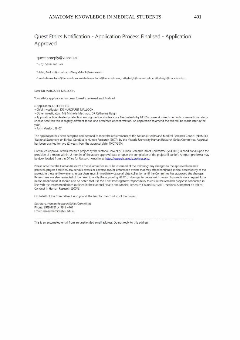
ANATOMY KNOWLEDGE IN MEDICAL STUDENTS 401

ANATOMY KNOWLEDGE IN MEDICAL STUDENTS 402

ANATOMY KNOWLEDGE IN MEDICAL STUDENTS 403
APPENDIX D
Research Flyer to Students

ANATOMY KNOWLEDGE IN MEDICAL STUDENTS 404

ANATOMY KNOWLEDGE IN MEDICAL STUDENTS 405
APPENDIX E
List of Anatomy objectives covered in Year A

ANATOMY KNOWLEDGE IN MEDICAL STUDENTS 406
Regional topic areas of clinically relevant anatomy examined
Abdomen & Pelvis
1. Organs affected by ischaemia from occlusion of foregut, midgut & hindgut arteries.
2. Significance and diagnosis of a perforated peptic ulcer and the resulting haematemesis
and/or malena resulting from damaged vessels.
3. Endangerment of spleen from lower left rib fractures.
4. Potential hernia sites, noting difference between indirect and direct inguinal hernia.
5. Site of pain referral associated with hernias.
6. Site of pancreatic cancer and its association with obstructive jaundice.
7. Significance of gallstones and where they are most likely to lodge.
8. Lowest parts of peritoneal cavity in supine male and female, noting significance of pus
or blood potentially accumulating at these sites.
9. Visceral sources of pain for epigastric pain, umbilical pain, suprapubic pain, perineal
pain and loin pain.
10. Lymph drainage in tumour spread from abdominal organ
11. Site of pain referral from pelvic structures via obturator nerve
12. Significance of tumour spread from prostate cancer to spine via internal vertebral
venous plexus
13. Relationship and significance of ureters to cervix and to uterine arteries
14. Importance of pelvic floor in maintaining faecal continence.
Thorax
1. Distribution of coronary artery and significance of dominance in coronary artery
occlusion
2. Ideal site for intercostal catheterisation via anterior and lateral approach, noting sites
where thoracic wall nerves and vessels are endangered.
3. Effect of severed thoracic duct with ligated thoracic duct
4. Sites of pain referral over thorax, noting specifically how cardiac pain and oesophageal
pain may mimic each other.
5. Significance of inhaled material to pass into right main bronchus.
6. Significance of cardiac tamponade and acceptable site for pericardial tap.
7. Significance of deviation of trachea.
8. Significance of lymph drainage of lungs, noting significance of widening of carina on
bronchoscopy.
9. Surface projections of fissures and lobes of lungs in diagnosis of lung disease.
10. Lymph drainage of breast noting implications for tumour spread
11. Significance of venous drainage of breast and venous spread of tumours and infection
12. Arterial supply and anastomoses of breast
Back
1. Indicate particular spinal nerve which emerges from each intervertebral foramen (noting
that there are 8 cervical spinal nerves, yet only 7 cervical vertebrae).
2. Sites, mechanism, degrees and effects of lumbar disc herniation, noting where discs are
least supported.
3. Meninges & associated spaces, level of termination of spinal cord and ideal site for
lumbar puncture, noting required posture of patient and bony surface landmarks to
detect most appropriate level.
4. Structures potentially affected in back injuries and anatomical basis for reflex muscle
spasm.

ANATOMY KNOWLEDGE IN MEDICAL STUDENTS 407
Head & Neck
1. Potential complications of thyroid surgery, noting endangerment of arterial and nerve
supplies.
2. Ideal sites for cricothyroidotomy.
3. Lymph node biopsies in posterior triangle of neck noting endangerment of accessory
nerve and subsequent effects.
4. Lesions of cranial nerves IX, X, XI and subsequent effects
5. Motor effects of strokes involving main blood vessels of brain
6. Cranial nerves supplying extra ocular muscles of eye and how these are tested.
7. Sites of emissary veins, noting significance as potential avenues of intracranial spread
for face and scalp lacerations.
8. Contents of floor of mouth and relation of vessels to submandibular gland
9. Branches of facial nerve and sites where motor nerves are endangered with facial
lacerations and incisions
10. Nerve endangered with depressed fracture of zygomatic bone.
Upper Limb
1. Role of dorsal digital expansion in finger movements and significance of injury to it.
2. Motor and sensory impairments from lesions to median nerve at wrist and in carpal
tunnel
3. Motor and sensory impairments from lesions to ulnar nerve at wrist and entrapment in
cubital tunnel.
4. Rotator cuff muscles noting sites and structures involved in painful arc syndrome
5. Note key neurovascular contents in axilla region and effects of damage to structures.
6. Motor and sensory impairments of damage to posterior cord
7. Mechanism, components torn and effects of dislocated acromioclavicular joint, noting
structures endangered.
8. Key neurovascular contents in palm and dorsum of hand and importance of it for
cannulation.
9. Mechanism, effect and structures endangered in fractured mid-shaft of humerus.
Lower Limb
1. Sites where major lower limb nerves are endangered by laceration (where arteries are
palpable and across large superficial veins).
2. Acceptable site for central venous catheterisation, comprehending boundaries of femoral
triangle.
3. Mechanism and effects of fractured neck of femur, noting supply to its head.
4. Factors resulting in positive Trendelenburg sign.
5. Sites where nerves are endangered by entrapment, particularly under inguinal ligament
and in tarsal tunnel.
6. Motor and sensory impairments from lesions to tibial nerve entrapped in tarsal tunnel;
common fibular nerve in popliteal fossa and behind head of fibula via laceration or
compression.
7. Surface markings of dorsalis pedis artery.
8. Diagnosis and potential consequences of compartment syndrome in leg (particularly
anterior compartment).
9. Mechanism and effects of damage to cruciate ligaments and how to test integrity of
these ligaments.

ANATOMY KNOWLEDGE IN MEDICAL STUDENTS 408
APPENDIX F
Anatomy Learning Survey & Anatomy Test Questions

ANATOMY KNOWLEDGE IN MEDICAL STUDENTS 409
ANATOMY LEARNING QUESTIONNAIRE
1. My previous educational background was
A. Anatomy in a professional course (Physiotherapy/Occupational therapy)
B. Anatomy in a biomedical science course
C. Biological Science
D. Other science
E. No science
2. The resource I use/used the most for learning anatomy is/was
A. Textbooks
B. An@tomedia
C. Anatomy days at Clayton
D. Tutorials
E. Lectures
F. Study groups
G. Other (please specify)
3. The resource I find the most useful for learning anatomy is
A. Textbooks
B. Anatomedia
C. Anatomy days at Clayton
D. Tutorials
E. Lectures
F. Study groups
G. Other (please specify)
4. The volume of anatomy knowledge that I needed to learn in Year A was daunting
A. Strongly agree
B. Agree
C. Unsure
D. Disagree
E. Strongly disagree
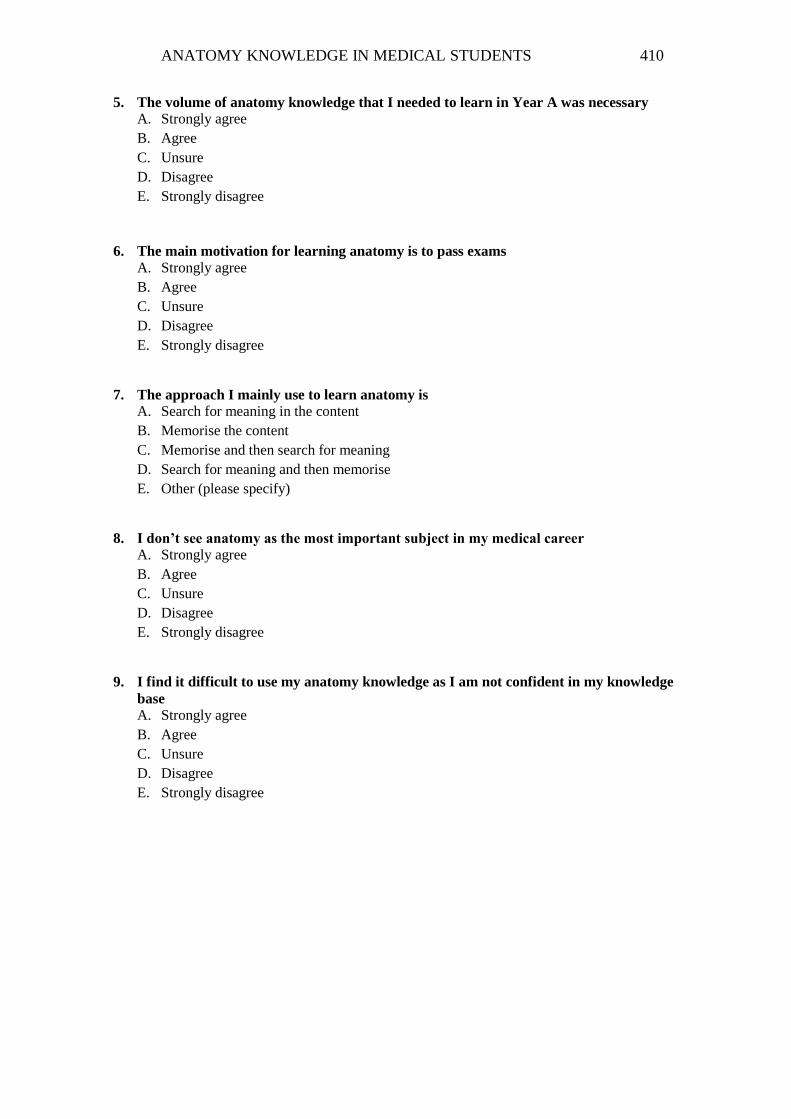
ANATOMY KNOWLEDGE IN MEDICAL STUDENTS 410
5. The volume of anatomy knowledge that I needed to learn in Year A was necessary
A. Strongly agree
B. Agree
C. Unsure
D. Disagree
E. Strongly disagree
6. The main motivation for learning anatomy is to pass exams
A. Strongly agree
B. Agree
C. Unsure
D. Disagree
E. Strongly disagree
7. The approach I mainly use to learn anatomy is
A. Search for meaning in the content
B. Memorise the content
C. Memorise and then search for meaning
D. Search for meaning and then memorise
E. Other (please specify)
8. I don’t see anatomy as the most important subject in my medical career
A. Strongly agree
B. Agree
C. Unsure
D. Disagree
E. Strongly disagree
9. I find it difficult to use my anatomy knowledge as I am not confident in my knowledge
base
A. Strongly agree
B. Agree
C. Unsure
D. Disagree
E. Strongly disagree

ANATOMY KNOWLEDGE IN MEDICAL STUDENTS 411
10. I frequently use my anatomy knowledge during clinical skills sessions
A. Strongly agree
B. Agree
C. Unsure
D. Disagree
E. Strongly disagree
11. I see anatomy as an important part of becoming a doctor
A. Strongly agree
B. Agree
C. Unsure
D. Disagree
E. Strongly disagree
12. The challenges I perceive in learning anatomy are
A. The amount of material that needs to be learned
B. Application of anatomical knowledge to clinical cases
C. Visualisation of anatomical structures
D. Location of anatomical structures
E. Other (please specify)
13. Prior to commencing Year A, I had a solid foundation in anatomy (or particular
systems e.g., the musculoskeletal system – if the latter, please specify)
A. Strongly agree
B. Agree
C. Unsure
D. Disagree
E. Strongly disagree

ANATOMY KNOWLEDGE IN MEDICAL STUDENTS 412
ABDOMEN/PELVIS
1. Leah, a 27-year old female bodybuilder presents to her GP with pain in her left lower
abdominal area following a session of heavy weight lifting at the gym. On examination, the
GP finds a small protrusion in the groin area located right above and lateral to the mid-
point of the inguinal ligament. Which of the following hernias is Leah MOST LIKELY to
be diagnosed with?
A. Diaphragmatic hernia
B. Direct inguinal hernia
C. Femoral hernia
D. Incisional hernia
E. Indirect inguinal hernia
Objective Define the term ‘hernia’ (noting mechanism, diagnosis & effects). List
potential hernial sites from the abdomen through orifices normally
present & through areas of weakness. Contrast an ‘indirect’ with a
‘direct’ inguinal hernia.
Taxonomy Application
Borderline 0.5
2. Leah is admitted for a hernia procedure. During the surgery, which of the following
structures would be found passing through the deep inguinal ring and traversing the
inguinal canal?
A. Femoral branch of genitofemoral nerve
B. Iliohypogastric nerve
C. Ilioinguinal nerve
D. Ovarian artery
E. Round ligament of uterus
Objective Outline surface markings of the superficial inguinal ring & of the deep
inguinal ring. Comprehend the walls of inguinal canal & structures
transmitted
Taxonomy Recall
Borderline 0.3
3. Following a laparoscopic inguinal hernia repair, a 47-year old man complains to his
doctor about pain in the scrotum. Examination reveals pain and paresthesia in the
anterolateral surface of the scrotum. Which of the following nerves was MOST
LIKELY injured during this hernia repair?
A. Anterior femoral cutaneous nerve
B. Femoral branch of genitofemoral nerve
C. Iliohypogastric nerve
D. Ilioinguinal nerve
E. Subcostal nerve

ANATOMY KNOWLEDGE IN MEDICAL STUDENTS 413
Objective Indicate the site of pain referral (noting it is often not at the source
which may obscure diagnosis, hence hernial sites should be examined
in abdominal pain)
Taxonomy Application
Borderline 0.3
4. A 55-year old male is admitted to hospital following complaints of severe abdominal
pain. Radiological examination reveals the obstruction to be at one of the branches of
the inferior mesenteric artery. Which of the following organs is MOST LIKELY to
have been affected by the ischaemia?
A. Ileum
B. Ascending colon
C. Caecum
D. Descending colon
E. Transverse colon
Objective Comprehend the arteries of foregut, midgut & hindgut (& their
distribution)
Taxonomy Recall
Borderline 0.7
5. Emma presents to the Emergency room with hematemesis (vomiting blood). Endoscopy
reveals an ulcer that has perforated the posterior wall of the first part of the duodenum.
Which of the following arteries is MOST LIKELY to be damaged as a result of this?
A. Gastroduodenal artery
B. Gastro-omental artery
C. Left gastric artery
D. Short gastric artery
E. Splenic artery
Objective Explain the significance and diagnosis of a perforated peptic ulcer.
Explain how a massive haematemesis &/or malena can result from a
peptic ulcer (indicating the artery directly behind the stomach and the
artery directly behind the first part of the duodenum)
Taxonomy Recall
Borderline 0.4

ANATOMY KNOWLEDGE IN MEDICAL STUDENTS 414
6. During an AFL game, one of the players, Matthew, is injured when his opponent
smashes into him. The physician on site discovers that Matthew has sustained rib
fractures on the left side and is rushed to hospital. Radiological examination reveals a
fractured 9th and 10th rib on the left side near the angle of the rib. Which of the
following organs is MOST LIKELY the first to be injured?
A. Liver
B. Pancreas
C. Left Kidney
D. Spleen
E. Stomach
Objective Explain how the spleen is endangered in fractures of the lower left ribs.
Taxonomy Application
Borderline 0.7
Questions 7 and 8 relate to the following options. Each option may be used once, twice or
not at all.
A. Body of pancreas
B. Common bile duct
C. Cystic duct
D. Head of pancreas
E. Hepatopancreatic ampulla (Ampulla of Vater)
F. Neck of pancreas
G. Pancreatic duct
H. Right hepatic duct
I. Tail of pancreas
J. Uncinate process
7. Jim, a 62-year-old man develops severe jaundice and is taken to the hospital by his son.
On history taking and ordering further tests and radiological examination, the doctor
makes a diagnosis of pancreatic cancer. At which of the following pancreatic sites is the
tumour MOST LIKELY to be found?
Answer - D
Objective Explain why obstructive jaundice tends to occur with cancer of the head
of the pancreas (noting the structure intimately related to it)
Taxonomy Application
Borderline 0.4
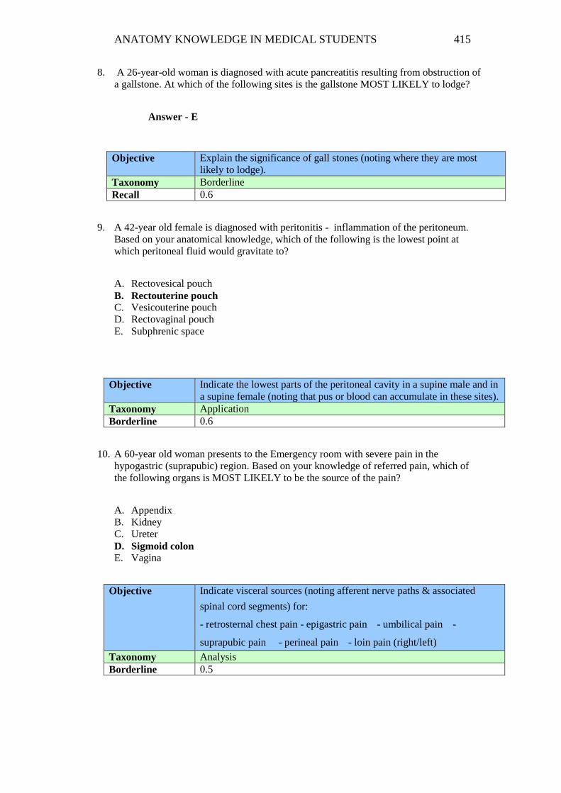
ANATOMY KNOWLEDGE IN MEDICAL STUDENTS 415
8. A 26-year-old woman is diagnosed with acute pancreatitis resulting from obstruction of
a gallstone. At which of the following sites is the gallstone MOST LIKELY to lodge?
Answer - E
Objective Explain the significance of gall stones (noting where they are most
likely to lodge).
Taxonomy Borderline
Recall 0.6
9. A 42-year old female is diagnosed with peritonitis - inflammation of the peritoneum.
Based on your anatomical knowledge, which of the following is the lowest point at
which peritoneal fluid would gravitate to?
A. Rectovesical pouch
B. Rectouterine pouch
C. Vesicouterine pouch
D. Rectovaginal pouch
E. Subphrenic space
Objective Indicate the lowest parts of the peritoneal cavity in a supine male and in
a supine female (noting that pus or blood can accumulate in these sites).
Taxonomy Application
Borderline 0.6
10. A 60-year old woman presents to the Emergency room with severe pain in the
hypogastric (suprapubic) region. Based on your knowledge of referred pain, which of
the following organs is MOST LIKELY to be the source of the pain?
A. Appendix
B. Kidney
C. Ureter
D. Sigmoid colon
E. Vagina
Objective Indicate visceral sources (noting afferent nerve paths & associated
spinal cord segments) for:
- retrosternal chest pain - epigastric pain - umbilical pain -
suprapubic pain - perineal pain - loin pain (right/left)
Taxonomy Analysis
Borderline 0.5

ANATOMY KNOWLEDGE IN MEDICAL STUDENTS 416
11. Which of the following would be the first palpable lymph node in a patient with
stomach cancer?
A. Superficial inguinal lymph node
B. Axillary lymph node
C. Right supraclavicular lymph node
D. Left supraclavicular lymph node
E. Deep inguinal lymph node
Objective Trace the path of lymph drainage (noting significance in tumour
spread): - from stomach - from testis. Explain why the first palpable
lymph node tends to be the left supraclavicular node (noting it is a late
sign although often the first sign of cancer in these organs)
Taxonomy Application
Borderline 0.7
12. Jim, a 77-year old has been diagnosed with prostate cancer and is undergoing a pelvic
lymph node dissection in order to determine if the cancer has spread to the lymph
nodes. A day following the surgery, Jim is unable to feel sensation around the medial
aspect of his thigh. Which of the following nerves has MOST LIKELY been injured
during the surgery?
A. Femoral nerve
B. Saphenous nerve
C. Obturator nerve
D. Pudendal nerve
E. Genitofemoral nerve
Objective Comprehend the boundaries of femoral triangle, gluteal region,
popliteal fossa & tarsal tunnel and the key neurovascular contents in
each of the above major regions
Taxonomy Recall
Borderline 0.5
13. Which of the following arteries provides the main supply for midgut structures?
A. Celiac trunk
B. Splenic artery
C. Inferior mesenteric artery
D. Superior mesenteric artery
E. Ileocolic artery
Objective Comprehend the arteries of foregut, midgut & hindgut (& their
distribution)
Taxonomy Recall
Borderline 0.7

ANATOMY KNOWLEDGE IN MEDICAL STUDENTS 417
14. Anna, a 45-year-old woman presents to the Emergency Department with severe pain in
the left lumbar and iliac fossa region. On history taking, Anna tells the doctor that she
has been voiding (urinating) quite frequently and that the colour of her urine is slightly
brown in color. Radiological imaging reveals the presence of cancer in an organ directly
related to the termination of the left ureter. In which of the following structures is the
cancer MOST LIKELY to be found?
A. Apex of bladder
B. Fundus of uterus
C. Cervix of uterus
D. Recto-sigmoid junction
E. Ovary
Objective Indicate the relationship of the ureters to the cervix and to the uterine
arteries. Explain the significance of each.
Taxonomy Application
Borderline 0.5
15. Following delivery of her first born child, Melinda, complains problems with
defecation. On history taking, the doctor suspects damage to the pelvic floor muscles
during delivery resulting in a diagnoses of faecal incontinence. Which of the following
muscles is MOST LIKELY to contribute to faecal continence and hence has been
damaged?
A. Pubococcygeus
B. Puborectalis
C. Iliococcygeus
D. Internal anal sphincter
E. Obturator internus
Objective Explain role of the pelvic floor (particularly puborectalis) for faecal
continence (noting why pelvic viscera normally do not prolapse during
a Valsalva manoeuvre).
Taxonomy Recall
Borderline 0.5

ANATOMY KNOWLEDGE IN MEDICAL STUDENTS 418
THORAX
1. George, a 45-year old man presents to the Emergency Room with angina, and
hypotension. A coronary angiogram reveals that George’s coronary circulation is right-
dominant and there is ischemia around the apex of the heart. Which of the following
arteries is the MOST LIKELY cause of this ischemia?
A. Anterior interventricular (descending)
B. Circumflex
C. Left marginal
D. Posterior interventricular (descending)
E. Right marginal
Objective Outline the area of distribution of each coronary artery (noting the
components of the conducting system supplied). Indicate which
coronary artery is typically dominant.
Taxonomy Recall
Borderline 0.6
2. A 53-year old woman in a car accident develops a hemopneumothorax on the left side.
An intercostal catheter (chest tube) is inserted in order to remove air and drain the
pleural fluid. Which of the following sites is the MOST APPROPRIATE insertion point
for this procedure?
A. Superior to the 5th left rib in the mid-clavicular line
B. Inferior to the 5th left rib in the mid-axillary line
C. Superior to the 6th left rib in the mid-axillary line
D. Inferior to the 6th left rib in the mid-axillary line
E. Superior to the 11th left rib in the scapular line
Objective Indicate the sites where thoracic wall vessels & nerves are endangered
by a penetrating injury. Indicate the ideal site for intercostal
catheterisation via an anterior approach and via a lateral approach.
Taxonomy Application
Borderline 0.5
3. A 60-year old man wakes up at night with retrosternal pain. He is rushed to the hospital
where the physician performs a cardiovascular assessment and is unable to find any
abnormalities. Which of the following structures is the next MOST LIKELY source of
his pain?
A. Parietal layer of serous pericardium
B. Mediastinal pleura
C. Oesophagus
D. Diaphragmatic pleura
E. Fibrous pericardium

ANATOMY KNOWLEDGE IN MEDICAL STUDENTS 419
Objective Indicate potential sources of referred pain to dermatomes over thorax
(noting why cardiac pain and oesophageal pain may mimic each other)
and from thoracic structures (particularly myocardium, pericardium &
pleura) to dermatomes over other sites.
Taxonomy Analysis
Borderline 0.3
4. As a medical student, you are about to observe a doctor perform pericardiocentesis on a
patient suspected of having cardiac tamponade. As the doctor prepares to drain excess
blood from the pericardial cavity, he asks you to indicate the site at which the needle
should be inserted. The MOST APPROPRIATE site to do so would be
A. Right 4th intercostal space, sternal margin
B. Left 4th intercostal space, sternal margin
C. Right xiphisternal junction
D. Left xiphisternal junction
E. Left 5th intercostal space, midclavicular line
Objective Indicate the effects of accumulation of fluid in the pericardial cavity
(noting the significance of ‘cardiac tamponade’, particularly from a
sudden bleed into it). Indicate an acceptable site for a pericardial tap.
Taxonomy Application
Borderline 0.4
5. A 44-year old man was stabbed in the right side of his neck, approximately 3 cm
superior to the middle of the clavicle. He develops a pneumothorax as a result of the
injury and is rushed to hospital. Which of the following signs would be MOST
APPARENT during a physical exam?
A. Decreased breath sounds on both sides of thorax
B. Deviation of trachea and apex beat to right side
C. Deviation of trachea and apex beat to left side
D. Increased breath sounds on right side of thorax
E. Decreased breath sounds on left side of thorax.
Objective Explain the significance of deviation of the trachea.
Taxonomy Analysis
Borderline 0.4
6. During a surgical procedure in the posterior mediastinum, the structure between the
descending aorta and azygos vein is inadvertently lacerated. Which of the following is
the MOST LIKELY structure to be injured?
A. Left recurrent laryngeal nerve
B. Hemiazygos vein
C. Pulmonary artery
D. Right recurrent laryngeal nerve
E. Thoracic duct

ANATOMY KNOWLEDGE IN MEDICAL STUDENTS 420
Objective Contrast the effect of a severed thoracic duct with that of a ligated
thoracic duct
Taxonomy Recall
Borderline 0.5
7. A 41-year old man who presents with fever and shortness of breath is diagnosed with
lobar pneumonia. Examination reveals bronchial breathing confined to the right axillary
region. Which of the following lobes is MOST LIKELY affected?
A. Hilum
B. Inferior lobe
C. Lingula
D. Middle lobe
E. Superior lobe
Objective Map the surface projections of the fissures and lobes of the lungs
Taxonomy Analysis
Borderline 0.4
8. A 35-year woman patient is diagnosed with cancer of the right breast. Which of the
following lymphatic structures would majority of the cancer cells first spread to?
A. Axillary lymph nodes
B. Parasternal lymph nodes
C. Left supraclavicular lymph nodes
D. Right lymphatic duct
E. Thoracic duct
Objective Outline the lymph drainage of the breast contrasting lateral with
medial quadrants (noting the significance of overlap across the nipple
line vertical watershed and the implications for tumour spread)
Taxonomy Application
Borderline 0.6
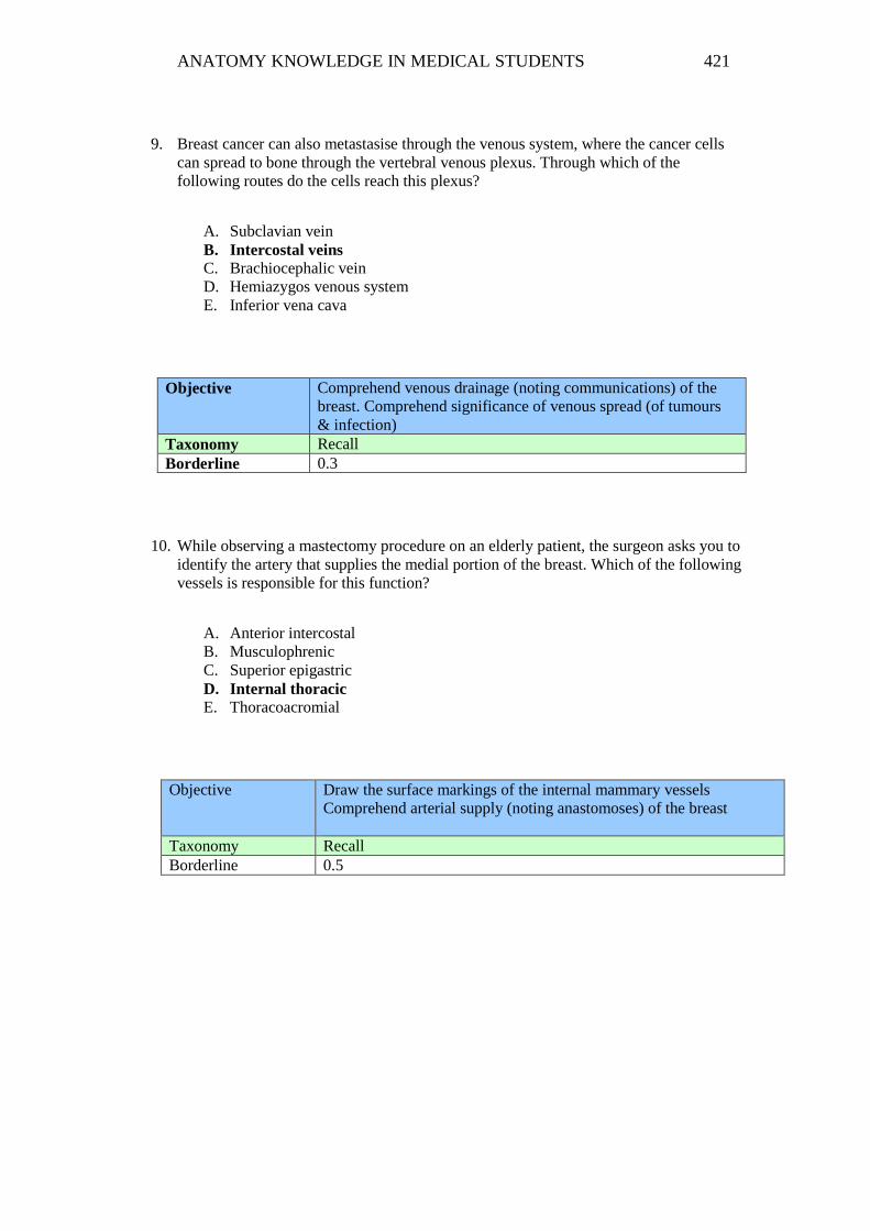
ANATOMY KNOWLEDGE IN MEDICAL STUDENTS 421
9. Breast cancer can also metastasise through the venous system, where the cancer cells
can spread to bone through the vertebral venous plexus. Through which of the
following routes do the cells reach this plexus?
A. Subclavian vein
B. Intercostal veins
C. Brachiocephalic vein
D. Hemiazygos venous system
E. Inferior vena cava
Objective Comprehend venous drainage (noting communications) of the
breast. Comprehend significance of venous spread (of tumours
& infection)
Taxonomy Recall
Borderline 0.3
10. While observing a mastectomy procedure on an elderly patient, the surgeon asks you to
identify the artery that supplies the medial portion of the breast. Which of the following
vessels is responsible for this function?
A. Anterior intercostal
B. Musculophrenic
C. Superior epigastric
D. Internal thoracic
E. Thoracoacromial
Objective Draw the surface markings of the internal mammary vessels
Comprehend arterial supply (noting anastomoses) of the breast
Taxonomy Recall
Borderline 0.5

ANATOMY KNOWLEDGE IN MEDICAL STUDENTS 422
UPPER LIMB
1. During a basketball game, the ball makes contact with the tip of Raoul’s extended middle
finger causing a forceful flexion at the distal interphalangeal joint. When examined by the
sports physician, Raoul is diagnosed with a mallet finger. Which of the following structures
is MOST LIKELY to have been affected?
A. Flexor digitorium superficialis
B. Extensor digitorium
C. Flexor digitorium profundus
D. Lumbricals 3 and 4.
E. Interosseous muscles
Objective Demonstrate and explain: the role of the dorsal digital expansion in
finger movements (noting the intrinsic muscles of palm used in
finger extension)
Taxonomy Application
Borderline 0.7
2. Tracy, 27-years of age is expecting her first child and is 4-months pregnant. She has
developed some weakness, numbness and pain sensations at night when her right wrist is
flexed. On visiting the doctor, she is diagnosed with carpal tunnel syndrome. Based on your
knowledge of anatomy, which of the following signs would be characteristic of this
syndrome?
A. Loss of sensation over the thenar eminence.
B. Loss of innervation to flexor digitorium superficialis.
C. Loss of innervation to flexor pollicis brevis.
D. Loss of innervation to flexor digitorium profundus.
E. Loss of sensation to skin of the palm of the hand
Objective Indicate motor & sensory impairments (noting any reflex lost &
deformity produced) from lesions to major nerves of the upper limb
at sites where endangered by: compression (median at elbow
compared to in carpal tunnel)
Taxonomy Application
Borderline 0.3

ANATOMY KNOWLEDGE IN MEDICAL STUDENTS 423
3. Tracy has a carpal tunnel release operation. The day following the surgery, she is unable to
abduct and flex her thumb. Which of the following nerves is MOST LIKELY to have been
injured?
A. Anterior interosseous nerve
B. Palmar cutaneous branch of median nerve
C. Deep branch of ulnar nerve
D. Superficial branch of ulnar nerve
E. Recurrent thenar branch of median nerve
Objective Indicate motor & sensory impairments (noting any reflex lost &
deformity produced) from lesions to major nerves of the upper limb
at sites where endangered by: lacerations (median at wrist compared
to of its thenar branch in the palm)
Taxonomy Recall
Borderline 0.5
4. A patient with multiple lacerations to her anterior wrist and palm presents to the Emergency
Room with severe bleeding. On examination, the patient is unable to hold a piece of paper
placed between the thumb and index finger. The function of which of the following nerves
is MOST LIKELY being tested?
A. Median nerve
B. Ulnar nerve
C. Recurrent thenar branch of median nerve
D. Superficial branch of ulnar nerve
E. Deep branch of ulnar nerve
Objective Indicate motor & sensory impairments (noting any reflex lost &
deformity produced) from lesions to major nerves of the upper limb
at sites where endangered by: entrapment (ulnar in cubital tunnel
compared to in ulnar canal)
Taxonomy Analysis
Borderline 0.3

ANATOMY KNOWLEDGE IN MEDICAL STUDENTS 424
5. George, a frequent tennis player is diagnosed with cubital tunnel syndrome due to
compression of the nerve that sits posterior to the medial epicondyle of the humerus. Which
of the following muscles would be affected by this syndrome?
A. Flexion of proximal interphalangeal joints of digits 2-5
B. Flexion of thumb
C. Adduction of thumb
D. Abduction of the thumb
E. Flexion of distal interphalangeal joints of digits 2-5
Objective Indicate motor & sensory impairments (noting any reflex lost &
deformity produced) from lesions to major nerves of the upper limb
at sites where endangered by: entrapment (ulnar in cubital tunnel
compared to in ulnar canal)
Taxonomy Application
Borderline 0.5
6. David, a swimming athlete visits his GP complaining of pain in his right shoulder region.
On inspection, you notice a difference between the two sides, particularly superior to the
spine of the scapula, with the right side appearing smaller than the left. There is no winging
of the scapula but on examination, David is unable to externally rotate and initiate
abduction of his arm. However, if the arm is assisted through the first 45 degrees of
abduction, David is able to complete full abduction. Given this information, which of the
following nerves would you suspect has been damaged?
A. Axillary nerve
B. Upper subscapular nerve
C. Suprascapular nerve
D. Dorsal scapular nerve
E. Lower subscapular nerve
Objective Identify ‘rotator cuff muscles’ of the shoulder joint and explain their
major role. Indicate the sites & structures involved in: ‘painful arc
syndrome’ (of shoulder)
Taxonomy Analysis
Borderline 0.4
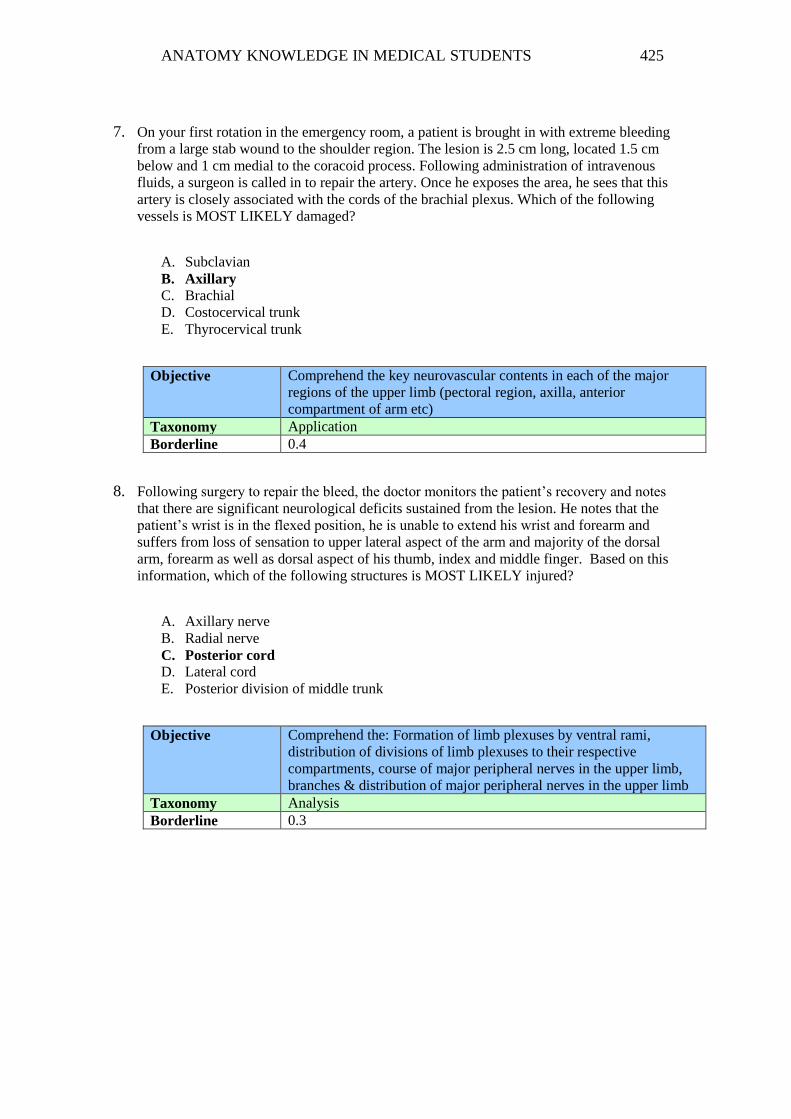
ANATOMY KNOWLEDGE IN MEDICAL STUDENTS 425
7. On your first rotation in the emergency room, a patient is brought in with extreme bleeding
from a large stab wound to the shoulder region. The lesion is 2.5 cm long, located 1.5 cm
below and 1 cm medial to the coracoid process. Following administration of intravenous
fluids, a surgeon is called in to repair the artery. Once he exposes the area, he sees that this
artery is closely associated with the cords of the brachial plexus. Which of the following
vessels is MOST LIKELY damaged?
A. Subclavian
B. Axillary
C. Brachial
D. Costocervical trunk
E. Thyrocervical trunk
Objective Comprehend the key neurovascular contents in each of the major
regions of the upper limb (pectoral region, axilla, anterior
compartment of arm etc)
Taxonomy Application
Borderline 0.4
8. Following surgery to repair the bleed, the doctor monitors the patient’s recovery and notes
that there are significant neurological deficits sustained from the lesion. He notes that the
patient’s wrist is in the flexed position, he is unable to extend his wrist and forearm and
suffers from loss of sensation to upper lateral aspect of the arm and majority of the dorsal
arm, forearm as well as dorsal aspect of his thumb, index and middle finger. Based on this
information, which of the following structures is MOST LIKELY injured?
A. Axillary nerve
B. Radial nerve
C. Posterior cord
D. Lateral cord
E. Posterior division of middle trunk
Objective Comprehend the: Formation of limb plexuses by ventral rami,
distribution of divisions of limb plexuses to their respective
compartments, course of major peripheral nerves in the upper limb,
branches & distribution of major peripheral nerves in the upper limb
Taxonomy Analysis
Borderline 0.3

ANATOMY KNOWLEDGE IN MEDICAL STUDENTS 426
9. While driving home from work one afternoon, Julie comes across an accident and is
transported to hospital due to a shoulder injury. On inspection, the lateral end of her right
clavicle is elevated and separated from the acromion process. In addition, her right upper
limb drops when not supported by her left arm. Which of the following ligaments would
have to be completely ruptured for this to occur?
A. Superior glenohumeral ligament
B. Coracoacromial ligament
C. Coracoclavicular ligament
D. Sternoclavicular ligament
E. Transverse humeral ligament
Objective Explain the mechanism, components torn and effect of a
dislocated: acromioclavicular joint (in contrast to a subluxation) and
shoulder joint (noting structures endangered & implication for
reduction)
Taxonomy Application
Borderline 0.6
10. You observe an intern doctor inserting an IV into the distal end of the forearm on the radial
side. Which of the following veins is MOST LIKELY cannulated?
A. Cephalic vein
B. Basilic vein
C. Brachial vein
D. Median cubital vein
E. Median antebrachial vein
Objective Comprehend the key neurovascular contents in each of the major
regions of the upper limb (palm of hand, dorsum of hand etc)
Taxonomy Recall
Borderline 0.6

ANATOMY KNOWLEDGE IN MEDICAL STUDENTS 427
BACK
EMQ 1 and 2 relate to the following options. Each option may be used once, twice or none
at all.
A. C3
B. C4
C. C5
D. C6
E. C7
F. L1
G. L2
H. L3
I. L4
J. L5
K. S1
1. A rugby player suffers from a herniated (ruptured) intervertebral disk in the region of
his neck. On imaging, the disk appears to be compressing the spinal nerve exiting the
intervertebral foramen between the 4th and 5th cervical vertebrae. Which of the
following spinal nerves has been injured?
Answer – C
Objective Indicate the particular spinal nerve which emerges from each
intervertebral foramen (noting that there are 8 cervical spinal nerves
yet only 7 cervical vertebrae).
Taxonomy Recall
Borderline 0.6
2. A woman presents with a herniated intervertebral disc in the lumbar region between L5
and S1. If the spinal nerve immediately posterior to this disc is compressed, which
nerve is affected?
Answer – J
Objective Explain sites, mechanism, degrees and effects of lumbar disc
herniation (noting where discs are least supported).
Taxonomy Application
Borderline 0.4
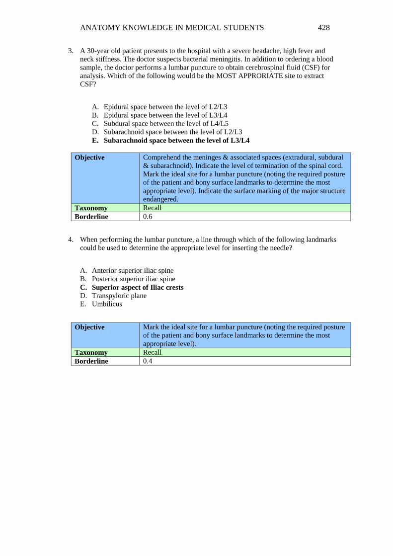
ANATOMY KNOWLEDGE IN MEDICAL STUDENTS 428
3. A 30-year old patient presents to the hospital with a severe headache, high fever and
neck stiffness. The doctor suspects bacterial meningitis. In addition to ordering a blood
sample, the doctor performs a lumbar puncture to obtain cerebrospinal fluid (CSF) for
analysis. Which of the following would be the MOST APPRORIATE site to extract
CSF?
A. Epidural space between the level of L2/L3
B. Epidural space between the level of L3/L4
C. Subdural space between the level of L4/L5
D. Subarachnoid space between the level of L2/L3
E. Subarachnoid space between the level of L3/L4
Objective Comprehend the meninges & associated spaces (extradural, subdural
& subarachnoid). Indicate the level of termination of the spinal cord.
Mark the ideal site for a lumbar puncture (noting the required posture
of the patient and bony surface landmarks to determine the most
appropriate level). Indicate the surface marking of the major structure
endangered.
Taxonomy Recall
Borderline 0.6
4. When performing the lumbar puncture, a line through which of the following landmarks
could be used to determine the appropriate level for inserting the needle?
A. Anterior superior iliac spine
B. Posterior superior iliac spine
C. Superior aspect of Iliac crests
D. Transpyloric plane
E. Umbilicus
Objective Mark the ideal site for a lumbar puncture (noting the required posture
of the patient and bony surface landmarks to determine the most
appropriate level).
Taxonomy Recall
Borderline 0.4

ANATOMY KNOWLEDGE IN MEDICAL STUDENTS 429
5. Charles, a 45-year old man presents to the GP with intense back pain region following a
golf session with his friends the previous day. On inspection and examination, the
doctor concludes that the pain must be due to spasm of the erector spinae muscle.
Which of the following structures is responsible for innervating this muscle?
A. Dorsal root of spinal nerve
B. Dorsal ramus of spinal nerve
C. Ventral root of spinal nerve
D. Ventral ramus of spinal nerve
Objective Indicate the structures potentially affected in back injuries. Explain
the anatomical basis for reflex muscle spasm (of erector spinae).
Taxonomy Recall
Borderline 0.5

ANATOMY KNOWLEDGE IN MEDICAL STUDENTS 430
HEAD/NECK
1. Daisy has a thyroidectomy for a suspected malignancy in the right lobe of the thyroid
gland. A month after surgery, Daisy’s speech is normal but she informs her doctor that
her voice fatigues easily and that she has difficulty producing high-pitched sounds.
Which of the following nerves is MOST LIKELY to have been damaged for Daisy to
have these symptoms?
A. Phrenic nerve
B. Cervical sympathetic nerves
C. External branch of superior laryngeal nerve
D. Internal branch of superior laryngeal nerve
E. Recurrent laryngeal nerve
Objective List the potential complications of thyroid surgery (noting the effects
of each).
Taxonomy Application
Borderline 0.3
2. One afternoon, a patient comes in to the clinic with extreme inspiratory stridor and
acute upper airway obstruction. As it would take nearly 30 minutes to get to the nearest
hospital, the doctor acts immediately and creates an emergency surgical airway in order
to restore breathing. Which of the following sites is the MOST APPROPRIATE site to
create an airway?
A. At the jugular notch
B. Through the 2nd tracheal ring
C. Just above the thyroid cartilage
D. Just below the thyroid cartilage
E. Just below the cricoid cartilage
Objective Indicate the ideal site for a: - cricothyroidotomy
The ideal site for a tracheostomy (in contrast to an emergency
cricothyroidotomy) noting the layers pierced and structures
endangered (particularly in a child).
Taxonomy Application
Borderline 0.5
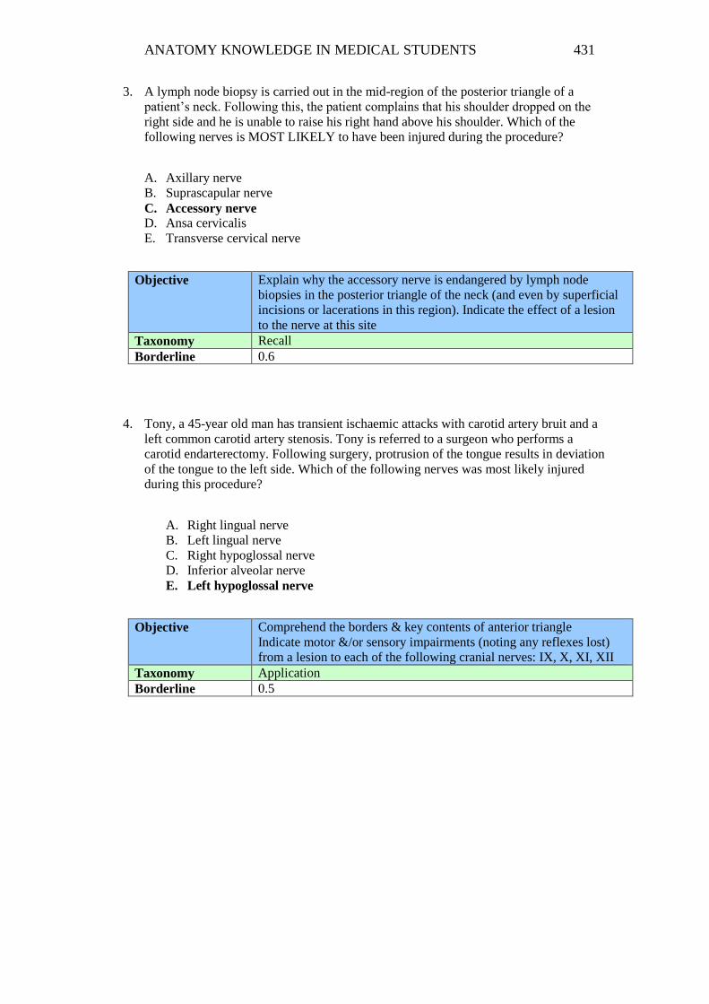
ANATOMY KNOWLEDGE IN MEDICAL STUDENTS 431
3. A lymph node biopsy is carried out in the mid-region of the posterior triangle of a
patient’s neck. Following this, the patient complains that his shoulder dropped on the
right side and he is unable to raise his right hand above his shoulder. Which of the
following nerves is MOST LIKELY to have been injured during the procedure?
A. Axillary nerve
B. Suprascapular nerve
C. Accessory nerve
D. Ansa cervicalis
E. Transverse cervical nerve
Objective Explain why the accessory nerve is endangered by lymph node
biopsies in the posterior triangle of the neck (and even by superficial
incisions or lacerations in this region). Indicate the effect of a lesion
to the nerve at this site
Taxonomy Recall
Borderline 0.6
4. Tony, a 45-year old man has transient ischaemic attacks with carotid artery bruit and a
left common carotid artery stenosis. Tony is referred to a surgeon who performs a
carotid endarterectomy. Following surgery, protrusion of the tongue results in deviation
of the tongue to the left side. Which of the following nerves was most likely injured
during this procedure?
A. Right lingual nerve
B. Left lingual nerve
C. Right hypoglossal nerve
D. Inferior alveolar nerve
E. Left hypoglossal nerve
Objective Comprehend the borders & key contents of anterior triangle
Indicate motor &/or sensory impairments (noting any reflexes lost)
from a lesion to each of the following cranial nerves: IX, X, XI, XII
Taxonomy Application
Borderline 0.5

ANATOMY KNOWLEDGE IN MEDICAL STUDENTS 432
5. A 64-year old woman recently suffered a stroke, which paralysed parts of the right side
of the body. In addition, she was having difficulty forming complete sentences.
Neurological imaging reveal that she suffered from a thromboembolic episode that
occluded blood supply to a part of the brain. Which of the following vessels was MOST
LIKELY affected?
A. Anterior cerebral artery
B. Internal carotid artery
C. Middle cerebral artery
D. Middle meningeal artery
E. Posterior cerebral artery
Objective Describe the main arterial supply of the brain and spinal
cord(vertebral-basilar systems and circle of willis). Recognise the
motor effects of strokes involving the main blood vessels of the brain.
Taxonomy Application
Borderline 0.6
6. With a patients’ eyes directed straight ahead, which direction would you ask the patient
to divert his gaze to in order to test the integrity of the trochlear nerve?
A. Medial and superiorly
B. Medial and inferiorly
C. Lateral and superiorly
D. Lateral and inferiorly
Objective Demonstrate eye movements (indicating the extra ocular muscles
involved)
Indicate the cranial nerves that supply the extra ocular muscles
Taxonomy Application
Borderline 0.4
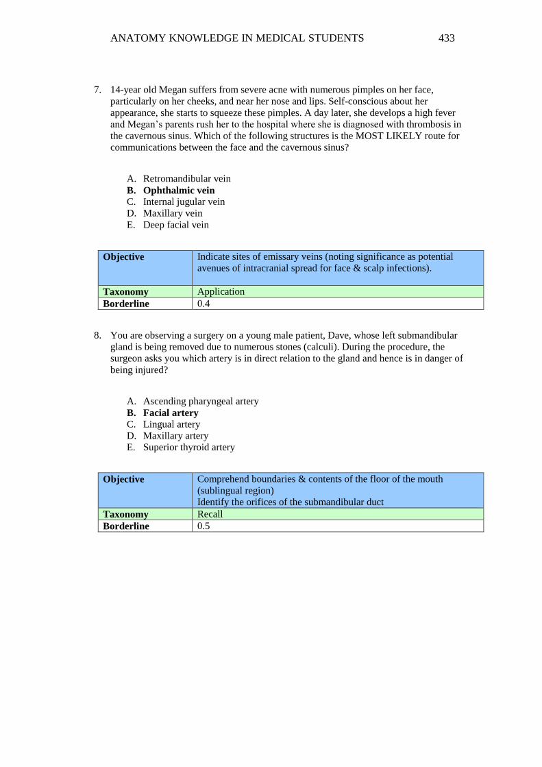
ANATOMY KNOWLEDGE IN MEDICAL STUDENTS 433
7. 14-year old Megan suffers from severe acne with numerous pimples on her face,
particularly on her cheeks, and near her nose and lips. Self-conscious about her
appearance, she starts to squeeze these pimples. A day later, she develops a high fever
and Megan’s parents rush her to the hospital where she is diagnosed with thrombosis in
the cavernous sinus. Which of the following structures is the MOST LIKELY route for
communications between the face and the cavernous sinus?
A. Retromandibular vein
B. Ophthalmic vein
C. Internal jugular vein
D. Maxillary vein
E. Deep facial vein
Objective Indicate sites of emissary veins (noting significance as potential
avenues of intracranial spread for face & scalp infections).
Taxonomy Application
Borderline 0.4
8. You are observing a surgery on a young male patient, Dave, whose left submandibular
gland is being removed due to numerous stones (calculi). During the procedure, the
surgeon asks you which artery is in direct relation to the gland and hence is in danger of
being injured?
A. Ascending pharyngeal artery
B. Facial artery
C. Lingual artery
D. Maxillary artery
E. Superior thyroid artery
Objective Comprehend boundaries & contents of the floor of the mouth
(sublingual region)
Identify the orifices of the submandibular duct
Taxonomy Recall
Borderline 0.5

ANATOMY KNOWLEDGE IN MEDICAL STUDENTS 434
9. Following the removal of the submandibular gland, Dave, finds that he is unable to
fully depress the angle of his mouth on his left side. Which of the following nerves is
MOST LIKELY injured in this case?
A. Buccal nerve
B. Facial nerve
C. Mandibular nerve
D. Marginal mandibular nerve
E. Ophthalmic nerve
Objective Explain why special care is taken with incisions on the face and
indicate the sites where motor nerve branches are endangered
Taxonomy Application
Borderline 0.4
10. A boxer comes to the ER to be treated for a fractured zygomatic bone. His right
cheekbone is depressed and on examination, there is no sensation to his right lower
eyelid, lateral aspect of the nose and upper lip. Which of the following nerves is MOST
LIKELY injured?
A. Supraorbital nerve
B. Zygomatic nerve
C. Infraoribital nerve
D. Superior alveolar nerve
E. Buccal nerve
Objective Explain how a depressed fracture of the cheekbone can be detected
(noting the bone involved). Indicate the nerve endangered and how
its function is tested
Taxonomy Application
Borderline 0.6
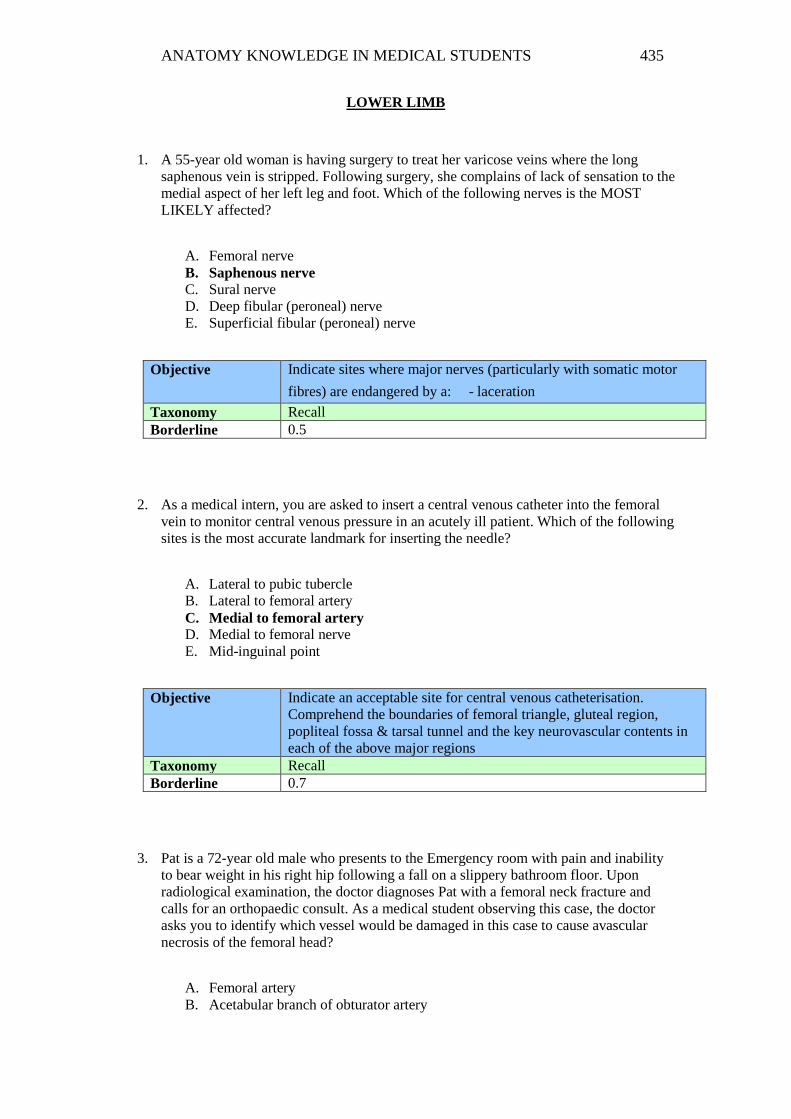
ANATOMY KNOWLEDGE IN MEDICAL STUDENTS 435
LOWER LIMB
1. A 55-year old woman is having surgery to treat her varicose veins where the long
saphenous vein is stripped. Following surgery, she complains of lack of sensation to the
medial aspect of her left leg and foot. Which of the following nerves is the MOST
LIKELY affected?
A. Femoral nerve
B. Saphenous nerve
C. Sural nerve
D. Deep fibular (peroneal) nerve
E. Superficial fibular (peroneal) nerve
Objective Indicate sites where major nerves (particularly with somatic motor
fibres) are endangered by a: - laceration
Taxonomy Recall
Borderline 0.5
2. As a medical intern, you are asked to insert a central venous catheter into the femoral
vein to monitor central venous pressure in an acutely ill patient. Which of the following
sites is the most accurate landmark for inserting the needle?
A. Lateral to pubic tubercle
B. Lateral to femoral artery
C. Medial to femoral artery
D. Medial to femoral nerve
E. Mid-inguinal point
Objective Indicate an acceptable site for central venous catheterisation.
Comprehend the boundaries of femoral triangle, gluteal region,
popliteal fossa & tarsal tunnel and the key neurovascular contents in
each of the above major regions
Taxonomy Recall
Borderline 0.7
3. Pat is a 72-year old male who presents to the Emergency room with pain and inability
to bear weight in his right hip following a fall on a slippery bathroom floor. Upon
radiological examination, the doctor diagnoses Pat with a femoral neck fracture and
calls for an orthopaedic consult. As a medical student observing this case, the doctor
asks you to identify which vessel would be damaged in this case to cause avascular
necrosis of the femoral head?
A. Femoral artery
B. Acetabular branch of obturator artery
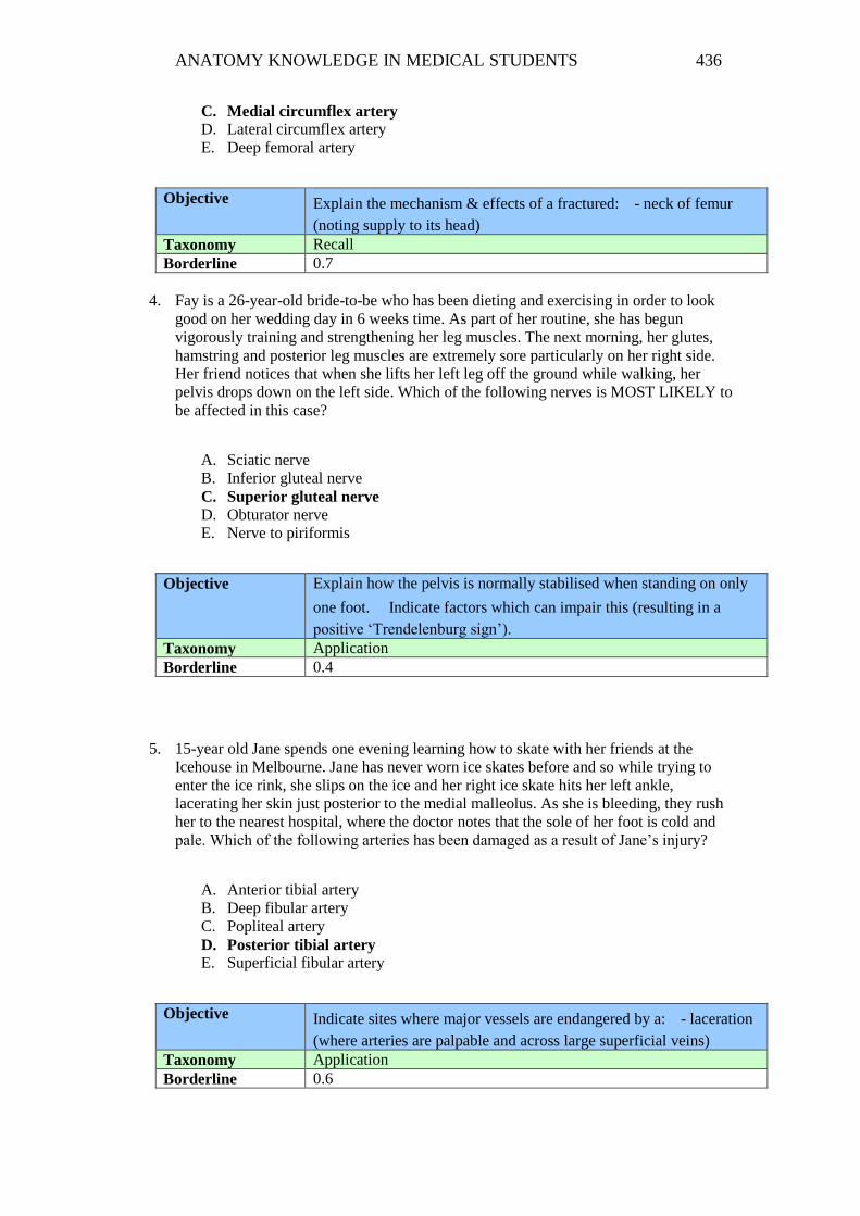
ANATOMY KNOWLEDGE IN MEDICAL STUDENTS 436
C. Medial circumflex artery
D. Lateral circumflex artery
E. Deep femoral artery
Objective Explain the mechanism & effects of a fractured: - neck of femur
(noting supply to its head)
Taxonomy Recall
Borderline 0.7
4. Fay is a 26-year-old bride-to-be who has been dieting and exercising in order to look
good on her wedding day in 6 weeks time. As part of her routine, she has begun
vigorously training and strengthening her leg muscles. The next morning, her glutes,
hamstring and posterior leg muscles are extremely sore particularly on her right side.
Her friend notices that when she lifts her left leg off the ground while walking, her
pelvis drops down on the left side. Which of the following nerves is MOST LIKELY to
be affected in this case?
A. Sciatic nerve
B. Inferior gluteal nerve
C. Superior gluteal nerve
D. Obturator nerve
E. Nerve to piriformis
Objective Explain how the pelvis is normally stabilised when standing on only
one foot. Indicate factors which can impair this (resulting in a
positive ‘Trendelenburg sign’).
Taxonomy Application
Borderline 0.4
5. 15-year old Jane spends one evening learning how to skate with her friends at the
Icehouse in Melbourne. Jane has never worn ice skates before and so while trying to
enter the ice rink, she slips on the ice and her right ice skate hits her left ankle,
lacerating her skin just posterior to the medial malleolus. As she is bleeding, they rush
her to the nearest hospital, where the doctor notes that the sole of her foot is cold and
pale. Which of the following arteries has been damaged as a result of Jane’s injury?
A. Anterior tibial artery
B. Deep fibular artery
C. Popliteal artery
D. Posterior tibial artery
E. Superficial fibular artery
Objective Indicate sites where major vessels are endangered by a: - laceration
(where arteries are palpable and across large superficial veins)
Taxonomy Application
Borderline 0.6

ANATOMY KNOWLEDGE IN MEDICAL STUDENTS 437

ANATOMY KNOWLEDGE IN MEDICAL STUDENTS 438
6. Following the skating accident, Jane complains of pain sensations to the sole of her foot
including her toes. Which of the following nerves is MOST LIKELY to be damaged?
A. Anterior tibial nerve
B. Common peroneal nerve
C. Posterior tibial nerve
D. Sural nerve
E. Tibial nerve
Objective Indicate sites where nerves are endangered by entrapment
(particularly lateral femoral cutaneous, under the inguinal ligament
and tibial, in tarsal tunnel)
Taxonomy Application
Borderline 0.7
7. Chris had a fractured tibia and fibula which was treated via reduction and
immobilisation. That night, Chris, complains of severe pain in his leg. You are
concerned that he has anterior compartment syndrome of the leg. Which of the
following findings would be the MOST PROMINENT with this syndrome?
A. Loss of sensation to the dorsum of the foot
B. Loss of eversion
C. Loss of dorsiflexion of foot
D. Loss of plantarflexion of the foot
E. Loss of inversion
Objective Explain the diagnosis & potential consequences of compartment
syndrome in the leg (particularly the anterior compartment)
Taxonomy Application
Borderline 0.5
8. Which of the following is the MOST APPROPRIATE site for finding the dorsalis pedis
pulse?
A. Posterior to popliteal fossa
B. Posterior to medial malleolus
C. Anterior to lateral malleolus
D. Lateral to extensor hallicus tendon
E. Anterior to medial malleolus
Objective Palpate and draw surface markings of the: - dorsalis pedis artery
(noting that it is occasionally absent)
Taxonomy Recall
Borderline 0.6

ANATOMY KNOWLEDGE IN MEDICAL STUDENTS 439
9. Frank is crossing a pedestrian walk when a car that brakes hard to stop at the red light
suddenly hits him. The car strikes him on the lateral side of his leg. Following the
accident, Frank suffers from a foot drop. Every time he lifts his foot off the ground, it
hangs loosely in plantar flexion. Frank is unable to extend his toes and evert his foot
although inversion of the foot and flexion of the toes is unaffected. Given this
information, which of the following is the MOST LIKELY nerve to be damaged?
A. Tibial
B. Common fibular
C. Superficial fibular
D. Deep fibular
E. Medial plantar
Objective Indicate sites where major nerves (particularly with somatic motor
fibres) are endangered by a: - laceration (common fibular in
popliteal fossa & behind head of fibula)
Indicate the site where a major nerve is endangered by external
compression (common fibular around neck of fibula, from a plaster
cast that is too far up the leg)
Taxonomy Analysis
Borderline 0.6
10. A football player complains of discomfort and pain in his right knee. To test the
integrity of the ligaments of the knee joint, the GP applies an anterior force to the
proximal tibia of the patient’s flexed knee. The ensuing result is excessive displacement
of the tibia anteriorly. This would indicate damage to which of the following ligaments?
A. Tibial (medial) collateral ligament
B. Fibular (lateral) collateral ligament
C. Anterior cruciate ligament
D. Posterior cruciate ligament
E. Meniscofemoral ligament
Objective Explain the mechanism & effects (noting blood supply & healing) of
a ruptured: - cruciate ligament (anterior & posterior) of knee
Test the integrity of: cruciate ligaments (anterior & posterior) of knee
Taxonomy Application
Borderline 0.6

ANATOMY KNOWLEDGE IN MEDICAL STUDENTS 440
APPENDIX G
Research Interview Questions

ANATOMY KNOWLEDGE IN MEDICAL STUDENTS 441
Interview Questions for Phase Two
M.Machado –Ph.D
1 – Which year level of your 4-year graduate entry MBBS course are you currently in?
2 – What field is your undergraduate degree in?
3 – How satisfied are or were you with the teaching of anatomy during Year A of the MBBS
course? (Elaboration probe)
4 – Do / did you find the resources available in Year A useful in learning anatomy?
5a – What contribution does cadaveric exposure make to learning anatomy?
5b – What other approaches to teaching supported your learning of anatomy in Year A e.g.,
body painting, projection of anatomical images onto a living body, imaging
(ultrasonography, radiography), practising physical examination techniques etc.?
6a – Do you feel that the anatomy covered in Year A was adequate/sufficient? (for Year A
participant)
6b – Do you feel that the anatomy covered in Year A was sufficient to help you through
your clinical years? (for Year B/C/D participant)
6c – Have you received any subsequent formal teaching in anatomy? (elaboration probe)
7a – How do you learn anatomy?
7b – Have you attempted to keep your anatomical knowledge current? If so, how? (Link to
10?)
7c – What experiences have expanded your anatomical knowledge during your clinical
years? (Year B/C/D participant)
8 – How you do rate the importance of anatomy relative to other basic science courses?
9a – Did you perform well in anatomy in Semester One of Year A (for year A participant)
9b – Did you perform well in anatomy during Year A (if participants are in Year B/C/D)
10a – How do you find your anatomy knowledge to be now compared to Semester one?
(For Year A participant)
10b – How do you find your anatomy knowledge to be now compared to Year A (For
participants in Year B/C/D)
11 – Is there a particular area within anatomy that you feel you need to work on? Why?
12a – Are there personal or professional challenges that have impacted your performance
in anatomy in Year A? If so, can you explain? (Elaboration probe)
12b – Are there any personal or professional challenges that have impacted your
performance in anatomy in the clinical setting? If so, can you explain? (Elaboration probe)
(for Year B/C/D participant)


