79
Introduction to the Prolonged Field Care Prehospital Clinical Practice Guideline Series
Sean Keenan, MD
THIS FIRST CLINICAL PRACTICE GUIDELINE (CPG) was produced through a collaboration of the SOMA Prolonged Field Care Working Group (PFCWG) and the Joint Trauma System (JTS) at the U.S. Army Insti-tute of Surgical Research (USAISR) in San Antonio. Of note, this effort is the result from requests for informa-tion and guidance through the PFC website (PFCare.org) and from the Joint Special Operations Medical Training Center instructors located at Fort Bragg, North Carolina.
We are excited to introduce the first prehospital (pre-surgical) CPG specifically aimed at providing guidance
beyond the initial evaluation and treatment of casual-ties in a PFC operational environment. This and fu-ture CPGs are aimed at serious clinical problems seen less frequently (e.g., crush injury, burns) or where fur-ther advanced practice recommendations are required (e.g., pain and sedation recommendations beyond TCCC recommendations, traumatic brain injury).
We hope that this collaboration of experienced op-erational practitioners and true subject matter ex-perts, operating under the guidance set forth in past JTS CPG editorial standards, will bring practical and applicable clinical recommendations to the advanced practice first responders and Role 1 providers in the field. For feedback or additional input, please visit PFCare.org.
An Ongoing Series
Management of Crush Syndrome Under Prolonged Field Care
Thomas Walters, PhD; Douglas Powell, MD; Andrew Penny, NREMT-P; Ian Stewart, MD; Kevin Chung, MD; Sean Keenan, MD; Stacy Shackelford, MD
This Role 1, prolonged field care (PFC) guideline is intended to be used after Tactical Combat Ca-sualty Care (TCCC) Guidelines when evacuation
to higher level of care is not immediately possible. A provider of PFC must first and foremost be an expert in TCCC. This Clinical Practice Guideline (CPG) is meant to provide medical professionals who encounter crush syndrome in austere environments with evidence-based guidance for how to manage the various aspects of crush injury care and monitoring. Recommendations follow a “minimum,” “better,” “best” format that provides al-ternate or improvised methods when optimal hospital options are unavailable.
Crush syndrome is a life and limb-threatening condi-tion that can occur as a result of entrapment of the ex-tremities accompanied by extensive damage of a large muscle mass. It can develop following as little as 1 hour of entrapment. Effective medical care is required to re-duce the risk of kidney damage, cardiac arrhythmia, and death.
Crush syndrome is a reperfusion injury that leads to traumatic rhabdomyolysis. Reperfusion results in the release of muscle cell components, including myoglobin and potassium, that can be lethal. Myoglobin release results in rhabdomyolysis, with risk of kidney damage. Hyperkalemia can cause kidney damage and cardiac ar-rhythmias. Calcium is taken up by injured muscle cells and this can cause hypocalcemia, contributing to cardiac arrhythmias. The risks are increased with large areas of tissue crushed (one or both lower extremities) and the length of time the casualty is pinned prior to extrication. The primary treatment is aggressive fluid administration.
Reperfusion after prolonged tourniquet application (>2 hours), extremity compartment syndrome, and severe limb trauma involving blunt trauma can also result in rhabdomyolysis by the same mechanisms as crush syn-drome, and the treatment is the same.
Telemedicine: Management of crush syndrome is complex. Establish telemedicine consult as soon as possible.
All articles published in the Journal of Special Operations Medicine are protected by United States copyright law and may not be reproduced, distributed, transmitted, displayed, or otherwise published without the prior written permission
of Breakaway Media, LLC. Contact [email protected].

80 Journal of Special Operations Medicine Volume 15, Edition 3/Fall 2016
Fluid Resuscitation
The principles of hypotensive resuscitation accord-ing to TCCC DO NOT apply in the setting of extremity crush injury requiring extrication. However:
In the setting of a crush injury associated with noncompressible hemorrhage, aggressive fluid resusci-tation may result in increased hemorrhage. Balancing the risk of uncontrolled hemorrhage against the risk of cardiotoxic levels of potassium should ideally be guided by expert medical advice (in-person or telemedicine).
Fluids1–5
Goal: Correct hypovolemia to prevent myoglobin in-jury to the kidneys and dilute toxic concentrations of potassium to reduce risk of kidney damage and lethal arrhythmias.• Best: IV crystalloids
o Start intravenous (IV) or intraosseous (IO) admin-istration IMMEDIATELY (before extrication). Rate and volume: initial bolus, 2L; initial rate: 1L/h, ad-just to urine output (UOP) goal of >100–200mL/h (see below)
• Better: oral intake of electrolyte solution o Sufficient volume replacement may require “coached” drinking on a schedule.6
• Minimum: rectal infusion of electrolyte solution o Rectal infusion of up to 500mL/h can be supple-mented with oral hydration.6,7
Life-threatening hyponatremia can result from large-volume administration of plain water. If using oral or rectal fluids because of unavailability of IV fluids or access, they must be in the form of a premixed or impro-vised electrolyte solution to reduce this risk.6
Examples of mixed or improvised electrolyte solutions include the following:
• World Health Organization (WHO) oral rehydration salts (ORS): preferred
• Pedialyte® (Abbott Laboratories, https://pedialyte.com)• Per 1L water: 8 tsp sugar, 0.5 tsp salt, 0.5 tsp baking soda• Per quart Gatorade® (Stokely-Van Camp Inc, www
.gatorade.com): 0.25 tsp salt, 0.25 tsp baking soda
MonitoringGoal: maintain high UOP, detect cardiotoxicity, ade-quate oxygenation and ventilation, avoid hypotension, trend response to resuscitation. Document blood pres-sure (BP), heart rate (HR), fluid input, urine output (UOP), mental status, pain, pulse oximetry, and tem-perature on a flowsheet.
Urine Output8,9
Goal: UOP of 100–200mL/h. The fluid rate should be adjusted to maintain this level of UOP.• Best: place Foley catheter.• Minimum: capture urine in premade or improvised
graduated cylinder (e.g., Nalgene® bottle [Thermo Fisher Scientific, nalgene.com]).
• Maintain goal UOP until myoglobin can be moni-tored and normalized. o If UOP is below goal at IV fluid rate of 1L/h for >2 hours, kidneys may be damaged and unable to respond to fluid resuscitation. Consider:
Teleconsultation, if available• Decreasing the fluid rate to reduce risks of volume
overload (e.g., pulmonary edema)• Hemorrhage or third spacing may cause hypovole-
mia. Consider: o Increasing the fluid rate
Urine Myoglobin10–13
Goal: Monitor for worsening condition• Best: laboratory monitoring of urine myoglobin• Better: urine dipstick monitoring of erythrocyte/he-
moglobin (Ery/Hb)10
• Urine dipstick Ery/Hb will be positive in patients with myoglobinuria.
• Minimum: monitor urine color. Darker urine (red, brown, or black), either consistently or worsening over time, is associated with increasing myoglobin-uria and increased risk of kidney damage.
Hyperkalemia and Cardiac ArrhythmiasRelease of potassium from tissue damage and kidney damage can result in hyperkalemia (5.5mEq/L), re-sulting in life-threating cardiac arrhythmias or heart failure14–17
Goal: Monitor for life-threatening hyperkalemia• Best: laboratory monitoring of potassium levels, 12-
lead electrocardiogram (ECG), cardiac monitor (e.g., ZOLL® [ZOLL Medical Corp, www.zoll.com]; Tempus Pro™ [Remote Diagnostic Technologies, http://www .rdtltd.com])
• Better: laboratory monitoring of potassium levels, cardiac monitor (e.g. ZOLL®, Tempus Pro™)
• Minimum: close monitoring of vital signs and circula-tory examination
• Frequency: every 15 minutes for initial 1–2 hours• Decrease frequency to every 30 minutes, then hourly
if stable or if urine is clearing• Monitor for premature ventricular contractions (PVCs;
skipped beats), bradycardia, decreased peripheral pulse strength, hypotension
• Specific ECG signs: sinus bradycardia (primary sign); peaked T waves, lengthening PR interval (early signs),
All articles published in the Journal of Special Operations Medicine are protected by United States copyright law and may not be reproduced, distributed, transmitted, displayed, or otherwise published without the prior written permission
of Breakaway Media, LLC. Contact [email protected].
Management of Crush Syndrome Under Prolonged Field Care 81
prolonged QRS interval, PVCs or runs of ventricu- lar tachycardia, conduction block (bundle branch, fascicular)
• If PVCs become more frequent, the patient develops bradycardia, peripheral pulse strength decreases, or potassium levels are >5.5mEq/L or rising, treat ur-gently for hyperkalemia.
• Insulin and 50% dextrose (D50); calcium gluconate; albuterol (see treatment instructions below)
Consider teleconsultation or more urgent evacua-tion to facility with laboratory and ECG monitoring, if possible.• Use tourniquets to isolate limb(s) (see Tourniquets
below)
Treatments for Cardiac Arrhythmias Due to HyperkalemiaTreat if potassium level is >5.5mEq/L or there are car-diac arrhythmias (see above). Note that a normal ECG may occur in patients with hyperkalemia.
Goal: Restore normal ECG/prevent fatal cardiac complications
Treatment for Hyperkalemia• Best: calcium gluconate; insulin + D50; albuterol; so-
dium polystyrene sulfonate• Better: calcium gluconate; insulin + D50• Minimum: any individual or combination of treat-
ments, as available• Calcium gluconate (calcium replacement): Increases
serum calcium to overcome the effect of hyperkale-mia on cardiac function.18 Alternate: may use calcium chloride, which is more irritating when administered via peripheral IV.
o Treatment instructions: Administer 10 mL (10%) calcium gluconate or calcium chloride IV over 2–3 minutes. Onset of effect: immediate. Duration of action: 30–60 minutes.
• Insulin and glucose: Insulin is given to lower the se-rum potassium level by driving it back into the cells; glucose is given to prevent hypoglycemia.18
o Treatment instructions: give 10 units of regular in-sulin followed immediately by 50mL of D50. Onset of effect: 20 minutes. Duration of action: 4–6 hours.
• Albuterol: Lowers serum potassium level by driving it back into the cells; effect is additive with insulin.19
o Treatment instructions: Administer 12mL of alb-uterol sulfate inhalation solution, 0.083% (2.5mg/ 3mL) in nebulizer. Onset of effect: 30 minutes. Du-ration of action: 2 hours.
• Sodium polystyrene sulfonate (Kayexalate®; Concor-dia Pharmaceuticals, http://concordiarx.com): Low-ers serum potassium level by removing potassium from the gut.18
o Treatment instructions: 15–30g suspended in 50–100mL liquid. Oral or rectal. Onset of action: >2 hours. Duration of action: 4–6 hours.
• Bicarbonate: Although routinely recommended as mainstay treatment to reduce kidney damage by rais-ing the urine pH and diminishing intratubular pig-ment cast formation, and uric acid precipitation; to correct metabolic acidosis; and to reduce potassium levels, there is no clear evidence that bicarbonate re-duces kidney damage20, and the effect of reducing po-tassium is slow and unsustained.21
Sodium polystyrene sulfonate removes potassium from the body. All other treatments temporarily lower po-tassium by shifting it out of circulation and into the cells. Continue to monitor and repeat treatment when needed.
Tourniquets for Management of Crush
Tourniquets may delay the life-threating complications of a reperfusion injury if immediate fluid resuscitation or monitoring is not initially available. Consider tourni-quet placement for crush injury before extrication if the length of entrapment exceeds 2 hours and crush injury protocol cannot be initiated immediately.22–24
Goal: Delay acute toxicity until after fluid resuscitation and monitoring are available.• Best: Apply two tourniquets side by side and proxi-
mal to the injury immediately before extrication• Minimum: Apply two tourniquets side-by-side proxi-
mal to the injury immediately after extrication• Initiate crush injury protocol before loosening tour-
niquet, and then only if the patient meets criteria for tourniquet conversion or removal given in the TCCC guideline
A limb that is cool, insensate, tensely swollen, and pulseless is likely dead. Patient may develop shock and kidney damage, and may die. Consider fasciotomy. If no improvement, place two tourniquets side by side and proximal to the injury and do not remove. Amputation anticipated.
Fasciotomy
Extremity compartment syndrome must be anticipated with crush injury and reperfusion injury.25–27
Goal: Decompress muscle, restore blood flow.• Best: Perform fasciotomy (only if there are clinical
signs of compartment syndrome). The earliest sign is limb swelling with severe pain with or without pas-sive motion, persisting despite adequate analgesia, followed by paresthesia, pallor, paralysis, poikilother-mia, and pulselessness.
All articles published in the Journal of Special Operations Medicine are protected by United States copyright law and may not be reproduced, distributed, transmitted, displayed, or otherwise published without the prior written permission
of Breakaway Media, LLC. Contact [email protected].
82 Journal of Special Operations Medicine Volume 15, Edition 3/Fall 2016
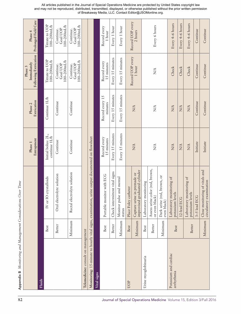
Ap
pen
dix
B
Mon
itor
ing
and
Man
agem
ent
Con
side
rati
ons
Ove
r T
ime
Phas
e 1
E
ntra
pmen
tPh
ase
2
Ext
rica
tion
Phas
e 3
Im
med
iate
ly
Follo
win
g E
xtri
cati
onPh
ase
4Pr
olon
ged
Fiel
d C
are
Flui
ds
Bes
tIV
or
IO c
ryst
allo
ids
Init
ial b
olus
: 2L
, co
ntin
ue 1
L/h
Con
tinu
e 1L
/hT
itra
te t
o U
OP
100–
200m
L/h
Tit
rate
to
UO
P
100–
200m
L/h
Bet
ter
Ora
l ele
ctro
lyte
sol
utio
nC
onti
nue
Con
tinu
eC
onti
nue.
G
oal U
OP
10
0–20
0mL
/h
Con
tinu
e.
Goa
l UO
P
100–
200m
L/h
Min
imum
Rec
tal e
lect
roly
te s
olut
ion
Con
tinu
eC
onti
nue
Con
tinu
e.
Goa
l UO
P
100–
200m
L/h
Con
tinu
e.
Goa
l UO
P
100–
200m
L/h
Tel
emed
icin
e: c
onsu
lt o
n m
anag
emen
t
Mon
itor
ing:
15-
min
ute
to h
ourl
y vi
tal s
igns
, exa
min
atio
n, u
rine
out
put
docu
men
ted
on f
low
shee
t
Vit
al s
igns
Bes
tPo
rtab
le m
onit
or w
ith
EC
GR
ecor
d ev
ery
15
min
utes
Rec
ord
ever
y 15
m
inut
esR
ecor
d ev
ery
15
min
utes
Rec
ord
ever
y
1 ho
ur
Bet
ter
Che
ck in
term
itte
nt v
ital
sig
nsE
very
15
min
utes
Eve
ry 1
5 m
inut
esE
very
15
min
utes
Eve
ry 1
hou
r
Min
imum
Mon
itor
pul
se a
nd m
enta
l st
atus
Eve
ry 1
5 m
inut
esE
very
15
min
utes
Eve
ry 1
5 m
inut
esE
very
1 h
our
UO
PB
est
Plac
e Fo
ley
cath
eter
N/A
N/A
Rec
ord
UO
P ev
ery
1
hour
Rec
ord
UO
P ev
ery
2
hour
sM
inim
umC
aptu
re u
rine
in p
rem
ade
or
impr
ovis
ed g
radu
ated
cyl
inde
r
Uri
ne m
yogl
obin
uria
Bes
tL
abor
ator
y m
onit
orin
g
N/A
N/A
N/A
Eve
ry 6
hou
rsB
ette
rA
sses
s ur
ine
colo
r (r
ed, b
row
n,
or e
ven
blac
k)
Min
imum
Dar
k ur
ine
(red
, bro
wn,
or
even
bla
ck)
Pota
ssiu
m a
nd c
ardi
ac
arrh
ythm
iaB
est
Lab
orat
ory
mon
itor
ing
of
pota
ssiu
m le
vels
N/A
N/A
Che
ckE
very
4–6
hou
rs
12-l
ead
EC
GN
/AN
/AC
heck
Eve
ry 4
–6 h
ours
Bet
ter
Lab
orat
ory
mon
itor
ing
of
pota
ssiu
m le
vels
N/A
N/A
Che
ckE
very
4–6
hou
rs
3–5
lead
EC
GIn
itia
teC
onti
nue
Con
tinu
eC
onti
nue
Min
imum
Clo
se m
onit
orin
g of
vit
als
and
circ
ulat
ory
exam
inat
ion
Init
iate
Con
tinu
eC
onti
nue
Con
tinu
e
Tre
atm
ents
for
Hyp
erka
lem
ia (
>5.5
mE
q/L
) or
Car
diac
Arr
hyth
mia
Cal
cium
glu
cona
te (
10%
)
Bes
t
10m
L I
V o
ver
2–3
min
utes
N/A
N/A
Mon
itor
; rep
eat
as r
equi
red
Insu
lin (
regu
lar)
and
D50
10
uni
ts I
V p
ush
+ 50
mL
D50
N/A
N/A
Alb
uter
ol (
2.5m
g/3m
L v
ial)
10m
g (4
via
ls)
in n
ebul
izer
N/A
N/A
Sodi
um p
olys
tyre
ne s
ulfo
nate
(K
ayex
alat
e)15
–30g
sus
pend
ed in
50–
100m
L li
quid
, ora
l or
rect
alN
/AN
/A
Cal
cium
glu
cona
te 1
0%A
ltern
ate:
cal
cium
chl
orid
e 10
%B
ette
r10
mL
IV
ove
r 10
min
utes
N/A
N/A
Insu
lin (
regu
lar)
and
D50
10 u
nits
IV
pus
h +
50m
L D
50N
/AN
/AM
onit
or; r
epea
t as
req
uire
dA
ny in
divi
dual
or
com
bina
tion
of
abo
ve, a
s av
aila
ble
Min
imum
See
abov
eN
/AN
/A
Man
agem
ent
of I
njur
ed E
xtre
mit
y
Ext
rem
ity
com
part
men
t sy
ndro
me
Bes
tC
linic
al a
sses
smen
t•
6 Ps
*•
Rig
id c
ompa
rtm
ent
——
Fasc
ioto
my:
onl
y if
qua
lifie
d m
edic
al
pers
onne
l or
tele
cons
ulta
tion
ava
ilabl
e
Min
imum
Coo
l lim
b (e
vapo
rati
ve o
r
envi
ronm
enta
l coo
ling,
no
ice/
snow
)
Tour
niqu
et(f
or c
rush
man
agem
ent)
Bes
tIf
ade
quat
e fl
uids
are
un
avai
labl
e, o
r ar
rhyt
hmia
ca
nnot
be
man
aged
dur
ing
entr
apm
ent
and
extr
icat
ion
If e
ntra
pmen
t ti
me
>2 h
ours
, con
side
r to
urni
quet
. Pla
ce t
wo
tour
niqu
ets
side
by
side
and
pro
xim
al t
o th
e in
jury
If t
he p
atie
nt m
eets
cri
teri
a fo
r to
urni
quet
con
vers
ion
or r
emov
al,
and
flui
ds a
re a
vaila
ble,
init
iate
cru
sh in
jury
pro
toco
l bef
ore
loos
enin
g to
urni
quet
.
Tour
niqu
et
(for
irre
vers
ible
inju
ry)
A li
mb
that
is c
ool,
inse
nsat
e,
tens
ely
swol
len,
and
pul
sele
ss is
lik
ely
dead
.
Pati
ent
may
dev
elop
sho
ck a
nd
kidn
ey d
amag
e, a
nd m
ay d
ie.
Con
side
r fa
scio
tom
y. I
f no
im
prov
emen
t, p
lace
tw
o to
urni
quet
s si
de b
y si
de a
nd
prox
imal
to
the
inju
ry. A
mpu
tati
on
anti
cipa
ted
Pain
Per
TC
CC
Per
TC
CC
Per
TC
CC
Ref
er t
o Pa
in/
seda
tion
CPG
Infe
ctio
n co
ntro
l
Ant
ibio
tics
Bes
tPo
rtab
le m
onit
or w
ith
EC
GE
rtap
enem
, 1g
IV/d
ay (
1g, 1
0mL
sal
ine
or s
teri
le w
ater
)
Bet
ter
Che
ck in
term
itte
nt v
ital
sig
nsC
efaz
olin
, 2g
IV e
very
6 t
o 8
hour
s; c
linda
myc
in (
300–
450m
g by
mou
th t
hree
tim
es d
aily
or
600m
g IV
eve
ry 8
hou
rs);
or
mox
iflo
xaci
n (4
00m
g/da
y; I
V o
r by
mou
th)
Min
imum
Mon
itor
pul
se a
nd m
enta
l st
atus
——
Ens
ure
wou
nds
clea
ned
and
dres
sed,
and
hy
gien
e of
wou
nds
and
pati
ent
opti
miz
ed
to t
he e
xten
t po
ssib
le g
iven
env
iron
men
t.
N/A
, not
app
licab
le; U
OP,
uri
ne o
utpu
t. *
6 Ps
: Pai
n pe
rsis
ting
des
pite
ade
quat
e an
alge
sia
is m
ost
impo
rtan
t sy
mpt
om, f
ollo
wed
by
pare
sthe
sia,
pal
lor,
para
lysi
s, p
oiki
loth
erm
ia, p
ulse
less
ness
All articles published in the Journal of Special Operations Medicine are protected by United States copyright law and may not be reproduced, distributed, transmitted, displayed, or otherwise published without the prior written permission
of Breakaway Media, LLC. Contact [email protected].

Management of Crush Syndrome Under Prolonged Field Care 83
Ap
pen
dix
B
Mon
itor
ing
and
Man
agem
ent
Con
side
rati
ons
Ove
r T
ime
Phas
e 1
E
ntra
pmen
tPh
ase
2
Ext
rica
tion
Phas
e 3
Im
med
iate
ly
Follo
win
g E
xtri
cati
onPh
ase
4Pr
olon
ged
Fiel
d C
are
Flui
ds
Bes
tIV
or
IO c
ryst
allo
ids
Init
ial b
olus
: 2L
, co
ntin
ue 1
L/h
Con
tinu
e 1L
/hT
itra
te t
o U
OP
100–
200m
L/h
Tit
rate
to
UO
P
100–
200m
L/h
Bet
ter
Ora
l ele
ctro
lyte
sol
utio
nC
onti
nue
Con
tinu
eC
onti
nue.
G
oal U
OP
10
0–20
0mL
/h
Con
tinu
e.
Goa
l UO
P
100–
200m
L/h
Min
imum
Rec
tal e
lect
roly
te s
olut
ion
Con
tinu
eC
onti
nue
Con
tinu
e.
Goa
l UO
P
100–
200m
L/h
Con
tinu
e.
Goa
l UO
P
100–
200m
L/h
Tel
emed
icin
e: c
onsu
lt o
n m
anag
emen
t
Mon
itor
ing:
15-
min
ute
to h
ourl
y vi
tal s
igns
, exa
min
atio
n, u
rine
out
put
docu
men
ted
on f
low
shee
t
Vit
al s
igns
Bes
tPo
rtab
le m
onit
or w
ith
EC
GR
ecor
d ev
ery
15
min
utes
Rec
ord
ever
y 15
m
inut
esR
ecor
d ev
ery
15
min
utes
Rec
ord
ever
y
1 ho
ur
Bet
ter
Che
ck in
term
itte
nt v
ital
sig
nsE
very
15
min
utes
Eve
ry 1
5 m
inut
esE
very
15
min
utes
Eve
ry 1
hou
r
Min
imum
Mon
itor
pul
se a
nd m
enta
l st
atus
Eve
ry 1
5 m
inut
esE
very
15
min
utes
Eve
ry 1
5 m
inut
esE
very
1 h
our
UO
PB
est
Plac
e Fo
ley
cath
eter
N/A
N/A
Rec
ord
UO
P ev
ery
1
hour
Rec
ord
UO
P ev
ery
2
hour
sM
inim
umC
aptu
re u
rine
in p
rem
ade
or
impr
ovis
ed g
radu
ated
cyl
inde
r
Uri
ne m
yogl
obin
uria
Bes
tL
abor
ator
y m
onit
orin
g
N/A
N/A
N/A
Eve
ry 6
hou
rsB
ette
rA
sses
s ur
ine
colo
r (r
ed, b
row
n,
or e
ven
blac
k)
Min
imum
Dar
k ur
ine
(red
, bro
wn,
or
even
bla
ck)
Pota
ssiu
m a
nd c
ardi
ac
arrh
ythm
iaB
est
Lab
orat
ory
mon
itor
ing
of
pota
ssiu
m le
vels
N/A
N/A
Che
ckE
very
4–6
hou
rs
12-l
ead
EC
GN
/AN
/AC
heck
Eve
ry 4
–6 h
ours
Bet
ter
Lab
orat
ory
mon
itor
ing
of
pota
ssiu
m le
vels
N/A
N/A
Che
ckE
very
4–6
hou
rs
3–5
lead
EC
GIn
itia
teC
onti
nue
Con
tinu
eC
onti
nue
Min
imum
Clo
se m
onit
orin
g of
vit
als
and
circ
ulat
ory
exam
inat
ion
Init
iate
Con
tinu
eC
onti
nue
Con
tinu
e
Tre
atm
ents
for
Hyp
erka
lem
ia (
>5.5
mE
q/L
) or
Car
diac
Arr
hyth
mia
Cal
cium
glu
cona
te (
10%
)
Bes
t
10m
L I
V o
ver
2–3
min
utes
N/A
N/A
Mon
itor
; rep
eat
as r
equi
red
Insu
lin (
regu
lar)
and
D50
10
uni
ts I
V p
ush
+ 50
mL
D50
N/A
N/A
Alb
uter
ol (
2.5m
g/3m
L v
ial)
10m
g (4
via
ls)
in n
ebul
izer
N/A
N/A
Sodi
um p
olys
tyre
ne s
ulfo
nate
(K
ayex
alat
e)15
–30g
sus
pend
ed in
50–
100m
L li
quid
, ora
l or
rect
alN
/AN
/A
Cal
cium
glu
cona
te 1
0%A
ltern
ate:
cal
cium
chl
orid
e 10
%B
ette
r10
mL
IV
ove
r 10
min
utes
N/A
N/A
Insu
lin (
regu
lar)
and
D50
10 u
nits
IV
pus
h +
50m
L D
50N
/AN
/AM
onit
or; r
epea
t as
req
uire
dA
ny in
divi
dual
or
com
bina
tion
of
abo
ve, a
s av
aila
ble
Min
imum
See
abov
eN
/AN
/A
Man
agem
ent
of I
njur
ed E
xtre
mit
y
Ext
rem
ity
com
part
men
t sy
ndro
me
Bes
tC
linic
al a
sses
smen
t•
6 Ps
*•
Rig
id c
ompa
rtm
ent
——
Fasc
ioto
my:
onl
y if
qua
lifie
d m
edic
al
pers
onne
l or
tele
cons
ulta
tion
ava
ilabl
e
Min
imum
Coo
l lim
b (e
vapo
rati
ve o
r
envi
ronm
enta
l coo
ling,
no
ice/
snow
)
Tour
niqu
et(f
or c
rush
man
agem
ent)
Bes
tIf
ade
quat
e fl
uids
are
un
avai
labl
e, o
r ar
rhyt
hmia
ca
nnot
be
man
aged
dur
ing
entr
apm
ent
and
extr
icat
ion
If e
ntra
pmen
t ti
me
>2 h
ours
, con
side
r to
urni
quet
. Pla
ce t
wo
tour
niqu
ets
side
by
side
and
pro
xim
al t
o th
e in
jury
If t
he p
atie
nt m
eets
cri
teri
a fo
r to
urni
quet
con
vers
ion
or r
emov
al,
and
flui
ds a
re a
vaila
ble,
init
iate
cru
sh in
jury
pro
toco
l bef
ore
loos
enin
g to
urni
quet
.
Tour
niqu
et
(for
irre
vers
ible
inju
ry)
A li
mb
that
is c
ool,
inse
nsat
e,
tens
ely
swol
len,
and
pul
sele
ss is
lik
ely
dead
.
Pati
ent
may
dev
elop
sho
ck a
nd
kidn
ey d
amag
e, a
nd m
ay d
ie.
Con
side
r fa
scio
tom
y. I
f no
im
prov
emen
t, p
lace
tw
o to
urni
quet
s si
de b
y si
de a
nd
prox
imal
to
the
inju
ry. A
mpu
tati
on
anti
cipa
ted
Pain
Per
TC
CC
Per
TC
CC
Per
TC
CC
Ref
er t
o Pa
in/
seda
tion
CPG
Infe
ctio
n co
ntro
l
Ant
ibio
tics
Bes
tPo
rtab
le m
onit
or w
ith
EC
GE
rtap
enem
, 1g
IV/d
ay (
1g, 1
0mL
sal
ine
or s
teri
le w
ater
)
Bet
ter
Che
ck in
term
itte
nt v
ital
sig
nsC
efaz
olin
, 2g
IV e
very
6 t
o 8
hour
s; c
linda
myc
in (
300–
450m
g by
mou
th t
hree
tim
es d
aily
or
600m
g IV
eve
ry 8
hou
rs);
or
mox
iflo
xaci
n (4
00m
g/da
y; I
V o
r by
mou
th)
Min
imum
Mon
itor
pul
se a
nd m
enta
l st
atus
——
Ens
ure
wou
nds
clea
ned
and
dres
sed,
and
hy
gien
e of
wou
nds
and
pati
ent
opti
miz
ed
to t
he e
xten
t po
ssib
le g
iven
env
iron
men
t.
N/A
, not
app
licab
le; U
OP,
uri
ne o
utpu
t. *
6 Ps
: Pai
n pe
rsis
ting
des
pite
ade
quat
e an
alge
sia
is m
ost
impo
rtan
t sy
mpt
om, f
ollo
wed
by
pare
sthe
sia,
pal
lor,
para
lysi
s, p
oiki
loth
erm
ia, p
ulse
less
ness
All articles published in the Journal of Special Operations Medicine are protected by United States copyright law and may not be reproduced, distributed, transmitted, displayed, or otherwise published without the prior written permission
of Breakaway Media, LLC. Contact [email protected].

84 Journal of Special Operations Medicine Volume 15, Edition 3/Fall 2016
Only if qualified medical personnel or teleconsul-tation (ideally with real-time video capability) available.
o Then only if wound care available. o Regional anesthesia with nerve block or IV seda-tion required.
• Minimum: Cool limb to reduce extremity edema (evaporative or environmental cooling only, do not pack limb in ice or snow because of risk of further tissue damage).
• Pain management: Refer to TCCC Guidelines for an-algesia on the battlefield.28
Infection
For infection due to associated wounds and not crush injury itself, follow the Joint Theater Trauma System Infection Control Guidelines: “Prevent Infection in Combat-Related Injuries for Extremity Wounds.”29
Goal: Prevent infection.• Best: Ertapenem, 1 gm IV/day (1g, 10 ml saline or
sterile water)• Better: Cefazolin, 2g IV every 6 to 8 hours; clindamy-
cin (300–450 mg by mouth three times daily or 600 mg IV every 8 hours); or moxifloxacin (400 mg/day; IV or by mouth)
• Minimum: Ensure wounds are cleaned and dressed, and hygiene of wounds and patient optimized to the extent possible given environment.
Two appendices accompany this article: Appendix A presents a summary of fluid and equipment planning considerations; Appendix B comprises three tables pre-senting monitoring and management considerations relative to time.
References
1. Brochard L, Abroug F, Brenner M, et al. An official ATS/ERS/ESICM/SCCM/SRLF Statement: prevention and management of acute renal failure in the ICU patient: an international con-sensus conference in intensive care medicine. Am J Respir Crit Care Med. 2010;181:1128–1155.
2. Greaves I, Porter K, Smith JE, et al. Consensus statement on the early management of crush injury and prevention of crush syndrome. J R Army Med Corps. 2003;149:255–259.
3. Greaves I, Porter KM. Consensus statement on crush injury and crush syndrome. Accid Emerg Nurs. 2004;12:47–52.
4. Gunal AI, Celiker H, Dogukan A, et al. Early and vigorous fluid resuscitation prevents acute renal failure in the crush vic-tims of catastrophic earthquakes. J Am Soc Nephrol. 2004;15: 1862–1867.
5. Sever MS, Vanholder R. Management of crush victims in mass disasters: highlights from recently published recommendations. Clin J Am Soc Nephrol. 2013;8:328–335.
6. Michell MW, Oliveira HM, Kinsky MP, et al. Enteral resuscita-tion of burn shock using World Health Organization oral rehy-dration solution: a potential solution for mass casualty care. J Burn Care Res. 2006;27:819–825.
Appendix A Fluid and equipment planning considerations
Best:
• Fluids: IV fluid to provide 1L/h for 24 to 48 hours (depending on evacuation availability)
• Equipment: ECG, laboratory tests for serum potas-sium and urine myoglobin, Foley catheter with grad-uated collection system, tourniquets
• Medications: hyperkalemia*: calcium gluconate (5 x 10mL vial or Bristojet), insulin: 1 vial Humulin R (500 units; Lilly USA, www.humulin.com), D50 (120mL), albuterol (24 vials), Kayexalate (360g; Concordia Pharmaceuticals, http://concordiarx.com)
• Pain: refer to Analgesia, Sedation Clinical Practice Guidelines (CPG)
• Antibiotics: ertapenem• Monitoring: Continuous monitoring with portable
monitor; 15-minute to hourly vital signs, examina-tion, urine output documented on flowsheet
Communications: real-time video telemedicine consultation
Better:
• Fluids: IV fluid to provide 1L/h for 24 to 48 hours• Equipment: Dipstick urine tests to monitor urine,
graduated container to monitor urine output, tourniquets
• Medications: hyperkalemia: calcium gluconate (5 x 10mL vial or Bristojet), insulin: 1 vial Humulin R (500 units), D50 (120mL)
• Pain medications• Antibiotics• Monitoring: 15-minute to hourly vital signs, exami-
nation, urine output documented on flowsheet
Communications: telephone, possibly e-mail tele-medicine consultation
Minimum:
• Fluids: IV fluid for initial bolus resuscitation (2L), then oral or rectal fluid resuscitation with commer-cial or improvised electrolyte solution
• Equipment: Guaduated container to monitor urine output, tourniquets
• Medications: hyperkalemia: calcium gluconate (5 x 10mL vial or Bristojet)
• Pain medications• Antibiotics• Monitoring: 15-minute to hourly vital signs, exami-
nation, urine output documented on flowsheet or other written format
Communications: telemedicine by telephone*Calculated quantities based on treating one patient for 48 hours.
All articles published in the Journal of Special Operations Medicine are protected by United States copyright law and may not be reproduced, distributed, transmitted, displayed, or otherwise published without the prior written permission
of Breakaway Media, LLC. Contact [email protected].
Management of Crush Syndrome Under Prolonged Field Care 85
7. Foex BA, Dark P, Rees Davies R. Fluid replacement via the rectum for treatment of hypovolaemic shock in an animal model. Emerg Med J. 2007;24:3–4.
8. Li W, Qian J, Liu X, et al. Management of severe crush injury in a front-line tent ICU after 2008 Wenchuan earthquake in China: an experience with 32 cases. Crit Care. 2009;13:R178.
9. Huang KC, Lee TS, Lin YM, et al. Clinical features and out-come of crush syndrome caused by the Chi-Chi earthquake. J Formo Med Assoc. 2002;101:249–256.
10. Alavi-Moghaddam M, Safari S, Najafi I, et al. Accuracy of urine dipstick in the detection of patients at risk for crush-in-duced rhabdomyolysis and acute kidney injury. Eur J Emerg Med. 2012;19:329–332.
11. Better OS. The crush syndrome revisited (1940-1990). Neph-ron. 1990;55:97–103.
12. Better OS, Abassi ZA. Early fluid resuscitation in patients with rhabdomyolysis. Nat Rev Nephrol. 2011;7:416–422.
13. Malinoski DJ, Slater MS, Mullins RJ. Crush injury and rhab-domyolysis. Crit Care Clin. 2004;20:171–192.
14. Huerta-Alardin AL, Varon J, Marik PE. Bench-to-bedside re-view: rhabdomyolysis—an overview for clinicians. Cri Care. 2005;9:158–169.
15. Nespoli A, Corso V, Mattarel D, et al. The management of shock and local injury in traumatic rhabdomyolysis. Minerva Anestesiol. 1999;65:256–262.
16. Polderman KH. Acute renal failure and rhabdomyolysis. Int J Artif Organs. 2004;27:1030–1033.
17. Zimmerman JL, Shen MC. Rhabdomyolysis. Chest. 2013;144: 1058–1065.
18. Weisberg LS. Management of severe hyperkalemia. Crit Care Med. 2008;36:3246–3251.
19. McCullough PA, Beaver TM, Bennett-Guerrero E, et al. Acute and chronic cardiovascular effects of hyperkalemia: new insights into prevention and clinical management. Rev Cardiovasc Med. 2014;15:11–23.
20. Scharman EJ, Troutman WG. Prevention of kidney injury fol-lowing rhabdomyolysis: a systematic review. Ann Pharmaco-ther. 2013;47:90–105.
21. Parham WA, Mehdirad AA, Biermann KM, et al. Hyperkale-mia revisited. Tex Heart Inst J. 2006;33:40–47.
22. Porter K, Greaves I. Crush injury and crush syndrome: a con-sensus statement. Emerg Nurse. 2003;11:26–30.
23. Schwartz DS, Weisner Z, Badar J. Immediate lower extrem-ity tourniquet application to delay onset of reperfusion injury after prolonged crush injury. Prehosp Emerg Care. 2015;19: 544–547.
24. Centers for Disease Control and Prevention. Crush injury and crush syndrome. http://www.acep.org/MobileArticle.aspx?id=46079&parentid=740
25. Chen X, Zhong H, Fu P, et al. Infections in crush syndrome: a retrospective observational study after the Wenchuan earth-quake. Emerg Med J. 2011;28:14–17.
26. Guner SI, Oncu MR. Evaluation of crush syndrome patients with extremity injuries in the 2011 Van Earthquake in Tur-key. J Clin Nurs. 2014;23:243–249.
27. Michaelson M, Taitelman U, Bursztein S. Management of crush syndrome. Resuscitation. 1984;12:141–146.
28. US Army Institute of Surgical Research. Tactical Combat Ca-sualty Care Guidelines. 2014. http://www.usaisr.amedd.army.mil/pdfs/TCCC_Guidelines_140602.pdf
29. US Army Institute of Surgical Research. Guidelines to prevent infection in combat-related injuries. Joint Theater Trauma System Clinical Practice Guideline. 2012. http://www.usaisr.amedd.army.mil/cpgs/Infection_Control_2_Apr_12.pdf.
Dr Walters is a research physiologist and a member of the Extremity Trauma and Regenerative Medicine Task Area at the US Army Institute of Surgical Research (USAISR), where he conducts human and animal research focused on limb sal-vage following combat-related muscle trauma. E-mail: [email protected].
MAJ Powell is an intensive care physician currently serving as the 4th Battalion 3rd Special Forces Group (Airborne) sur-geon and a staff intensivist at Womack Army Medical Center. E-mail: [email protected].
SFC Penny, NREMT-P is a senior medic and instructor at the Special Operations Combat Medic Course, U.S. Army John F. Kennedy Special Warfare Center and School. E-mail: [email protected]
LTC(P) Chung is a board-certified internist and intensivist and currently the critical care consultant to the Army Surgeon General. He has spent the last 11 years at the US Army Insti-tute of Surgical Research in various capacities. He is an SME for burns, critical care, and organ failure. E-mail: [email protected].
COL Keenan is a board-certified emergency medicine phy-sician and is currently serving as command surgeon, Special Operations Command, Europe. He has previously served as battalion surgeon in both 1st and 3rd SFG(A) and as group surgeon, 10th SFG(A). He is the coordinator for the SOMA Prolonged Field Care Working Group. E-mail: [email protected].
COL Shackelford, MC, USAF, is a trauma surgeon, cur-rently serving as the chief of performance improvement, Joint Trauma System, San Antonio, Texas. She is a member of the Committee on TCCC and has previously deployed as the di-rector of the Joint Theater Trauma System. E-mail: [email protected].
All articles published in the Journal of Special Operations Medicine are protected by United States copyright law and may not be reproduced, distributed, transmitted, displayed, or otherwise published without the prior written permission
of Breakaway Media, LLC. Contact [email protected].