
Alalwan, Hasanain Kahtan Abdulkhalik (2018) Combining nanofabrication
with natural antimicrobials to control denture plaque. PhD thesis.
https://theses.gla.ac.uk/30751/
Copyright and moral rights for this work are retained by the author
A copy can be downloaded for personal non-commercial research or study,
without prior permission or charge
This work cannot be reproduced or quoted extensively from without first
obtaining permission in writing from the author
The content must not be changed in any way or sold commercially in any
format or medium without the formal permission of the author
When referring to this work, full bibliographic details including the author,
title, awarding institution and date of the thesis must be given
Enlighten: Theses
https://theses.gla.ac.uk/

Combining nanofabrication with natural
antimicrobials to control denture plaque
Hasanain Kahtan Abdulkhalik Alalwan
(B.D.S., M.Sc.)
Submitted in fulfilment of the requirements for the
degree of Doctor of Philosophy
Medicine, Dentistry and Nursing School
College of Medical, Veterinary and Life Sciences
University of Glasgow
May 2018

II
Abstract Management of fungal biofilms represents a significant challenge to oral
healthcare. As a preventive approach, minimising adhesion between intra-oral
devices and microorganisms would be an important step forward. Denture
stomatitis (DS) is a multifactorial denture-associated inflammation of the oral
mucosa where candidal biofilms are one of the contributing factors. Therefore,
understanding candidal biofilms on dentures and finding novel strategies to
control these biofilms are of significance. Interference with the adhesion step of
biofilm formation is hypothetically effective strategy to control biofilms.
To understand the relationship between denture candidal load, denture material
type and C. albicans biofilm forming heterogeneity in DS, quantitative polymerase
chain reaction (qPCR) molecular method and crystal violet (CV) assay were used.
This study investigated two novel strategies to control C. albicans biofilms through
interfering with adhesion: natural polyphenol curcumin (CUR) and modifying the
topography of the denture material surface. Based on the optimised effective CUR
concentrations, CUR adsorption to PMMA denture material was
spectrophotometrically analysed. Based on these data, the effect of adsorbed CUR
to PMMA and CUR pre-exposure on adhesion of C. albicans were assessed. The
effect of CUR on Candida-Candida adhesion was investigated and the expression
profile of selected adhesion and aggregation-associated genes was assessed using
qPCR method. Micro/nano-fabricated polycarbonate and PMMA materials were
replicated using injection and compression moulding techniques, respectively and
were characterised using scanning electron microscopy (SEM). Adhesion of C.
albicans on the micro and nano-scaled patterns was assessed using microscopic
and qPCR molecular methods, respectively. The physical characteristics of the
materials were assessed using theta tensiometer and a white light profiler.
The data demonstrated that although C. albicans was detected in greater
quantities in diseased individuals, it was not associated with increased biofilm
biomass. Denture substrata were shown to influence biofilm biomass, with
poly(methyl methacrylate) providing the most suitable environment for C.
albicans to reside.

III
Subsequent studies showed that CUR concentrations of 50 μg/ml could prevent
adhesion to PMMA. This effect was enhanced by the CUR pre-treatment of yeast
cells (>90% inhibition, p < 0.001). Investigation of the biological impact of CUR
showed that it preferentially affected immature morphological forms (yeast and
germlings), and actively promoted aggregation of the cells. Transcriptional
analyses showed that CUR temporally modulated adhesion and aggregation
associated genes. Finally, PMMA denture material was replicated to show nano
features. These topographies influenced adhesion of C. albicans, depending on
the candidal morphological form and the shape. Nano-pit spatial arrangements
variably affect the adhesion of C. albicans, where SQ arrangement demonstrated
a significant anti-adhesive capacity. Differential adhesin expression was observed
on these surfaces, which were affected by the wettability and roughness of
surfaces tested.
In summary, C. albicans is an important determinant of denture disease, so
preventing its adhesion and biofilm formation were worthwhile objectives. This
thesis has shown that CUR molecules and SQ nano-pit topographies reduced C.
albicans adhesion, demonstrating that chemical and physical inhibition strategies
are useful. The data presented in this thesis showed the high potential of the novel
strategies to be used against C. albicans biofilms, and encourages the further
investigation of these approaches against polymicrobial denture biofilms.

IV
List of contents
Abstract ………………………………………………………………………………………………………………. II
List of tables ………………………………………………………………………………………………………VIII
List of figures ………………………..………………………………………………………………………….IX
Acknowledgments …………………………………………………………………………………………… XII
Author’s declaration …….………………………………………………………………………………….XIV
Abbreviations …………………………………………………………………………………………….……. XV
1 Introduction …….………………………………………………………………………………..……………1
1.1 Introduction …………………………………………………………………………………………………… 2
1.2 Evolution and importance of removable prosthodontic appliances …………….. 3
1.3 Mechanopathological responses to removable prosthodontic appliances .….. 4
1.4 Denture stomatitis …………………………………………………………………………………………. 8
1.4.1 Signs and symptoms and classification ………………………………………………………. 8
1.4.2 Epidemiology and aetiology ……………………………………………………………………….. 9
1.4.3 Diagnosis …………………………………………………………………………………………………….10
1.5 Denture plaque and the role of Candida in DS ……………………………………………. 12
1.6 Candida albicans virulence attributes ………………………………………………………….18
1.7 Treatment strategies of DS ………………………………………………………………………… 22
1.7.1 Physical strategies ……………………………………………………………………………………. 23
1.7.2 Chemical strategies ………………………………………………………………………………… 24
1.8 The polyphenol curcumin ………………………………………………………………………………27
1.8.1 The structure and biomedical significance of curcumin …………………………..27
1.8.2 The antimicrobial effect and mechanism of curcumin ……………………………. 29
1.9 Preventive antimicrobial strategies in denture material ……………………………. 35
1.9.1 Chemical modification ……………………………………………………………………….......35
1.9.2 Physical modification ………………………………………………………………………........ 39
1.10 Hypothesis & Aims ………………………………………………………………………………… 45
2 Candida albicans biofilms formation on denture materials …………………… 46
2.1 Introduction ………………………………………………………………………………………………. 47

V
2.2 Aims ………………………………………………………………………………….................... 49
2.3 Materials and Methods …………………………………………………………………………. …. 50
2.3.1 Molecular quantification of Candida from dentures ………………………….... 50
2.3.2 Standardised C. albicans biofilm assessment of denture isolates ………… 54
2.3.3 Investigation of biofilm formation upon denture materials …………………. 55
2.3.4 Statistical analysis ………………………………………………………………………………. 59
2.4 Results ………………………………………………………………………………………………………. 60
2.4.1 Denture carriage of Candida in health and disease …….………………………. 60
2.4.2 Variability of denture isolates in biofilm formation ……………………………… 62
2.4.3 Characterisation of in-vitro C. albicans biofilms …………………………………… 63
2.4.4 Impact of denture substratum type on candidal colonisation ………………. 70
2.5 Discussion …………………………………………………………………………………………………… 73
3 The anti-adhesive activity of curcumin on Candida albicans biofilms on
PMMA denture material .…………………………………………………………………………….. 79
3.1 Introduction …………………………………………………………………………………………… 80
3.2 Aims ……………………………………………………………………………………………………… 82
3.3 Materials and Methods …………………………………………………………………………. 84
3.3.1 Culture conditions and standardisation ……………………………………………. 84
3.3.2 Antifungal susceptibility testing ……………………………………………………. 84
3.3.3 Investigating the effect of CUR on C. albicans growth kinetics ……….. 85
3.3.4 Investigating the capacity for adsorption of CUR onto denture material
………………………………………………………………………………………………………………………… 86
3.3.5 Investigating the effect of CUR adsorption on adhesion of C. albicans to
PMMA …………………………………………………………………………………………………………………86
3.3.6 Microscopic imaging of adherent C. albicans to CUR-adsorbed PMMA
…………………………………………………………………………………………………………………87
3.3.7 Investigating the biological effect of CUR on C. albicans adhesion and
biofilm formation …………………………………………………………………………………………..88
3.3.8 Investigating the aggregative effect of CUR ….…………………………………….89
3.3.9 Investigating the effect of CUR on cell surface hydrophobicity …………. 89
3.3.10 Assessing the molecular impact of CUR on adhesion and biofilm
formation …………………………………………………… ……………………………………………….90

VI
3.3.11 Investigating the effect of CUR incorporation on adhesion ……………….. 94
3.3.12 Statistical Analysis …………………………………………………………………………….. 94
3.4 Results ………………………………………………………………………………………………………. 95
3.4.1 CUR can be adsorbed onto denture material up to effective
concentrations ………………………………………………………………………………………………. 95
3.4.2 CUR adsorption reduces Candida albicans adhesion ………………………...... 98
3.4.3 CUR inhibits biofilm formation and enhances Candida albicans
aggregation ……………………………………………………………………………………………………. 100
3.4.4 CUR reduces cell surface hydrophobicity of Candida albicans …………… . 106
3.4.5 CUR affects the temporal expression of Candida albicans adhesins …… 107
3.4.6 PMMA-incorporated CUR does not inhibit Candida albicans adhesion …. 109
3.5 Discussion ………………………………………………………………………………………………….. 111
4 The effect of micro and nano-patterned surfaces on Candida albicans
colonisation ……………………………………………………………………………………………………. 119
4.1 Introduction …………………………………………………………………………………………......120
4.2 Aims ……..…………………………………………………………………………………………………… 123
4.3 Materials and methods …………………………………………………………………………………125
4.3.1 Replication of micro/nano-featured materials…………………………………..... 125
4.3.2 Candida albicans adhesion on the micro-pillar patterns………………………….129
4.3.3 Candida albicans adhesion on the micro-pit patterns …………………….......130
4.3.4 Candida albicans adhesion on the nano-pit patterns ……………………………..131
4.3.5 Development of biofilms on the SQ nano-pit pattern ……………………………. 133
4.3.6 Measurement of surface properties of the materials tested ………………… 133
4.3.7 Candidal colonisation on CUR-adsorbed nano surfaces…………………………….134
4.3.8 Adsorption of CUR to SQ nano-pit topographies……………………………….......135
4.3.9 Statistical analysis…………………………………………………………………………………… 136
4.4. Results……………………………………………………………………………………………………….. 137
4.4.1 Adhesion of C. albicans to micro-patterned topographies……………………… 137
4.4.2 Adhesion of C. albicans to nano-pit patterned topographies………………… 144
4.4.3 Evaluation of surface roughness and wettability properties…………………. 150
4.4.4 Biofilm formation onto SQ nano-pit topographies ……………………………….. 152

VII
4.4.5 Combination of CUR and SQ nanotopographies ……………………………………. 153
4.5 Discussion …………………………………………………………………………………………………..155
5 Final discussion ………………………………………………………………………………………. 161
5.1 Introduction ………………………………………………………………………………………………. 162
5.2 Candidal denture biofilm and DS: Impacts of quantity and heterogeneity
………………………………………………………………………………………………………………. 163
5.3 Why CUR could be a promising polyphenol for denture wearers? …………….165
5.4 Micro and nanopatterned surfaces: the quest for antifouling denture
surface ……………………………………………………………………………………………………………. 169
5.5 Combining nanotopography with adsorbing CUR …………………………………….. 173
5.6 Future work ………………………………………………………………………………………………. 173
References …………………………………………………………………………………………………….. 176

VIII
List of tables
Table 3.1: Primers used for real time qPCR transcriptional analysis of Candida
albicans …………………………………………………………………………………………………………………93

IX
Table of Figures
Figure 1.1: The multiple contributing factors in oral health of denture wearers. 7
Figure 1.2: SEM image of non-polished acrylic denture base material with and
without yeast cells (scale = 20um). ...................................................... 8
Figure 1.3: Denture stomatitis clinical presentations according to Newton’s
classification. ............................................................................... 9
Figure 1.4: Schematic representation of Candida albicans biofilm formation on
different device substrates. ............................................................. 14
Figure 1.5: The Candida-bacteria colonisation of denture surface. ............... 16
Figure 1.6: The main morphological forms of C. albicans. .......................... 19
Figure 1.7: Virulence factors of C. albicans: A schematic plot. .................... 21
Figure 1.8: Chemical structure of curcumin. .......................................... 28
Figure 1.9: The multiple targeted antifungal mechanism of curcumin. ........... 34
Figure 2.1: Candida carriage in dentures in respect to DS status. ................. 61
Figure 2.2: Quantification of biofilm biomass from denture isolates. ............. 62
Figure 2.3: Optimisation of C. albicans early biofilm on PMMA and polystyrene. 63
Figure 2.4: Effect of different inoculum densities on RPMI-developed mature
biofilms. .................................................................................... 64
Figure 2.5: Optimisation of the CFU/ml concentration for development of
candidal biofilm. .......................................................................... 65
Figure 2.6: Optimisation of the incubation medium for development of candidal
biofilm. ..................................................................................... 67
Figure 2.7: The effect of strain variability on development of candidal biofilm.
............................................................................................... 69
Figure 2.8: CV staining of early biofilms developed on different denture
materials. ................................................................................... 70
Figure 2.9: Effect of the investigated denture materials on the biofilm formation
of C. albicans. ............................................................................. 72
X
Figure 3.1: Growth kinetics of half PMIC CUR-treated C. albicans. ................ 96
Figure 3.2: The adsorption capacity of CUR onto PMMA denture material. ...... 97
Figure 3.3: The impact of CUR adsorption to PMMA on Candida albicans adhesion.
............................................................................................... 99
Figure 3.4: The impact of single and dual-treatment of CUR on C. albicans
adhesion. .................................................................................. 100
Figure 3.5: Impact of longer-term CUR pre-exposure on candidal adhesion. ... 101
Figure 3.6: The impact of CUR on C. albicans biofilm formation. ................ 103
Figure 3.7: The impact of CUR on C. albicans biofilm architecture. ............ 104
Figure 3.8: The impact of CUR on aggregation of C. albicans. .................... 105
Figure 3.9: The impact of CUR on cell surface hydrophobicity of C. albicans. . 106
Figure 3.10: Transcriptional expression of CUR-treated C. albicans. ............. 108
Figure 3.11: Visual transcriptional analysis of CUR treated C. albicans. ......... 109
Figure 3.12: The non-effective role of CUR incorporated to PMMA on Candida
albicans adhesion. ........................................................................ 110
Figure 4.1: Silicon master wafer including micro-fabricated patterns. .......... 126
Figure 4.2: Replication of micro/nano-patterns on PMMA denture material. .. 128
Figure 4.3: Polycarbonate replicated micro-patterns. .............................. 129
Figure 4.4: Polycarbonate nano-pit patterned sections. ........................... 131
Figure 4.5: Tensiometer device loaded with sample. ............................... 134
Figure 4.6: Optimisation of the micro-scale replicability of PMMA denture
material. ................................................................................... 138
Figure 4.7: Characterisation of polycarbonate replicated micro-pillars. ........ 139
Figure 4.8: Adhesion of C. albicans to micro-pillar topographies. ................ 140
Figure 4.9: Characterisation of PMMA denture material micro-pit patterns. .... 141
Figure 4.10: Adhesion of C. albicans to micro-pit PMMA denture material. ..... 143
Figure 4.11: Characterisation of polycarbonate nano-pit patterns. ............. 144

XI
Figure 4.12: Quantification of adhered C. albicans to polycarbonate
nanotopographies. ........................................................................ 146
Figure 4.13: Adhered yeasts on SQ nanotopography. ............................... 147
Figure 4.14: Visual transcriptional analysis of adhered C. albicans to
polycarbonate nanotopographies. ..................................................... 148
Figure 4.15. Characterisation of nanoimprinted PMMA denture material. ....... 149
Figure 4.16. Quantification of adhered C. albicans to PMMA denture material
nanotopographies. ........................................................................ 149
Figure 4.17: Visual transcriptional analysis of adhered C. albicans to PMMA
denture material nanotopographies. .................................................. 150
Figure 4.18: Surface roughness of flat surfaces of materials tested. ............. 151
Figure 4.19: Wettability of the flat and nanopatterned materials tested. ...... 151
Figure 4.20: Evaluation of metabolic activity of C. albicans biofilms on SQ
nanotopographies. ........................................................................ 152
Figure 4.21: Evaluation of biomass of C. albicans biofilms on SQ
nanotopographies. ........................................................................ 152
Figure 4.22: Effect of combining SQ nanotopographies with CUR on C. albicans
colonisation. .............................................................................. 154
Figure 4.23: Adsorption of CUR to SQ nanotopography. ............................ 154
Figure 5.1: Schematic representation of the relationship of candidal denture
load, denture stomatitis and C. albicans heterogeneity............................ 164
Figure 5.2: Impact of CUR on C. albicans. ............................................ 168
Figure 5.3: Schematic representation of adhesion of yeast and hyphae cells on
relatively hydrophobic and hydrophilic surfaces. .................................... 172

XII
Acknowledgments
I have been extremely blessed to live in one of the friendliest cities in the world
‘Glasgow’ and to have the opportunity to join a marvellous group of people at
Glasgow Dental School and Hospital, many thanks to your help that contributed in
producing this thesis after four years of efforts and bittersweet experience.
First, I would like to express my very great gratitude and appreciation to Professor
Gordon Ramage. You have my deepest thanks for offering me the opportunity to
follow my dream and complete this PhD. You were also my source for enthusiasm,
encouragement and invaluable advice. You supported and guided me with great
patience and genuine supervision and I do not even have the words to thank you.
I would like to offer my special thanks to my other supervisors Dr Douglas
Robertson, Dr Chris Nile at Glasgow Dental School and Professor Nikolaj Gadegaard
at Engineering School-University of Glasgow for their continuous support and
helping me to complete this piece of work, truly I cannot thank you enough. The
expertise of Dr Robertson and Dr Nile backed my make over from a clinician into
a scientist smoothly and being a ‘scientician’. The micro/nanofabrication
technology was not accessible without the motivation, support and help that have
been given by Professor Gadegaard.
I would like to present my sincere gratitude to the Iraqi cultural attaché in
London/Iraqi Ministry of Higher Education and Scientific Research for financial
sponsorship. Many thanks to University of Baghdad for nominating me to obtain
PhD degree.
I am grateful for Mr Mike Broad and Mr Robert McKerlie for their welcoming me to
work in the prosthodontic lab at GDS&H. Special thanks to Dr Paul Reynolds, Dr
Anwer Saeed and Ms Rachel Love at Engineering School-University of Glasgow for
their support in the nanofabrication lab and help to carry out some material
surface experiments. My special and sincere thanks to Dr David Lappin for
providing technical and statistical advice kindly. Many thanks to the other
members of the Oral Sciences Research Group at GDS&H who have supported my
research include Professor Jeremy Bagg, Dr Shauna Culshaw and Mr Steven
Milligan. Many thanks to Dr Marcello Riggio and Dr Andrea Sherriff for supporting
and reviewing my progress. I would like to Acknowledge Mrs Margaret Mullin and

XIII
Mr Peter Chung at the Joseph Black and Gregory Buildings, respectively for all
their help in imaging and processing the samples for scanning electron microscopy.
Many thanks to all my friends in the lab who have big-heartedly supported me and
their help was truthfully priceless: Lindsay O’Donnell, Gareth Calvert, Ranjith
Rajendran, Leighann Sherry, Ryan Kean; as well as Emma Millhouse, Eleanor
Townsend, Tracy Show and Chris Delaney. Thank you again Ryan for your
instrumental proofreading.
I am particularly grateful for my friend Mohammed Al-jarsha for his generous help
to find an accommodation. Thank you to my financial guarantors in Iraq Ya'rub,
Shubbar and Amani. Many thanks for all my brothers and sisters for their
continuing encouragement and support. Special thanks to Ya'rub, Zainab, Hayder,
Mohammed and Ali for help in sorting things out in Iraq. Finally, I cannot find
enough words that describing my gratefulness and thankfulness to Sura ‘my wife’
the inspiring person in my life who have borne the responsibility of four amazing
children and alleviated the nostalgic feelings and the gloom and doom in some
days of my study.

XIV
Author’s Declaration
I declare that I have carried out the work described in this thesis unless otherwise
acknowledged or cited, under the supervision of Professor Gordon Ramage, Dr
Douglas Robertson, Dr Christopher Nile and Professor Nikolaj Gadegaard.
I further declare that this thesis has not in whole or in part, been submitted for
any other degree.
Hasanain Alalwan
May 2018

XV
Abbreviations
AAF1: Adhesion-aggregation factor 1
ALS: Agglutinin-like sequences
AMP: Antimicrobial peptide
ANOVA: Analysis of variance
ATP: Adenosine triphosphate
AS: Artificial saliva
BSA: Bovine serum albumin
cDNA: Complementary deoxyribonucleic acid
CFU: Colony forming units
CHX: Chlorhexidine
CLSI: Clinical and laboratory standards institute
COPD: Chronic obstructive pulmonary disease
CSH: Cellular surface hydrophobicity
Ct: Cycle threshold
CUR: Curcumin
CV: Crystal violet
ddH2O: Double distilled water
DS: Denture stomatitis
DMSO: Dimethyl sulfoxide
DNA: Deoxyribonucleic acid
EAP1: Epithelial adhesion protein 1
EDTA: Ethylene diamine tetraacetic acid
ELISA: Enzyme linked immunosorbent assay
F: Flat
G: Germling
H: Hyphae
HBD: human beta defensin
HBF: High biofilm former
HEX: Hexagonal
HIS: Histatin
HMDS: Hexamethyldisilazane
HSP: Heat shock protein

XVI
IL-8: Interleukin-8
INF: Intermediate biofilm formation
LBF: Low biofilm former
MAP: Mitogen activated protein
MIC: Minimum inhibitory concentration
MOLLO: Molloplast
MWD: Microwave disinfection
NC: Negative control
NRT: No reverse transcriptase
NSQ: Near square
NTC: Non-template control
OD: Optical density
OMIG: Oral microbiology and immunology group
PBS: Phosphate buffered saline
PCR: Polymerase chain reaction
PDT: Photodynamic therapy
PEM/THFM: Poly (ethyl methacrylate)/tetrahydrofurfuryl methacrylate
PMIC: Planktonic minimum inhibitory concentration
PMMA: Poly (methylmethacrylate)
qPCR: Quantitative polymerase chain reaction
rDNA: Ribosomal DNA
RNA: Ribonucleic acid
ROS: Reactive oxygen species
rpm: revolutions per minute
RPMI-1640: Roswell Park Memorial Institute - 1640 media
RT: Reverse transcription
RT-qPCR: Real time-qPCR
SAB: Sabouraud dextrose agar
SAP: Secreted aspartyl proteinases
SD: Standard deviation
SEM: Scanning electron microscopy
SEM: Standard error of mean
SMIC: Sessile minimum inhibitory concentration
SOD: Superoxide dismutase

XVII
SQ: Square
TBAEMA: Tert butylaminoethyl methacrylate
TUP1: Thymidine uptake 1
UFI: Ufi gel
WCA: Water contact angle
XTT: 2,3 bis(2-methoxy-4-nitro-5-sulfo-phenyl)-2H-tetrazolium-5-carboxanilide
Y: Yeast
YPD: Yeast peptone dextrose

1 Introduction

Chapter 1: Introduction
2
1.1 Introduction
As the elderly population expands to a predicted two billion by 2050, the number
of denture wearers will concomitantly rise. Edentulousness is an irreversible
clinical condition that can be described as an ultimate marker of oral disease
burden (Cunha-Cruz et al., 2007). It is also associated with socioeconomic factors,
with higher prevalence reported in the poor and women (Bedos et al., 2003). All
types of dentures (complete, partial and overdenture) can be associated with oral
mucosal lesions with predisposing factors that can be physical such as chronic
irritation, immunological such as hypersensitivity or microbiological (Jainkittivong
et al., 2010).
Currently, around 20% of the UK population wear removable dentures of some
form, with 70% of UK adults older than 75 years old wearing dentures (Hannah et
al., 2017), with many of these individuals suffering from denture stomatitis (DS),
an inflammation of the palate (Gendreau & Loewy, 2011). Poor oral hygiene is
frequently observed within this patient group and several factors can impact the
onset of DS such as salivary flow, denture cleanliness, age of denture, smoking
and diet (Martori et al., 2014b). A large proportion (>two-thirds) of individuals
who wear removable complete dentures may suffer from DS, though most
individuals are asymptomatic (Gendreau & Loewy, 2011). DS represents the most
frequent oral mucous lesion in elderly individuals (Rivera et al., 2017). Denture-
oral hygiene instruction and professional guidance is required, which is more
significant in the elderly population who could suffer from cognitive and manual
dexterity obstacles (Zenthofer et al., 2013). Only a minority of sufferers
experience pain, itching or burning sensation, discomfort or taste disturbance.
Soft tissue inflammation below or above the denture, as a result of persistent
exposure to microorganisms, is characteristic of DS (O’Donnell et al., 2017)
A recent systematic review showed that there is no well-defined treatment
strategy of DS because of the multi-causative nature of this inflammatory response
(Yarborough et al., 2016). This may rationalise why research was focused on
prevention and inhibition of denture microbial biofilms (Park et al., 2015;Tsutsumi
et al., 2016). Nevertheless, endeavours to create antimicrobial denture materials

Chapter 1: Introduction
3
often result in a collateral damage of the mechanical properties that may fail the
prosthesis (Paleari et al., 2011). This chapter will review the influences of
removable prosthodontic appliances on health, focus on how various factors
influence a move towards disease, and introduce existing and novel clinical
management strategies.
1.2 Evolution and importance of removable prosthodontic appliances
Prosthodontic appliances have been used since ancient times right through to our
own contemporary period. The ancient Egyptians in 2500 BC tried to replace
missing teeth using natural teeth connected to one another by gold wires
(Forshaw, 2009). The first endeavour to fabricate a realistic denture, including
denture base, was in the fifteenth century in Europe using blocks of ivory or bone
(Murray & Darvell, 1993). US president George Washington had ivory dentures
(Ring, 2010). The use of ivory or bone as raw materials for denture fabrication
continued until the year 1780 when it was replaced by porcelain, and latterly in
1820 gold had been introduced to prosthodontic field as a denture base (Murray &
Darvell, 1993). Vulcanite then superseded all the previous denture base materials
in 1850 when Goodyear was granted a patent for mixing sulphur with natural
rubber, then celluloid and bakelite (a phenol-formaldehyde resin) were
introduced, though were not typical substitutes for vulcanite. After around a
century of vulcanite reigning, in 1935 the acrylic resins, specifically the poly
methylmethacrylate (PMMA) were introduced. Having evolved after more than 3
decades of extensive research it had many advantages over vulcanite, including
dimensional and colour stability, inertness, and the possibility of chemical bonding
of artificial teeth made from the same material. Given these important
properties, in 1946 approximately 95% of fabricated dentures were manufactured
from PMMA, with cobalt-chromium base metal alloys playing an adjunct role in
denture fabrication (Corrado, 1990;Murray & Darvell, 1993;Williams, 2015).
Despite the general trend of declining tooth loss, the world-wide demographic
change to an aging population has seen an overall net gain in the demand for
dental prostheses (Douglass & Watson, 2002).

Chapter 1: Introduction
4
Dentures are removable prosthetic replacement of missing teeth and associated
soft and hard tissues for complete or partially edentulous patients. Functional
(mastication and phonetics) and aesthetic restoration, and therefore restoration
of the somatic and psychological health, are the main advantages of denture wear,
where oral health-related quality of life is usually enhanced after prosthodontic
treatment (Montero et al., 2013;Swelem et al., 2014). Several studies have
revealed a positive impact of prosthodontic treatment on the temporomandibular
joint disorders (Goiato et al., 2010;Abdelnabi & Swelem, 2015). Moreover,
improvement of oral functions after head and neck cancer surgery, such as
hemimaxillectomy, can be achieved by obturator dentures that aim to close the
defect. The obturator denture separates the oral and nasal cavities and prevents
nasal regurgitation of food and liquids and avoid hyper-nasal speech, besides the
facial profile support and ordinary benefits of dentures (Chen et al., 2016a). Thus,
dentures are considered of significant importance for the general wellbeing of
denture wearers, though are not without their disadvantages.
1.3 Mechanopathological responses to removable prosthodontic appliances
Through the last two decades, intensive research had been performed to confirm
the link between the denture and oral, as well as systemic health (Nikawa et al.,
1998b). Poorly fitted dentures may reduce chewing and masticatory performance,
which in turn negatively impact the general health and can deteriorate the
nutritional status of the denture wearer (Garrett et al., 1996;Sahyoun & Krall,
2003). Wearing a removable oral prosthesis alongside poor denture plaque
management could influence systemic health. Although causality has not been
well established, vigilant hygienic measures should be considered for patients with
systemic diseases (Le Bars et al., 2015). Indeed, there is even suggestions that
treatment of DS is able to improve endothelial function and minimise risk of
atherosclerosis and hypertension (Osmenda et al., 2017). It has also been reported
that denture plaque has a role in initiating unanticipated lung infections such as
aspiration pneumonia in immunocompromised and medicated elderly population
(Nikawa et al., 1998b). Poor oral hygiene was significantly related to nosocomial
and aspiration pneumonia, as several serious pathogenic microbes were isolated

Chapter 1: Introduction
5
from the oral cavity of institutionalized elderly persons, especially those in
hospital ICUs and nursing home settings (Scannapieco, 2006;Kuyama et al.,
2010;Iinuma et al., 2014). Furthermore, a systematic review indicates that teeth
brushing after meals, a thorough cleaning of denture at least once a day, in
addition to professional oral hygiene care once a week, are all necessary to reduce
aspiration pneumonia and associated mortality in fragile elderly people individuals
(van der Maarel-Wierink et al., 2013). Further studies report that life-threatening
pneumonia was doubled in elderly denture wearers, indicating a potential
relationship between denture wear and respiratory infections (Iinuma et al.,
2014). This is endorsed by a recent report demonstrating the abundance of several
potential respiratory pathogens on denture surface of healthy and diseased
individuals using a molecular approach (O'Donnell et al., 2016). Therefore, the
denture has an inherent capacity to be a hazardous reservoir of infectious
pathogens, that have the potential under certain circumstances to influence
systemic health. An association between denture plaque and chronic obstructive
pulmonary disease (COPD) has also been reported by Przybylowska et al. (2014),
where 90% of the study patients had pathogenic pulmonary microorganisms in
their denture plaque, with 75% of these having a yeast.
From another systemic perspective, and given the association between Candida
species and denture wearing (Webb et al., 1998b;Radford et al., 1999;Ramage et
al., 2006), a positive relationship between oral Candida species and occurrence
of oral cancer was uncovered by Alnuaimi et al. (2015). That study observed a
highly significant oral Candida carriage rate in oral cancer subjects, which has
been corroborated by Uittamo et al. (2009). It was reported that C. albicans is
associated with the formation of acetaldehyde (a potent carcinogenic compound)
via metabolism of glucose. Moreover, the carcinogenesis process and metastasis
could be developed and progressed by the C. albicans through several
mechanisms, such as the production of carcinogenic nitrosamines and activation
of CD4 T-cells to produce specific interleukins that stimulate the angiogenesis of
the tumors (Ramirez-Garcia et al., 2016). Collectively, denture wearing has more
profound systemic implications than we may generally acknowledge.

Chapter 1: Introduction
6
Locally, the potential oral pathological response to denture wearing is
multifactorial. Studies have shown a significant role of medication on oral health
of denture wearers (Carr et al., 1993), a significant physical effect of denture
surface and biomechanics on denture plaque accumulation and associated dental
and periodontal status (Drake & Beck, 1993), the positive role of maintenance of
a good denture hygiene (Shay, 2000), and the negative role of the inappropriate
use of a product or a technique (Verran et al., 2014). Besides, other factors may
complicate the denture-oral cavity relationship, such as salivary flow, dietary
effect and microbial colonisation (Turner et al., 2008;Altarawneh et al.,
2013;Martori et al., 2014a). These contributing factors are summarised in Figure
1.1.
Denture biomechanics can contribute to oral disease, where poorly fitted and
fabricated complete or partial dentures could lead to deterioration of the hard
tissues (teeth and bone), such as exposure of the abutment teeth to excessive and
poorly distributed forces that produce bone resorption and increased teeth
mobility (Aydin & Tekkaya, 1992). In some cases, poorly fitted partial dentures
may lead to root fracture of the abutment tooth (Mizuno et al., 2016). Another
example on the local mechano-pathological response to dentures is combination
syndrome, which is characterised by increased bone loss in the anterior region
under the maxillary complete denture opposing a lower anterior natural teeth,
causing a hyperplastic flabby ridge (Palmqvist et al., 2003).
Dentures might also physically affect soft tissue (oral mucosa), and spots of
traumatic ulcers could evolve due to high extension of the borders of denture
flanges and occlusal instability. Additionally, hyperplastic fibrous tissue, or
denture irritation hyperplasia, could evolve in response to chronic irritation from
poorly fitted denture or overextended flanges (Carlsson, 1997). Burning mouth
syndrome, which is characterised with painful burning sensation in the oral cavity
without any obvious lesion, was positively associated with denture wearing,
though it is of multifactorial aetiology (Svensson & Kaaber, 1995;Mukatash-Nimri
et al., 2017).

Chapter 1: Introduction
7
Figure 1.1 The multiple contributing factors in oral health of denture wearers.
From a microbiological point of view, denture-associated detriments might be
more complicated and recalcitrant to therapy. This is attributed to the oral
environment, host immunological response and associated microbiome, where the
oral cavity represents a typical milieu for microbial colonisation (Preshaw et al.,
2011;Benso et al., 2013). Furthermore, denture material surfaces especially the
unpolished fitting surface that is in an intimate contact with the mucosa
exaggerate such a problem because of the inherent roughness, voids and crevices
that could provide a shelter for the microbes and increase their capacities to shear
forces, which can clearly observed by scanning electron microscopy (Verran &
Maryan, 1997;Ramage et al., 2004), as shown in Figure 1.2.

Chapter 1: Introduction
8
Figure 1.2 SEM image of non-polished acrylic denture base material with and without yeast cells. Scale bar is 20 um.
1.4 Denture stomatitis
1.4.1 Signs and symptoms and classification
DS is a chronic erythematous and oedematous inflammation of oral mucosa
localized to areas in direct contact with the fitting surfaces of the removable
prosthodontic appliances, although it could be detected in association with
orthodontic appliances and obturators (Webb et al., 1998a). The denture bearing
area of the maxillary mucosa is the typical inflammation site. Usually, it is
asymptomatic, although it could be accompanied by swelling and bleeding of the
mucosa, dryness, burning sensation and unpleasant taste and halitosis of the oral
cavity. Moreover, it was reported that one to two thirds of the denture stomatitis
patients complain discomfort in their oral cavity (Arendorf & Walker, 1987;Webb
et al., 1998a). Newton's classification has divided its clinical manifestation into
three types based on the grade of inflammation severity. Type I: localized
inflammation or pinpoint hyperaemia, Type II: diffuse hyperaemia
(erythematous), where more diffused erythema involving large part or entire
denture covered mucosa; Type III: granular with papillary hyperplasia distributing
over the centre of the hard palate and the alveolar ridge (Scully & Felix, 2005)
(Figure 1.3).

Chapter 1: Introduction
9
(i) (ii) (iii)
Figure 1.3 Denture stomatitis clinical presentations according to Newton’s classification. (i) localised hypermedia: Grade I (ii), widespread erythematous inflammation: Grade II, (iii)Granular and papillary hyperplasia: Grade III (da Silva et al., 2011).
1.4.2 Epidemiology and aetiology
The epidemiological reports indicate a varied prevalence of DS among denture
wearers ranging between 15->70% (Gendreau & Loewy, 2011). Age and gender are
important factors in incidence of DS, where elderly people and women are more
liable to this disease and showed higher incidence rate (Gendreau & Loewy, 2011).
Moreover, DS is a frequent oral mucosal disorder correlated with institutionalized
elderly people (Magalhães & Moreira, 2010), as well as those with a cognitive
impairment due to dementia (Bramanti et al., 2015). Socioeconomic status (Evren
et al., 2011) and levels of education (Baran & Nalcaci, 2009), also negatively
impact prevalence rates for DS.
DS is a multifactorial disease, initially described with three main aetiological
factors driving its initiation: trauma, infection and allergy (Budtz-JöRgensen,
1974). However, malnutrition, hormonal disturbances and antibiotics are also
considered as predisposing factors (Jeganathan & Lin, 1992). Candida species,
smoking and nocturnal wear are also correlated to DS (Barbeau et al. (2003).
Salerno et al. (2011) attributed DS into systemic and local factors. These systemic
factors included diabetes, deficiency of nutritional factors, kidney disease and
xerostomia, while the local factors included trauma, saliva, pH of the oral cavity,
the permeability of the acrylic resins, and the presence of microbial plaque. Given

Chapter 1: Introduction
10
that DS is proposed to be a type of oral candidiasis, it can therefore be influenced
by systemic contributory factors that drive thrush, including cigarette/tobacco
smoking, antibiotic treatment, cytotoxic/ radiotherapy, and diabetes mellitus
(Soysa et al., 2004;Soysa & Ellepola, 2005;Soysa et al., 2006).
Trauma from ill-fitted denture can be considered one of the causes, which is
endorsed by research showing the using of implant supported over-dentures. The
over-denture reduces the direct forces on the mucosa and facilitates a good
distribution of forces, which reduces the trauma of mucosa and subsequently the
possibility of DS occurrence (Emami et al., 2008). Moreover, poor denture fit, poor
denture hygiene, and night-time wearing, in addition to denture surface
imperfections and using silicon denture liners, facilitate the establishment of
denture microbial biofilm and plaque formation (Gendreau & Loewy, 2011).
Regular sugar consumption, low salivary pH and the presence of Candida in the
oral cavity were also shown as risk factors and correlated with establishment of
DS (Martori et al., 2014a). Finally, the microbiome and level of dentition in the
oral cavity can have a significant impact on DS development and exacerbation,
where O'Donnell et al. (2015b) showed an associated with DS in partially edentate
patients, suggesting bacterial-fungi inter-kingdom interactions play an important
role. Overall, the factors that drive DS are multifactorial, which makes the clinical
management problematic.
1.4.3 Diagnosis
The diagnosis of DS is fundamentally based on clinical features of the palatal
mucosa, such as direct visual observation for the presence of pin-point or diffuse
erythematous lesions, and papillary hyperplasia that correspond to the fitting
surface of the denture (Puryer, 2016). Optical devices have also been advocated,
such as an erythema meter that can be used to measure the degree of erythema
of the palatal mucosa (Cross et al., 1998;Cross et al., 2004). Any suspicious
immunocompromising condition should be excluded, such as diabetes and HIV
infection (Scully, 2013). Laboratory tests are also useful for confirmation,
including haematological evaluation (full blood count and iron, folate and vitamin
B12 screening) and histological evaluation (via obtaining biopsy), where significant
differences in palatal epithelial thickness and haematological abnormalities are

Chapter 1: Introduction
11
reported (Jennings & MacDonald, 1990). Microbiological and immunological
diagnostic approaches that detect the presence of C. albicans and the antibody
titre to its antigen are often used (Jeganathan & Lin, 1992).
Microbiologically, direct microscopy of smears of the palate and the denture,
culturing of palatal swabs, and use of imprint culture are all useful (Webb et al.,
1998a). Historically, smears were examined by staining with Periodic acid-Schiff
stain for fungal yeasts detection (Davenport, 1970;Budtz-Jorgensen, 1972), and
for culture, swabs taken from the mucosa or denture are plated onto Sabouraud’s
agar to detect Candida (Cawson, 1965). Imprint culture can also be performed by
using a foam pad that is pressed against the area of mucosal interest, then
removed and pressed firmly on to the Sabouraud’s plate and incubated (Arendorf
& Walker, 1980). The oral rinse culture is a method that involves instructing the
patients to rinse their mouth with 10 ml of phosphate buffer saline for 1 min,
which is collected, centrifuged and resuspended into 1ml (concentrated method)
prior to plating on Sabouraud’s agar, is also regularly used for candida diagnostics
(Samaranayake et al., 1986). In today’s microbiology laboratories, Candida
chromogenic agars are used that enable quantitative and qualitative assessment
of different species, such as C. albicans and C. glabrata (Coco et al. (2008b).
To expedite conventional microbial diagnostics beyond the concentrated oral rinse
methodology then the successful use of real time quantitative polymerase chain
reaction (RT-qPCR) with a high level of sensitivity has been reported (1-10
CFU/ml) (White et al., 2004). The use of RT-qPCR has also been recently reported
to detect the presence of pathogenic pulmonary bacteria in dentures using this
advanced technique (O'Donnell et al., 2016). Given the general move in clinical
microbiology to become more molecular, then rapid and specific detection of key
pathogenic culprits is likely, though we still need live organisms in order to
perform sensitivity testing to inform the best chemotherapeutic intervention.
Immunological biomarkers for candidal detection could also be additionally
helpful, though are not widely implemented. These include radioimmunoassay and
enzyme-linked immunosorbent assay (ELISA), which have been reported to
recognise Candida serologically (Byadarahally Raju & Rajappa, 2011). For

Chapter 1: Introduction
12
example, significant increases of salivary anti-Candida IgA antibodies have been
detected in in DS patients (Jeganathan et al., 1987). Other immunological
biomarkers include elevated interleukin 2 (IL-2) (Rodriguez-Archilla et al., 1996),
the alteration in neutrophil morphology and activity (Gasparoto et al., 2012b),
and the modulation of other cytokines, such as IL-1β, IL-4,IL-10, IL-6, IL-12, TNF-
α, CXCL8, MCP-1(Gasparoto et al., 2012a;Pinke et al., 2016). However, at this
stage these are largely used in a research capacity and their general utility in
routine testing will always be limited.
1.5 Denture plaque and the role of Candida in DS
Microbial biofilms are associated with 65-80% of human infectious diseases (Joo &
Otto, 2012). These are characterised by highly structured microbial communities
that are attached to the colonised surfaces, in which the microbes communicate
to each other through a system called quorum sensing. Furthermore, production
of extra cellular matrix is a distinctive phenomenon of biofilms that give this
microbial community the utmost protection against shear forces, penetration of
antimicrobial agents and immune cells (Ramage et al., 2002b;Ramage et al.,
2009). Moreover, persister cells, a small sub-population of resilient cells, ensures
maintenance and repopulation following chemical insult (Lewis, 2005;LaFleur et
al., 2006). Biofilm development on dentures has been demonstrated by Ramage
et al. (2004) and others (Sachdeo et al., 2008;Susewind et al., 2015).
The presence of an oral prosthesis is significantly correlated with deteriorating
oral health, specifically mucosal integrity (O'Donnell et al., 2015b), but also
cariogenic and periodontal status (da Fonte Porto Carreiro et al., 2016). Oral
malodour, halitosis, is also a notable consequence of denture microflora (Verran,
2005;Nalcaci & Baran, 2008). Compositionally, the denture microflora is
heterogeneous and dynamic given its close proximity to other oral
microenvironments (O'Donnell et al., 2015b), and can influence or be influenced
by other surfaces. Indeed, Marsh et al. (1992) showed the capability of partial
dentures to increase cariogenic bacteria within the oral cavity. Moreover, the
prevalence of gingivitis and root caries was shown to increase with removable
partial dentures wearing (Preshaw et al., 2011), where fungal species also

Chapter 1: Introduction
13
contribute together with bacteria in such pathologic abnormalities (Coulthwaite
& Verran, 2007). Indeed, it has been shown that there is a synergistic relation
between C. albicans and Streptococcus mutans in developing virulent decay-
inducing biofilms (Koo & Bowen, 2014). Angular cheilitis, another common disease
in denture wearers, is characterized by erythematous fissures at the corners of
the mouth that is highly associated with C. albicans and Staphylococcus aureus
(Martori et al., 2014a). However, biofilms formed on denture surfaces often
include Candida species, which is thought and consistently reported to be a
principal player in denture biofilm-associated disease (Ramage et al., 2004;Taylor
et al., 2008;Øilo & Bakken, 2015). Figure 1.4 demonstrates the growth and
development of a C. albicans biofilm on denture and silicone substrates.

Chapter 1: Introduction
14
Figure 1.4: Schematic representation of Candida albicans biofilm formation on different device substrates. Schematics show biofilms formed on (a,b) PMMA denture strips or (c,d) silicone elastomer catheter disks. Panels a and c represent the substrate seen from the top, whereas panels b and d show the side view of biofilms formed on the PMMA strip and SE disk, respectively. ECM, extracellular material. This schematic was derived on the basis of data obtained from fluorescence and confocal microscopy analyses (Chandra et al., 2001).

Chapter 1: Introduction
15
It is suggested that Candida and bacteria are correlated with DS, where higher
numbers of yeasts and bacteria were cultured from DS patients (Budtz-Jorgensen
et al., 1983). Formation of the microbial biofilm is a successful survival strategy
that different microbes can follow, and it is associated with different infectious
diseases that may be related to indwelling medical devices (Donlan & Costerton,
2002). Denture material has the capability, by help of oral environment, to initiate
an inter-kingdom microbial biofilm (Cavalcanti et al., 2016). The diversity of
bacterial and fungal species (Candida species specifically) within the oral cavity
in health and disease is complex, and relies on the particular environment in which
they coexist, and the physical or chemical nature of cohabitation (O'Donnell et
al., 2015a). Maintenance of homeostasis in the oral cavity between bacteria and
fungi is important and influenced by the host environment (Xu & Dongari-
Bagtzoglou, 2015). Therefore, dysbiosis could cause pathological consequences,
such as that induced by antibiotics (McLean, 2014). The extensive microbiome of
the denture fitting surface in vivo ranges from 104 to 106/cm2, thus maximising
the significance of the denture biofilm ecosystem (Monsenego, 2000).
Furthermore, it was demonstrated that 1 mg of the denture plaque could contain
up to 1011 microbes (Nikawa et al., 1998a).
Generally, accumulation of microbial denture plaque on the denture fitting
surface is considered a significant factor in DS (Nikawa et al., 1998b). Higher
number of Candida, the initial positive effect of antimycotics, adhesion
capabilities to both denture surfaces and palatal mucosa and the much greater
mass of Candida in comparison to bacteria (> 50 times) make Candida an essential
element in DS occurrence and development. Nevertheless, the role of bacteria
should not be underestimated (Salerno et al., 2011). It was shown that
collaboration between the bacteria and Candida begins from the first stage of
biofilm formation ‘adhesion’ (Busscher et al., 2010). Adherence of the Candida
cells to surfaces is a mainstay in their virulence and ability for colonization and
reproduction and biofilm formation. Moreover, given the biofilm formation and
dimorphism capacities of C. albicans that could act as a scaffolding for other
microorganisms and instigate co-aggregative coexisting habitat onto dentures
(O’Donnell et al., 2015a) (Figure 1.5). Indeed, this is recently endorsed by Kean
et al. (2017), where Staphylococcus aureus benefited from C. albicans in

Chapter 1: Introduction
16
formation of a biofilm via using it as a structural scaffold. Bacterial adherence to
denture surfaces has also reported, where Streptococcus mutans and
Staphylococcus aureus were the most isolated bacterial microorganisms from the
upper complete denture with a rate of 53.3 and 34.4% respectively (Ribeiro et al.,
2012). Pereira-Cenci et al. (2008b) showed that Streptococcus mutans had a
negative effect on the hyphae formation capacity in Candida, despite significant
quantities of denture biofilm. Moreover, Streptococcus oralis adhesion to dentures
was reported elsewhere (Charman et al., 2009), which alongside C. albicans have
the potential to produce complex denture biofilms (Cavalcanti et al., 2016).
Figure 1.5 The Candida-bacteria colonisation of denture surface. A schematic drawing represents the Polymycrobial community with C. albicans in yeast and hyphal forms (O'Donnell et al., 2015a).
Several studies have shown that the pathogenicity of Candida can be synergised
by the presence of some types of bacteria within the oral cavity, such as
Streptococcus mutans and Staphylococcus aureus (Harriott & Noverr, 2009;Diaz et
al., 2012;Falsetta et al., 2014). This notion was supported by Cavalcanti et al.
(2015), where up-regulation of ALS3, EAP1, HWP1 and SAP6 Candida virulence-
significant genes was reported in presence of bacteria. Another report
demonstrated that lactic acid bacteria such as lactobacilli could induce
filamentation in opaque cell type C. albicans (Liang et al., 2016). This is relevant
as the presence of hyphae in the saliva or on the mucosa had a positive effect on

Chapter 1: Introduction
17
raising the load of C. albicans. Likewise, there was a statistically significant
relation between the hyphae incidence and number of lactobacilli, so this
bacterium appears to play an important adjunctive role in DS (Bilhan et al., 2009).
However, Lamfon et al. (2005) did not find a significant difference in the
quantities of the Candida, Lactobacillus, Streptococcus, Veillonella or
Actinomyces species in mucosa and dentures swabs of DS diagnosed patients group
and control group.
To demonstrate the importance of Candida spp in DS, Ramage et al. (2004) used
SEM to inspect development of candidal biofilms directly from dentures from DS
patients. Mesh of yeast and hyphal candidal forms in an entangled biofilm was
observed, with denture cracks and imperfections representing niches for
attachment, and biofilm thriving. The potential contribution of non-albicans types
of Candida in DS pathogenesis was explored by Coco et al. (2008b). Traditional
microbiological plating method with counting of colony forming units was followed
and revealed a significant difference between the total Candida burden of the
advanced grades of DS (grade II & III) and the control. In the same report, the
percentages of presence of C. albicans and C. glabrata throughout the healthy
and diseased groups were 75 and 30% of participating patients, while the rate of
Candida glabrata was 80% among the highest grade of Newton's DS classification,
with significant association with C. albicans, indicating an important co-
association in DS.
Further studies have shown that C. tropicalis was the focus of DS pathogenesis
deterioration. This was reported in a study evaluating the prevalence of Candida
species among DS diabetic type 2 and non-diabetic people. C. albicans showed a
prevalence of 81% with 15% for both C. glabrata and tropicalis. C. albicans and
tropicalis were higher in prevalence of DS group. Moreover, C. tropicalis had a
significant predilection for the highest degree of DS inflammation (Sanita et al.,
2011). Production of enzymes may be considered an explicit liaison between
Candida and DS. The role of secreted aspartyl proteinases (SAP protease enzymes)
in DS development and severity was explored by Ramage et al. (2012a).
Transcriptional quantitative PCR results showed a highly significant difference in
all SAP genes expressions between control and diseased group. Further studies
have shown that phospholipase production by Candida species of clinical isolates
Chapter 1: Introduction
18
was significantly related to DS and was limited to C. albicans and C. dubliniensis,
(Marcos-Arias et al., 2011). Indeed, there are a number of studies that have shown
that haemolytic activity, germ tube formation, induction of clot formation
(coagulase activity) and biofilm formation of Candida spp. may all contribute to
DS (Pereira et al., 2016), highlighting the importance of candida related virulence.
1.6 Candida albicans virulence attributes
C. albicans has sufficient ammunition to exhibit pathogenicity when given an
opportunity (Ramage et al., 2009). The adaptation of C. albicans to environmental
changes makes it a pleiotropic organism (Poulain, 2015), existing in two main
morphological forms: round to oval yeasts, or long mycelial form hyphae (Figure
1.6). The pathogenicity of Candida can be facilitated through many determinants
displayed in Figure 1.7, including adhesion, dimorphism, development of biofilms,
switching (phenotypic plasticity), metabolic flexibility (glycolysis,
gluconeogenesis), thigmotropism (directional hyphal growth), invasion, secretion
of proteases and rapid adaptation to pH environmental fluctuations (Mayer et al.,
2013). Moreover, a thick, protective (0.5 µm) cell wall in addition to the ability
to fight the free radicals released by immune cells through formation of
superoxide dismutase enzyme (Poulain, 2015). Thigmotropism can be described as
a representative of virulent contact sensing capacity of C. albicans. The contact
sensing definitive mechanism is still elusive, although mechanosensitive ion
channels and integrin-like signalling could be significant potential mechanisms
that elucidate this fundamental physio-pathogenic fungal process (Kumamoto,
2008).

Chapter 1: Introduction
19
(i) (ii) (iii)
Figure 1.6 The main morphological forms of C. albicans. (i) Yeast morphological form. (ii) Germling morphological form (iii) Hyphae morphological form. Images were acquired at 10,000, 4,000 and 2500 x magnifications, respectively.
Highly structured biofilm formation could be the most important strategy, as it
acts as a refuge, reservoir, scaffold and communication for microbes (Ramage et
al., 2009). In biofilm formation, the steps of biofilm formation include adherence
of yeasts, their proliferation, hyphal cells on the top of yeasts basal layer,
accumulation of extracellular matrix and dispersion of yeast cells away from the
biofilm complex (Figure 1.4), (Finkel & Mitchell, 2011). These dispersed yeasts
can then take part in the dissemination of candidiasis in different human body
organs (Uppuluri et al., 2010).
The first stage in biofilm formation is adhesion, which is principally driven by the
ALS genes (Hoyer & Cota, 2016). The proteins they encode control cell surface
hydrophobicity and the attachment to abiotic and biotic surfaces. The Als3 protein
plays a multifunctional and potent role in C. albicans pathogenicity. Alongside
Hwp1 and Als1, Als3 adhesins function together to form biofilms. It also has a role
in Candida iron acquisition via binding to ferritin. ALS3 gene is significantly
upregulated in C. albicans hyphae and pseudohyphae, and is inhibited by an
abundant nutrient environment. Moreover, Als3 could have a functional role in co-
aggregation with bacteria (Liu & Filler, 2011).
Following adhesion, biofilm formation is driven by the Efg1 protein, a morphogenic
regulator (Ramage et al., 2002a). Other key proteins include Hwp1 (hyphal wall
protein), Als3 (agglutinin-like sequence protein), and Sap4, Sap5, Sap6 (secreted
aspartic proteases) (Mayer et al., 2013). C. albicans can adapt to different pH,
changing morphology depending on its environment - pH >7 encourages the
Candida to grow with hyphal predominance, while pH <6 induce yeast form growth

Chapter 1: Introduction
20
(Mayer et al., 2013). Related to this hyphal transition is another important C.
albicans weapon recently discovered by Moyes et al. (2016). This hyphally
regulated peptide toxin is called candidalysin, which has been shown to impact
cell membranes of epithelial cells, damaging the permeability equilibrium. This
represents a critical determinant in breaching the epithelial tissue barrier role.
Regulation of these processes is in part driven by central regulators, that include
BCR1 and associated transcription factors, as well as adaptive protective
mechanisms. Mayer et al. (2013) demonstrated that the heat shock protein
(HSP90) gene has a role in dispersion of yeasts from the mature biofilm, increased
biofilm antifungal resistance, and is co-regulated with a network of genes to drive
biofilm formation. Moreover, HSP90 has a role in regulation of candidal
filamentation (Lu et al., 2011), a pivotal aspect of C. albicans biofilms
development. Many of these attributes are driven by the microenvironment that
they exist within, and nutrient availability.
In nutrient starvation, C. albicans modulates the extracellular acidity by
alkalinizing its surrounding to trigger an autoinducing hyphal form transformation
(Vylkova et al., 2011). This transformation could be associated with up taking of
surrounding amines containing molecules instead of glucose, though this may be
a strain dependant attribute. It has recently been reported that the metabolism
of the amino acids, such as aspartate, arginine, proline and glutamate, has been
upregulated in C. albicans strains capable of high biofilm formation, whereas
sucrose, starch and purine metabolism have been upregulated in low biofilm
formers (Rajendran et al., 2016b). The absence of glucose as a main source of
carbon may press the Candida to use an alternative source, which could increase
their virulence. Lactate grown C. albicans has a more rigid cell wall than the
glucose grown one, enabling it to deal more effectively with osmotic shock (Ene
et al., 2015). Moreover, lactate grown C. albicans is less detectable by the innate
immune cells (Ene et al., 2013). Overall, this shows that Candida has a genome
that is well adapted to survive and persist within the oral cavity.

Chapter 1: Introduction
21
Figure 1.7 Virulence factors of C. albicans: A schematic plot.

Chapter 1: Introduction
22
1.7 Treatment strategies of DS
A diligent sanitizing regimen, regular dentist visits, and overnight denture removal
are mandatory to reduce the incidence of DS. Nevertheless, development of anti-
biofilm strategies to reduce the microbial adhesion could be a significant measure
to ameliorate denture hygiene and reduce DS (Gendreau & Loewy, 2011).
However, commitment to strict sanitizing measures and appropriate cleaning
without compromising the denture surface can be considered as a difficult target
to attain within denture wearers (Jagger & Harrison, 1995). In a clinical study,
denture hygiene indices statistically deteriorated after long-term follow up in
comparison to last recall check-up. The patient sample of this study were elderly
people who were able to clean their dentures themselves, having passed manual
dexterity and cognitive capacity tests. This indicates the importance of the need
for periodical professional intervention with such a group, which gives an
indication that the status will be worse with people who need extensive care and
unable to perform denture hygiene care themselves.
Webb et al. (1998b) reviewed DS treatment modalities, which included
antimicrobial, antiseptic and disinfectant measures. They concluded that there
was not a comprehensively effective treatment per se, because of the
multifactorial etiological nature of DS, and asserted on patient’s compliance
playing an imperative role in DS management. Therefore, DS management require
multiple mutually supportive factors. Ramage (2015) reviewed the denture
cleansing chemical and mechanical procedures and their influence on oral
microbial response and associated denture stomatitis, clarifying some of their
drawbacks. In the same report, general guidelines for denture management from
a microbiological angle were presented, which could be summarised by the
following: reducing denture plaque using daily non-abrasive brushing and denture
cleansers, annual professional cleaning, and the potential beneficial role of
denture adhesives in increasing denture stability.
A recent systematic review by Yarborough et al. (2016) for the DS treatment
measures, reported the importance of antifungal treatment to the
immunocompromised individual, while focusing on prosthesis disinfection as an
effective strategy for healthy individuals. Multiple strategies were clinically

Chapter 1: Introduction
23
followed to reduce the pathogenic established biofilms. Furthermore, the
recurrence of DS remains as a problem although its prevalence is unknown.
1.7.1 Physical strategies
Microwave irradiation could be considered as a practical strategy in DS
management. Neppelenbroek et al. (2008) confirmed the significant impact of
microwave disinfection in treatment of DS in contrast to miconazole, besides the
recurrence in microwaved denture group was reduced. Microwave disinfection was
investigated in another study, where rinsing with nystatin and microwave
disinfection (MWD) were evaluated to treat DS. Three denture microwaving
treatment (650w-3min) weekly was as effective as 4 times daily nystatin.
Recurrence was reported after 3 months with approximately half of the
participants in every treatment modality (Silva et al., 2012). Three sequential
exposures of 60 sec had effectively disinfected maxillary dentures and reduced
the oral candidiasis of the denture wearers, although it could affect the
dimensional stability of the denture and it is an inappropriate disinfecting
technique to metal containing dentures (Banting & Hill, 2001). Furthermore,
several reports proved the detrimental effects of microwave denture disinfection
on the physico-mechanical properties (Seo et al., 2007;Hamouda & Ahmed,
2010;Vasconcelos et al., 2013).
Photodynamic therapy has emerged in multiple aspects of dentistry (Konopka &
Goslinski, 2007). As a helpful weapon to fight oral candidiasis, photosensitizer
erythrosine illustrated excellent impact on planktonic cells, though was less
effective against biofilms (Costa et al., 2011). Another report confirmed the
efficacy of photodynamic approach in reducing the virulence traits of Candida
species isolated from DS individuals (Pereira et al., 2015). In a clinical report,
harnessing of photodynamic therapy (photosensitizer: hematoporphyrin
derivative) to manage DS in vivo showed a positive response and it required several
sequential sessions, although recurrence was observed (Mima et al., 2011).
Further analysis has shown that C. albicans was less susceptible than Escherichia
coli and Staphylococcus aureus (Demidova & Hamblin, 2005).
Other strategies include ultrasonics, where in vitro and in vivo evidence suggests
positive benefits from ultrasonic cleaning of acrylic denture base to removing C.

Chapter 1: Introduction
24
albicans (>80%) (Kawasaki et al., 2014). Nishi et al. (2014) showed that distilled
water ultrasonic cleaning of dentures was not as effective as performing in the
presence of denture cleanser. Use of ultrasonic or cleanser alone led to significant
surviving of microorganisms in comparison to combining cleaning method.
1.7.2 Chemical strategies
It was demonstrated that patient responsibility for accurate denture cleanliness
is more difficult to do than it appears. This was established by clinical and
microbiological evidence, as treatment of DS without antifungals represented an
unrealistic management option. Using systemic itraconazole for 2 weeks to
manage DS, a significant myco-clinical enhancement was reported, while 50% of
subjects have recurred with DS after 6 months (Cross et al., 2000). Martin-
Mazuelos et al. (1997) showed that fluconazole and itraconazole antifungals had
improved the clinical status of DS, though fluconazole resistance was problematic.
Making new dentures as an alternative way to DS management has not show
clinical or mycological success in comparison to combined antifungal-antiseptic
methods that use systemic fluconazole and local application of chlorhexidine to
the fitting surface of dentures (Kulak et al., 1994). However, a meta-analysis of
randomized clinical trials indicated the significant possibility of using alternative
ways of managing DS rather than antifungal synthetic drugs. The alternative
methods were enumerated as disinfectants, mouthwashes, photodynamic
approaches, natural antimicrobials and microwave irradiation (Emami et al.,
2014). In a clinical study related to DS, the study participants were instructed to
rinse their mouths with chlorhexidine gluconate 0.12% and to soak their dentures
overnight in it for 24 days. The results showed a significant mycological and
clinical improvement, but once treatment stopped, the disease recurred from
both microbiological and clinical diagnostic angles (Kamalakshi et al., 1992).
However, evidence of significant dental staining drawback were reported with
using chlorhexidine (Van Strydonck et al., 2013).
To better understand how clinical management can be improved, we can draw on
laboratory investigations. Ramage et al. (2011) reported a significant efficacy of
some of the over-the-counter mouthwashes in comparison to some antifungal
drugs against developed biofilms. Caspofungin (an echinocandin drug) was as

Chapter 1: Introduction
25
effective to biofilm as to planktonic C. albicans, but it is limited by its intravenous
delivery method. Additionally, they showed that Corsodyl mouthwash (0.2%
chlorhexidine and 7% ethanol) exhibited ̴anti-biofilm activity to Candida. Caveats
to these approaches can be that mouthwashes can exhibit undesirable side
effects, such as taste problems and staining (Hong et al., 2009;Barao et al.,
2015;Sadat Sajadi et al., 2015). There are also links from oral mouthwashes to
head and neck cancer, which still remain controversial and need further
investigation (Conway, 2009;La Vecchia, 2009;Gandini et al., 2012;Ahrens et al.,
2014).
In an in vitro study, 10 different disinfection procedures were used for denture
soft liner cleaning. It was found that only sodium hypochlorite (1%), microwave
irradiation at 850W (6min), and effervescent cleansing were effective (Buergers
et al., 2008). Denture cleansers are a good, but not perfect strategy, for anti-
candidal biofilm approaches. Therefore, an adjunctive mechanical removal of the
residual biofilm to prevent denture recolonization seems imperative (Jose et al.,
2010). In an in vitro study, Ramage et al (2012) have reported the anti-biofilm
efficacy of two different sequential denture cleaning regimens. Logically, a 4 day
sequential continuous denture cleanser chemical approach was more effective
than a 4 day intermittent chemical/mechanical one. SEM of the day 2 showed
regrowth of DS clinically isolated C. albicans and this was attributed to retention
of Candida in denture material cracks or crevices and production of extracellular
protecting matrix. This could necessitate the need for enhanced chemical and
ultrasonic approaches to manage denture biofilms (Ramage et al., 2012c). In
another study, concerns were raised in respect to the influence of denture
cleanser on multi-species biofilm, where the counts of C. albicans increased 10
times in comparison to Streptococcus mutans after 7 days sequential daily 3 min
immersion in alkaline peroxide denture cleanser (Lucena-Ferreira et al., 2014).
Moreover, the alkaline peroxide effervescent denture cleansers were investigated
from the physical and mechanical point of view, and negative effects were
reported (Peracini et al., 2010). From a disinfection point of view, 0.5% sodium
hypochlorite has been suggested by Skupien et al. (2013) in their systematic
review as an efficacious anticandidal disinfectant for denture liners (denture
liners are used to reline the fitting surface of the denture). Furthermore,

Chapter 1: Introduction
26
disinfection of dentures with sodium hypochlorite soaking has promised a clinical
anti-DS effect and reduced number of Candida CFU, but metal containing
removable appliances represent a hurdle in respect to this method (Webb et al.,
2005).
Given the less toxic effect of phytomedicines, phytotherapeutics such as propolis,
Punica granatum, Melaleuca alternifolia and Vitis vinifera increasingly become
potential rival to conventional antifungals in treatment of DS, although further
clinical studies are needed to confirm a standard preparation (Casaroto & Lara,
2010). Zataria multiflora 0.1% gel and miconazole 2% gel were investigated for
their efficacy on DS subjects. The results were positive and comparable, while
relapse was observed after 4 weeks of completing the treatment, they have
suggested that dentures are the source of reinfection (Amanlou et al., 2006).
Likewise, Artemisia sieberi herbal mouthwash (1%) was equivalent to nystatin in
treatment of DS (Sefidgar et al., 2010).
Propolis extracts (ethanolic and non-ethanolic) with antifungal and anti-
inflammatory capacities have also been suggested for DS treatment (Santos et al.,
2005;Santos et al., 2008). The anti-planktonic and anti-biofilm effect of several
types of natural polyphenolic compounds against C. albicans were investigated by
Shahzad et al. (2014). They suggested that affinity of the oral surfaces to adsorb
the polyphenols and their potential saliva bioavailability may potentiate their oral
hygiene impact. It was concluded that curcumin and pyrogallol showed promise
against the planktonic and biofilm form cells. Other studies have investigated the
aqueous extract of garlic, Melaleuca alternifolia (tea tree oil) and Punica
granatum efficacy in treating DS. These studies indicated significant treating
action (Vasconcelos et al., 2003;Catalan et al., 2008;Bakhshi et al., 2012).

Chapter 1: Introduction
27
1.8 The polyphenol curcumin
1.8.1 The structure and biomedical significance of curcumin
Curcumin is a polyphenolic natural compound. It was isolated from turmeric (Asian
spice) two centuries ago. The turmeric was extracted from the rhizomes of
Curcuma longa plant in the thirteen century (Esatbeyoglu et al., 2012). The
curcumin (Diferuloylmethane) chemical formula is C21H20O6, International Union of
Pure and Applied Chemistry (IUPAC) nomenclature is (1E,6E)-1,7-bis(4-hydroxy-3-
methoxyphenyl)-1,6-heptadiene-3,5-dione, and its molecular weight is 368.38.
Curcumin consists of two phenolic O-methoxy containing groups that are
connected by a seven carbon conjugated (alternating single and multiple bonds)
linker consisting of an α,β-unsaturated β-diketone moiety. The B-diketone
functionality contribute in the intramolecular H+ atom transfer which is called
tautomerism, therefore, curcumin can be existed in two different forms enol form
and keto form (Figure 1.8) (Priyadarsini, 2009;Priyadarsini, 2014). Curcumin’s
physical-chemical properties can be summarised as solid, orange to yellow in
colour, non-flammable, hydrophobic, poorly soluble at physiological pH, behaves
as a proton donor at acidic pH where the keto form is dominated and as an
electron donor at basic pH, where the enol form is dominated, melting
temperature 170-175оC, light sensitive, odourless, soluble in ethanol, dimethyl
sulfoxide and acetic acid (Esatbeyoglu et al., 2012).

Chapter 1: Introduction
28
i.
ii.
Figure 1.8 Chemical structure of curcumin. (i) Keto form, (ii) Enol form.
Several clinical trials showed the non-toxicity, good toleration, and beneficial
preventive and therapeutic effects of curcumin on a plethora of human diseases
such as cancer, arthritis, cardiovascular diseases, Crohn’s disease, allergies,
ulcerative colitis, neurodegenerative diseases, diabetes, obesity, cerebral edema
and psoriasis among others (Gupta et al., 2013;Shehzad et al., 2013;Yang et al.,
2013;Furst & Zundorf, 2014;Panahi et al., 2017).
Anti-oxidative and anti-inflammatory effects, besides inducing molecular targets
such as transcription factors and enzymes could be considered as potential
mechanisms of its multiple biological effects, although a comprehensive
understanding is elusive so far (Mahmood et al., 2015). China, United States,
India, Korea, Thailand, Pakistan, South Africa, Nepal and Japan have used
curcumin as a supplement, although it is not permitted yet as a treatment (Gupta
et al., 2012). The daily dietary consumption of curcumin was 2.7-14.8 mg in Korea
while it has quadrupled in south Asia (Kwon, 2014).

Chapter 1: Introduction
29
In spite of its advantages, a disadvantage was frequently reported which is the
low bioavailability, while significant scientific research has been conducted to
reduce this drawback using nanonisation and conjugation strategies (Prasad et al.,
2014). Remarkably, it was demonstrated by Esatbeyoglu et al. (2015) that
degradation of curcumin at 180оC could not minimise its biological beneficial
effects because of the significant biological effects of its degradation products,
which are ferulic acid, 4-vinyl guaiacol and vanillin. In an in vivo study, it was
demonstrated that the efficacy of curcumin mouth rinse was higher than
chlorhexidine mouth wash in treating oral mucositis in cancer patients treated
with radio-chemotherapy (Patil et al., 2015). The safety of curcumin has been
reported by several studies (Lao et al., 2006;Chandran & Goel, 2012;Shehzad et
al., 2013;Sahebkar & Henrotin, 2016), all these studies share the support for the
good-toleration of curcumin and absence of adverse effects even with daily high
doses that may reach up to 6-8 g/day.
1.8.2 The antimicrobial effect and mechanism of curcumin
Epidemiological studies indicate the importance of the phenolic compounds in the
prevention of many diseases that are common to western communities.
Furthermore, the microbes could transfer the phenolic compounds into more
efficacious antimicrobials (Cueva et al., 2010). The activity of polyphenols against
the microbial causative agents of dental caries and periodontal disease was
recently reviewed (Slobodnikova et al., 2016), the review authors thought the
capability of these compounds might lead to their being introduced into practice
soon. In an in vitro study by Rai et al. (2008), it was shown that the bacterial
cytokinesis of Escherichia coli and Bacillus subtilis can be inhibited by curcumin
through interfering with polymerization of FtsZ protein assembly that forms the Z
ring constricting septum, which is very critical structure during the cytokinesis
process.
Periodontitis is a degenerative chronic disease of the gingiva and is highly
correlated with bacteria such as Porphyromonas gingivalis (Slots, 1986). In an in
vitro study conducted by Shahzad et al. (2015), curcumin was reported as the
most potent investigated polyphenol among 48 different types. Curcumin showed
an effective inhibition of planktonic growth of Porphyromonas gingivalis and

Chapter 1: Introduction
30
Aggregatibacter actinomycetemcomitans bacteria, which are commonly
associated with periodontitis. Besides, that study reported the high affinity of
curcumin to hydroxyl apatite crystal coated surfaces, which might add an
advantage for fighting periodontitis because of the dental biofilm-developing
nature of the periodontitis-associate bacteria.
Given that the dental caries-bacteria relationship is notorious, perhaps biofilm
formation and the associated cariogenicity of Streptococcus mutans could be
reduced by curcumin (Hu et al., 2013). Therefore, curcumin molecule could be a
significant potential candidate in caries prevention. Hu et al. (2013) showed the
inhibition of Sortase A enzyme that catalyses the tethering of some of adherence
proteins to the cell wall by curcumin, accompanied by reduced biofilm biomass.
This aforementioned report has confirmed a former in vitro study by Song et al.
(2012), where a significant effect of curcumin was observed on the vitality and
adherence of Streptococcus mutans to collagen and fibronectin human teeth
surfaces. It is noteworthy to mention that the anti-adherence capacity of
curcumin to the bacteria that is inferred by the Sortase A inhibiting capacity of
curcumin was firstly reported by Park et al. (2005) in Staphylococcus aureus.
Furthermore, a significant synergism between antibiotics and curcumin was
concluded by an in vitro study that investigated the possibility of reducing the
minimum inhibitory concentration (MIC) of several antibiotics on methicillin-
resistant Staphylococcus aureus (MRSA) (Mun et al., 2013). Moreover, Brown
(2015) prioritised some of the compounds that could be repurposed for
development of antibiotic resistance breaker strategies, where the curcumin was
selected and preferred for its significant bi-functionality (direct antibiotic and
anti-inflammatory effects) on both classes of bacteria (Gram positive and Gram
negative). The mechanism of curcumin antibacterial efficacy could be explained
by affecting bacterial cell membrane permeability, which was investigated by
propidium iodide uptake and calcein leakage fluorescence assays (Tyagi et al.,
2015), where all the investigated Gram-positive and negative bacteria
(Enterococcus faecalis, Staphylococcus aureus, Pseudomonas aeruginosa
Escherichia coli) showed a significant concentration and time dependent influence
by curcumin.

Chapter 1: Introduction
31
The prophylaxis of oral candidiasis could benefit from the potential antifungal
activities of some natural bioactive compounds, which could be added to the oral
health products (Rautemaa & Ramage, 2011). The antifungal capacity of
nutraceuticals and natural phenolic extracts have been reviewed by Rodrigues et
al. (2016), these types of extracts were considered as one of the growing novel
armamentarium and attractive prototypes in fighting Candida infection. The
antifungal capacity of crude Curcuma longa extracted oil was firstly reported
against dermatophytes mold type species (Apisariyakul et al., 1995). Another
report showed the anti-Candida (albicans and krusei) effect of curcumin,
especially when conjugated with glycine/alanine molecules that increased its
bioavailability (Mishra et al., 2005).
The proliferation of C. albicans can be inhibited by curcumin. This was confirmed
by a study that relied on flow cytometry technology to determine the level of S-
G2-M during the cell cycle, where a decline was demonstrated in these important
events, but that study used high curcumin concentrations (>1000 µg/ml) (Jianhua
& Hai, 2009). In an in vitro study conducted by Martins et al. (2009), results
suggested the potential clinical antifungal efficacy of curcumin and a significant
antifungal role of curcumin against Candida species (except glabrata) and
Paracoccidioides brasiliensis, while Aspergillus different species were
invulnerable to curcumin. Furthermore, Martins et al. (2009) microscopically
measured the number of adhered Candida cells to the buccal epithelial cells using
different species of Candida, and they noticed a remarkable reduction with
curcumin treated Candida cells.
In a study that interrogated the mechanism of the curcumin anticandidal capacity
against laboratory and clinical strains, Neelofar et al. (2011) reported the
effective capacity of curcumin on both types of strains and identified three
essential mechanisms which are: reduction of ATPase activity, inhibition of
proteinase production and interference with ergosterol biosynthesis, although all
the ergosterol targeted antifungals display upregulation of the proteinase activity.
Notably, Sharma et al. (2010b) have shown the anti-hyphal capacity of curcumin
to C. albicans and they related that to the up-regulation of global suppressor
thymidine uptake 1 gene (TUP1) in response to curcumin, where the tup1 null
mutant did not show an influence by curcumin. The antifungal mechanism of
Chapter 1: Introduction
32
curcumin was interrogated, where the level of the reactive oxygen species (ROS)
in all the tested species of Candida was raised after curcumin treatment.
Additionally, the oxidative stress associated genes were upregulated.
Consequently, they correlated these perturbing cellular events to early apoptosis
and cell growth inhibition of Candida species. Moreover, another antifungal
mechanism was reported by the same research group, where C. albicans
ergosterol depletion was associated with cell membrane disturbances, besides
accumulation of the ergosterol biogenisis precursors and the generation of ROS
(Sharma et al., 2012). The curcumin function in compromising fungal cell
membrane equilibrium was endorsed by a different study (Lee & Lee, 2014). In
that study, depolarisation of the membrane potential of the curcumin treated C.
albicans was shown which was potentially attributed to electrostatic and
hydrophobic interactions. Further, the potassium efflux of the tested cells was
increased after curcumin treatment, which compromising the cellular homeostasis
and leading to cell death. Another research group attributed the anti-Candida
mechanism of curcumin to the increased intracellular acidity that resulted from
the inhibition of the H+ efflux capability of the Candida cell suggesting the
disturbance of functional role of the cell membrane in controlling such activities
(Khan et al., 2012). Nevertheless, the notion of breaching the cell wall integrity
mechanism has emerged using candidacidal concentration. This notion was
reported by Kumar et al. (2014), who demonstrated the down-regulation of many
cell wall integrity genes and disruption of calcinurin and mitogen activated protein
(MAP) kinase pathways that are associated with cell wall homeostasis. Recently,
the dual anti-fungal and anti-carcinogenic mechanism of curcumin was
demonstrated (Chen et al., 2016b). Figure 1.9 summarises the aforementioned
literature suggested antifungal mechanisms.
The invasive type of fungal infection in cancer patients provokes a logical interest
in molecules of bi-pharmacological effects such as curcumin, specifically with its
high dose safety, besides its capacity to inhibit the efflux pump strategies that
are frequently used by cancer cells (Chearwae et al., 2006). The light sensitivity
feature of curcumin was used to fight the Candida species in a planktonic and
biofilm growth modes, displaying the possibility of curcumin use in photodynamic
therapy (Dovigo et al., 2011). All the investigated light excited-curcumin treated

Chapter 1: Introduction
33
Candida species were significantly inhibited, where the colony forming units of
the plated planktonic cells and the metabolic activity and the biofilm biomass
were reduced of the biofilms. The former report was endorsed by murine in vivo
study (Dovigo et al., 2013). However, a study conducted by Carmello et al. (2015)
indicated a temporary genotoxic effect after light excitement (without curcumin),
while a significant slowing in DNA repair was noticed with curcumin-light excited
Candida albicans. In a supporting context of the aforementioned potential
capacity of curcumin to augment existing conventional treatments, significant
synergism between nystatin, amphotericin B, ketoconazole, miconazole,
fluconazole, itraconazole, voriconazole antifungal drugs was reported by Sharma
et al. (2010a). Similarly, fluconazole and curcumin reduced the resistance of
clinical isolates of Candida albicans to fluconazole (Garcia-Gomes et al., 2012).

Chapter 1: Introduction
34
Figure 1.9 The multiple targeted antifungal mechanism of curcumin.

Chapter 1: Introduction
35
1.9 Preventive antimicrobial strategies in denture material
1.9.1 Chemical modification
Polymethyl methacrylate (PMMA) is the most utilized material for denture
fabrication, while soft liners of different chemical constituents are used to reline
fitting surface of the denture (Sakaguchi & Powers, 2012). There were many
experimental endeavours to modify the bulk chemical structure of polymer
denture base materials.
Microbial biofilm represents a major challenge for biomaterials. Incorporation of
antimicrobials and a sustained release approach have been used to manage
candidal biofilms. Examples are described below. A thin film polymer coating with
high or low porosity that was incorporated with chlorhexidine (CHX), nystatin or
amphotericin B was developed by Redding et al. (2009) to inhibit C. albicans
biofilm. The incorporation of chlorhexidine in the coated samples proved its
superiority in biofilm inhibition. Silver nano-particles were added to PMMA to
investigate the adherence and biofilm formation of C. albicans, though no
significant effect was observed in either case (Wady et al., 2012). This was
attributed to the non-release of Ag ion. However, another study reported a
significant reducing capacity of modified denture base material with silver nano-
particles although poor colour stability was reported as well (Nam et al., 2012),
which was recently confirmed by different group (Li et al., 2016). In spite of the
increased surface roughness, combined release of small amounts of F-, BO33-and
Na+ ions could be sufficient to reduce the adhesion of C. albicans to denture resin
material incorporated with glass ionomer filler (Tsutsumi et al., 2016). Salim et
al. (2013) investigated the anti-Candida biofilm efficacy of impregnation of CHX
or fluconazole to a poly (ethyl methacrylate)/tetrahydrofurfuryl methacrylate
(PEM/THFM) over 28 days. The CHX anti-biofilm efficacy was apparent, where the
biofilm biomass and the metabolic activity reduction were 75% and 84%
respectively, while in contrast the fluconazole impregnation was 8.8% and 12.6%,
respectively. The researchers suggested that the CHX-impregnated PEM/THFM
polymeric drug carrier system could potentially be used as a denture liner for the
treatment of DS. Another study investigated the incorporation of chlorhexidine

Chapter 1: Introduction
36
different salts in prevention of denture Candida biofilm. The chlorhexidine
diacetate seemed effective due to its release to the adjacent areas in comparison
with chlorhexidine hydrochloride which had not anti-Candida impact (Bertolini et
al., 2014). Pusateri et al. (2009) evaluated the anti-candidal effect of pre-coated
PMMA with 0.12% CHX gluconate, histatin-5 (His-5) or human β-defensin-3 (HBD-
3). The results showed that His-5 and CHX had an effect on biofilms, while HBD-3
had not shown any significant effect on biofilms. Moreover, immersion coating of
nanoparticles of CHX digluconate on dental silicones reduced proliferation of
Candida by releasing effective concentrations (Garner et al., 2015). An anti-C.
albicans effect of zinc oxide nano-particles incorporation as a composite or
coating to acrylic denture base material was reported by Cierech et al. (2016). A
successful rechargeable anti-Candida denture material was developed by (Cao et
al., 2010;Villar et al., 2013), through copolymerization of methacrylic acid and
diurethane dimethacrylate. The click-on click-off (recharging) miconazole
controlled release was investigated for elongated period up to 2 months that could
be washed off by EDTA to quench the antifungal drug. Likewise, a rechargeable
denture material using poly(N-vinyl-2-pyrrolidinone)-grafting polymerisation was
demonstrated by Sun et al. (2013). They reported a significant antifungal efficacy
of controlled sustained release that persisted for weeks with chlorhexidine and
months with miconazole. However, the antimicrobial release approach can be
criticised for the initial burst stage that being followed by release of low
ineffective concentrations (Huang & Brazel, 2001), besides the potential of short
period of activity and discontinuity of release (Kitagawa et al., 2014).
Contact killing surfaces could be more reliable and promising than antimicrobial
releasing surfaces (van de Lagemaat et al., 2017). Chemicals such as quaternary
ammonium and 2-tert butylaminoethyl methacrylate (TBAEMA) have been used for
that purpose. Incorporation of 2% quaternary ammonium polymer compounds in
heat cured acrylic resin have showed successful bacteriostatic and fungistatic
effects but it was stated that development of resistance ,toxicity and long-term
stability need further studies (Pesci-Bardon et al., 2006).
However, dangerous side effects were reported on quaternary ammonium, such
as negative effect on the reproductive system (Melin et al., 2014) and stimulation
of respiratory and skin allergic reactions (Lipinska & Walusiak, 2014). Another

Chapter 1: Introduction
37
study investigated the impregnation of 10 and 25% of TBAEMA, which is a poly-
cationic substance with pendant amino groups acting as a contact biocide, into
PMMA (Marra et al., 2012). An antibacterial efficacy against Streptococcus mutans
and Staphylococcus aureus biofilms was shown. There was no significant effect on
C. albicans biofilm formation, which was attributed to the rigid physical properties
of the Candida cell wall that prevented the biocidal compound from replacing the
Mg+2and Ca+2 of the cell membrane.
Studies using modification with anionic charged surfaces were also of interest.
The effect of copolymerization of methacrylic acid with PMMA on C. albicans
adherence was investigated by Park et al. (2003). The resulted negatively charged
surface showed candidal adherence inversely related to increasing the ratio of
methacrylic acid, which support the advocates of the effect of electrostatic
interaction on initial adherence of C. albicans. Similar results have been revealed
with modification of the acrylic resin surface charge and characteristics (Park et
al., 2008). Further, the absence of phosphate anions in PMMA may reduce the
adsorption of cationic salivary antimicrobials that could minimize and control
microbial colonization. That notion was investigated in an in vitro study, where
PMMA has been modified by incorporation of phosphate group using mixtures of
methyl methacrylate and methallyl phosphate as monomers. The anti-adherence
efficacy of the new polymer to C. albicans and their capability to adsorb histatin
5 was improved in comparison to the control group (Raj & Dentino, 2011).
A major problem with adding antimicrobials to biomaterials is the negative impact
on the mechanical properties, where incorporation of antimicrobial polymers to
denture base acrylic resins may compromise its flexural strength as in using of
tert-butylaminoethyl methacrylate (Paleari et al., 2011) or incorporating of
apatite-coated TiO2 photocatalyst (Shibata et al., 2007). Also, phosphate
containing monomer with an anti-Candida effect affected some of the PMMA
physico-mechanical properties (Dhir et al., 2007). Moreover, addition of
methacryloyloxyundecyl pyridinium bromide (MUPB) as an antimicrobial
monomer, showed a higher fibroblasts cytotoxicity than methyl methacrylate
(MMA) by 20 times (Regis et al., 2012).

Chapter 1: Introduction
38
These drawbacks could indicate that modification of the surface only would be
advantageous. Bazaka et al. (2011) reported that coating and surface modification
strategies for removable biomaterials seem to be more realistic in maintaining the
polymer bulk properties, and might enhance the surface bio-functionality and
biocompatibility. Graft polymerization of 2-methacryloyloxyethyl phosphoryl
choline (MPC) on acrylic denture surfaces create a covalently bonded durable
brushing-induced friction surface and anti-adhesive to Streptococcus mutans
biofilm. The grafting method had confirmed its superiority to the coating with
dipping method. Anti-adhesiveness was attributed to the lower liability for protein
adsorption where proteins produce a scaffold for bacterial adhesion (Takahashi et
al., 2014).
Lazarin et al. (2013) investigated the effect of different experimental photo
polymerized coatings containing hydrophilic monomers (2 hydroxyethyl
methacrylate, 3-hydroxypropyl methacrylate and 2-trimethylammonium ethyl
methacrylate chloride or zwitterionic monomer (sulfobetaine methacrylate) on
acrylic denture material. They noticed that the increased hydrophilicity led to a
decrease of the Candida adhesion and concluded that the physicochemical
properties of the substrate surface dictate the formation and composition of the
acquired pellicle that influences the biofilm development. In contrast to the
former study, coating the PMMA with Silane-SiO2 nanocomposite film reduced the
adherence of C. albicans though it decreased the surface energy and reduced its
hydrophilicity (Yodmongkol et al., 2014). Hydrophobic-fumed silica treated with
hexamethyl disilane was added to silicone soft liner, the results revealed lower
penetration percentage of Candida with increasing the filler percentage (Rodger
et al., 2010).
Improved bio-functionality of biomaterials may increase the opportunity to the
adsorption and bonding of bioactive molecules (Dang et al., 2014). Surface
modification of PMMA beads by copolymerisation with methyl methacrylate acid
had positively influenced on the adsorption and desorption of histatin 5 anti-
microbial peptide. It was concluded that controlled release of this from PMMA had
reduced the adherence of C. albicans while, adsorbed histatin had not shown any
candidacidal effect (Edgerton et al., 1995). Following on from this, Yoshinari et
al. (2006) used oxygen cold plasma to improve the adsorption of histatin 5 to

Chapter 1: Introduction
39
PMMA. The results showed a six times increase in the rate of adsorption of histatin
5 with plasma treatment that reduced colonisation by Candida. Recently, the
acrylic resin denture base was bio-functionalised by plasma initiated grafting
polymerisation of several coating polymers (Wen et al., 2016). The adsorption of
miconazole antifungal drug was significantly higher to the bio-functionalised
PMMA and the biofilm formation was inhibited.
1.9.2 Physical modification
To the best of my knowledge there is no research article that has investigated the
modification of the PMMA denture material surface physically using micro/nano
fabrication approaches. Therefore, given the relative novelty of the field as a
whole, a general view will be presented on the biomedical impact of micro/nano
patterned surfaces on mammalian cells and the microorganisms, then a brief
paragraph about the fabrication.
Nano fabricated surfaces are well-studied in the electronic engineering and
optical fields, while, in the biology and medicine fields they remain to some
extent novel and poorly explored, although initial research of cell-substrate
interaction displayed a response to the nano-structured materials (Anselme et al.,
2010). The surface energy of the micro-structured patterns is affected with Cassie-
Baxter model (air trapped between the microstructures) or Wenzel model (water
trapped between the microstructures). This could be more pronounced with
regularly nanostructured patterns presenting potential ambivalent phenomena
(the nano-patterned surface could increase the hydrophilicity or the
hydrophobicity of the surface) (Martines et al., 2005).
The Interactions between micro/nanotopographies and human cells have been
investigated in several studies. In orthopaedic/dental implant associated
surgeries, fibroblast and osteoblast cell adhesion, differentiation and survival are
critical (Chatakun et al., 2014). Downing et al. (2013) demonstrated the possibility
of the biophysical microtopographies represented by highly ordered microgrooves
replacing the biochemical molecules in reprogramming the somatic cells to
pluripotent cells. This is a critical issue in cell-engineering and regenerative
medicine. Furthermore, nano-patterned topographies of 100-200 nm diameter

Chapter 1: Introduction
40
nanodots effectively modulated the adherence capacity of the fibroblast cells and
induced apoptosis like events (Pan et al., 2009).
Biggs et al. (2007) reported a significant interaction between the nano-patterened
substrate and the cells. They showed a reduced adherence capacity of the primary
human osteoblast cells onto highly ordered arrays of nanopit topographies in
comparison to the planar control substrates, but on the contrary, the controlled
slightly disordered nano-patterned arrays displayed higher adhesion capacities.
They suggested a compromisation of the protein adsorption in addition to the
surface free energy changes as potential mechanisms that led to the reduction in
the focal adhesion (cell-extra cellular matrix adhesion). While, completely
random nanotopographies have led to an increase in the focal adhesion, which
was evidenced by modulation of the genetic expression (Biggs et al., 2009).
Support for regenerative therapies by controlling stem cell differentiation and cell
fate through nanotopographical systems was reviewed by Dalby et al. (2014). They
expected an evolution of the next generation of regenerative medicine
biomaterials using the knowledge base about nanotopographies-cells interaction.
Furthermore, micro/nanotopographies have been investigated for their potential
interaction with microorganisms, although it is notable to mention the presence
of only one (according to the best of our knowledge) research article that
investigated the interaction between such surfaces and fungal species (Whitehead
et al., 2005). Whitehead et al. (2005) interrogated micro and sub-micro titanium
engineered topographies to reveal the effect of different feature diameters on
the retention of C. albicans in addition to Staphylococcus aureus and Psuedomonas
aeruginosa. These titanium-engineered topographies did not show differences
with C. albicans, in opposite to the other two investigated microorganisms due to
the disparity of the sizes of theses microbes relative to the size of the features.
Generally, an increase in uncontrolled surface roughness could offer a shelter for
the microorganism from cleaning shear forces and by maximising the surface area
of attachment.
This was elucidated by Boyd & Verran (2002) as they demonstrated an increase
of the adherence capacity of Staphylococcus aureus to unpolished and randomly
abraded stainless steel surfaces. On the other hand, antifouling and self-cleaning

Chapter 1: Introduction
41
natural systems such as lotus leaf effect and shark skin that are characterised by
characteristic hydrophobicity and distinctive topographies at micro/nano scale,
are main drivers of interest in the interaction of micro-nano topographical
features with microorganisms in an endeavour to control the bacterial adhesion
and subsequent biofilm formation (Gu & Ren, 2014).
Inspiration by nature has frequently affected surface design engineering,
therefore self-cleaning windows, submarine low drag and wall-climbing robots
could be considered mimicking designs of the hydrophobic lotus leaf, dolphin
shape and gecko feet respectively. This is termed biological mimicry, which
describes the copying of natural designs to solve different challenges (Bixler &
Bhushan, 2012). The finer Sharklet AF™ microtopographies that are inspired by the
shark skin design reduced the adhesion of Ulva linza zoospores by 85%, and the
authors suggested the wettability thermodynamics as a causative mechanism
(Carman et al., 2006). The same microtopography (Sharklet AF™) was further
investigated by Chung et al. (2007) using polydimethyl siloxane elastomer
substrate. The authors reported a significant reduction in 7-21 day biofilm of
Staphylococcus aureus in comparison to the smooth surface control. In another
report, bacterial adhesion could be modulated by ordered micro/nano
topographical surfaces, where Pseudomonas fluorescens early stage biofilms were
hindered by using submicron ordered grooves (Diaz et al., 2009).
The traditional concept of the smoother surfaces can allow less adhered bacteria
could be endangered by Mitik-Dineva et al. (2009) study. They have reported the
increased adherence of Pseudomonas aeruginosa, Escherichia coli and
Staphylococcus aureus on nano-scale smoothened surfaces in comparison to the
control surfaces. This was attributed to the increased extra cellular
polysaccharides formation by the bacteria. Ploux et al. (2009) showed the
opposite behaviour of human osteoblast progenitor cells to the Escherichia coli
bacterial cells in adhesion to ridge-groove microtopography, where more
osteoblasts and fewer bacterial cells adhered at the initial stage of incubation.
However, a review discussed the adhesion of the human cells and bacteria to
micro-nanotopographies, the deformity of the cytoskeleton of the bacteria in
response to micro-nanostructures is less likely to occur in comparison to the
eukaryotic human cells, which could hinder the targeting of mechano-stimulus

Chapter 1: Introduction
42
mechanism driven by the contact with micro-nanostructures. Besides, the surface
chemical composition play a critical role in bacterial adhesion that may mask the
topography effect (Anselme et al., 2010). The notion of possible interaction
between the surface chemistry and topography was reported, when a
mathematical model was developed by Decuzzi & Ferrari (2010) that
demonstrated the possibility of tailoring the surface energy and the topography
of the biomedical surfaces to reduce bacterial adhesion to the nanotopographical
features. A significant coherent relation between surface energy, which might be
dictated by the surface chemical composition, and the nanotopography was
reported.
A silane based-fluorinated nanosilica superhydrophobic coating displayed
antibioadhesive properties against Staphylococcus aureus and Pseudomonas
aeruginosa (Privett et al., 2011). In contrast, a different study showed a massive
role for the topographical shape, size and feature in reduction of bacterial
adhesion and subsequent biofilm formation while the surface
hydrophobicity/hydrophilicity did not demonstrate a significant impact (Perera-
Costa et al., 2014).
Different bacteria show different behaviour towards nanostructured surfaces that
may complicate the evolution of universal conclusion (Hsu et al., 2013). In an
endeavour to mimic the superhydrophobic, antifouling, lotus leaf surface, a
titanium surface was fabricated in dual scale micro-nano projecting features
(Fadeeva et al., 2011). In that report, Staphylococcus aureus and Pseudomonas
aeruginosa were the testing models. The first type of bacteria was capable of
attaching to the surface while the other type was not. It was concluded that
different bacteria behave differently on superhydrophobic surfaces.
Cicada wings could represent a different nanofeatured antibacterial mechanism.
As shown by Ivanova et al. (2012), Pseudomonas aeruginosa could be killed within
minutes through a purely mechanical approach, where the cell membrane is
ruptured due to the high pressure applied by the tapered nano pillars of the wings
that cause stretching of the cell membrane although, the hydrophobic self-
cleaning nature of these wings did not prevent the bacteria from adhering.
Furthermore, the cicada wings model was a logical basis for investigating the

Chapter 1: Introduction
43
bactericidal effect of TiO2 nanowire via another study. Diu et al. (2014) showed a
significant killing of Pseudomonas aeruginosa, while Staphylococcus aureus was
not affected. The bactericidal mechanical-stretching mechanism of cicada wing
was confirmed, using a biophysical model and 3 different bacterial species
(Pogodin et al., 2013). This inherent physical antibacterial strategy has been
recently endorsed with dragonfly wings that presented a specific nanofeatured
surface (Bhadra et al., 2015;Mainwaring et al., 2016). However, in a different
study, a variety of submicron topographical arranged features showed higher
affinity to bacterial adherence, where the attached bacteria used different
mechanisms to maximise their attachment (Hsu et al., 2013).
The generation of micro/nano master patterns and the subsequent replication
methods will now briefly described. The fabrication technology of master patterns
encompasses a range of methodologies which are photolithography, electron beam
lithography, colloidal lithography, polymer demixing among others (Kearns et al.,
2011). The first and second methods present the possibility of elaboration of highly
ordered topographies. Photolithography technology depends on developing a film
of photosensitive layer (photoresist) on the substrate and applying a light that
transfer through a specific patterned mask to limit the illumination to the targeted
areas, and then an etchant is applied for removal of the photoresist and
preparation of the pattern in a synchronising manner. The electron beam
lithography method does not use the mask role in developing the patterns but use
the electron beam to directly develop the pattern on the photoresist. It elaborates
more accurate patterns but takes longer time and more expensive (Kearns et al.,
2011).
Moreover, the ‘soft lithography’ which is an alternative and complement to the
photo and electron beam lithography can be used as successful way to generate
the replicas in inexpensive and more convenient approach that facilitates the
introduction of the micro/nano topographies to the biomedical research field. Soft
lithography uses non-photolithography techniques (to avoid the optical diffraction
disadvantage and the subsequent low resolution) such as self-assembly monolayer,
microcontact printing, microtransfer molding, replica molding and embossing for
replicating features using mostly elastomeric ‘soft’ stamps or molds (Xia &
Whitesides, 1998).

Chapter 1: Introduction
44
The replication of polymer patterns via imprinting of the master patterns can be
divided into 2 main non-photolithographic approaches: Hot embossing technique,
which is simple and requires heating the polymer slightly above its glass transition
temperature then applying a given force. The second approach is the injection
moulding technique that is characterised by its capacity to produce larger number
of replicates in a shorter time but it requires a more complicated equipment (Xia
& Whitesides, 1998;Gadegaard et al., 2006b).

Chapter 1: Introduction
45
1.10 Hypothesis & Aims
The preceding introduction has demonstrated the importance of microorganisms
on the denture surface. The hypothesis of this study is that C. albicans plays a
critically important role in DS, and that new methods of controlling these
microorganisms will lead to improved clinical management. Therefore, this PhD
thesis aimed to undertake the following objectives:
To screen and assess the ability of Candida albicans to form biofilms on
clinically relevant surfaces
To evaluate whether the polyphenol curcumin could be used to manage
candidal adhesion
To evaluate whether physical manipulation of denture relevant substrates
could be used to manage candidal adhesion.

2 Candida albicans biofilms formation on denture materials

Chapter 2: Candida albicans biofilms formation on denture materials
47
2.1 Introduction
Tooth loss and edentulousness is related to age, and with changing population
demographics this problem is expected to continue to rise. Generally,
microorganisms living within biofilms are more resistant to antimicrobial agents,
therefore prevention of biofilm formation is a priority (Ramage et al.,
2004;Ramage et al., 2009). Within the biofilm, C. albicans co-aggregates with a
range of bacterial species forming the basis of denture plaque, which is often
associated with other oral diseases, including root caries, gingivitis, halitosis and
angular cheilitis (O'Donnell et al., 2015a). In severe cases, more serious systemic
effects are reported, such as aspiration pneumonia, which is a potentially life
threatening lung infection (van der Maarel-Wierink et al., 2013;Iinuma et al.,
2014;O'Donnell et al., 2016). In light of the local and systemic impact of denture
wearing, investigating the ability of microbes to colonize denture surfaces, in
particular C. albicans, is a worthwhile aim for oral health research.
DS is a common inflammatory condition of the oral mucosa that sits adjacent and
in direct contact with the fitting surface of a removable prosthesis. The
prevalence of DS ranges between 15 to 70% in these patients (Gendreau & Loewy,
2011), and is associated with multiple factors, including night-time denture
wearing, poor oral hygiene, denture surface imperfections, denture stability,
medications, salivary flow, smoking, poor general health, and diseases causing
compromised immunity (Jeganathan & Lin, 1992;Rodriguez-Archilla et al.,
1996;Soysa & Ellepola, 2005;Gendreau & Loewy, 2011). In addition to these
contributory factors, microbial infection with Candida species and other oral
bacterial pathogens is also key to the development of DS (Gendreau & Loewy,
2011;Salerno et al., 2011). The microorganism most pertinent to DS is C. albicans,
a commensal opportunistic yeast capable of colonizing different surfaces. Existing
as a biofilm on denture related surfaces C. albicans displays a number of adaptive
resistance mechanisms, making it difficult to manage solely through
chemotherapeutic intervention (Rautemaa & Ramage, 2011). Its capability of
expressing a range of key virulence determinants during this sessile lifestyle,
including morphogenetic switching (yeast to hyphae transition), the release of
hydrolytic enzymes, and the production of protective extracellular matrix

Chapter 2: Candida albicans biofilms formation on denture materials
48
components, makes this an important denture related pathogen (O’Donnell et al.,
2015b). Moreover, clinical isolate heterogeneity means that we cannot conclude
that all strains will be equally pathogenic (Tumbarello et al., 2007;Sherry et al.,
2014;Rajendran et al., 2016a;Kean et al., 2018), thus complicating our ability to
fully understand the basis of candidal induced DS.
Poly (methyl methacrylate) (PMMA) is one of the most commonly used denture
materials because of the appropriate mechanical, physical and economical
properties. Furthermore, the ease of manipulation, despite its varied topography
that promotes the development of microbial communities (Raj & Dentino,
2013;Jackson et al., 2014;Susewind et al., 2015). The rigid mechanical feature of
PMMA may cause discomfort to the underlying mucosa. Therefore, resilient soft
liners such as silicone- and acrylic-based liners are becoming increasingly popular.
These materials showed enhancement in the patient masticatory function and
minimised appearance of pressure sore spots inside the oral cavity and the
associated pain (Kimoto et al., 2007;Palla et al., 2015). However, these liners
cannot resist microbial colonisation and could deteriorate denture hygiene even
further (Nevzatoglu et al., 2007;Susewind et al., 2015). Attachment of Candida
can be influenced by a number of factors, though the physicochemical nature of
the denture substrate is important (Pereira-cenci et al., 2008a), and given the
variety of denture related substrates available then it is not surprising we observe
so much clinical variation. Whether this variation is biologically driven or material
driven remains to be determined.

Chapter 2: Candida albicans biofilms formation on denture materials
49
2.2 Aims
It is hypothesised that candidal denture biofilm formation is multifactorial,
influenced both by the surface and microorganisms, though which is the more
dominant is unclear. Therefore, the overall aim of this chapter was to:
1. Investigate the quantitative impact of denture candidal burden in DS patients.
2. To assess the biofilm-forming capacity of Candida albicans clinical denture
isolates.
3. To investigate the impact of different denture surface materials on the
adherence capacity and biofilm formation of different clinically isolated C.
albicans.
Part of the data presented in this chapter has been published in the Journal of
Medical Microbiology:
O’Donnell L, Alalwan H, Kean R, Calvert G, Nile C, Lappin D, Robertson D, Williams
C, Ramage G, Sherry L. 06/02/2017. J. Med. Microbiol. 66(1):54-60
doi:10.1099/jmm.0.000419
Findings from this chapter have been presented at the following academic
meetings:
1. Postgraduate research prize seminar held in Glasgow Dental School on 27th
of April, 2016.
2. Oral microbiology and immunology group (OMIG) postgraduate research
prize symposium held in the College of Clinical Dentistry at the University
of Sheffield on 8th of Feb, 2017.

Chapter 2: Candida albicans biofilms formation on denture materials
50
2.3 Materials and Methods
2.3.1 Molecular quantification of Candida from dentures
Denture plaque samples were collected from denture wearers attending Glasgow
Dental Hospital (NHS Greater Glasgow & Clyde). Removable prosthodontic
appliances from 129 patients were submitted to this study, as previously described
(O'Donnell et al., 2015b). Written informed consents were obtained from all
participants, with ethical approval granted by the West of Scotland Research
Ethics Service (12/WS/0121). Every patient was examined by a dentist to
determine the presence of DS. Patients with treatment history of antibacterial or
antifungal drug subscription within 6 weeks before sampling were excluded.
Eighty-one patients were categorized as healthy (no signs of oral inflammation in
denture-foundation areas) and 48 patients were categorized as diseased. Newton's
classification was utilised to subdivide the DS group (Newton, 1962). DS group was
sub grouped into Grade 1 inflammation (localised hyperemia [n=24]), Grade 2
(diffuse erythematous hyperemia[n=14]) and Grade 3 (diffuse granular
erythematous with papillary hyperplasia[n=10]). As a general instruction, every
patient was asked to not clean his/her dentures on the day of sample collecting.
After removal of the denture from the patient’s oral cavity, it was placed in a
plastic bag (Fisher Scientific, Loughborough, UK) filled with 50 ml 1x phosphate–
buffered saline (PBS [Sigma–Aldrich, Poole, UK]). Each appliance was sonicated in
a water bath at 35 kHz (Ultrawave, Cardiff, UK) for 5 min to collect the adherent
biofilm (Coco et al., 2008a). The denture sonicate of each sample was centrifuged
at 3700 × g for 10 min and the pellet resuspended in 2ml of RNAse free water®
(Qiagen, Manchester, UK), which was then frozen at -80°C to be used in further
experiments. Dr Lindsay O’Donnell undertook this component of the study during
her PhD thesis (O’Donnell, 2016).
2.3.1.1 DNA extraction of clinical samples
DNA purification to investigate Candida denture burden was performed using the
QIAGEN QIAamp mini kit for isolation of genomic DNA (QIAGEN, Germany). Frozen
samples were thawed from the -80°C freezer; vortexed for 30 s and 1 ml pipetted

Chapter 2: Candida albicans biofilms formation on denture materials
51
into a 1.5 ml Eppendorf tube. These were then centrifuged at 10000 rpm for 10
min. The supernatant was carefully pipetted, leaving the pellet intact. To
disintegrate proteins and for enzymatic lysis purposes, 20 µl of proteinase K and
180 µl of ATL Buffer (a tissue lysis buffer) was added to the pellet and vortexed.
The mixture was incubated in a heat block (Pierce Reacti-Therm® Heating Module)
at 55°C for 20 min. Further steps were undertaken to guarantee cell lysis, using a
bead beater [BeadBug™ microtube homogenizer (Sigma-Aldrich, Gillingham, UK)].
For this, 0.25 ml of 0.5mm diameter sterile glass beads (Thistle Scientific,
Glasgow, UK) were transferred to the lysis suspension within defined O-ring
screwcap microtubes and 3 × 30 s disruptions were performed at maximum
velocity (400 rpm), incubating for 10 s on ice between disruptions. Supernatants
were pipetted and transferred into a fresh Eppendorf tube after centrifuging at
7000 rpm for 10 min.
Thereafter, QIAmp mini DNA extraction kit steps were followed as per the
manufacturers protocol, beginning with the addition of 200 µl of Buffer AL (lysis
buffer) to the sample, pulse vortexing for 15 s, and incubating in the heat block
at 70°C for 10 min. Next, 200 µl of 100% ethanol was added and pulse vortexed
for 15 s. This was then pipetted in to a QIAmp mini spin column and centrifuged
at 8000 rpm for 1 min. The mini spin column was removed from its collecting tube,
the collecting tube containing the filtrate was disposed, and the mini spin column
was placed in a new collecting tube. Next, 500 µl of Buffer AW1 was added and
centrifuged at 8000 rpm for 1 min. Again, the mini spin column was removed from
its collecting tube and placed in a new collecting tube after disposal of the
collecting tube containing the filtrate. Five hundred microlitres of buffer AW2 was
then added and centrifuged at 14000 rpm for 4 min. The mini spin column was
transferred into a new eppendorf. Finally, 50 µL of AE buffer was added and
incubated for 1 min at room temperature, and centrifuged at 8000 rpm for 2 min.
Next, the mini spin column was discarded and the filtrate containing purified DNA
was stored in -20 ºC after assessment of the purified DNA quality and quantity by
a NanoDrop®-1000 spectrophotometer (ThermoScientific, Loughborough, UK) prior
to moving to the subsequent step with quantitative polymerase chain reaction
molecular method (qPCR).

Chapter 2: Candida albicans biofilms formation on denture materials
52
2.3.1.2 Culture conditions
Laboratory stocks of the reference strain C. albicans SC5314 were maintained
weekly on Sabouraud dextrose agar [Oxoid, Cambridge, UK] plates at 4°C. This
strain was selected because it is genetically well characterised as it is the origin
of strains conventionally used for molecular analysis (Fonzi & Irwin, 1993;Jones et
al., 2004;Thewes et al., 2008). Yeast peptone dextrose (YPD) broth medium [1%
w/v yeast extract, 2% w/v peptone, 2% w/v dextrose (Oxoid, UK)] was used for
propagation of the yeast cells. This was performed by inoculating a loopful of C.
albicans colonies in 10ml YPD in 50ml polyethylene tubes, and incubating
overnight in a shaking incubator with 150 rpm at 30oC. The overnight propagated
cells were centrifuged for 5 min at 3000 rpm and washed twice with 1 x PBS, then
resuspended in 10 ml of 1 x PBS.
2.3.1.3 Quantitative standard curve
Preparation of a standard curve is an essential step for qPCR analysis from clinical
specimens. To standardise C. albicans SC5314 cells to 1 × 10⁸ per/mL, the yeast
cells were counted using a Neubauer haemocytometer. Ten ml of 1 × 10⁸ CFU/ml
was distributed to a series of eppendorf tubes with 1 ml in each; DNA was
extracted as described in the previous section (2.3.1.1) for each one of the
eppendorfs. After Purified DNA was assessed for quantity and quality using a
NanoDrop-1000 spectrophotometer. It was ten-fold serially diluted with AE buffer
to 10-5, then stored at -20ºC.
2.3.1.4 Quantification of Candida CFEs in denture sonicates
A real time qPCR (RT-qPCR) approach was used to assess the dentures' candidal
colony forming equivalents (CFEs) burden. Following extraction of DNA, 129
denture sonicate DNA samples were distributed into MicroAmp™ Optical 96-Well
Reaction PCR Plates (Applied biosystems, USA). Briefly, a master mixture was
prepared according to the number of the targeted reactions by mixing 1µL of
forward (F) and reverse (R) universal Candida species primers (10 μM), 7 µL
Nuclease free water (Hyclone®, Utah, USA) and 10 µL Fast SYBR® Green PCR
Master Mix (Applied Biosystems, USA). These quantities were equivalent to only

Chapter 2: Candida albicans biofilms formation on denture materials
53
one targeted reaction. The 18S-rDNA primer sequences were as following: F -
CTCGTAGTTGAACCTTGGGC and R - GGCCTGCTTTGAACACTCTA, as previously
described (Rajendran et al., 2015). Nineteen microlitres of this mixture was
dispensed in every target well of a 96-well reaction PCR plate. Then, 1 µL of the
purified DNA per sample was added in duplicate. Negative control wells (no DNA
template) were set as duplicate. A duplicate of reaction wells was allocated for
each dilution of the ten-fold serially diluted standard curve-purified DNA, which
was isolated as annotated in 2.3.1.3. The 96- well PCR plate containing aliquots
was briefly centrifuged at 1000 rpm for 1 min to remove any trapped bubbles.
A thermal cycler [StepOnePlus™ Real-Time PCR System unit (Applied biosystems,
USA)] was used to monitor the amplification of the 18S rDNA candida specific gene
through monitoring the fluorescence of the DNA-intercalating fluorescent dye in
all the targeted reactions. This was performed under the following thermo-cycling
conditions: 50°C for 2 min, 95°C for 2 min followed by amplification procedure of
40 cycles of 95°C for 3s (denaturation) and 60°C for 30s (annealing/extension).
The analysis of the cycle threshold (Ct) values was carried out using StepOne
software v2.3. This was performed by generating a specific standard curve
equation for each PCR plate and analysing the data (Ct values) accordingly. The
number of the colony forming equivalents (CFEs) per denture was doubled to be
meaningful for the whole sample, because the CFEs was quantified for only 1 ml
of the finally centrifuged denture sonicate sample (2 ml), as previously annotated
in 2.3.1 and 2.3.1.1.
2.3.2 Standardised C. albicans biofilm assessment of denture isolates
The independent detection and identification of Candida species of the denture
sonicates was performed by Dr Lindsay O’Donnell during her PhD thesis (O’Donnell,
2016). Briefly, 100 µl of the denture sonicate was plated on Colorex Candida agar
(E & O labs, Bonnybridge, UK) and incubated at 30оC for 72 h. Species
identification was performed as per the manufacturers protocol, with green
colonies representing C. albicans, pink colonies representing C. glabrata and the
blue colonies representing C. tropicalis. Isolates were then were further sub-
cultured on sabouraud dextrose agar for purification purposes. These denture

Chapter 2: Candida albicans biofilms formation on denture materials
54
isolates were stored in Microbank vials (Pro-Lab Diagnostics) at -80оC, until further
use.
2.3.2.1 Biofilm formation from denture clinical isolates
To perform this biofilm assessment experiment, 31 and 37 C. albicans clinical
isolates were selected that were isolated from denture sonicates of healthy and
DS patients, respectively. The C. albicans cells were propagated in YPD, washed
twice with PBS, centrifuged and resuspended in PBS as annotated in section
(2.3.1.2). To develop C. albicans mature biofilm, the cell count was standardised
in RPMI-1640 medium to 1×10⁶ CFU/ml using Neubauer haemocytometer as
previously established for 96-well microtitre plates (Ramage et al., 2001;Pierce
et al., 2008). Briefly, 100 µl of the standardised suspension were inoculated in the
targeted wells of the 96-well flat-bottomed polystyrene plates (Corning
Incorporated, NY, USA) and the microtitre plates were covered with their original
lids and sealed with parafilm. Each isolate was tested in triplicate, and negative
controls containing media without C. albicans were included. Microtitre plates
were then statically incubated at 37оC for 24 h. Following growth, media were
carefully aspirated and the resultant biofilms were washed twice with PBS to
remove non-adherent cells from the biofilm.
2.3.2.2 Crystal violet assay for biofilm biomass assessment
The biomass of these biofilms was assessed using the crystal violet (CV) assay, as
described previously by our group (Mowat et al., 2007;Sherry et al., 2014). Briefly,
following washing, biofilms were left overnight to dry at room temperature. One
hundred microlitre of 0.05% weight/volume CV solution was then added to each
dried biofilm and incubated at room temperature for 10 min to allow absorption
of the dye. Following incubation, the CV solution was discarded, and biofilms were
carefully washed with running tap water to remove any unbound dye, and then
100 µl of 100% ethanol was applied to destain each biofilm. The contents of every
well were mixed thoroughly by pipetting, before 75 µl was transferred to a new
96-well plate for spectrophotometric measurement. Biomass was then
spectrophotometrically measured by reading absorbance at 570 nm in a microtitre
plate reader (FluoStar Omega, BMG Labtech). All absorbance values were blank

Chapter 2: Candida albicans biofilms formation on denture materials
55
corrected based upon two negative controls wells, where no biofilms were
formed. Grouping of the isolates were based on their level of biomass distribution.
All isolates observed below the first quartile (Q1, OD570=0.382) were classified as
low biofilm former (LBF), and isolates with a biomass higher than the third quartile
(Q3, OD570=1.192) were considered high biofilm former (HBF), as described
previously (Sherry et al., 2014;Rajendran et al., 2016a).
2.3.3 Investigation of biofilm formation upon denture materials
Development of candidal biofilms is usually assessed using RPMI-1640 medium and
a polystyrene plate as a substrate. The artificial nature of testing environments
of these biofilms can be modulated to be more clinically relevant using different
denture materials as substrates and artificial saliva (AS) as a medium. Two clinical
isolates [n=1 LBF (GSK106) and n=1 HBF (GSK090)] and one laboratory strain
(SC5314) were selected for observation and measurement of the initial (4 h) and
mature (24 h) biofilm formation upon different denture materials.
2.3.3.1 Fabrication of denture material samples
Discs of denture prosthetic materials (12 mm ±0.2 diameter × 1 mm thickness)
were fabricated using the lost wax technique (McCord, 2009). Discs of sheet wax
were invested in die stone molds in prosthodontic metal flasks. After boiling out
the wax, different denture materials were applied and processed using a
compression molding technique using a dental hydraulic press (Kavo, Germany).
Briefly, a die stone investing medium (Super yellow, John & Winter co. Ltd, UK),
was poured in the lower half of the metal flask, then the wax discs were placed
and left to set. Afterwards, a layer of a separating medium (DENTSPLY, USA) was
applied. Then, the upper half of the flask was fitted and the second layer of die
stone investing medium was poured. After reaching the final setting, the wax was
eliminated with 5 min boiling water immersion and washing. Three denture
materials were utilized to prepare the samples: heat cured
polymethylmethacrylate PMMA (C&J De-luxe denture base polymer, Surrey, UK),
Molloplast B® (GmbH & Co. KG, Germany) heat cured silicone-based denture soft
liner and Ufi Gel® SC (VOCO GmbH, Germany) cold cured acrylic based denture
soft liner. The heat cured samples were fabricated in a hot water bath (QD, UK)

Chapter 2: Candida albicans biofilms formation on denture materials
56
according to the manufacturer's instruction for each material. After completion
of the specific curing regimen for each material, the discs were deflasked and
finished by removing the border flashes with an acrylic bur.
In order to reflect the clinical situation where the fitting surface of the denture
is not highly polished, no further finishing was carried out on the surface of discs.
Each disc was visually examined and discs with visible internal or external
imperfections were excluded. Discs were then immersed in distilled water for 7
days to remove any remnants of residual monomer. The discs were thoroughly
disinfected by sonication in distilled water at 35KHz- for 15 min before immersion
in 100% ethanol for one hour. Discs were then re-sonicated as above for a further
15 minutes before exposure to ultraviolet light (TripleRed®, NUAIRE cabinet,
Plymouth, UK). Both sides of each disc side were exposed to 15 minutes UV light
to complete the process (Ramage et al., 2012c). The prepared discs were then
stored in dry sterile tubes until use.
2.3.3.2 Preparation of artificial saliva
Artificial saliva (AS) was prepared to be used as an incubation medium as
previously defined (Pratten et al., 1998;Millhouse et al., 2014), this is to relatively
simulate the oral cavity environment. AS was prepared by mixing of porcine
stomach mucins 2.5 g, sodium chloride 3.5 g, potassium chloride 0.2 g, calcium
chloride dihydrate CaCl2.2H2O 0.2 g (Sigma- Aldrich); and yeast extract 2.0 g, Lab
Lemco powder 1.0 g, proteose peptone 5.0 g (Oxoid). Next, 1 litre of sterile
distilled water was added to mix the ingredients thoroughly. The mixture was
autoclaved at 121°C for 15 min. Following autoclaving, 1.25 mL of 40% sterile Urea
v/v (Oxoid) was added to AS which was aliquoted into 50 mL volumes, kept out of
the light and stored at 4°C for a maximum of two weeks. Prior to every use, it
was vortexed to homogenise the precipitated contents.

Chapter 2: Candida albicans biofilms formation on denture materials
57
2.3.3.3 Biofilm formation on different denture materials
The selected C. albicans isolates (GSK106 and GSK090) and the laboratory strain
SC5314 were maintained, propagated and washed as explained in 2.3.1.2 and
2.3.1.3. Colony forming units (CFU) were standardized using a haemocytometer
and light microscope to 1×10⁷ CFU/ml. Discs of each denture material were
distributed in triplicate for each C. albicans strain and denture material in
polystyrene 24-wells plates (Corning Incorporated, NY, USA), with AS used as
medium of incubation. One millilitre of the standardised AS-C. albicans suspension
was dispensed in each well containing discs, then the plates were covered with
their original lids and sealed with parafilm and incubated for 4 or 24 h at 37ºC in
static condition. The experiment was repeated on 3 independent occasions.
2.3.3.4 Metabolic activity quantification assay
For in vitro candidal biofilm optimisation and characterisation purposes the XTT
assay was used. Quantitative assessment of C. albicans adhesion and biofilm
formation was assessed using an XTT [2, 3-bis (2-methoxy-4-nitro-5-sulfo-phenyl)-
2H-tetrazolium-5-carboxanilide]formazan salt-based metabolic reduction assay
(Ramage et al., 2001). Briefly, XTT (Sigma-Aldrich, Dorset, UK) was prepared as a
solution of 0.25 g/L in distilled water and placed on a magnetic stirring platform
for 30 min and wrapped in tin foil to ensure complete darkness. To ensure sterility,
0.22 μm filters (Cole-Parmer CA Syringe Filters, UK) were used to filter the
solution, which was aliquoted and stored at -80oC. Prior to use, XTT solution was
thawed and menadione (Sigma-Aldrich, Dorset, UK), prepared in acetone to 10
mM, was added to a final concentration of 1 μM. After washing, developed biofilms
with 1 ml of PBS, 1 mL of XTT/menadione mixed solution was aliquoted to all
washed biofilms, including negative control wells. Blanks were included for
background correction purposes. Plates were incubated in the dark for 3 h at 37°C.
Then, 750 µl was pipetted and transferred to a new 24 wells plate. A colorimetric
change in XTT reduction was measured at 492 nm in a microtitre plate reader
(FluoStar Omega, BMG Labtech, Aylesbury, Buckinghamshire, UK).

Chapter 2: Candida albicans biofilms formation on denture materials
58
2.3.3.5 Macroscopic visual inspection
The ability of C. albicans to form biofilms on denture materials was visually
inspected by eye through staining with crystal violet in a way analogous to dental
plaque disclosing agents (Wu et al., 2013). Early (4h) and mature (24h) biofilms
were developed at optimal densities of cells (107 CFU/ml) in AS at 37°C on all the
three denture materials tested using three C. albicans strains, grown as described
above in 2.3.3.3. Negative control (NC) discs (immersed in AS only) were also
included. Following incubation, biofilms were washed with 1 ml of PBS and left to
dry overnight. One ml of crystal violet stain 0.05% w/v was added to each well
and incubated in room temperature for 10 min. Then, the discs were washed
thoroughly by dipping five times in two Petri dishes each containing 40 ml of
distilled water to eliminate unbound stain. Then, the discs were air-dried and
photographic images were taken using a digital camera (Sony, Japan).
2.3.3.6 In vitro molecular quantification
For molecular analysis, C. albicans DNA from 4 and 24h biofilms was extracted.
Biofilms were washed once with 1 ml of PBS gently to remove the non-adherent
cells. Discs were picked with sterile tweezer and put in sterile bijous (Sterilin Ltd,
Newport, UK) containing 1 ml of PBS. Bijous were sonicated in water bath
(Ultrawave, Cardiff, UK) for 10 min to collect the adherent cells then the
suspension was pipetted into Eppendorf ® microtubes, which were centrifuged at
10000 rpm for 10 min. The supernatant was pipetted carefully; leaving the pellet
intact. Then, DNA extraction was performed as described in section 2.3.1.1.
Quantification of the adherent colony forming equivalents by Real Time-
quantitative PCR assay was performed as described in 2.3.1.4.

Chapter 2: Candida albicans biofilms formation on denture materials
59
2.3.4 Statistical analysis
Data distribution, statistical analysis and graphics were performed using GraphPad
Prism version 5.0 (GraphPad Software Inc., La Jolla, CA). All data were
investigated for being normally distributed or not using D'Agostino-Pearson
omnibus and Shapiro-Wilk tests. Level of significance with 0.05 was used as an
accepted probability of 5% for incorrectly rejecting the null hypothesis.
Transformation of the data, if necessary, and selection of the specific statistical
test are described in the results sections of this chapter.

Chapter 2: Candida albicans biofilms formation on denture materials
60
2.4 Results
2.4.1 Denture carriage of Candida in health and disease
To determine whether there was an association between denture
carriage/colonisation of Candida spp. and DS, qPCR was employed to investigate
levels of Candida in denture sonicates from 81 healthy and 48 diseased patients
with DS. Data were not normally distributed and therefore log10 transformation
was undertaken. Figure 2.1(i) demonstrates that Candida quantities were greater
on dentures from DS patients compared to healthy denture wearers (p<0.001).
Nevertheless, an observable overlap was shown between these two groups.
Candidal carriage for healthy patients ranged from 2.45×10² to 5.03×10⁷ CFEs and
from 4.57×10³ to 9.77×10⁷ CFEs for DS patients, across the 129 dentures tested.
When the DS patients were stratified based on Newton’s grade of inflammation,
no significant difference between Candida carriage of healthy denture wearers
and patients classified with Newton’s grade 1 was observed. However, significant
differences were observed between healthy denture wearers and patients
classified with Newton’s grade 2 (p<0.01) and Newton’s grade 3 (p<0.01) (Figure
2.1[ii]).

Chapter 2: Candida albicans biofilms formation on denture materials
61
(i)
(ii)
Figure 2.1: Candida carriage in dentures in respect to DS status. (i) Candida cells in colony forming equivalents (CFEs) in health and diseased groups (N=129). Higher number was observed in diseased group samples. An unpaired t-test was performed to compare the independent groups, and horizontal line refer to mean. (ii) Candida carriage in dentures in respect to the DS grades of inflammation. One-way ANOVA showed a high significant difference with p value of <0.0001. Bonferroni multiple comparison test analysed the statistical relation of healthy group samples with different sub groups of diseased group and between the different grades of inflammation themselves. Horizontal lines represent means.

Chapter 2: Candida albicans biofilms formation on denture materials
62
2.4.2 Variability of denture isolates in biofilm formation
C. albicans clinical isolates have various capabilities to form biofilms. Evaluation
of denture candidal biofilm forming heterogeneity with respect to DS was
investigated through assessment of the capacity of the denture C. albicans isolates
to form biofilms. These isolates were cultured from the dentures of 31 and 37
selected healthy and DS diseased study participants, respectively. CV assay was
used to evaluate the biofilm-forming ability by measuring the biofilm biomass.
Figure 2.2 shows the differential biofilm formation of these clinical isolates. The
isolates were categorised into three classes: low, intermediate and high biofilm
formers. Isolates with optical densities of the first quartile (Q1, OD570=0.382) were
deemed as low biofilm formers (LBF), isolates of the third quartile (Q3,
OD570=1.192) were deemed as high biofilm formers (HBF) and the in-between
optical densities were considered as intermediate biofilm formers. The range of
optical densities for healthy patients was from 0.09 to 2.81 and from 0.1 to 2.06
for DS patients, through the 68 isolates assessed. A high variability in the
biomasses of healthy and DS patients was noticed, and a statistical significance
was not observed between them (P>0.05). This confirmed the heterogeneous
nature of C. albicans biofilm forming ability within both groups.
Figure 2.2: Quantification of biofilm biomass from denture isolates. C. albicans isolated from dentures of healthy (n=31) and DS (n=37) patients were grown as biofilms for 24 hr and their biofilm biomass assessed using the crystal violet assay. Unpaired t-test was used to analyse the data (p>0.05). The dotted lines represent demarcation of LBF and HBF isolates by the lower and upper quartiles, respectively.

Chapter 2: Candida albicans biofilms formation on denture materials
63
2.4.3 Characterisation of in-vitro C. albicans biofilms
To observe the effect of variable conditions associated with the development of
C. albicans biofilms, XTT and CV assays were used for quantifying the biofilm
metabolism and biomass, respectively. Initially, early biofilms were developed on
PMMA denture material and standard polystyrene plates using clinically relevant
medium (AS) to determine any possible differences. Figure 2.3(i) showed that an
inoculum of 1×10⁷ CFU/ml resulted in a significantly greater metabolism of early
AS-developed biofilms on PMMA denture material than 1×10⁶ CFU/ml (p<0.01),
1×10⁵ CFU/ml and 1×10⁴ CFU/ml (p<0.001). Duplication of the experiment on the
standard polystyrene 24 well culture plates displayed a similar trend, as shown in
Figure 2.3(ii). Therefore, prospectively further optimisation and characterisation
experiments were performed using polystyrene 24 well plates for feasibility
purposes.
(i) (ii)
Figure 2.3: Optimisation of C. albicans early biofilm on PMMA and polystyrene. XTT metabolic assay was used to assess the biofilms tested, with the experiment repeated in 3 independent occasions in triplicate. (i) PMMA discs were colonised for 4 hr with different CFU/ml concentrations using AS medium. (ii) Replication of the same aforementioned experiment using polystyrene as substrate. One-way ANOVA test displayed P value < 0.0001. Bonferroni post hoc assay was used for further analysis. *= P< 0.05, **= P<0.01, ***= P< 0.001. Error bars represent standard error of mean.

Chapter 2: Candida albicans biofilms formation on denture materials
64
2.4.3.1 The effect of the inoculum density on biofilm formation
For an initial screening purpose, first, several inoculum densities were
investigated for any differences in mature biofilm biomass, which were developed
on 24 well plates and using the standard RPMI-1640 medium. Data showed a
significantly higher biomass for the inoculum density of 5×10⁵ over 2.5×10⁵
(p<0.05) but not for any of the other densities investigated (Figure 2.4). These
data indicated that CV assay is not the most appropriate to detect any differences
among biofilms developed from different inoculum densities. Based on these data,
metabolic activity of early and mature biofilms were interrogated for two
inoculum densities 5×10⁵ or 1×10⁷ CFU/ml using the XTT assay in two media (AS &
RPMI). These densities were selected according to the previous data shown in
Figures 2.3 and 2.4. Figure 2.5 (i) and (iii) show the non-significant difference
between the early (4 hr) and mature (24hr) biofilms when the 1×10⁷ CFU/ml cell
density was used with all strains tested in both AS and RPMI media. In contrast,
the 5×10⁵ CFU/mL inoculum density showed a significantly greater metabolic
activity for all of the strains tested and in both media used (AS and RPMI) as shown
in Figure 2.5 (ii) and (iv), respectively. Therefore, 5×10⁵ CFU/ml inoculum density
significantly increased the metabolic activity of the mature biofilms (24 hr) in
comparison to their early counterparts (4 hr).
Figure 2.4: Effect of different inoculum densities on RPMI-developed mature biofilms. Biofilms of C. albicans SC5314 were grown for 24 h in RPMI media at varying inoculum densities. Following growth, biofilm biomass was assessed using the crystal violet assay. The experiment repeated in 3 independent occasions using triplicates. One-way ANOVA test displayed P value < 0.05. Bonferroni post hoc assay was used for further analysis. * = P< 0.05, Error bars represent standard error of mean.

Chapter 2: Candida albicans biofilms formation on denture materials
65
SC5314 GSK106 GSK0900.0
0.1
0.2
0.3
4
24
(i)
Strain Type of C. albicans
Ab
so
rban
ce a
t 492n
m
SC5314 GSK106 GSK0900.0
0.1
0.2
0.3
4
24**
*
**
(ii)
Strain Type of C. albicans
Ab
so
rban
ce a
t 492n
m
SC5314 GSK106 GSK0900.00
0.05
0.10
0.15
0.20
0.25
4
24
(iii)
Strain Type of C. albicans
Ab
so
rban
ce a
t 492n
m
SC5314 GSK106 GSK0900.0
0.5
1.0
1.5
2.0
2.5
4
24
*
**
**
(iv)
Strain Type of C. albicans
Ab
so
rban
ce a
t 492n
m
Figure 2.5: Optimisation of the CFU/ml concentration for development of candidal biofilm. XTT assay was used to assess the metabolism of the biofilms tested. The experiment repeated in 3 independent occasions using triplicates. (i) and (ii) represent the metabolic activity of AS-developed biofilms with 1×10⁷ and 5×10⁵ CFUs/ml, respectively. (iii) and (iv) represent the metabolic activity of RPMI-developed biofilms with 1×10⁷ and 5×10⁵ CFUs/ml, respectively. Student’s t test was used for statistical analysis. * = P< 0.05, **= P< 0.01. Error bars represent standard error of mean.

Chapter 2: Candida albicans biofilms formation on denture materials
66
2.4.3.2 The effect of incubation medium on biofilm formation
The comparison of AS medium to RPMI medium for biofilm development was
statistically interrogated (student’s t test) using the data sets derived from the
5×10⁵ CFU/ml inoculum density. Figure 2.6 shows the higher capacity of RPMI
medium to develop early and mature biofilms relative to AS, where significant
differences were observed between the AS and the RPMI media for all tested
strains at both time points. Thus, RPMI medium considerably promotes biofilm
metabolism in comparison to the AS.

Chapter 2: Candida albicans biofilms formation on denture materials
67
(i)
(ii)
(iii)
Figure 2.6: Optimisation of the incubation medium for development of candidal biofilm. XTT assay was used to analyse the metabolism of the biofilms tested. (i) represents the biofilm metabolism for the reference strain SC5314, while (ii) and (iii) represent the biofilm metabolisms for the clinical isolates GSK106 and GSK090 respectively. Student t test was used to analyse the data statistically. * = P< 0.05, **= P< 0.01, ***= P< 0.001. Error bars represent standard error of mean.

Chapter 2: Candida albicans biofilms formation on denture materials
68
2.4.3.3 The effect of strain variability on biofilm formation
Interrogation of the effect of strain variability on biofilm development requires
an alternative assay, as the XTT assay is not optimal for comparison of different
strains due to the possible difference in the metabolic rate among different strains
(Ramage, 2016;Azeredo et al., 2017). Therefore, the CV assay was used for
measurement of the biofilm biomass and comparing between different strains
using two media (AS and RPMI) and incubation periods (4 and 24 hr). The statistical
analysis by two-way ANOVA signified the important roles of strain variability and
incubation period as significant sources of variation in candidal biofilms.
Bonferroni’s multiple comparison post hoc test clarified the non-significant effect
of strain variability on 4hr early biofilms as opposed to 24 hr mature biofilms
(Figure 2.7). Within the mature biofilms, the clinical isolate GSK106 showed a
significant low biofilm forming capacity in comparison to the other investigated
strains. Accordingly, strain variability was a vital source of variation in the
development of mature biofilms but not early biofilms.

Chapter 2: Candida albicans biofilms formation on denture materials
69
(i)
(ii)
Figure 2.7: The effect of strain variability on development of candidal biofilm. CV assay was used to assess the biofilm biomass of the biofilms tested. Data collected from 3 replicates in 3 independent occasions. The biofilm biomass in AS-developed biofilm (i) and biofilm biomass in RPMI-developed biofilm (ii). Two-way Anova followed by Bonferroni multiple comparison test were used for statistical analysis. * = P< 0.05, **= P< 0.01, ***= P< 0.001. Error bars represent standard error of mean.

Chapter 2: Candida albicans biofilms formation on denture materials
70
2.4.4 Impact of denture substratum type on candidal colonisation
The influence of the denture material on candidal attachment and colonisation
has revealed conflicting data (Verran & Maryan, 1997;Pereira-cenci et al.,
2008a;Lazarin et al., 2013). Therefore, this study aimed to evaluate colonisation
of C. albicans on three different denture materials using a variety of
methodologies. The effect of denture material upon the formation of C. albicans
early and mature biofilms was investigated using three types of denture materials,
two clinical isolates and one laboratory strain of C. albicans.
2.4.4.1 Visual investigation
Crystal violet staining of 4 h colonised C. albicans laboratory and clinical C.
albicans strains did not show clear visual differences between the different
denture materials. Although it revealed the capability of C. albicans to colonise
the denture material surfaces irrespective of denture material (Figure 2.8).
Figure 2.8: CV staining of early biofilms developed on different denture materials. SC5314, GSK106 and GSK090 C. albicans were colonised on the denture materials for 4 hr. PMMA, MOLLO and UFI stand for the denture substrata used polymethyl methacrylate, Molloplast® and Ufi gel®, respectively. NC represent negative control (no biofilms were developed). Scale bar is 1 cm.

Chapter 2: Candida albicans biofilms formation on denture materials
71
2.4.4.2 Molecular quantification
The number of the colonising C. albicans on denture materials tested were
quantified by molecular qPCR methodology. For statistical analysis, one-way
ANOVA and post hoc Bonferroni multiple comparison test were undertaken after
data log10 transformation, with data is shown in figure 2.9(i). Comparison of early
(4h) biofilms revealed no statistical significance observed among the three
substrata tested (P>0.05). At the mature stage (24h) of biofilm formation, PMMA
denture material revealed an increased affinity for C. albicans colonisation. A
colonising average of 4.21×10⁸ C. albicans CFEs for PMMA was 2.8 times and 4.1
times greater than those of Molloplast® (P<0.05) and Ufi gel® (P<0.05),
respectively. Expectedly, C. albicans burden was significantly higher in the mature
biofilms than in their early counterparts in all denture materials tested: PMMA
(P<0.001), Molloplast® (P<0.05) and Ufi gel® (P<0.001). However, there was no
statistical significance among the tested strains on early or mature biofilms of C.
albicans that were colonised onto different denture materials, as shown in figure
2.9(ii) and (iii).
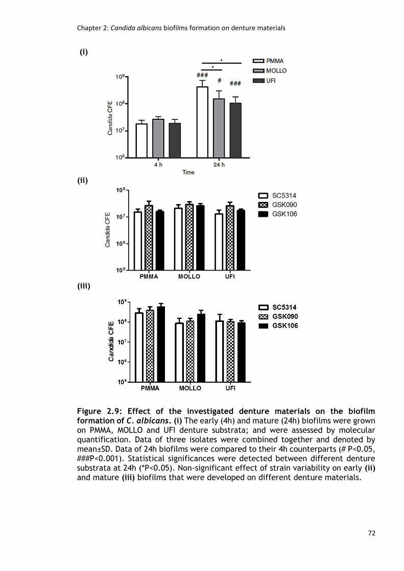
Chapter 2: Candida albicans biofilms formation on denture materials
72
(i)
(ii)
(iii)
Figure 2.9: Effect of the investigated denture materials on the biofilm formation of C. albicans. (i) The early (4h) and mature (24h) biofilms were grown on PMMA, MOLLO and UFI denture substrata; and were assessed by molecular quantification. Data of three isolates were combined together and denoted by mean±SD. Data of 24h biofilms were compared to their 4h counterparts (# P<0.05, ###P<0.001). Statistical significances were detected between different denture substrata at 24h (*P<0.05). Non-significant effect of strain variability on early (ii) and mature (iii) biofilms that were developed on different denture materials.

Chapter 2: Candida albicans biofilms formation on denture materials
73
2.5 Discussion
Over the last fifty years, there has been significant advances in our understanding
of oral hygiene. Nevertheless, with an ageing population, prosthodontic treatment
with partial or complete denture remains an inevitable option for a vast majority
of adult society. Improvements in conventional therapy for edentulous people is
important because the demand for full prosthodontic appliances is unlikely to
decrease in the near future. The necessity of conventional dentures is still high
because osseointegration-associated therapies are region-specific. Furthermore,
these implant-associated therapies could involve a supplementary conventional
prosthodontic treatment as an overdenture option (Carlsson & Omar, 2010).
Moreover, dentures showed high esthetic, acceptable chewing capability, comfort
and phonetics satisfaction in denture wearers that may exceed the patient
expectations (Santos et al., 2015;McCunniff et al., 2017). Therefore, the necessity
of studying biomedical associated-diseases of denture wearing is still of great
importance. The high prevalence of DS (15-<70%) and its high recurrence makes
its prevention a real need (Gendreau & Loewy, 2011). It has been reported that
Candida species, specifically C. albicans, play a vital role in progression of DS and
denture associated diseases (Webb et al., 1998a;Dagistan et al., 2009;Salerno et
al., 2011).
This chapter reports that the denture candidal load has a significant role in DS, in
agreement with earlier investigations (Barbeau et al., 2003;Dagistan et al.,
2009;Salerno et al., 2011). Traditional microbiological techniques (culturing and
staining techniques), have revealed higher quantities of Candida cells from
dentures of the DS diseased group in comparison to those of the healthy group,
especially dentures taken from patients diagnosed with moderate to severe grades
of inflammation. While biofilm forming heterogeneity of C. albicans different
strains (strain variability) did not show such a significant role in respect to DS,
denture material appeared to have an impact on C. albicans mature biofilms.
The biofilm forming heterogeneity phenomenon is a clear and intriguing
observation shown among the denture C. albicans clinical isolates investigated,
displaying a yet, uncharacterised role in DS. From a clinical view, the hypothesis

Chapter 2: Candida albicans biofilms formation on denture materials
74
that biofilm forming heterogeneity of C. albicans can contribute in deciding
morbidity and mortality levels is deserving of examination concerning the oral
cavity (Kean et al., 2018). Our group revealed a relationship with this phenotype
and mortality in a cohort of candidaemia patients in Scotland (Rajendran et al.,
2016a), therefore examining this in a cohort of denture patients was a reasonable
hypothesis. Despite the fact that biofilm forming heterogeneity was observed
utilizing in-vitro models, a relationship with patient clinical presentation was not
observed. Failure to exhibit a connection might be clarified by the way that DS in
immunocompetent people is milder than candidaemia from an extremely
immunocompromised population. Nonetheless, a significant relationship between
the denture candidal load and DS was observed, proposing a notion that the
physical cooperation between extensive quantities of yeasts and hyphae on the
denture surface was more vital than the phenotype per se, and this is possibly
impacted by the various microbiota in the oral cavity as previously described
(O'Donnell et al., 2015b). Undoubtedly, there is a growing body of evidence to
suggest that Candida and bacteria compose polymicrobial communities, where
various bacterial species that are communal to the oral environment can influence
the pathogenicity of each other (Harriott & Noverr, 2009;Diaz et al., 2012;Falsetta
et al., 2014;O'Donnell et al., 2015a), and particular oral microbes can stimulate
the transition of C. albicans LBF into a HBF (Arzmi et al., 2016).
Real time qPCR quantification was previously reported for quantifying fungal cells
(Lievens et al., 2006;Willger et al., 2016). Our research group has recently used
qPCR methodology to quantify respiratory pathogens residing on dentures
(O'Donnell et al., 2016). Yet, according to the best of our knowledge, this is the
first study to quantify denture-adherent Candida cells associated with DS using
molecular methodologies. It has been observed that candidal denture biofilms are
characterised by yeast-hyphae cellular aggregations (Ramage et al., 2004;Coco et
al., 2008a;Bilhan et al., 2009), thus, traditional CFU counting methods could
underestimate the real number of the denture-colonising cells. For this reason,
qPCR offers specific advantages over conventional microbiology techniques
(Azeredo et al., 2017).

Chapter 2: Candida albicans biofilms formation on denture materials
75
Conflicting results were previously reported in the literature regarding the
adhesion and colonisation capacity of Candida on denture liners (Pereira-cenci et
al., 2008). Therefore, an experiment was designed to address this problem. AS
medium was used to mimic the oral environment and prolonged incubation periods
were not targeted because dentures are generally frequently cleaned (Soll &
Daniels, 2016). A high inoculum density of 1×10⁷ CFU/ml was used as previously
defined in the literature (Verran & Maryan, 1997;Chandra' et al., 2001;Raj &
Dentino, 2011;Marra et al., 2012;Lazarin et al., 2013;Jackson et al.,
2014;Yodmongkol et al., 2014). Moreover, this high cell density induces the
development of a mature biofilm with less interlocking hyphae and more yeasts
(Mayer et al., 2013). Therefore, this high cell density may facilitate the process
of collecting the biofilm as it was reported that it is difficult to collect the hyphae
form of C. albicans (Hoyer & Cota, 2016). The qPCR methodology was undertaken
to prevent the negative influence of the results by clumping of the cells, as we
have used 4 and 24hr incubation periods that considered enough to develop an
early and mature biofilm that could facilitate this clumping. The reason for
selecting the denture materials investigated was based on a clinical standpoint.
PMMA is the most routinely used denture material because of its low cost and easy
handling, despite its varied surface, which clearly impacts on microbial
establishment and biofilm growth (Jackson et al., 2014;Susewind et al., 2015).
Discomfort could be promoted by the close contact of PMMA denture material to
the mucosa because of its stiffness. As a result, resilient, less rigid denture liners
are progressively become popular. Reports suggest that denture liners improve
patient masticatory function and decrease discomfort (Kimoto et al., 2007;Palla
et al., 2015), yet denture liners are liable for microbial colonisation (Bulad et al.,
2004;Vural et al., 2010), and biofilm accumulation (Nevzatoglu et al.,
2007;Valentini et al., 2013). In this chapter, samples were fabricated in stone
molds, and were not polished. This was in order to simulate the ordinary clinical
manipulation of the intaglio surface of the dentures, which is usually not polished
to preserve the surface details and prevent compromising denture retention.
However, investigating linear (polished) PMMA surfaces can be suggested for
further research because of the possible capability of C. albicans to form biofilms
on these surfaces.

Chapter 2: Candida albicans biofilms formation on denture materials
76
Data shown in this chapter revealed no statistical difference in candidal
colonization upon various denture materials among the early in-vitro biofilms,
which is in agreement with previous studies (Bulada et al., 2004;Pereira-Cenci et
al., 2007;Hahnel et al., 2012). However, at a later phase of biofilm development,
PMMA was demonstrated to be the denture material surface that is most
susceptible to biofilm development. A conceivable explanation as to why
increased numbers were observed on PMMA is potentially because the porosity of
soft liners are significantly more than that of hard acrylics (Wright et al.,
1998;Pereira-cenci et al., 2008a;Øilo & Bakken, 2015). Hence, large quantities of
microorganisms could be embedded in these porous surfaces (Taylor et al., 1998).
Consequently, even an forceful strategy for biofilm removal such as sonication,
may not remove all microorganisms colonising soft liner cracks and crevices.
Therefore, traditional quantification techniques used in previous studies could
explain the differences observed in this study (Radford et al., 1998;Bal et al.,
2008;Mutluay et al., 2010).
The heterogeneity of clinical isolates of C. albicans has a potential role in
complicating the clinical presentation of diseases (Sherry et al., 2014;Pereira et
al., 2016;Rajendran et al., 2016b), besides a potential role in their adhesion
capacity (Cannon et al., 1995). Therefore, it was important to investigate early
and mature biofilms on denture materials using clinical isolates together with a
commonly used reference strain. Contrary to Waters et al. (1997), the data
displayed a non-significant impact of strain variability on early and mature
biofilms formed on all materials investigated.
Investigation of in vitro biofilm models is critical and consideration of the
variables that are related to the biofilm formation is of high importance. This is
firstly because of the association of Candida biofilm formation with DS (Ramage
et al., 2004;Rautemaa & Ramage, 2011;Pereira et al., 2016), and secondly the
positive link between in vitro biofilm models and their in vivo counterparts
obtained from DS patients (Nobile & Johnson, 2015). There is clear disparity in the
literature regarding the starting cell density inoculum used in candidal biofilm
formation models; ranging from ~ 1×10⁷ through 1×10⁶ to 1×10⁴ cells/ml (Hawser
& Douglas, 1994;Ramage et al., 2004;Johnson et al., 2012). Consequently,

Chapter 2: Candida albicans biofilms formation on denture materials
77
investigation of 1×10⁷ and 5×10⁵ cell densities after a brief preliminary
optimization was performed. The highest cell density did not show significant
biofilm development in either of the considered media for all investigated strains.
This can be interpreted by terms of the quorum-sensing systems that are used by
Candida in response to the density of cell population. In high cell densities,
quorum-sensing molecules such as farnesol and tyrosol inhibit biofilm formation
and filamentation (Ramage et al., 2002b;Albuquerque & Casadevall, 2012). The
positive effects of RPMI-1640 medium on promoting C. albicans adhesion, biofilm
development and antifungal susceptibility testing are well established in
comparison to yeast nitrogen base and other media (Kucharikova et al., 2011;Soll
& Daniels, 2016;Weerasekera et al., 2016). Therefore, RPMI-1640 medium was
used as a standard medium for development of C. albicans biofilm. However, the
effect of AS medium on biofilm development is still not fully established. Data
shown in this chapter revealed RPMI as an appropriate medium to develop
filamentous biofilms characterized with high metabolic activity. While, AS-grown
biofilms demonstrated a less filamentous phenotype and lower metabolism, which
could be resulted from the rich-nutrient nature of the AS. This is in line with Arzmi
et al. (2016), although they used a low concentration of AS (25%). Moreover,
mucins that are found in AS may suppress Candida cells during biofilm
development (Kavanaugh et al., 2014). However, this is a controversial issue,
because some authors reported a supportive role for the mucin in C. albicans
adhesion (Edgerton et al., 1993;Burgers et al., 2010). Irrespective of the type of
medium of incubation, the characterisation data shows the importance of strain
variability in outlining the biomass of the mature biofilms, while this is not the
case in early biofilms. In conclusion, the biofilm characterisation data presented
herein shows the significance of a rational approach to selecting the appropriate
cell density, medium of incubation for the development of in vitro biofilms.
Additionally, it highlights that strain variability should be considered as it has an
impact on mature biofilms.

Chapter 2: Candida albicans biofilms formation on denture materials
78
Chapter findings
Molecular quantification revealed the importance of Candida denture
burden in DS.
Heterogeneity in biofilm formation of the denture isolated C. albicans
strains was observed, although no significant association with DS was
observed.
Characterisation data of C. albicans in vitro biofilms showed the
importance of a rationale in selection of the biofilm development variables.
The XTT assay was more sensitive than CV assay in detecting differences
among C. albicans biofilms developed from various inoculum densities. In
contrast to the low inoculum density and RPMI medium, high inoculum
density and AS medium did not show significant differences between early
and mature biofilms. Strain variability showed significant effect on biofilm
development in mature biofilms only.
Early C. albicans biofilms were not influenced by denture material,
however their mature counterparts were, with PMMA denture materials
yielding the highest quantity of C. albicans.
Strain variability (3 strains levels of biofilm forming capability) did not
significantly affect early or mature phases of C. albicans biofilm
development on denture materials using clinically relevant medium.

3 The anti-adhesive activity of curcumin on Candida albicans biofilms on PMMA denture material

Chapter 3: The anti-adhesive activity of curcumin on Candida albicans
80
3.1 Introduction
Increasingly, there are fewer effective antimicrobials available to treat clinical
infections (Thabit et al., 2015). This can be attributed to the increased prevalence
of anti-microbial resistance, driven largely by poor antibiotic stewardship,
combined with a gradual decrease in investment in antimicrobial development
from the pharmaceutical industry (Renwick et al., 2016). The failure of the
development of effective antimicrobials to keep pace with emerging bacterial
resistance has the potential to reverse the medical, surgical and social
advancements of the last century and place us in a pre-penicillin era. Microbial
diseases deemed unimportant now, may pose a greater threat in the future. We
must therefore aim to be proactive and consider novel antimicrobial strategies
(Crofts et al., 2017).
Nature has an abundance of possibilities, where botanically isolated
chemotherapeutic extracts can provide benefits over purely synthetic agents due
to their co-evolution alongside harmful microbes. Polyphenolic natural compounds
are intriguing potential sources in this context. The macromolecular composition
of polyphenols and the characteristic phenolic functional group or “phenolic
moiety” are found in many plants and can be synthetically prepared (Shahzad et
al., 2014). The polyphenol curcumin (diferuloylmethane), isolated from the
rhizomes of the Curcuma longa plant (Mahmood et al., 2015) is one such compound
which merits consideration. Curcumin (CUR) is the vital element of turmeric used
in Asiatic cuisine and as a dietary supplement or food additive in the food industry.
Turmeric contains up to 5% CUR (Esatbeyoglu et al., 2012;Kwon, 2014). Gupta et
al. (2012) indicated the relative safety and active biological potentials of CUR,
including inhibition of the damaging oxidation and inflammation reactions.
Critically, CUR shows wide range antimicrobial activities (Moghadamtousi et al.,
2014), including antifungal (Martins et al., 2009;Khan et al., 2012), antibacterial
activities (Shahzad et al., 2015;Tyagi et al., 2015), and additionally a potential
capacity to impact biofilm and adhesion properties (Shahzad et al., 2014;Shahzad
et al., 2015).

Chapter 3: The anti-adhesive activity of curcumin on Candida albicans
81
Research from the Ramage laboratory has demonstrated the capability of CUR to
modify the attachment of periodontal microbes to the hydroxyapatite-coated
substrates and influence biofilm development (Shahzad et al., 2015). In parallel,
investigations on the denture pathogen C. albicans showed that CUR was highly
effective against sessile biofilm cells (Shahzad et al., 2014). Kumar et al. (2014)
further demonstrated that CUR could specifically affect the biosynthesis and
permeability of the C. albicans cell wall through targeting of the calcineurin-
mediated signalling and mitogen activated protein kinase pathway.
The globally important opportunistic pathogen C. albicans is known for its ability
to form clinically important biofilms (Ramage et al., 2006;Uppuluri et al.,
2009;Rajendran et al., 2016c). Moreover, this property is complemented by a
range of colonisation determinants, including adhesion and invasion proteins, such
as agglutinin-like sequences (ALS) and secreted aspartyl proteases (SAPs), that
encourage attachment and invasion into the host (O’Donnell et al., 2015a). The
yeasts capacity to alter morphology and exhibit dimorphism and thigmotropic
tendencies makes it a very well-adapted and tenacious pathogen. C. albicans has
the capacity to form biofilms on both biotic and abiotic surfaces making this
microorganism particularly important in oral diseases. To survive and compete in
the oral cavity the organism possesses an array of phenotypic characteristics that
provide physical protection from internal and external antimicrobial mechanisms
(Ramage et al., 2014). Given that C. albicans plays a significant role in DS means
we need to consider antimicrobial strategies that target this microorganism as a
basic prerequisite for DS prevention. Hence, it seems plausible that CUR might
promote oral health through counteracting or limiting the effect of C. albicans to
denture wearers.

Chapter 3: The anti-adhesive activity of curcumin on Candida albicans
82
3.2 Aims
It is hypothesised that CUR could be used to actively inhibit C. albicans biofilm
formation on denture materials through interfering with the initial adhesion
phases. The aim of this chapter was to assess this hypothesis using the following
key investigations:
1. To investigate the capacity of PMMA denture material to adsorb CUR.
2. To investigate the impact of adsorbed CUR on adhesion of C. albicans to
PMMA denture material.
3. To investigate the capability of CUR pre-treated C. albicans to adhere to
PMMA denture material and the possibility of synergistic combination with
CUR adsorbed on to the surfaces.
4. To investigate the influence of CUR on different morphological forms of C.
albicans and biofilm formation.
5. To investigate the effect of CUR on the inter-C. albicans adhesive
interactions.
6. To assess the response of C. albicans to CUR at the molecular level through
investigation of the expression of selected adhesion-aggregation associated
genes.
7. To investigate the impact of CUR incorporation in the PMMA material on
candidal adhesion.

Chapter 3: The anti-adhesive activity of curcumin on Candida albicans
83
Part of the data presented in this chapter has been published in Frontiers in
Microbiology journal.
Alalwan H, Rajendran R, Lappin DF, Combet E, Shahzad M, Robertson D, Nile CJ,
Williams C, Ramage G. Front Microbiol. 2017 Apr 20;8:659. doi:
10.3389/fmicb.2017.00659.
Findings from this chapter have been presented at the following academic
meetings:
1. Postgraduate research prize seminar held in Glasgow Dental School on 27th
of April 2016.
2. OMIG postgraduate research prize symposium held in College of Clinical
Dentistry in University of Sheffield on 8th of Feb, 2017.

Chapter 3: The anti-adhesive activity of curcumin on Candida albicans
84
3.3 Materials and Methods
3.3.1 Culture conditions and standardisation
C. albicans SC5314 laboratory strain was used for these studies. Operational stocks
of yeast cells were prepared on fresh Sabouraud agar (Sigma-Aldrich, UK),
cultivated for 48 h at 30°C, and maintained at 4oC as annotated in chapter 2
(section 2.3.1.2). Using one colony, the cells were cultured in yeast-peptone-
dextrose (YPD) medium (Sigma-Aldrich) for 18 h at 30°C and 150 rpm in an orbital
shaker. The cells were washed twice by centrifuging in sterile phosphate buffered
saline (PBS) (Sigma-Aldrich, UK), then standardised to the required colony-forming
unit per millilitre (CFU/ml) using a Neubauer haemocytometer.
3.3.2 Antifungal susceptibility testing
The two cellular modes of growth (planktonic free-floating cells and sessile biofilm
cells) were first examined for their sensitivity to the polyphenol curcumin (CUR)
(HPLC grade, Acros Organics, Belgium). Prior to each experiment, a standard stock
of CUR was prepared, using a non-antimicrobial concentration of dimethyl
sulfoxide (DMSO) as a solvent, and adjusted to <5% v/v in RPMI-1640 medium
(Sigma-Aldrich, UK) for preparation of working concentrations (Shahzad et al.,
2015). At the commencement, standardised CLSI M-27A broth microdilution
methodology was conducted for planktonic yeast cultures in 96 well round
bottomed microtitre plates (Corning Incorporated, NY, USA) (CLSI-M27-A, 2008).
Briefly, serial doubling dilutions were performed using RPMI-1640 medium after
addition of 200 µl of CUR to the first column of the microtitre plate and 100 µl of
medium in the next nine columns. Thereafter, 100 µl of 1×10⁴ of standardised C.
albicans cells were inoculated into appropriate wells. The treatment
concentration range was (0.39 - 200µg/ml). Plates were incubated at 37oC for 24
h. Negative (medium only) and positive controls (medium + cells only) were
included. Clear wells with no visible growth were considered as the planktonic
minimum inhibitory concentration (PMIC).
For sessile susceptibility testing, pre-formed mature biofilms were treated with
CUR using standardised sessile antifungal testing (Ramage et al., 2001;Pierce et

Chapter 3: The anti-adhesive activity of curcumin on Candida albicans
85
al., 2008). Briefly, 100 µl of 1×10⁶ of standardised C. albicans cells were grown in
96 wells flat-bottomed microtitre plates (Corning Incorporated, NY, USA) at 37oC
for 24 h using RPMI-1640 medium. Then, biofilms were washed with PBS and
treated with 100 µl of CUR for a further 24 h at 37oC, prepared using serial doubling
dilutions as described above. The same treatment regimen described above was
used, where CUR was double diluted in a fresh 96 well microtitre plate
immediately prior to the treatment of the biofilms. Subsequently, treated biofilms
were washed with PBS and assessed for reduction of tetrazolium to formazan using
the XTT assay (Ramage et al., 2001) as described in chapter 2 section (2.3.3.4),
and the optical densities measured at 492 nm using a microtitre plate reader
(FluoStar Omega, BMG Labtech, UK). Negative (medium only) and positive
(medium + cells only) controls were included. The anti-sessile impact was
calculated via comparing the experimental wells data to the positive control data
to reveal the SMIC80, i.e. where the optical density is reduced more than 80% in
comparison to the positive control optical density. These experiments were
repeated in 3 independent occasions with triplicate wells for each condition.
3.3.3 Investigating the effect of CUR on C. albicans growth kinetics
The growth kinetics of CUR-treated C. albicans was assessed to determine if 0.5 x
PMIC concentration could influence the rate of 24 h growing yeasts. The cells were
standardised to 1 × 10⁴ CFU/ml in YPD, with 200 μl inoculated to the appropriate
well of a 96 well round-bottomed plate. Alongside, CUR-treated cells (50 µg/ml=
0.5 ×PMIC) were inoculated. The microtitre plate was incubated at 37°C for 24 h
and every 1 h the absorbance was measured at 530 nm after automated shaking
for 30 s at 100 rpm. This experiment was repeated on two independent occasions
using six replicates. Negative controls (medium only) and (medium + CUR) were
included for appropriate background correction of the untreated and treated cell,
respectively.

Chapter 3: The anti-adhesive activity of curcumin on Candida albicans
86
3.3.4 Investigating the capacity for adsorption of CUR onto denture material
The possible affinity of CUR to fabricated PMMA denture material was explored by
measuring the quantity of the adsorbed CUR spectrophotometrically. Heat cured
PMMA denture base material (Chaperlin and Jacobs Ltd, Surrey, UK) was used to
produce 12 mm diameter discs using the dental compression moulding technique
as described in chapter 2 (2.3.3.1). PMMA sections were immersed in ddH2O for
one week to ensure removal of any residual toxic monomers. To prepare varying
operational concentrations of CUR suspensions, stock CUR was diluted in RPMI-
1640 medium to 200, 400 and 800 µg/ml. The PMMA sections were distributed in
the appropriate wells of 24 well plates (Costar, Corning Incorporated, USA) and 1
ml of the CUR suspension was added. The plates were then incubated at room
temperature for a series of time points (1, 5, 10, 30, 60, 120, 240 and 1440 min),
covered with tin foil to prevent light exposure. After the incubation period the
CUR suspension was removed. The PMMA sections were then transferred with
tweezers to fresh wells and washed by gentle immersion in 1 ml of distilled water,
then distributed to fresh well. Next, 1 ml of DMSO (100%) was added for 5 min to
dissolve the adsorbed CUR. For quantification of the released CUR, a standard
curve was developed by serially diluting CUR in DMSO to prepare concentrations
ranging from 100 to 0.39 µg/ml. Based on that standard curve, the DMSO- dissolved
CUR was measured at 436 nm using the spectrophotometer (FluoStar Omega, BMG
Labtech) including appropriate blank correction wells.
3.3.5 Investigating the effect of CUR adsorption on adhesion of C. albicans to PMMA
Based on the previous experimental data, PMMA denture material sections were
each immersed in 1 ml of 800 µg/ml of RPMI-CUR suspension for 10 min in order
to obtain approximately 50 ug/ml of adsorbed CUR, then washed with ddH2O to
eliminate the unadsorbed molecules. The CUR-adsorbed PMMA sections were
distributed in a 24 well plate and immediately inoculated with 1 ml of 5 x 10⁵ CFU
C. albicans SC5314 cells, then incubated for 30 min at 37°C. Positive controls were
included, which were initially immersed in RPMI medium only for 10 min then
inoculated with the cells. When the incubation period was completed, the sections

Chapter 3: The anti-adhesive activity of curcumin on Candida albicans
87
were gently washed twice in PBS and sonicated in bijou tubes contained 1 ml of
PBS at 35 kHz for 10 min (Ultrasonic bath, Fisher scientific, UK) to collect the
adherent cells. These collected cells were counted using the Miles and Misra plate
counting method (Miles et al., 1938). In brief, using PBS, ten times serial dilutions
were performed of the collected cells (10-1 to 10-4). A twenty microlitre drop of
each diluted suspension was plated in triplicate on SAB agar plates. Plates were
incubated for 24-30 h at 37°C. The most clearly separated colonies in the least
dilution were considered in counting.
The following equation was applied to quantify the adhered cells:
Number of colonies × dilution factor × 50 = colony forming units per cm2 PMMA
The final cell number was then compared to a CUR untreated control. All
experiments were performed in triplicate on 3 independent occasions.
3.3.6 Microscopic imaging of adherent C. albicans to CUR-adsorbed PMMA
To examine the impact of adsorbed CUR on candidal adhesion visually, scanning
electron microscopy (SEM) was also performed using the same experimental
parameters that were described in the aforementioned section (3.3.5), then
processed and imaged, as described previously (Erlandsen et al., 2004;Sherry et
al., 2016). Briefly, C. albicans cells were grown on CUR adsorbed PMMA sections.
These sections were washed twice with PBS, before being fixed for 20 h in 2%
paraformaldehyde, 2% glutaraldehyde, 0.15M sodium cacodylate, and 0.15% w/v
alcian blue, at pH 7.4, and prepared for SEM on the next day. In brief, sections
were triple washed with 0.15M sodium cacodylate buffer, then incubated in 1%
osmium tetroxide for 1 h. After triple washing with distilled water, 0.5% uranyl
acetate was added and incubated for 1 h. After this, a sequential ethanol
dehydration was followed with 30%, 50%, 70%, 90%, absolute alcohol and dried
absolute alcohol. Later, HMDS (Hexamethyldisilazane) was added for 5 min. The
samples were incubated in a desiccator at room temperature for 24 h. Next, the
samples were sputter-coated with a layer of gold-palladium (15-20 nm) and
inspected under a JEOL JSM-6400 scanning electron microscope.

Chapter 3: The anti-adhesive activity of curcumin on Candida albicans
88
3.3.7 Investigating the biological effect of CUR on C. albicans adhesion and biofilm formation
Following CUR adsorption to PMMA denture material, which was conducted using
same experimental parameters as described above, the PMMA discs [untreated
and treated (PMMA- and PMMA+, respectively)] were distributed in the appropriate
wells of 24 well plates and inoculated with 1 ml of 5 x 10⁵ CFU C. albicans SC5314
cells. The inoculated cells were either + 3 min CUR (50 µg/ml) pretreated or PBS
(control) pretreated at 37°C. These discs then were incubated for 30 min at 37°C.
Subsequently, the discs were gently washed twice with PBS. Candidal adhesion
was then assessed and quantified using the Miles and Misra plate counting method
as described in section (3.3.5). The levels of adhesion were expressed as a
proportion of the control (PMMA-/C. albicans).
In parallel, an assessment was conducted to determine whether extended CUR
pretreatment time negatively influenced adhesion via treating cells for 3, 30 and
90 min, then the levels of adhesion to PMMA were quantified as described above.
CUR treatment of C. albicans cells at different growth phases was assessed to
determine whether or not can CUR played a role in biofilm development, with the
hypothesis that there might be dissimilarities in how yeast (Y), germlings (G) or
hyphae (H) reacted to this molecule. Briefly, yeast cells were grown overnight,
standardised to 1 x 10⁶ CFU/ml in RPMI and inoculated into a 96-well microtitre
plate as described in chapter 2 (section 2.3.2.1). Cells were then exposed to 50,
100 or 200 ug/ml CUR at either 0 h (Y), 2 h (G) or 4 h (H) post inoculation and
incubated for a further 24 h at 37oC. Subsequently, the developed biofilms were
washed with PBS and the resultant biofilm assessed using an XTT metabolic
reduction and crystal violet assays as described in chapter 2 (sections 2.3.3.4 and
2.3.2.2, respectively). Moreover, the developed biofilms were microscopically
evaluated using a light microscope (Model BX40F4; Olympus, Tokyo, Japan). All
experiments were performed in duplicate on 3 independent occasions.

Chapter 3: The anti-adhesive activity of curcumin on Candida albicans
89
3.3.8 Investigating the aggregative effect of CUR
With the purpose of assessing whether CUR has further effects on C. albicans
yeasts, its impact on aggregation was assessed. SC5314 laboratory strain cells (Y)
were standardised (1 x 10⁶ cells) in PBS and exposed ± to 50 µg/ml of CUR (sub-
inhibitory concentration) for 90 min at 37oC in an orbital shaker (200 rpm).
Following incubation, vortexing of the standardised inoculum was avoided in order
to reduce any disaggregating forces. The cells were serially ten-fold diluted in PBS
and plated onto Sabouraud agar using the Miles and Misra method (Miles et al.,
1938). Next, the plates were incubated for 24-30 h at 37oC and the colonies
counted as annotated in section (3.3.5). In parallel, a light microscope was used
to observe the cells and to evaluate aggregation visually. All experiments were
performed in triplicate on 4 independent occasions.
3.3.9 Investigating the effect of CUR on cell surface hydrophobicity
Microbial adhesion to the hydrocarbon xylene was utilised to measure the cell
surface hydrophobicity (CSH) of ± CUR exposed C. albicans (SC5314) using a
modified version of the CSH assay (Yoshijima et al., 2010;Sherry et al., 2014).
Cells were propagated overnight and standardised to 1.0 at OD 600 in PBS using a
spectrophotometer and semi-micro cuvettes. 1.1 ml of the standardised cells were
added to a 1.5 ml Eppendorf and spun for 10 min at 7000 r/min, the supernatant
was discarded leaving the pellet intact. CUR (50 µg/ml) diluted in PBS was added
to the pelleted samples. Control samples (cells not exposed to CUR) were
included. Afterwards, the samples were vortexed and incubated at 37°C for 3, 30
and 90 minutes in an agitated state (200 rpm). Next, samples were washed twice
with 20% DMSO to remove the CUR, and controls were similarly treated. Later,
xylene (0.275 ml) was added to form 1/5 of the final volume, and vortexed for 2
min and left for 30 min phase separation. The lower aqueous layer was carefully
pipetted and its optical density was measured at OD600. Negative controls (PBS
only + xylene) were included for background correction. The optical densities
were measured for the standardised treated and untreated samples that did not
contain any xylene (incubated at 37°C and 150 r/m) at the specific time point and
considered as OD before xylene overlay.

Chapter 3: The anti-adhesive activity of curcumin on Candida albicans
90
The following equation was applied to calculate the CSH percentage: ([OD before
Xylene overlay- OD after Xylene overlay] / OD before Xylene overlay) × 100. The
experiment was independently repeated six times using duplicates.
3.3.10 Assessing the molecular impact of CUR on adhesion and biofilm formation
Y and H morphological forms of C. albicans SC5314 cells were prepared via an
initial inoculum of 1 x 10⁸ and 5 x 10⁵ CFU/ml in RPMI-1640 medium, respectively.
For H cells, these were colonised on PMMA denture material sections within 24
wells plates for 4 h. Both Y and H cells were then exposed ± CUR (50ug/ml) in
RPMI for 3, 30 and 90 min and incubated at 37°C. After incubation, cells were
either centrifuged (for Y cells) or sonicated (for H cells) in a 35 kHz for 10 min
(Ultrasonic bath, Fisher scientific, UK) to harvest the cells. The harvested cells
were then washed by centrifugation and RNA was extracted from these cells.
RNA extraction was performed using a combined mechanical disruption (0.5mm
glass beads) and chemical TRIzol™ method (Invitrogen, Paisley, UK). The extracted
RNA was further treated to remove any residual DNA, and then were processed for
production of complementary DNA, which are quantitatively analysed for the
expression profile of targeted genes using qPCR. This multiple procedure is
described in detail in the following sub-sections:
3.3.10.1 Extraction of total RNA
The first step in RNA extraction was the addition of 1 ml of TRIzol™ solution
(Invitrogen, Paisley, UK), which is a monophasic solution of phenol and guanidine
isothiocyanate, to the pelleted candidal cells (Y and H). The mixture was vortexed
for 30 seconds, then transferred to screw cap microcentrifuge tubes containing
0.25 ml of 0.5mm diameter sterile glass beads (Thistle Scientific, Glasgow, UK) to
facilitate disruption of the cell wall, using a Bead beater [BeadBug™ microtube
homogenizer (Sigma-Aldrich, Gillingham, UK)]. Three cycles of bead beating were
performed, each cycle lasted for 30 seconds at maximum velocity (400 rpm), the
tubes were kept on ice in-between the cycles.

Chapter 3: The anti-adhesive activity of curcumin on Candida albicans
91
Next, 100 µl of 1-bromo-3-chloropropane was added and vortexed for 30 seconds,
then left for 3 minutes. The next step was to centrifuge at 13,000 rpm for 15 min
at 4°C (Heraeus centrifuge) to isolate an upper aqueous clear layer. This layer
was transferred to a sterile RNase-free microtube, and 500 μl of absolute
isopropanol was added in order to precipitate the RNA. The tubes were then
inverted 30-40 times, and placed at -20°C overnight to increase RNA precipitation.
The next day, sedimentation of RNA was implemented by centrifuging for 10 min
at 13,000 rpm at 4°C. The supernatant was discarded, and the remaining pellet
washed with 800 µL of ice cold 70% ethanol to be centrifuged for 10 min at 6500
rpm at 4°C. Ethanol was carefully pipetted, and samples were air dried for 20-30
min. The final step was RNA resuspension in 20 µL of RNase free distilled water
and incubation at 65°C in a heat block (Techne, Staffordshire, UK) for 5 min to
facilitate RNA recovery.
3.3.10.2 DNA digestion by DNase
RNase-free DNase kit (QIAGEN GmbH, Germany) was used to eliminate residual
genomic DNA. One and half µl of DNase was added to every RNA sample and
incubated at room temperature for 30 minutes, then incubated at 75°C in a heat
block (Techne, Staffordshire, UK) for 10 min to inactivate the DNase enzyme.
Finally, the RNA samples were stored at -80°C.
3.3.10.3 Synthesis of complementary DNA (cDNA)
RNA concentration and purity was measured by NanoDrop™ ND-1000
spectrophotometer (Labtech International, East Sussex, UK) to standardise RNA
quantity reverse transcripted to cDNA. cDNA synthesis was performed using High-
Capacity RNA-to-cDNA™ reverse transcription (RT) (Applied Biosystems, UK). The
mastermix was prepared by mixing 2 µL of RT Buffer, 2 µL of RT Random Primers,
0.8 µL of dNTP Mix, 4.2 µL RNase free distilled water and 1 µL of MultiScribe®
Reverse Transcriptase enzyme. Ten microliters of mastermix was added to 10 µL
of RNA-distilled water standardised solution to obtain a final volume of 20 µL in
200 µL dome-capped PCR microtubes (Abgene, ThermoFisher, Surrey, UK).

Chapter 3: The anti-adhesive activity of curcumin on Candida albicans
92
Solutions were prepared containing 500 and 50 ng of the purified RNA from the
yeasts and hyphae samples, respectively. NRT (No Reverse transcriptase) samples
were included to monitor the efficacy of the Reverse transcriptase enzyme. Next,
the samples were placed in a Thermo-cycler (Bio-Rad, Hertfordshire, UK). The
cycle program consisted of 10 min at 25°C, 120 min at 37°C, 5 min at 85°C and a
final hold stage at 4°C. cDNA was then stored at -20°C until used in subsequent
PCR assays.
3.3.10.4 Primers used
Oligonucleotide primer synthesis was conducted by (Invitrogen, Paisley, UK). The
primers were designed against the ALS1, ALS3, ALS5 (agglutanin-like sequence 1,
3 and 5), EAP1 (epithelial adhesion protein 1) and AAF1 (adhesion and aggregation
factor 1) genes. The primers were synthesised to match the sequences taken from
peer-reviewed papers. Except, AAF1 which was designed by the Oral Sciences
Research Group in Glasgow Dental Hospital and School. The primers are listed in
Table 3.1.

Chapter 3: The anti-adhesive activity of curcumin on Candida albicans
93
Table 3.1: Primers used for real time qPCR transcriptional analysis of Candida
albicans
Gene Sequence (5’ to 3’) Reference
ALS1 F - TTCTCATGAATCAGCATCCACAA (Nailis et al., 2009)
R - CAGAATTTTCACCCATACTTGGTTTC
ALS3 F - CAACTTGGGTTATTGAAACAAAAACA (Nailis et al., 2009)
R - AGAAACAGAAACCCAAGAACAACCT
ALS5 F - CTGCCGGTTATCGTCCATTTA (Green et al., 2005)
R - ATTGATACTGGTTATTATCTGAGGGAGAAA
EAP1 F - ACCACCACCGGGTATACAAA (Sherry et al., 2014)
R - GCCATCACATTTGGTGACAG
AAF1 F - CTGCCCTTGTTGGTACATCT This study
R - TGGGATAGTTGGTGGAGGAG
ACT1 F - AAGAATTGATTTGGCTGGTAGAGA (Ricardo et al., 2009)
R - TGGCAGAAGATTGAGAAGAAGTTT
3.3.10.5 Gene expression assessment and analysis utilising real time PCR
Fast SYBR® Green PCR Master Mix (Applied biosystems, USA), intercalating,
fluorescent dye was harnessed to perform RT-q PCR, using the same procedure
and thermo-cycler program annotated in chapter 2 (2.3.1.4). Each parameter was
analysed in duplicate using the StepOnePlus™ Real-Time PCR System unit (Applied
biosystems, USA)] and StepOne software v2.3. The primers listed in Table 3.1 were
used and 1 µl of synthesised cDNA was added instead of genomic DNA. No cDNA
template and no reverse transcriptase controls were included.
ACT1 housekeeping gene was selected for normalisation; all the generated Ct
values of the interrogated genes were relative to the Ct values of that standard
housekeeping gene. 2−ΔΔCt arithmetical method (Percentage of expression= 2−ΔCt,
where ΔCt= Ct of targeted gene – Ct of ACT1 housekeeping gene) was undertaken,
to quantify the relative expression (Livak & Schmittgen, 2001). A heatmap was
created for the differential expression of genes (log2) over the period of 3 to 90

Chapter 3: The anti-adhesive activity of curcumin on Candida albicans
94
min from the untreated control compared to CUR exposed cells. Maps and clusters
were generated in R with the use of heatmap.2 function from the gplots package.
Dr Ranjith Rajendran supported these analysis.
3.3.11 Investigating the effect of CUR incorporation on adhesion
The capacity of CUR-incorporated PMMA denture material to reduce adhesion of
C. albicans was explored. Prior to fabrication of PMMA discs, stock CUR was mixed
with the monomer to obtain a standardised solution of 200 µg/ml. Then, the discs
were fabricated as annotated in chapter2 (2.3.3.1). Control discs were included
taking into consideration the CUR dissolvent (DMSO) concentration. All discs were
appropriately distributed into a 24 well plate. C. albicans SC5314 was propagated
and standardised in RPMI-1640 medium. One ml of 5 × 10⁵ cells were inoculated
and statically incubated for 30 min at 37°C. After the incubation, the sections
were washed in PBS, sonicated to collect the adherent cells and the collected
cells were counted using the Miles and Misra plate counting method (Miles et al.,
1938) as annotated in section (3.3.5). Three replicates were used and the
experiments were repeated on five independent occasions. In parallel, the release
of CUR from the fabricated discs was investigated via immersion of the targeted
discs in DMSO solvent for 24 h and measuring released CUR spectrophotometrically
as annotated in section (3.3.4).
3.3.12 Statistical Analysis
As we were unable to ascertain that the data conformed to a Gaussian distribution
data analysis was performed on non-parametric data using either a Mann-Whitney
test or a Kruskal-Wallis test with Dunn’s multiple comparison post-test. All
independent data points were presented, with error bars representing the median
with interquartile range. Where proportional data were presented, analysis was
performed on the original data sets. All statistics and figures were produced using
GraphPad Prism v.5 (GraphPad Software Inc., La Jolla, CA).

Chapter 3: The anti-adhesive activity of curcumin on Candida albicans
95
3.4 Results
3.4.1 CUR can be adsorbed onto denture material up to effective concentrations
Initially, the potential inhibitive capacity of CUR to planktonic and biofilm C.
albicans cells was tested to establish biologically active working concentrations
appropriate for use in downstream analyses. The planktonic minimum inhibitory
concentration (PMIC) was 100 µg/ml of CUR, while the sessile (biofilm) minimum
inhibitory concentration that produced an ≥80% reduced metabolic activity
(SMIC80) was 200 µg/ml, representing a significant reduction in the biofilm’s
activity and/or viability. Furthermore, the growth kinetics of 0.5 x PMIC CUR-
treated C. albicans was investigated. Figure 3.1 shows that the start of
exponential phase of CUR-treated cells was delayed for a period of 4 h compared
to the untreated control (6h v 10h). Both sets of cells reached the stationary phase
at the same time (20 h), with approximately same cellular quantity. Statistical
analysis did not show any difference (p>0.05), which could clarify that the
selected concentration of CUR did not influence growth of C. albicans and
subsequently did not interfere with the results of the next adhesion investigations.
According to these data, it was important to evaluate whether these effective
concentrations of CUR could be adsorbed to PMMA denture material to interfere
with C. albicans adhesion. Therefore, the PMMA samples were immersed in 200,
400 and 800 µg/ml CUR for different time periods and the adsorbed concentrations
were measured using an elution method in conjunction with an optimised standard
curve. Figure 3.2(i) displays the visible light absorption spectrum of the CUR in
DMSO. This figure shows the absorbability of CUR at 436 nm at ≤100 µg/ml
concentrations. The kinetics of adsorption for each concentration is presented in
figure 3.2(ii). PMMA had the capacity to adsorb biologically effective CUR
concentrations and immersion in 800 µg/ml CUR solution for 90 min was required
to achieve adsorbed concentrations with anti-biofilm activity (200 µg/ml).
Nonetheless, 50 µg/ml and 100 µg/ml concentrations were adsorbed from this
initial concentration when immersed for 10 and 30 min. The lower concentration
CUR solution (400 µg/ml) was able to attain 50 µg/ml and PMIC levels, though this
approximately required 90 and 240 min immersion. Finally, the lowest

Chapter 3: The anti-adhesive activity of curcumin on Candida albicans
96
concentration CUR solution (200 µg/ml) did not show the capability to achieve any
antimicrobial level concentrations, even after 24 h adsorption. Consequently,
using of 800 µg/ml of CUR in adsorption experiment appeared biologically
effective and efficient.
Figure 3.1: Growth kinetics of half PMIC CUR-treated C. albicans. The blue and the red dotted lines represent tangents to the start points of exponential and the stationary phases, respectively. Mann-Whitney statistical test was used to analyse the data.

Chapter 3: The anti-adhesive activity of curcumin on Candida albicans
97
(i)
(ii)
Figure 3.2: The adsorption capacity of CUR onto PMMA denture material. (i) Visible light spectrophotometric analysis of CUR in DMSO using a range of CUR concentrations, where the vertical line that contacts the peaks represents the absorption wavelength. (ii) Time and concentration dependent adsorption of CUR to PMMA (blue dotted line) at 0.5 x PMIC (50 µg/ml), PMIC (100 µg/ml) and SMIC80 (200 µg/ml).
200 300 400 500 600 7000
5
10
200 400 600 800
-5
0
5
10
15
200 400 600 800
-5
0
5
10
15100 g/ml
50 g/ml
25 g/ml
12.5 g/ml
6.25 g/ml
3.12 g/ml
1.56 g/ml
0.78 g/ml
Wave length in nm
Op
tica
l de
ns
ity
Wave length in nm
Op
tic
al d
en
sity
Wavelength in nm
Op
tical d
en
sity

Chapter 3: The anti-adhesive activity of curcumin on Candida albicans
98
3.4.2 CUR adsorption reduces Candida albicans adhesion
Based on the data in the previous section, the focus was on immersion samples in
800 µg/ml of CUR for 10 min, which achieved 50 µg/ml on the surface of the PMMA
for downstream analysis. Selection of a 0.5 x PMIC concentration was aimed to
ensure exposure of the cells to a non-inhibitory concentration.
Next, the adherence capability of C. albicans SC5314 to PMMA denture material
for 30 min was evaluated, where surfaces adsorbed with CUR (50 µg/ml) were
compared to an unadsorbed control [Figure 3.3(i)]. A three-fold significant
reduction in adhesion of C. albicans was observed (p<0.004), where the median
number of adherent cells were reduced from approximately 1.56 x 10⁵ to 4.8 x 10⁴
cells/cm2 with interquartile ranges of 1.5-1.75 x 10⁵ and 4-5.9 x 10⁴, respectively.
Besides, SEM analysis showed a visible decline of adherent yeasts cells on the
PMMA surfaces [Figure 3. 3(ii)].

Chapter 3: The anti-adhesive activity of curcumin on Candida albicans
99
(i)
(ii)
Figure 3.3: The impact of CUR adsorption to PMMA on Candida albicans adhesion. (i) C. albicans anti-adhesive capacity of approximately 50 μg/ml adsorbed CUR onto PMMA (+CUR) compared to non-adsorbed control surfaces, where a Mann-Whitney test was performed on data from nine independent experiments (n=9). All independent data points are presented, with error bars representing the median with interquartile range (**= p < 0.01). (ii) SEM micrographs of 30 min adherent C. albicans cells onto CUR adsorbed (+CUR) and non-adsorbed (control) PMMA surfaces. Scale bar is 20 µm.
Given that CUR adsorption showed capacity for a direct anti-adhesive effect, we
explored the possibility that combining a short exposure to CUR with that of CUR
adsorbed to PMMA would result in a synergistic increase in overall activity and
prevent adhesion and colonization. C. albicans was exposed to CUR 50 µg/ml for
3 min (CA + CUR) then compared to both PMMA adsorbed with CUR (PMMA + CUR)
or a combination of CUR exposed C. albicans and CUR adsorbed PMMA (PMMA CA

Chapter 3: The anti-adhesive activity of curcumin on Candida albicans
100
+ CUR). Figure 3.4 shows that the adhesion was reduced by 27% only with the
short, direct exposure of C. albicans alone, which was lower than CUR adsorption
alone (70% reduction), where a significant difference (p<0.05) was observed
between these reductions. Moreover, when both the cell and surface were co-
exposed to CUR a significant reduction of 93% (p<0.001) was observed.
Figure 3.4: The impact of single and dual-treatment of CUR on C. albicans adhesion. A synergistic anti-adhesive capacity of (PMMA CA +CUR) was observed, where short pre-exposure of cells to CUR (CA +CUR) with PMMA adsorbed CUR (PMMA +CUR) were combined. The statistical analysis was performed using a Kruskal-Wallis test with Dunn’s multiple comparison post-test performed on the data from nine independent experiments. The percentages shown relative to the control. All independent data points (n=9) are presented, with error bars representing the median with interquartile range (* p < 0.05, ** p < 0.01, *** p < 0.001).
3.4.3 CUR inhibits biofilm formation and enhances Candida albicans aggregation
The data above displayed a positive anti-adhesive impact to C. albicans with
respect to the adsorption of CUR to PMMA surfaces, and displayed that a brief CUR
pre-exposure (3 min) resulted in diminished adhesion of C. albicans, proposing
that sub-inhibitory concentrations can provoke some biological activity. To further
investigate this, the CUR pre-exposure time on C. albicans was extended. It was
shown that increasing the pre-exposure time from 3 to 30 and 90 min,

Chapter 3: The anti-adhesive activity of curcumin on Candida albicans
101
respectively, significantly improved anti-adhesion (p>0.01). However, between 30
to 90 min there were no significant enhancements in anti-adhesive capabilities
(Figure 3.5).
Figure 3.5: Impact of longer-term CUR pre-exposure on candidal adhesion. C.
albicans SC5314 was pre-exposed to 0.5 x PMIC sub-inhibitory concentration of
CUR (50 μg/ml) for 3, 30 and 90 min and adhesion to PMMA was evaluated in
triplicate in 3 independent experiments. The percentages shown are relative to
their respective controls. All independent data points are presented, with error
bars representing the median with interquartile range (* p < 0.05, ** p < 0.01).
Next, the longer-term impact of CUR and response of the different morphological
forms of C. albicans to CUR and how this could impact biofilm formation was
investigated. To achieve this aim, yeast cells (Y = 0 min), germlings (G = 120 min)
and hyphae (H = 240 min) were prepared prior to CUR exposure at 50, 100 and 200
µg/ml, which were then incubated for 24 h to develop biofilms. The developed
biofilms were assessed using metabolic and biomass data [Figures 3.6(i) and (ii)].
The resultant metabolic and biomass data mostly showed similar patterns, where
at lower sub-inhibitory concentrations (50 µg/ml) and PMIC levels (100 µg/ml) the
anti-candidal properties moderately impacted the overall biofilm metabolism and
biomass with a lesser effect on the latter. At SMIC levels (200 µg/ml) a significant
drop in biofilm metabolism was observed for Y and G of approximately >90% of the
control (p<0.01), though H cells were least impacted (p<0.05).

Chapter 3: The anti-adhesive activity of curcumin on Candida albicans
102
That considerable anti-biofilm effect of SMIC concentration on metabolism was
less noticeable on biofilm biomass, with statistically significant 85% to 54%
reduction in Y and G (p<0.05), respectively, dropping to an insignificant 11%
reduction in H cells. The inhibited biofilms were microscopically examined, where
the light microscope images showed an anti-biofilm impact on all biofilms
investigated in a concentration and morphological form dependent manner (Figure
3.7).

Chapter 3: The anti-adhesive activity of curcumin on Candida albicans
103
(i)
(ii)
Figure 3.6: The impact of CUR on C. albicans biofilm formation. Three
morphological forms of C. albicans SC5314 (Y = yeast; G = germlings; H = hyphae)
were exposed to CUR at different concentrations 50,100 and 200 μg/ml, and after
24 h the resultant biofilm formation assessed through metabolic (i) and biomass
(ii) analytical assays. Data represents six independent experiments, Kruskal-Wallis
test with Dunn’s multiple comparison post-test were used for statistical analysis.

Chapter 3: The anti-adhesive activity of curcumin on Candida albicans
104
Figure 3.7: The impact of CUR on C. albicans biofilm architecture. Light
microscope images of biofilms tested, which show a reduction in the biofilm
mycelial quantity and density in a concentration and morphological form
dependent manner, images acquired at 250 X magnification. Scale bar 18 µm. (Y
= yeast; G = germlings; H = hyphae)

Chapter 3: The anti-adhesive activity of curcumin on Candida albicans
105
To assess whether CUR induces aggregation through alteration of the cell wall
surface a plate counting based approach was performed. To quantify aggregation
it was hypothesized that sub-MIC levels would lower the resultant CFU compared
to its respective control, where each aggregate would produce only one CFU and
reduce the overall homogeneity of the suspension. Indeed, a significant drop in
CFU counts was demonstrated in the CUR group (p<0.01) [Figure 3.8(i)], which was
additionally confirmed microscopically [Figure 3.8(ii)].
(i)
(ii)
Figure 3.8: The impact of CUR on aggregation of C. albicans. (i) Aggregation of
C. albicans SC5314 treated with (50 ug/ml) was evaluated by total viable cell
counts, analysed using a Mann-Whitney test on triplicate data from 4 independent
experiments, All independent data points are presented, with error bars
representing the median with interquartile range (* p < 0.05). Additionally,
validation of the phenotype by light microscopy (400 X magnification) was
performed (ii), where aggregation possibility was observed. Scale bar is 17 µm.

Chapter 3: The anti-adhesive activity of curcumin on Candida albicans
106
3.4.4 CUR reduces cell surface hydrophobicity of Candida albicans
Given the potential positive relationship between the microbial CSH and its
adhesive capability (Wang et al., 2015), this was tested for CUR with respect to
C. albicans using the aqueous-hydrocarbon biphasic partitioning assay. The
resultant data showed an overall significant reduction in the CSH of CUR exposed
C. albicans at all time-points investigated, with 13, 51 and 37% reduction (Figure
3.9). Additionally, 30 min of CUR exposure showed a statistically significant
difference compared to 3 min (p< 0.05). These data suggested reduction of C.
albicans CSH as a potential biological mechanism of CUR antiadhesive capability.
Figure 3.9: The impact of CUR on cell surface hydrophobicity of C. albicans.
Kruskal-Wallis test with Dunn’s multiple comparison post-test were used for
statistical analysis. All independent data points are presented, with error bars
representing the median with interquartile range (* p < 0.05, ** p < 0.01). Red
asterisks represent the significant difference between each unexposed control
and its Cur exposed at the relevant time point.

Chapter 3: The anti-adhesive activity of curcumin on Candida albicans
107
3.4.5 CUR affects the temporal expression of Candida albicans adhesins
The data above suggested that CUR induced biological effects on C. albicans, most
considerably on preventing biofilm development, influencing aggregation and
reducing CSH. Therefore, the aim was to investigate a panel of related genes
through transcriptional analysis. To perform this, Y and H cells at 3, 30 and 90 min
post CUR exposure were prepared. Graphical and visual representation of the data
obtained were featured in bar charts (Figure 3.10) and heat map analysis with
hierarchical clustering (Figure 3.11).
It was shown that that Y cells (Figures 3.10i, ii & iii) exposed to CUR showed
temporal changes in gene expression, most notably the progressive down-
regulation of the most potent adhesion-related gene ALS3 that became
statistically significant after 90 min of CUR exposure. Whereas ALS1, which was
related to ALS3 as the hierarchical clustering illustrated, showed a variable
modulation in response to CUR, where it was significantly down-regulated at 3 min
(p< 0.01) and then up-regulated at 30 and 90 min, though it was significant at 30
min only. However, the clustered aggregative and flocculation genes AAF1, EAP1,
and ALS5 transcripts were all significantly up-regulated (p< 0.05-0.01) in a time-
dependent manner.
The clustering of expression for the H cells (Figures 3.10iv, v & vi) was similar to
that of Y cells, and the patterns showed moderate similarity. More specifically,
ALS3 gene expression was significantly down-regulated (p<0.05) at 30 and 90 min
time points though it was non-significantly up-regulated at 3 min. Whereas, ALS1
showed non-significant up-regulation up to 30 min and down-regulation at 90 min.
ALS5, EAP1 and AAF1 were considerably upregulated with AAF1 showing the
highest levels of expression at 30 min in comparison to the control, and
reciprocally ALS3 being the most down-regulated. The heat map clustering (figure
3.11) showed the relative contrast between the main two gene clusters in
response to CUR exposure, where the expression of ALS5 and ALS3 genes were
clear examples. Levels of differential expression were consistently higher in the
Y than H cells.

Chapter 3: The anti-adhesive activity of curcumin on Candida albicans
108
(i) (ii) (iii)
(iv) (v) (vi) Figure 3.10: Transcriptional expression of CUR-treated C. albicans. Y cells (i, ii & iii) were prepared and exposed to CUR for 3, 30 and 90 min, respectively. The same was also performed to the H cells (iv, v & vi). Expression of ALS1, ALS3, ALS5, EAP1 and AAF1 were then assessed using qPCR and relative gene expression assessed the ACT1 housekeeping gene. An unpaired t test was used (*p < 0.05, **p < 0.01), where a natural log transformation was performed on the data. Error bars represent standard error of mean.
ALS1 ALS3 ALS5 EAP1 AAF1
100
101
102
103
104
Control
CUR**
*
**
Genes
% e
xp
res
sio
n r
ela
tiv
e t
o A
CT
1
ALS1 ALS3 ALS5 EAP1 AAF1
100
101
102
103
104
Control
CUR
** **
**
**
Genes
% e
xp
res
sio
n r
ela
tiv
e t
o A
CT
1
ALS1 ALS3 ALS5 EAP1 AAF1
100
101
102
103
104
Control
CUR
**
** *
**
Genes
% e
xp
res
sio
n r
ela
tiv
e t
o A
CT
1
ALS1 ALS3 ALS5 EAP1 AAF1
100
101
102
103
104
PC
CUR
Genes
% e
xp
res
sio
n r
ela
tiv
e t
o A
CT
1
ALS1 ALS3 ALS5 EAP1 AAF1
100
101
102
103
104
PC
CUR
*
*
Genes
% e
xp
res
sio
n r
ela
tiv
e t
o A
CT
1
ALS1 ALS3 ALS5 EAP1 AAF1
100
101
102
103
104
PC
CUR
*
*
*
Genes
% e
xp
res
sio
n r
ela
tiv
e t
o A
CT
1

Chapter 3: The anti-adhesive activity of curcumin on Candida albicans
109
(i) (ii)
Figure 3.11 Visual transcriptional analysis of CUR treated C. albicans. (i) Y and (ii) H cells. Heatmap and clustering was performed for the differential expression of genes (log2).
3.4.6 PMMA-incorporated CUR does not inhibit Candida albicans adhesion
Based on the previous data, adhesion of C. albicans was reduced when freely
adsorbed CUR was permitted to physically adsorb to the already cured (completely
polymerized) PMMA surface. Therefore, it was hypothesized that incorporation of
CUR within PMMA may provide longer sustained release of bioactive
concentrations. The effect of incorporation of CUR into the uncured PMMA was
tested by adding CUR to the monomer (200 µg/ml) before mixing it with the
powder, and candidal adhesion (30 min) tested using Miles and Mirsa plate
counting methodology. Figure 3.12 shows the non-significant difference in the
number of the counted CFU between the CUR-incorporated and control (p>0.05).
Thus, incorporation of CUR during fabrication of PMMA denture material did not
show the predicted impact on C. albicans adhesion. To investigate why this was
not the case, CUR release from the processed CUR-incorporated PMMA was
spectrophotometrically assessed. No CUR release was observed, suggesting
covalent chemical interaction between CUR and PMMA monomers that could
restrict CUR solubility and release.

Chapter 3: The anti-adhesive activity of curcumin on Candida albicans
110
Figure 3.12: The non-effective role of CUR incorporated to PMMA on Candida albicans adhesion. C. albicans anti-adhesive capacity incorporated CUR to PMMA (CUR-incorporated) compared to non-incorporated control surfaces, and a Mann-Whitney test was performed on data from 5 independent occasions, where 3 surfaces were used as replicates and an average was considered (p>0.05). The averaged independent data are presented, with error bars representing the median with interquartile range.

Chapter 3: The anti-adhesive activity of curcumin on Candida albicans
111
3.5 Discussion
The potent biological activities of the polyphenol CUR has enabled some
researchers to describe it as a modern biological regulator (Esatbeyoglu et al.,
2012). The data presented in this chapter display the possibility for use of CUR
within the perspective of oral health for denture wearers. It has been shown that
CUR can adsorb to denture substrates and reduce C. albicans adhesion, rather
than actively kill or inhibit the microorganisms. Remarkably, exposure of C.
albicans to CUR elicited cellular aggregation, an effect that also reduced its
adhesion capacity, besides the decline in the cell surface hydrophobicity.
Transcriptional analyses revealed that the key ALS3 adhesin [the most important
‘king’ of the ALS family as illustrated by Hoyer et al. (2008)] was negatively
impacted, whereas genes associated with aggregation were positively impacted.
Collectively, the data shown reveal that CUR has the potential to be used in
denture care as a means of preventing C. albicans biofilms and subsequently
denture-induced stomatitis (O'Donnell et al., 2015a).
Initially, the aim was to evaluate and confirm CUR antimicrobial capabilities.
These data are in general agreement with others (Martins et al., 2009;Sharma et
al., 2010b;Khan et al., 2012), displaying that concentrations around 100 µg/ml
are necessary to inhibit cellular growth. Besides, 200 µg/ml demonstrated a
considerable anti-biofilm activity (Shahzad et al., 2014). For further confirmation,
0.5 x PMIC concentration (50 µg/ml) showed no impact on the cellular growth
kinetics (Neelofar et al., 2011). Any discrepancies noted might be attributed to
the protocols used for the broth microdilution method, variability of strains used,
variability of CUR source and purity and proportions of curcuminiods involved.
Considering the principal interest of this research was in preventing C. albicans,
rather than actively inhibiting and killing C. albicans, then the focus was on using
a lower sub-inhibitory concentration (half PMIC) of 50 ug/ml. This was motivated
by the hypothesis that CUR could be clinically used as a supplement, or as part of
a nutritional regimen, where, the concentration of CUR could be maintained
through salivation along with adsorbing on to the oral tissues and the related
prostheses.

Chapter 3: The anti-adhesive activity of curcumin on Candida albicans
112
The results presented herein indicate that approximately 6% of the CUR provided
can be adsorbed to the prosthesis surface within 10 min. Theoretically, polyphenol
CUR might be directly and indirectly delivered to the oral cavity. The direct
method would be during ingestion, and indirectly when it is absorbed by the
digestive system and becomes bioavailable to bodily fluids, i.e. blood and saliva.
Indeed, literature showed that polyphenols have high affinity for the salivary
proteins, especially the proline-rich proteins and histatins (Bennick, 2002;Soares
et al., 2011). The daily CUR consumption is very variable across cultures, for
example, in Nepal and India daily consumption of CUR can reach up to 100 mg
(Shahzad et al., 2015), and in South Korea this may only reach 15 mg (Kwon, 2014),
whereas, in the United Kingdom, according to the European Food Safety Authority,
it may reach up to 0.9 to 3.3 mg/kg of the body weight daily, which is equivalent
to 60-230 mg/day (EFSA, 2010). Therefore, providing an anti-candidal
concentration just through nutritional approach is not without challenges, though
the potential of achieving an anti-adhesive concentration is a more conceivable
goal. Indeed, it was possible to show that 50 µg/ml could be adsorbed onto PMMA,
the polymer most commonly used to construct denture prostheses, within a
relatively short time. The resultant adsorption optimisation process showed that
the optimised CUR-adsorbed PMMA reduced C. albicans adhesion by up to 70%,
which was further improved to 93% with a short CUR pre-exposure of C. albicans.
This combination revealed that CUR has dual functionality, through surface
adsorption and directly against C. albicans producing a synergistic-like effect.
For further understanding of the biological function of the sub-inhibitory CUR
effect, a series of studies were undertaken to define its impact on the kinetics of
adhesion, whilst also evaluating how its influence on the dissimilar morphological
forms of C. albicans. The Ramage laboratory have formerly shown that other
natural molecules, such as tea tree oil (TTO) derivatives and carbohydrate-derived
fulvic acid (CHD-FA) affect C. albicans colonisation and growth depending on the
stage of biofilm development (Ramage et al., 2012b;Sherry et al., 2012).
Therefore, it was reasoned that CUR may also act on C. albicans in a similar
manner. It was shown that prolonged exposure (30 and 90 min) of C. albicans yeast
cells considerably minimised its adhesive capability onto PMMA, signifying that
CUR was capable of modifying its adhesive capability in some manner, which was

Chapter 3: The anti-adhesive activity of curcumin on Candida albicans
113
further investigated through observing cells grown to different phases of
morphological development, namely yeast, germling and hyphae cells. It was
hypothesised that the morphological phase of C. albicans at which the cells were
treated with CUR could influence overall development and integrity of the biofilm.
Indeed, it was shown that all treated morphological forms exhibited reduced
general biofilm formation, though only the inhibitory concentrations, specifically
200 µg/ml, reduced the yeast and germling cells developed biofilms in a
significant way in comparison to the sub-inhibitory concentration. However,
hyphae cells were not influenced in a concentration dependent manner,
suggesting that the efficacy of CUR was more apparent against immature
morphological phases. Remarkably, though the biofilms were generally reduced
compared to control levels, there were still significant biofilms remaining,
indicating once more the advantage of early preventative intervention. A key
limitation of this interpretation is the sample sizes used during these analyses
(Vaux, 2012). Indeed, it raises questions whether the statistical analyses are
valuable, which is why individual data points are presented. Nonetheless, when
the data is examined in its entirety there are indicative trends that CUR
demonstrates positive biological influences, though further research is required
to confirm these data.
The anti-oxidative and hydrophobic capacities of the polyphenol CUR molecules
(Priyadarsini, 2013;Mirzaei et al., 2017) might give an explanation why it
preferentially adsorbs to PMMA and the C. albicans cell wall. It was hypothesised
that the hydrophobic nature of the CUR molecule might initiate an aggregation
process of the coated C. albicans with one another. This notion could be of
importance within the context of oral delivery with the saliva, specifically if the
possibility of creating complexes of cells that reduce their interaction with the
denture surface was considered, which also may minimise the opportunity for
individual cells to adhere and be shielded inside denture surface crevices and
imperfections. Indeed, this was verified both quantitatively and visually, which
could also explain why a synergistic inhibition of adhesion at sub-inhibitory
concentrations was observed.

Chapter 3: The anti-adhesive activity of curcumin on Candida albicans
114
Further mechanistic causes of microbial aggregation might result from failure of
daughter cells separation, as in Candida auris (Borman et al., 2016). The
possibility of this to occur in response to CUR in this study is low because PBS was
used as a non-nourishing medium although the phenomenon might occur when
CUR- exposed C. albicans tested in nourishing media. The other potential cause is
an increase of the cell surface hydrophobicity (CSH). Therefore, the CSH of CUR-
exposed (50 µg/ml) C. albicans was tested over different periods of exposure.
In parallel, the effect of sub-inhibitory concentration of CUR on reducing the CSH
of C. albicans was demonstrated. Several expressed proteins participate in CSH
modulation (Bujdáková et al., 2013). The CSH experiment helped to achieve two
objectives in a single action. It suggested a potential mechanism for the anti-
adhesive effect of sub inhibitory concentration of CUR, and also these experiments
supported the notion of coating of the cells with CUR, because the CSH experiment
was modified by washing the cells with diluted DMSO to remove the proposed
coating CUR. This data comes in line with several studies that confirmed the
positive association between CSH and adhesion to surfaces (Krasowska & Sigler,
2014;Ellepola et al., 2016;Wang et al., 2017b), though some reports did not
confirm this relationship (Bujdáková et al., 2013;Silva-Dias et al., 2015) and
others presented a complicated role for CSH in adhesion (Hoyer & Cota, 2016).
We can suggest that the decrease of C. albicans CSH in response to CUR exposure
is an evasion endeavor from these cells to distance themselves from the organic
hydrophobic molecules, which resembles a mechanism observed in hydrophilic
microorganisms that protect themselves from attachment of organic molecules
(Krasowska & Sigler, 2014).
An inherent range of genetic and morphological properties of C. albicans provide
it with enhanced capacity for colonisation and biofilm formation (Blankenship &
Mitchell, 2006). Finding strategic approaches to controlling these properties offers
potential for innovative anti-candidal treatments. Mechanistically, it was
intriguing to understand how CUR elicited C. albicans specific effects. A previous
study has demonstrated that CUR has the capability to modulate the global
repressor of filamentation TUP1 (Sharma et al., 2010b). Indeed, Ramage’s
research group showed that HWP1, which is a key hyphal wall associated element,

Chapter 3: The anti-adhesive activity of curcumin on Candida albicans
115
was down-regulated when a focused study on mature biofilms was conducted
(Shahzad et al., 2014). To this end, a transcriptional approach was employed to
assess genes involved in adhesion and aggregation. CUR downregulated the ALS3
gene in both yeast and hyphae cells; ALS3 has a well-established and key role in
C. albicans adhesion, and plays a pivotal role in biofilm formation and invasion
(Zhao et al., 2004;Nobile & Mitchell, 2005;Hoyer et al., 2008;Liu & Filler, 2011).
The down- regulation of such an important gene results in a reduction in cellular
adhesive capacity. However, the ALS1 gene was variably modulated by CUR and
its transcriptional data did not steadily come in a line with the adhesion data.
Indeed, it was reported that ALS1 has less impact on adhesion than ALS3 and
complicated interrelationships among ALS family members have been observed
(Zhao et al., 2004). Nevertheless, both genes have been shown to have significant
functions in early biofilm events (Nailis et al., 2009;Fox et al., 2015).
There is controversy about the role of the ALS5 gene in initial biofilm formation
and adhesion of C. albicans (Hoyer et al., 2008). ALS5 has a less well-defined
function within the ALS family, and even though defined as an adhesin,
functionally it appears to have amyloid characteristics and the capability to
enhance aggregation (Rauceo et al., 2004;Garcia et al., 2011). This aggregation-
driving role might explain why it is up-regulated following exposure of the C.
albicans cells to CUR and this corresponds to the observed phenotype in this study.
Furthermore, the expression of AAF1 was also up-regulated by CUR in both
morphological forms tested, which is a gene highly associated with flocculation
and aggregation (Fu et al., 1998). It was interesting that AAF1 appears to have an
insignificant adhesive function (Rieg et al., 1999), subsequently further supports
the developing concept of the phenotypes and anti-adhesive capacities detected
next to exposure to CUR.
Surprisingly, EAP1 gene expression manifested comparable trends to ALS5,
although this gene expresses a protein recognised to improve adhesive properties
and biofilm formation (Li et al., 2007;Fox et al., 2015). It was expected that we
would see a similar level of down-regulation to that of ALS3. This unexpected data
suggests that EAP1, whereas displaying these adhesion driving capacities, might
have supplementary functions in cellular aggregation, nevertheless this

Chapter 3: The anti-adhesive activity of curcumin on Candida albicans
116
necessitates further research. Collectively, these molecular data reveal the
capacity of sub-inhibitory concentrations of CUR to elicit the expression of
biological properties that could be beneficial in reducing colonisation of C.
albicans.
Finally, it was intriguing to investigate the possibility of CUR-incorporated denture
material to induce effective anti-adhesive capability. CUR-incorporated polymer
conjugates have previously shown beneficial biological activity (Yoncheva et al.,
2015;Requejo-Aguilar et al., 2016), where water-soluble polymers were
considered to facilitate the delivery of the drugs. Hence, it was not surprising that
incorporation of CUR inside the water-insoluble PMMA denture material during
fabrication did not demonstrate the potential anti-adhesive capacity.
Furthermore, this also could be attributed to the very low solubility of CUR in
water (Esatbeyoglu et al., 2012), and the potential chemistry between the CUR
molecules and PMMA polymer, where CUR may be bound within the PMMA. Indeed,
submersion of the incorporated PMMA in DMSO solvents for 24 h showed absolutely
no CUR release, which further supports our explanation.
While it is true that existing advice on denture cleaning will largely allow patients
to maintain oral health these do not appear to be being followed consistently
leading to a high prevalence in disease. This study explores the opportunities to
circumvent to a certain extent the poor compliance with existing denture hygiene
measures enhancing the effectiveness of existing approaches through dietary
intake of biologically significant polyphenols such as CUR. Not only is CUR active
against C. albicans, but against other pathogens of the oral cavity such as
periodontopathic bacteria (Shahzad et al., 2015). Taking into consideration the
high diversity of the microbiome and mycobiome of denture wearers (O'Donnell et
al., 2015b), then the wide-spectrum of activity shown means that CUR has the
potential to be beneficial in areas of oral health beyond denture health. The
identified biological activity of CUR would support further investigation and
development of a CUR based solution as a denture soak. Such a solution could
have the potential to effectively manage adhesion of these pathogenic biofilm
related microorganisms. It is also possible that CUR could be used in conjunction
with other effective strategies for example photoactivation. There is a potential
Chapter 3: The anti-adhesive activity of curcumin on Candida albicans
117
to promote the CUR antimicrobial activity induced via photoactivation (Cieplik et
al., 2015), which may yield both a dual preventative and decontaminative
strategy. Indeed, clinical research has previously demonstrated an additive
advantage to photodynamic therapy (PDT) alone (Pereira et al., 2015), as well as
in conjunction with CUR in the context of oral healthcare measures (Leite et al.,
2014). The mechanistic philosophy of PDT works through locally acting light-
activated photo-antimicrobial particles that generate excessive quantities of
highly reactive oxygen species (ROS). The dramatic increase of ROS has a damaging
effect on the targeted site of action (Wainwright et al., 2017). For this reason,
PDT could provide an additional advantage in improving the low concentrations of
orally supplied natural antimicrobials, where it might be light-activated bi-daily
or more frequently.
Nevertheless, establishment and maintenance of activatable concentrations that
exert an anti-adhesive influence might represent a hurdle. Consideration must be
given to the route of delivery of these active particles. Another potential way to
use this molecule would be to partner with nanotechnological strategies, such as
the formation of nanosized CUR, which has previously been demonstrated to
enhance the biological activity and cellular response (Gopal et al., 2016), and
would optimise our capability to deliver biologically active concentrations.
Another hurdle is in the potential discoloration of the dental prosthesis and
related oral tissues due to the yellowish colour of CUR, though some CUR
analogous or derivatives such as the tetrahydrocurcumin have overcome this
chemical property (Aggarwal et al., 2014).

Chapter 3: The anti-adhesive activity of curcumin on Candida albicans
118
Chapter findings
1. CUR can be adsorbed to PMMA denture material up to effective
concentrations in a time and concentration dependant manner.
2. Adsorption of CUR to PMMA reduces C. albicans adhesion.
3. CUR has the biological activity to reduce the adhesion of the pre-exposed
C. albicans.
4. Combination of adsorption of CUR to PMMA with pre-exposure of C.
albicans to CUR provides a synergistic antiadhesive-like effect.
5. CUR inhibits the metabolic and biomass of C. albicans biofilms in a
morphological form, concentration and time point dependent manner.
6. CUR induces C. albicans aggregation.
7. CUR reduces the CSH of C. albicans.
8. CUR modulates adhesion and aggregation associated genes within a
relatively short time.
9. Incorporation of CUR inside PMMA denture material does not induce
biological activity.

119
4 The effect of micro and nano-patterned surfaces on Candida albicans colonisation

Chapter 4: The effect of micro and nano-patterned surfaces
120
4.1 Introduction
Current anti-biofilm strategies for dental biomaterials, such as coating and
sustained release of antimicrobial molecules, remain under-developed and
unexploited (Øilo & Bakken, 2015). Scientists have recognised that surface
modification is an effective strategy to fight microbial biofilms, though chemical
alteration using polyethylene glycol brush-like surfaces, and incorporation of
antimicrobial compounds such as antibiotics, silver ions, cationic peptides and
quaternary ammonium, whilst transiently effective, eventually lose efficacy
(Perera-Costa et al., 2014). Moreover, the functional chemical moieties of the
surface can be masked by the microbial secretions and adsorbed host proteins
(Perera-Costa et al., 2014), and there is also the potential for associated
cytotoxicity (Wang et al., 2017a). Therefore, developing anti-fouling mechanisms
through a physical alteration appears promising, as this would negate some of
these negative consequences of chemical modification.
The commensal C. albicans has the capacity to switch morphology and become a
versatile opportunistic pathogen through its capacity to produce hyphae and a
range of adapted virulence factors (Nobile & Johnson, 2015). The adhesion
capacity of C. albicans to biotic and abiotic surfaces represents a key step in a
key virulent attribute, i.e. biofilm formation (Fox et al., 2015). Interference with
C. albicans adhesion and biofilm formation through a natural chemotherapeutic
approach was presented in the previous chapter (Chapter 3). Chemical
modification of the denture material surface has been reported elsewhere (Park
et al., 2008), which involve changing the chemical properties of the polymer
surface. Physico-chemical modification of plastic polymers is also reported with
plasma modification through bombarding the surface with a beam of gas in a
plasma status to modify the surface roughness, wettability and adhesion
properties (Ozdemir et al., 1999). More specifically, plasma physico-chemical
treatment for denture polymers was also reported, which was designed to increase
the surface free energy and minimise the microbial adhesion (Zamperini et al.,
2013;Qian et al., 2016). However, there is lack of information within the literature
regarding purely physical modification approaches of denture material surface

Chapter 4: The effect of micro and nano-patterned surfaces
121
that is devoid of any chemical processing. Thus, focussing on this area of research
seems appropriate and timely.
The problem that exists with denture substrates is that the increase of
uncontrolled surface roughness offers shelter and protection for microorganisms
from the host salivary dynamics and mastication, as well as adjunct cleaning shear
forces. Moreover, uncontrolled surface topography enhances the surface area of
attachment to support further microbial retention (Charman et al., 2009). This
was elucidated by Boyd and Verran, as they demonstrated the increase of the
adherence capacity of Staphylococcus aureus to unpolished and randomly abraded
stainless steel surfaces (Boyd & Verran, 2002). On the other hand, antifouling and
self-cleaning natural systems such as lotus leaf effect and shark skin, which are
characterised by characteristic hydrophobicity and distinctive well organised
topographies at micro/nano scale are main drivers in the interest of the
interaction of micro-nano-topographical features with microorganisms and
subsequent biofilm formation (Gu & Ren, 2014). Therefore, controlled surface
topographies may provide a positive benefit for these clinical problems.
Nano-fabricated surfaces are firmly established in the electronic engineering and
optical studies, while in the biology and clinical fields this remains a poorly
explored area of research (Anselme et al., 2010). Pioneer research endeavours on
cell- nano-structured substrate (Dalby et al., 2002;Teixeira et al., 2003) and
microorganism- nano-structured substrate (Diaz et al., 2007;Ploux et al., 2009)
interactions reported the induction of a biological response to the nano-structured
materials, suggesting that similar approaches could have potential antimicrobial
effects on nano-structured denture surfaces. During the last decade, several
studies have investigated the biomedical interaction of the biomimetic and
bioinspired micro-nano patterned topographies with the bacteria that showed
promising antifouling results (Ploux et al., 2009;Crawford et al., 2012;Diu et al.,
2014;Gu & Ren, 2014;Perera-Costa et al., 2014). However, conflicting results have
also been observed with microbial attachment to engineered surfaces (Hsu et al.,
2013). To date, at the time of writing this thesis, only one study investigated
adhesion of a fungal species with micrometre and sub-micrometre dimensionally
patterned surfaces (Whitehead et al., 2005). Here, titanium-coated surfaces were

Chapter 4: The effect of micro and nano-patterned surfaces
122
used that did not show any significant differences between the regularly
distributed micrometre and non-regularly distributed sub-micrometre featured
surfaces on the retention of C. albicans. This chapter aimed to take this one step
further and investigate the interaction between micro/nano topographies and C.
albicans.

Chapter 4: The effect of micro and nano-patterned surfaces
123
4.2 Aims
The general objective of this chapter is to explore the possibility of development
of an engineered patterned denture material surface that could enhance the anti-
adhesive capacity of C. albicans colonisation. Therefore, it was important to
explore the possibility of the most widely used denture material (PMMA polymer)
to replicate topographies of micro-, sub-micro and nano-scale features.
The specific objectives are as follows:
1. To investigate the capacity of PMMA denture material to replicate micro-,
submicro- and nano-scale features.
2. To investigate the adhesion of C. albicans on engineered surfaces of micro
and sub-micro features of different forms.
3. To investigate the adhesion of C. albicans on nano-scale featured
surfaces.
4. To evaluate the molecular response of C. albicans to the engineered
surfaces.
5. To evaluate the wettability of the engineered surfaces and the surface
roughness of the non-patterned polymers.
6. To assess the efficacy of combining the engineered-patterned surface
with CUR adsorption on the adhesion and biofilm formation of C. albicans.

Chapter 4: The effect of micro and nano-patterned surfaces
124
Some of the shown data in this chapter has been published in “Nanomedicine:
Nanotechnology, Biology and Medicine” journal.
Hasanain Alalwan, Christopher J. Nile, Ranjith Rajendran, Robert McKerlie, Paul
Reynolds, Nikolaj Gadegaard, Gordon Ramage. Volume 14, Issue 3, April 2018,
Pages 1045-1049.
Findings from this chapter have been presented at the following academic
meetings:
1. Oral presentation for OMIG Postgraduate Research Prize Symposium in
College of Clinical Dentistry in University of Sheffield on 8th of Feb, 2017.
2. Poster presentation in the annual conference of the British Society of
Prosthodontics (BSSPD) in London on 6th April, 2017.

Chapter 4: The effect of micro and nano-patterned surfaces
125
4.3 Materials and methods
4.3.1 Replication of micro/nano-featured materials
The polymer surfaces that were used and colonised with C. albicans in this study
were engineered topographies of thermoplastic polycarbonate and PMMA denture
materials. Production of the master patterns and micro- and the nano-fabricated
polycarbonate topographies was performed by Dr Paul Reynolds in collaboration
with Professor Nikolaj Gadegaard lab, Biomedical Engineering, School of
Engineering, University of Glasgow.
4.3.1.1 Replication of micro/nano-featured polycarbonate material
Generally, the process consists of three sequential steps: Patterning (pattern
generation), die fabrication and replication. The injection molding technique was
the replication method of choice for polycarbonate polymers, because of the high
temperature required for melting the polycarbonate, the capacity of introducing
of very small sub 10 nm features and the successful high throughput production.
For pattern generation, electron beam lithography was used to prepare the metal
patterned master substrates (Gadegaard et al., 2003b). In brief, silicon substrates
were coated with PMMA and exposed in an electron beam lithography tool (Vistec
VB6 UHRWF). After development, the substrates were electroplated to form nickel
shims that were made of a nickel vanadium alloy (7% vanadium) (Gadegaard et
al., 2003a). These shims were used as die for replication with injection moulding
(Victory 28, Engel GmbH) of polycarbonate (Makrolon OD2015) substrates after
melting it at 280°C (Reynolds et al., 2012).
All the materials tested for C. albicans colonisation were sterilised by immersion
in 70% ethanol for 5 min, left over night to dry in the hood in an oblique position
(Johansson et al., 2002), then exposed to UV light for 5 min per aspect (ULTRA-
LUM UVC-508).

Chapter 4: The effect of micro and nano-patterned surfaces
126
4.3.1.2 Investigating the replicability of micro/sub-micro-featured PMMA denture material
Initially the feasibility of using PMMA denture material in the replication of micro-
scale features was evaluated. A photo lithographically fabricated master silicon
wafer (Figure 4.1) was supplied by the Professor Nikolaj Gadegaard lab
(Biomedical Engineering, School of Engineering, University of Glasgow) to enable
an initial screening of the possibility of generating micro features in a heat cure-
high viscosity dough of PMMA denture material. The silicon master wafer had 0.04
cm2 micro-patterned surface area of micro-patterned pits, therefore it will
produce micro pillars on replication. Powder/liquid ratio of heat cure PMMA
denture material (C&J De-luxe denture base polymer, Surrey, UK) was mixed and
left at room temperature to reach the dough stage as annotated in manufacturer
instructions. The silicon wafer was placed on a glass slide and a small dough of
the PMMA was applied over it, sandwiched with another glass slide, and pressed
with thumb pressure until the engraved micro- imprinted pattern of the master
was clearly shown through the material. Then the material was cured in a water
bath following to the manufacturer instructions.
Figure 4.1: Silicon master wafer including micro-fabricated patterns. Scale bar
is 2 mm.

Chapter 4: The effect of micro and nano-patterned surfaces
127
4.3.1.3 Replication of micro/nano-featured PMMA denture material
Alternatively, the traditional dental compression moulding technique using dental
flasks and dental hydraulic press was used. A polydimethyl siloxane resilient pad
was fabricated in customised manner for the fragile silicon wafer and was placed
underneath the master silicon wafer to avoid the risk of its fracture because the
resilient pad will absorb and evenly distribute the applied forces. Subsequently,
because of the fragility of the silicon master pattern, metal master patterns were
used. The inherent nature of the metal for non-breakability cancelled the need
for a resilient pad underneath the master pattern. Therefore, a nickel shim was
moulded directly in die stone material. The micro-patterned nickel shim has a
micro-patterned surface area of 0.25 cm2 (0.5×0.5 cm) of micro-pillars, therefore,
it will produce micro-pits when replicated. After moulding the nickel shim in the
lower half of the dental flask, a polythene sheet was placed over the lower half
of the flask covering the nickel shim so as to separate it from the moulding wax.
Then, to provide a universal thickness for the replicated PMMA, 2 mm thickness of
the moulding wax was applied upon the pattern and the upper half was articulated
with the lower half for completing the mould. The die stone was poured into the
upper half and left to set at room temperature for 1 h. Then, the two halves of
the flask were separated and the wax was removed using boiling water. An
alginate separating medium was applied to the set die stone moulds and left to
dry for 30 min. At that moment, the die stone mould containing the nickel shim
was ready for the replication process.
The PMMA denture material dough was prepared as described above and directly
placed upon the nickel shim and pressed using a dental hydraulic press with 1250
psi for 5 min (Consani et al., 2002). Then, the flask was immersed in a water bath
for curing according to the manufacturer instructions. After completing the curing
cycle, the flask was bench cooled for 3 h, and then the cured replicated specimen
was deflasked and edges finished using acrylic burs. For PMMA denture material
replication of nano-patterns, the same aforementioned procedure was followed
as illustrated in Figure 4.2. The surface area of the nano-patterned area was 1
cm2 (1×1 cm) of nano-pillars.

Chapter 4: The effect of micro and nano-patterned surfaces
128
After completing the replication process, the nickel shim was directly placed in
absolute isopropanol and cleaned using overnight 50°C hot acetone, then
sonicated at 35 kHz for 5 min (Ultrasonic bath, Fisher scientific, UK), transferred
to absolute isopropanol and sonicated for further 5 min. Then, the nickel shim was
kept in fresh isopropanol until next use. Before every use, it was removed from
the isopropanol and left at the bench to dry for 30 min. All the work was
performed in the laboratory hood.
Figure 4.2: Replication of micro/nano-patterns on PMMA denture material. The
process was performed using dental flask compression molding technique. Scale
bar is 1 cm.

Chapter 4: The effect of micro and nano-patterned surfaces
129
4.3.2 Candida albicans adhesion on the micro-pillar patterns
Polycarbonate micro-patterned pillars of 0.04 cm2 of three different dimensions
(2, 1 and 0.5 µm in diameter) and two different arrangement forms (square form
(SQ) and near square (NSQ) form of arrangements) were challenged for adhesion
of C. albicans (Figure 4.3). Before the adhesion experiment, these polycarbonate
sections were characterised by scanning electron microscopy and the sections
were prepared and coated with a layer of gold-palladium (15 nm). C. albicans
SC5314 was propagated in yeast-peptone-dextrose (YPD) medium (Sigma-Aldrich)
for 18 h at 30°C in an orbital shaker at 150 rpm. The cells were washed by
centrifugation in sterile phosphate buffered saline (PBS, Sigma-Aldrich, UK) and
standardised to an inoculum density of 1×10⁴ CFU/ml in RPMI-1640 medium
(Sigma-Aldrich, UK). A duplicate of sections of every form of arrangement were
distributed in petri dishes and 1.5 ml of the C. albicans-medium suspension was
dispensed upon the section. The inoculated cells were allowed to adhere to the
targeted sections and incubated for 30 and 90 min at 37оC statically. This
incubation period was sufficient to obtain yeast (Y) and germling (G) C. albicans
morphological forms
Figure 4.3: Polycarbonate replicated micro-patterns. Every square represents
different dimention. The blank area is demarcated at a microsopic scale to
represent the control flat area. Scale bar is 2 mm.

Chapter 4: The effect of micro and nano-patterned surfaces
130
After completing the incubation period, the medium-cells suspensions were
aspirated and the sections were washed twice with 1.5 ml of PBS to remove any
planktonic non-adherent cells. Then, the sections were left to dry at room
temperature for 6 h and stained with 1.5 ml of crystal violet dye (0.05% v/v) for
10 min at room temperature. Afterwards, the dye was removed, and to remove
any unbound stain the sections were comprehensively washed ten times through
five consecutive immersions in two Petri dishes each containing 40 ml of distilled
water. Then, the sections were air-dried for 30 min and examined under a light
microscope at 400 x magnification for counting the total number of adherent cells
in every targeted area. Counting was performed in 16 fields of view and repeated
twice where an average was considered. The experiment was repeated on four
independent occasions.
4.3.3 Candida albicans adhesion on the micro-pit patterns
PMMA denture material with micro-pit patterns were replicated using the dental
compression moulding technique as described in section 4.3.1.3. The replicated
sample dimensions were 5×5×2 mm. Two forms of micro-pit inter-distance were
used (short and long micro-pits inter-distance). The replicated samples were
characterised by scanning electron microscopy (SEM) and the pit diameter was
0.63 µm±0.02, the inter-pit distances were 0.3 and 0.8 µm±0.02 and the depth
was 1.3 µm±0.02. C. albicans were propagated as annotated in the previous
section and standardised to 1×10⁶ CFU/ml in RPMI-1640 medium. Control (flat
surface) PMMA denture material were involved. The sections were distributed in
24 well plate and 1 ml of the cells-medium suspension was dispensed and
statistically incubated for 30 and 90 min to allow adherence of the C. albicans and
to obtain yeast (Y) and germling (G) morphological forms. Then, the incubated
suspensions were pipetted and the sections were washed twice by gentle dipping
in fresh 1 ml of PBS. Subsequently, the C. albicans containing samples were
prepared for scanning electron microscopy imaging as annotated in chapter 3 (
section 3.3.6). After completing the gold-palladium coating procedure, every
sample was imaged at 500 x magnification in four random fields of view.

Chapter 4: The effect of micro and nano-patterned surfaces
131
The experiment was duplicated on two independent occasions. For counting the
number of the adhered cells, the acquired images were processed and analysed
by ImageJ 1.51h software (National Institutes of Health, USA).
4.3.4 Candida albicans adhesion on the nano-pit patterns
At the beginning, polycarbonate nano-pit patterned samples of 22×22×2 mm
dimensions were challenged for C. albicans adhesion (Figure 4.4). Three different
arrangement forms of nano-pit arrays were tested. These arrays were square (SQ),
near square (NSQ50) and hexagonal (HEX). The pits were of 120 nm diameter, 100
nm depth and 300 nm pitch (pit centre to pit centre) with an offset of ±50 nm for
the NSQ50 topography. The nano-pit topographies were characterised with SEM.
The cells were propagated and standardised in RPMI-1640 medium as annotated
in the previous section. The sections were distributed in 35 mm diameter petri
dishes (Thermo Fisher scientific, USA). Four ml of the standardised cell-medium
suspension was aliquoted. Control (flat surface) specimens were involved.
Figure 4.4: Polycarbonate nano-pit patterned sections. The sections were manufactured using injection moulding machine. The bevel up surface was the surface of interest (nano-patterned and tested). The nano patterned sections are completely identical to the non-patterned (flat) sections when they were macroscopically examined (vision with unaided eye). Scale bar is 1 cm.

Chapter 4: The effect of micro and nano-patterned surfaces
132
The inoculated cells were allowed to adhere to the targeted sections and
incubated for 30 and 90 min at 37оC in static position. These sections were then
washed with PBS and retained cells removed through sonication at 35 kHz for 10
min (Ultrasonic bath, Fisher scientific, UK), followed by 15 sec vortex.
DNA and RNA were extracted using a combination of mechanical (disruption with
0.5mm glass beads) and chemical methods [(QIAGEN QIAamp mini kit for isolation
of genomic DNA (QIAGEN, Germany)] and (TRIzolTM, Invitrogen, Paisley, UK for
isolation of RNA) as described in chapter 2 (2.3.1.1) and chapter 3 (3.3.10.1),
respectively. The adherent cells were quantified using qPCR through amplification
of the Candida-specific 18S DNA as described in chapter 2 (2.3.1.4). The
expression of specific C. albicans adhesion genes (ALS1, ALS3 and EAP1) was also
investigated at each experimental parameter using real-time qPCR as described
in chapter 3 (3.3.10.5). The gene expression data were analysed and processed
via 2-ΔΔCT method (Livak & Schmittgen, 2001). Generation of maps and clusters was
through R programming language with the use of heatmap 0.2 function from the
gplots package. The experiment was repeated at least in four independent
occasions using two sample replicates.
After assessment of the results, the SQ form of arrangement was selected for
replication into PMMA denture material. Therefore, nano-pit replicated PMMA
specimens were fabricated as described in section (4.3.1.3). The dimensions of
the replicated nano-imprinted PMMA were 10×10×2 mm. Flat surfaces PMMA were
fabricated as described in chapter 2 (2.3.3.1) and used as controls. The samples
were distributed into 24 well plate and 1 ml of the standardised cell-medium
suspension was aliquoted. The same incubation parameters were used; 30 and 90
min. Then, DNA and RNA of the C. albicans were extracted to evaluate the
adhesion capacity and related molecular response of the adherent cells. Two
replicates were used in this experiment and it was repeated, at least, in three
independent occasions.

Chapter 4: The effect of micro and nano-patterned surfaces
133
4.3.5 Development of biofilms on the SQ nano-pit pattern
SQ nano-pit polycarbonate were challenged for development of C. albicans
biofilms using RPMI-1640 medium. Measurement of biofilm metabolism and
biomass were considered for evaluation of the biofilms developed. The cells were
propagated and standardised as described in chapter 2 (2.3.2.1). The substrates
were distributed in 35 mm petri dishes and 5 ml of the cell-medium suspensions
were inoculated. Flat surfaces were involved as control. These petri dishes were
incubated for 4 and 24 h at 37оC in static position. Then, the substrates were
washed twice with 5 ml of PBS and placed in fresh 35mm petri dishes for adding 5
ml of the XTT solution. And the XTT assay was performed as described in chapter
2 (2.3.3.4). The same biofilm formation procedure was repeated for CV assay,
which was performed as described in chapter 2 (2.3.2.2).
4.3.6 Measurement of surface properties of the materials tested
Both of the materials tested (polycarbonate and PMMA denture material) were
examined for their surface roughness and wettability. Non-contact 3D optical
profiling was performed for the flat sections of both materials using Contour GT-
X 3D optical profiler microscope (Bruker, UK), which is based on white light
interferometry, to measure the surface roughness metrological property. Five
samples were interrogated and average values were obtained from two
measurements. The images were corrected to a line-wise plane and the average
surface roughness (Ra), which is the most popular parameter typically tested (Choi
et al., 2014), was calculated from 180×180 µm acquired images via Vision 64TM
analysis software.
For wettability evaluation purposes the measurement of static water contact
angle (WCA) was conducted for the patterned and flat sections using a Theta
optical tensiometer (Biolin Scientific, Stockholm, Sweden). At room temperature,
a drop of 3 µl water was dispensed on the surface of the section on a vibration-
free stage through an automated flat tipped needle, as illustrated in Figure 4.5.
The WCA was calculated by averaging the measurement of 12 WCA/sec within 30
sec for the right and left sides of the drop. The experiment was performed for 4
different samples with at least 3 measurements.

Chapter 4: The effect of micro and nano-patterned surfaces
134
Figure 4.5: Tensiometer device loaded with sample.
4.3.7 Candidal colonisation on CUR-adsorbed nano surfaces
It was a subsequent aim to interrogate the effect of combining the nano scale
modified surface with CUR adsorption of sub-inhibitory concentration on the
adherence and biofilm formation of C. albicans. Polycarbonate nano-pit patterned
sections of the SQ form of arrangement were used and flat surfaces were used as
controls.
In the adherence experiment, C. albicans were propagated and standardised to
1×10⁶ CFU/ml in PBS. Samples were distributed in 35 mm petri dishes and five ml
of CUR (800 µg/ml and diluted with RPMI-1640 medium) was allowed to adsorb to
the nano patterned and flat surfaces for 10 min at room temperature and covered
with aluminium foil to prevent light exposure to mimic what was conducted in
chapter 3 (3.3.4). Control samples were considered that were immersed in RPMI
medium only. After completing the adsorption time, the sections tested were
gently dipped in five ml of distilled water to remove the non-adsorbed molecules
and transferred to fresh petri dishes. Then, five ml of standardised C. albicans-
PBS suspensions were added to all targeted sections [control flat without CUR (F-
), control SQ nano pattern without CUR (SQ-), Flat with CUR (F+) and SQ nano
pattern with CUR (SQ+)] and incubated for 30 min at 37 °C static situation.

Chapter 4: The effect of micro and nano-patterned surfaces
135
Next, the incubated cellular suspension was removed, and the sections were
washed twice by gentle dipping in five ml of PBS to remove the non-adherent
cells. Afterwards, the cells were collected from the sections through sonication in
15 ml of PBS for 10 min at 35 kHz and vortex for 15 sec. The collected cells were
counted using the Miles and Misra plate counting method, as described in chapter
3 (3.3.5) (Miles et al., 1938). In this experiment, three surface replicates were
considered and the work repeated in four independent occasions.
In the remainder of the experiment, biofilm formation was targeted. C. albicans
were standardised to 1×10⁶ CFU/ml in RPMI-1640 medium. Same aforementioned
parameters were undertaken and the C. albicans loaded samples were incubated
for 24 h at 37°C in static station. The developed biofilms were washed twice in
five ml of PBS and transferred to fresh 35 mm petri dishes. The developed biofilms
were evaluated from their metabolic activity and biomass themes using XTT
metabolic and CV staining assays, respectively. These assays were previously
annotated in chapter 2 (2.3.3.4) and chapter 2 (2.3.2.2), respectively. In these
assays, 5 ml of the assay’s reagent was used and only one ml was
spectrophotometrically measured in fresh 24 well plate using plate reader
(FluoStar Omega, BMG Labtech, UK). This experiment was independently repeated
three times using three replicates.
4.3.8 Adsorption of CUR to SQ nano-pit topographies
To test the effect of the SQ nano-pit surfaces on the capacity of CUR adsorption,
SQ nano-pit polycarbonate specimens were used in comparison to flat specimens.
The CUR adsorption parameters used in the previous section were used. The
adsorbed CUR was measured as explained in chapter 3 (3.3.4). The experiment
was replicated using three specimens and repeated in three independent
occasions.

Chapter 4: The effect of micro and nano-patterned surfaces
136
4.3.9 Statistical analysis
GraphPad prism 5 was used to analyse data and generate associated figures. Any
non-normally distributed data were analysed with non-parametric statistical tests
and further were Log10 transformed and then analysed with parametric statistical
tests, if any disagreement between them it will be reported. Proportional data
were natural log transformed, then analysed. Tests used will be individually
reported in the appropriate results sections.

Chapter 4: The effect of micro and nano-patterned surfaces
137
4.4 Results
The adhesion capacity of C. albicans to engineered topographies was investigated,
using micro-scale to nano-scale featured surfaces that can influence the size of
the C. albicans yeast form. Therefore, two micrometre diameter feature and four
micrometre pitch area (the area between the features) were selected and used
as maximum dimensions.
4.4.1 Adhesion of C. albicans to micro-patterned topographies
At the outset, there was a tendency to explore the feasibility of using the most
commonly used denture material (PMMA) to replicate features of micron scale.
The importance of this investigation originates from the inherent high viscosity
(dough stage) of the polymer during the replication that may impede the
replication process. It was not possible to use the same injection-moulding
machine used for polycarbonate replication to replicate the PMMA denture
material patterned substrates, because of the PMMA being supplied in a
powder/liquid form and the high viscosity of the resultant mixture. For this
reason, other ways of replication were attempted. Two ways were undertaken,
thumb embossing and conventional dental compression moulding technique. The
capacity for replication was examined through scanning electron microscopy
(Figure 4.6). Using of the dental compression moulding technique showed a well
replicated topography, in contrast to the thumb embossing technique. The
replication of features including the base and top was clearly affected. For this
reason, the dental compression moulding technique was undertaken.

Chapter 4: The effect of micro and nano-patterned surfaces
138
(i) (ii)
Figure 4.6: Optimisation of the micro-scale replicability of PMMA denture
material. SEM micrographs of micro-pillar replicated topography using thumb
embossing technique (i) and dental compression technique (ii). Scale bar is 1 µm
and images acquired at 10000 and 25000 x magnifications.
First, it was important to analyse the factors that could influence the adhesion of
C. albicans to the micron and submicron patterned topographies. The adhered
cells were visually quantified using light or scanning electron microscopy by direct
counting or through images, respectively. Thirty min incubation was sufficient to
ensure adherence of Y form C. albicans, while adhesion of 90 min in RPMI-1640
medium stimulated C. albicans cells to the G morphological form (Figure 1.6-
Chapter 1).

Chapter 4: The effect of micro and nano-patterned surfaces
139
SEM characterisation of the micro-pillar topographies showed the successful
replication and the spatial arrangement of features (Figure 4.7). The adhesion
percentage (in comparison to the flat topographies) of the Y and G morphological
forms to the micro-pillar polycarbonate topographies showed opposing results, as
shown in Figure 4.8. Adhesion of the Y cells to the patterned micro-pillars was
approximately 4-6 times (374-645%) and significantly (p<0.01) greater than that
of the non-patterned flat surfaces. However, G cells showed a non-significant
reduced adhesion capability (66-85%) to these surfaces. These opposing results
between Y and G cells were statistically significant (p<0.0001).
In both morphological forms of C. albicans, non-significant differences were
observed between the two arrangement forms tested (SQ and NSQ). Similarly, the
different feature sizes (0.5, 1 and 2 µm) did not show statistical differences,
though there was a trend for more adhesion capacity on surfaces of larger features
and inter-feature pitches, which was clear in adhesion of yeasts to the SQ
arrangement.
Figure 4.7: Characterisation of polycarbonate replicated micro-pillars. The
upper panel of SEM micrographs represents the near square (NSQ) form of
arrangement and the lower panel represents the square (SQ) form of arrangement.
Scale bar is 5 µm and the magnification used was 5000 X.

Chapter 4: The effect of micro and nano-patterned surfaces
140
(i)
(ii)
Figure 4.8: Adhesion of C. albicans to micro-pillar topographies. (i) Adhesion
data of Y form cells. (ii) Adhesion data of G form cells. Topographies used were
of different arrangement forms and different feature size. Data obtained from
eight surfaces (2 samples in 4 independent occasions). Mann-Whitney test was
used to compare patterned and non-patterned surfaces and unpaired t test was
used after natural log transformation of proportions to analyse different
arrangement forms and feature dimensions. Error bars represent standard error
of mean.

Chapter 4: The effect of micro and nano-patterned surfaces
141
Given the high levels of adhesion of the yeast cells to micro-pillar patterns, then
future analysis was stopped, and studies diverted to sub-micron pit features. Two
different pit-inter-distances were investigated. The topographies were
successfully replicated in PMMA denture material as described in section 4.3.1.3,
and characterised via scanning electron microscopy as illustrated in Figure 4.9.
(i) (ii)
Figure 4.9: Characterisation of PMMA denture material micro-pit patterns. SEM
micrographs of micro-pit replicated topography. Pit diameter is 0.63 µm and depth
is 1.3 µm. (i) Inter-pit distance (the shortest straight line between two horizontally
adjacent pits) is 0.8 µm±0.02 (ii) 0.3 µm±0.02. Scale bar is 1 µm and images
acquired at 10000 and 25000 x magnifications.

Chapter 4: The effect of micro and nano-patterned surfaces
142
Similar C. albicans morphological forms (Y and G) were allowed to adhere to the
micro-pit replicated surfaces and SEM micrographs were acquired as shown in
Figure 4.10(i) representative images. Dimensions of the analysed images were 225
x 165 µm. In Figure 4.11(ii), the percentages of Y form adherence onto sub-micron
pit topography relative to non-patterned (flat) surfaces (128%) showed a clear
drop in comparison to that of the sub-micron pillar topography shown in Figure
4.8. This reflected the relative inherent anti-adherent capacity of pit form
topography in comparison to the pillar form. An interesting observation from
Figure 4.10(ii) was the decrease of the percentage of Y adherence from 128% to
86% in respect of the two different pit-inter-distance topographies, with a
reduction of yeast form adhesion to closely located pits. However, it was not
statistically significant (p=0.077). Furthermore, both of the sub-micron pit
topographies showed slight anti-adherence capacity to the G morphological form,
with 83-84% adhesion relative to the flat topographies. Collectively, these data
suggested using pit-patterned topographies of smaller dimensions.

Chapter 4: The effect of micro and nano-patterned surfaces
143
(i)
A B C
D E F
(ii)
Figure 4.10: Adhesion of C. albicans to micro-pit PMMA denture material. (i)
SEM micrographs of adhered Y (upper panel) and G (lower panel) forms on flat
(A,D), 0.8 µm inter-pit distance (B,E) and 0.3 µm inter-pit distance topographies
(C,F). Scale bar is 50 µm and magnification used is 500 x. (ii) Adhesion to the
micro-pits relative to that of the flat unpatterned PMMA. Data obtained from 8
values (4 values in 2 independent occasions). Unpaired t test was used to compare
patterned and non-patterned surfaces and natural log transformation of
proportions was performed to compare different morphologies and pit inter-
distances. Error bars represent standard error of mean.
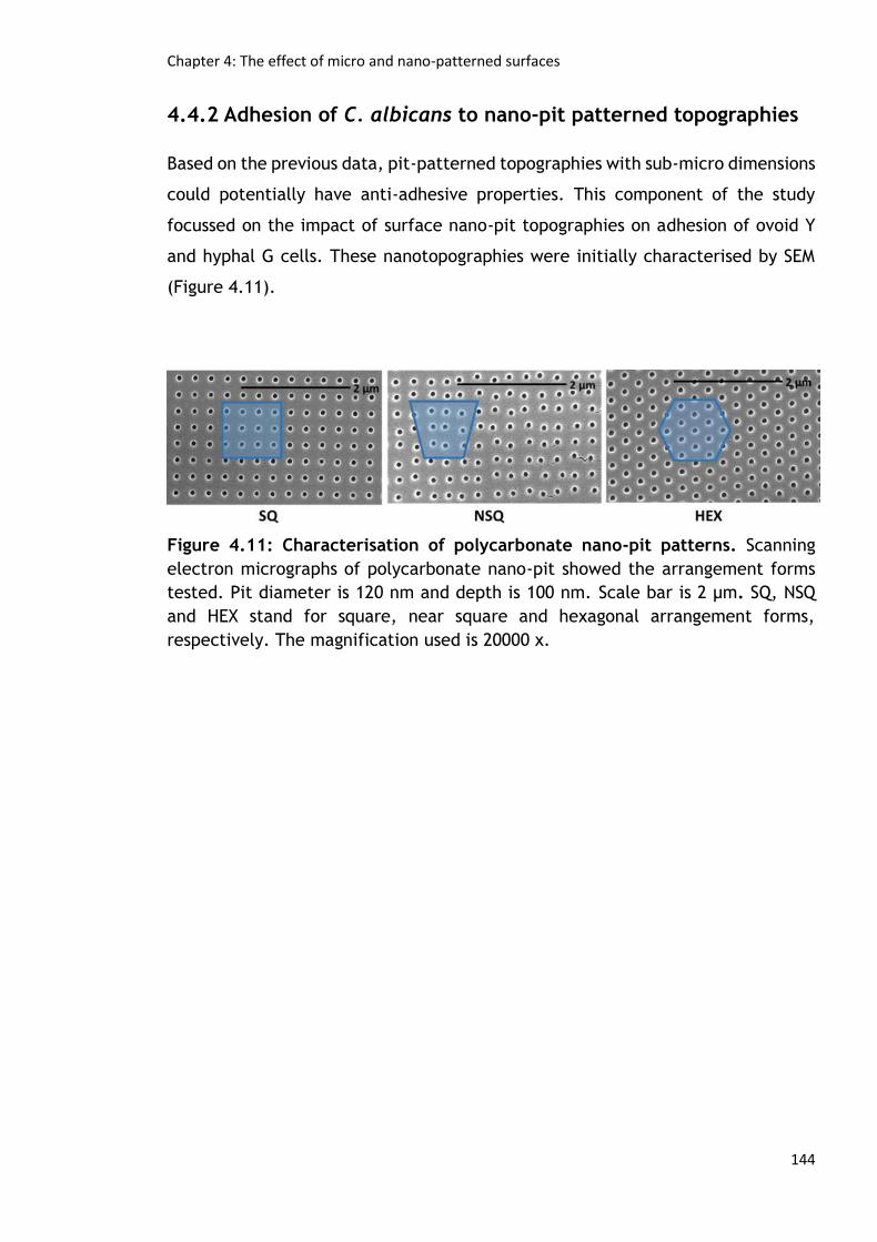
Chapter 4: The effect of micro and nano-patterned surfaces
144
4.4.2 Adhesion of C. albicans to nano-pit patterned topographies
Based on the previous data, pit-patterned topographies with sub-micro dimensions
could potentially have anti-adhesive properties. This component of the study
focussed on the impact of surface nano-pit topographies on adhesion of ovoid Y
and hyphal G cells. These nanotopographies were initially characterised by SEM
(Figure 4.11).
Figure 4.11: Characterisation of polycarbonate nano-pit patterns. Scanning
electron micrographs of polycarbonate nano-pit showed the arrangement forms
tested. Pit diameter is 120 nm and depth is 100 nm. Scale bar is 2 µm. SQ, NSQ
and HEX stand for square, near square and hexagonal arrangement forms,
respectively. The magnification used is 20000 x.

Chapter 4: The effect of micro and nano-patterned surfaces
145
Assessment of these two morphologies adhering to SQ, NSQ and HEX nano-pits on
polycarbonate was quantified using qPCR. The three arrangement forms tested of
the nanotopographies showed different adhesive properties of C. albicans. NSQ
nanotopography showed a significant increase in the adhesion capacity relative to
the other nanotopographies tested, especially in Y morphology form (Figure 4.12
i). However, the highly ordered SQ arrangement showed an contrasting
observation, where it was shown that SQ arrangement significantly reduced
adhesion (relative to flat surfaces) of both Y cells (p=0.038), with ~48% reduction
in adhesion, and G cells (p=0.020), with approximately̴ 64% reduction adhesion as
illustrated in Figure 4.12 i and ii, respectively. Unpaired t test were used for
statistical analysis and any non-parametric data were Log10 transformed prior to
analysis, which were preceded by one-way ANOVA that showed significant
differences (p<0.01). For further analysis, non-parametric data were analysed by
Mann Whitney test.
NSQ and HEX had no significant anti-adhesion properties for either cellular
morphology in comparison to the flat, where NSQ showed the highest adhesion
capability for C. albicans among the surfaces tested. Figure 4.13 shows a
microscopic observation of the adhered yeasts on flat and SQ pattern, where the
cells (and even the substrate surface) seemed covered with cell wall adhesins in
relatively higher abundance in flat topography.

Chapter 4: The effect of micro and nano-patterned surfaces
146
(i)
(ii)
Figure 4.12: Quantification of adhered C. albicans to polycarbonate
nanotopographies. Quantification of adherent colony forming equivalents (CFEs)
of Y (i), where (n=8) and G (ii), where (n=10). The horizontal line represents mean
and error bar represents standard error of mean. Data were analysed by unpaired
t test, * and ** represent (p<0.05) and (p<0.01), respectively.

Chapter 4: The effect of micro and nano-patterned surfaces
147
(i) (ii)
Figure 4.13: Adhered yeasts on SQ nanotopography. SEM micrographs of Y cells
on flat (i) and SQ pattern (ii). Scale bar is 2 µm and magnification used is 6000 x.
In parallel, expression profiles of key adhesion genes (ALS1, ALS3 and EAP1) were
assessed by real-time qPCR and presented as heat-map relative to the
housekeeping gene ACT1 (Figure 4.14). Here, it was shown that all 3 adhesion
genes were down-regulated in the polycarbonate nanotopographies in comparison
to the flat topographies in both Y and G cells, but notably for the SQ arrangement
ALS1 was significantly down-regulated with (p=0.043) and (p=0.036) in Y and G
cells, respectively.

Chapter 4: The effect of micro and nano-patterned surfaces
148
Figure 4.14: Visual transcriptional analysis of adhered C. albicans to
polycarbonate nanotopographies. Heat map transcriptional data of selected
adhesion genes (ALS1, ALS3 and EAP1) of Y and G cells, where gene expression
data are represented in Log2 colour key values. Unpaired t test was used after
natural log transformation of the data. * represent (p<0.05).
Based on these data, nanoimprinted (SQ form arrangement) PMMA denture
material was developed using the dental compression moulding. The validity of
this approach for replication of pits at a nano-scale was investigated by scanning
electron microscopy (Figure 4.15), where the presence of imprinted nanopits were
observed regularly spaced. As with polycarbonate surfaces, we demonstrated a
significant reduction of adherence of both Y (p=0.025) and G (p=0.001) cells on
PMMA with approximately 35 and 55% reduction in adhesion, respectively, using
same abovementioned statistical tests (Figure 4.16 i& ii). Analysis of the adhesion
expression profiles showed no difference between the control and SQ PMMA
surface (Figure 4.17).

Chapter 4: The effect of micro and nano-patterned surfaces
149
Figure 4.15. Characterisation of nanoimprinted PMMA denture material. SEM
micrograph of replicated SQ nano-pit topography (scale bar is 200 nm) and
magnification used was 50000 and 100000 x.
(i) (ii)
Figure 4.16. Quantification of adhered C. albicans to PMMA denture material
nanotopographies. Quantification of adherent colony forming equivalents (CFEs)
of Y (ii) and G (ii). Data obtained from four independent occasions (n=8). The
horizontal line at mean and error bar represents standard error of mean). Data
were analysed by unpaired t test, * and ** represent (p<0.05) and (p<0.01),
respectively.

Chapter 4: The effect of micro and nano-patterned surfaces
150
Figure 4.17: Visual transcriptional analysis of adhered C. albicans to PMMA
denture material nanotopographies. Heat map transcriptional data of selected
adhesion genes (ALS1, ALS3 and EAP1) of Y and G cells, where gene expression
data are represented in Log2 colour key values. Unpaired t test was used after
natural log transformation of the data.
4.4.3 Evaluation of surface roughness and wettability properties
The physical surface properties of the two materials (polycarbonate and PMMA)
tested were explored with regard to surface roughness and wettability. A
statistical difference of high significance (p<0.001) was detected between the
surface roughness average of flat polycarbonate (Ra 4.1 nm) and that of the flat
PMMA denture material (Ra 1549 nm) (Figure 4.18). The surface roughness of the
patterned material was not measured because the depth of the nano-pits was
already identified. On the other hand, when the water contact angle (WCA) was
evaluated for the nanopit SQ replicated imprints a significant difference was
observed in the patterned topographies relative to the flat (p<0.05), and a higher
WCA was observed in the polycarbonate material relative to the PMMA denture
material in both flat and patterned topographies (p<0.01) (Figure4.19).

Chapter 4: The effect of micro and nano-patterned surfaces
151
Figure 4.18: Surface roughness of flat surfaces of materials tested. Surface
roughness average was measured with Contour GT-X 3D microscope and the
micrograph images with scale bar of 50 µm. Data were analysed by unpaired t
test, *** represents p< 0.001.
Figure 4.19: Wettability of the flat and nanopatterned materials tested.
Static WCA of all investigated materials (error bar shows the standard error of
mean), Images acquired at the 30th second by the tensiometer camera. Data
were analysed by unpaired t test, *, ** and *** represent p<0.05, p<0.01 and p<
0.001, respectively.

Chapter 4: The effect of micro and nano-patterned surfaces
152
4.4.4 Biofilm formation onto SQ nano-pit topographies
Biofilm formation on SQ nanotopographies were evaluated for metabolic activity
and biofilm biomass outlooks using XTT and CV assays, respectively. Initial (4 h)
and mature (24 h) C. albicans biofilms were tested. In both assays and in both
biofilms developed, there were no significant differences (Figures 4.20, 4.21),
which clarified the incapability of nanotopographies to prevent C. albicans from
developing an interlocking hyphal recalcitrant net. This may instigate the
necessity for exploration of the effect of CUR combination with nanotopographies
on adhesion and biofilm formation.
Figure 4.20: Evaluation of metabolic activity of C. albicans biofilms on SQ
nanotopographies. Early and mature biofilms were analysed with XTT assay. Data
were statistically analysed using unpaired t test. Error bars represent standard
error of mean.
Figure 4.21: Evaluation of biomass of C. albicans biofilms on SQ
nanotopographies. Early and mature biofilms were analysed with CV assay. Data
were statistically analysed using unpaired t test. Error bars represent standard
error of mean.

Chapter 4: The effect of micro and nano-patterned surfaces
153
4.4.5 Combination of CUR and SQ nanotopographies
A potential synergistic effect of combining adsorption of sub inhibitory
concentration of CUR with polycarbonate SQ nanotopographies on C. albicans
adhesion and biofilm formation was assessed using colony counting method and
biofilm metabolic activity and biomass assessment assays. Figure 4.22 shows a
highly significant effect (p<0.001) of the CUR adsorption on reduction of adhesion
(89-90%) and biofilm formation (73-77% metabolic activity) and (85-86% biomass)
in both flat (F+CUR) and SQ (SQ+CUR) surfaces, respectively. However, a non-
significant (p>0.05) impact was observed for combining the adsorbed CUR with
nanotopographies over the CUR adsorbed flat topographies that might indicate the
absence of synergism between CUR and nanotopographies.
Next, when the impact of nanotopographies on capacity of CUR adsorption was
assessed, a non-significant difference was observed (p>0.05), though adsorption
to nanotopographies (33.1 µg/ml) was slightly lower than that to flat topographies
(36.7 µg/ml) (Figure 4.23).

Chapter 4: The effect of micro and nano-patterned surfaces
154
(i) (ii) (iii)
Figure 4.22: Effect of combining SQ nanotopographies with CUR on C. albicans
colonisation. (i) Reduction in adhesion. Reduction in biofilm metabolic activity
(ii) and biomass (iii). F+CUR and SQ+CUR represent reduction in C. albicans
colonisation of CUR-adsorbed flat relative to CUR-free flat surfaces and CUR-
adsorbed nanotopographies relative to CUR-free nanotopographies, respectively.
Error bars represent standard error of mean and *** asterisks represents p<0.001.
Figure 4.23: Adsorption of CUR to SQ nanotopography. The concentration of
adsorbed CUR (µg/ml) was measured spectrophotometrically. Unpaired t test was
used to analyse the data statistically (p>0.05). Error bars represent standard error
of mean.

Chapter 4: The effect of micro and nano-patterned surfaces
155
4.5 Discussion
Adherence of fungi and consequent biofilm formation is an important clinical
problem (Rajendran et al., 2016a), though these properties can also be exploited
for novel therapeutic targets of fungi. Fungal attachment to biotic and abiotic
surfaces can trigger a physicochemical interaction, and this contact sensing
interaction can be ascribed to fungal-specific mechanisms permitting C. albicans
to respond to different topographies in discriminative ways (Kumamoto, 2008).
One promising approach is to reduce the available contact area to the C. albicans
attachment through engineering of the surfaces. The “attachment point” theory
(Hoipkemeier-Wilson et al., 2004;Kearns et al., 2011) might provide a support for
explanation of the mechanical impact of the topographies tested on the capability
of C. albicans adhesion. This theory assumes a stronger adherence of the
microorganism if it is smaller than the topography feature and vice versa.
Therefore, the aim of this chapter was to allow C. albicans adherence to
topographies with relatively smaller features. Here, it was shown for the first time
the ability to replicate sub-micro/nano-scale features using compression moulding
technique on PMMA denture material to reduce candidal adhesion.
According to this study, for C. albicans Y form, micro-pillar and micro-pit
topographies increased the adhesion capacity, though it was slight for the latter.
A trend of higher adhesion percentages was observed onto the larger features of
the highly ordered pillars only, which could be attributed to the consistent
increase in the dimensions of the pitches (inter-pillar space). These pitches could
offer shelter for the Y form cells.
For G form, the adhesion capacity was slightly decreased. In detail, the micro-
pillar data disagree with studies performed on Staphylococcus epidermidis/aureus
that showed reduction in adhesion with micron and sub-micron pillars (Xu &
Siedlecki, 2012a;Xu & Siedlecki, 2014), which could be attributed to the
difference in the adhesion methodology used or kind of the microorganism, but
particularly, the massive difference in size and mass between bacteria and yeast
(Salerno et al., 2011).

Chapter 4: The effect of micro and nano-patterned surfaces
156
The difference between the adhesion capacity of oval-shaped yeasts on micro-
scale pillar and pit topographies can be explained by understanding these two
topographies using simple geometric and arithmetic analysis. As an analogy, the
inter-pillar field works in a manner similar to an egg box, where the area of the
inter-pillar field is four times larger in terms of surface area compared to pit
topography. Thus, pit featured topography has superior anti-adhesive potential
relative to pillar topography, which was endorsed with the data presented above,
suggesting the importance of spatial arrangement.
Cell morphology also plays an important role, as hyphal-shaped germling cells
adhered to both micro-scale pillar and pit topographies with slightly reduced
capacities. This may be in line with Whitehead & Verran (2006) and Perera-Costa
et al. (2014), where they revealed the importance of shape and even the size of
bacteria on retention on micro and sub-microtopographies. It is noteworthy to
mention that G form has more adhesion capacity than Y form due to over-
expression of some cell wall adhesins that support adherence and biofilm integrity
(Nobile & Mitchell, 2005).
Nevertheless, microtopographies of maximum 2 µm features reduced their
adhesion relative to that on flat indicating the importance of microorganism
dimensions and shape on adhesion to engineered topographies. It was also shown
that smaller feature dimensions and pit form topographies showed lower adhesion
capacities relative to their larger and micro-pillar counterparts, which was the
main driver for exploring smaller dimension materials. The highly order arrays of
nanopits with a SQ form arrangement that were replicated on two different
substrates showed the capacity to significantly reduce the adhesion of the
pathogenic yeast C. albicans in both Y and G morphological forms. The disparity
in the data presented in this chapter and that reported by Whitehead et al (2005),
may simply be because of an order of scale from micro- to nano-topographies
(Whitehead et al., 2005).

Chapter 4: The effect of micro and nano-patterned surfaces
157
An explanation for the anti-adhesive capacity of nanopits may be related to fluid
dynamics, where air entrapment in the pit features may cause the surge in the
hydrophobicity of the nanoimprinted topographies relative to their flat
counterparts (Martines et al., 2005), and given the dynamic lower resistance of
fluid flow on hydrophobic surfaces (low fluid drag) (Rothstein, 2010). Accordingly,
the nanoimprinted topographies might permit for larger removal of adherent C.
albicans due to the lower resistance to fluids flow. This explanation coincides with
the Whitehead & Verran (2006) concept about the role of hydrophobic surfaces
in reduction of bacterial adhesion.
Furthermore, a recent study showed the anti-adhesive capacity to C. albicans of
PMMA denture material coated with trimethylsilane hydrophobic film that
increased the surface energy (Liu et al., 2017). Moreover, the anti-adhesive
capacity of nano-pit surfaces was not influenced by the wettability differences of
the polycarbonate and PMMA denture material surfaces tested, where both
nanoimprinted polymer types showed an anti-adhesive capability. This is in
agreement with Perera-Costa et al. (2014) study that showed an anti-adhesive
capacity of spatially organised micro engineered surfaces relative to flat surfaces
on bacterial adhesion regardless of the surfaces hydrophilicity/ hydrophobicity of
the microtopographies. Therefore, the trapped air in the nano-pits increased the
hydrophobicity of the surface and it is one of the probable mechanisms for the
antiadhesive capacity. While, we can suggest that the primary antiadhesive
mechanism of the nano-pits is the physical mechanism where less surface area
available for the adhesins to attach.
When the data of gene expression were analysed, interesting observations have
confirmed the substrate differential impact on the expression of key adhesins.
This may propose that the biology-related physical characteristics of the SQ nano-
pit topographies were sufficient to challenge bio-molecular interactions between
C. albicans morphological types, their adhesins, and the topography. The absence
of total resemblance observed in gene expression profiles between both substrates
tested might be clarified by the fact that Y and G cells are hydrophilic and
hydrophobic, respectively (Rodrigues et al., 1999), and from our data that PMMA
and polycarbonate are hydrophilic and hydrophobic relative to each other,

Chapter 4: The effect of micro and nano-patterned surfaces
158
respectively. Therefore, in a selective manner, induction of stress and differential
induction of adhesins might be attributed to these physicochemical interactions
(Fox et al., 2015). Indeed, Krasowska & Sigler (2014) emphasised that
hydrophobic microorganisms favourably adhere to hydrophobic substrates and the
hydrophilic microorganisms adhere to hydrophilic substrates.
We could not explain the differences among the different nano-pit spatial
arrangements tested using the gene expression data, though the highly ordered
SQ and HEX arrangements showed relatively similar results. Nevertheless, several
studies showed the potential inherent anti-adhesive capacity of nano-pit
substrates to in-vitro mammalian cells adhesion formation (Martines et al.,
2004;Biggs et al., 2007;Biggs et al., 2009).
The SQ nanotopography was tested against harsh biofilm environments
characterised with a dense interlocking network of candidal hyphae. These
nanotopographies did not show significant anti-biofilm capability, that may be
caused by the entangling filamental nature of the biofilms tested and the static
environments that permit the candidal cells to adhere, settle, and form a cohesive
community for a sufficient time prior to submitting to dislodging challenges. This
stimulated investigation of the dual effect of combination the adsorbed CUR
molecules with nano-surfaces. Additionally, given the proposed relative increase
in the available surface area within nanotopographies for CUR adsorption that may
suggest a synergistic effect. However, a nanotopography-CUR adsorption
synergistic effect was not observed. The absence of a topography effect could be
attributed to the larger effect of CUR that may preclude quantification of the
topography effect. Besides, the adsorbed molecules may reduced the topography
anti-adhesive influence. Furthermore, the nanotopographies did not adsorbed
larger quantities of CUR as expected, which might be ascribed to the molecule
size of the CUR used.
Non-patterned PMMA denture material showed high Ra, while the Ra of the
nanopatterned PMMA was not measured because of the specific nanofabrication
technique used in this study and the known depth of the nano-pits, nevertheless,
it can be suggested to measure the Ra of nano-pit topographies in further studies.

Chapter 4: The effect of micro and nano-patterned surfaces
159
Translationally, fabricating topography with SQ nanoimprinted pits onto PMMA
denture material has its advantages over unpatterned surfaces, whose associated
large surface roughness becomes negligible when nanoimprinted, taking into
consideration the fabrication method of the fitting surface of the denture that is
characterised by a non-polished surface. Instead, crucial biophysical
characteristics are induced through the SQ pit nanotopography, where adhesion
of C. albicans is significantly decreased. CUR adsorption has a substantial effect
on candidal colonisation as shown in the last chapter. Indeed, the adsorption of
CUR onto nanotopographies may add an advantage, especially if nanonisation
technologies is considered, where nano-scale CUR molecules are harnessed.
Regarding denture production, this has a potential value where the denture
stomatitis affecting millions of denture wearing individuals globally. Overcoming
the obstacles to manufacturer these will prove challenging, yet the potential
impact to oral health is enormous.
Chapter findings
PMMA denture material can replicate sub-micro and nano features.
Adhesion of C. albicans to micro and sub-micro featured topographies
depends on the candidal morphological form and the shape of the features.
C. albicans variably respond to the spatial arrangement of nano-pit
topographies, where SQ arrangement showed an anti-adhesive capacity for
both candidal morphological forms tested.
Hyphal candidal biofilms were not affected by the SQ nanotopography.
The Molecular response to nanotopographies is complex and can be
affected by the hydrophobicity/hydrophilicity and surfaces roughness of
surfaces tested.

Chapter 4: The effect of micro and nano-patterned surfaces
160
Nano-pit surfaces showed higher water contact angle. Flat PMMA tested
showed a rougher surface and lower water contact angle than their
polycarbonate counterpart.
Combination of natural antimicrobials with surface topography through
adsorption of CUR to SQ nanotopographies did not significantly enhance the
anti-adhesive and anti-biofilm capability. Similarly, SQ nanotopographies
did not improve the CUR adsorption capacity to the surfaces.

161
5 Final discussion

Chapter 5: Final discussion
162
5.1 Introduction
Removable dentures are beneficial prosthetic devices, which improve dental
aesthetics and function although they can be detrimental and associated with oral
and systemic diseases. PMMA denture materials have many beneficial features,
such as good mechanical and physical properties and a relatively low production
cost yet they are highly susceptible to accumulation of microbial biofilms
(Sivakumar et al., 2014).
Denture stomatitis is a multifactorial disease that affects more than half of all
denture wearers who form a considerable percentage of the population due to the
fact that one-fifth (20%) of the UK population have removable dentures (Hannah
et al., 2017) and given increases in life expectancy, this percentage is likely to
rise. Indeed, the World Health Organisation predict that the percentage of people
over 60 years old will approximately double from 11.7 in 2013 to 21.1% in 2050
(UN, 2013). Control of prosthesis associated biofilms is an essential strategy in the
management of DS (Yarborough et al., 2016). Candida species, and especially C.
albicans, has been identified as a pivotal microbial contributing factor in DS
(Gendreau & Loewy, 2011). Candidal denture carriage in health and disease has
previously been investigated using traditional microbiological methods (Barbeau
et al., 2003;Coco et al., 2008b;Dagistan et al., 2009;Salerno et al., 2011). In this
study, a molecular-based qPCR was employed to obtain data and quantify
adherent cells. Furthermore, given the heterogeneity (strain variability) of C.
albicans biofilm formation, it was uncertain whether this property is associated
with DS (Kean et al., 2018).
Reports have shown that dentures are a potential reservoir for pathogenic
respiratory bacteria that cause pneumonia and are associated with a high
mortality rate (Przybylowska et al., 2014;O'Donnell et al., 2016). In polymicrobial
environments, such as the oral cavity, C. albicans acts as a scaffold and via cell-
cell communication can modify pathogenicity of either cell (Kean et al., 2017).
Therefore, discovering innovative strategies to control adhesion and biofilm
formation of C. albicans is a worthwhile goal.

Chapter 5: Final discussion
163
The development of novel therapeutic strategies that could augment the current
strategies used in prevention and treating DS could improve the clinical
management of denture-associated diseases.
5.2 Candidal denture biofilm and DS: Impacts of quantity and heterogeneity
Many studies have reported the association between Candida and DS, though,
often denture carriage importance was not the specific focus. This study for the
first time according to our knowledge reports the use of a molecular quantitative
method (Real-time qPCR) to quantify the denture adherent candida cells, though
previous studies used this molecular method to quantify bacteria in dental-related
research (Blome et al., 2008;Kim et al., 2013;O'Donnell et al., 2016). Here we
confirm the significance of the candidal load on dentures in DS, though our data
showed several dentures of healthy and diseased patients shared approximately
similar candidal carriage quantities. These data, alongside the recently reported
concepts of C. albicans biofilm formation heterogeneity (strain variability)
associated with increasing resistance, pathogenicity and mortality (Sherry et al.,
2014;Rajendran et al., 2016a), have driven this research to investigate the
potential association between biofilm formation heterogeneity and DS.
The data described in this thesis did not demonstrate an active role for C. albicans
biofilm forming heterogeneity in DS, which could be attributed to the relatively
mild nature of DS in comparison to the systemic candedemia, which is often
associated with immunocompromised patients, rather than the immunocompetent
patient cohort examined in this study. Figure 5.1 summarises the obtained data
regarding the candidal denture load, DS and biofilm formation heterogeneity.

Chapter 5: Final discussion
164
Figure 5.1: Schematic representation of the relationship of candidal denture load, denture stomatitis and C. albicans heterogeneity. Higher candidal load is associated with denture stomatitis. However, C. albicans biofilm formation heterogeneity does not represent a key determinant, where both HBF and LBF are observed similarly in isolates from DS and healthy participants. HBF and LBF represent high and low biofilm formers.
Challenges in the development of a globally standardised in vitro candidal biofilm
model are numerous due to the adaptability of C. albicans to various
environmental conditions (Soll & Daniels, 2016), therefore experiments to
optimise and characterise biofilms were first undertaken in this thesis. This
research mainly concentrated on the use of RPMI-1640 as a well-established
medium to test the heterogeneity of biofilm formation and to develop recalcitrant
biofilms. RPMI-1640 is capable of inducing different candidal morphological forms
and subsequently it will offer the capability to investigate adhesion capacities of
these various forms. However, this research also demonstrates that artificial
saliva has the possibility of being used to recreate clinically relevant conditions in
future studies.
Given that candidal carriage was found to be significant in DS, the question
remained whether denture material has an impact on the early and mature stages
of biofilm formation? There is little reliable evidence to show whether the denture
material has an impact on early and mature biofilm formation (Pereira-Cenci et
al., 2008b). Therefore, a qPCR based methodology was used alongside
representative clinical isolates with defined biofilm formation heterogenic
capabilities. For all isolates, PMMA showed a greater capacity than soft liners
tested to induce the formation of mature biofilms, but not early stage biofilms,

Chapter 5: Final discussion
165
suggesting that the adhesion stage of biofilm formation is not influenced by
denture material, though only limited number and type of denture base materials
were tested. For this reason, studying the adhesion of C. albicans using
representative denture related strains may provide a more clinically relevant
approach to studying the capability of C. albicans to colonise denture surfaces.
5.3 Why CUR could be a promising polyphenol for denture wearers?
As of 2015, over 125 clinical trials were registered with the US National Institutes
of Health investigating CUR biological activity (Ahmad et al., 2017), which
indicates the intriguing nature of this molecule in modern research. Cancer,
Alzheimer and cardiovascular malfunction are destructive age-related diseases
(Bennett & Leurgans, 2010). Given its potential anti-cancerous, anti-Alzheimer
and beneficial cardiovascular-related CUR is a rational target for geriatric-related
research (Hamaguchi et al., 2010;Marchiani et al., 2014;Hu et al., 2015;Venigalla
et al., 2016;Yao et al., 2016;Sun et al., 2017;Larasati et al., 2018;Reddy et al.,
2018). Indeed, most denture wearers are classified as being within the older
population, thus introducing CUR to oral healthcare and denture-related research
seems interesting on multiple levels.
Given the reported broad-spectrum antimicrobial effect of CUR (Moghadamtousi
et al., 2014;Vaughn et al., 2017), one focus of this thesis was to investigate the
potential interaction between CUR and one of the most common opportunistic
microorganisms related to denture-associated diseases, i.e. C. albicans. The
overarching hypothesis was that CUR could be used in a clinical context to manage
and minimise the attachment of the organism and impact on downstream denture
plaque development. The studies presented in this thesis showed that CUR
effectively inhibited C. albicans in both modes of growth (planktonic and biofilm)
in a concentration dependent manner. Nonetheless, biofilms showed signs of
surviving even in the presence of high CUR concentrations, suggesting that CUR
could be better employed in prevention rather than treatment of established
denture biofilms.

Chapter 5: Final discussion
166
Data presented in this thesis showed the affinity of CUR to adsorb to denture
material polymers, and these adsorbed sub-inhibitory concentrations showed an
anti-adherent impact on C. albicans. This impact was augmented by CUR pre-
exposure to create a significant synergistic-like effect (Figure 5.2i). Therefore,
using CUR in denture care soaks or direct oral consumption of CUR either as a
dietary supplement, or through normal daily ingestion, may allow CUR to adsorb
on to the denture material and exert its biological effect. The affinity of CUR to
PMMA denture material could be driven by the hydrophobic interaction theory,
where the hydrophobic substance is attracted to the hydrophobic surface of the
denture in water-based biological environments (Israelachvili & Pashley, 1984).
Furthermore, given the bonding affinity between salivary proteins/peptides and
polyphenols, specifically proteins rich in the amino acid proline (Charlton et al.,
2002), the opportunity of interaction between the denture material and adsorbed
CUR could be enhanced in natural (oral) environments due to deposition of the
bonded complexes upon the denture material.
The dimorphic capacity of C. albicans is an important virulence factor expressed
by this opportunistic microorganism and interrogating CUR capacity against the
different forms appeared intriguing. Data presented in this thesis showed a
greater effect on the immature form (yeast) in comparison to the mature form
(hyphae). Mechanistically, the biological effects of CUR are many and complex
(Zhou et al., 2011). Amongst others, CUR acts as an iron chelating agent and this
suggests that CUR has the potential to act as a divalent cation chelator such as
ethylene diamine tetraacetic acid (EDTA) (Jiao et al., 2006). The inhibitory effect
of EDTA on a C. albicans biofilms was demonstrated through inhibition of
filamentation and depletion of metal ions that are important to candidal biofilms
integrity (Ramage et al., 2007).
CUR has the capacity to modulate various molecular targets through alteration of
gene expression and signalling pathways or by direct interaction with biological
molecules (Shishodia, 2013). Indeed, CUR modulated some of the key adhesion
and aggregation-associated genes in both yeast and hyphae morphological forms
of C. albicans. The ALS3 gene, which is involved in adhesion, dimorphism, invasion
(Liu & Filler, 2011) and aggregation with bacteria (Peters et al., 2012;Bamford et
Chapter 5: Final discussion
167
al., 2015;O'Donnell et al., 2015a), was down-regulated in the presence of CUR.
This suggests a possible vital role for CUR in interfering with biofilm formation and
therefore pathogenicity crucial for in vivo polymicrobial biofilm formation.
However, ALS5 and AAF1 are aggregation associated genes that showed a
considerable up-regulation. This coincides with the observed quantified
aggregation that was elicited in response to CUR. ALS1 gene expression was also
affected, but the data was found to be inconclusive and varied depending on
length of exposure to CUR. These data indicate the high adaptability of C. albicans
to external modulating molecules. Previously, ALS5 and ALS1 have been shown to
play a role in aggregation of C. albicans cells to themselves and to other bacteria
(Klotz et al., 2007). Therefore, the data here in could suggest a compensatory
function to the down-regulated powerful ALS3. Unexpectedly, Eap1, which has an
adhesin function and is regulated by hyphal inducing EFG1 gene (Li & Palecek,
2003) showed a considerable up-regulation suggesting a noticeable cell-cell
aggregation role (Li & Palecek, 2008) or potential compensatory regulative role
due to the down-regulation of important hyphal inducing genes.
CUR molecules that coat the exposed cells may act as glue that keep the cells
aggregated in a direct way and this direct physical aggregation could drive gene
expression targets to elicit an indirect biological aggregation. Overall, CUR could
contribute to cell-cell aggregation and less to cell-substrate adhesion, but this
requires further studies to confirm (Figure 5.2).
CUR has been reported to have anti-amyloid effects through producing non-
effective amyloid aggregates with less density and toxicity. Mechanistically, CUR
can bind to the amyloid beta monomers or penetrate the amyloid beta
polymerised sheets leading to weaker amyloid aggregates (Gupta et al., 2011;Rao
et al., 2015;Reddy et al., 2018). Given the amyloid-like clustering of the ALS
adhesins on the cell wall (Lipke et al., 2012) occurrence of these potential
physico-biological events in the interaction between C. albicans and CUR is
possible. Moreover, the amyloidogenic domain sequence that exists in several
members of the ALS gene family (Garcia et al., 2011) suggests the possibility of
formation of weak aggregates due to the potential conformational changes in the
expressed proteins. These weak cellular aggregates reduce the capability of C.

Chapter 5: Final discussion
168
albicans to withstand shear stresses. This notion could explain why the cellular
aggregates did not outweigh the anti-adherence capacity. Additionally, the
relative hydrophobicity of CUR could prevent formation of the amyloid-like ALS
clusters on the cell wall by acting as a macromolecular crowder (Latshaw & Hall,
2015). The inability to form these macromolecular adhesin clusters could reduce
cell wall hydrophobicity, which indeed, was observed in the data shown in this
thesis.
Figure 5.2: Impact of CUR on C. albicans. (i) Adsorption of CUR to PMMA reduces the number of adhered cells, elicits aggregation, reduces filamentation and induces a synergistic effect from the pre-treatment of the cells with sub-inhibitory concentrations. (ii) The expressed molecular response to CUR treatment.
Modification of denture acrylics with active and antimicrobial macromolecules is
an important field for research but it is essential that researchers are mindful of
the risks of unwanted sequelae (Sivakumar et al., 2014). In spite of the expected
colour alteration and the potential weakening effect on the mechanical properties
of PMMA, it was interesting to investigate the opportunity of modifying the PMMA
denture material with a safe and active molecule through creating simple CUR-
incorporated PMMA to determine the subsequent effect on C. albicans adhesion.

Chapter 5: Final discussion
169
The data showed no effect on the adhesion capacity. Investigation of CUR release
from the polymerised PMMA in the appropriate solvent showed lack of detectable
CUR release, suggest that there is a strong chemical bonding between CUR and
PMMA. Nonetheless, using colourless curcuminoid analogues or derivatives and
using conjugate chemistry needs further investigation (Tang et al., 2010;Requejo-
Aguilar et al., 2016). In the context of the reported safety and multiple beneficial
effects of CUR, designing clinical trials for DS-related research that include CUR
as main component of a mouth wash, food supplement or denture soak are rational
suggestions.
5.4 Micro and nanopatterned surfaces: the quest for antifouling denture surface
In the development of a new generation of biomedical materials, researchers must
give consideration to the topography of surface as vital interactions take place
with the biological environment at that interface (Li et al., 2014). Biomedical
material surfaces with a micro- and nano- fabricated surface have shown promise
in a range of beneficial areas (Ploux et al., 2009;Kearns et al., 2011), such as
antimicrobial properties (Xu & Siedlecki, 2012b) and the capacity to control stem
cell adhesion and fate, which contributes to the evolution of the next generation
of regenerative therapy (Dalby et al., 2014). This significant impact on cellular
behaviour has boosted the potential of fabrication of denture material surfaces to
control the microbial fouling process. Micro- and nano-patterned denture surfaces
could interfere with colonisation of C. albicans on dentures.
To this end, several micro- and nano-patterned surfaces were investigated in a
rational manner, where the focus was on the first crucial stage of microbial biofilm
formation ‘adhesion’. The main morphological forms of C. albicans within the
adhesion stage (yeast and germling) were tested because these forms have
different shapes (ovoid and hyphal-like) and dimensions that could influence the
interaction between cells and surface topography. In addition to the already
successfully used injection moulding technique to replicate micro- and nano-scale
features in polycarbonates (Reynolds et al., 2012), data presented in this thesis
showed the capacity of dental compression moulding to replicate micro, sub-micro

Chapter 5: Final discussion
170
and nano-scale features in a heat cured PMMA denture. These data have paved
the way to interrogate the adherence of C. albicans to these replicated
topographies.
Review of the literature showed the potential antimicrobial properties of pillar
shaped micro, sub-micro and nano-features (Ma et al., 2011;Ivanova et al.,
2012;Xu & Siedlecki, 2012b;Xu & Siedlecki, 2014). Therefore, it was interesting to
investigate adhesion to micro and sub-micro pillar topographies. In contrast to the
literature, the data showed a considerable increase in the adhesion capability of
the yeast morphological form to the micron and sub-micron pillars. This may be
explained by differences in feature dimensions, material types, microorganisms
and methodologies used. Nevertheless, germling forms of C. albicans showed a
reduced adhesion capability. The form and dimensions of the adhered cells could
be a potential explanation for this difference.
The data described above indirectly suggested that using the opposite of pillar
topography, the ‘pit form’, may have more beneficial effects. Indeed, pit form
microtopography displayed an anti-adhesive capability indicating an influential
impact for this geometric form of patterned topography on adhesion of C.
albicans. In light of the potential relative anti-adherent nature of pit
microtopographies to C. albicans, nano-pit topographies in various arrangements
were investigated, where the highly ordered arrangement forms, especially the
SQ form, manifested anti-adhesive capability to both yeast and germling
morphological forms in both denture materials (polycarbonate and PMMA denture
material).
The gene expression profile of adherent cells on the nano-polycarbonate surfaces
showed a down-regulation in the genes investigated, though such down-regulation
was not observed in PMMA suggesting a greater role for physical mechanism in
candidal adhesion. Investigation of the surface properties of the materials tested
helped to understand the molecular response of the C. albicans and the
differences observed between the materials, where relative
hydrophobicity/hydrophilicity of the material and the adherent cells may elicit
different stresses and interactions (Figure 5.3). PMMA denture material, which

Chapter 5: Final discussion
171
was fabricated in a way that is relevant to prosthodontic clinical practice, showed
high roughness average. Therefore, imprinting of the denture surface material
with 100 nm depth pits appeared to be relatively negligible to the surface
roughness effect, and theoretically produces PMMA denture surface with lower
surface roughness that reduces the adhesion potential.
These data force us to consider the clinical feasibility of producing dentures with
nanoimprinted features. Production of nanoimprinted polymer extra-thin sheets
and the possibility of using them as a denture surface lining could be a promising
suggestion. Indeed, production of nanoimprinted polymer sheets is interesting and
reveals significant implications (Gadegaard et al., 2006a). Another potential
suggested idea is harnessing of direct nanoimprinting, where it was proven as a
successful method to generate nanoimprinted topographies, even using much
harder metal surfaces whether planar or curved (Greer et al., 2013).

Chapter 5: Final discussion
172
(i)
(ii)
Figure 5.3: Schematic representation of adhesion of yeast and hyphae cells on relatively hydrophobic and hydrophilic surfaces. A simplification of our notion of the interactive adhesion between the surfaces and C. albicans morphological forms, and how this could shape the expression of adhesins in flat (i) and nanoimprinted surfaces (ii). Yellow circles represent adhesins.

Chapter 5: Final discussion
173
5.5 Combining nanotopography with adsorbing CUR
Given the anti-adhesive capability that was separately observed against C.
albicans from CUR adsorbed and nanoimprinted surfaces, it was intriguing to
combine these different anti-adhesive strategies in an endeavour to obtain a
synergistic-like effect. Indeed, this idea has been inspired by nature, where anti-
fouling surfaces manifest both chemical and physical properties (Magin et al.,
2010). For instance, shark skin and lotus leaves harness the combination of surface
topography and surface chemicals to synergise their anti-fouling effect (Sun et
al., 2005;Ma et al., 2011;Bixler & Bhushan, 2012). A research group from China
reported the potential biological diverse benefits of combining patterned
topographies with chemically modified polymers and the possibility of obtaining
optimal results (Li et al., 2014). Therefore, CUR was adsorbed onto SQ nano-pit
topographies, and adhesion and biofilm formation was investigated. Anti-adhesive
or anti-biofilm synergism was not observed, and the CUR effect may control the
combination data. The nanotopographies showed a trend of reduced adsorption of
CUR, which may explain this observation. Furthermore, the concealed role of the
nanotopography might be attributed to the size and accumulation of adsorbed
CUR molecules that may reduce the topography effect suggesting using nano-sized
CUR particles as a promising strategy.
5.6 Future work
The structure of this thesis was focused on the interaction between DS and
candidal biofilms on denture material, besides interrogation of novel strategies to
control these tenacious biofilms. The studies presented herein potentially show
that reducing denture candidal carriage through nature-inspired concepts of
combining physical or chemical approaches could reduce the likelihood of patients
developing DS. Accordingly, further investigation and development of these
strategies will increase their efficacy to reduce microbial biofilms and this will
form the basis of future work. It would be interesting to investigate the nano-pits
of SQ arrangement of different dimensions such as smaller diameter or smaller
pitches (pit centre to pit centre). Another suggestion is to design and investigate
SQ nano-pits of extreme hydrophilic and hydrophobic nature.

Chapter 5: Final discussion
174
Taking the research into different dimensions could reveal a promising field of
research, such as challenging the strategies used in this research with relevant
bacterial biofilms (Streptococcus mutans, Staphylococcus aureus) and more
intricate polymicrobial biofilms to investigate the potential possibility of CUR
molecules and highly ordered nano-pit topographies to control diverse biofilms.
Indeed, Ramage’s lab has a previous experience with similar diverse Harnessing
polymicrobial biofilms (Sherry et al., 2013;Millhouse et al., 2014;Sherry et al.,
2016) and this lab showed the potential antibacterial efficacy of CUR against
periodontal pathogens (Shahzad et al., 2015). Furthermore, several reports
showed the potential antibacterial capacity of submicron and nanopatterned
topographies against polymicrobial biofilms (Ploux et al., 2009;Xu & Siedlecki,
2012b). Thus, investigation of polymicrobial biofilms is a worthwhile future
exercise.
CUR sensitivity to light can be utilised to increase its efficacy (photodynamic
therapy) and to reduce its staining effect. Given the safety of CUR, there is an
opportunity to design a clinical trial to investigate the preventive effect of CUR
on the adhesion of C. albican and the possible incidence of DS in healthy denture
wearers using various ways of delivery to the oral cavity.
The use of nano-sized modified CUR, conjugated CUR, CUR modified through
liposomes or polymeric micelles could amplify the biological efficacy and enhance
the low oral bioavailability that are observed in clinical trials using free non-
modified CUR. Indeed, a recent study showed the effectivity of functionalising the
fitting surface of the PMMA denture with polycaprolactone microspheres
containing Amphotericin-B for anti-candidal therapy using a 3D printing (fused
filament fabrication) technique (Nagrath et al., 2018). In addition, using CUR
analogues or derivatives characterised with colourless or neutral colours and
investigating their biological activity on adhesion and biofilm formation of C.
albicans on denture materials is also worthy of future investigation. If this strategy
reveals promising results there will be a chance to integrate these analogues or
derivatives in the denture materials through incorporation of conjugates or
coating strategies, where the resultant denture material could be nanoimprinted
to maximise the beneficial anti-biofouling impact.

Chapter 5: Final discussion
175
Finally, in light of increasingly emerging multi drug-resistant candida pathogens,
even against the potent echinochandins (Arendrup & Patterson, 2017;Xiao et al.,
2018) the search for new preventative strategies takes on even greater
importance. Failure to prevent unnecessary use of antifungals and reduce
investments in searching for new antifungals are critical issues that enhance the
growth of resistant pathogens. Accordingly, focusing on development of
preventive strategies is important. Using host-derived molecules for instance,
acetylcholine (Rajendran et al., 2015) alongside micronutrients to control these
complex biofilms as an alternative to traditional antimicrobials appears to be a
potential strategy.
Thesis findings
The multifactorial nature of DS cannot conceal the impact of denture
candidal carriage that shows superior significance relative to the candidal
heterogeneity, thus reducing that carriage is a worthwhile target.
Nature inspired strategies of combining safe, natural, chemical molecules
with nanofabrication physical approaches to control microbial biofilms are
intriguing and worth further interrogative work. Using CUR molecules with
sub-inhibitory concentration and SQ arrangement form of nano-pit
topographies reduced adhesion of C. albicans to PMMA denture material.
However, adsorbing CUR to the nano-pit topographies did not show
significant synergistic-like effect.
The obtained data from the strategies used showed antifouling capability
against C. albicans biofilms and encourage investigation against more
complicated polymicrobial biofilms.

176
References
ABDELNABI, M. H. & SWELEM, A. A. 2015. Influence of defective complete dentures renewal on TMD; an MRI and clinical controlled prospective study. Gerodontology 32: 211-221.
AGGARWAL, B. B., DEB, L. & PRASAD, S. 2014. Curcumin differs from tetrahydrocurcumin for molecular targets, signaling pathways and cellular responses. Molecules 20: 185-205.
AHMAD, B., BORANA, M. S. & CHAUDHARY, A. P. 2017. Understanding curcumin-induced modulation of protein aggregation. Int J Biol Macromol 100: 89-96.
AHRENS, W., POHLABELN, H., FORAITA, R., NELIS, M., LAGIOU, P., LAGIOU, A., BOUCHARDY, C., SLAMOVA, A., SCHEJBALOVA, M., MERLETTI, F., RICHIARDI, L., et al. 2014. Oral health, dental care and mouthwash associated with upper aerodigestive tract cancer risk in Europe: the ARCAGE study. Oral Oncol 50: 616-625.
ALBUQUERQUE, P. & CASADEVALL, A. 2012. Quorum sensing in fungi--a review. Med Mycol 50: 337-345.
ALNUAIMI, A. D., WIESENFELD, D., O'BRIEN-SIMPSON, N. M., REYNOLDS, E. C. & MCCULLOUGH, M. J. 2015. Oral Candida colonization in oral cancer patients and its relationship with traditional risk factors of oral cancer: a matched case-control study. Oral Oncol 51: 139-145.
ALTARAWNEH, S., BENCHARIT, S., MENDOZA, L., CURRAN, A., BARROW, D., BARROS, S., PREISSER, J., LOEWY, Z. G., GENDREAU, L. & OFFENBACHER, S. 2013. Clinical and histological findings of denture stomatitis as related to intraoral colonization patterns of Candida albicans, salivary flow, and dry mouth. Journal of Prosthodontics : Official Journal of the American College of Prosthodontists 22: 13-22.
AMANLOU, M., BEITOLLAHI, J. M., ABDOLLAHZADEH, S. & TOHIDAST-EKRAD, Z. 2006. Miconazole gel compared with Zataria multiflora Boiss. gel in the treatment of denture stomatitis. Phytotherapy Research : PTR 20: 966-969.
ANSELME, K., DAVIDSON, P., POPA, A. M., GIAZZON, M., LILEY, M. & PLOUX, L. 2010. The interaction of cells and bacteria with surfaces structured at the nanometre scale. Acta Biomater 6: 3824-3846.
APISARIYAKUL , A., VANITTANAKOM , N. & BUDDHASUKH, D. 1995. Antifungal activity of turmeric oil extracted from Curcuma longa (Zingiberaceae). Journal of Ethnopharmacology 49: 163-169.
ARENDORF, T. & WALKER, D. 1987. Denture stomatitis: a review. J Oral Rehabil 14: 217-227.
ARENDORF, T. M. & WALKER, D. M. 1980. The prevalence and intra-oral distribution of Candida albicans in man. Archives of Oral Biology 25: 1-10.
ARENDRUP, M. C. & PATTERSON, T. F. 2017. Multidrug-Resistant Candida: Epidemiology, Molecular Mechanisms, and Treatment. The Journal of Infectious Diseases 216: S445-s451.
ARZMI, M. H., ALNUAIMI, A. D., DASHPER, S., CIRILLO, N., REYNOLDS, E. C. & MCCULLOUGH, M. 2016. Polymicrobial biofilm formation by Candida albicans, Actinomyces naeslundii, and Streptococcus mutans is Candida albicans strain and medium dependent. Med Mycol 54: 856-864.

177
AYDIN, A. K. & TEKKAYA, A. E. 1992. Stresses induced by different loadings around weak abutments. J Prosthet Dent 68: 879-884.
AZEREDO, J., AZEVEDO, N. F., BRIANDET, R., CERCA, N., COENYE, T., COSTA, A. R., DESVAUX, M., DI BONAVENTURA, G., HEBRAUD, M., JAGLIC, Z., KACANIOVA, M., et al. 2017. Critical review on biofilm methods. Crit Rev Microbiol 43: 313-351.
BAKHSHI, M., TAHERI, J. B., SHABESTARI, S. B., TANIK, A. & PAHLEVAN, R. 2012. Comparison of therapeutic effect of aqueous extract of garlic and nystatin mouthwash in denture stomatitis. Gerodontology 29: e680-684.
BAL, B., YAVUZYILMAZ , H. & YÜCEL, M. 2008. A pilot study to evaluate the adhesion of oral microorganisms to temporary soft lining materials. Journal of Oral Science 50: 1-8.
BAMFORD, C. V., NOBBS, A. H., BARBOUR, M. E., LAMONT, R. J. & JENKINSON, H. F. 2015. Functional regions of Candida albicans hyphal cell wall protein Als3 that determine interaction with the oral bacterium Streptococcus gordonii. Microbiology 161: 18-29.
BANTING, D. W. & HILL, S. A. 2001. Microwave disinfection of dentures for the treatment of oral candidiasis. Special Care in Dentistry : official publication of the American Association of Hospital Dentists, the Academy of Dentistry for the Handicapped, and the American Society for Geriatric Dentistry 21: 4-8.
BARAN, I. & NALCACI, R. 2009. Self-reported denture hygiene habits and oral tissue conditions of complete denture wearers. Arch Gerontol Geriatr 49: 237-241.
BARAO, V. A., OGAWA, E. S., MORENO, A., MESQUITA, M. F., WEE, A. G. & ASSUNCAO, W. G. 2015. Long-term clinical evaluation of the color stability and stainability of acrylic resin denture teeth. J Prosthet Dent 113: 628-635.
BARBEAU, J., SEGUIN, J., GOULET, J. P., DE KONINCK, L., AVON, S. L., LALONDE, B., ROMPRE, P. & DESLAURIERS, N. 2003. Reassessing the presence of Candida albicans in denture-related stomatitis. Oral surgery, Oral medicine, Oral pathology, Oral radiology, and Endodontics 95: 51-59.
BAZAKA, K., JACOB, M. V., CRAWFORD, R. J. & IVANOVA, E. P. 2011. Plasma-assisted surface modification of organic biopolymers to prevent bacterial attachment. Acta Biomater 7: 2015-2028.
BEDOS, C., BRODEUR, J. M., BOUCHERON, L., RICHARD, L., BENIGERI, M., OLIVIER, M. & HADDAD, S. 2003. The dental care pathway of welfare recipients in Quebec. Social Science & Medicine 57: 2089-2099.
BENNETT, D. A. & LEURGANS, S. 2010. Is there a link between cancer and Alzheimer disease? Neurology 74: 100-101.
BENNICK, A. 2002. Interaction of plant polyphenols with salivary proteins. Critical Reviews in Oral Biology and Medicine : an official publication of the American Association of Oral Biologists 13: 184-196.
BENSO, B., KOVALIK, A. C., JORGE, J. H. & CAMPANHA, N. H. 2013. Failures in the rehabilitation treatment with removable partial dentures. Acta Odontol Scand 71: 1351-1355.
BERTOLINI, M. M., PORTELA, M. B., CURVELO, J. A., SOARES, R. M., LOURENCO, E. J. & TELLES, D. M. 2014. Resins-based denture soft lining materials modified by chlorhexidine salt incorporation: an in vitro analysis of antifungal activity, drug release and hardness. Dent Mater 30: 793-798.
BHADRA, C. M., TRUONG, V. K., PHAM, V. T., AL KOBAISI, M., SENIUTINAS, G., WANG, J. Y., JUODKAZIS, S., CRAWFORD, R. J. & IVANOVA, E. P. 2015.

178
Antibacterial titanium nano-patterned arrays inspired by dragonfly wings. Sci Rep 5: 16817.
BIGGS, M. J., RICHARDS, R. G., GADEGAARD, N., WILKINSON, C. D. & DALBY, M. J. 2007. Regulation of implant surface cell adhesion: characterization and quantification of S-phase primary osteoblast adhesions on biomimetic nanoscale substrates. J Orthop Res 25: 273-282.
BIGGS, M. J., RICHARDS, R. G., GADEGAARD, N., MCMURRAY, R. J., AFFROSSMAN, S., WILKINSON, C. D., OREFFO, R. O. & DALBY, M. J. 2009. Interactions with nanoscale topography: adhesion quantification and signal transduction in cells of osteogenic and multipotent lineage. J Biomed Mater Res A 91: 195-208.
BILHAN, H., SULUN, T., ERKOSE, G., KURT, H., ERTURAN, Z., KUTAY, O. & BILGIN, T. 2009. The role of Candida albicans hyphae and Lactobacillus in denture-related stomatitis. Clin Oral Investig 13: 363-368.
BIXLER, G. D. & BHUSHAN, B. 2012. Biofouling: lessons from nature. Philos Trans A Math Phys Eng Sci 370: 2381-2417.
BLANKENSHIP, J. R. & MITCHELL, A. P. 2006. How to build a biofilm: a fungal perspective. Current Opinion in Microbiology 9: 588-594.
BLOME, B., BRAUN, A., SOBARZO, V. & JEPSEN, S. 2008. Molecular identification and quantification of bacteria from endodontic infections using real-time polymerase chain reaction. Oral microbiology and immunology 23: 384-390.
BORMAN, A. M., SZEKELY, A. & JOHNSON, E. M. 2016. Comparative Pathogenicity of United Kingdom Isolates of the Emerging Pathogen Candida auris and Other Key Pathogenic Candida Species. mSphere 1: e00189-00116.
BOYD, R. D. & VERRAN, J. 2002. Use of the Atomic Force Microscope To Determine the Effect of Substratum Surface Topography on Bacterial Adhesion. Langmuir 18: 2343-2346.
BRAMANTI, E., BRAMANTI, A., MATACENA, G., BRAMANTI, P., RIZZI, A. & CICCIU, M. 2015. Clinical evaluation of the oral health status in vascular-type dementia patients. A case-control study. Minerva Stomatologica 64: 167-175.
BROWN, D. 2015. Antibiotic resistance breakers: can repurposed drugs fill the antibiotic discovery void? NATURE REVIEWS DRUG DISCOVERY 14: 821–832.
BUDTZ-JORGENSEN, E. 1972. Denture stomatitis v candida agglutinins in human sera. Acta Odontol Scand 30: 313-325.
BUDTZ-JORGENSEN, E., THEILADE, E. & THEILADE, J. 1983. Quantitative relationship between yeast and bacteria in denture-induced stomatitis. Scandinavian Journal of Dental Research 91: 134-142.
BUDTZ-JÖRGENSEN, E. 1974. The significance of Candida albicans in denture stomatitis. European Journal of Oral Sciences 82: 151-190.
BUERGERS, R., ROSENTRITT, M., SCHNEIDER-BRACHERT, W., BEHR, M., HANDEL, G. & HAHNEL, S. 2008. Efficacy of denture disinfection methods in controlling Candida albicans colonization in vitro. Acta Odontol Scand 66: 174-180.
BUJDÁKOVÁ, H., DIDIÁŠOVÁ, M., DRAHOVSKÁ, H. & ČERNÁKOVÁ, L. 2013. Role of cell surface hydrophobicity in Candida albicans biofilm. Central European Journal of Biology 8: 259-262.
BULAD, K., TAYLOR, R. L., VERRAN, J. & MCCORD, J. F. 2004. Colonization and penetration of denture soft lining materials by Candida albicans. Dental Materials 20: 167-175.

179
BURGERS, R., HAHNEL, S., REICHERT, T. E., ROSENTRITT, M., BEHR, M., GERLACH, T., HANDEL, G. & GOSAU, M. 2010. Adhesion of Candida albicans to various dental implant surfaces and the influence of salivary pellicle proteins. Acta Biomater 6: 2307-2313.
BUSSCHER, H. J., RINASTITI, M., SISWOMIHARDJO, W. & VAN DER MEI, H. C. 2010. Biofilm formation on dental restorative and implant materials. Journal of Dental Research 89: 657-665.
BYADARAHALLY RAJU, S. & RAJAPPA, S. 2011. Isolation and identification of Candida from the oral cavity. ISRN Dent 2011: 487921.
CANNON, R. D., NAND , A. K. & JENKINSON, H. F. 1995. Adherence of Candida albicans to human salivary components adsorbed to hydroxyI apatite. Microbiolgy 141: 213-219.
CAO, Z., SUN, X., YEH, C. K. & SUN, Y. 2010. Rechargeable infection-responsive antifungal denture materials. Journal of Dental Research 89: 1517-1521.
CARLSSON, G. E. 1997. Clinical morbidity and sequelae of treatment with complete dentures. J Prosthet Dent 79: 17-23.
CARLSSON, G. E. & OMAR, R. 2010. The future of complete dentures in oral rehabilitation. A critical review. J Oral Rehabil 37: 143-156.
CARMAN, M. L., ESTES, T. G., FEINBERG, A. W., SCHUMACHER, J. F., WILKERSON, W., WILSON, L. H., CALLOW, M. E., CALLOW, J. A. & BRENNAN, A. B. 2006. Engineered antifouling microtopographies – correlating wettability with cell attachment. Biofouling 22: 11-21.
CARMELLO, J. C., PAVARINA, A. C., OLIVEIRA, R. & JOHANSSON, B. 2015. Genotoxic effect of photodynamic therapy mediated by curcumin on Candida albicans. FEMS Yeast Res 15: fov018.
CARR, L., LUCAS, V. S. & BECKER, P. J. 1993. Diseases, medication, and postinsertion visits in complete denture wearers. The Journal of Prosthetic Dentistry 70: 257-260.
CASAROTO, A. R. & LARA, V. S. 2010. Phytomedicines for Candida-associated denture stomatitis. Fitoterapia 81: 323-328.
CATALAN, A., PACHECO, J. G., MARTINEZ, A. & MONDACA, M. A. 2008. In vitro and in vivo activity of Melaleuca alternifolia mixed with tissue conditioner on Candida albicans. Oral surgery, Oral medicine, Oral pathology, Oral radiology, and Endodontics 105: 327-332.
CAVALCANTI, I. M., NOBBS, A. H., RICOMINI-FILHO, A. P., JENKINSON, H. F. & DEL BEL CURY, A. A. 2016. Interkingdom cooperation between Candida albicans, Streptococcus oralis and Actinomyces oris modulates early biofilm development on denture material. Pathog Dis 74.
CAVALCANTI, Y. W., MORSE, D. J., DA SILVA, W. J., DEL-BEL-CURY, A. A., WEI, X., WILSON, M., MILWARD, P., LEWIS, M., BRADSHAW, D. & WILLIAMS, D. W. 2015. Virulence and pathogenicity of Candida albicans is enhanced in biofilms containing oral bacteria. Biofouling 31: 27-38.
CAWSON, R. A. 1965. Symposium on denture sore mouth. II. The role of Candida. The Dental practitioner and dental record 16: 138-142.
CHANDRA', J., MUKHERJEEL, P. K., LEIDICHL, S. D., FADDOUL, F. F., HOYER, L. L., DOUGLAS, L. J. & GHANNOUML, M. A. 2001. Antifungal Resistance of Candidal Biofilms Formed on Denture Acrylic in vitro. Journal of Dental Research 80: 903-908.
CHANDRA, J., KUHN, D. M., MUKHERJEE, P. K., HOYER, L. L., MCCORMICK, T. & GHANNOUM, M. A. 2001. Biofilm Formation by the Fungal Pathogen

180
Candida albicans: Development, Architecture, and Drug Resistance. Journal of Bacteriology 183: 5385-5394.
CHANDRAN, B. & GOEL, A. 2012. A randomized, pilot study to assess the efficacy and safety of curcumin in patients with active rheumatoid arthritis. Phytotherapy Research : PTR 26: 1719-1725.
CHARLTON, A. J., NICOLA J. BAXTER, M. LOKMAN KHAN, ARTHUR J. G. MOIR, EDWIN HASLAM, ALAN P. DAVIES & WILLIAMSON, M. P. 2002. Polyphenol/Peptide Binding and Precipitation. J Agric Food Chem 50: 1593-1601.
CHARMAN, K. M., FERNANDEZ, P., LOEWY, Z. & MIDDLETON, A. M. 2009. Attachment of Streptococcus oralis on acrylic substrates of varying roughness. Letters in Applied Microbiology 48: 472-477.
CHATAKUN, P., NÚÑEZ-TOLDRÀ, R., DÍAZ LÓPEZ, E. J., GIL-RECIO, C., MARTÍNEZ-SARRÀ, E., HERNÁNDEZ-ALFARO, F., FERRÉS-PADRÓ, E., GINER-TARRIDA, L. & ATARI, M. 2014. The effect of five proteins on stem cells used for osteoblast differentiation and proliferation: a current review of the literature. Cellular and Molecular Life Sciences 71: 113-142.
CHEARWAE, W., SHUKLA, S., LIMTRAKUL, P. & AMBUDKAR, S. V. 2006. Modulation of the function of the multidrug resistance-linked ATP-binding cassette transporter ABCG2 by the cancer chemopreventive agent curcumin. Mol Cancer Ther 5: 1995-2006.
CHEN, C., REN, W., GAO, L., CHENG, Z., ZHANG, L., LI, S. & ZHI, P. K. 2016a. Function of obturator prosthesis after maxillectomy and prosthetic obturator rehabilitation. Braz J Otorhinolaryngol 82: 177-183.
CHEN, J., HE, Z. M., WANG, F. L., ZHANG, Z. S., LIU, X. Z., ZHAI, D. D. & CHEN, W. D. 2016b. Curcumin and its promise as an anticancer drug: An analysis of its anticancer and antifungal effects in cancer and associated complications from invasive fungal infections. Eur J Pharmacol 772: 33-42.
CHOI, S. H., KANG, D. Y. & HWANG, C. J. 2014. Surface roughness of three types of modern plastic bracket slot floors and frictional resistance. The Angle Orthodontist 84: 177-183.
CHUNG, K. K., SCHUMACHER, J. F., SAMPSON, E. M., BURNE, R. A., ANTONELLI, P. J. & BRENNAN, A. B. 2007. Impact of engineered surface microtopography on biofilm formation of Staphylococcus aureus. Biointerphases 2: 89-94.
CIEPLIK, F., PUMMER, A., REGENSBURGER, J., HILLER, K. A., SPATH, A., TABENSKI, L., BUCHALLA, W. & MAISCH, T. 2015. The impact of absorbed photons on antimicrobial photodynamic efficacy. Front Microbiol 6: 706.
CIERECH, M., KOLENDA, A., GRUDNIAK, A. M., WOJNAROWICZ, J., WOZNIAK, B., GOLAS, M., SWOBODA-KOPEC, E., LOJKOWSKI, W. & MIERZWINSKA-NASTALSKA, E. 2016. Significance of polymethylmethacrylate (PMMA) modification by zinc oxide nanoparticles for fungal biofilm formation. Int J Pharm 510: 323-335.
CLSI-M27-A. 2008. Clinical Laboratory Standards Institute. CLSI reference method for broth dilution antifungal susceptibility testing of yeast; approved standard—third edition. CLSI document M27-A3.
COCO, B. J., BAGG, J., CROSS, L. J., JOSE, A., CROSS, J. & RAMAGE, G. 2008a. Mixed Candida albicans and Candida glabrata populations associated with the pathogenesis of denture stomatitis. Oral Microbiology Immunology 23: 377-383.

181
COCO, B. J., BAGG, J., CROSS, L. J., JOSE, A., CROSS, J. & RAMAGE, G. 2008b. Mixed Candida albicans and Candida glabrata populations associated with the pathogenesis of denture stomatitis. Oral Microbiology Immunology 23: 377–383.
CONSANI, R. L., DOMITTI, S. S. & CONSANI, S. 2002. Effect of a new tension system, used in acrylic resin flasking, on the dimensional stability of denture bases. J Prosthet Dent 88: 285-289.
CONWAY, D. 2009. Oral health, mouthwashes and cancer--what is the story? Evidence-based dentistry 10: 6-7.
CORRADO, O. J. 1990. Dentures. BMJ : British Medical Journal 301: 1265-1268. COSTA, A. C., DE CAMPOS RASTEIRO, V. M., PEREIRA, C. A., DA SILVA
HASHIMOTO, E. S., BELTRAME, M., JR., JUNQUEIRA, J. C. & JORGE, A. O. 2011. Susceptibility of Candida albicans and Candida dubliniensis to erythrosine- and LED-mediated photodynamic therapy. Arch Oral Biol 56: 1299-1305.
COULTHWAITE, L. & VERRAN, J. 2007. Potential pathogenic aspects of denture plaque. British Journal of Biomedical Science 64: 180-189.
CRAWFORD, R. J., WEBB, H. K., TRUONG, V. K., HASAN, J. & IVANOVA, E. P. 2012. Surface topographical factors influencing bacterial attachment. Advances in Colloid and Interface Science 179-182: 142-149.
CROFTS, T. S., GASPARRINI, A. J. & DANTAS, G. 2017. Next-generation approaches to understand and combat the antibiotic resistome. Nature Reviews Microbiology 15: 422.
CROSS, L., BAGG, J. & MOSELEY, H. 1998. Evaluation of an optical instrument for objective assessment of oral mucosal erythema. Journal of Oral Rehabilitation 25: 496-501.
CROSS, L., WILLIAMS, D., SWEENEY, C., JACKSON, M., LEWIS, M. & BAGG, J. 2004. Evaluation of the recurrence of denture stomatitis and Candida colonization in a small group of patients who received itraconazole. Oral Surgery, Oral Medicine, Oral Pathology, Oral Radiology & Endodontics 97: 351-358.
CROSS, L. J., BAGG, J. & AITCHISON, T. C. 2000. Efficacy of the Cyclodextrin Liquid Preparation of Itraconazole in Treatment of Denture Stomatitis: Comparison with Itraconazole Capsules. Antimicrobial Agents and Chemotherapy 44: 425-427.
CUEVA, C., MORENO-ARRIBAS, M. V., MARTIN-ALVAREZ, P. J., BILLS, G., VICENTE, M. F., BASILIO, A., RIVAS, C. L., REQUENA, T., RODRIGUEZ, J. M. & BARTOLOME, B. 2010. Antimicrobial activity of phenolic acids against commensal, probiotic and pathogenic bacteria. Res Microbiol 161: 372-382.
CUNHA-CRUZ, J., HUJOEL, P. P. & NADANOVSKY, P. 2007. Secular trends in socio-economic disparities in edentulism: USA, 1972-2001. Journal of Dental Research 86: 131-136.
DA FONTE PORTO CARREIRO, A., DE CARVALHO DIAS, K., CORREIA LOPES, A. L., BASTOS MACHADO RESENDE, C. M. & LUZ DE AQUINO MARTINS, A. R. 2016. Periodontal Conditions of Abutments and Non-Abutments in Removable Partial Dentures over 7 Years of Use. Journal of Prosthodontics : Official Journal of the American College of Prosthodontists.
DA SILVA, H. F., MARTINS-FILHO, P. R. S. & PIVA, M. R. 2011. Denture-related oral mucosal lesions among farmers in a semi-arid Northeastern Region of Brazil. Medicina Oral Patología Oral y Cirugia Bucal e740-e744.

182
DAGISTAN, S., AKTAS, A. E., CAGLAYAN, F., AYYILDIZ, A. & BILGE, M. 2009. Differential diagnosis of denture-induced stomatitis, Candida, and their variations in patients using complete denture: a clinical and mycological study. Mycoses 52: 266-271.
DALBY, M. J., GADEGAARD, N. & OREFFO, R. O. 2014. Harnessing nanotopography and integrin-matrix interactions to influence stem cell fate. Nature Materials 13: 558-569.
DALBY, M. J., RIEHLE, M. O., JOHNSTONE, H., AFFROSSMAN, S. & CURTIS, A. S. G. 2002. In vitro reaction of endothelial cells to polymer demixed nanotopography. Biomaterials 23: 2945-2954.
DANG, T. T., NIKKHAH, M., MEMIC, A. & KHADEMHOSSEINI, A. 2014. Polymeric Biomaterials for Implantable Prostheses. 309-331.
DAVENPORT, J. C. 1970. The oral distribution of candida in denture stomatitis. Br Dent J 129: 151-156.
DECUZZI, P. & FERRARI, M. 2010. Modulating cellular adhesion through nanotopography. Biomaterials 31: 173-179.
DEMIDOVA, T. N. & HAMBLIN, M. R. 2005. Effect of cell-photosensitizer binding and cell density on microbial photoinactivation. Antimicrob Agents Chemother 49: 2329-2335.
DHIR, G., BERZINS, D. W., DHURU, V. B., PERIATHAMBY, A. R. & DENTINO, A. 2007. Physical Properties of Denture Base Resins Potentially Resistant to Candida Adhesion. J Prosthodont 16: 465–472.
DIAZ, C., SCHILARDI, P. L. & SALVAREZZA, R. C. 2007. Nano/Microscale Order Affects the Early Stages of Biofilm Formation on Metal Surfaces. Langmuir 23: 11206-11210.
DIAZ, C., SCHILARDI, P. L., DOS SANTOS CLARO, P. C., SALVAREZZA, R. C. & FERNANDEZ LORENZO DE MELE, M. A. 2009. Submicron trenches reduce the Pseudomonas fluorescens colonization rate on solid surfaces. ACS Applied Materials & Interfaces 1: 136-143.
DIAZ, P. I., XIE, Z., SOBUE, T., THOMPSON, A., BIYIKOGLU, B., RICKER, A., IKONOMOU, L. & DONGARI-BAGTZOGLOU, A. 2012. Synergistic interaction between Candida albicans and commensal oral streptococci in a novel in vitro mucosal model. Infect Immun 80: 620-632.
DIU, T., FARUQUI, N., SJOSTROM, T., LAMARRE, B., JENKINSON, H. F., SU, B. & RYADNOV, M. G. 2014. Cicada-inspired cell-instructive nanopatterned arrays. Sci Rep 4: 7122.
DONLAN, R. M. & COSTERTON, J. W. 2002. Biofilms: Survival Mechanisms of Clinically Relevant Microorganisms. Clinical Microbiology Reviews 15: 167-193.
DOUGLASS, C. & WATSON, A. 2002. Will there be a need for complete dentures in the United States in 2020? J Prosthet Dent 87: 9-14.
DOVIGO, L. N., PAVARINA, A. C., CARMELLO, J. C., MACHADO, A. L., BRUNETTI, I. L. & BAGNATO, V. S. 2011. Susceptibility of clinical isolates of Candida to photodynamic effects of curcumin. Lasers Surg Med 43: 927-934.
DOVIGO, L. N., CARMELLO, J. C., DE SOUZA COSTA, C. A., VERGANI, C. E., BRUNETTI, I. L., BAGNATO, V. S. & PAVARINA, A. C. 2013. Curcumin-mediated photodynamic inactivation of Candida albicans in a murine model of oral candidiasis. Med Mycol 51: 243-251.

183
DOWNING, T. L., SOTO, J., MOREZ, C., HOUSSIN, T., FRITZ, A., YUAN, F., CHU, J., PATEL, S., SCHAFFER, D. V. & LI, S. 2013. Biophysical regulation of epigenetic state and cell reprogramming. Nature Materials 12: 1154-1162.
DRAKE, C. W. & BECK, J. D. 1993. The oral status of elderly removable partial denture wearers. J Oral Rehabil 20: 53-60.
EDGERTON, M., RAJ, T. P. A. & LEVINE, M. J. 1995. Surface-modified poly(methy1 methacrylate) enhances adsorption and retains anticandidal activities of salivary histatin 5. Journal of Biomedical Materials Research 29: 1277-1286.
EDGERTON, M., SCANNAPIECO, F. A., REDDY, M. S. & LEVINE, M. J. 1993. Human submandibular-sublingual saliva promotes adhesion of Candida albicans to polymethylmethacrylate. Infect Immun 61: 2644-2652.
EFSA. 2010. European Food Safety Authority; "Scientific Opinion on the re-evaluation of curcumin (E 100) as a food additive". EFSA Journal 8: 1679.
ELLEPOLA, A. N., CHANDY, R., KHAN, Z. U. & SAMARANAYAKE, L. P. 2016. Caspofungin-induced in-vitro post-antifungal effect and its impact on adhesion related traits of oral Candida dubliniensis and Candida albicans isolates. Microbiology and Immunology 60: 160-167.
EMAMI, E., KABAWAT, M., ROMPRE, P. H. & FEINE, J. S. 2014. Linking evidence to treatment for denture stomatitis: a meta-analysis of randomized controlled trials. J Dent 42: 99-106.
EMAMI, E., DE GRANDMONT, P., ROMPRE, P. H., BARBEAU, J., PAN, S. & FEINE, J. S. 2008. Favoring trauma as an etiological factor in denture stomatitis. Journal of Dental Research 87: 440-444.
ENE, I. V., CHENG, S. C., NETEA, M. G. & BROWN, A. J. 2013. Growth of Candida albicans cells on the physiologically relevant carbon source lactate affects their recognition and phagocytosis by immune cells. Infect Immun 81: 238-248.
ENE, I. V., WALKER, L. A., SCHIAVONE, M., LEE, K. K., MARTIN-YKEN, H., DAGUE, E., GOW, N. A., MUNRO, C. A. & BROWN, A. J. 2015. Cell Wall Remodeling Enzymes Modulate Fungal Cell Wall Elasticity and Osmotic Stress Resistance. Mbio 6: e00986.
ERLANDSEN, S. L., KRISTICH, C. J., DUNNY, G. M. & WELLS, C. L. 2004. High-resolution Visualization of the Microbial Glycocalyx with Low-voltage Scanning Electron Microscopy: Dependence on Cationic Dyes. Journal of Histochemistry & Cytochemistry 52: 1427–1435.
ESATBEYOGLU, T., ULBRICH, K., REHBERG, C., ROHN, S. & RIMBACH, G. 2015. Thermal stability, antioxidant, and anti-inflammatory activity of curcumin and its degradation product 4-vinyl guaiacol. Food Funct 6: 887-893.
ESATBEYOGLU, T., HUEBBE, P., ERNST, I. M., CHIN, D., WAGNER, A. E. & RIMBACH, G. 2012. Curcumin--from molecule to biological function. Angew Chem Int Ed Engl 51: 5308-5332.
EVREN, B. A., ULUDAMAR, A., ISERI, U. & OZKAN, Y. K. 2011. The association between socioeconomic status, oral hygiene practice, denture stomatitis and oral status in elderly people living different residential homes. Arch Gerontol Geriatr 53: 252-257.
FADEEVA, E., TRUONG, V. K., STIESCH, M., CHICHKOV, B. N., CRAWFORD, R. J., WANG, J. & IVANOVA, E. P. 2011. Bacterial retention on superhydrophobic titanium surfaces fabricated by femtosecond laser ablation. Langmuir 27: 3012-3019.

184
FALSETTA, M., KLEIN, M., COLONNE, P., SCOTT-ANNE, K., GREGOIRE, S., PAI, C., GONZALEZ-BEGNE, M., WATSON, G., KRYSAN, D., BOWEN, W. & KOO, H. 2014. Symbiotic relationship between Streptococcus mutans and Candida albicans synergizes virulence of plaque biofilms in vivo. Infect Immun 82: 1968-1981.
FINKEL, J. S. & MITCHELL, A. P. 2011. Genetic control of Candida albicans biofilm development. Nat Rev Microbiol 9: 109-118.
FONZI, W. A. & IRWIN, M. Y. 1993. Isogenic strain construction and gene mapping in Candida albicans. Genetics 134: 717-728.
FORSHAW, R. J. 2009. The practice of dentistry in ancient Egypt. Br Dent J 206: 481-486.
FOX, E. P., BUI, C. K., NETT, J. E., HARTOONI, N., MUI, M. C., ANDES, D. R., NOBILE, C. J. & JOHNSON, A. D. 2015. An expanded regulatory network temporally controls Candida albicans biofilm formation. Mol Microbiol 96: 1226-1239.
FU, Y., FILLER, S. G., SPELLBERG, B. J., FONZI, W., IBRAHIM, A. S., KANBE, T., GHANNOUM, M. A. & EDWARDS, J. E. 1998. Cloning and characterization of CAD1/AAF1, a gene from Candida albicans that induces adherence to endothelial cells after expression in Saccharomyces cerevisiae. Infection and Immunity 66: 2078-2084.
FURST, R. & ZUNDORF, I. 2014. Plant-derived anti-inflammatory compounds: hopes and disappointments regarding the translation of preclinical knowledge into clinical progress. Mediators Inflamm 2014: 146832.
GADEGAARD, N., MOSLER, S. & LARSEN, N. B. 2003a. Biomimetic Polymer Nanostructures by Injection Molding. Macromol Mater Eng 288: 76-83.
GADEGAARD, N., MARTINES, E., RIEHLE, M. O., SEUNARINE, K. & WILKINSON, C. D. W. 2006a. Applications of nano-patterning to tissue engineering. Microelectronic Engineering 83: 1577-1581.
GADEGAARD, N., THOMS, S., MACINTYRE, D. S., MCGHEE, K., GALLAGHER, J., CASEY, B. & WILKINSON, C. D. W. 2003b. Arrays of nano-dots for cellular engineering. Microelectronic Engineering 67: 162-168.
GADEGAARD, N., DALBY, M. J., MARTINES, E., SEUNARINE, K., RIEHLE, M. O., CURTIS, A. S. G. & WILKINSON, C. D. W. 2006b. Nano patterned surfaces for biomaterial applications. Advances in Science and Technology 53: 107-115.
GANDINI, S., , E. N., , P. B. & CARLO LA VECCHIA, P. B. 2012. Mouthwash and Oral Cancer Risk –Quantitative Meta-analysis of Epidemiologic Studies. Annals of Agricultural and Environmental Medicine 2012, Vol 19, No 2 19: 173-180.
GARCIA-GOMES, A. S., CURVELO, J. A., SOARES, R. M. & FERREIRA-PEREIRA, A. 2012. Curcumin acts synergistically with fluconazole to sensitize a clinical isolate of Candida albicans showing a MDR phenotype. Med Mycol 50: 26-32.
GARCIA, M. C., LEE, J. T., RAMSOOK, C. B., ALSTEENS, D., DUFRENE, Y. F. & LIPKE, P. N. 2011. A role for amyloid in cell aggregation and biofilm formation. PLoS One 6: e17632.
GARNER, S. J., NOBBS, A. H., MCNALLY, L. M. & BARBOUR, M. E. 2015. An antifungal coating for dental silicones composed of chlorhexidine nanoparticles. J Dent 43: 362-372.

185
GARRETT, N. R., PEREZ, P., ELBERT, C. & KAPUR, K. K. 1996. Effects of improvements of poorly fitting dentures and new dentures on masseter activity during chewing. J Prosthet Dent 76: 394-402.
GASPAROTO, T. H., DE OLIVEIRA, C. E., VIEIRA, N. A., PORTO, V. C., GASPAROTO, C. T., CAMPANELLI, A. P. & LARA, V. S. 2012a. The pattern recognition receptors expressed on neutrophils and the associated cytokine profile from different aged patients with Candida-related denture stomatitis. Experimental gerontology 47: 741-748.
GASPAROTO, T. H., DE OLIVEIRA, C. E., VIEIRA, N. A., PORTO, V. C., CUNHA, F. Q., GARLET, G. P., CAMPANELLI, A. P. & LARA, V. S. 2012b. Activation pattern of neutrophils from blood of elderly individuals with Candida-related denture stomatitis. European Journal of Clinical Microbiology & Infectious Diseases : Official Publication of the European Society of Clinical Microbiology 31: 1271-1277.
GENDREAU, L. & LOEWY, Z. G. 2011. Epidemiology and etiology of denture stomatitis. Journal of prosthodontics : Official Journal of the American College of Prosthodontists 20: 251-260.
GOIATO, M. C., GARCIA, A. R., DOS SANTOS, D. M. & PESQUEIRA, A. A. 2010. TMJ vibrations in asymptomatic patients using old and new complete dentures. Journal of prosthodontics : Official Journal of the American College of Prosthodontists 19: 438-442.
GOPAL, J., MUTHU, M. & CHUN, S. C. 2016. Water soluble nanocurcumin extracted from turmeric challenging the microflora from human oral cavity. Food Chem 211: 903-909.
GREEN, C. B., ZHAO, X. & HOYER, L. L. 2005. Use of green fluorescent protein and reverse transcription-PCR to monitor Candida albicans agglutinin-like sequence gene expression in a murine model of disseminated candidiasis. Infect Immun 73: 1852-1855.
GREER, A. I., SEUNARINE, K., KHOKHAR, A. Z., MACLAREN, I., BRYDONE, A. S., MORAN, D. A. & GADEGAARD, N. 2013. Increased efficiency of direct nanoimprinting on planar and curved bulk titanium through surface modification. Microelectron Eng 112: 67-73.
GU, H. & REN, D. 2014. Materials and surface engineering to control bacterial adhesion and biofilm formation: A review of recent advances. Frontiers of Chemical Science and Engineering 8: 20-33.
GUPTA, S. C., PATCHVA, S. & AGGARWAL, B. B. 2013. Therapeutic roles of curcumin: lessons learned from clinical trials. AAPS J 15: 195-218.
GUPTA, S. C., PATCHVA, S., KOH, W. & AGGARWAL, B. B. 2012. Discovery of curcumin, a component of golden spice, and its miraculous biological activities. Clin Exp Pharmacol Physiol 39: 283-299.
GUPTA, S. C., PRASAD, S., KIM, J. H., PATCHVA, S., WEBB, L. J., PRIYADARSINI, I. K. & AGGARWAL, B. B. 2011. Multitargeting by curcumin as revealed by molecular interaction studies. Nat Prod Rep 28: 1937-1955.
HAMAGUCHI, T., ONO, K. & YAMADA, M. 2010. REVIEW: Curcumin and Alzheimer's disease. CNS neuroscience & Therapeutics 16: 285-297.
HAMOUDA, I. M. & AHMED, S. A. 2010. Effect of microwave disinfection on mechanical properties of denture base acrylic resin. Journal of the Mechanical Behavior of Biomedical Materials 3: 480-487.
HANNAH, V. E., O'DONNELL, L., ROBERTSON, D. & RAMAGE, G. 2017. Denture Stomatitis: Causes, Cures and Prevention. Primary Dental Journal 6: 46-51.

186
HARRIOTT, M. M. & NOVERR, M. C. 2009. Candida albicans and Staphylococcus aureus form polymicrobial biofilms: effects on antimicrobial resistance. Antimicrob Agents Chemother 53: 3914-3922.
HAWSER , S. P. & DOUGLAS, L. J. 1994. Biofilm Formation by Candida Species on the Surface of Catheter Materials In Vitro. Infection and Immunity 62: 915-921.
HOIPKEMEIER-WILSON, L., SCHUMACHER, J. F., CARMAN, M. L., GIBSON, A. L., FEINBERG, A. W., CALLOW, M. E., FINLAY, J. A., CALLOW, J. A. & BRENNAN, A. B. 2004. Antifouling Potential of Lubricious, Micro-engineered, PDMS Elastomers against Zoospores of the Green Fouling Alga Ulva (Enteromorpha). Biofouling 20: 53-63.
HONG, G., AI LI, Y., MURATA, H., SADAMORI, S. & HAMADA, T. 2009. Influence of denture cleansers on the color stability of three types of denture base acrylic resin. J PROSTHET DENT 101: 205-213.
HOYER, L. L. & COTA, E. 2016. Candida albicans Agglutinin-Like Sequence (Als) Family Vignettes: A Review of Als Protein Structure and Function. Front Microbiol 7: 280.
HOYER, L. L., GREEN, C. B., OH, S. H. & ZHAO, X. 2008. Discovering the secrets of the Candida albicans agglutinin-like sequence (ALS) gene family--a sticky pursuit. Med Mycol 46: 1-15.
HSU, L. C., FANG, J., BORCA-TASCIUC, D. A., WOROBO, R. W. & MORARU, C. I. 2013. Effect of micro- and nanoscale topography on the adhesion of bacterial cells to solid surfaces. Appl Environ Microbiol 79: 2703-2712.
HU, A., HUANG, J.-J., LI, R.-L., LU, Z.-Y., DUAN, J.-L., XU, W.-H., CHEN, X.-P. & FAN, J.-P. 2015. Curcumin as therapeutics for the treatment of head and neck squamous cell carcinoma by activating SIRT1. Scientific Reports 5: 13429.
HU, P., HUANG, P. & CHEN, M. W. 2013. Curcumin reduces Streptococcus mutans biofilm formation by inhibiting sortase A activity. Arch Oral Biol 58: 1343-1348.
HUANG, X. & BRAZEL, C. S. 2001. On the importance and mechanisms of burst release in matrix-controlled drug delivery systems. Journal of Controlled Release 73: 121-136.
IINUMA, T., ARAI, Y., ABE, Y., TAKAYAMA, M., FUKUMOTO, M., FUKUI, Y., IWASE, T., TAKEBAYASHI, T., HIROSE, N., GIONHAKU, N. & KOMIYAMA, K. 2014. Denture Wearing during Sleep Doubles the Risk of Pneumonia in the Very Elderly. Journal of Dental Research.
ISRAELACHVILI, J. N. & PASHLEY, R. M. 1984. Measurement of the hydrophobic interaction between two hydrophobic surfaces in aqueous electrolyte solutions. Journal of Colloid and Interface Science 98: 500-514.
IVANOVA, E. P., HASAN, J., WEBB, H. K., TRUONG, V. K., WATSON, G. S., WATSON, J. A., BAULIN, V. A., POGODIN, S., WANG, J. Y., TOBIN, M. J., LOBBE, C., et al. 2012. Natural bactericidal surfaces: mechanical rupture of Pseudomonas aeruginosa cells by cicada wings. Small 8: 2489-2494.
JACKSON, S., COULTHWAITE, L., LOEWY, Z., SCALLAN, A. & VERRAN, J. 2014. Biofilm development by blastospores and hyphae of Candida albicans on abraded denture acrylic resin surfaces. J Prosthet Dent 112: 988-993.
JAGGER, D. C. & HARRISON, A. 1995. Denture cleansing--the best approach. Br Dent J 178: 413-417.
JAINKITTIVONG, A., ANEKSUK, V. & LANGLAIS, R. P. 2010. Oral mucosal lesions in denture wearers. Gerodontology 27: 26-32.

187
JEGANATHAN, S. & LIN, C. 1992. Denture stomatitis--a review of the aetiology, diagnosis and management. Aust Dent J 37: 107-114.
JEGANATHAN, S., UFOMATA, D., HOBKIRK, J. A. & IVANYI, L. 1987. Immunoglobulin A1 and A2 subclass of salivary antibodies to Candida albicans in patients with oral candidosis. Clinical and experimental immunology 70: 316-321.
JENNINGS , K. J. & MACDONALD, D. G. 1990. Histological, microbiological and haematological investigations in denture-induced stomatitis. J Dent 18: 102-106.
JIANHUA, W. & HAI, W. 2009. Antifungal susceptibility analysis of berberine, baicalin, eugenol and curcumin on Candida albicans. Journal of Medical Colleges of PLA 24: 142–147.
JIAO, Y., WILKINSON, J. T., CHRISTINE PIETSCH, E., BUSS, J. L., WANG, W., PLANALP, R., TORTI, F. M. & TORTI, S. V. 2006. Iron chelation in the biological activity of curcumin. Free Radical Biology & Medicine 40: 1152-1160.
JOHANSSON, B.-L., LARSSON, A., OCKLIND, A. & ÖHRLUND, Å. 2002. Characterization of air plasma-treated polymer surfaces by ESCA and contact angle measurements for optimization of surface stability and cell growth. Journal of Applied Polymer Science 86: 2618-2625.
JOHNSON, C. C., YU, A., LEE, H., FIDEL, P. L., JR. & NOVERR, M. C. 2012. Development of a contemporary animal model of Candida albicans-associated denture stomatitis using a novel intraoral denture system. Infect Immun 80: 1736-1743.
JONES, T., FEDERSPIEL, N. A., CHIBANA, H., DUNGAN, J., KALMAN, S., MAGEE, B. B., NEWPORT, G., THORSTENSON, Y. R., AGABIAN, N., MAGEE, P. T., DAVIS, R. W., et al. 2004. The diploid genome sequence of Candida albicans. Proceedings of the National Academy of Sciences of the United States of America 101: 7329-7334.
JOO, H.-S. & OTTO, M. 2012. Molecular basis of in-vivo biofilm formation by bacterial pathogens. Chemistry & biology 19: 1503-1513.
JOSE, A., COCO, B. J., MILLIGAN, S., YOUNG, B., LAPPIN, D. F., BAGG, J., MURRAY, C. & RAMAGE, G. 2010. Reducing the incidence of denture stomatitis: are denture cleansers sufficient? Journal of Prosthodontics : official journal of the American College of Prosthodontists 19: 252-257.
KAMALAKSHI, ROBERT, A., LAL, B., A R. PETER, P. RENNER, S., JERRY & J. POLLOCK. 1992. Assessment of antimicrobial treatment of denture stomatitis using an in vivo replica model system: therapuetic efficacy of an oral rinse. J Prosthet Dent 67: 72-77.
KAVANAUGH, N. L., ZHANG, A. Q., NOBILE, C. J., JOHNSON, A. D. & RIBBECK, K. 2014. Mucins suppress virulence traits of Candida albicans. Mbio 5: e01911.
KAWASAKI, K., KAMIKAWA, Y. & SUGIHARA, K. 2014. In vitro and in vivo removal of oral Candida from the denture base. Gerodontology.
KEAN, R., DELANEY, C., RAJENDRAN, R., SHERRY, L., METCALFE, R., THOMAS, R., MCLEAN, W., WILLIAMS, C. & RAMAGE, G. 2018. Gaining Insights from Candida Biofilm Heterogeneity: One Size Does Not Fit All. J Fungi (Basel) 4.
KEAN, R., RAJENDRAN, R., HAGGARTY, J., TOWNSEND, E. M., SHORT, B., BURGESS, K. E., LANG, S., MILLINGTON, O., MACKAY, W. G., WILLIAMS, C. & RAMAGE, G. 2017. Candida albicans Mycofilms Support
188
Staphylococcus aureus Colonization and Enhances Miconazole Resistance in Dual-Species Interactions. Front Microbiol 8: 258.
KEARNS, V. R., MCMURRAY, R. J. & DALBY, M. J. 2011. Biomaterial surface topography to control cellular response: technologies, cell behaviour and biomedical applications. Surface Modification of Biomaterials,p.^pp. 169-201. Woodhead Publishing Limited.
KHAN, N., SHREAZ, S., BHATIA, R., AHMAD, S. I., MURALIDHAR, S., MANZOOR, N. & KHAN, L. A. 2012. Anticandidal activity of curcumin and methyl cinnamaldehyde. Fitoterapia 83: 434-440.
KIM, S. Y., SHIN, Y., LEE, C. Y. & JUNG, I. Y. 2013. In vivo quantitative evaluation of live and dead bacteria in root canal infection by using propidium monoazide with real-time PCR. Journal of Endodontics 39: 1359-1363.
KIMOTO, S., KIMOTO, K., GUNJI, A., KAWAI, Y., MURAKAMI, H., TANAKA, K., SYU, K., AOKI, H., TOYODA, M. & KOBAYASHI, K. 2007. Clinical effects of acrylic resilient denture liners applied to mandibular complete dentures on the alveolar ridge. J Oral Rehabil 34: 862-869.
KITAGAWA, H., TAKEDA, K., KITAGAWA, R., IZUTANI, N., MIKI, S., HIROSE, N., HAYASHI, M. & IMAZATO, S. 2014. Development of sustained antimicrobial-release systems using poly(2-hydroxyethyl methacrylate)/trimethylolpropane trimethacrylate hydrogels. Acta Biomater 10: 4285-4295.
KLOTZ, S. A., GAUR, N. K., DE ARMOND, R., SHEPPARD, D., KHARDORI, N., EDWARDS, J. E., JR., LIPKE, P. N. & EL-AZIZI, M. 2007. Candida albicans Als proteins mediate aggregation with bacteria and yeasts. Med Mycol 45: 363-370.
KONOPKA, K. & GOSLINSKI, T. 2007. Photodynamic Therapy in Dentistry. Journal of dental research 86: 694-707.
KOO, H. & BOWEN, W. H. 2014. Candida albicans and Streptococcus mutans: a potential synergistic alliance to cause virulent tooth decayin children. Future Microbiol 12: 1295–1297.
KRASOWSKA, A. & SIGLER, K. 2014. How microorganisms use hydrophobicity and what does this mean for human needs? Front Cell Infect Microbiol 4: 112.
KUCHARIKOVA, S., TOURNU, H., LAGROU, K., VAN DIJCK, P. & BUJDAKOVA, H. 2011. Detailed comparison of Candida albicans and Candida glabrata biofilms under different conditions and their susceptibility to caspofungin and anidulafungin. J Med Microbiol 60: 1261-1269.
KULAK, Y., ARIKAN, A. & DELIBALTA, N. 1994. Comparison of three different treatment methods for generalized denture stomatitis. J Prosthetic Dentistry 72: 283-288.
KUMAMOTO, C. A. 2008. Molecular mechanisms of mechanosensing and their roles in fungal contact sensing. Nat Rev Microbiol 6: 667-673.
KUMAR, A., DHAMGAYE, S., MAURYA, I. K., SINGH, A., SHARMA, M. & PRASAD, R. 2014. Curcumin targets cell wall integrity via calcineurin-mediated signaling in Candida albicans. Antimicrob Agents Chemother 58: 167-175.
KUYAMA, K., SUN, Y. & YAMAMOTO, H. 2010. Aspiration pneumonia: With special reference to pathological and epidemiological aspects, a review of the literature. Japanese Dental Science Review 46: 102-111.
KWON, Y. 2014. Estimation of curcumin intake in Korea based on the Korea National Health and Nutrition Examination Survey (2008-2012). Nutr Res Pract 8: 589-594.
LA VECCHIA, C. 2009. Mouthwash and oral cancer risk: an update. Oral Oncol 45: 198-200.

189
LAFLEUR, M. D., KUMAMOTO, C. A. & LEWIS, K. 2006. Candida albicans biofilms produce antifungal-tolerant persister cells. Antimicrob Agents Chemother 50: 3839-3846.
LAMFON, H., AL-KARAAWI, Z., MCCULLOUGH, M., PORTER, S. R. & PRATTEN, J. 2005. Composition of in vitro denture plaque biofilms and susceptibility to antifungals. FEMS microbiology letters 242: 345-351.
LAO, C. D., RUFFIN, M. T. T., NORMOLLE, D., HEATH, D. D., MURRAY, S. I., BAILEY, J. M., BOGGS, M. E., CROWELL, J., ROCK, C. L. & BRENNER, D. E. 2006. Dose escalation of a curcuminoid formulation. BMC Complement Altern Med 6: 10.
LARASATI, Y. A., YONEDA-KATO, N., NAKAMAE, I., YOKOYAMA, T., MEIYANTO, E. & KATO, J.-Y. 2018. Curcumin targets multiple enzymes involved in the ROS metabolic pathway to suppress tumor cell growth. Scientific Reports 8: 2039.
LATSHAW, DAVID C. & HALL, CAROL K. 2015. Effects of Hydrophobic Macromolecular Crowders on Amyloid β (16–22) Aggregation. Biophysical Journal 109: 124-134.
LAZARIN, A. A., MACHADO, A. L., ZAMPERINI, C. A., WADY, A. F., SPOLIDORIO, D. M. & VERGANI, C. E. 2013. Effect of experimental photopolymerized coatings on the hydrophobicity of a denture base acrylic resin and on Candida albicans adhesion. Arch Oral Biol 58: 1-9.
LE BARS, P., KOUADIO, A. A., N'GORAN J, K., BADRAN, Z. & SOUEIDAN, A. 2015. Relationship between removable prosthesis and some systemics disorders. J Indian Prosthodont Soc 15: 292-299.
LEE, W. & LEE, D. G. 2014. An antifungal mechanism of curcumin lies in membrane-targeted action within Candida albicans. IUBMB Life 66: 780-785.
LEITE, D. P., PAOLILLO, F. R., PARMESANO, T. N., FONTANA, C. R. & BAGNATO, V. S. 2014. Effects of photodynamic therapy with blue light and curcumin as mouth rinse for oral disinfection: a randomized controlled trial. Photomed Laser Surg 32: 627-632.
LEWIS, K. 2005. Persister cells and the riddle of biofilm survival. Biochemistry Biokhimiia 70: 267-274.
LI, D., ZHENG, Q., WANG, Y. & CHEN, H. 2014. Combining surface topography with polymer chemistry: exploring new interfacial biological phenomena. Polym Chem 5: 14-24.
LI, F. & PALECEK, S. P. 2003. EAP1, a Candida albicans gene involved in binding human epithelial cells. Eukaryot Cell 2: 1266-1273.
LI, F. & PALECEK, S. P. 2008. Distinct domains of the Candida albicans adhesin Eap1p mediate cell-cell and cell-substrate interactions. Microbiology 154: 1193-1203.
LI, F., SVAROVSKY, M. J., KARLSSON, A. J., WAGNER, J. P., MARCHILLO, K., OSHEL, P., ANDES, D. & PALECEK, S. P. 2007. Eap1p, an adhesin that mediates Candida albicans biofilm formation in vitro and in vivo. Eukaryot Cell 6: 931-939.
LI, Z., SUN, J., LAN, J. & QI, Q. 2016. Effect of a denture base acrylic resin containing silver nanoparticles on Candida albicans adhesion and biofilm formation. Gerodontology 33: 209-216.
LIANG, W., GUAN, G., DAI, Y., CAO, C., TAO, L., DU, H., NOBILE, C. J., ZHONG, J. & HUANG, G. 2016. Lactic acid bacteria differentially regulate filamentation in two heritable cell types of the human fungal pathogen Candida albicans. Mol Microbiol 102: 506-519.

190
LIEVENS, B., BROUWER, M., VANACHTER, A. C. R. C., CAMMUE, B. P. A. & THOMMA, B. P. H. J. 2006. Real-time PCR for detection and quantification of fungal and oomycete tomato pathogens in plant and soil samples. Plant Science 171: 155-165.
LIPINSKA, O. A. & WALUSIAK, S. J. 2014. [Quaternary ammonium compounds--new occupational hazards]. Medycyna pracy 65: 675-682.
LIPKE, P. N., GARCIA, M. C., ALSTEENS, D., RAMSOOK, C. B., KLOTZ, S. A. & DUFRENE, Y. F. 2012. Strengthening relationships: amyloids create adhesion nanodomains in yeasts. Trends Microbiol 20: 59-65.
LIU, T., XU, C., HONG, L., GARCIA-GODOY, F., HOTTEL, T., BABU, J. & YU, Q. 2017. Effects of trimethylsilane plasma coating on the hydrophobicity of denture base resin and adhesion of Candida albicans on resin surfaces. J Prosthet Dent.
LIU, Y. & FILLER, S. G. 2011. Candida albicans Als3, a multifunctional adhesin and invasin. Eukaryot Cell 10: 168-173.
LIVAK, K. J. & SCHMITTGEN, T. D. 2001. Analysis of relative gene expression data using real-time quantitative PCR and the 2(-Delta Delta C(T)) Method. Methods 25: 402-408.
LU, Y., SU, C., WANG, A. & LIU, H. 2011. Hyphal development in Candida albicans requires two temporally linked changes in promoter chromatin for initiation and maintenance. PLoS biology 9: e1001105.
LUCENA-FERREIRA, S. C., RICOMINI-FILHO, A. P., SILVA, W. J., CURY, J. A. & CURY, A. A. 2014. Influence of daily immersion in denture cleanser on multispecies biofilm. Clin Oral Investig 18: 2179-2185.
MA, J., SUN, Y., GLEICHAUF, K., LOU, J. & LI, Q. 2011. Nanostructure on Taro Leaves Resists Fouling by Colloids and Bacteria under Submerged Conditions. Langmuir 27: 10035-10040.
MAGALHÃES, R. C. F. S. D. & MOREIRA, A. N. 2010. Oral mucosal alterations among the institutionalized elderly in Brazil. Braz Oral Res 24: 296-302.
MAGIN, C. M., COOPER, S. P. & BRENNAN, A. B. 2010. Non-toxic antifouling strategies. Materials Today 13: 36-44.
MAHMOOD, K., ZIA, K. M., ZUBER, M., SALMAN, M. & ANJUM, M. N. 2015. Recent developments in curcumin and curcumin based polymeric materials for biomedical applications: A review. Int J Biol Macromol 81: 877-890.
MAINWARING, D. E., NGUYEN, S. H., WEBB, H., JAKUBOV, T., TOBIN, M., N. LAMB, R., WU, A. H.-F., MARCHANT, R., J., C. & IVANOVA, E. P. 2016. The nature of inherent bactericidal activity: insights from the nanotopology of three species of dragonfly. Nanoscale 8: 6527-2534.
MARCHIANI, A., ROZZO, C., FADDA, A., DELOGU, G. & RUZZA, P. 2014. Curcumin and curcumin-like molecules: from spice to drugs. Current medicinal chemistry 21: 204-222.
MARCOS-ARIAS, C., ERASO, E., MADARIAGA, L., AGUIRRE, J. M. & QUINDOS, G. 2011. Phospholipase and proteinase activities of Candida isolates from denture wearers. Mycoses 54: e10-16.
MARRA, J., PALEARI, A. G., RODRIGUEZ, L. S., PERIN LEITE, A. R., PERO, A. C. & COMPAGNONI, M. A. 2012. Effect of an acrylic resin combined with an antimicrobial polymer on biofilm formation. J Appl Oral Sci 20: 643-648.
MARSH, P. D., PERCIVAL, R. S. & CHALLACOMBE, S. J. 1992. The influence of denture-wearing and age on the oral microflora. Journal of Dental Research 71: 1374-1381.

191
MARTIN-MAZUELOS, E., ALLER, A. I., ROMERO, M. J., RODRIGUEZ ARMIJO, A., GUTIERREZ, M. J., BERNAL, S. & MONTERO, O. 1997. Response to fluconazole and itraconazole of Candida spp. in denture stomatitis. Mycoses 40: 283-289.
MARTINES, E., MCGHEE, K., WILKINSON, C. & CURTIS, A. 2004. A parallel-plate flow chamber to study initial cell adhesion on a nanofeatured surface. IEEE Trans Nanobioscience 3: 90-95.
MARTINES, E., SEUNARINE, K., MORGAN, H., GADEGAARD, N., WILKINSON, C. D. W. & RIEHLE, M. O. 2005. Superhydrophobicity and Superhydrophilicity of Regular Nanopatterns. Nano Letters 5: 2097-2103.
MARTINS, C. V., DA SILVA, D. L., NERES, A. T., MAGALHAES, T. F., WATANABE, G. A., MODOLO, L. V., SABINO, A. A., DE FATIMA, A. & DE RESENDE, M. A. 2009. Curcumin as a promising antifungal of clinical interest. J Antimicrob Chemother 63: 337-339.
MARTORI, E., AYUSO-MONTERO, R., MARTINEZ-GOMIS, J., VINAS, M. & PERAIRE, M. 2014a. Risk factors for denture-related oral mucosal lesions in a geriatric population. J Prosthet Dent 111: 273-279.
MARTORI, E., AYUSO-MONTERO, R., MARTINEZ-GOMIS, J., VIÑAS, M. & PERAIRE, M. 2014b. Risk factors for denture-related oral mucosal lesions in a geriatric population. Journal of Prosthetic Dentistry 111: 273-279.
MAYER, F. L., WILSON, D. & HUBE, B. 2013. Candida albicans pathogenicity mechanisms. Virulence 4: 119-128.
MCCORD, J. F. 2009. Contemporary Techniques for Denture Fabrication. Journal of Prosthodontics 18: 106-111.
MCCUNNIFF, M., LIU, W., DAWSON, D. & MARCHINI, L. 2017. Patients’ esthetic expectations and satisfaction with complete dentures. The Journal of Prosthetic Dentistry.
MCLEAN, R. J. 2014. Normal bacterial flora may inhibit Candida albicans biofilm formation by Autoinducer-2. Front Cell Infect Microbiol 4: 117.
MELIN, V. E., POTINENI, H., HUNT, P., GRISWOLD, J., SIEMS, B., WERRE, S. R. & HRUBEC, T. C. 2014. Exposure to common quaternary ammonium disinfectants decreases fertility in mice. Reproductive toxicology (Elmsford, NY) 50: 163-170.
MILES, A. A., MISRA, S. S. & IRWIN, J. O. 1938. The estimation of the bactericidal power of the blood. J Hyg (Lond) 38: 732-749.
MILLHOUSE, E., JOSE, A., SHERRY, L., LAPPIN, D. F., PATEL, N., MIDDLETON, A. M., PRATTEN, J., CULSHAW, S. & RAMAGE, G. 2014. Development of an in vitro periodontal biofilm model for assessing antimicrobial and host modulatory effects of bioactive molecules. BMC oral health 14: 80.
MIMA, E. G., PAVARINA, A. C., SILVA, M. M., RIBEIRO, D. G., VERGANI, C. E., KURACHI, C. & BAGNATO, V. S. 2011. Denture stomatitis treated with photodynamic therapy: five cases. Oral surgery, Oral medicine, Oral pathology, Oral radiology, and Endodontics 112: 602-608.
MIRZAEI, H., SHAKERI, A., RASHIDI, B., JALILI, A., BANIKAZEMI, Z. & SAHEBKAR, A. 2017. Phytosomal curcumin: A review of pharmacokinetic, experimental and clinical studies. Biomed Pharmacother 85: 102-112.
MISHRA, S., NARAIN, U., MISHRA, R. & MISRA, K. 2005. Design, development and synthesis of mixed bioconjugates of piperic acid-glycine, curcumin-glycine/alanine and curcumin-glycine-piperic acid and their antibacterial and antifungal properties. Bioorg Med Chem 13: 1477-1486.

192
MITIK-DINEVA, N., WANG, J., TRUONG, V. K., STODDART, P., MALHERBE, F., CRAWFORD, R. J. & IVANOVA, E. P. 2009. Escherichia coli, Pseudomonas aeruginosa, and Staphylococcus aureus attachment patterns on glass surfaces with nanoscale roughness. Curr Microbiol 58: 268-273.
MIZUNO, Y., GONDA, T., TAKAHASHI, T., TOMITA, A. & MAEDA, Y. 2016. Root Fracture of Abutment Teeth for Partial Removable Dental Prostheses. The International Journal of Prosthodontics 29: 461-466.
MOGHADAMTOUSI, S. Z., KADIR, H. A., HASSANDARVISH, P., TAJIK, H., ABUBAKAR, S. & ZANDI, K. 2014. A review on antibacterial, antiviral, and antifungal activity of curcumin. BioMed research international.
MONSENEGO, P. 2000. Presence of microorganisms on the fitting denture complete surface: study ‘in vivo’. Journal of Oral Rehabilitation 27: 708–713.
MONTERO, J., CASTILLO-OYAGUE, R., LYNCH, C. D., ALBALADEJO, A. & CASTANO, A. 2013. Self-perceived changes in oral health-related quality of life after receiving different types of conventional prosthetic treatments: a cohort follow-up study. J Dent 41: 493-503.
MOWAT, E., BUTCHER, J., LANG, S., WILLIAMS, C. & RAMAGE, G. 2007. Development of a simple model for studying the effects of antifungal agents on multicellular communities of Aspergillus fumigatus. J Med Microbiol 56: 1205-1212.
MOYES, D. L., WILSON, D., RICHARDSON, J. P., MOGAVERO, S., TANG, S. X., WERNECKE, J., HOFS, S., GRATACAP, R. L., ROBBINS, J., RUNGLALL, M., MURCIANO, C., et al. 2016. Candidalysin is a fungal peptide toxin critical for mucosal infection. Nature 532: 64-68.
MUKATASH-NIMRI, G. E., AL-NIMRI, M. A., AL-JADEED, O. G., AL-ZOBE, Z. R., ABURUMMAN, K. K. & MASARWA, N. A. 2017. Patients with burning mouth sensations. A clinical investigation of causative factors in a group of "compete denture wearers" Jordanian population. The Saudi Dental Journal 29: 24-28.
MUN, S. H., JOUNG, D. K., KIM, Y. S., KANG, O. H., KIM, S. B., SEO, Y. S., KIM, Y. C., LEE, D. S., SHIN, D. W., KWEON, K. T. & KWON, D. Y. 2013. Synergistic antibacterial effect of curcumin against methicillin-resistant Staphylococcus aureus. Phytomedicine 20: 714-718.
MURRAY, M. D. & DARVELL, B. W. 1993. The evolution of the complete denture base. Theories of complete denture retention A review. Part 1. Australian Dental Journal 38: 216-219.
MUTLUAY, M. M., OGUZ, S., ORSTAVIK, D., FLOYSTRAND, F., DOGAN, A., SODERLING, E., NARHI, T. & OLSEN, I. 2010. Experiments on in vivo biofilm formation and in vitro adhesion of Candida species on polysiloxane liners. Gerodontology 27: 283-291.
NAGRATH, M., SIKORA, A., GRACA, J., CHINNICI, J. L., RAHMAN, S. U., REDDY, S. G., PONNUSAMY, S., MADDI, A. & ARANY, P. R. 2018. Functionalized prosthetic interfaces using 3D printing: Generating infection-neutralizing prosthesis in dentistry. Materials Today Communications 15: 114-119.
NAILIS, H., VANDENBROUCKE, R., TILLEMAN, K., DEFORCE, D., NELIS, H. & COENYE, T. 2009. Monitoring ALS1 and ALS3 gene expression during in vitro Candida albicans biofilm formation under continuous flow conditions. Mycopathologia 167: 9-17.
NALCACI, R. & BARAN, I. 2008. Oral malodor and removable complete dentures in the elderly. Oral surgery, Oral medicine, Oral pathology, Oral radiology, and Endodontics 105: e5-9.

193
NAM, K. Y., LEE, C. H. & LEE, C. J. 2012. Antifungal and physical characteristics of modified denture base acrylic incorporated with silver nanoparticles. Gerodontology 29: e413-419.
NEELOFAR, K., SHREAZ, S., RIMPLE, B., MURALIDHAR, S., NIKHAT, M. & KHAN, L. A. 2011. Curcumin as a promising anticandidal of clinical interest. Can J Microbiol 57: 204-210.
NEPPELENBROEK, K. H., PAVARINA, A. C., PALOMARI SPOLIDORIO, D. M., SGAVIOLI MASSUCATO, E. M., SPOLIDORIO, L. C. & VERGANI, C. E. 2008. Effectiveness of microwave disinfection of complete dentures on the treatment of Candida-related denture stomatitis. J Oral Rehabil 35: 836-846.
NEVZATOGLU, E. U., OZCAN, M., KULAK-OZKAN, Y. & KADIR, T. 2007. Adherence of Candida albicans to denture base acrylics and silicone-based resilient liner materials with different surface finishes. Clin Oral Investig 11: 231-236.
NEWTON, A. V. 1962. Denture sore mouth, a possible aetiology. British Dental Journal 357.
NIKAWA, H., HAMADA, T. & YAMAMOTO, T. 1998a. Denture plaque — past and recent concerns. Journal of Dentistry 26: 299–304.
NIKAWA, H., HAMADA, T. & YAMAMOTO, T. 1998b. Denture plaque - past and recent concerns. J of Dentistry 26: 299-304.
NISHI, Y., SETO, K., KAMASHITA, Y., KAJI, A., KURONO, A. & NAGAOKA, E. 2014. Survival of microorganisms on complete dentures following ultrasonic cleaning combined with immersion in peroxide-based cleanser solution. Gerodontology 31: 202-209.
NOBILE, C. J. & MITCHELL, A. P. 2005. Regulation of cell-surface genes and biofilm formation by the C. albicans transcription factor Bcr1p. Curr Biol 15: 1150-1155.
NOBILE, C. J. & JOHNSON, A. D. 2015. Candida albicans Biofilms and Human Disease. Annu Rev Microbiol 69: 71-92.
O'DONNELL, L. E., MILLHOUSE, E., SHERRY, L., KEAN, R., MALCOLM, J., NILE, C. J. & RAMAGE, G. 2015a. Polymicrobial Candida biofilms: friends and foe in the oral cavity. FEMS Yeast Res 15.
O'DONNELL, L. E., SMITH, K., WILLIAMS, C., NILE, C. J., LAPPIN, D. F., BRADSHAW, D., LAMBERT, M., ROBERTSON, D. P., BAGG, J., HANNAH, V. & RAMAGE, G. 2016. Dentures are a Reservoir for Respiratory Pathogens. Journal of Prosthodontics : official journal of the American College of Prosthodontists 25: 99-104.
O'DONNELL, L. E., ROBERTSON, D., NILE, C. J., CROSS, L. J., RIGGIO, M., SHERRIFF, A., BRADSHAW, D., LAMBERT, M., MALCOLM, J., BUIJS, M. J., ZAURA, E., et al. 2015b. The Oral Microbiome of Denture Wearers Is Influenced by Levels of Natural Dentition. PLoS One 10: e0137717.
O’DONNELL, L. 2016. The impact of denture related disease on the oral microbiome of denture wearers. Thesis, University of Glasgow.
O’DONNELL, L. E., ROBERTSON, D. & RAMAGE, G. 2015a. Candida Virulence Factors. Oral Candidosis: Physiopathology, Decision Making, and Therapeutics,p.^pp. 7-19. © Springer-Verlag Berlin Heidelberg.
O’DONNELL, L. E., ROBERTSON, D. & RAMAGE, G. 2015b. Candida Virulence Factors. Oral Candidosis: Physiopathology, Decision Making, and Therapeutics,p.^pp. 7-19. © Springer-Verlag Berlin Heidelberg
O’DONNELL, L. E., ALALWAN, H. K. A., KEAN, R., CALVERT, G., NILE, C. J., LAPPIN, D. F., ROBERTSON, D., WILLIAMS, C., RAMAGE, G. & SHERRY,

194
L. 2017. Candida albicans biofilm heterogeneity does not influence denture stomatitis but strongly influences denture cleansing capacity. Journal of Medical Microbiology 66: 54-60.
ØILO, M. & BAKKEN, V. 2015. Biofilm and Dental Biomaterials. Materials 8: 2887-2900.
OSMENDA, G., MACIĄG, J., WILK, G., MACIĄG, A., NOWAKOWSKI, D., LOSTER, J., DEMBOWSKA, E., ROBERTSON, D., GUZIK, T. & CZEŚNIKIEWICZ-GUZIK, M. 2017. Treatment of denture-related stomatitis improves endothelial function assessed by flow-mediated vascular dilation. Archives of Medical Science : AMS 13: 66-74.
OZDEMIR, M., YURTERI, C. U. & SADIKOGLU, H. 1999. Physical polymer surface modification methods and applications in food packaging polymers. Critical Reviews in Food Science and Nutrition 39: 457-477.
PALEARI, A. G., , J. M., , A. C. P., , L. S. R., ADHEMAR RUVOLO-FILHO & COMPAGNONI, M. A. 2011. Effect of incorporation of 2-tert-butylaminoethyl methacrylate on flexural strength of a denture base acrylic resin. J Appl Oral Sci 19: 195-199.
PALLA, E. S., KARAOGLANI, E., NAKA, O. A. & ANASTASSIADOU, V. 2015. Soft denture liners’ effect on the masticatory function in patients wearing complete dentures: A systematic review. Journal of Dentistry 43: 1403-1410.
PALMQVIST, S., CARLSSON, G. E. & OWALL, B. 2003. The combination syndrome: a literature review. J Prosthet Dent 90: 270-275.
PAN, H. A., HUNG, Y. C., SU, C. W., TAI, S. M., CHEN, C. H., KO, F. H. & STEVE HUANG, G. 2009. A Nanodot Array Modulates Cell Adhesion and Induces an Apoptosis-Like Abnormality in NIH-3T3 Cells. Nanoscale Res Lett 4: 903-912.
PANAHI, Y., KIANPOUR, P., MOHTASHAMI, R., JAFARI, R., SIMENTAL-MENDIA, L. E. & SAHEBKAR, A. 2017. Efficacy and Safety of Phytosomal Curcumin in Non-Alcoholic Fatty Liver Disease: A Randomized Controlled Trial. Drug research 67: 244-251.
PARK, B.-S., KIM, J.-G., KIM, M.-R., LEE, S.-E., TAKEOKA, G. R., OH, K.-B. & KIM, J.-H. 2005. Curcuma longa L. constituents inhibit sortase A and Staphylococcus aureus cell adhesion to fibronectin. JAgricFood Chem 53: 9005−9009.
PARK, J. B., PRODDUTURI, S., MOROTT, J., KULKARNI, V. I., JACOB, M. R., KHAN, S. I., STODGHILL, S. P. & REPKA, M. A. 2015. Development of an antifungal denture adhesive film for oral candidiasis utilizing hot melt extrusion technology. Expert Opin Drug Deliv 12: 1-13.
PARK, S. E., PERIATHAMBY, A. R. & LOZA, J. C. 2003. Effect of surface-charged poly(methyl methacrylate) on the adhesion of Candida albicans1. Journal of Prosthodontics 12: 249-254.
PARK, S. E., BLISSETT, R., SUSARLA, S. M. & WEBER, H. P. 2008. Candida albicans adherence to surface-modified denture resin surfaces. Journal of Prosthodontics : official journal of the American College of Prosthodontists 17: 365-369.
PATIL, K., GULEDGUD, M. V., KULKARNI, P. K., KESHARI, D. & TAYAL, S. 2015. Use of Curcumin Mouthrinse in Radio-Chemotherapy Induced Oral Mucositis Patients: A Pilot Study. J Clin Diagn Res 9: ZC59-62.
PERACINI, A., DAVI, L. R., DE QUEIROZ RIBEIRO, N., DE SOUZA, R. F., LOVATO DA SILVA, C. H. & DE FREITAS OLIVEIRA PARANHOS, H. 2010. Effect of

195
denture cleansers on physical properties of heat-polymerized acrylic resin. Journal of Prosthodontic research 54: 78-83.
PEREIRA-CENCI, T., DEL BEL CURY, A., CURY, A. D. B., CRIELLARD, W. C. & TEN KATE, J. M. 2008a. Development of Candida-associated Denture Stomatitis: New Insights. J Appl Oral Sci 16: 86-94.
PEREIRA-CENCI, T., DENG, D. M., KRANEVELD, E. A., MANDERS, E. M., DEL BEL CURY, A. A., TEN CATE, J. M. & CRIELAARD, W. 2008b. The effect of Streptococcus mutans and Candida glabrata on Candida albicans biofilms formed on different surfaces. Arch Oral Biol 53: 755-764.
PEREIRA, C. A., DOMINGUES, N., SILVA, M. P., COSTA, A. C., JUNQUEIRA, J. C. & JORGE, A. O. 2015. Photodynamic inactivation of virulence factors of Candida strains isolated from patients with denture stomatitis. J Photochem Photobiol B 153: 82-89.
PEREIRA, C. A., DOMINGUES, N., ARAUJO, M. I., JUNQUEIRA, J. C., BACK-BRITO, G. N. & JORGE, A. O. 2016. Production of virulence factors in Candida strains isolated from patients with denture stomatitis and control individuals. Diagnostic Microbiology and Infectious Disease 85: 66-72.
PERERA-COSTA, D., BRUQUE, J. M., GONZALEZ-MARTIN, M. L., GOMEZ-GARCIA, A. C. & VADILLO-RODRIGUEZ, V. 2014. Studying the influence of surface topography on bacterial adhesion using spatially organized microtopographic surface patterns. Langmuir 30: 4633-4641.
PESCI-BARDON, C., FOSSE, T., SERREAND, D. & MADINIER, I. 2006. In vitro antiseptic properties of an ammonium compound combined with denture base acrylic resin. Gerodontology 23: 111–116.
PETERS, B. M., OVCHINNIKOVA, E. S., KROM, B. P., SCHLECHT, L. M., ZHOU, H., HOYER, L. L., BUSSCHER, H. J., VAN DER MEI, H. C., JABRA-RIZK, M. A. & SHIRTLIFF, M. E. 2012. Staphylococcus aureus adherence to Candida albicans hyphae is mediated by the hyphal adhesin Als3p. Microbiology 158: 2975-2986.
PIERCE, C. G., UPPULURI, P., TRISTAN, A. R., WORMLEY, F. L., JR., MOWAT, E., RAMAGE, G. & LOPEZ-RIBOT, J. L. 2008. A simple and reproducible 96-well plate-based method for the formation of fungal biofilms and its application to antifungal susceptibility testing. Nat Protoc 3: 1494-1500.
PINKE, K. H., FREITAS, P., VIERA, N. A., HONORIO, H. M., PORTO, V. C. & LARA, V. S. 2016. Decreased production of proinflammatory cytokines by monocytes from individuals presenting Candida-associated denture stomatitis. Cytokine 77: 145-151.
PLOUX, L., ANSELME, K., DIRANI, A., PONCHE, A., SOPPERA, O. & ROUCOULES, V. 2009. Opposite responses of cells and bacteria to micro/nanopatterned surfaces prepared by pulsed plasma polymerization and UV-irradiation. Langmuir 25: 8161-8169.
POGODIN, S., HASAN, J., BAULIN, V. A., WEBB, H. K., TRUONG, V. K., PHONG NGUYEN, T. H., BOSHKOVIKJ, V., FLUKE, C. J., WATSON, G. S., WATSON, J. A., CRAWFORD, R. J., et al. 2013. Biophysical model of bacterial cell interactions with nanopatterned cicada wing surfaces. Biophys J 104: 835-840.
POULAIN, D. 2015. Candida albicans, plasticity and pathogenesis. Crit Rev Microbiol 41: 208-217.
PRASAD, S., GUPTA, S. C., TYAGI, A. K. & AGGARWAL, B. B. 2014. Curcumin, a component of golden spice: from bedside to bench and back. Biotechnology advances 32: 1053-1064.

196
PRATTEN, J., WILLS, K., BARNETT, P. A. & WILSON, M. 1998. In vitro studies of the effect of antiseptic-containing mouthwashes on the formation and viability of Streptococcus sanguis biofilm. Journal of Applied Microbiology 84: 1149–1155.
PRESHAW, P. M., WALLS, A. W., JAKUBOVICS, N. S., MOYNIHAN, P. J., JEPSON, N. J. & LOEWY, Z. 2011. Association of removable partial denture use with oral and systemic health. J Dent 39: 711-719.
PRIVETT, B. J., YOUN, J., HONG, S. A., LEE, J., HAN, J., SHIN, J. H. & SCHOENFISCH, M. H. 2011. Antibacterial fluorinated silica colloid superhydrophobic surfaces. Langmuir 27: 9597-9601.
PRIYADARSINI, K. I. 2009. Photophysics, photochemistry and photobiology of curcumin: Studies from organic solutions, bio-mimetics and living cells. Journal of Photochemistry and Photobiology C: Photochemistry Reviews 10: 81-95.
PRIYADARSINI, K. I. 2013. Chemical and structural features influencing the biological activity of curcumin. Curr Pharm Des 19: 2093-2100.
PRIYADARSINI, K. I. 2014. The chemistry of curcumin: from extraction to therapeutic agent. Molecules 19: 20091-20112.
PRZYBYLOWSKA, D., MIERZWINSKA-NASTALSKA, E., SWOBODA-KOPEC, E., RUBINSZTAJN, R. & CHAZAN, R. 2014. Potential respiratory pathogens colonisation of the denture plaque of patients with chronic obstructive pulmonary disease. Gerodontology.
PURYER, J. 2016. Denture stomatitis − A clinical update. Dent Update 43: 529–535.
PUSATERI, C. R., MONACO, E. A. & EDGERTON, M. 2009. Sensitivity of Candida albicans biofilm cells grown on denture acrylic to antifungal proteins and chlorhexidine. Arch Oral Biol 54: 588-594.
QIAN, K., PAN, H., LI, Y., WANG, G., ZHANG, J. & PAN, J. 2016. Time-related surface modification of denture base acrylic resin treated by atmospheric pressure cold plasma. Dent Mater J 35: 97-103.
RADFORD, D. R., CHALLACOMBE, S. J. & WALTER, J. D. 1999. Denture plaque and adherence of Candida albicans to denture-base materials in vivo and in vitro. Critical Reviews in Oral Biology and Medicine : an official publication of the American Association of Oral Biologists 10: 99-116.
RADFORD, D. R., SWEET, S. P., CHALLACOMBE, S. J. & WALTER, J. D. 1998. Adherence of Candida albicans to denture-base materials with different surface finishes. J Dent 26: 577-583.
RAI, D., SINGH, J. K., ROY, N. & PANDA, D. 2008. Curcumin inhibits FtsZ assembly: an attractive mechanism for its antibacterial activity. Biochem J 410: 147-155.
RAJ, P. A. & DENTINO, A. R. 2011. New phosphated poly(methyl methacrylate) polymers for the prevention of denture-induced microbial infection: an in vitro study. Clinical, Cosmetic and Investigational Dentistry 3: 25-32.
RAJ, P. A. & DENTINO, A. R. 2013. Denture polymers with antimicrobial properties: a review of the development and current status of anionic poly(methyl methacrylate) polymers. Future medicinal chemistry 5: 1635-1645.
RAJENDRAN, R., BORGHI, E., FALLENI, M., PERDONI, F., TOSI, D., LAPPIN, D. F., O'DONNELL, L., GREETHAM, D., RAMAGE, G. & NILE, C. 2015. Acetylcholine Protects against Candida albicans Infection by Inhibiting Biofilm Formation and Promoting Hemocyte Function in a Galleria mellonella Infection Model. Eukaryot Cell 14: 834-844.

197
RAJENDRAN, R., SHERRY, L., NILE, C. J., SHERRIFF, A., JOHNSON, E. M., HANSON, M. F., WILLIAMS, C., MUNRO, C. A., JONES, B. J. & RAMAGE, G. 2016a. Biofilm formation is a risk factor for mortality in patients with Candida albicans bloodstream infection-Scotland, 2012-2013. Clin Microbiol Infect 22: 87-93.
RAJENDRAN, R., MAY, A., SHERRY, L., KEAN, R., WILLIAMS, C., JONES, B. L., BURGESS, K. V., HERINGA, J., ABELN, S., BRANDT, B. W., MUNRO, C. A., et al. 2016b. Integrating Candida albicans metabolism with biofilm heterogeneity by transcriptome mapping. Sci Rep 6: 35436.
RAJENDRAN, R., MAY, A., SHERRY, L., KEAN, R., WILLIAMS, C., JONES, B. L., BURGESS, K. V., HERINGA, J., ABELN, S., BRANDT, B. W., MUNRO, C. A., et al. 2016c. Integrating Candida albicans metabolism with biofilm heterogeneity by transcriptome mapping. Scientific Reports 6: 35436.
RAMAGE, G. 2015. Microbial cities on denture surfaces: implications for denture care. Oral Health Report 8-11.
RAMAGE, G. 2016. In vitro biofilm production of Candida bloodstream isolates: any association with clinical characteristics? J Med Microbiol 65: 272-277.
RAMAGE, G., MARTINEZ, J. P. & LOPEZ-RIBOT, J. L. 2006. Candida biofilms on implanted biomaterials: a clinically significant problem. FEMS Yeast Res 6: 979-986.
RAMAGE, G., WICKES, B. L. & LOPEZ-RIBOT, J. L. 2007. Inhibition on Candida albicans biofilm formation using divalent cation chelators (EDTA). Mycopathologia 164: 301-306.
RAMAGE, G., ROBERTSON, S. N. & WILLIAMS, C. 2014. Strength in numbers: antifungal strategies against fungal biofilms. International Journal of Antimicrobial Agents 43: 114-120.
RAMAGE, G., VANDE WALLE, K., WICKES, B. L. & LOPEZ-RIBOT, J. L. 2001. Standardized method for in vitro antifungal susceptibility testing of Candida albicans biofilms. Antimicrob Agents Chemother 45: 2475-2479.
RAMAGE, G., VANDEWALLE, K., LOPEZ-RIBOT, J. L. & WICKES, B. L. 2002a. The filamentation pathway controlled by the Efg1 regulator protein is required for normal biofilm formation and development in Candida albicans. FEMS microbiology letters 214: 95-100.
RAMAGE, G., SAVILLE, S. P., WICKES, B. L. & LOPEZ-RIBOT, J. L. 2002b. Inhibition of Candida albicans Biofilm Formation by Farnesol, a Quorum-Sensing Molecule. Applied and Environmental Microbiology 68: 5459-5463.
RAMAGE, G., TOMSETT, K., WICKES, B. L., LOPEZ-RIBOT, J. L. & REDDING, S. W. 2004. Denture stomatitis: a role for Candida biofilms. Oral surgery, Oral medicine, Oral pathology, Oral radiology, and Endodontics 98: 53-59.
RAMAGE, G., MOWAT, E., JONES, B., WILLIAMS, C. & LOPEZ-RIBOT, J. 2009. Our current understanding of fungal biofilms. Crit Rev Microbiol 35: 340-355.
RAMAGE, G., COCO, B., SHERRY, L., BAGG, J. & LAPPIN, D. F. 2012a. In vitro Candida albicans biofilm induced proteinase activity and SAP8 expression correlates with in vivo denture stomatitis severity. Mycopathologia 174: 11-19.
RAMAGE, G., JOSE, A., COCO, B., RAJENDRAN, R., RAUTEMAA, R., MURRAY, C., LAPPIN, D. F. & BAGG, J. 2011. Commercial mouthwashes are more effective than azole antifungals against Candida albicans biofilms in vitro. Oral surgery, Oral medicine, Oral pathology, Oral radiology, and Endodontics 111: 456-460.

198
RAMAGE, G., MILLIGAN, S., LAPPIN, D. F., SHERRY, L., SWEENEY, P., WILLIAMS, C., BAGG, J. & CULSHAW, S. 2012b. Antifungal, cytotoxic, and immunomodulatory properties of tea tree oil and its derivative components: potential role in management of oral candidosis in cancer patients. Front Microbiol 3: 220.
RAMAGE, G., ZALEWSKA, A., CAMERON, D. A., SHERRY, L., MURRAY, C., FINNEGAN, M. B., LOEWY, Z. G. & JAGGER, D. C. 2012c. A comparative in vitro study of two denture cleaning techniques as an effective strategy for inhibiting Candida albicans biofilms on denture surfaces and reducing inflammation. Journal of Prosthodontics : official journal of the American College of Prosthodontists 21: 516-522.
RAMIREZ-GARCIA, A., REMENTERIA, A., AGUIRRE-URIZAR, J. M., MORAGUES, M. D., ANTORAN, A., PELLON, A., ABAD-DIAZ-DE-CERIO, A. & HERNANDO, F. L. 2016. Candida albicans and cancer: Can this yeast induce cancer development or progression? Crit Rev Microbiol 42: 181-193.
RAO, P. P., MOHAMED, T., TECKWANI, K. & TIN, G. 2015. Curcumin Binding to Beta Amyloid: A Computational Study. Chem Biol Drug Des 86: 813-820.
RAUCEO, J. M., GAUR, N. K., LEE, K. G., EDWARDS, J. E., KLOTZ, S. A. & LIPKE, P. N. 2004. Global cell surface conformational shift mediated by a Candida albicans adhesin. Infect Immun 72: 4948-4955.
RAUTEMAA, R. & RAMAGE, G. 2011. Oral candidosis--clinical challenges of a biofilm disease. Crit Rev Microbiol 37: 328-336.
REDDING, S., BHATT, B., RAWLS, H. R., SIEGEL, G., SCOTT, K. & LOPEZ-RIBOT, J. 2009. Inhibition of Candida albicans biofilm formation on denture material. Oral surgery, Oral medicine, Oral pathology, Oral radiology, and Endodontics 107: 669-672.
REDDY, P. H., MANCZAK, M., YIN, X., GRADY, M. C., MITCHELL, A., TONK, S., KURUVA, C. S., BHATTI, J. S., KANDIMALLA, R., VIJAYAN, M., KUMAR, S., et al. 2018. Protective Effects of Indian Spice Curcumin Against Amyloid-beta in Alzheimer's Disease. J Alzheimers Dis 61: 843-866.
REGIS, R. R., DELLA VECCHIA, M. P., PIZZOLITTO, A. C., COMPAGNONI, M. A., SOUZA, P. P. & DE SOUZA, R. F. 2012. Antimicrobial properties and cytotoxicity of an antimicrobial monomer for application in prosthodontics. Journal of Prosthodontics : official journal of the American College of Prosthodontists 21: 283-290.
RENWICK, M. J., SIMPKIN, V. & MOSSIALOS, E. 2016. European Observatory Health Policy Series. Targeting innovation in antibiotic drug discovery and development: The need for a One Health - One Europe - One World Framework,p.^pp. European Observatory on Health Systems and Policies
(c) World Health Organization 2016 (acting as the host organization for, and secretariat of, the European Observatory on Health Systems and Policies). Copenhagen (Denmark).
REQUEJO-AGUILAR, R., ALASTRUE-AGUDO, A., CASES-VILLAR, M., LOPEZ-MOCHOLI, E., ENGLAND, R., VICENT, M. J. & MORENO-MANZANO, V. 2016. Combined polymer-curcumin conjugate and ependymal progenitor/stem cell treatment enhances spinal cord injury functional recovery. Biomaterials 113: 18-30.
REYNOLDS, P. M., PEDERSEN, R. H., RIEHLE, M. O. & GADEGAARD, N. 2012. A dual gradient assay for the parametric analysis of cell-surface interactions. Small 8: 2541-2547.

199
RIBEIRO, D. G., PAVARINA, A. C., DOVIGO, L. N., MACHADO, A. L., GIAMPAOLO, E. T. & VERGANI, C. E. 2012. Prevalence of Candida spp. associated with bacteria species on complete dentures. Gerodontology 29: 203-208.
RICARDO, E., COSTA-DE-OLIVEIRA, S., DIAS, A. S., GUERRA, J., RODRIGUES, A. G. & PINA-VAZ, C. 2009. Ibuprofen reverts antifungal resistance on Candida albicans showing overexpression of CDR genes. FEMS Yeast Res 9: 618-625.
RIEG, G., FU, Y., IBRAHIM, A. S., ZHOU, X., FILLER, S. G. & EDWARDS, J. E. 1999. Unanticipated heterogeneity in growth rate and virulence among Candida albicans AAF1 null mutants. Infection and Immunity 67: 3193-3198.
RING, M. E. 2010. John Greenwood, dentist to President Washington. Journal of the California Dental Association 38: 846-851.
RIVERA, C., DROGUETT, D. & ARENAS-MARQUEZ, M. J. 2017. Oral mucosal lesions in a Chilean elderly population: A retrospective study with a systematic review from thirteen countries. Journal of Clinical and Experimental Dentistry 9: e276-e283.
RODGER, G., TAYLOR, R. L., PEARSON, G. J. & VERRAN, J. 2010. In vitro colonization of an experimental silicone by Candida albicans. Journal of biomedical materials research Part B, Applied biomaterials 92: 226-235.
RODRIGUES, A. G., MÅRDH, P. A., PINA-VAZ, C., MARTINEZ-DE-OLIVEIRA, J. & FONSECA, A. F. 1999. Germ tube formation changes surface hydrophobicity of Candida cells. Infect Dis Obstet Gynecol 7: 222–226.
RODRIGUES, M. E., SILVA, S., AZEREDO, J. & HENRIQUES, M. 2016. Novel strategies to fight Candida species infection. Crit Rev Microbiol 42: 594-606.
RODRIGUEZ-ARCHILLA, A., URQUIA, M., CUTANDO, A. & ASENCIO, R. 1996. Denture stomatitis: quantification of interleukin-2 production by mononuclear blood cells cultured with Candida albicans. J Prosthet Dent 75: 426-431.
ROTHSTEIN, J. P. 2010. Slip on Superhydrophobic Surfaces. Annual Review of Fluid Mechanics 42: 89-109.
SACHDEO, A., HAFFAJEE, A. D. & SOCRANSKY, S. S. 2008. Biofilms in the edentulous oral cavity. Journal of prosthodontics : official journal of the American College of Prosthodontists 17: 348-356.
SADAT SAJADI, F., MORADI, M., PARDAKHTY, A., YAZDIZADEH, R. & MADANI, F. 2015. Effect of Fluoride, Chlorhexidine and Fluoride-chlorhexidine Mouthwashes on Salivary Streptococcus mutans Count and the Prevalence of Oral Side Effects. Journal of Dental Research, dental clinics, dental prospects 9: 49-52.
SAHEBKAR, A. & HENROTIN, Y. 2016. Analgesic Efficacy and Safety of Curcuminoids in Clinical Practice: A Systematic Review and Meta-Analysis of Randomized Controlled Trials. Pain Med 17: 1192-1202.
SAHYOUN, N. R. & KRALL, E. 2003. Low dietary quality among older adults with self-perceived ill-fitting dentures. Journal of the American Dietetic Association 103: 1494-1499.
SAKAGUCHI, R. L. & POWERS, J. M. 2012. Restorative Materials-Composites and Polymers. Craig's Restorative Dental Materials (Sakaguchi RL & Powers JM, eds.), p.^pp. 191-192. Elsevier Inc., Philadelphia.
SALERNO, C., PASCALE, M., CONTALDO, M., ESPOSITO, V., BUSCIOLANO, M., MILILLO, L., GUIDA, A., PETRUZZI, M. & SERPICO, R. 2011. Candida-associated denture stomatitis. Medicina Oral Patología Oral y Cirugia Bucal e139-e143.

200
SALIM, N., SILIKAS, N., SATTERTHWAITE, J. D., MOORE, C., RAMAGE, G. & RAUTEMAA, R. 2013. Chlorhexidine-impregnated PEM/THFM polymer exhibits superior activity to fluconazole-impregnated polymer against Candida albicans biofilm formation. Int J Antimicrob Agents 41: 193-196.
SAMARANAYAKE, L. P., MACFARLANE, T. W., LAMEY, P. J. & FERGUSON, M. M. 1986. A comparison of oral rinse and imprint sampling techniques for the detection of yeast, coliform and Staphylococcus aureus carriage in the oral cavity. Journal of Oral Pathology 15: 386-388.
SANITA, P. V., PAVARINA, A. C., GIAMPAOLO, E. T., SILVA, M. M., MIMA, E. G., RIBEIRO, D. G. & VERGANI, C. E. 2011. Candida spp. prevalence in well controlled type 2 diabetic patients with denture stomatitis. Oral surgery, Oral medicine, Oral pathology, Oral radiology, and Endodontics 111: 726-733.
SANTOS, B. F. O., DOS SANTOS, M. B. F., SANTOS, J. F. F. & MARCHINI, L. 2015. Patients’ Evaluations of Complete Denture Therapy and Their Association with Related Variables: A Pilot Study. Journal of Prosthodontics 24: 351-357.
SANTOS, V. R., PIMENTA, F. J., AGUIAR, M. C., DO CARMO, M. A., NAVES, M. D. & MESQUITA, R. A. 2005. Oral candidiasis treatment with Brazilian ethanol propolis extract. Phytotherapy Research : PTR 19: 652-654.
SANTOS, V. R., GOMES, R. T., DE MESQUITA, R. A., DE MOURA, M. D., FRANCA, E. C., DE AGUIAR, E. G., NAVES, M. D., ABREU, J. A. & ABREU, S. R. 2008. Efficacy of Brazilian propolis gel for the management of denture stomatitis: a pilot study. Phytotherapy Research : PTR 22: 1544-1547.
SCANNAPIECO, F. A. 2006. Pneumonia in nonambulatory patients. The Journal of the American Dental Association 137: S21-S25.
SCULLY, C. 2013. 40 - Denture-related stomatitis. Oral and Maxillofacial Medicine (Third Edition),p.^pp. 264-267. Churchill Livingstone.
SCULLY, C. & FELIX, D. H. 2005. Oral Medicine — Update for the dental practitioner Red and pigmented lesions. British Dental Journal 199: 639-645.
SEFIDGAR, S. A. A., MOGHADAMNIA, A. A., SAHEBJAMI , A. A. T. M., HEYDARI , M. & MOTALLEBNEJAD, M. 2010. Evaluation of the effect of Artemisia Sieberi mouthwash 1% on denture stomatitis (A preliminary study). Casp J Intern Med 1: 47-49.
SEO, R. S., VERGANI, C. E., PAVARINA, A. C., COMPAGNONI, M. A. & MACHADO, A. L. 2007. Influence of microwave disinfection on the dimensional stability of intact and relined acrylic resin denture bases. The Journal of Prosthetic Dentistry 98: 216-223.
SHAHZAD, M., SHERRY, L., RAJENDRAN, R., EDWARDS, C. A., COMBET, E. & RAMAGE, G. 2014. Utilising polyphenols for the clinical management of Candida albicans biofilms. Int J Antimicrob Agents 44: 269-273.
SHAHZAD, M., MILLHOUSE, E., CULSHAW, S., EDWARDS, C. A., RAMAGE, G. & COMBET, E. 2015. Selected dietary (poly)phenols inhibit periodontal pathogen growth and biofilm formation. Food Funct 6: 719-729.
SHARMA, M., MANOHARLAL, R., NEGI, A. S. & PRASAD, R. 2010a. Synergistic anticandidal activity of pure polyphenol curcumin I in combination with azoles and polyenes generates reactive oxygen species leading to apoptosis. FEMS Yeast Res 10: 570-578.
SHARMA, M., MANOHARLAL, R., PURI, N. & PRASAD, R. 2010b. Antifungal curcumin induces reactive oxygen species and triggers an early apoptosis but prevents hyphae development by targeting the global repressor TUP1 in Candida albicans. Biosci Rep 30: 391-404.

201
SHARMA, M., DHAMGAYE, S., SINGH, A. & PRASAD, R. 2012. Lipidome analysis reveals antifungal polyphenol curcumin affects membrane lipid homeostasis. Front Biosci (Elite Ed) 4: 1195–1209.
SHAY, K. 2000. Denture hygiene: a review and update. The Journal of Contemporary Dental Practice 1: 28-41.
SHEHZAD, A., REHMAN, G. & LEE, Y. S. 2013. Curcumin in inflammatory diseases. BioFactors 39: 69-77.
SHERRY, L., MILLHOUSE, E., LAPPIN F, D., MURRAY, C., CULSHAW, S., NILE, C. J. & RAMAGE, G. 2013. Investigating the biological properties of carbohydrate derived fulvic acid (CHD-FA) as a potential novel therapy for the management of oral biofilm infections. BMC Oral Health 13.
SHERRY, L., JOSE, A., MURRAY, C., WILLIAMS, C., JONES, B., MILLINGTON, O., BAGG, J. & RAMAGE, G. 2012. Carbohydrate Derived Fulvic Acid: An in vitro Investigation of a Novel Membrane Active Antiseptic Agent Against Candida albicans Biofilms. Front Microbiol 3: 116.
SHERRY, L., LAPPIN, G., O'DONNELL, L. E., MILLHOUSE, E., MILLINGTON, O. R., BRADSHAW, D. J., AXE, A. S., WILLIAMS, C., NILE, C. J. & RAMAGE, G. 2016. Viable Compositional Analysis of an Eleven Species Oral Polymicrobial Biofilm. Frontiers in Microbiology 7.
SHERRY, L., RAJENDRAN, R., LAPPIN, D. F., BORGHI, E., PERDONI, F., FALLENI, M., TOSI, D., SMITH, K., WILLIAMS, C., JONES, B., NILE, C. J., et al. 2014. Biofilms formed by Candida albicans bloodstream isolates display phenotypic and transcriptional heterogeneity that are associated with resistance and pathogenicity. Bmc Microbiol 14: 182.
SHIBATA, T., HIDEFUMI KUMADA, HAMADA, N., UMEMOTO, T., KIMOTO, K. & TOYODA, M. 2007. Antifungal Effect of Acrylic Resin Containing Apatite-
coated TiO2 Photocatalyst. Dental Materials Journal 26: 437-444.
SHISHODIA, S. 2013. Molecular mechanisms of curcumin action: gene expression. Biofactors 39: 37-55.
SILVA-DIAS, A., MIRANDA, I. M., BRANCO, J., MONTEIRO-SOARES, M., PINA-VAZ, C. & RODRIGUES, A. G. 2015. Adhesion, biofilm formation, cell surface hydrophobicity, and antifungal planktonic susceptibility: relationship among Candida spp. Frontiers in Microbiology 6: 205.
SILVA, M. M., MIMA, E. G., COLOMBO, A. L., SANITA, P. V., JORGE, J. H., MASSUCATO, E. M. & VERGANI, C. E. 2012. Comparison of denture microwave disinfection and conventional antifungal therapy in the treatment of denture stomatitis: a randomized clinical study. Oral surgery, oral medicine, oral pathology and oral radiology 114: 469-479.
SIVAKUMAR, I., ARUNACHALAM, K. S., SAJJAN, S., RAMARAJU, A. V., RAO, B. & KAMARAJ, B. 2014. Incorporation of antimicrobial macromolecules in acrylic denture base resins: a research composition and update. Journal of prosthodontics : official journal of the American College of Prosthodontists 23: 284-290.
SKUPIEN, J. A., VALENTINI, F., BOSCATO, N. & PEREIRA-CENCI, T. 2013. Prevention and treatment of Candida colonization on denture liners: a systematic review. J Prosthet Dent 110: 356-362.
SLOBODNIKOVA, L., FIALOVA, S., RENDEKOVA, K., KOVAC, J. & MUCAJI, P. 2016. Antibiofilm Activity of Plant Polyphenols. Molecules 21.
SLOTS, J. 1986. Bacterial specificity in adult periodontitis. A summary of recent work. Journal of Clinical Periodontology 13: 912-917.

202
SOARES, S., VITORINO, R., OSORIO, H., FERNANDES, A., VENANCIO, A., MATEUS, N., AMADO, F. & DE FREITAS, V. 2011. Reactivity of human salivary proteins families toward food polyphenols. J Agric Food Chem 59: 5535-5547.
SOLL, D. R. & DANIELS, K. J. 2016. Plasticity of Candida albicans Biofilms. Microbiol Mol Biol Rev 80: 565-595.
SONG, J., CHOI, B., JIN, E. J., YOON, Y. & CHOI, K. H. 2012. Curcumin suppresses Streptococcus mutans adherence to human tooth surfaces and extracellular matrix proteins. European journal of Clinical Microbiology & Infectious Diseases : Official Publication of the European Society of Clinical Microbiology 31: 1347-1352.
SOYSA, N. & ELLEPOLA, A. 2005. The impact of cigarette/tobacco smoking on oral candidosis: an overview. Oral Diseases 11: 268–273.
SOYSA, N. S., SAMARANAYAKE, L. P. & ELLEPOLA, A. N. 2004. Cytotoxic drugs, radiotherapy and oral candidiasis. Oral Oncol 40: 971-978.
SOYSA, N. S., SAMARANAYAKE, L. P. & ELLEPOLA, A. N. 2006. Diabetes mellitus as a contributory factor in oral candidosis. Diabet Med 23: 455-459.
SUN, S.-W., TONG, W.-J., GUO, Z.-F., TUO, Q.-H., LEI, X.-Y., ZHANG, C.-P., LIAO, D.-F. & CHEN, J.-X. 2017. Curcumin enhances vascular contractility via induction of myocardin in mouse smooth muscle cells. Acta Pharmacologica Sinica 38: 1329.
SUN, T., FENG, L., GAO, X. & IIANG, L. 2005. Bioinspired Surfaces with Special Wettability. Acc Chem Res 38: 644-652.
SUN, X., CAO, Z., YEH, C.-K. & SUN, Y. 2013. Antifungal activity, biofilm-controlling effect, and biocompatibility of poly(N-vinyl-2-pyrrolidinone)-grafted denture materials. Colloids and Surfaces B: Biointerfaces 110: 96-104.
SUSEWIND, S., LANG, R. & HAHNEL, S. 2015. Biofilm formation and Candida albicans morphology on the surface of denture base materials. Mycoses 58: 719–727.
SVENSSON, P. & KAABER, S. 1995. General health factors and denture function in patients with burning mouth syndrome and matched control subjects. J Oral Rehabil 22: 887-895.
SWELEM, A. A., GUREVICH, K. G., FABRIKANT, E. G., HASSAN, M. H. & AQOU, S. 2014. Oral health-related quality of life in partially edentulous patients treated with removable, fixed, fixed-removable, and implant-supported prostheses. The International Journal of Prosthodontics 27: 338-347.
TAKAHASHI, N., IWASA, F., INOUE, Y., MORISAKI, H., ISHIHARA, K. & BABA, K. 2014. Evaluation of the durability and antiadhesive action of 2-methacryloyloxyethyl phosphorylcholine grafting on an acrylic resin denture base material. J Prosthet Dent 112: 194-203.
TANG, H., MURPHY, C. J., ZHANG, B., SHEN, Y., VAN KIRK, E. A., MURDOCH, W. J. & RADOSZ, M. 2010. Curcumin polymers as anticancer conjugates. Biomaterials 31: 7139-7149.
TAYLOR, R., MARYAN, C. & VERRAN, J. 1998. Retention of oral microorganisms on cobalt-chromium alloy and dental acrylic resin with different surface finishes. J Prosthet Dent 80: 592-597.
TAYLOR, R. L., BULAD, K., VERRAN, J. & MCCORD, J. F. 2008. Colonization and deterioration of soft denture lining materials in vivo. The European Journal of Prosthodontics and Restorative Dentistry 16: 50-55.
TEIXEIRA, A. I., ABRAMS, G. A., MURPHY, C. J. & NEALEY, P. F. 2003. Cell behavior on lithographically defined nanostructured substrates. Journal of

203
Vacuum Science & Technology B: Microelectronics and Nanometer Structures 21: 683.
THABIT, A. K., CRANDON, J. L. & NICOLAU, D. P. 2015. Antimicrobial resistance: impact on clinical and economic outcomes and the need for new antimicrobials. Expert Opinion on Pharmacotherapy 16: 159-177.
THEWES, S., MORAN, G. P., MAGEE, B. B., SCHALLER, M., SULLIVAN, D. J. & HUBE, B. 2008. Phenotypic screening, transcriptional profiling, and comparative genomic analysis of an invasive and non-invasive strain of Candida albicans. Bmc Microbiol 8: 187.
TSUTSUMI, C., TAKAKUDA, K. & WAKABAYASHI, N. 2016. Reduction of Candida biofilm adhesion by incorporation of prereacted glass ionomer filler in denture base resin. J Dent 44: 37-43.
TUMBARELLO, M., POSTERARO, B., TRECARICHI, E. M., FIORI, B., ROSSI, M., PORTA, R., DE GAETANO DONATI, K., LA SORDA, M., SPANU, T., FADDA, G., CAUDA, R., et al. 2007. Biofilm production by Candida species and inadequate antifungal therapy as predictors of mortality for patients with candidemia. J Clin Microbiol 45: 1843-1850.
TURNER, M., JAHANGIRI, L. & SHIP, J. A. 2008. Hyposalivation, xerostomia and the complete denture: a systematic review. Journal of the American Dental Association 139: 146-150.
TYAGI, P., SINGH, M., KUMARI, H., KUMARI, A. & MUKHOPADHYAY, K. 2015. Bactericidal activity of curcumin I is associated with damaging of bacterial membrane. PLoS One 10: e0121313.
UITTAMO, J., SIIKALA, E., KAIHOVAARA, P., SALASPURO, M. & RAUTEMAA, R. 2009. Chronic candidosis and oral cancer in APECED-patients: production of carcinogenic acetaldehyde from glucose and ethanol by Candida albicans. International Journal of Cancer Journal International du Cancer 124: 754-756.
UN (2013) World Population Ageing Department of Economic and Social Affairs, Population Division ST/SEA/SER.A/348, United Nations publication.
UPPULURI, P., PIERCE, C. G. & LÓPEZ-RIBOT, J. L. 2009. Candida albicans
Biofilm Formation and its Clinical Consequences. Future Microbiology 4: 1235-1237.
UPPULURI, P., CHATURVEDI, A. K., SRINIVASAN, A., BANERJEE, M., RAMASUBRAMANIAM, A. K., KOHLER, J. R., KADOSH, D. & LOPEZ-RIBOT, J. L. 2010. Dispersion as an important step in the Candida albicans biofilm developmental cycle. PLoS Pathog 6: e1000828.
VALENTINI, F., LUZ, M. S., BOSCATO, N. & PEREIRA-CENCI, T. 2013. Biofilm formation on denture liners in a randomised controlled in situ trial. J Dent 41: 420-427.
VAN DE LAGEMAAT, M., GROTENHUIS, A., VAN DE BELT-GRITTER, B., ROEST, S., LOONTJENS, T. J. A., BUSSCHER, H. J., VAN DER MEI, H. C. & REN, Y. 2017. Comparison of methods to evaluate bacterial contact-killing materials. Acta Biomaterialia 59: 139-147.
VAN DER MAAREL-WIERINK, C. D., VANOBBERGEN, J. N., BRONKHORST, E. M., SCHOLS, J. M. & DE BAAT, C. 2013. Oral health care and aspiration pneumonia in frail older people: a systematic literature review. Gerodontology 30: 3-9.

204
VAN STRYDONCK, D., SLOT, D., VAN DER VELDEN, U. & VAN DER WEIJDEN, F. 2013. Chlorhexidine mouthwash reduces plaque and gingivitis. Evidence-Based Dentistry 14: 17-18.
VASCONCELOS, L. C. S. D. S., SAMPAIO, M. C. L. C., SAMPAIO, F. B. C. & HIGINO, J. S. 2003. Use of Punica granatum as an antifungal agent against candidosis associated with denture stomatitis. Mycoses 46: 192–196.
VASCONCELOS, L. R., CONSANI, R. L., MESQUITA, M. F. & SINHORETI, M. A. 2013. Effect of chemical and microwave disinfection on the surface microhardness of acrylic resin denture teeth. Journal of Prosthodontics : official journal of the American College of Prosthodontists 22: 298-303.
VAUGHN, A. R., HAAS, K. N., BURNEY, W., ANDERSEN, E., CLARK, A. K., CRAWFORD, R. & SIVAMANI, R. K. 2017. Potential Role of Curcumin Against Biofilm-Producing Organisms on the Skin: A Review. Phytotherapy research : PTR 31: 1807-1816.
VAUX, D. L. 2012. Research methods: Know when your numbers are significant. Nature 492: 180-181.
VENIGALLA, M., SONEGO, S., GYENGESI, E., SHARMAN, M. J. & MUNCH, G. 2016. Novel promising therapeutics against chronic neuroinflammation and neurodegeneration in Alzheimer's disease. Neurochemistry International 95: 63-74.
VERRAN, J. 2005. Malodour in denture wearers: an ill-defined problem. Oral Dis 11 Suppl 1: 24-28.
VERRAN, J. & MARYAN, C. J. 1997. Retention of Candida albicans on acrylic resin and silicone of different surface topography. J Prosthet Dent 77: 535-539.
VERRAN, J., JACKSON, S., COULTHWAITE, L., SCALLAN, A., LOEWY, Z. & WHITEHEAD, K. 2014. The effect of dentifrice abrasion on denture topography and the subsequent retention of microorganisms on abraded surfaces. J Prosthet Dent 112: 1513-1522.
VILLAR, C. C., LIN, A. L., CAO, Z., ZHAO, X. R., WU, L. A., CHEN, S., SUN, Y. & YEH, C. K. 2013. Anticandidal activity and biocompatibility of a rechargeable antifungal denture material. Oral Dis 19: 287-295.
VURAL, C., GUVEN OZDEMIR, HUSEYIN KURTULMUS, OVUL KUMBULOGLU & ÖZCAN, M. 2010. Comparative effects of two different artificial body fluids on Candida albicans adhesion to soft lining materials. Dental Materials Journal 29: 206–212.
VYLKOVA, S., CARMAN, A. J., DANHOF, H. A., COLLETTE, J. R., ZHOU, H. & LORENZ, M. C. 2011. The Fungal Pathogen Candida albicans Autoinduces Hyphal Morphogenesis by Raising Extracellular pH. Mbio 2.
WADY, A. F., MACHADO, A. L., ZUCOLOTTO, V., ZAMPERINI, C. A., BERNI, E. & VERGANI, C. E. 2012. Evaluation of Candida albicans adhesion and biofilm formation on a denture base acrylic resin containing silver nanoparticles. J Appl Microbiol 112: 1163-1172.
WAINWRIGHT, M., MAISCH, T., NONELL, S., PLAETZER, K., ALMEIDA, A., TEGOS, G. P. & HAMBLIN, M. R. 2017. Photoantimicrobials-are we afraid of the light? Lancet Infect Dis 17: e49-e55.
WANG, C. H., XIE, X. R., LIU, W. S., HOU, G. G., SUN, J. F., ZHAO, F., CONG, W., LI, H. J. & XIN, W. Y. 2017a. Quaternary ammonium salts substituted by 5-phenyl-1,3,4-oxadiazole-2-thiol as novel antibacterial agents with low cytotoxicity. Chem Biol Drug Des 90: 943-952.
WANG, Y., LEE, S. M. & DYKES, G. 2015. The physicochemical process of bacterial attachment to abiotic surfaces: Challenges for mechanistic studies,

205
predictability and the development of control strategies. Crit Rev Microbiol 41: 452-464.
WANG, Y., BANDARA, H., MIKKELSEN, D. & SAMARANAYAKE, L. P. 2017b. Effects of tea extracts on the colonization behaviour of Candida species: attachment inhibition and biofilm enhancement. J Med Microbiol.
WATERS, M. G. J., WILLIAMS, D. W., JAGGER, R. G. & LEWIS, M. A. O. 1997. Adherence of Candida aibicans to experimental denture soft lining materials. J Prosthet Dent 77: 306-312.
WEBB, B., THOMAS, C., WILLCOX, M., HARTY, D. & KNOX, K. 1998a. Candida-associated denture stomatitis. Aetiology and management: a review. Part 2. Oral diseases caused by Candida species. Australian Dental Journal 43: 160-166.
WEBB, B. C., THOMAS, C. J. & WHITTLE, T. 2005. A 2-year study of Candida-associated denture stomatitis treatment in aged care subjects. Gerodontology 22: 168–176.
WEBB, B. C., THOMAS, C. J., WILLCOX, M. D. P., HARTY, D. W. S. & KNOX, K. W. 1998b. Candida-associated denture stomatitis. Aetiology and management : Areview. Part 3. Treatment of oral candidosis. Australian Dental Journal 43.
WEERASEKERA, M. M., WIJESINGHE, G. K., JAYARATHNA, T. A., GUNASEKARA, C. P., FERNANDO, N., KOTTEGODA, N. & SAMARANAYAKE, L. P. 2016. Culture media profoundly affect Candida albicans and Candida tropicalis growth, adhesion and biofilm development. Mem Inst Oswaldo Cruz 111: 697-702.
WEN, J., JIANG, F., YEH, C.-K. & SUN, Y. 2016. Controlling fungal biofilms with functional drug delivery denture biomaterials. Colloids and Surfaces B: Biointerfaces 140: 19-27.
WHITE, P. L., WILLIAMS, D. W., KURIYAMA, T., SAMAD, S. A., LEWIS, M. A. O. & BARNES, R. A. 2004. Detection of Candida in Concentrated Oral Rinse Cultures by Real-Time PCR. Journal of Clinical Microbiology 42: 2101-2107.
WHITEHEAD, K. A. & VERRAN, J. 2006. The Effect of Surface Topography on the Retention of Microorganisms. Food and Bioproducts Processing 84: 253-259.
WHITEHEAD, K. A., COLLIGON, J. & VERRAN, J. 2005. Retention of microbial cells in substratum surface features of micrometer and sub-micrometer dimensions. Colloids and surfaces B, Biointerfaces 41: 129-138.
WILLGER, S. D., GRIM, S. L., EMILY L DOLBEN, SHIPUNOVA, A., HAMPTON, T. H., MORRISON, H. G., FILKINS, L. M., O‘TOOLE1, G. A., MOULTON, L. A., , A. A., SOGIN, M. L., et al. 2016. Characterization and quantification of the fungal microbiome in serial samples from individuals with cystic fibrosis. Microbiome 40.
WILLIAMS, B. 2015. Dentures: Necessity or Vanity? British Dental Journal 218: 94-95.
WRIGHT, P. S., YOUNG, K. A., RIGGS, P. D., PARKER, S. & KALACHANDRA, S. 1998. Evaluating the effect of soft lining materials on the growth of yeast. J Prosthet Dent 79: 404-409.
WU, T., HU, W., GUO, L., FINNEGAN, M., BRADSHAW, D. J., WEBSTER, P., LOEWY, Z. G., ZHOU, X., SHI, W. & LUX, R. 2013. Development of a new model system to study microbial colonization on dentures. Journal of Prosthodontics : Official Journal of the American College of Prosthodontists 22: 344-350.

206
XIA, Y. & WHITESIDES, G. M. 1998. Soft Lithography. Angew Chem Int 37: 550-557.
XIAO, M., FAN, X., HOU, X., CHEN, S. C., WANG, H., KONG, F., SUN, Z. Y., CHU, Y. Z. & XU, Y. C. 2018. Clinical characteristics of the first cases of invasive candidiasis in China due to pan-echinocandin-resistant Candida tropicalis and Candida glabrata isolates with delineation of their resistance mechanisms. Infection and drug resistance 11: 155-161.
XU, H. & DONGARI-BAGTZOGLOU, A. 2015. Shaping the oral mycobiota: interactions of opportunistic fungi with oral bacteria and the host. Current Opinion in Microbiology 26: 65-70.
XU, L.-C. & SIEDLECKI, C. A. 2012a. Submicron-textured biomaterial surface reduces staphylococcal bacterial adhesion and biofilm formation. Acta Biomaterialia 8: 72-81.
XU, L. C. & SIEDLECKI, C. A. 2012b. Submicron-textured biomaterial surface reduces staphylococcal bacterial adhesion and biofilm formation. Acta Biomater 8: 72-81.
XU, L. C. & SIEDLECKI, C. A. 2014. Staphylococcus epidermidis adhesion on hydrophobic and hydrophilic textured biomaterial surfaces. Biomedical materials 9: 035003.
YANG, C., SU, X., LIU, A., ZHANG, L., YU, A., XI, Y. & ZHAI, G. 2013. Advances in clinical study of curcumin. Curr Pharm Des 19: 1966-1973.
YAO, Y., WANG, W., LI, M., REN, H., CHEN, C., WANG, J., WANG, W. E., YANG, J. & ZENG, C. 2016. Curcumin Exerts its Anti-hypertensive Effect by Down-regulating the AT1 Receptor in Vascular Smooth Muscle Cells. Scientific Reports 6: 25579.
YARBOROUGH, A., COOPER, L., DUQUM, I., MENDONCA, G., MCGRAW, K. & STONER, L. 2016. Evidence Regarding the Treatment of Denture Stomatitis. Journal of Prosthodontics : Official Journal of the American College of Prosthodontists 25: 288-301.
YODMONGKOL, S., CHANTARACHINDAWONG, R., THAWEBOON, S., THAWEBOON, B., AMORNSAKCHAI, T. & SRIKHIRIN, T. 2014. The effects of silane-SiO2 nanocomposite films on Candida albicans adhesion and the surface and physical properties of acrylic resin denture base material. J Prosthet Dent 112: 1530-1538.
YONCHEVA, K., KONDEVA-BURDINA, M., TZANKOVA, V., PETROV, P., LAOUANI, M. & HALACHEVA, S. S. 2015. Curcumin delivery from poly(acrylic acid-co-methyl methacrylate) hollow microparticles prevents dopamine-induced toxicity in rat brain synaptosomes. International Journal of Pharmaceutics 486: 259-267.
YOSHIJIMA, Y., MURAKAMI, K., KAYAMA, S., LIU, D., HIROTA, K., ICHIKAWA, T. & MIYAKE, Y. 2010. Effect of substrate surface hydrophobicity on the adherence of yeast and hyphal Candida. Mycoses 53: 221-226.
YOSHINARI, M., KATO, T., MATSUZAKA, K., HAYAKAWA, T., INOUE, T., ODA, Y., OKUDA, K. & SHIMONO, M. 2006. Adsorption behavior of antimicrobial peptide histatin 5 on PMMA. Journal of Biomedical Materials Research Part B, Applied biomaterials 77: 47-54.
ZAMPERINI, C. A., CARNEIRO HDE, L., RANGEL, E. C., CRUZ, N. C., VERGANI, C. E. & MACHADO, A. L. 2013. In vitro adhesion of Candida glabrata to denture base acrylic resin modified by glow-discharge plasma treatment. Mycoses 56: 134-144.

207
ZENTHOFER, A., DIEKE, R., DIEKE, A., WEGE, K. C., RAMMELSBERG, P. & HASSEL, A. J. 2013. Improving oral hygiene in the long-term care of the elderly--a RCT. Community Dentistry and Oral Epidemiology 41: 261-268.
ZHAO, X., OH, S. H., CHENG, G., GREEN, C. B., NUESSEN, J. A., YEATER, K., LENG, R. P., BROWN, A. J. & HOYER, L. L. 2004. ALS3 and ALS8 represent a single locus that encodes a Candida albicans adhesin; functional comparisons between Als3p and Als1p. Microbiology 150: 2415-2428.
ZHOU, H., BEEVERS, C. S. & HUANG, S. 2011. Targets of curcumin. Current Drug Targets 12: 332-347.

ORIGINAL RESEARCHpublished: 20 April 2017
doi: 10.3389/fmicb.2017.00659
Frontiers in Microbiology | www.frontiersin.org 1 April 2017 | Volume 8 | Article 659
Edited by:
Luis Cláudio Nascimento da Silva,
Universidade Ceuma, Brazil
Reviewed by:
Fabian Cieplik,
University of Regensburg, Germany
Tom Coenye,
Ghent University, Belgium
*Correspondence:
Gordon Ramage
Specialty section:
This article was submitted to
Antimicrobials, Resistance and
Chemotherapy,
a section of the journal
Frontiers in Microbiology
Received: 01 February 2017
Accepted: 31 March 2017
Published: 20 April 2017
Citation:
Alalwan H, Rajendran R, Lappin DF,
Combet E, Shahzad M, Robertson D,
Nile CJ, Williams C and Ramage G
(2017) The Anti-Adhesive Effect of
Curcumin on Candida albicans
Biofilms on Denture Materials.
Front. Microbiol. 8:659.
doi: 10.3389/fmicb.2017.00659
The Anti-Adhesive Effect ofCurcumin on Candida albicansBiofilms on Denture MaterialsHasanain Alalwan 1, 2, Ranjith Rajendran 1, David F. Lappin 1, Emilie Combet 3,
Muhammad Shahzad 1, 3, 4, Douglas Robertson 3, Christopher J. Nile 1, Craig Williams 5 and
Gordon Ramage 1*
1Oral Sciences Research Group, Glasgow Dental School, University of Glasgow, Glasgow, UK, 2Department of
Prosthodontics, College of Dentistry, University of Baghdad, Baghdad, Iraq, 3Department of Human Nutrition, School of
Medicine, Dentistry and Nursing, College of Medical, Veterinary and Life Sciences, University of Glasgow, Glasgow, UK,4 Institute of Basic Medical Sciences, Khyber Medical University, Peshawar, Pakistan, 5 Institute of Healthcare Policy and
Practice, University of West of Scotland, Paisley, UK
The use of natural compounds as an alternative source of antimicrobials has become a
necessity given the growing concern over global antimicrobial resistance. Polyphenols,
found in various edible plants, offers one potential solution to this. We aimed to
investigate the possibility of using curcumin within the context of oral health as a way
of inhibiting and preventing the harmful development of Candida albicans biofilms. We
undertook a series of adsorption experiments with varying concentrations of curcumin,
showing that 50 µg/ml could prevent adhesion. This effect could be further synergized
by the curcumin pre-treatment of yeast cells to obtain significantly greater inhibition
(>90%, p < 0.001). Investigation of the biological impact of curcumin showed that it
preferentially affected immature morphological forms (yeast and germlings), and actively
promoted aggregation of the cells. Transcriptional analyses showed that key adhesins
were down-regulated (ALS1 and ALS3), whereas aggregation related genes (ALS5 and
AAF1) were up-regulated. Collectively, these data demonstrated that curcumin elicits
anti-adhesive effects and that induces transcription of genes integrally involved in the
processes related to biofilm formation. Curcumin and associated polyphenols therefore
have the capacity to be developed for use in oral healthcare to augment existing
preventative strategies for candidal biofilms on the denture surface.
Keywords: Curcumin, polyphenol, Candida albicans, adhesion, adsorption
INTRODUCTION
Increasingly there are fewer antimicrobial options available to treat life-threatening infections, duelargely to therapeutic mismanagement actively driving resistance, coupled with disinvestment inantimicrobial drug development from the pharmaceutical industry. The antimicrobial resistancedebate suggests an imminent return to an era of uncertainty and limited therapeutic options,suggesting that we should stop our wavering and tackle this threat head on.
Naturally derived chemotherapeutic agents are an attractive option, particularly thosebotanically derived molecules, which offer advantages over synthetic derivatives due to theirnatural evolution and diminished likelihood of resistance. An interesting active plant extractworth consideration is the polyphenol curcumin (diferuloylmethane), extracted from the

Alalwan et al. Curcumin Is Beneficial to Denture Wearers
rhizomes of the Curcuma longa plant (Mahmood et al., 2015). Itis the active ingredient of turmeric and copiously used in Asia asa food additive or dietary supplement, though it forms typically<5% composition (Esatbeyoglu et al., 2012; Kwon, 2014).Curcumin (CUR) shows safe and effective biological activitiessuch as anti-inflammatory, antioxidant, anti-proliferation, withpotential efficacy against many human diseases as suggested byanimal studies (Gupta et al., 2012). Importantly, CUR displaysbroad-spectrum antimicrobial properties (Moghadamtousi et al.,2014), including antibacterial (Shahzad et al., 2015; Tyagi et al.,2015) and antifungal properties (Martins et al., 2009; Khan et al.,2012), as well as the ability to influence adhesive and biofilmproperties (Shahzad et al., 2014, 2015).
Studies by our group have shown that CUR has thecapacity to alter the adhesion of key periodontal pathogens, andimpact overall biofilm formation (Shahzad et al., 2015). Parallelstudies on the primary denture pathogen Candida albicansdemonstrated that CUR exhibited anti-biofilm properties at highconcentrations, as well as antifungal activity against planktonicand biofilm cells (Shahzad et al., 2014). It is thought that theseelevated concentrations directly impact cell wall permeabilitythrough signaling of the MAP kinase and calcineurin-mediatedsignaling, pathways which maintains cell wall integrity (Kumaret al., 2014). C. albicans is a major global opportunisticpathogen, armed with recognized virulence determinants thatinclude colonization factors (adhesins, hyphae and thigmotropicproperties), as well as the release of invasins, such as hydrolyticproteins that facilitate invasion into the host (O’Donnell et al.,2015c). The ability to adhere to both biological and inertsubstrates and form biofilms makes this organism of particularinterest in the context of oral disease (O’Donnell et al., 2016).Biofilm etiology in this environment is a primary mechanismof persistence and survival in the oral cavity, providing physicalprotection from endogenous and exogenous antimicrobialfactors (Ramage et al., 2014). Most significantly, C. albicansprominent role in inducing inflammation to cause dentureinduced stomatitis means we have a keen interest in developingways to manipulate and interfere with biofilm development,as this is critical in preventing this disease. Therefore, thisstudy aimed to investigate whether CUR could be used throughdirect interaction with materials and the yeast C. albicans tointerfere with early adhesion events on a clinically relevantsubstrate.
MATERIALS AND METHODS
Culture Conditions and StandardizationThe laboratory based C. albicans SC5314 was used in this study(O’Donnell et al., 2016). Yeast cells were cultivated as workingstocks on fresh Sabouraud agar (Sigma-Aldrich, UK) for 48 h at30◦C and maintained at 4◦C. One unique colony was used togrow the cells in yeast-peptone-dextrose (YPD) medium (Sigma-Aldrich) for 18 h at 30◦C and 150 rpm orbital shaker. Thecells were washed twice by centrifuging in sterile phosphatebuffered saline (PBS; Sigma-Aldrich, UK) and standardized usinga Neubauer haemocytometer.
Antifungal Susceptibility TestingPlanktonic and sessile cells were first investigated for theirsusceptibility to the polyphenol CUR (HPLC grade, AcrosOrganics, Belgium). Stock CUR was prepared immediatelypreceding the experiment using an non-antimicrobialconcentration of dimethyl sulfoxide (DMSO) as a solventand adjusted to <5% v/v in RPMI-1640 medium (Sigma-Aldrich, UK; Shahzad et al., 2015). Standardized CLSI M-27Abroth microdilution methodology was initially undertaken forplanktonic yeast cultures in 96 well round bottomed microtitreplates (CLSI-M27-A, 2008). Clear wells with no visible growthwere considered as the minimum inhibitory concentration(MIC). For sessile susceptibility testing, pre-formed 24 h biofilmswere challenged with CUR using standardized sessile antifungaltesting (Ramage et al., 2001; Pierce et al., 2008). Reduction oftetrazolium to formazan through an XTT assay was used, andthe optical densities quantified at 492 nm using a microtitreplate reader (FluoStar Omega, BMG Labtech, UK). Negativeand positive controls were included, and the experimental wellsdata were compared to the positive control data to reveal theSMIC80, where the optical density is reduced more than 80%in comparison to the positive control optical density, reflectingsignificant bioactivity against the biofilm. These procedures wererepeated in three independent occasions where three replicateshave been considered.
Investigating the Effect of CUR Adsorptionon AdhesionThe potential capability of CUR to be adsorbed to denturematerial was investigated. Heat cure poly methyl methacrylate(PMMA) denture base material (Chaperlin and Jacobs Ltd,Surrey, UK) was used to fabricate 12mm diameter discs usingthe dental compression molding technique. These discs wereimmersed in ddH2O for 7 days to ensure excess toxic monomerswere removed. CUR was diluted in RPMI-1640 medium to200, 400, and 800 µg/ml. Discs were distributed in 24 wellsplates (Costar, Corning Incorporated, USA) and 1ml of theCUR suspension was added. The plates were incubated atroom temperature for a series of time points (1, 5, 10, 30,60, 120, 240, and 1,440 min). Next, discs were transferredto fresh wells and washed with 1ml of distilled water andthen 1ml of DMSO was added to dissolve the adsorbed CUR.To quantify the released CUR, a standard curve (0.39–100µg/ml serially double diluted) was developed and measuredat 436 nm using the spectrophotometer. Based on these data,PMMA discs were immersed in 1 ml of 800 µg/ml of CURfor 10 min (equivalent to 50 µg/ml) then washed with PBSto remove the unabsorbed molecules. These treated discs wereinoculated with 1ml of 5 × 10⁵ CFU of C. albicans SC5314cells and incubated for 30 min at 37◦C. Following the initialadhesion, cells were washed in PBS and the adherent cellsremoved by sonication at 35 kHz for 10min (Ultrasonic bath,Fisher scientific, UK), and enumerated using the Miles andMisra plate counting method (Miles et al., 1938). The final cellnumber was expressed per cm2 PMMA, which was comparedto a CUR negative control. All experiments were performed
Frontiers in Microbiology | www.frontiersin.org 2 April 2017 | Volume 8 | Article 659
Alalwan et al. Curcumin Is Beneficial to Denture Wearers
with three independent sections on three independent occasions.Scanning electron microscopy (SEM) was also performed usingthe same experimental parameters, then processed and imaged,as described previously by our group. Briefly, biofilms weregrown on ThermanoxTM coverslips or hydrogel cellulose matrixand treated, as previously described. Biofilms were washed twicewith PBS, before being fixed in 2% para-formaldehyde, 2%glutaraldehyde, 0.15M sodium cacodylate, and 0.15% w/v alcianblue, at pH 7.4, and prepared for SEM as previously described(Erlandsen et al., 2004). The specimens were sputter-coated withgold and viewed under a JEOL JSM-6400 scanning electronmicroscope.
Investigating the Biological Effect of CURon Adhesion and Biofilm FormationDiscs were adsorbed with CUR as described above, then the discs(untreated and treated) were inoculated with either 1 ml of 5 ×
105 CFU + 3min CUR (50 µg/ml) or PBS (negative control)treated C. albicans SC5314 cells, and incubated for 30min at37◦C. Adhesion of C. albicans was then assessed and quantifiedas described in the previous section, with the levels of adhesionexpressed as a proportion of the negative control (PMMA-/C.albicans-). In parallel we then assessed whether longer CURexposure time negatively impacted adhesion by treating cells for3, 30, and 90min, and the levels of adhesion to PMMAquantified.Finally, we assessed whether or not CUR exposure to C. albicanscells at different growth phases played a role, with the hypothesisthat there may be differences in how yeast (Y), germlings (G), orhyphae (H) responded to this molecule. Briefly, cells were grownovernight, standardized to 1× 10⁶ CFU in RPMI and inoculatedinto a 96-well microtitre plate. Cells were then exposed to 50, 100,or 200 µg/ml CUR at either 0 h (Y), 2 h (G), or 4 h (H) post-inoculation and incubated for a further 24 h at 37◦C. Thereafter,the cells were washed in PBS and the resultant biofilm quantifiedusing an XTT metabolic reduction assay. All experiments wereperformed in duplicate on three independent occasions.
Investigating the Aggregative Effect ofCURIn order to assess whether CUR has additional effects on thephysicality of these yeasts, we assessed its impact on aggregation.C. albicans SC5314 cells (Y) were standardized (1 × 10⁶ cells)in PBS and exposed ± to subinhibitory concentrations of CUR(50 µg/ml) for 90 min at 37◦C under constant agitation (200rpm). Following incubation the cells were serially 10 diluted inPBS diluent and plated onto Sabouraud agar using the Miles andMisra methodology. The plates were then incubated overnightat 37◦C and the colonies enumerated. In parallel, cells wereexamined under a light microscope to evaluate aggregationvisually. All experiments were performed in triplicate on threeindependent occasions.
Investigating the Molecular Effect of CURon Adhesion and Biofilm FormationPreparation of Y and H cells was performed using an initialinoculum of 1 × 10⁸ cells and 5 × 10⁵ cells of C. albicans
SC5314 in RPMI, respectively. For H cells these were incubatedon PMMA sections within 24 wells plates for 4 h. Both Yand H cells were then treated ± CUR (50 µg/ml) in RPMIfor 3, 30, and 90min, after which they were prepared forRNA extraction. Cells were either centrifuged or sonicated ina 35 kHz for 10min (Ultrasonic bath, Fisher scientific, UK) toharvest the cells. These were then washed by centrifugationprior to RNA extraction using a combinedmechanical disruption(0.5mm glass beads) and chemical TRIzolTM method (Invitrogen,Paisley, UK). After DNase treatment (Qiagen, Crawley, UK) andpurification (RNeasy MinElute clean up kit, Qiagen, Crawley,UK), cDNA was synthesized using a High Capacity RNA tocDNA kit (Life Technologies, Paisley, UK), and quantitative PCRperformed using a SYBR R© GreenERTM assay (Life TechnologiesLtd, Paisley, UK). The primers used for quantitative PCR wereALS1, ALS3, ALS5 (agglutanin-like sequence 1, 3, and 5),EAP1 (epithelial adhesion protein 1), and AAF1 (adhesion andaggregation factor 1). Each parameter was analyzed in duplicateusing MxProP Quantitative PCR machine and MxProP 3000software (Stratagene, Amsterdam,Netherlands). Gene expressionwas normalized to the housekeeping gene ACT1 accordingto 2−11CT method (Livak and Schmittgen, 2001). Table 1
summarizes all the primer details used in this study. A heatmapwas created for the differential expression of genes (log2) over theperiod of 3–90min from the untreated control compared to CURexposed cells. Maps and clusters were generated in R with the useof heatmap 0.2 function from the gplots package. All experimnetswere performed in triplicate on three independent occasions.
Statistical AnalysisAs we were unable to ascertain that the data conformedto a Gaussian distribution data analysis was performed onnon-parametric data using either a Mann–Whitney test or aKruskal–Wallis test with Dunn’s multiple comparison post-test. All independant data points are presented, with errorbars representing the median with interquartile range. Where
TABLE 1 | Primers used for real time qPCR transcriptional analysis of
Candida albicans.
Gene Sequence (5′–3′) References
ALS1 F—TTCTCATGAATCAGCATCCACAA Nailis et al., 2009
R—CAGAATTTTCACCCATACTTGGTTTC
ALS3 F—CAACTTGGGTTATTGAAACAAAAACA Nailis et al., 2009
R—AGAAACAGAAACCCAAGAACAACCT
ALS5 F—CTGCCGGTTATCGTCCATTTA Green et al.,
2005
R—ATTGATACTGGTTATTATCTGAGGGAGAAA
EAP1 F—ACCACCACCGGGTATACAAA Sherry et al.,
2014
R—GCCATCACATTTGGTGACAG
AAF1 F—CTGCCCTTGTTGGTACATCT This study
R—TGGGATAGTTGGTGGAGGAG
ACT1 F—AAGAATTGATTTGGCTGGTAGAGA Ricardo et al.,
2009
R—TGGCAGAAGATTGAGAAGAAGTTT
Frontiers in Microbiology | www.frontiersin.org 3 April 2017 | Volume 8 | Article 659

Alalwan et al. Curcumin Is Beneficial to Denture Wearers
proportional data is presented, analysis was performed on theoriginal data sets. All statistics and figures were produced usingGraphPad Prism v.5 (GraphPad Software Inc., La Jolla, CA).
RESULTS
Curcumin Adsorption Reduces Candidaalbicans AdhesionFirst, we tested the potential of CUR to inhibit and killplanktonic and biofilm cells to establish biologically activeworking concentrations suitable for use in downstream analyses.The planktonic MIC (PMIC) was shown to be 100 µg/ml of CURfor SC5314 and two other clinical strains tested (data not shown),whereas the sessile (biofilm) MIC that caused an 80% reducedmetabolic activity (SMIC80) was ≥ 200 µg/ml, demonstratingthat the biofilm’s activity and/or viabilty are significantly reduced.
Based on these data we wanted to evaluate whether theselevels of CUR could be adsorbed to PMMA material to preventC. albicans adhesion. We therefore adsorbed 200, 400, and 800µg/ml of CUR to PMMA sections over different time periods
and quantified these adsorbed concentrations using an elutionmethod alongside an optimized standard curve. Figure 1A
illustrates the kinetics of adsorption for each concentration.It was shown that 800 µg/ml CUR was required to achieveconcentrations with anti-biofilm activity (200 µg/ml), though 90min adsorption was required to achieve this. Nevertheless, after10 and 30min adsorption, 50 and 100µg/ml concentrations wereachieved from this initial concentration, respectively. The lowerconcentration of 400 µg/ml was able to achieve PMIC levels,though this took∼90min adsorption. Finally, 200µg/ml was notable to achieve any antimicrobial level concentrations, even after24 h adsorption. Based on these data we focussed on adsorptionof 800 µg/ml for 10min, which was able to achieve 50 µg/mlon the surface of the PMMA for downstream analysis. Next, weevaluated the capacity of C. albicans to attach to PMMA for 30min adsorbed with CUR (50 µg/ml) and compared a control(Figure 1B). Here we showed a significant three-fold reduction inadhesion of C. albicanswas observed (p< 0.004). When analyzedby SEM the visible reduction of yeasts cells can be shown on thePMMA surfaces (Figure 1C).
FIGURE 1 | The impact of CUR adsorption to PMMA and its impact on Candida albicans SC5314 adhesion. (A) Time and concentration dependant
adsorption of CUR to PMMA (blue dotted line) at half the MIC (50 µg/ml), MIC (100 µg/ml), and SMIC80 200 µg/ml. (B) C. albicans inhibitory capability of 50 µg/ml
adsorbed CUR onto PMMA compared to untreated control and a Mann–Whitney test was performed on data from nine independent experiments. (C) SEM images of
30min adherent C. albicans cells onto CUR adsorbed (+CUR) and non-adsorbed PMMA control. (D) Single and dual-treatment of CUR on C. albicans adhesion,
which were analyzed using a Kruskal–Wallis test with Dunn’s multiple comparison post-test performed on data from nine independent experiments. All independant
data points are presented, with error bars representing the median with interquartile range (*p < 0.05, **p < 0.01, ***p < 0.001).
Frontiers in Microbiology | www.frontiersin.org 4 April 2017 | Volume 8 | Article 659

Alalwan et al. Curcumin Is Beneficial to Denture Wearers
Given that CUR was shown to elicit antimicrobial effects, bothplanktonically and to a lesser extent against sessile biofilms cells,and that adsorption appeared to influence adhesive capacity, wedecided to investigate whether combining an early direct effecton C. albicans with that of adsorption to PMMAwould synergizeCUR activity in prevention of adhesion and colonization. C.albicans was treated for 3min ± CUR 50 µg/ml (CA + CUR),which was compared to both PMMA adsorbed with CUR(PMMA + CUR), or a combination of CUR treated C. albicansand CUR adsorbed PMMA (PMMA CA + CUR). Figure 1Ddemonstrates that the direct treatment of C. albicans alone onlyinhibited 30min adhesion by 27%, which was significantly lowerthanCUR adsorption alone (70%, p< 0.05). However, combiningthe effect on C. albicans with adsorbed CUR resulted in asignificant reduction than adsorption alone of 93% (p < 0.001),thus improving the anti-adhesive capacity of C. albicans.
Curcumin Prevents Biofilm Formation andPromotes Candida albicans AggregationOur data above showed a positive anti-candidal effect withrespect to surface adsorption, but also showed that a brief CURpre-exposure (3min) resulted in reduces adhesion of C. albicans,
suggesting sub-inhibitory concentrations elicited some biologicalactivity. We therefore sought to further investigate this effecton C. albicans by extending the CUR pre-exposure time. Wewere able to show that extending the time from 3 to 30 and90min significantly enhanced anti-adhesion (p > 0.01). Though,between 30 and 90min there were no significant improvementsin anti-adhesion properties (Figure 2A).
In order to understand the longer term effects of CURtreatment on C. albicans and how this could impact biofilmformation, we prepared yeast cells (Y = 0 min), germlings (G =
120 min) and hyphae (H = 240 min) prior to CUR exposure at50, 100, and 200 µg/ml, which were then allowed to develop forbiofilm for 24 h (Figure 2B). The resultant data showed that atlower sub-inhibitory concentrations (50 µg/ml) and PMIC levels(100 µg/ml) the anti-biofilm effects moderately impacted theoverall biofilm metabolism, though at SMIC levels (200 µg/ml)a significant reduction in biofilm formation was observed for Yand G to approximately >90% of the control (p < 0.01), thoughH cells were least impacted (p < 0.05).
Next, we sought to determine whether CUR exhibited anyadditional effects on C. albicans. The premise of the experimentwas to assess whether CUR induced aggregation through
FIGURE 2 | The impact of CUR on C. albicans adhesion, biofilm formation, and aggregation. (A) C. albicans was pre-treatment with sub-inhibitory
concentration of CUR (50 µg/ml) for 3, 30, and 90min and adhesion to PMMA assessed on data from triplicate data from three independent experiments. (B)
Different morphological forms of C. albicans (Y, yeast; G, germlings; H, hyphae) were pre-treated with CUR at 50, 100, and 200 µg/ml, and the resultant biofilm
formation assessed metabolically after 24 h. Data represents six independent experiments, which was analyzed using a Kruskal–Wallis test with Dunn’s multiple
comparison post-test. (C) Aggregation of C. albicans exposed to CUR (50 µg/ml) was assessed by total viable cell counts, analyzed using a Mann–Whitney test on
triplicate data from four independent experiments, and the phenotype validated by light microscopy (400 × magnification). All independent data points are presented,
with error bars representing the median with interquartile range (*p < 0.05, **p < 0.01, ***p < 0.001).
Frontiers in Microbiology | www.frontiersin.org 5 April 2017 | Volume 8 | Article 659

Alalwan et al. Curcumin Is Beneficial to Denture Wearers
alteration of the cell wall surface. We reasoned that using aplate counting based approach of cells treated at sub-inhibitoryconcentrations, then if aggregation occurred then the CFUwouldbe lower than the respective control as each aggregate wouldresult in only one CFU due to a heterogeneous population of cells.Indeed, we demonstrated a significant reduction in cell counts inthe CUR group (p < 0.01), which was further confirmed throughlight microscopy observations (Figure 2C).
Curcumin Affects the Temporal Expressionof Candida albicans AdhesinsOur data above suggest that CUR elicits biological effects onC. albicans, most significantly on preventing biofilm formationand impacting aggregation. We therefore aimed to assess a panelof associated genes through transcriptional analysis. To do thiswe focussed on Y and H cells at 3, 30, and 90min post CURexposure. Visual representation of these patterns was illustratedusing heatmap analysis and hierarchical clustering (Figure 3; rawCT data profiles are presented in Supplementary Figure 1). Wedemonstrated that Y cells treated with CUR showed temporalchanges in gene expression, most notably the down-regulationof the adhesin ALS3, and minimal impact on its related ALS1.Whereas, the clustered aggregative and flocculation genes AAF1,EAP1, and ALS5 transcripts were all up-regulated in a timedependant manner. Similar patterns and clustering of expressionwere also observed for the H cells, with AAF1 showing thehighest levels of expression at 30min compared to the control,and reciprocally ALS3 being the most down-regulated. Overallthough the levels of differential expression were consistentlylower in the H cells (Figure 3B) than Y cells (Figure 3A).
DISCUSSION
CUR is a polyphenol with potent biological effects, and has beendescribed as a modern biological regulator (Esatbeyoglu et al.,
2012). The data presented herein demonstrate the potential forits use within the context of oral health for denture wearers.We have shown that CUR has the capacity to adsorb to denturerelevant substrates and to inhibit C. albicans adhesion, ratherthan actively kill or inhibit the microorganisms. Interestingly, C.albicans exposed to CUR induced cellular aggregation, and effectthat also reduced its adhesion capacity. Transcriptional analysesrevealed that key adhesins were negatively impacted whereasgenes associated with aggregation were positively impacted.Collectively, these data demonstrate that CUR has the potentialto be used in denture care as a means of preventing denture-induced stomatitis, a disease associated with C. albicans biofilms(O’Donnell et al., 2015a).
Initially, we wanted to evaluate and confirm the antimicrobialproperties of CUR. Our data is in general agreement with others,showing that concentrations around 100 µg/ml are requiredto inhibit cellular growth (Martins et al., 2009; Sharma et al.,2010; Khan et al., 2012), and 200 µg/ml to elicit any anti-biofilm activity (Shahzad et al., 2014). Any deviations canbe accounted for in terms of variability of CUR source andpurity, ratios of curcuminiods involved, and protocols used forbroth microdilution method and the strains used. Given thatour primary interest was in preventing C. albicans opposedto actively inhibiting and killing C. albicans then we focussedon lower sub-inhibitory concentrations of 50 µg/ml. This wasdriven through the translational possibility that CUR could betaken as part of a diet or supplement, so could be maintainedwithin saliva as well as adsorbing to hard tissues and prosthesesin the oral cavity. Here, we showed that ∼6% of the CURprovided adsorped to the surface within 10min. Conceptually,CUR could be delivered directly during ingestion, and thenback through the blood into saliva indirectly. In Nepal andIndia for example, daily CUR consumption can reach up to100mg in Nepal and India (Shahzad et al., 2015), whereasin South Korea this may only reach 15mg (Kwon, 2014).
FIGURE 3 | Transcriptional analysis of CUR treated C. albicans. (A) Y and (B) H cells were prepared and exposed ± CUR for 3, 30, and 90 min. Expression of
ALS1, ALS3, ALS5, EAP1, and AAF1 were then assessed using qPCR and relative gene expression assessed the ACT1 housekeeping gene. A heatmap and
clustering was created for the differential expression of genes (log2).
Frontiers in Microbiology | www.frontiersin.org 6 April 2017 | Volume 8 | Article 659

Alalwan et al. Curcumin Is Beneficial to Denture Wearers
So delivering an antimicrobial concentration solely througha diet is not without challenges, though reaching an anti-adhesive concentration is possible. To this end we were ableto demonstrate that 50 µg/ml could be readily adsorbed ontoPMMA, the polymer used to construct denture prostheses.The optimized PMMA adsorbed with CUR was shown toreduce C. albicans adhesion by up to 70%, which was furtherreduced by 93% when a 3 min pre-treated C. albicans wereinoculated on the optimized CUR adsorbed PMMA denturematerial. This synergized effect demonstrated that CUR has dualfunctionality, through surface adsorption and directly against C.albicans.
To further understand the biological basis of the sub-inhibitory CUR effect, we undertook a series of experimentsto determine its effects on the kinetics of adhesion, whilstalso evaluating how its impact on different morphologicalforms of C. albicans. We have previously shown that othernatural compounds, such as tea tree oil (TTO) derivatives andcarbohydrate-derived fulvic acid (CHD-FA) affect C. albicansdevelopment depending on the stage of biofilm growth (Ramageet al., 2012; Sherry et al., 2012), therefore we reasoned thatCUR could also interact with C. albicans in a similar manner.We were able to show that prolonged exposure (30min) of C.albicans yeast cells significantly reduced its adhesive capacityonto PMMA, suggesting that CURwas able tomodify its adhesivecapacity in some way. We further investigated this throughlooking at cells grown to different stages of morphologicalmaturation, namely Y, G, and H cells. We hypothesized thatdepending on the stage of growth that the cells were exposedto CUR then this may affect overall biofilm development.Indeed, we showed that all morphological forms displayedreduced overall biofilm formation, though only the 200 µg/mlsignificantly reduced the Y and G cells in comparison to theother concentrations. H cells were not affected in a concentrationdependant manner, suggesting that CUR was more effectiveagainst immature morphological forms. Interestingly, althoughbiofilm formation was generally inhibited compared to controllevels, there was still significant biofilm remaining, againsuggesting early preventative intervention was most beneficial.The major limitation of this interpretation is the sample sizesused during these analyses (Vaux, 2012). Indeed, it begs thequestion whether the statistical analyses are worthwhile, hencewhy individual data-points are presented. Nevertheless, whenwe look at the data in its entirety, there are certainly trendssuggestive that CUR exhibits positive biological effects, thoughfurther studies are required to confirm our observations.
CUR is a polyphenol with both antioxidant and hydrophobicproperties (Priyadarsini, 2013; Mirzaei et al., 2017), which mayexplain why it preferentially adsorbs to PMMA and the cellwall of C. albicans. We hypothesized that the hydrophobicnature of the molecule could drive the coated C. albicanscells to aggregate with one another, and if we consider thiswithin the context oral delivery within saliva then there is thepossibility of creating complexes of cells that minimize theirinteraction with the denture surface. Indeed, we were able todemonstrate this both quantitatively and visually, which mayexplain why we observe synergized inhibition of adhesion at
sub-inhibitory concentrations. C. albicans possesses a range ofmorphological and genetic attributes suited to colonization andbiofilm formation (Blankenship and Mitchell, 2006). Findingways of impacting these offers possibilities for novel anti-candidaltherapeutics. Mechanistically, we were intrigued to understandhow CUR induced C. albicans specific effects. Previous studieshave shown that CUR has the ability to modulate of globalrepressor of filamentation TUP1 (Sharma et al., 2010). Indeed,our own focussed studies onmature biofilms showed that HWP1,a key hyphal wall associated element, was down-regulated(Shahzad et al., 2014). To this end we employed a transcriptionalapproach to assess genes implicated in adhesion and aggregation.CUR appeared to down-regulate the ALS agglutanins (ALS1 andALS3), in both Y and H cells, suggesting it minimized theiradhesive capacity. Both genes have been shown to be importantin early biofilm events (Nailis et al., 2009; Fox et al., 2015). Ofthese ALS3 appeared to the most affected in these cells, whichencodes the protein with a superior adhesive role and pivotalrole in biofilm formation (Zhao et al., 2004; Nobile and Mitchell,2005; Hoyer et al., 2008). ALS5 is a less well-defined member ofthis family, and although defined as an adhesin, functionally itappears to have amyloid properties and the capacity to improveaggregation (Rauceo et al., 2004; Garcia et al., 2011). This mayexplain why it is up-regulated following CUR treatment and thisfits with the phenotype we observe. Moreover, AAF1 was alsoup-regulated by CUR in both Y and H cells, which is a genehighly related to the aggregation and flocculation (Fu et al., 1998).Interestingly it appears to have a minimal role in adhesion (Rieget al., 1999), so further supports the notion of the phenotypes andanti-adhesive properties observed following induction by CUR.EAP1 showed similar trends to ALS5, though this encodes aprotein known to enhance adhesion and biofilm formation (Liet al., 2007; Fox et al., 2015). This data is somewhat surprising, aswe would have expected a similar level of down-regulation to thatof ALS3. This suggests that EAP1, while exhibiting these adhesiveproperties, may have supplementary roles in cell-cell adhesion,though this requires further investigation. Collectively, these datademonstrate that CUR has the capacity at low concentrationsto induce meaningful biological effects beneficial to minimizingcandidal colonization.
In summary, given the strict denture hygiene regimen ofbrushing and cleansing required by an aging denture wearingglobal population, then this research provides opportunities toaugment existing oral health strategies through dietary intakeof important polyphenols like CUR. Not only is CUR effectiveagainst C. albicans, but also other oral pathogens (Shahzad et al.,2015). Given that the microbiome and mycobiome of denturewearers is highly diverse (O’Donnell et al., 2015b), then thebroad-spectrum profile increases the overall appeal of this oralhealthcare strategy. Indeed, there is merit to consider using aCUR based solution as a denture soak that has the potential toact and minimize further adhesion of these pathogenic biofilmrelated microorganisms, which with the enhanced antimicrobialactivity induced through photactivation (Cieplik et al., 2015),may provide both a dual decontamination and preventativestrategy. Indeed, clinical studies have already shown an additionalbenefit to photodynamic therapy (PDT) alone (Pereira et al.,
Frontiers in Microbiology | www.frontiersin.org 7 April 2017 | Volume 8 | Article 659

Alalwan et al. Curcumin Is Beneficial to Denture Wearers
2015), as well as alongside CUR in the context of oralhealth (Leite et al., 2014). Mechanistically PTD works throughlocally acting light-activated photoantimicrobials molecules thatproduce highly reactive oxygen species, which are harmful tothe site of action (Wainwright et al., 2017). This approach couldtherefore provide an augmentative benefit in enhancing lowerconcentrations of orally delivered antimicrobials, which could belight-activated bi-daily or more frequently. The hurdles will bein establishing and maintaining activatable concentrations thatcontinue to exert an anti-adhesive effect. Careful considerationon the delivery of these molecules is required, and partnering upwith nanotechnological approaches seems an obvious avenue ofinvestigation, such as the creation of nanosized curcumin, whichhas already been shown to improve cellular interaction (Gopalet al., 2016), and would optimize our ability to deliver biologicallyrelevant concentrations.
AUTHOR CONTRIBUTIONS
HA, RR, and MS participated in study design and experimentalprocedures and were responsible for preparation of the
manuscript. DL consulted and performed the statistical analysisCW, DR and CN contributed to study design and manuscriptpreparation. GR and EC conceived the study, participatedin study design and was responsible for producing the finalmanuscript. All authors have read and approved the finalmanuscript.
ACKNOWLEDGMENTS
HA is supported by the Iraqi Ministry of Higher Educationand Scientific Research. RR is supported by the Wellcome TrustStrategic Award for Medical Mycology and Fungal Immunology097377/Z/11/Z. We thank Margaret Mullin from the Universityof Glasgow for her expert assistance with scanning electronmicroscopy.
SUPPLEMENTARY MATERIAL
The Supplementary Material for this article can be foundonline at: http://journal.frontiersin.org/article/10.3389/fmicb.2017.00659/full#supplementary-material
REFERENCES
Blankenship, J. R., and Mitchell, A. P. (2006). How to build a biofilm: a
fungal perspective. Curr. Opin. Microbiol. 9, 588–594. doi: 10.1016/j.mib.2006.
10.003
Cieplik, F., Pummer, A., Regensburger, J., Hiller, K. A., Spath, A., Tabenski, L.,
et al. (2015). The impact of absorbed photons on antimicrobial photodynamic
efficacy. Front. Microbiol. 6:706. doi: 10.3389/fmicb.2015.00706
CLSI-M27-A (2008). CLSI Reference Method for Broth Dilution Antifungal
Susceptibility Testing of Yeast; Approved Standard—3rd Edn. Clinical
Laboratory Standards Institute. CLSI document M27–A3.
Erlandsen, S. L., Kristich, C. J., Dunny, G. M., and Wells, C. L. (2004). High-
resolution visualization of the microbial glycocalyx with low-voltage scanning
electron microscopy: dependence on cationic dyes. J. Histochem. Cytochem. 52,
1427–1435. doi: 10.1369/jhc.4A6428.2004
Esatbeyoglu, T., Huebbe, P., Ernst, I. M., Chin, D., Wagner, A. E., and Rimbach, G.
(2012). Curcumin–from molecule to biological function. Angew. Chem. Int. Ed
Engl. 51, 5308–5332. doi: 10.1002/anie.201107724
Fox, E. P., Bui, C. K., Nett, J. E., Hartooni, N., Mui, M. C., Andes, D. R., et al. (2015).
An expanded regulatory network temporally controls Candida albicans biofilm
formation.Mol. Microbiol. 96, 1226–1239. doi: 10.1111/mmi.13002
Fu, Y., Filler, S. G., Spellberg, B. J., Fonzi,W., Ibrahim, A. S., Kanbe, T., et al. (1998).
Cloning and characterization of CAD1/AAF1, a gene from Candida albicans
that induces adherence to endothelial cells after expression in Saccharomyces
cerevisiae. Infect. Immun. 66, 2078–2084.
Garcia, M. C., Lee, J. T., Ramsook, C. B., Alsteens, D., Dufrene, Y. F., and Lipke, P.
N. (2011). A role for amyloid in cell aggregation and biofilm formation. PLoS
ONE 6:e17632. doi: 10.1371/journal.pone.0017632
Gopal, J., Muthu, M., and Chun, S. C. (2016). Water soluble nanocurcumin
extracted from turmeric challenging the microflora from human oral cavity.
Food Chem. 211, 903–909. doi: 10.1016/j.foodchem.2016.05.140
Green, C. B., Zhao, X., and Hoyer, L. L. (2005). Use of green fluorescent protein
and reverse transcription-PCR to monitor Candida albicans agglutinin-like
sequence gene expression in amurinemodel of disseminated candidiasis. Infect.
Immun. 73, 1852–1855. doi: 10.1128/IAI.73.3.1852-1855.2005
Gupta, S. C., Patchva, S., Koh, W., and Aggarwal, B. B. (2012). Discovery
of curcumin, a component of golden spice, and its miraculous
biological activities. Clin. Exp. Pharmacol. Physiol. 39, 283–299.
doi: 10.1111/j.1440-1681.2011.05648.x
Hoyer, L. L., Green, C. B., Oh, S. H., and Zhao, X. (2008). Discovering the secrets
of the Candida albicans agglutinin-like sequence (ALS) gene family–a sticky
pursuit.Med. Mycol. 46, 1–15. doi: 10.1080/13693780701435317
Khan, N., Shreaz, S., Bhatia, R., Ahmad, S. I., Muralidhar, S., Manzoor, N.,
et al. (2012). Anticandidal activity of curcumin and methyl cinnamaldehyde.
Fitoterapia 83, 434–440. doi: 10.1016/j.fitote.2011.12.003
Kumar, A., Dhamgaye, S., Maurya, I. K., Singh, A., Sharma, M., and Prasad,
R. (2014). Curcumin targets cell wall integrity via calcineurin-mediated
signaling in Candida albicans. Antimicrob. Agents Chemother. 58, 167–175.
doi: 10.1128/AAC.01385-13
Kwon, Y. (2014). Estimation of curcumin intake in Korea based on the Korea
National Health and Nutrition Examination Survey (2008-2012). Nutr. Res.
Pract. 8, 589–594. doi: 10.4162/nrp.2014.8.5.589
Leite, D. P., Paolillo, F. R., Parmesano, T. N., Fontana, C. R., and Bagnato, V. S.
(2014). Effects of photodynamic therapy with blue light and curcumin asmouth
rinse for oral disinfection: a randomized controlled trial. Photomed. Laser Surg.
32, 627–632. doi: 10.1089/pho.2014.3805
Li, F., Svarovsky, M. J., Karlsson, A. J., Wagner, J. P., Marchillo, K., Oshel, P., et al.
(2007). Eap1p, an adhesin that mediates Candida albicans biofilm formation in
vitro and in vivo. Eukaryot. Cell 6, 931–939. doi: 10.1128/EC.00049-07
Livak, K. J., and Schmittgen, T. D. (2001). Analysis of relative gene expression
data using real-time quantitative PCR and the 2−11CT Method. Methods 25,
402–408. doi: 10.1006/meth.2001.1262
Mahmood, K., Zia, K. M., Zuber, M., Salman, M., and Anjum, M. N. (2015).
Recent developments in curcumin and curcumin based polymeric materials
for biomedical applications: a review. Int. J. Biol. Macromol. 81, 877–890.
doi: 10.1016/j.ijbiomac.2015.09.026
Martins, C. V., Da Silva, D. L., Neres, A. T., Magalhaes, T. F., Watanabe, G. A.,
Modolo, L. V., et al. (2009). Curcumin as a promising antifungal of clinical
interest. J. Antimicrob. Chemother. 63, 337–339. doi: 10.1093/jac/dkn488
Miles, A. A., Misra, S. S., and Irwin, J. O. (1938). The estimation of the bactericidal
power of the blood. J. Hyg. 38, 732–749. doi: 10.1017/S002217240001158X
Mirzaei, H., Shakeri, A., Rashidi, B., Jalili, A., Banikazemi, Z., and
Sahebkar, A. (2017). Phytosomal curcumin: a review of pharmacokinetic,
experimental and clinical studies. Biomed. Pharmacother. 85, 102–112.
doi: 10.1016/j.biopha.2016.11.098
Moghadamtousi, S. Z., Kadir, H. A., Hassandarvish, P., Tajik, H., Abubakar, S., and
Zandi, K. (2014). A review on antibacterial, antiviral, and antifungal activity of
curcumin. Biomed Res. Int. 2014:86864. doi: 10.1155/2014/186864.
Frontiers in Microbiology | www.frontiersin.org 8 April 2017 | Volume 8 | Article 659

Alalwan et al. Curcumin Is Beneficial to Denture Wearers
Nailis, H., Vandenbroucke, R., Tilleman, K., Deforce, D., Nelis, H., and Coenye, T.
(2009). Monitoring ALS1 and ALS3 gene expression during in vitro Candida
albicans biofilm formation under continuous flow conditions. Mycopathologia
167, 9–17. doi: 10.1007/s11046-008-9148-6
Nobile, C. J., and Mitchell, A. P. (2005). Regulation of cell-surface genes and
biofilm formation by the C. albicans transcription factor Bcr1p. Curr. Biol. 15,
1150–1155. doi: 10.1016/j.cub.2005.05.047
O’Donnell, L. E., Alalwan, H. K., Kean, R., Calvert, G., Nile, C. J., Lappin, D. F.,
et al. (2016). Candida albicans biofilm heterogeneity does not influence denture
stomatitis but strongly influences denture cleansing capacity. J. Med. Microbiol.
66, 54–60. doi: 10.1099/jmm.0.000419
O’Donnell, L. E., Millhouse, E., Sherry, L., Kean, R., Malcolm, J., Nile, C. J., et al.
(2015a). Polymicrobial Candida biofilms: friends and foe in the oral cavity.
FEMS Yeast Res. 15:fov077. doi: 10.1093/femsyr/fov077
O’Donnell, L. E., Robertson, D., Nile, C. J., Cross, L. J., Riggio, M., Sherriff, A.,
et al. (2015b). The oral microbiome of denture wearers is influenced by levels of
natural dentition. PLoS ONE 10:e0137717. doi: 10.1371/journal.pone.0137717
O’Donnell, L. E., Robertson, D., and Ramage, G. (2015c). “Candida virulence
factors,” in Oral Candidosis: Physiopathology, Decision Making, and
Therapeutics, ed E. A. R. Rosa (Berlin; Heidelberg: Springer-Verlag), 7–19.
Pereira, C. A., Domingues, N., Silva, M. P., Costa, A. C., Junqueira, J. C., and Jorge,
A. O. (2015). Photodynamic inactivation of virulence factors of Candida strains
isolated from patients with denture stomatitis. J. Photochem. Photobiol. B Biol.
153, 82–89. doi: 10.1016/j.jphotobiol.2015.08.029
Pierce, C. G., Uppuluri, P., Tristan, A. R., Wormley, F. L. Jr., Mowat, E., Ramage,
G., et al. (2008). A simple and reproducible 96-well plate-based method for
the formation of fungal biofilms and its application to antifungal susceptibility
testing. Nat. Protoc. 3, 1494–1500. doi: 10.1038/nprot.2008.141
Priyadarsini, K. I. (2013). Chemical and structural features influencing
the biological activity of curcumin. Curr. Pharm. Des. 19, 2093–2100.
doi: 10.2174/1381612811319110010
Ramage, G., Milligan, S., Lappin, D. F., Sherry, L., Sweeney, P., Williams, C., et al.
(2012). Antifungal, cytotoxic, and immunomodulatory properties of tea tree oil
and its derivative components: potential role in management of oral candidosis
in cancer patients. Front. Microbiol. 3:220. doi: 10.3389/fmicb.2012.00220
Ramage, G., Robertson, S. N., and Williams, C. (2014). Strength in numbers:
antifungal strategies against fungal biofilms. Int. J. Antimicrob. Agents 43,
114–120. doi: 10.1016/j.ijantimicag.2013.10.023
Ramage, G., Vande Walle, K., Wickes, B. L., and Lopez-Ribot, J. L. (2001).
Standardized method for in vitro antifungal susceptibility testing of
Candida albicans biofilms. Antimicrob. Agents Chemother. 45, 2475–2479.
doi: 10.1128/AAC.45.9.2475-2479.2001
Rauceo, J. M., Gaur, N. K., Lee, K. G., Edwards, J. E., Klotz, S. A., and Lipke,
P. N. (2004). Global cell surface conformational shift mediated by a Candida
albicans adhesin. Infect. Immun. 72, 4948–4955. doi: 10.1128/IAI.72.9.4948-49
55.2004
Ricardo, E., Costa-De-Oliveira, S., Dias, A. S., Guerra, J., Rodrigues, A. G.,
and Pina-Vaz, C. (2009). Ibuprofen revertsantifungal resistance on Candida
albicans showing overexpression of CDR genes. FEMS Yeast Res. 9, 618–625.
doi: 10.1111/j.1567-1364.2009.00504.x
Rieg, G., Fu, Y., Ibrahim, A. S., Zhou, X., Filler, S. G., and Edwards, J. E. (1999).
Unanticipated heterogeneity in growth rate and virulence among Candida
albicans AAF1 null mutants. Infect. Immun. 67, 3193–3198.
Shahzad, M., Millhouse, E., Culshaw, S., Edwards, C. A., Ramage, G., and Combet,
E. (2015). Selected dietary (poly)phenols inhibit periodontal pathogen growth
and biofilm formation. Food Funct. 6, 719–729. doi: 10.1039/C4FO01087F
Shahzad, M., Sherry, L., Rajendran, R., Edwards, C. A., Combet, E., and
Ramage, G. (2014). Utilising polyphenols for the clinical management
of Candida albicans biofilms. Int. J. Antimicrob. Agents 44, 269–273.
doi: 10.1016/j.ijantimicag.2014.05.017
Sharma, M., Manoharlal, R., Puri, N., and Prasad, R. (2010). Antifungal curcumin
induces reactive oxygen species and triggers an early apoptosis but prevents
hyphae development by targeting the global repressor TUP1 in Candida
albicans. Biosci. Rep. 30, 391–404. doi: 10.1042/BSR20090151
Sherry, L., Jose, A., Murray, C., Williams, C., Jones, B., Millington, O., et al. (2012).
Carbohydrate derived fulvic acid: an in vitro investigation of a novel membrane
active antiseptic agent against Candida albicans biofilms. Front. Microbiol.
3:116. doi: 10.3389/fmicb.2012.00116
Sherry, L., Rajendran, R., Lappin, D. F., Borghi, E., Perdoni, F., Falleni, M.,
et al. (2014). Biofilms formed by Candida albicans bloodstream isolates display
phenotypic and transcriptional heterogeneity that are associated with resistance
and pathogenicity. BMCMicrobiol. 14:182. doi: 10.1186/1471-2180-14-182
Tyagi, P., Singh, M., Kumari, H., Kumari, A., and Mukhopadhyay, K. (2015).
Bactericidal activity of curcumin I is associated with damaging of bacterial
membrane. PLoS ONE 10:e0121313. doi: 10.1371/journal.pone.0121313
Vaux, D. L. (2012). Research methods: know when your numbers are significant.
Nature 492, 180–181. doi: 10.1038/492180a
Wainwright, M., Maisch, T., Nonell, S., Plaetzer, K., Almeida, A., Tegos, G. P., et al.
(2017). Photoantimicrobials-are we afraid of the light? Lancet Infect. Dis. 17,
e49–e55. doi: 10.1016/S1473-3099(16)30268-7
Zhao, X., Oh, S. H., Cheng, G., Green, C. B., Nuessen, J. A., Yeater, K., et al.
(2004). ALS3 and ALS8 represent a single locus that encodes aCandida albicans
adhesin; functional comparisons between Als3p and Als1p. Microbiology 150,
2415–2428. doi: 10.1099/mic.0.26943-0
Conflict of Interest Statement: The authors declare that the research was
conducted in the absence of any commercial or financial relationships that could
be construed as a potential conflict of interest.
Copyright © 2017 Alalwan, Rajendran, Lappin, Combet, Shahzad, Robertson, Nile,
Williams and Ramage. This is an open-access article distributed under the terms
of the Creative Commons Attribution License (CC BY). The use, distribution or
reproduction in other forums is permitted, provided the original author(s) or licensor
are credited and that the original publication in this journal is cited, in accordance
with accepted academic practice. No use, distribution or reproduction is permitted
which does not comply with these terms.
Frontiers in Microbiology | www.frontiersin.org 9 April 2017 | Volume 8 | Article 659


