A sketch of the central nervous system and its origins
G. E. Schneider 2014 Part 9: Hypothalamus & Limbic System
MIT 9.14 Class 30 Hormonal and other influences on brain development and plasticity
(Limbic system 3)
Book chapter 27
1
Major topics:
– Sexual differentiation of the human hypothalamus • Emergence of female-male brain differences • Determinants of sexual orientation/attraction
– Bird song: Hormonal and other determinants • Cycles of neurogenesis and cell death in mature brain
2

Other relevant topics:
– Estrogen action during neural development: • This may be mediated, in part, by interactions with the
neurotrophins and their receptors. – Organotypic culture of the developing cerebral
cortex and hypothalamus: Relevance to sexual differentiation
– Sex steroids promote neurite growth in mes-encephalic tyrosine hydroxylase immunoreactive neurons in vitro.
3
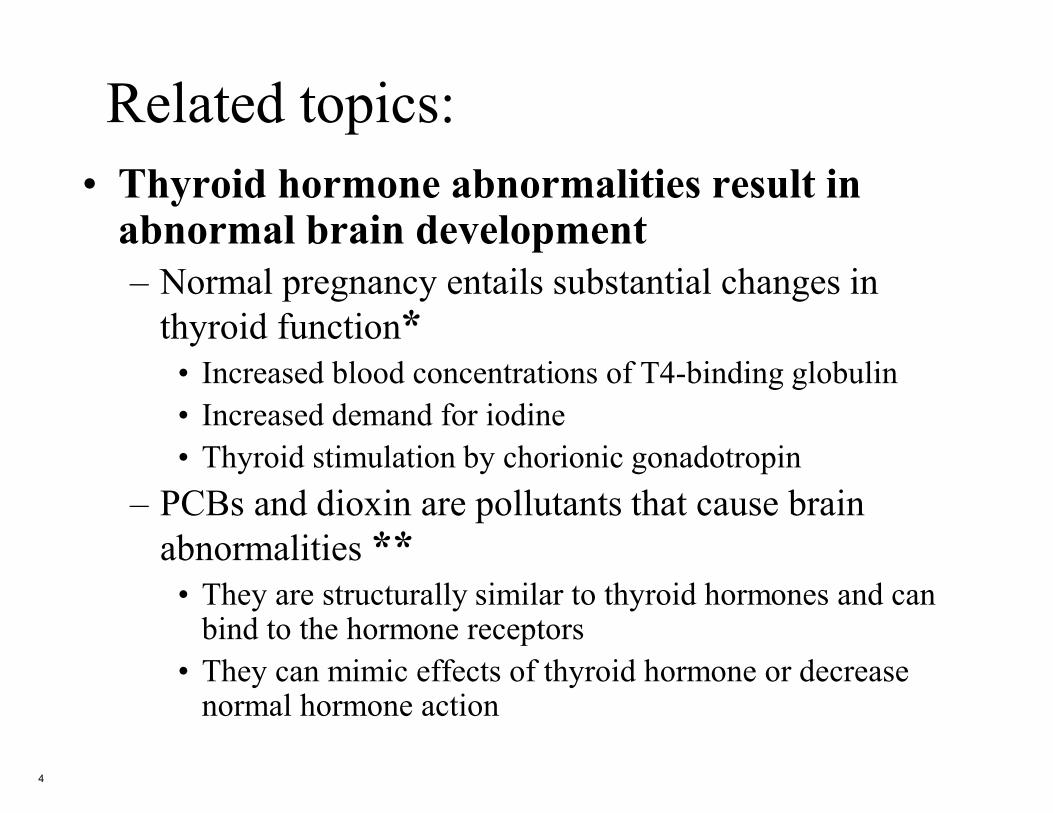
Related topics: • Thyroid hormone abnormalities result in
abnormal brain development – Normal pregnancy entails substantial changes in
thyroid function* • Increased blood concentrations of T4-binding globulin • Increased demand for iodine • Thyroid stimulation by chorionic gonadotropin
– PCBs and dioxin are pollutants that cause brain abnormalities **
• They are structurally similar to thyroid hormones and can bind to the hormone receptors
• They can mimic effects of thyroid hormone or decrease normal hormone action
4

Sources
* Colorado State Univ web site: http://arbl.cvmbs.colostate.edu/hbooks/pathphys/endocrine/thyroid/thyroid _preg.html
**Susan Porterfield, Environ Health Perspect 102(Suppl 2):125-130 (1994);
**Eric Montie, Woods Hole Oceanographic Institute, Ph.D. thesis (2006) on marine mammals, their brain development and effects of environmental pollutants in the ocean, especially polychlorinated biphenols. He has published, together with collaborators, a series of papers related to this topic.
5
Questions, chapter 27:
1) What are three factors that are likely to play roles in the development of sex differences in the central nervous system of mammals including humans?
2) Neuroanatomical studies of sex differences in the human brain were initially focused on what region? More recent studies have used methods for imaging the brain of living persons. Give an example of findings using this method.
6

Genes, hormones and environment: What determines sexual orientation?
What are the apparent relative contributions of genetic factors, hormonal influences, and inputs from the external environment in the determination of sexual orientation?
See Swaab & Hofman, p. 264, right-hand column.
• Strong support for genetic and hormonal influences
• Little support for social influences
7

Sex differences in human pathological syndromes
Studies have given very clear examples of neurological or psychiatric disease where there are dramatic sex differences.
Why might this be?
8
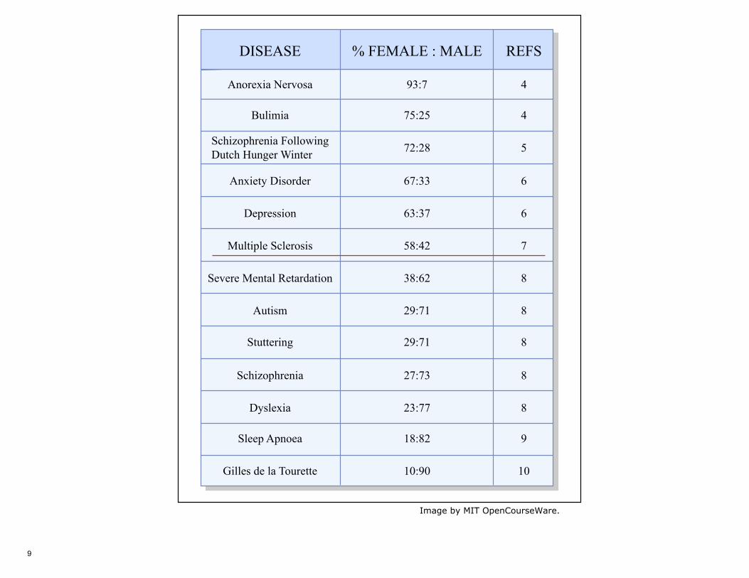
9
DISEASE % FEMALE : MALE REFS
Anorexia Nervosa
Bulimia
Schizophrenia FollowingDutch Hunger Winter
Anxiety Disorder
Depression
Multiple Sclerosis
Severe Mental Retardation
Autism
Stuttering
Schizophrenia
Dyslexia
Sleep Apnoea
Gilles de la Tourette
93:7
75:25
72:28
67:33
63:37
58:42
38:62
29:71
29:71
27:73
23:77
18:82
10:90
4
4
5
6
6
7
8
8
8
8
8
9
10
Image by MIT OpenCourseWare.

Changing levels of gonadal hormones in humans
When are sex differences in gonadal hormone levels at a peak in human development (three periods of time)?
When do morphological differences in the hypothalamus appear?
See Swaab & Hofman p. 265, left column: • Hormonal differences during 1st half of gestation, again
perinatally, again at puberty. • Hypothalamic differences after age 4 (next figure)
10

One particular structural difference:
Development and sexual differentiation of the sexually dimorphic nucleus of the preoptic area of human hypothalamus.
(Swaab & Hofman, TINS ’95)
11

Growth of the sexually dimorphic nucleus of the preoptic area of human hypothalamus in males (filled squares and circles) and females (unfilled). Data from homosexual males are shown as squares.
Note log-log scales Fig 27-1 12
Figure removed due to copyright restrictions.Please see course textbook or: Swaab, D. F., and M. A. Hofman. "Sexual differentiation of the humanhypothalamus in relation to gender and sexual orientation." Trends in neurosciences 18, no. 6 (1995): 264-70.
Problems with using human postmortum brain tissues
What is a major technical factor in studying morphological differences in the hypothalamus of postmortem human brains? What is the Swaab and Hofman solution?
See Swaab & Hofman pp. 266-267: Volume of a tissue region can vary with post-mortum factors:
• Tissue decay; time before fixation, • Nature of fixative used
Solution: cell counts (not easy to do)
13
More from Swaab & Hofman (1995)
• What is a cell group of the human hypothalamus which differs between the sexes? Is it also different in hetero- and homosexuals? [Answered in previous slides, but additional structures have been found—see following slides]
• Which other nucleus of the hypothalamus may be involved in sexual orientation according to a study in rats? How is this cell group different in human males of different sexual orientation? [following slides]
• There are also sex differences in CNS outside the hypothalamus.
14

15
Structures of the Limbic System
Level of following figure
15

Loci of anatomical differences in hypothalamus correlated with sex and sexual preferences
Differences have also been found in INAH 3, located caudal to this level.
III = Third ventricle AC = Anterior commissure INAH = Interstitial nucleus of the anterior hypothalamus OC = Optic chiasm PVN = Paraventricular nucleus SCN = Suprachiasmatic nucleus SDN = Sexually dimorphic nucleus SON = Supraoptic nucleus
Figure removed due to copyright restrictions.
Please see course textbook or: Swaab, D. F., and M. A. Hofman. "Sexual
Differentiation of the Human Hypothalamus in Relation to Gender and
Sexual Orientation." Trends in Neurosciences 18, no. 6 (1995): 264-70.
16
Fig 27-2 (From Swaab & Hofman , TINS ’95)
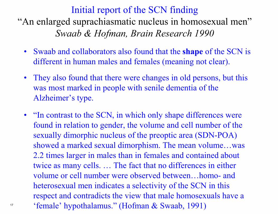
Initial report of the SCN finding “An enlarged suprachiasmatic nucleus in homosexual men”
Swaab & Hofman, Brain Research 1990 �
• Swaab and collaborators also found that the shape of the SCN is different in human males and females (meaning not clear).
• They also found that there were changes in old persons, but this was most marked in people with senile dementia of the Alzheimer’s type.
• “In contrast to the SCN, in which only shape differences were found in relation to gender, the volume and cell number of the sexually dimorphic nucleus of the preoptic area (SDN-POA) showed a marked sexual dimorphism. The mean volume…was 2.2 times larger in males than in females and contained about twice as many cells. … The fact that no differences in either volume or cell number were observed between…homo- and heterosexual men indicates a selectivity of the SCN in this respect and contradicts the view that male homosexuals have a ‘female’ hypothalamus.” (Hofman & Swaab, 1991) 17

Questions, chapter 27:
4) What appears to be a major cause of variation in sexual orientation in humans?
18

Other differences between male and female brains:
• Additional reports of sex differences in hypothalamic anatomy, e.g., – Differences in spine details
– Differences in length of dendrites of neurons in specific cell groups.
– What do these findings imply? [Next slide]
19
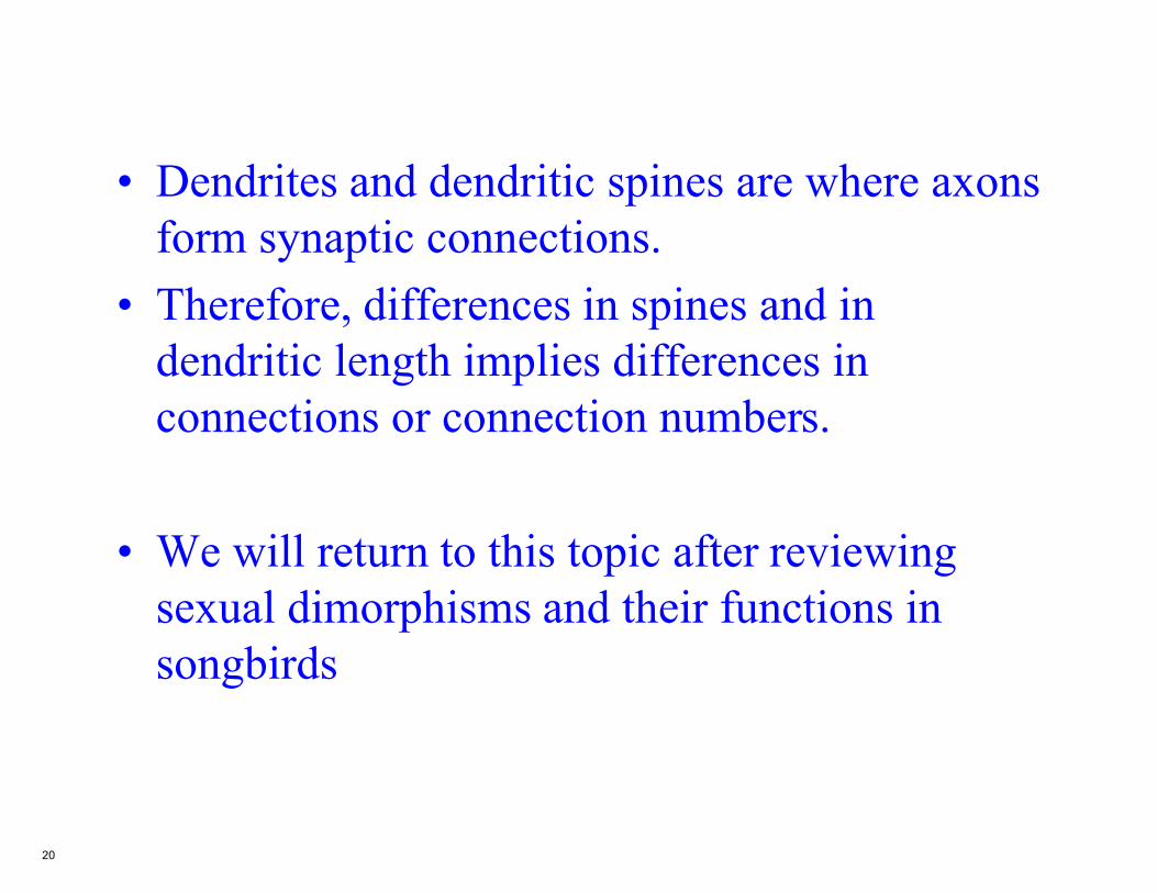
• Dendrites and dendritic spines are where axons form synaptic connections.
• Therefore, differences in spines and in dendritic length implies differences in connections or connection numbers.
• We will return to this topic after reviewing sexual dimorphisms and their functions in songbirds
20

There are also differences outside the hypothalamus:
Questions, chapter 27:
3) Name and describe the location of a cell group in the spinal cord where sexual dimorphism is very marked.
21
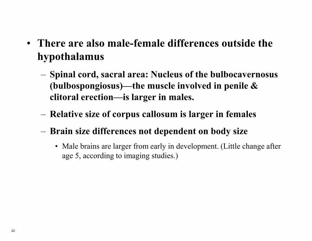
• There are also male-female differences outside the hypothalamus – Spinal cord, sacral area: Nucleus of the bulbocavernosus
(bulbospongiosus)—the muscle involved in penile & clitoral erection—is larger in males.
– Relative size of corpus callosum is larger in females
– Brain size differences not dependent on body size • Male brains are larger from early in development. (Little change after
age 5, according to imaging studies.)
22
Questions, chapter 27:
5) What were the striking findings made in Fernando Nottebohm’s lab at Rockefeller University in their studies of canary brains? There were two major findings (actually pairs of findings).
23

Figure removed due to copyright restrictions.
24
The robust nucleus of the arcopallium in frontal sections

Canary brain: song control structures (the most direct pathway)
(Nottebohm, Sci. Amer. ’89)
Figure removed due to copyright restrictions.
Please see: Nottebohm, Fernando. "From Bird Song to
Neurogenesis." Scientific American 260, no. 2 (1989): 74-9.
25

Direct vocal control pathway in songbird, For control of innate behavior
(sexually dimorphic structures ) 26
HVC
RA
L1
L2
L3
Syrinx
Pons - Medulla
ICoMLd
Mesencephalon
Telencephalon
Higher Vocal Center
Field L
Nuc Robustus of archistriatum
Hypoglossal nerve nucleus, tracheosyringeal division
Intercollicular nuc
Image by MIT OpenCourseWare.

Striatal Area X, the Vocal Control Center
Syrinx
HVC
LMAN
Area XRA
L1
L2
L3
Syrinx
Pons - Medulla
ICoMLd
Mesencephalon
Thalamus
Telencephalon
Indirect vocal control pathway in songbird, For the learning of song syllables & patterns:
striatum-dependent habit learning 27
Image by MIT OpenCourseWare.
“A brain for all seasons”
– What is the nature of the evidence that the male canary endbrain changes from season to season? (Fernando Nottebohm et al.)
– What did he think the change was caused by? – How has the work of Sarah Bottjer modified this
view?
28

Volume of song-control cell groups in male canaries varies with age (Nottebohm, Sci. Amer. ’89)
Figure removed due to copyright restrictions.
Please see: Nottebohm, Fernando. "From Bird Song to
Neurogenesis." Scientific American 260, no. 2 (1989): 74-9.
29

New song syllables and testosterone levels vary with season of year in male canaries
(Nottebohm, Sci. Amer. ’89)
Figure removed due to copyright restrictions.
Please see: Nottebohm, Fernando. "From Bird Song to
Neurogenesis." Scientific American 260, no. 2 (1989): 74-9.
30

Newborn neurons migrating in canary forebrain (Nottebohm, Sci. Amer. ’89)
Figure removed due to copyright restrictions.
Please see: Nottebohm, Fernando. "From Bird Song to
Neurogenesis." Scientific American 260, no. 2 (1989): 74-9.
31
Neurogenesis in adult canary brain: 3H-thymidine study
Figure removed due to copyright restrictions.
Please see: Nottebohm, Fernando. "From Bird Song to
Neurogenesis." Scientific American 260, no. 2 (1989): 74-9.
32

Sex differences in brains are multiple; many have been found in mammals, and many more will probably be found.
Another example: The findings of Toran-Allerand indicate that sex differences in the brain are probably much more widespread than has been found in studies of cell-stained human post-mortem material.
She has done in vitro studies:
1) Differentiation of mesencephalic DA neurons is affected by sex steroids
2) There is interaction between estrogens and neurotrophins.
33
Questions, chapter 27:
6) What in addition to hormonal factors may explain male-female differences in brain structure and function?
34

More recent findings:
Multiple genes important in sex determination • Since the 1970s, scientists have believed that estrogen and
testosterone were wholly responsible for sexually organizing the brain: a fetal brain simply needed to produce more testosterone to become male. But recent evidence indicates that hormones cannot explain everything about sexual differences between male and female brains.
• Eric Vilain and his colleagues explored whether genetic influences could explain the variations between male and female brains. Using two genetic testing methods, they compared the expression of over 12,000 genes in male and female brains in embryonic mice — long before the animals developed sex organs.
• To their surprise, the researchers found 54 genes expressed in different amounts in male and female mouse brains, prior to hormonal influence. Eighteen of the genes were expressed at higher levels in the male brains; 36 were expressed at higher levels in the female brains.
35
*Brain Res Mol Brain Res. 2003, 118: 82-90.*Excerpts were edited from a news report (UCLA, 10-22-2003)

More from Eric Vilain (2006) • One critical gene is Sry, identified in 1990 on the Y
chromosome. – Sex-determining region on the Y chromosome – Also known as the testis-determining gene
• It is found in the substantia nigra in rats, and is involved in dopamine secretion. – Reduction of the gene’s activity results in Parkinson’s –like
symptoms. • Does this explain why men are more susceptible to
Parkinson’s disease than woman? (This assumes that humans are similar to rats in the presence and function of Sry.)
• In women, how do these same neurons secrete dopamine, without Sry?
• Problem: This area of research has become secretive as a number of patent applications have been filed
36
Gene expression variations may explain some differences in sexual orientation: More from Eric Vilain and collaborators (2006):
• “Extreme skewing of X chromosome inactivation in mothers of homosexual men”*
• Findings “support a role for the X chromosome in regulating sexual orientation in a subgroup of gay men.”
• Problem reported in 2008: such skewing is not transmitted to offspring
*Bocklandt S, Horvath S, Vilain E, Hamer DH, Hum Genet (2006) 118: 691-694.
37
Quantitative variations in human brains• Recent neuroimaging data from longitudinal studies
by Judith L. Rapoport at NIMH (using large Ns) – Sex differences in overall hemisphere size are considerable
at all ages. – Size variations have both genetic and environmental
origins, and they vary from one part of the brain to another – Cerebellum seems to show greater variation, and this is
probably due to experiential factors. – Differences from normals are found in schizophrenics and
in young people with ADHD. Differences are region specific.
38

REVIEW
Frontal sections: the limbic system of rodent
Courtesy of MIT Press. Used with permission.Schneider, G. E. Brain Structure and its Origins: In the Development and in
Evolution of Behavior and the Mind. MIT Press, 2014. ISBN: 9780262026734.
39

40
OB, olfactory bulb Cerebral PF, prefrontal cortex hemisphere, Cing, Cingulate cortex medial view RS, retrosplenial cortex
(caudal cingulate)
S, septal area
fx, fornix
st, stria terminalis
DB(B), diagonal band of Anterior Broca
Commissure Am, amygdala
EC, entorhinal cortex
O Tub, olfactory tubercle
SI, substantia innominata
Acc, nuc. Accumbens
BNST, bed nucleus of the stria terminalis
A, anterior nuclei of thalamus
Cb, cerebellum
MD, mediodorsal nucleus of thalamus
mm, mammillary body
mt, mammillothalamic tract
m teg, mammillotegmental tract Fig 26-5 SC, superior colliculus
Papez’ Circuit Corpus Callosum Cingulate cx
Retrosplenial Cortex
Basal The underlying brainstem forebrain
Pituitary
Courtesy of MIT Press. Used with permission.Schneider, G. E. Brain Structure and its Origins: In the Development and in
Evolution of Behavior and the Mind. MIT Press, 2014. ISBN: 9780262026734.

Review: Papez’ circuit brought up to date: What are the inputs to this circuit? What are the outputs?
Association areas (neocortex)
Subiculum
Hippocampus
Dentate gyrus
Tegmental
Cingulate cortex Paralimbic areas, entorhinal area
Anterior nuclei of thalamus
fx mt
fxnuclei Mammillary Hypothalamus Septal
(Ach) bodies area
Hippocampal formation Courtesy of MIT Press. Used with permission.Schneider, G. E. Brain Structure and its Origins: In the Development and in
Evolution of Behavior and the Mind. MIT Press, 2014. ISBN: 9780262026734.
mt = mammillothalamic tract fx = fornix bundle (output of hippocampus) Ach= acetylcholine used as neurotransmitter
41

Papez’ circuit brought up to date: What are the inputs to this circuit? What are the outputs?
Association areas (neocortex)
Cingulate cortex Paralimbic areas, entorhinal area
Anterior nuclei of thalamus
fx mt
fxMammillary Hypothalamus Septal(Ach) bodies area
Subiculum
Hippocampus
Dentate gyrus
Tegmental nuclei
Courtesy of MIT Press. Used with permission. Hippocampal formation Schneider, G. E. Brain Structure and its Origins: In the Development and in
Evolution of Behavior and the Mind. MIT Press, 2014. ISBN: 9780262026734.
42
mt = mammillothalamic tract fx = fornix bundle (output of hippocampus) Ach= acetylcholine used as neurotransmitter
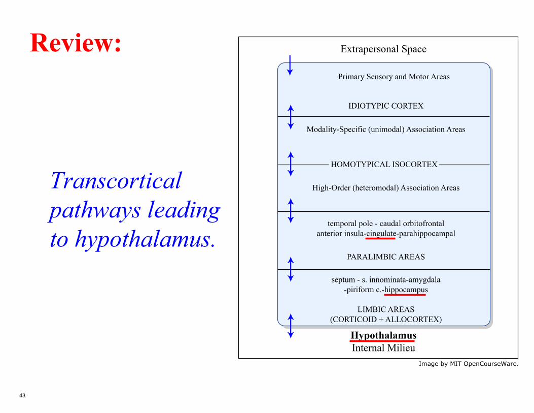
Review:
Transcortical pathways leading to hypothalamus.
43
Primary Sensory and Motor Areas
IDIOTYPIC CORTEX
Modality-Specific (unimodal) Association Areas
HOMOTYPICAL ISOCORTEX
High-Order (heteromodal) Association Areas
temporal pole - caudal orbitofrontalanterior insula-cingulate-parahippocampal
PARALIMBIC AREAS
septum - s. innominata-amygdala-piriform c.-hippocampus
LIMBIC AREAS(CORTICOID + ALLOCORTEX)
HypothalamusInternal Milieu
Extrapersonal Space
Image by MIT OpenCourseWare.

They try to say what you are, spiritual or sexual?
They wonder about Solomon and all his wives.
In the body of the world, they say, there is a soul
and you are that.
But we have ways within each other
that will never be said by anyone.
- by Rumi (1207-1273), translated by Coleman Barks
44

MIT OpenCourseWarehttp://ocw.mit.edu
9.14 Brain Structure and Its OriginsSpring 2014
For information about citing these materials or our Terms of Use, visit: http://ocw.mit.edu/terms.


