
Tamara Al-Azzeh + Asmaa Aljeelani
.....
28
Faisal
Moh Tarek + Suhayb
1 | P a g e
Digestion of dietary lipids
Lipid digestion and absorption are complex processes. They involve soluble enzymes,
substrates with different degree of solubility, and occur primarily in the stomach and
small intestine.
+++ : ( Dietary lipids are : triglycerides 90% , phospholipids, steroids, especially cholesterol and cholesterol
esters, fat-soluble vitamins, namely: vitamin A, D, E and K, and carotenoids.)
1) TAG with short or medium chain Fatty Acids (FA) (<=12 carbon)
Begins in the stomach (acid environment) so they are acid stable lipases
Both: Lingual Lipase and Gastric Lipase hydrolyze FA from TAG
Always remember that milk has medium chain of FA, so these Lipases play a
particularly important role in NEONATES whom milk fat is primary source of
calories.
Also, they become important digestive enzymes in individual with
PANCREATIC INSUFFICIENCY in which they work to digest those lipids in the
absence of pancreatic lipases.
They remove 1 FA producing = FA + DAG
2) In the small intestine
A- Emulsification in the duodenum –the first part of the small intestine-
Lipids are hydrophobic so they aren’t soluble in the aqueous environment like in
the intestine, to solve this problem Emulsification occurs. It’s happened by bile acids or bile salts made in liver and stored in Gallbladder
secreted with pancreatic lipases to the duodenum (more details in the next
lecture).
Emulsification breaks the fat globule to smaller molecule called emulsion droplets.
Emulsification increases the surface area of the hydrophobic lipid droplets so the
digestive enzymes (pancreatic lipases) can act effectively.
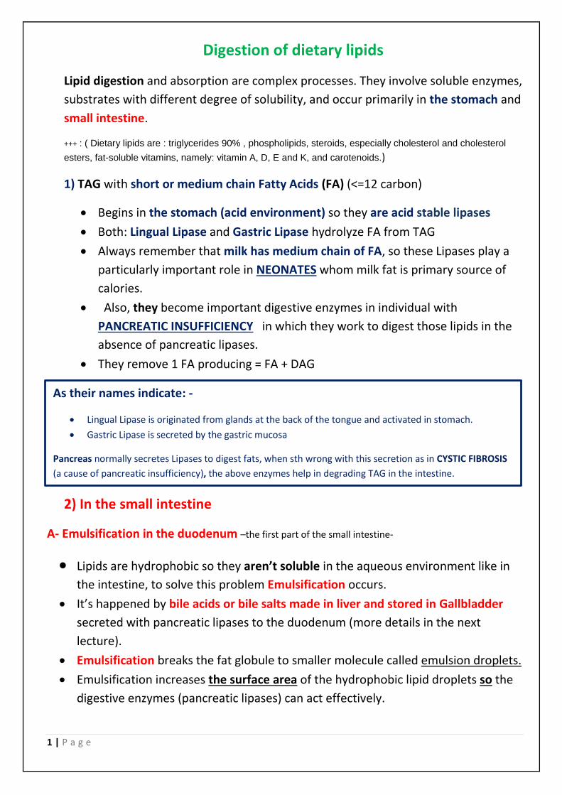
As their names indicate: -
Lingual Lipase is originated from glands at the back of the tongue and activated in stomach.
Gastric Lipase is secreted by the gastric mucosa
Pancreas normally secretes Lipases to digest fats, when sth wrong with this secretion as in CYSTIC FIBROSIS
(a cause of pancreatic insufficiency), the above enzymes help in degrading TAG in the intestine.
2 | P a g e
Bile salts and acids stabilize lipid particles as they become smaller and preventing
them from coalescing.
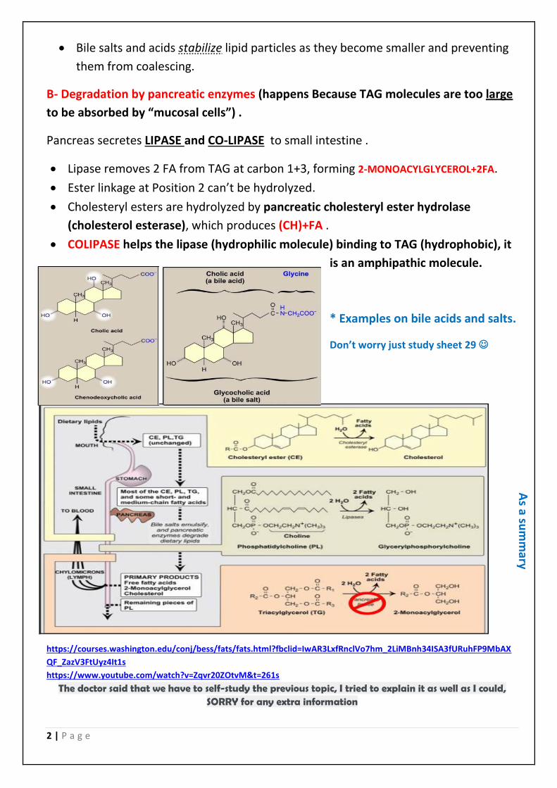
B- Degradation by pancreatic enzymes (happens Because TAG molecules are too large
to be absorbed by “mucosal cells”) .
Pancreas secretes LIPASE and CO-LIPASE to small intestine .
Lipase removes 2 FA from TAG at carbon 1+3, forming 2-MONOACYLGLYCEROL+2FA.
Ester linkage at Position 2 can’t be hydrolyzed.
Cholesteryl esters are hydrolyzed by pancreatic cholesteryl ester hydrolase
(cholesterol esterase), which produces (CH)+FA .
COLIPASE helps the lipase (hydrophilic molecule) binding to TAG (hydrophobic), it
is an amphipathic molecule.
* Examples on bile acids and salts.
Don’t worry just study sheet 29
https://courses.washington.edu/conj/bess/fats/fats.html?fbclid=IwAR3LxfRnclVo7hm_2LiMBnh34ISA3fURuhFP9MbAX
QF_ZazV3FtUyz4It1s
https://www.youtube.com/watch?v=Zqvr20ZOtvM&t=261s
The doctor said that we have to self-study the previous topic, I tried to explain it as well as I could, SORRY for any extra information
As a su
mm
ary
3 | P a g e
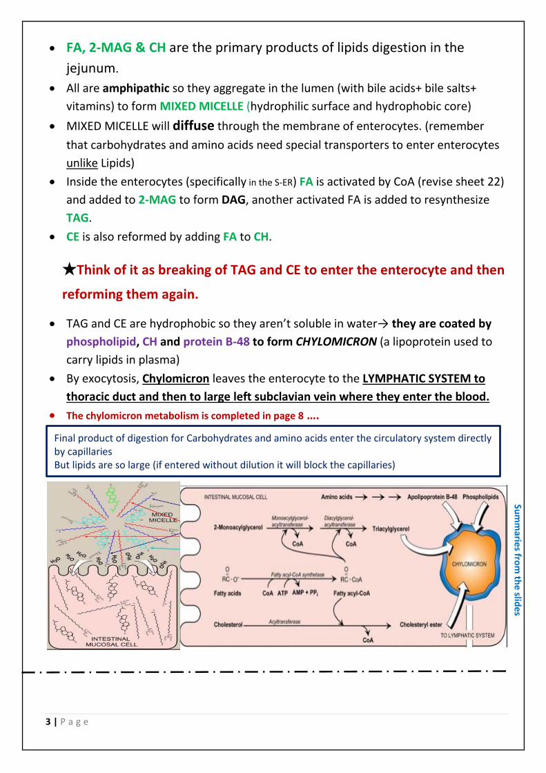
FA, 2-MAG & CH are the primary products of lipids digestion in the
jejunum.
All are amphipathic so they aggregate in the lumen (with bile acids+ bile salts+
vitamins) to form MIXED MICELLE (hydrophilic surface and hydrophobic core)
MIXED MICELLE will diffuse through the membrane of enterocytes. (remember
that carbohydrates and amino acids need special transporters to enter enterocytes
unlike Lipids)
Inside the enterocytes (specifically in the S-ER( FA is activated by CoA (revise sheet 22)
and added to 2-MAG to form DAG, another activated FA is added to resynthesize
TAG.
CE is also reformed by adding FA to CH.
★Think of it as breaking of TAG and CE to enter the enterocyte and then
reforming them again.
TAG and CE are hydrophobic so they aren’t soluble in water→ they are coated by
phospholipid, CH and protein B-48 to form CHYLOMICRON (a lipoprotein used to
carry lipids in plasma)
By exocytosis, Chylomicron leaves the enterocyte to the LYMPHATIC SYSTEM to
thoracic duct and then to large left subclavian vein where they enter the blood.
The chylomicron metabolism is completed in page 8 ….
Final product of digestion for Carbohydrates and amino acids enter the circulatory system directly by capillaries But lipids are so large (if entered without dilution it will block the capillaries)
Sum
maries fro
m th
e slides

4 | P a g e
LIPOPROTEINS
Glycerol is a very hydrophilic molecule with 3 carbon atoms + 3 hydroxyl groups
(soluble)
Fatty Acid (FA) has a negative charge on its carboxyl group, it’s amphipathic
(soluble)
When combining together and forming TriAcylGlycerol (TAG), it’s neither polar
nor amphipathic (ester bond is no longer hydrophilic)
(completely insoluble)
We know that 90% of the plasma is water, so we need a carrier to transport lipids
to and from tissues, this transport system is called LIPOPROTEINS
LIPOPROTEINS are composed of multimolecular complexes of lipids and proteins.
-lipids are
1) TAG, Cholesterol Ester (CE), both are non-polar so they are existed in the core of the
LIPOPROTEINS
2 )Cholesterol (CH), Phospholipids (PL), both are
amphipathic so they are existed on the surface (the
shell) with their polar part exposed on the surface to
make it soluble in water.
-Proteins : are called Apo-lipoproteins
They are amphipathic, exists of the surface (the
shell)
Several classes ( Apo A , Apo B-48 , Apo c , Apo E)
some will discussed later.
They have structural role (can’t be removed)
Or Regulatory role (can be transferred
between different lipoproteins)
Or Binding to cell surface receptors.
recall from the summer :-
if the protein is bound to something
else the protein part of that complex
is called apoprotein

5 | P a g e
(FLOATS)
(In-between)
(Sinks)
(Sinks)
(Sinks)
LIPOPRETEINS are Similar to Micelles
★ Lipoproteins are composed of a neutral lipid core
(containing TAG & CE) surrounded by a shell of
amphipathic Apolipoproteins, Phospholipids & free
Cholesterol.
LIPOPROTEINS are classified according to their densities
.centrifugationso we can separate them by
Remember that density of water=1g/cm3,
oil (fat) always float in water, so its density<1, therefore increasing lipid
component will decrease the density of lipoprotein and the opposite for increasing
protein components.
TAG is in the core so 85% of Chylomicrons is a core lipid therefore it has a low
surface area ratio to volume (bigger in size), the name micron indicates this also.
While PL and proteins are on the surface, so 70% of HDL is a shell therefore high
surface area ratio to volume (smaller in size).
★As an overview :- increasing the size will increase the volume and surface area,
but the increase in volume is much bigger than the increase in surface area.
VLDL: very low density lipoprotein
density lipoprotein-Intermediate: IDL
LDL: low density lipoprotein
HDL: high density lipoprotein
-Please don’t memorize the
numbers.
-You should know the arrangement
according to density, size, lipid and
protein percentage. (IDL>LDL and so
on..)
6 | P a g e
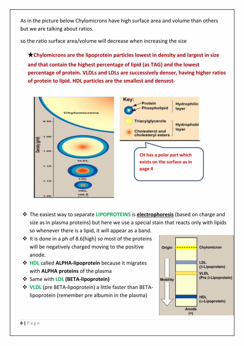
As in the picture below Chylomicrons have high surface area and volume than others
but we are talking about ratios.
so the ratio surface area/volume will decrease when increasing the size
★Chylomicrons are the lipoprotein particles lowest in density and largest in size
and that contain the highest percentage of lipid (as TAG) and the lowest
percentage of protein. VLDLs and LDLs are successively denser, having higher ratios
of protein to lipid. HDL particles are the smallest and densest .
The easiest way to separate LIPOPROTEINS is electrophoresis (based on charge and
size as in plasma proteins) but here we use a special stain that reacts only with lipids
so whenever there is a lipid, it will appear as a band.
It is done in a ph of 8.6(high) so most of the proteins
will be negatively charged moving to the positive
anode.
HDL called ALPHA-lipoprotein because it migrates
with ALPHA proteins of the plasma
Same with LDL (BETA-lipoprotein)
VLDL (pre BETA-lipoprotein) a little faster than BETA-
lipoprotein (remember pre albumin in the plasma)
CH has a polar part which
exists on the surface as in
page 4
7 | P a g e
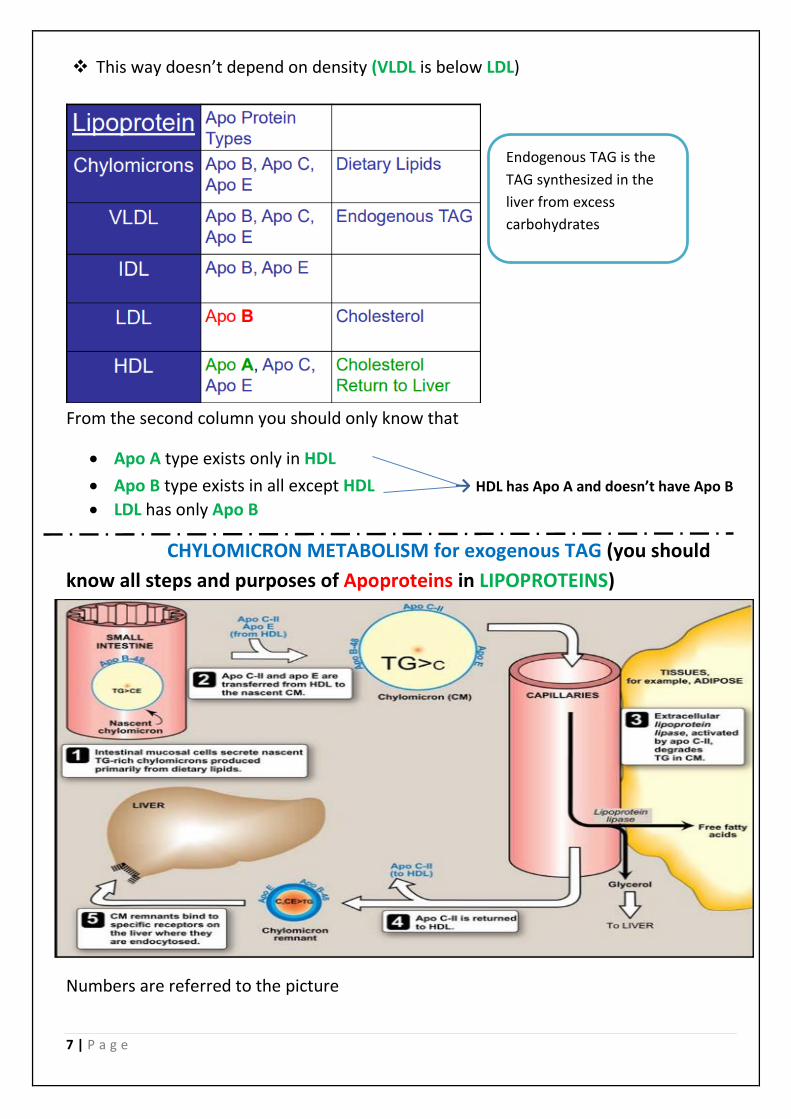
This way doesn’t depend on density (VLDL is below LDL)
From the second column you should only know that
Apo A type exists only in HDL
Apo B type exists in all except HDL → HDL has Apo A and doesn’t have Apo B
LDL has only Apo B
CHYLOMICRON METABOLISM for exogenous TAG (you should
know all steps and purposes of Apoproteins in LIPOPROTEINS)
Numbers are referred to the picture
Endogenous TAG is the
TAG synthesized in the
liver from excess
carbohydrates

8 | P a g e
The newly synthesized chylomicrons are called “nascent chylomicrons".
3- Most of the TAG is broken down by LIPOPROTEIN LIPASE (LPL)
synthesized by adipose tissue and skeletal muscle, it is an extracellular enzyme
requires an activator which is protein C-II comes from HDL in step 2
The products are ↓
FA diffuses to skeletal muscle to be oxidized or to adipose tissue to be oxidized or
re-esterified to produce TAG
Glycerol goes to the liver (which has glycerol kinase) to form glycerol 3-phosphate
Then it enters GLYCOLYSIS or GLUCONEOGENESIS or be used in TAG synthesis
Connect the previous lectures.
After this step TAG is decreased in chylomicron so the size is decreased
The density increases due to removing lipids (density lower than water)
focus on the changing of chylomicron shape in the picture
5- liver has a receptor for Apo E, when binding, endocytosis happens to the remnant of chylomicron (بقايا).
“chylomicron remnants “contains larger amounts of Cholesterol and cholesterol esters than TAG
which have been already degraded by LPL.
This metabolism takes several hours, so in order to measure cholesterol, the patient
must fast 12-14 hours, otherwise the cholesterol measured in the plasma= the
cholesterol we eat (very high)
Summary from book: Chylomicrons are assembled in intestinal mucosal cells from dietary
lipids (primarily TAG). Each nascent chylomicron particle has one molecule of apolipoprotein (apo) B-48. They are released from the cells into the lymphatic system and travel to the blood, where they receive apo C-II and apo E from HDLs. Apo C-II activates endothelial lipoprotein lipase (LPL), which degrades the TAG in chylomicrons to fatty acids and glycerol. The fatty acids that are released are stored (in the adipose) or used for energy (by the muscle). The glycerol is metabolized by the liver.
9 | P a g e
VLDL metabolism (for endogenous TAG), again you should know the steps
Almost the same as CHYLOMICRON except It is synthesized in the liver and
released directly to the blood (small in size), CHYLOMICRON (is assembled in the
small intestine and doesn’t enter the blood directly) When TAG is degraded the
density is increased as mentioned in chylomicron metabolism
This converts VLDL to IDL
IDL is taken by endocytosis or continues until the density increases a lot to
become LDL which has higher CE and C than TAG (denser).
All the components of LDL is taken by endocytosis to extrahepatic tissues or
liver.
Know that both chylomicrons
and VLDL lose TAG in the
blood
becomesChylomicron
remnant while Chylomicron
VLDL is converted to IDL
BECAUSE of the different
proteins on their surfaces
An
oth
er sum
mary fro
m th
e bo
ok
10 | P a g e
CHOLESTEROL
CHOLE= gallbladder Ster=steroid Ol= alcohol
so it’s the steroid alcohol of the gall bladder
cholesterol is related to atherosclerosis, strokes and many diseases,
A lot researches have been done on it.
★ Cholesterol was firstly isolated from gall bladder
stones in 1774
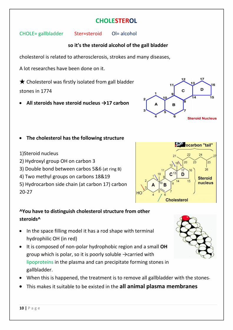
All steroids have steroid nucleus →17 carbon
The cholesterol has the following structure
1)Steroid nucleus
2) Hydroxyl group OH on carbon 3
3) Double bond between carbos 5&6 (at ring B)
4) Two methyl groups on carbons 18&19
5) Hydrocarbon side chain (at carbon 17) carbon
20-27
^You have to distinguish cholesterol structure from other
steroids^
In the space filling model it has a rod shape with terminal
hydrophilic OH (in red)
It is composed of non-polar hydrophobic region and a small OH
group which is polar, so it is poorly soluble →carried with
lipoproteins in the plasma and can precipitate forming stones in
gallbladder.
When this is happened, the treatment is to remove all gallbladder with the stones .
This makes it suitable to be existed in the all animal plasma membranes
11 | P a g e
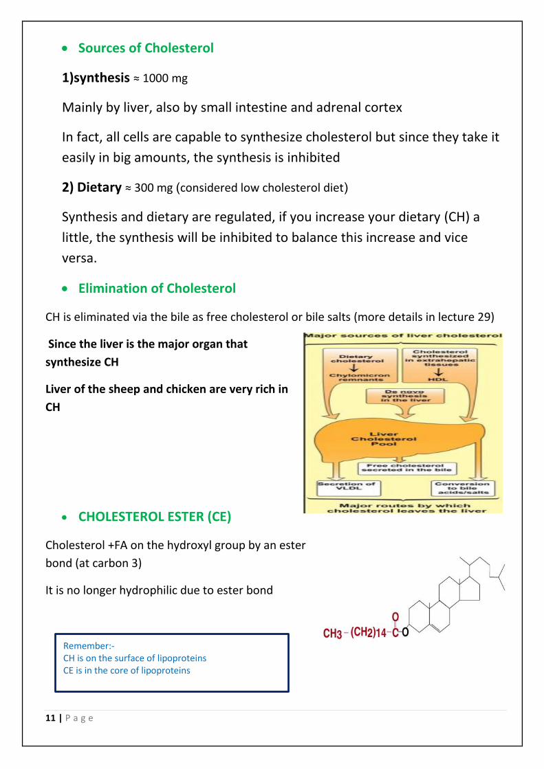
Sources of Cholesterol
1)synthesis ≈ 1000 mg
Mainly by liver, also by small intestine and adrenal cortex
In fact, all cells are capable to synthesize cholesterol but since they take it
easily in big amounts, the synthesis is inhibited
2) Dietary ≈ 300 mg )considered low cholesterol diet(
Synthesis and dietary are regulated, if you increase your dietary (CH) a
little, the synthesis will be inhibited to balance this increase and vice
versa.
Elimination of Cholesterol
CH is eliminated via the bile as free cholesterol or bile salts (more details in lecture 29)
Since the liver is the major organ that
synthesize CH
Liver of the sheep and chicken are very rich in
CH
CHOLESTEROL ESTER (CE)
Cholesterol +FA on the hydroxyl group by an ester
bond (at carbon 3)
It is no longer hydrophilic due to ester bond
Remember:- CH is on the surface of lipoproteins CE is in the core of lipoproteins
12 | P a g e
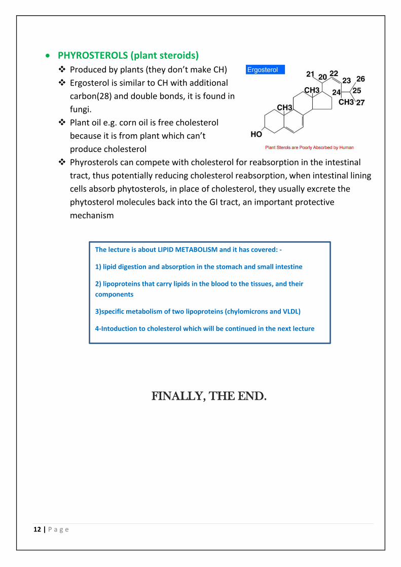
PHYROSTEROLS (plant steroids)
Produced by plants (they don’t make CH)
Ergosterol is similar to CH with additional
carbon(28) and double bonds, it is found in
fungi.
Plant oil e.g. corn oil is free cholesterol
because it is from plant which can’t
produce cholesterol
Phyrosterols can compete with cholesterol for reabsorption in the intestinal
tract, thus potentially reducing cholesterol reabsorption, when intestinal lining
cells absorb phytosterols, in place of cholesterol, they usually excrete the
phytosterol molecules back into the GI tract, an important protective
mechanism
FINALLY, THE END.
The lecture is about LIPID METABOLISM and it has covered: -
1) lipid digestion and absorption in the stomach and small intestine
2) lipoproteins that carry lipids in the blood to the tissues, and their
components
3)specific metabolism of two lipoproteins (chylomicrons and VLDL)
4-Intoduction to cholesterol which will be continued in the next lecture


