Download - 1 Brain I.Overview II.Brain Stem III.Cerebellum IV.Diencephalon V.Cerebrum VI.Cranial Nerves
1
Brain
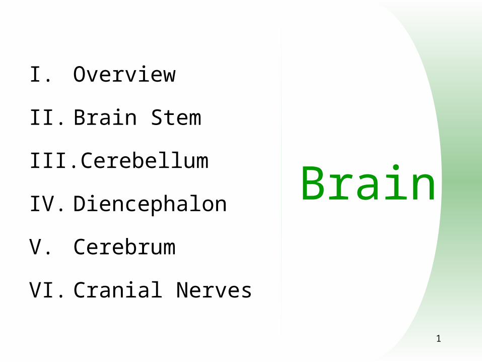
I. Overview
II. Brain Stem
III. Cerebellum
IV. Diencephalon
V. Cerebrum
VI. Cranial Nerves

2
Brain
I. Overview
A. Function
B. Protection
C. Blood Supply
D. Cerebrospinal Fluid
II. Brain Stem
III. Cerebellum
IV. Diencephalon
V. Cerebrum
VI. Cranial Nerves
3
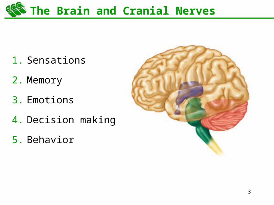
The Brain and Cranial Nerves
1. Sensations
2. Memory
3. Emotions
4. Decision making
5. Behavior

4
Principal Parts of the Brain
1. Cerebrum
2. Diencephalon
a) Thalamus
b) Hypothalamus
3. Cerebellum
4. Brainstem
a) Medulla
b) Pons
c) Midbrain Cerebell
um
Brain Stem
Cerebrum
Diencephalon

5
Protective Coverings of the Brain
Pia Mater
Bone
Periosteum
Meninges same as around the spinal cord
1. dura mater
2. arachnoid mater
3. pia materArachnoid
Mater
PeriosteumAnd Bone
Dura Mater

6
Protective Coverings of the Brain
Dura mater extensions
1. falx cerebri
2. tentorium cerebelli
3. falx cerebelli
Falx cerebri
Falx cerebelli
Tentorium cerebelli

7
Blood Supply to Brain
Arterial blood supply is branches from circle of Willis on base of brain
Vessels on surface of brain----penetrate tissue
Uses 20% of our bodies oxygen & glucose needs
blood flow to an area increases with activity in that area
deprivation of O2 for 4 min does permanent injury
at that time, lysosome release enzymes
waste
Capillary
Ependymal cells
O2
Fluid
Elec.
Glucose
8
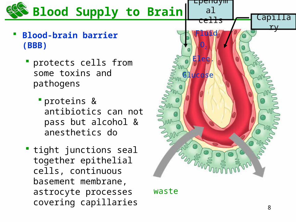
Blood Supply to Brain
Blood-brain barrier (BBB)
protects cells from some toxins and pathogens
proteins & antibiotics can not pass but alcohol & anesthetics do
tight junctions seal together epithelial cells, continuous basement membrane, astrocyte processes covering capillaries
waste
Capillary
Ependymal cells
O2
Fluid
Elec.
Glucose

9
Cerebrospinal Fluid (CSF)
80-150 ml (3-5oz)
Clear liquid containing glucose, proteins, & ions
Functions
1. mechanical protection
floats brain & softens impact with bony walls
2. chemical protection
optimal ionic concentrations for action potentials
3. circulation
nutrients and waste products to and from bloodstream

10
Origin of CSF
Choroid plexus = capillaries covered by ependymal cells
2 lateral ventricles, one within each cerebral hemisphere
roof of 3rd ventricle
fourth ventricle
Choroid plexus
Lateral Ventricle
s
Third Ventricle
sFourth
Ventricles

11
Drainage of CSF from Ventricles
One median aperture & two lateral apertures allow CSF to exit from the interior of the brain Lateral
apertureMedian aperture
15
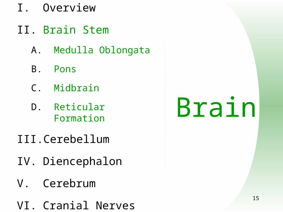
Brain
I. Overview
II. Brain Stem
A. Medulla Oblongata
B. Pons
C. Midbrain
D. Reticular Formation
III. Cerebellum
IV. Diencephalon
V. Cerebrum
VI. Cranial Nerves
16
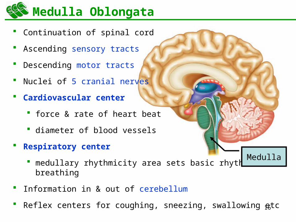
Medulla Oblongata
Continuation of spinal cord
Ascending sensory tracts
Descending motor tracts
Nuclei of 5 cranial nerves
Cardiovascular center
force & rate of heart beat
diameter of blood vessels
Respiratory center
medullary rhythmicity area sets basic rhythm of breathing
Information in & out of cerebellum
Reflex centers for coughing, sneezing, swallowing etc
Medulla

17
Ventral Surface of Medulla Oblongata
Ventral surface bulge
pyramids
large motor tract
decussation of most fibers
left cortex controls right muscles
Olive = olivary nucleus
neurons send input to cerebellum
proprioceptive signals
gives precision to movements

18
Dorsal Surface of Medulla Oblongata
Nucleus gracilis & nucleus cuneatus = sensory neurons
relay information to thalamus on opposite side of brain
5 cranial nerves arise from medulla -- 8 thru 12
Nucleus gracilis
Nucleus cutaneou
s
CN 7CN 8CN 9CN 10CN 11
CN 12

19
Injury to the Medulla
Hard blow to the back of the head may be fatal
Cranial nerve malfunctions on same side as injury;loss of sensation or paralysis of throat or tongue; irregularities in breathing and heart rhythm
20
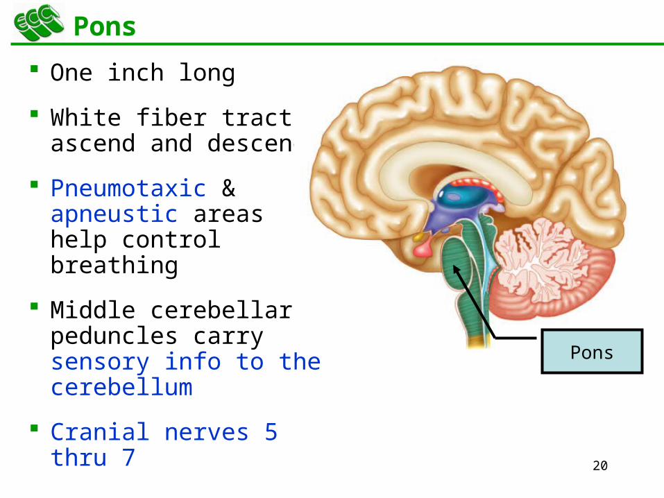
Pons
One inch long
White fiber tracts ascend and descend
Pneumotaxic & apneustic areas help control breathing
Middle cerebellar peduncles carry sensory info to the cerebellum
Cranial nerves 5 thru 7
Pons

21
Midbrain
One inch in length
Extends from pons to diencephalon
Cerebral aqueduct connects 3rd ventricle above to 4th ventricle below
Midbrain
Third ventricle
Fourth ventricle
Cerebral aqueduc
t
22
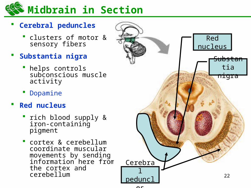
Midbrain in Section
Cerebral peduncles
clusters of motor & sensory fibers
Substantia nigra
helps controls subconscious muscle activity
Dopamine
Red nucleus
rich blood supply & iron-containing pigment
cortex & cerebellum coordinate muscular movements by sending information here from the cortex and cerebellum Cerebral
peduncles
Red nucleus
Substantia nigra

23
Dorsal Surface of Midbrain
Corpora quadrigemina
superior colliculi
inferior colliculi
coordinate eye movements with visual stimuli
coordinate head movements with auditory stimuli
Superior colliculi
Inferior colliculi

24
Midbrain
Superior, middle & inferior peduncles attach to brainstem
inferior carries sensory information from spinal cord
middle carries sensory fibers from cerebral cortex & basal ganglia
superior carries motor fibers that extend to motor control areas
Inferior peduncle
s
Superior
peduncles

25
Reticular Formation
Scattered nuclei in medulla, pons & midbrain
Reticular activating system
alerts cerebral cortex to sensory signals (sound of alarm, flash light, smoke or intruder) to awaken from sleep
maintains consciousness & helps keep you awake with stimuli from ears, eyes, skin and muscles
Motor function is involvement with maintaining muscle tone
Reticular formatio
n

26
Brain
I. Overview
II. Brain Stem
III. Cerebellum
IV. Diencephalon
V. Cerebrum
VI. Cranial Nerves

27
Cerebellum
2 cerebellar hemispheres and vermis (central area)
Function
correct voluntary muscle contraction and posture based on sensory data from body about actual movements
sense of equilibrium
Vermis

28
Cerebellum
Transverse fissure between cerebellum & cerebrum
Cerebellar cortex (folia) & central nuclei are grey matter
Arbor vitae = tree of life = white matter

29
Brain
I. Overview
II. Brain Stem
III. Cerebellum
IV. Diencephalon
A. Thalamus
B. Hypothalamus
C. Epithalamus
D. Circumventricular Organs
V. Cerebrum
VI. Cranial Nerves

30
Diencephalon Surrounds 3rd Ventricle
Surrounds 3rd ventricle
Superior part of walls is thalamus
Inferior part of walls & floor is hypothalamus
Thalamus
(enclosed in 3rd
ventricle)
Hypothalamus
31
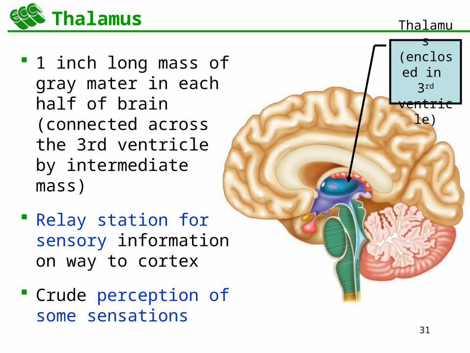
ThalamusThalamu
s (enclosed in 3rd
ventricle)
1 inch long mass of gray mater in each half of brain (connected across the 3rd ventricle by intermediate mass)
Relay station for sensory information on way to cortex
Crude perception of some sensations

32
Thalamic Nuclei
Nuclei have different roles
relays auditory and visual impulses, taste and somatic sensations
receives impulses from cerebellum or basal ganglia
anterior nucleus concerned with emotions, memory and acquisition of knowledge (cognition)
Emotions
Motor
Visual
Auditory
Emotions
Emotions,
Alertness,
Memory
Integrates with other
nuclei

33
Hypothalamus
Dozen or so nuclei in 4 major regions
mammillary bodies are relay station for olfactory reflexes
infundibulum suspends the pituitary gland
Major regulator of homeostasis
receives somatic and visceral input, taste, smell & hearing information; monitors osmotic pressure, temperature of blood
SmellConnects
to pituitary
gland
Pituitary gland

34
Functions of Hypothalamus
Controls and integrates activities of the ANS which regulates smooth, cardiac muscle and glands
Synthesizes regulatory hormones that control the anterior pituitary
Contains cell bodies of axons that end in posterior pituitary where they secrete hormones
Regulates rage, aggression, pain, pleasure & arousal
Feeding, thirst & satiety centers
Controls body temperature
Regulates daily patterns of sleep
SmellConnects
to pituitary
gland
Pituitary gland

35
Epithalamus
Pineal gland
endocrine gland the size of small pea
secretes melatonin during darkness
promotes sleepiness & sets biological clock
Habenular nuclei
emotional responses to odors
Pineal gland
Habenular nuclei
36
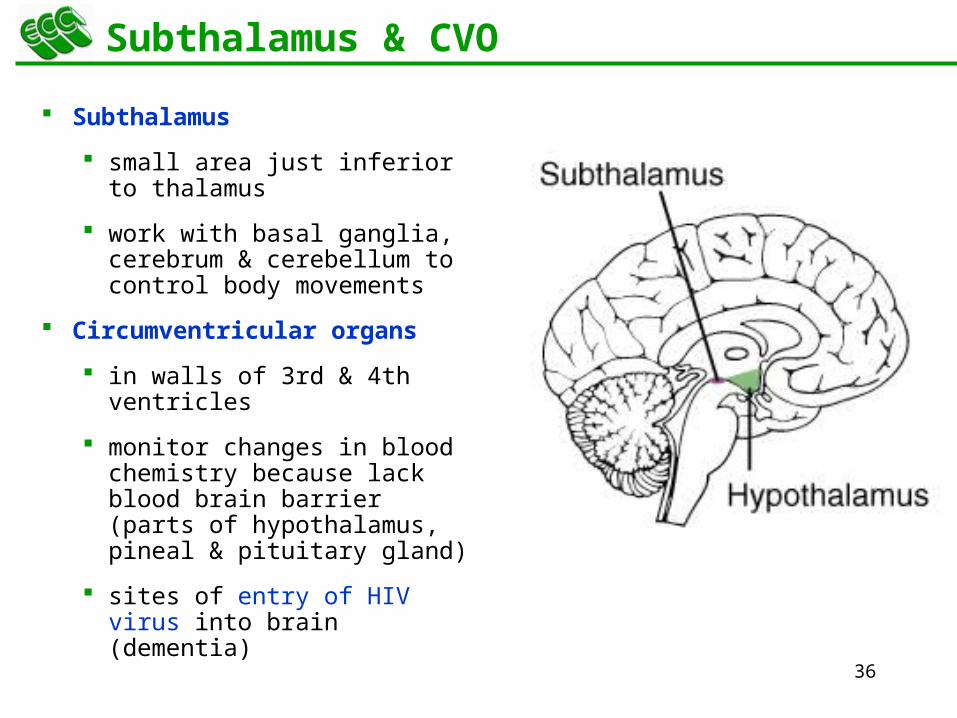
Subthalamus & CVO
Subthalamus
small area just inferior to thalamus
work with basal ganglia, cerebrum & cerebellum to control body movements
Circumventricular organs
in walls of 3rd & 4th ventricles
monitor changes in blood chemistry because lack blood brain barrier (parts of hypothalamus, pineal & pituitary gland)
sites of entry of HIV virus into brain (dementia)
37
Brain
I. Overview
II. Brain Stem
III. Cerebellum
IV. Diencephalon
V. Cerebrum
A. Lobes
B. White Matter
C. Basal Ganglia
D. Limbic System
E. Function of Cortex
1. Sensory
2. Motor
3. Association
4. Lateralization
5. Brain Waves
VI. Cranial Nerves

38
Cerebrum (Cerebral Hemispheres)
Cerebral cortex is gray matter overlying white matter
2-4 mm thick containing billionsof cells
grew so quickly formed folds(gyri) and grooves (sulci or fissures)
Gray matter
White matter
39
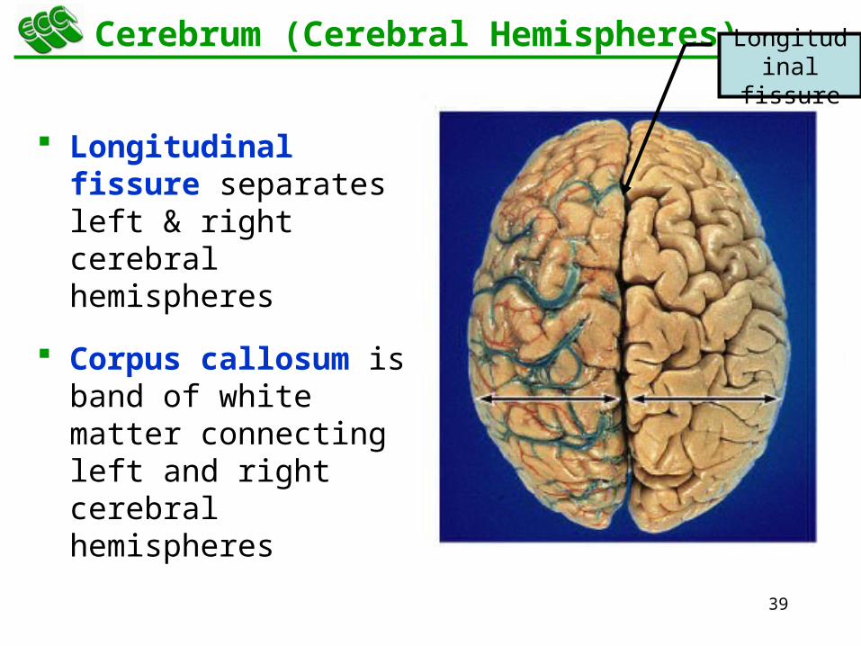
Cerebrum (Cerebral Hemispheres)
Longitudinal fissure separates left & right cerebral hemispheres
Corpus callosum is band of white matter connecting left and right cerebral hemispheres
Corpus callosum
Longitudinal
fissure

40
Lobes and Fissures Each hemisphere is subdivided into 4
lobes
1. Frontal lobe
Motor area
Personality, behavior
Emotions
Memory
2. Parietal lobe
Somatosensory – skin and muscle
3. Occipital lobe
Vision
4. Temporal lobe
Hearing
Smell
Taste
Frontal Parietal
Temporal
Occipital

41
Lobes and Fissures
Central sulcus (black)
precentral & postcentral gyrus
Parieto-occipital sulcus (red)
Lateral sulcus (yellow)
Frontal Parietal
Temporal
Occipital

43
Cerebral White Matter
1. Association fibers between gyri in same hemisphere
2. Commissural fibers from one hemisphere to other
3. Projection fibers form descending & ascending tracts
Association fibers
Commisural fibers
Projection fibers
44
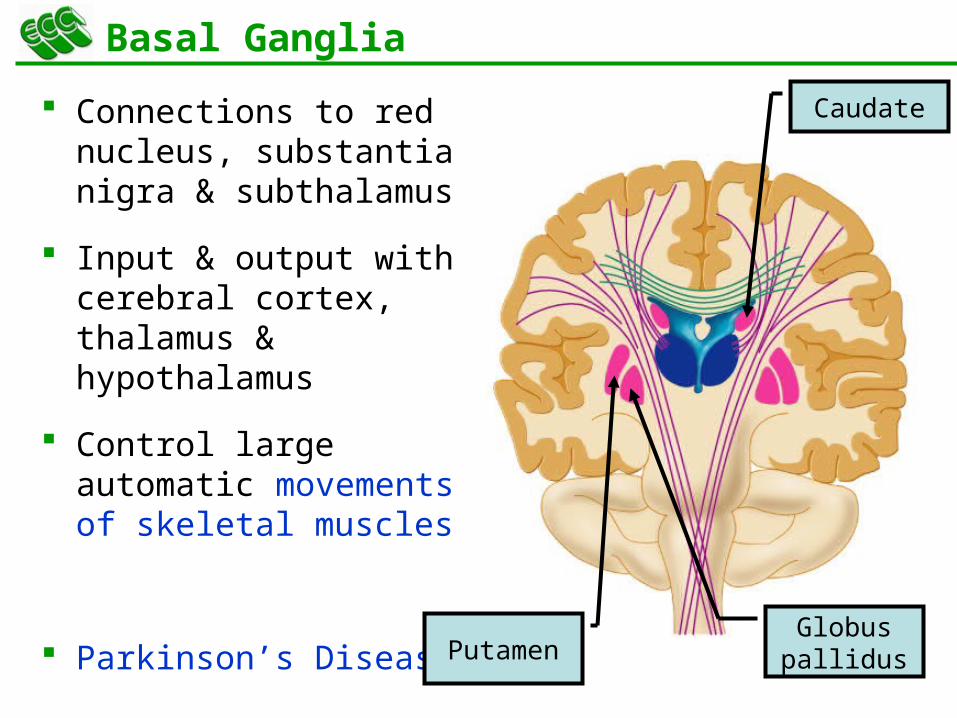
Basal Ganglia
Connections to red nucleus, substantia nigra & subthalamus
Input & output with cerebral cortex, thalamus & hypothalamus
Control large automatic movements of skeletal muscles
Parkinson’s Disease
Caudate
Globus pallidusPutamen
45
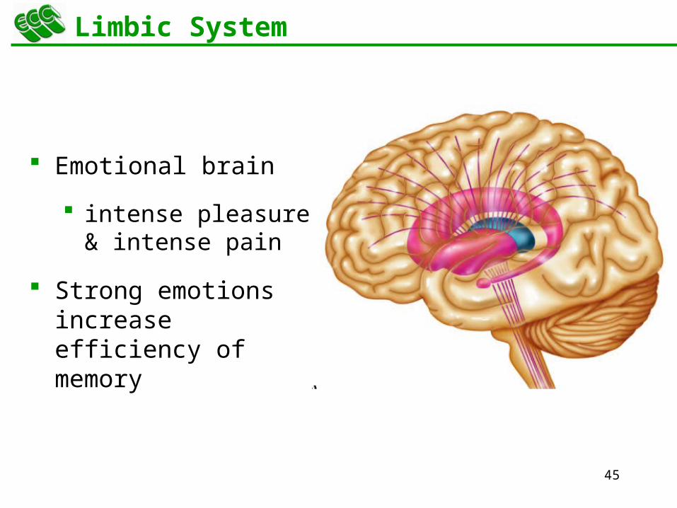
Limbic System
Emotional brain
intense pleasure & intense pain
Strong emotions increase efficiency of memory

47
Sensory Areas of Cerebral Cortex
Receive sensory information from the thalamus
Primary somatosensory area = postcentral gyrus = 1,2,3
Primary visual area = 17
Primary auditory area = 41 & 42
Primary gustatory area = 43

48
Motor Areas of Cerebral Cortex
Voluntary motor initiation
Primary motor area = 4 = precentral gyrus
controls voluntary contractions of skeletal muscles on other side
Motor speech area = 44 = Broca’s area
production of speech -- control of tongue & airway
49
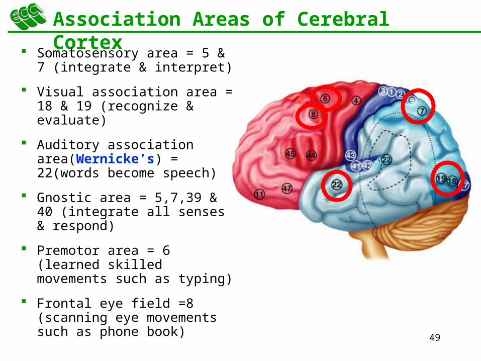
Association Areas of Cerebral Cortex
Somatosensory area = 5 & 7 (integrate & interpret)
Visual association area = 18 & 19 (recognize & evaluate)
Auditory association area(Wernicke’s) = 22(words become speech)
Gnostic area = 5,7,39 & 40 (integrate all senses & respond)
Premotor area = 6 (learned skilled movements such as typing)
Frontal eye field =8 (scanning eye movements such as phone book)

51
Hemispheric Lateralization
Functional specialization of each hemisphere more pronounced in men
Females have larger connections between 2 sides
Damage to left side produces aphasia
Damage to same area on right side produces speech with little emotional inflection

52
Electroencephalogram (EEG)
Brain waves are millions of nerve action potentials in cerebral cortex
diagnosis of brain disorders (epilepsy)
brain death (absence of activity in 2 EEGs 24 hours apart)
1. Alpha -- awake & resting
2. Beta -- mental activity
3. Theta -- emotional stress
4. Delta -- deep sleep

53
Brain
I. Overview
II. Brain Stem
III. Cerebellum
IV. Diencephalon
V. Cerebrum
VI. Cranial Nerves
54
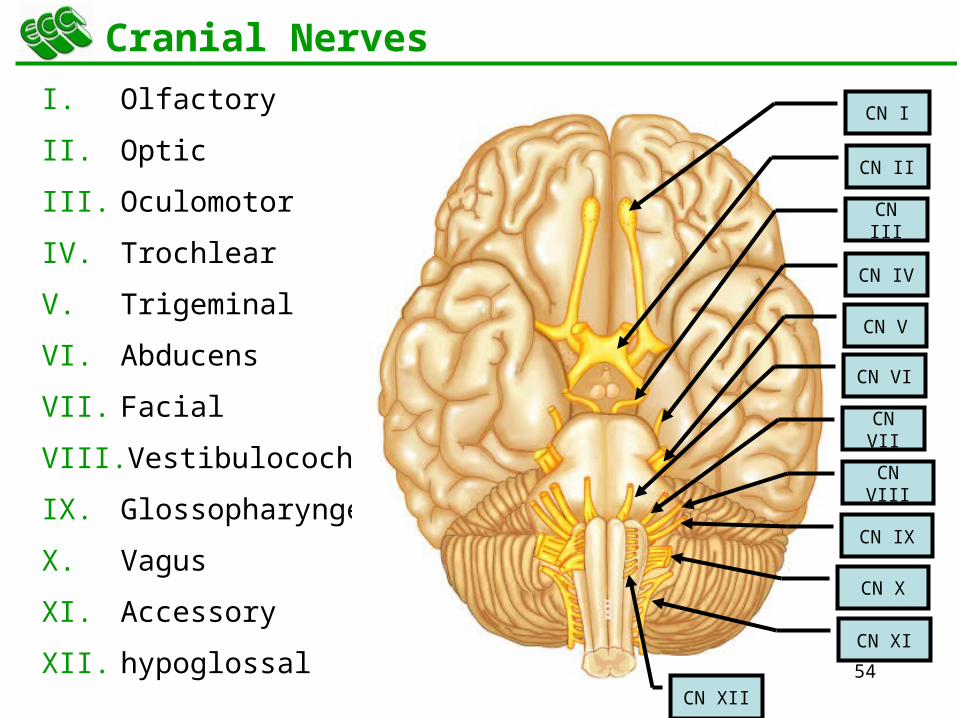
Cranial Nerves
I. Olfactory
II. Optic
III. Oculomotor
IV. Trochlear
V. Trigeminal
VI. Abducens
VII. Facial
VIII. Vestibulocochlear
IX. Glossopharyngeal
X. Vagus
XI. Accessory
XII. hypoglossal
CN I
CN II
CN III
CN IV
CN V
CN VI
CN VII
CN VIII
CN IX
CN X
CN XI
CN XII

55
I -- Olfactory Nerve (S)
Extends from olfactory mucosa of nasal cavity to olfactory bulb
Sense of smell
Anosmia – loss of smell

56
II -- Optic Nerve (S)
Connects to retina supplying vision
Defect in
Visual acuity
Visual field
Visual field defect
Visual acuity defect

57
III = Oculomotor Nerve (S/M)
Sensory:
Proprioception – sense of position
Motor:
Levator palpebrae raises eyelid
Ptosis – drooping of eyelid
4 extrinsic eye muscles
Superior, medial, inferior, and inferior oblique
2 intrinsic eye muscles
accomodation for near vision (changing shape of lens during reading)
constriction of pupil
Diplopia – double vision

58
IV = Trochlear Nerve (M)
Sensory:
Proprioception – sense of position
Motor:
Superior oblique eye muscle
59
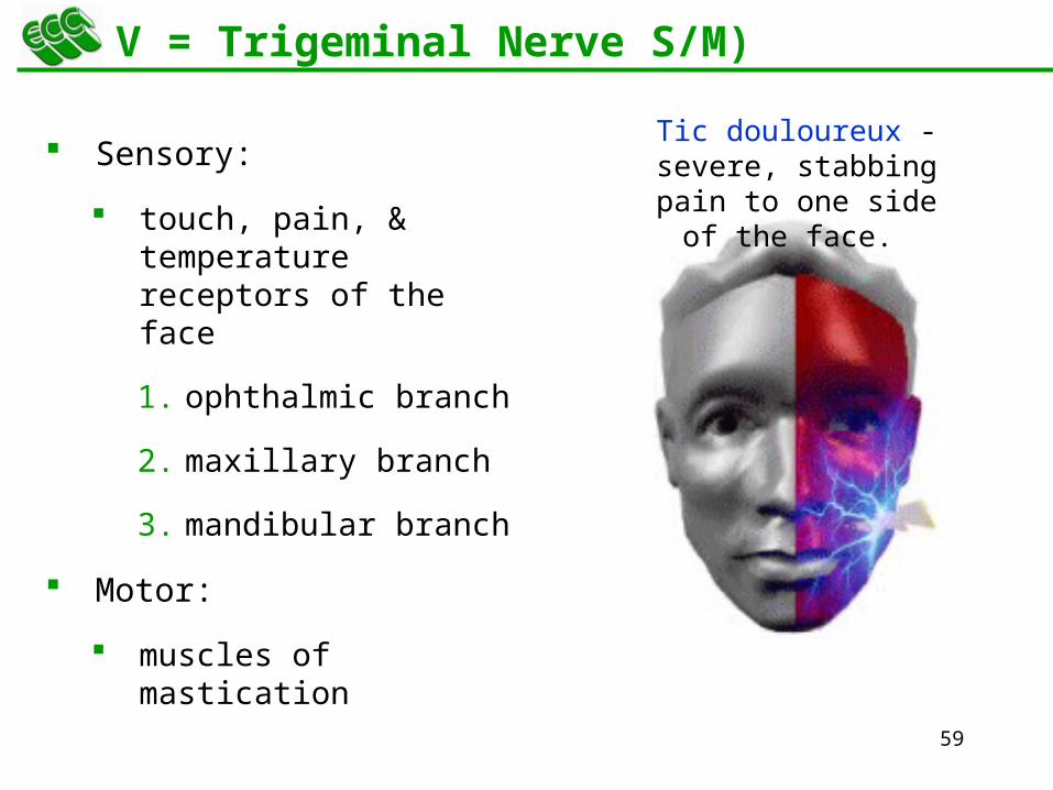
V = Trigeminal Nerve S/M)
Sensory:
touch, pain, & temperature receptors of the face
1. ophthalmic branch
2. maxillary branch
3. mandibular branch
Motor:
muscles of mastication
Tic douloureux - severe, stabbing
pain to one side of the face.

60
VI = Abducens Nerve (S/M)
Sensory:
Proprioception – sense of position
Motor:
Lateral rectus eye muscle

61
VII = Facial Nerve (S/M)
Sensory:
taste buds on anterior 2/3’s of tongue
Motor:
facial muscles
salivary & nasal and oral mucous glands & tears
Bell’s Palsy

62
VIII = Vestibulocochlear Nerve (S/M)
Cochlear branch begins in medulla
receptors in cochlea
hearing
if damaged deafness or tinnitus (ringing) is produced
Vestibular branch begins in pons
receptors in vestibular apparatus
sense of balance
vertigo (feeling of rotation)
ataxia (lack of coordination)
63
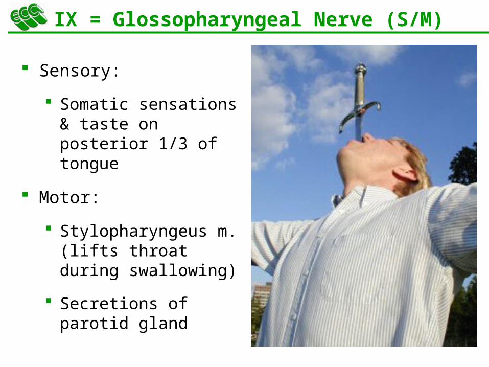
IX = Glossopharyngeal Nerve (S/M)
Sensory:
Somatic sensations & taste on posterior 1/3 of tongue
Motor:
Stylopharyngeus m. (lifts throat during swallowing)
Secretions of parotid gland

64
X = Vagus Nerve (S/M)
Sensory:
Receives sensations from viscera
Motor:
Controls cardiac muscle and smooth muscle of the viscera
Controls secretion of digestive fluids
Bradycardia
65
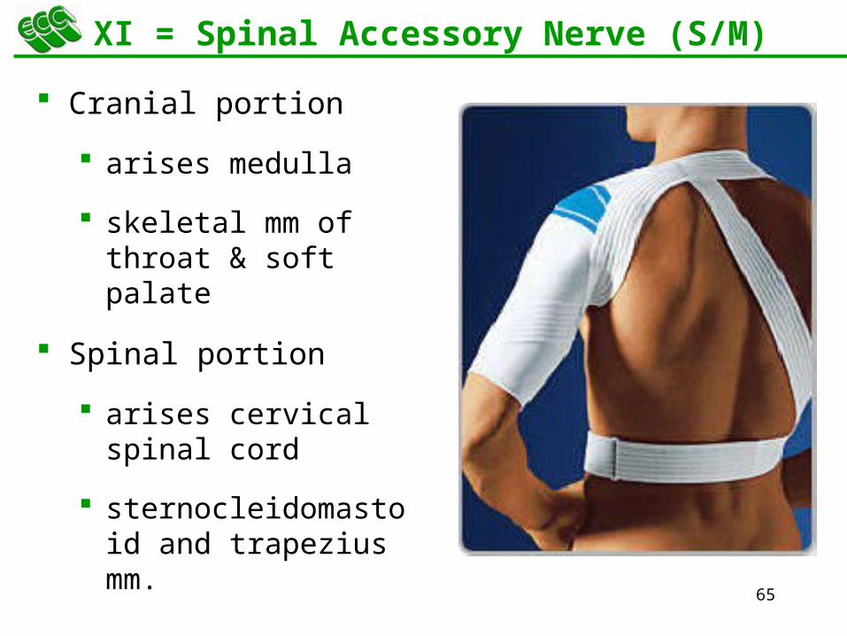
XI = Spinal Accessory Nerve (S/M)
Cranial portion
arises medulla
skeletal mm of throat & soft palate
Spinal portion
arises cervical spinal cord
sternocleidomastoid and trapezius mm.
66
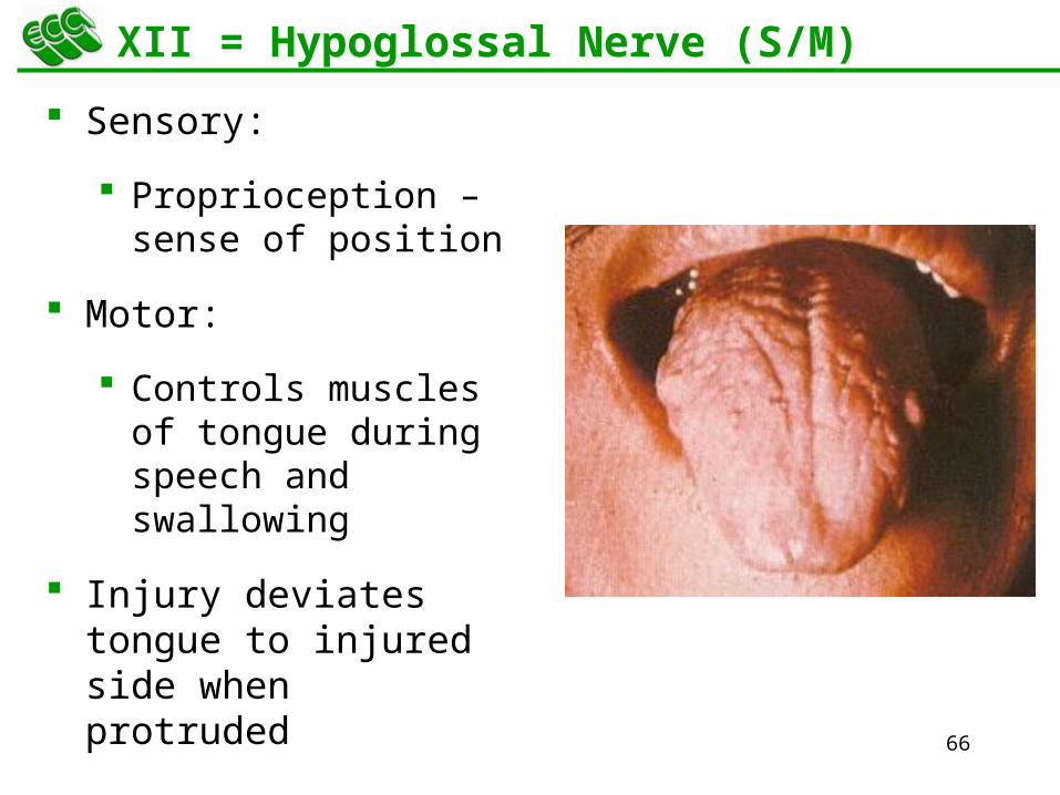
XII = Hypoglossal Nerve (S/M)
Sensory:
Proprioception – sense of position
Motor:
Controls muscles of tongue during speech and swallowing
Injury deviates tongue to injured side when protruded


