download - eurosurveillance
TRANSCRIPT

www.eurosurveillance.org
Vol. 18 | Weekly issue 30 | 25 July 2013
E u r o p e ’ s j o u r n a l o n i n f e c t i o u s d i s e a s e e p i d e m i o l o g y, p r e v e n t i o n a n d c o n t r o l
Editorials Epidemiological surveillance of leishmaniasis in the European Union: operational and research challenges 2by L Gradoni
Surveillance and outbreak reports Re-emergence of leishmaniasis in Spain: community outbreak in Madrid, Spain, 2009 to 2012 5by A Arce, A Estirado, M Ordobas, S Sevilla, N García, L Moratilla, S de la Fuente, AM Martínez, AM Pérez, E Aránguez, A Iriso, O Sevillano, J Bernal, F Vilas
Imported leishmaniasis in the Netherlands from 2005 to 2012: epidemiology, diagnostic techniques and sequence-based species typing from 195 patients 14by A Bart, PP van Thiel , HJ de Vries, CJ Hodiamont, T Van Gool
Research articles Molecular typing of Leishmania infantum isolates from a leishmaniasis outbreak in Madrid, Spain, 2009 to 2012 22by C Chicharro, IP Llanes-Acevedo, E García, J Nieto, J Moreno, I Cruz
Leishmania infantum in free-ranging hares, Spain, 2004-2010 35by F Ruiz-Fons, E Ferroglio, C Gortázar
Heat-shock protein 70 gene sequencing for Leishmania species typing in European tropical infectious disease clinics 40by G Van der Auwera , I Maes, S De Doncker, C Ravel, L Cnops, M Van Esbroeck, A Van Gompel, J Clerinx, JC Dujardin
Perspectives Leishmaniasis in the era of tumor necrosis factor alpha antagonist therapy – a research agenda for Europe 49by P Zanger , S Gabrysch
Review articles The role of indigenous phlebotomine sandflies and mammals in the spreading of leishmaniasis agents in the Mediterranean region 54by M Antoniou, M Gramiccia, R Molina, V Dvorak, P Volf
News LeishMan: harmonising diagnostic and clinical management of leishmaniasis in Europe 62by J Blum

2 www.eurosurveillance.org
Editorials
Epidemiological surveillance of leishmaniasis in the European Union: operational and research challenges
L Gradoni ([email protected])1
1. Unit of Vector-borne Diseases and International Health, MIPI Department, Istituto Superiore di Sanità, Rome, Italy
Citation style for this article: Gradoni L. Epidemiological surveillance of leishmaniasis in the European Union: operational and research challenges. Euro Surveill. 2013;18(30):pii=20539. Available online: http://www.eurosurveillance.org/ViewArticle.aspx?ArticleId=20539
Article submitted on 22 July 2013 / published on 25 July 2013
Leishmaniasis is complex of vector-borne diseases caused by protozoan parasites of the genus Leishmania transmitted by the bite of phlebotomine sandflies. A dozen nosogeographical entities – characterised by different parasite, vector and reservoir host spe-cies, geographical distribution and clinical features in humans – affect 101 countries in tropical, subtropical and temperate zones of the world [1,2]. More than 90% of 200,000–400,000 global cases of visceral leish-maniasis (VL), the most severe form, are estimated to occur annually in India, Bangladesh, Sudan, South Sudan, Ethiopia and Brazil. A less severe form, cuta-neous leishmaniasis (CL), is more widely distributed, accounting for 0.7–1.2 million cases each year in coun-tries of Latin America, Mediterranean basin, Middle East and Central Asia.
Even though many physicians and public health experts still consider leishmaniasis a tropical disease, two entities associated with several Phlebotomus spe-cies are endemic in southern Europe: (i) zoonotic VL and CL caused by L. infantum throughout the region, having dogs as reservoir host; and (ii) anthroponotic CL caused by L. tropica, which occurs sporadically in Greece. More recently, a third parasite species (L. donovani, assumed to be anthroponotic) has been recorded in Cyprus, where it causes both VL and CL [3].
VL is endemic in nine countries of the European Union (EU). The World Health Organization’s Department for the Control of Neglected Tropical Diseases has esti-mated a total VL incidence of approximately 410–620 cases each year during 2003 to 2008 in these endemic countries, adjusted to take into account a ‘mild’ 1.2–1.8-fold under-reporting [2]. Recent experiences from six of the nine countries – Bulgaria, Greece, Croatia, Italy, France and Spain – are presented in this special issue.
Zoonotic CL usually occurs in the same areas endemic for VL, but there are probably many more cases than those registered (2.8–4.6-fold under-reporting has been estimated for the EU region [2]). As pointed out by Lachaud et al. for France [4], but also applicable to
other EU countries endemic for CL, cutaneous lesions due to L. infantum are often benign and patients are seen by general practitioners or dermatologists who generally do not report these cases or notify them even when mandatory.
Despite provoking a limited number of overt clinical cases – in comparison with global leishmaniasis fig-ures – L. infantum represents a latent public health threat in the EU because studies performed in sev-eral endemic foci have disclosed a high prevalence of asymptomatic parasite carriers [5]. A recent exam-ple is provided for Croatia by Šiško-Kraljević et al. [6]. Hence, immunosuppressive conditions, either due to co-morbidities (e.g. human immunodeficiency virus (HIV) infection) or therapies (e.g. organ transplantation or treatment of immunological disorders [7]) may result in the reactivation of latent infections. In this regard, it should be emphasised that dermotropic L. infantum genotypes – the usual agents of benign CL – may dis-seminate to cause severe VL in immunosuppressed individuals [8]. Such elevated prevalence of human infections could have been predicted from two strands of evidence: humans are frequently bitten by sandflies and L. infantum infections are widespread in dogs, a highly susceptible host [9]. In large parts of countries of southern EU, canine seroprevalence rates are esti-mated to be in the range of 5–30%, which means that infection rates may reach values of 40–80% [10].
Some European countries at the north of regions with natural transmission of leishmaniasis have reported large series of VL and CL imported cases, many of which have acquired the parasitic infection during holidays in southern Europe [11-14]. In several instances, a defini-tive diagnosis of VL proved difficult and for one case, the period before symptom onset and specific treatment was longer than a year. Delay in diagnosis or misdiag-nosis can also occur in southern European countries endemic for VL, but in parts where cases occur rarely, as has been reported from a northern Italian region [15]. These observations suggest that awareness about leishmaniasis endemicity in Europe should be greatly increased among general practitioners and clinicians.
3www.eurosurveillance.org
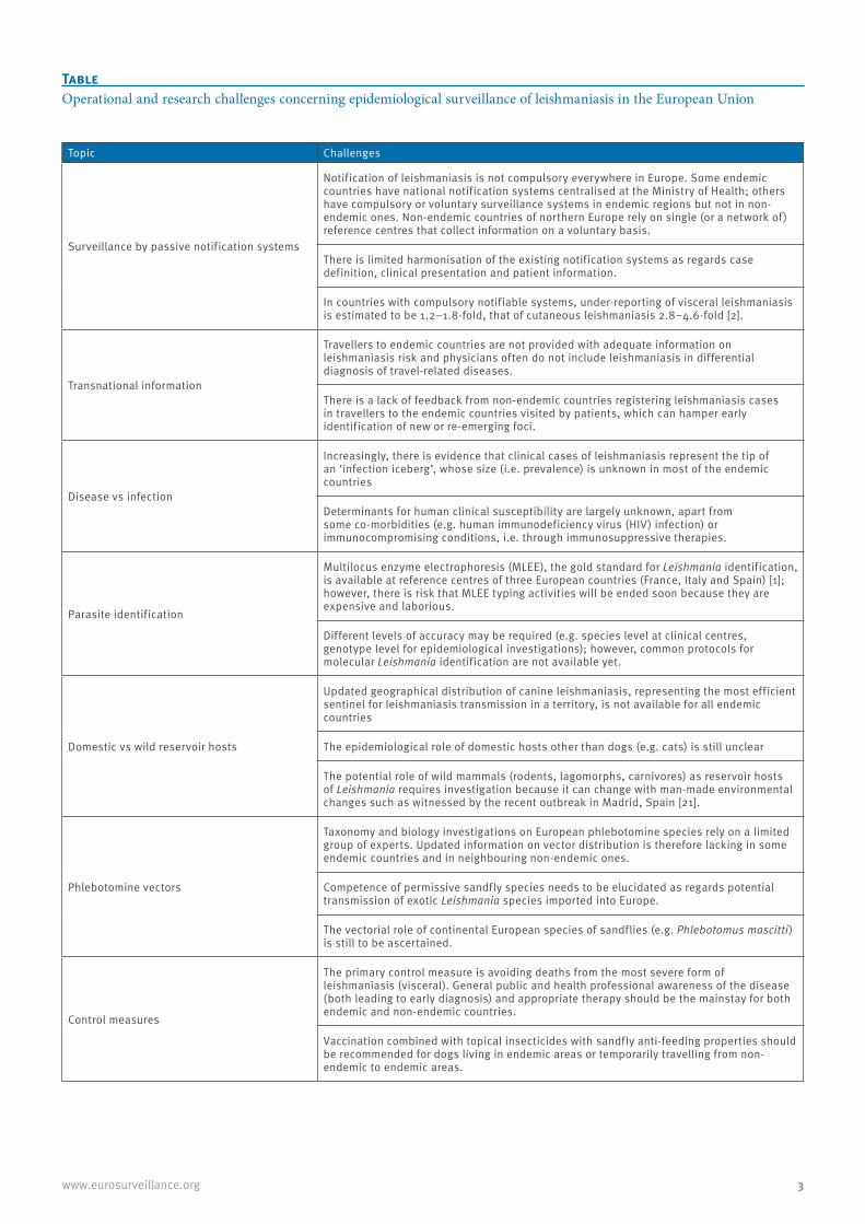
Table Operational and research challenges concerning epidemiological surveillance of leishmaniasis in the European Union
Topic Challenges
Surveillance by passive notification systems
Notification of leishmaniasis is not compulsory everywhere in Europe. Some endemic countries have national notification systems centralised at the Ministry of Health; others have compulsory or voluntary surveillance systems in endemic regions but not in non-endemic ones. Non-endemic countries of northern Europe rely on single (or a network of) reference centres that collect information on a voluntary basis.
There is limited harmonisation of the existing notification systems as regards case definition, clinical presentation and patient information.
In countries with compulsory notifiable systems, under-reporting of visceral leishmaniasis is estimated to be 1.2–1.8-fold, that of cutaneous leishmaniasis 2.8–4.6-fold [2].
Transnational information
Travellers to endemic countries are not provided with adequate information on leishmaniasis risk and physicians often do not include leishmaniasis in differential diagnosis of travel-related diseases.
There is a lack of feedback from non-endemic countries registering leishmaniasis cases in travellers to the endemic countries visited by patients, which can hamper early identification of new or re-emerging foci.
Disease vs infection
Increasingly, there is evidence that clinical cases of leishmaniasis represent the tip of an ‘infection iceberg’, whose size (i.e. prevalence) is unknown in most of the endemic countries
Determinants for human clinical susceptibility are largely unknown, apart from some co-morbidities (e.g. human immunodeficiency virus (HIV) infection) or immunocompromising conditions, i.e. through immunosuppressive therapies.
Parasite identification
Multilocus enzyme electrophoresis (MLEE), the gold standard for Leishmania identification, is available at reference centres of three European countries (France, Italy and Spain) [1]; however, there is risk that MLEE typing activities will be ended soon because they are expensive and laborious.
Different levels of accuracy may be required (e.g. species level at clinical centres, genotype level for epidemiological investigations); however, common protocols for molecular Leishmania identification are not available yet.
Domestic vs wild reservoir hosts
Updated geographical distribution of canine leishmaniasis, representing the most efficient sentinel for leishmaniasis transmission in a territory, is not available for all endemic countries
The epidemiological role of domestic hosts other than dogs (e.g. cats) is still unclear
The potential role of wild mammals (rodents, lagomorphs, carnivores) as reservoir hosts of Leishmania requires investigation because it can change with man-made environmental changes such as witnessed by the recent outbreak in Madrid, Spain [21].
Phlebotomine vectors
Taxonomy and biology investigations on European phlebotomine species rely on a limited group of experts. Updated information on vector distribution is therefore lacking in some endemic countries and in neighbouring non-endemic ones.
Competence of permissive sandfly species needs to be elucidated as regards potential transmission of exotic Leishmania species imported into Europe.
The vectorial role of continental European species of sandflies (e.g. Phlebotomus mascitti) is still to be ascertained.
Control measures
The primary control measure is avoiding deaths from the most severe form of leishmaniasis (visceral). General public and health professional awareness of the disease (both leading to early diagnosis) and appropriate therapy should be the mainstay for both endemic and non-endemic countries.
Vaccination combined with topical insecticides with sandfly anti-feeding properties should be recommended for dogs living in endemic areas or temporarily travelling from non-endemic to endemic areas.

4 www.eurosurveillance.org
As an endemic country comprises known areas or foci of endemicity, it is interestingly to note that in some instances, travellers became infected after visiting an area that was not considered as endemic by the health authorities of the country visited [16]. This should encourage the development of systems for appropri-ate transnational information following leishmaniasis diagnosis in travellers.
Deaths due to VL, although possible, are rare. The dis-ease has a slow chronic course, so that fatal cases may be patients with individual risk factors such as severe co-morbidities or, in case of young children, malnu-trition associated with late diagnosis. On the other hand, deaths due to inappropriate use of VL drugs can be even more frequent. In some European countries, antimonial drugs are still in use for some categories of patients because of the high cost of liposomal ampho-tericin B [17] and it is well known that overdose of pen-tavalent antimony in adults can cause severe cardiac failures in addition to pancreatitis.
This special issue of Eurosurveillance, published in two parts, is a useful instrument to review diverse aspects of leishmaniasis in Europe related to topics such as the information and surveillance systems in place in countries within the EU, the current epidemiological situation and novel aspects related to parasite identi-fication [18,19], domestic and wild reservoir hosts [20] and vectors [9]. The main challenges associated with these topics are summarised in the Table.
In conclusion, leishmaniasis, a neglected disease, is rare in some countries of Europe, but endemic in oth-ers, having a great impact on individuals and the poten-tial to spread further. The disease should be monitored carefully and systems for its notification should be har-monised at both national and transnational levels.
References1. World Health Organization (WHO). Control of the
leishmaniases: report of a meeting of the WHO Expert Committee on the Control of Leishmaniases, Geneva, 22-26 March 2010. Geneva: WHO; 2010. WHO technical report series; no. 949. Available from: http://whqlibdoc.who.int/trs/WHO_TRS_949_eng.pdf
2. Alvar J, Vélez ID, Bern C, Herrero M, Desjeux P, Cano J, et al. Leishmaniasis worldwide and global estimates of its incidence. PLoS ONE 2012;7(5):e35671. http://dx.doi.org/10.1371/journal.pone.0035671 PMid:22693548 PMCid:PMC3365071
3. Antoniou M, Haralambous C, Mazeris A, Pratlong F, Dedet JP, Soteriadou K. Leishmania donovani leishmaniasis in Cyprus. Lancet Infect Dis. 2008;8(1):6-7. http://dx.doi.org/10.1016/S1473-3099(07)70297-9
4. Lachaud L, Dedet JP, Marty P, Faraut F, Buffet P, Gangneux JP, et al. Surveillance of leishmaniases in France, 1999 to 2012. Euro Surveill. 2013;18(29):pii=20534. Available from: http://www.eurosurveillance.org/ViewArticle.aspx?ArticleId=20534
5. Michel G, Pomares C, Ferrua B, Marty P. Importance of worldwide asymptomatic carriers of Leishmania infantum (L. chagasi) in human. Acta Trop. 2011;119(2-3):69-75. http://dx.doi.org/10.1016/j.actatropica.2011.05.012 PMid:21679680
6. Šiško-Kraljević K, Jerončić A, Mohar B, Punda-Polić V. Asymptomatic Leishmania infantum infections in humans living in endemic and non-endemic areas of Croatia, 2007 to 2009. Euro Surveill. 2013;18(29):pii=20533. Available from: http://www.eurosurveillance.org/ViewArticle.aspx?ArticleId=20533
7. Zanger P, Gabrysch S. Leishmaniasis in the era of tumor necrosis factor alpha antagonist therapy – a research agenda for Europe. Euro Surveill. 2013;18(30):pii=20542. Available from: http://www.eurosurveillance.org/ViewArticle.aspx?ArticleId=20542
8. Gradoni L, Gramiccia M. Leishmania infantum tropism: strain genotype or host immune status? Parasitol Today. 1994;10(7):264-7. http://dx.doi.org/10.1016/0169-4758(94)90142-2
9. Antoniou M, Gramiccia M, Molina R, Dvorak V, Volf P. The role of indigenous phlebotomine sandflies and mammals in the spreading of leishmaniasis agents in the Mediterranean region. Euro Surveill. 2013;18(30):pii=20540. Available from: http://www.eurosurveillance.org/ViewArticle.aspx?ArticleId=20540
10. Franco AO, Davies CR, Mylne A, Dedet JP, Gállego M, Ballart C, et al. Predicting the distribution of canine leishmaniasis in western Europe based on environmental variables. Parasitology. 2011;138:1878-91. http://dx.doi.org/10.1017/S003118201100148X PMid:21914251
11. Wall EC, Watson J, Armstrong M, Chiodini PL, Lockwood DN. Epidemiology of imported cutaneous leishmaniasis at the Hospital for Tropical Diseases, London, United Kingdom: use of polymerase chain reaction to identify the species. Am J Trop Med Hyg. 2012;86(1):115-8. http://dx.doi.org/10.4269/ajtmh.2012.10-0558 PMid:22232460 PMCid:PMC3247118
12. Malik AN, John L, Bruceson AD, Lockwood DN. Changing pattern of visceral leishmaniasis, United Kingdom, 1985-2004. Emerg Infect Dis. 2006;12(8):1257-9. http://dx.doi.org/10.3201/eid1708.050486 PMid:16965709 PMCid:PMC3291201
13. Weitzel T, Mühlberger N, Jelinek T, Schunk M, Ehrhardt S, Bogdan C, et al. Imported leishmaniasis in Germany 2001-2004: data of the SIMPID surveillance network. Eur J Clin Microbiol Infect Dis. 2005;24(7):471-6. http://dx.doi.org/10.1007/s10096-005-1363-1 PMid:15997368
14. Bart A, van Thiel PP, de Vries HJ, Hodiamont CJ, Van Gool T. Imported leishmaniasis in the Netherlands from 2005 to 2012: epidemiology, diagnostic techniques and sequence-based species typing from 195 patients. Euro Surveill. 2013;18(30):pii=20544. Available from: http://www.eurosurveillance.org/ViewArticle.aspx?ArticleId=20544
15. Varani S, Cagarelli R, Melchionda F, Attard L, Salvadori C, Finarelli AC, et al. Ongoing outbreak of visceral leishmaniasis in Bologna Province, Italy, November 2012 to May 2013. Euro Surveill. 2013;18(29):pii=20530. Available from: http://www.eurosurveillance.org/ViewArticle.aspx?ArticleId=20530
16. Faber WR, Hoekzema R, Bart A, Zeegelaar JE, de Vries HJ. Cutaneous leishmaniasis acquired in Jura, France. Emerg Infect Dis. 2012;18(1):183-4. http://dx.doi.org/10.3201/eid1801.110408 PMid:22257720 PMCid:PMC3310094
17. Gradoni L, Soteriadou K, Louzir H, Dakkak A, Toz SO, Jaffe C, et al. Drug regimens for visceral leishmaniasis in Mediterranean countries. Trop Med Int Health. 2008;13(10):1272-6. http://dx.doi.org/10.1111/j.1365-3156.2008.02144.x PMid:18764817
18. Van der Auwera G, Maes I, De Doncker S, Ravel C, Cnops L, Van Esbroeck M, Van Gompel A, Clerinx J, Dujardin JC. Heat-shock protein 70 gene sequencing for Leishmania species typing in European tropical infectious disease clinics. Euro Surveill. 2013;18(30):pii=20543. Available from: http://www.eurosurveillance.org/ViewArticle.aspx?ArticleId=20543
19. Chicharro C, Llanes-Acevedo IP, García E, Nieto J, Moreno J, Cruz I. Molecular typing of Leishmania infantum isolates from a leishmaniasis outbreak in Madrid, Spain, 2009 to 2012. Euro Surveill. 2013;18(30):pii=20545. Available from: http://www.eurosurveillance.org/ViewArticle.aspx?ArticleId=20545
20. Ruiz-Fons F, Ferroglio E, Gortázar C. Leishmania infantum in free-ranging hares, Spain, 2004-2010. Euro Surveill. 2013;18(30):pii=20541. Available from: http://www.eurosurveillance.org/ViewArticle.aspx?ArticleId=20541
21. Arce A, Estirado A, Ordobas M, Sevilla S, García N, Moratilla L, de la Fuente S, Martínez AM, Pérez AM, Aránguez E, Iriso A, Sevillano O, Bernal J, Vilas F. Re-emergence of leishmaniasis in Spain: community outbreak in Madrid, Spain, 2009 to 2012. Euro Surveill. 2013;18(30):pii=20546. Available from: http://www.eurosurveillance.org/ViewArticle.aspx?ArticleId=20546

5www.eurosurveillance.org
Surveillance and outbreak reports
Re-emergence of leishmaniasis in Spain: community outbreak in Madrid, Spain, 2009 to 2012
A Arce ([email protected])1, A Estirado1, M Ordobas1, S Sevilla1, N García1, L Moratilla1, S de la Fuente2, A M Martínez2, A M Pérez1, E Aránguez2, A Iriso2, O Sevillano2, J Bernal2, F Vilas2
1. Division of Epidemiology, Health Promotion and Prevention Subdirectorate, Primary Care Directorate, Madrid, Spain2. Division of Health Environmental, Ordination and Inspection Directorate, Health Department, Madrid, Spain
Citation style for this article: Arce A, Estirado A, Ordobas M, Sevilla S, García N, Moratilla L, de la Fuente S, Martínez AM, Pérez AM, Aránguez E, Iriso A, Sevillano O, Bernal J, Vilas F. Re-emergence of leishmaniasis in Spain: community outbreak in Madrid, Spain, 2009 to 2012. Euro Surveill. 2013;18(30):pii=20546. Available online: http://www.eurosurveillance.org/ViewArticle.aspx?ArticleId=20546
Article submitted on 24 August 2012 / published on 25 July 2013
Since July 2009, there has been a community outbreak of leishmaniasis in the south-west area of the Madrid autonomous community, Spain, affecting residents from four towns that are geographically close together and share extensive park areas. As of December 2012, 446 cases were reported (6 in 2009, 97 in 2010, 196 in 2011 and 147 in 2012), a mean incidence rate of 22.2 per 100,000 inhabitants during July 2009 and December 2012. The mean age was 44 years (range: 2 months to 95 years); 61.0% were male. A total of 68 (15.2%) had immunosuppressive conditions; 160 (35.9%) had visceral leishmaniasis and 286 (64.1%) cutaneous. A total of 421 (94.4%) cases were confirmed. Leishmania infantum was identified as the agent. Monitoring revealed high densities of the vector Phlebotomus perniciosus. The surveillance system for canine leish-maniasis did not detect any increase in prevalence during the period. Environmental control measures have been taken, such as improvements in sanitation and disinsection in the risk areas and control of the overpopulation of Leporidae, as xenodiagnosis stud-ies have shown that hares play a role as active reser-voirs. This is the largest reported community outbreak of leishmaniasis in Europe. The discovery of the new reservoir stands out in the multifactorial aetiology of the outbreak. Epidemiological research and environ-mental intervention measures are continuing.
IntroductionHuman leishmaniasis is a zoonotic disease endemic in the Mediterranean basin, including Spain [1-4]. In Spain, the vector involved in the transmission of the parasite (genus Leishmania) is a sandfly of the Phlebotomus genus (primarily P. perniciosus), which is active between May and October and dogs are the main reservoir [3-5].
There is a formal system for reporting all compulsorily notifiable diseases, with notification protocols includ-ing case definitions. The notification process starts from physicians, primary care and hospitals, or from microbiology laboratories, which report to the Spanish
and Madrid Epidemiological Surveillance Network. All cases are reviewed by an epidemiologist. In the Madrid autonomous community, leishmaniasis has been moni-tored through the notifiable diseases surveillance system since 1997, although state-level reporting of this disease is not compulsory [6]. The Spanish Public Health Department’s approach to the disease calls for coordinated research and control actions, both epide-miological and environmental. The services in charge of environmental research are developing surveillance programmes for vectors and canine leishmaniasis in the community’s animal protection centres [7].
During 2000 to 2009, between 12 and 25 leishmaniasis cases have been reported per year in the region (with an annual incidence rate of around 0.5 per 100,000 inhabitants) [6]. During the last quarter of 2010, a fivefold increase was detected in the number of cases compared with the number seen in the whole year of previous years. Subsequent research confirmed that an outbreak of leishmaniasis had been occurring since July 2009 in the south-west area of the region of Madrid [8].
The aim of this article is to describe the epidemiologi-cal characteristics of the urban community outbreak of leishmaniasis and the control measures adopted.
MethodsAfter detecting an unusual increase in the number of leishmaniasis cases in Madrid, the Epidemiological Surveillance Network intensified surveillance using different strategies. Coordination was strengthened through periodic meetings with the professionals involved, in both primary and secondary health care, and active case finding was conducted. A retrospec-tive search for cases was performed using information from microbiology laboratories and hospital discharge records. Epidemiological research was intensified using a questionnaire administered by telephone, to gather information on patients’ place of residence, their work environment and leisure activities. Patients

6 www.eurosurveillance.org
were asked about the presence of dogs, sick dogs, mosquitoes (oriented on the habitat an characteris-tics of the sandflies), waste and rubbish dumps, and livestock farms in these environments during last year. Questions were also asked about their travel history during the incubation period to areas that were highly endemic for the disease.
A specific case definition was established for the out-break: a case was a person who met the clinical and laboratory criteria for leishmaniasis defined by the Epidemiological Surveillance Network, with residence in the towns located on the south-west area of the region of Madrid and with onset date of symptoms between 1 July 2009 and 31 December 2012. People affected lived in four towns – defined as the epidemic area – located geographically close together (Fuenlabrada, Leganés, Getafe and Humanes de Madrid), which share large urban parks and have a population over half a million inhabitants. It was considered that 1 July 2009 was the onset date of the outbreak because from that date, a steady increase in the number of cases was detected in the epidemic area; in the first six months of 2009, no cases were reported in this area. The Epidemiological Surveillance Network uses the case definition of leish-maniasis in the Notification system manual of notifiable diseases [9]. A probable case is a person that meets the clinical criteria of the case definition and may also have a positive serology (one-time positivity or titre
increase of IgG). According to the manual, confirma-tory diagnosis is made through demonstration of the presence of the parasite (visualisation, polymerase chain reaction (PCR) in aspirated samples or biopsy material obtained from the edges of a skin lesion (cutaneous leishmaniasis) or in a case of visceral leishmaniasis, from bone marrow, liver, spleen, lymph nodes or blood, or by the isolation of the parasite [9]. Laboratory analyses were carried out in the reference hospitals attended by each case and most cases were confirmed in the National Reference Laboratory for Leishmaniasis in Madrid (Instituto de Salud Carlos III, WHO Collaborating Centre for Leishmaniasis), where the pathogen was also classified.
We carried out a descriptive analysis of the epidemio-logical variables studied: sex, age, country of origin, onset date of symptoms, clinical presentation, classifi-cation of cases, diagnostic tests, intrinsic risk factors (immunosuppressive disease and/or immunosuppres-sive treatment), extrinsic risk factors (environmental exposure to the common vector and/or reservoir) and reporting delay. We analysed all the cases, separated according to their clinical presentation. The cases were georeferenced using the patients’ place of residence.
Incidence rates for the period were calculated per town as the number of cases per 100,000 inhabitants. The population given in the continuous census for 2009
Figure 1Outbreak cases of leishmaniasis by month of symptom onset and clinical presentation, region of Madrid, Spain, July 2009–December 2012 (n=446)
Num
ber o
f cas
es
3231 Cutaneous leishmaniasis (n=286)3029 Visceral leishmaniasis (n=160)28272625242322212019181716151413121110987654321
Month of symptom onset
2009 2010 2011 2012
The months in which the vector is active (May to October) are shown in red.

7www.eurosurveillance.org
to 2012 published by the Institute of Statistics of the Community of Madrid [10] was used as denominator.
In environmental research, regional actions included in the canine leishmaniasis programme were adopted and specific measures were intensified in the outbreak area (monitoring of known and potential reservoirs and control measures). A sandfly surveillance system was implemented in the Madrid region in 2008 [7], involv-ing 10 stations in various towns from May to October each year. Surveillance activities were intensified in the epidemic area, following the start of the outbreak.
Results
Epidemiological investigationFrom 1 July 2009 to 31 December 2012, 542 cases of leishmaniasis were reported in the region of Madrid to the Epidemiological Surveillance Network, of which 446 (82.3%) met the outbreak case definition: 6 were identified in 2009, 97 in 2010, 196 cases in 2011 and 147 cases in 2012. The mean incidence rate in the
epidemic area was 22.2 cases per 100,000 inhabitants during the period under investigation. The outbreak is under control but new cases (fewer) are being reported.
The patients lived in the following towns in the region of Madrid: Fuenlabrada (366 cases; 52.7 per 100,000 inhabitants), Leganés (48 cases; 7.3 per 100,000 inhab-itants), Getafe (26 cases; 4.4 per 100,000 inhabitants) and Humanes de Madrid (6 cases; 9.2 per 100,000 inhabitants). During 2000 to 2009, between 1 and 6 cases per year were detected in these four towns, with an incidence rate below 1.0 per 100,000 inhabitants.
The clinical presentation of patients in the outbreak was 35.9% visceral leishmaniasis (160 cases; 8.0 per 100,000 inhabitants). Of these, 140 had classical dis-ease and 20 atypical presentations (18 localised lym-phadenopathic leishmaniasis and two with mucosal leishmaniasis). The remaining 64.1% had cutaneous leishmaniasis (286 cases: 14.2 per 100,000 inhabit-ants). The epidemic curve by date of symptom onset and clinical presentation (Figure 1) and spatial distribution
Figure 2Spatial distribution of cases by place of residence and clinical presentation, community outbreak of leishmaniasis in the region of Madrid, Spain, July 2009–December 2012 (n=446)

8 www.eurosurveillance.org
of cases by place of residence and clinical presentation (Figure 2) are shown.
The median reporting delay was 151 days (41 days for visceral leishmaniasis, with a minimum of 9 days and 183 days for cutaneous leishmaniasis, with a minimum of 35 days).
The distribution of cases by sex, age group and clini-cal presentation is shown in Figure 3. A total of 272 (61.0%) of cases were male. The mean age of all cases was 44 years (40 years for the visceral leishmaniasis cases and 46 years for the cutaneous cases), ranging from 2 months to 95 years. It is worth noting that 15
cases were infants under 1 year of age (11 with vis-ceral leishmaniasis and 4 with cutaneous leishmania-sis) and 8 cases were aged between 12 and 23 months (7 with visceral leishmaniasis and 1 with cutaneous leishmaniasis).
The main clinical and epidemiological characteristics of the cases are shown in the Table. Some 68 (15.2%) of cases were of foreign origin: of these, 44 had visceral forms and 24 cutaneous forms. A total of 36 patients (8.1% of all cases) were born in sub-Saharan Africa (mostly from Equatorial Guinea and Nigeria), of which 32 had visceral leishmaniasis (20.0% of all the visceral leishmaniasis cases). The number of cases who were
Figure 3Distribution by sex, age group and clinical presentation, community outbreak of leishmaniasis in the region of Madrid, Spain, July 2009–December 2012 (n=446)
0
10
20
30
40
50
60
70
80
90
100
<2 2–14 15–29 30–44 45–59 ≥ 60
Num
ber o
f cas
es
Age groups (years)
MaleFemale
05
101520253035404550556065
<2 2–14 15–29 30–44 45–59 ≥ 60 <2 2–14 15–29 30–44 45–59 ≥ 60
Num
ber o
f cas
es
Age groups (years)
Visceral leishmaniasis
05
101520253035404550556065
Num
ber o
f cas
es
Age groups (years)
Cutaneous leishmanisis

9www.eurosurveillance.org
Table Clinical and epidemiological characteristics of leishmaniasis cases by clinical presentation, community outbreak in the region of Madrid, Spain, July 2009–December 2012 (n=446)
CharacteristicVisceral forms Cutaneous forms Total
Number of cases (%)a Number of cases (%)a Number of cases (%)
Total 160 (35.9) 286 (64.1) 446 (100.0)SexMale 117 (73.1) 155 (54.2) 272 (61.0)Female 43 (26.9) 131 (45.8) 174 (39.0)Age in years<2 18 (11.2) 5 (1.7) 23 (5.2)2–14 12 (7.5) 28 (9.8) 40 (9.0)15–29 19 (11.9) 20 (7.0) 39 (8.7)30–44 35 (21.9) 52 (18.2) 87 (19.5)45–59 40 (25.0) 117 (40.9) 157 (35.2)≥60 36 (22.5) 64 (22.4) 100 (22.4)Country of originSpain 116 (72.5) 262 (91.6) 378 (84.8)Sub-Saharan Africa 32 (20.0) 4 (1.4) 36 (8.1)Other countries 12 (7.5) 20 (7.0) 32 (7.2)Year the symptoms started2009 3 (1.9) 3 (1.0) 6 (1.3)2010 31 (19.4) 66 (23.1) 97 (21.8)2011 70 (43.7) 126 (44.1) 196 (43.9)2012 56 (35.0) 91 (31.8) 147 (33.0)ClassificationConfirmed 137 (85.6) 284 (99.3) 421 (94.4)Probable 23 (14.4) 2 (0.7) 25 (5.6)Diagnosis methodBiopsy/aspirate 126 (78.8) 283 (99.0) 409 (91.7)Culture 13 (8.1) 23 (8.0) 36 (8.1)Serology 100 (62.5) 0 (0.0) 100 (22.4)Hospitalisation Admitted to hospital 135 (84.4) 1 (0.3) 136 (30.5)Intrinsic risk factorsAll 50 (31.3) 18 (6.3) 68 (15.2)Immunosuppressive treatment 25 (15.6) 13 (4.5) 38 (8.5)HIV infection 16 (10.0) 2 (0.7) 18 (4.0)Other immunosuppressive conditions 20 (12.5) 6 (2.1) 26 (5.8)Alcoholism 13 (8.1) 3 (1.0) 16 (3.6)Drug injection 1 (0.6) 1 (0.3) 2 (0.4)Extrinsic risk factorsb
Contact with dogs 52 (32.5) 62 (21.7) 114 (25.6)Contact with sick dogs 7 (4.4) 10 (3.5) 17 (3.8)Presence of mosquitoesc 27 (16.9) 62 (21.7) 89 (20.0)Waste and rubbish dumps 6 (3.8) 10 (3.5) 16 (3.6)Walks near livestock farms 5 (3.1) 9 (3.1) 14 (3.1)Travel history during the incubation periodTravel to highly endemic areas 34 (21.3) 63 (22.0) 97 (21.7)
HIV: human immunodeficiency virus.
a Apart from the totals, the percentages shown use the number of visceral leishmaniasis cases or number of cutaneous leishmaniasis cases as appropriate.
b In domestic or peridomestic zones in the last year. c Questions were oriented on the habitat and characteristics of sandflies. The word ‘flebotomo’ [sandfly] was not used, as it is not known by
the general population.

10 www.eurosurveillance.org
born in sub-Saharan Africa was high and it should be noted that in the outbreak area, people of sub-Saharan origin represented less than 1% of the total population.
Most cases (n=421; 94.4%) were laboratory confirmed. L.infantum was identified as the causative agent. The remainder of the cases were probable.
Intrinsic risk factors that might decrease immunity were reported in 68 (15.2%) cases: 50 (31.3%) of cases of visceral leishmaniasis and 18 (6.3%) in the cutane-ous leishmaniasis cases, with more than one immuno-suppressive conditions or treatment occurring in the same patient.
Among the environmental risk factors analysed, it is noteworthy that 114 (25.6%) of cases had contact with dogs in one or more places in the domestic or perido-mestic environment. A total of 56 (12.6%) cases had a dog in the home as a pet and all the animals were correctly protected against sandfly bites. A total of 17 (3.8%) cases reported having had contact with dogs that were apparently sick – without specifying the ill-ness – which were subsequently checked to ensure that they were not affected by leishmaniasis.
Environmental research and control measures After the increase in the number of leishmaniasis cases was detected in 2010, many different environmental actions were initiated, aimed at researching and con-trolling the vector and reservoir.
Monitoring of the vector A sampling plan was developed in the epidemic area with the positioning, monitoring and analysis of both sticky and light traps for sandflies from May to October each year. In 2011, 37 stations were monitored with sticky traps (222 sampling sites) and 10,161 sand-flies were studied. In 2012, 24 sampling stations (120 sampling sites) were monitored with sticky traps and 23,160 sandflies were studied, detecting a predomi-nance of P. perniciosus (66.1%), the principal vector of Leishmania in the region. The mean density was very high, reaching 143.8 sandflies/m2, with more than 17 sampling stations having levels above this figure (one was above 1,000 sandflies/m2). Light traps were used in four stations, obtaining an average infection rate of 2.4% in the females collected.
The sandfly surveillance system implemented in the region of Madrid, which was intensified in the years following the start of the outbreak, showed an increase in the density of P. perniciosus in the epidemic area (16 sandflies/m2 in 2008, 30 sandflies/m2 in 2010 and 50 sandflies/m2 in 2012) [11].
Monitoring of dogs, the main known reservoirIn 2011 and 2012, we collected information from clini-cal veterinarians in the epidemic area. They reported that they had not recorded any increase in the leishma-niasis detection tests performed in their clinics, where
the prevalence of canine leishmaniasis was around 5%. In 2011, they performed leishmaniasis detection tests on 1,007 dogs during an anti-rabies vaccination cam-paign, using the rK39 blood test (BLK Fast Test, LETI), giving a prevalence of 1.0% in dogs that were house-hold pets and 3.6% in dogs that were in dog pounds, results that were similar to those estimated in other studies performed in the region of Madrid [12,13]. Complementary analyses were also carried out on four serologically positive dogs: all four were positive by PCR for Leishmania and the species was analysed in 3 of them, identifying L. infantum.
Since 2012, these veterinarians have been piloting a sentinel system for notifying canine leishmaniasis cases. In 2012, representative sampling of 561 pet dogs during the anti-rabies vaccination campaign in the epi-demic area revealed a Leishmania seroprevalence of 1.6%. Similarly, a sample of 502 dogs in potentially risky areas, such as dog pounds, hunting dog packs and livestock units, showed a prevalence of 2.0%.
Monitoring of other potential reservoirs, in view of the results obtained in dogsOther potential reservoirs are being investigated, such as hares, rabbits, cats and rats. Results obtained to date indicate that 30% of the hares studied in 2011 and 2012 were infected with the parasite and in xenodiag-nosis tests, evidence of the transmission of L. infantum from hares to sandflies has been obtained [14].
Environmental control measuresRisk areas in the epidemic towns have been identi-fied, in which environmental sanitation steps are being carried out (removal of vegetation debris, cleaning of wasteland, removal of rubble, as well as the issuing of recommendations to individuals and companies). Burrows are being destroyed in areas where this is feasible due to the land layout. A disinsection plan has been established in risk areas, in which periodical treatments with biological insecticides and pyrethroids are carried out (in 2012, there were four treatments: every two weeks in June, one in September and one in October). In some areas where higher sandfly den-sities were found, intensive treatment was carried out for seven days (in September), followed by treatment once a week until the end of vector activity in October.
The collection of abandoned animals was stepped up: 406 dogs and 381 cats were collected in 2011 and 880 dogs and cats in 2012.A control plan for the population of hares and rabbits in the environment has been set up, with around 1,000 hares having been caught to date using nets, grey-hounds and falcons, and legislation has been passed for some areas in the epidemic area to declare them as temporary emergency game zones [15].
In addition to reinforcing surveillance, more infor-mation has been given to professionals from veteri-nary centres, dog owners and the general public. The

11www.eurosurveillance.org
environmental actions have been carried out in coor-dination with the institutions involved (Departments of Health and the Environment, Town Councils in the area) and experts have given their advice (Instituto de Salud Carlos III Health Institute – WHO Collaborating Centre for Leishmaniasis, Veterinary Health Surveillance Centre, Veterinary Faculty and Biology Faculty of Complutense University in Madrid).
Discussion Regular epidemiological surveillance allowed an out-break of human leishmaniasis to be detected, which started in the second half of 2009. Up to December 2012, 446 cases were reported, representing over 80% of the cases reported in this period in the entire region of Madrid. To the best of our knowledge, this is the largest community outbreak described in Spain and in Europe. Furthermore, it occurred in an urban set-ting where the prevalence of leishmaniasis was previ-ously very low, a very different case to other outbreaks described in the literature [16-22].
Under-reporting of cases becomes apparent when mon-itoring the disease [1,2,23], which is more noticeable in the cutaneous form. In Madrid, over the past decade of monitoring this disease, 90% of the reported cases were visceral [6], whereas in the current outbreak, they represented 36% of the cases. Visceral leishmaniasis is a serious disease that requires a specific diagno-sis and treatment, normally with hospital admission, a factor that favours the notification of the disease to the surveillance network. Cutaneous leishmaniasis is a less serious disease, which can heal spontaneously, and where an aetiological diagnosis is not reached if the disease is not suspected and specific tests are not requested, such as PCR of the skin sample. Such cases are therefore generally under-represented in surveil-lance data. In this outbreak, given that the healthcare system in the south-west area of Madrid had been alerted, a thorough diagnosis was probably requested in patients with signs of cutaneous leishmaniasis.
The median time between the date of symptom onset and reporting to the Public Health Service was 41 days for cases of visceral leishmaniasis, as opposed to 183 days for cutaneous leishmaniasis cases. The delay arises from a number of factors that may be related to the patient (delay in seeking care) or the healthcare system (delay in diagnosis and reporting). The delay was greater for cases with cutaneous leishmaniasis due to the fact that patients take longer to request care and doctors take longer to consider the differential diagnosis of leishmaniasis and must wait for confirma-tion in order to be able to report the case [23].
Cases were found in all age groups. In those with vis-ceral leishmaniasis, more men have been affected in almost all the age groups – the sex difference being particularly obvious in those over 30 years of age. In the cutaneous forms, distribution according to sex was similar. The clinical manifestations were typical for the
disease (although it was remarkable that 11% of cases with visceral leishmaniasis had localised lymphad-enopathic leishmaniasis as the sole clinical presenta-tion) and the evolution was favourable after receiving the recommended treatment [2,4]. It is notable that 15 cases were infants under 1 year of age and 8 cases were aged between 12 and 23 months. It is also worth mentioning that 8% of the patients originated from sub-Saharan Africa, a percentage that rose to 20% for the visceral leishmaniasis cases.
During 2009 to 2012, there were four periods of sand-fly’s active life cycle, with most leishmaniasis cases occurring in the winter of 2010/11. The incubation period for the disease is variable [2]; it ranged from one week to several months and was generally longer in cases of visceral leishmaniasis, which may explain why these cases appeared more frequently during the cold months of the year. The epidemic curve allowed us to generate a hypothesis that favourable conditions for the transmission of Leishmania in the reservoir and/or vector began in the summer of 2009; it reached its peak in the summer of 2010 and continued in 2011. A gradual decrease in the number of cases was seen in 2012, following the introduction of control measures. Our hypothesis could be modified, depending on the evolution of the outbreak after 2012.
In most of the patients, there were no intrinsic risk factors that could alter their susceptibility to disease, although important differences were found accord-ing to the clinical form: 31% of visceral leishmaniasis cases and 6% of cutaneous leishmaniasis cases had intrinsic risk factors. In recent decades, leishmaniasis has been linked to decreased immunity and has been particularly associated with human immunodeficiency virus (HIV) infection [2-4,16]. In the outbreak described here, only 4% of all leishmaniasis cases were coin-fected with HIV.
None of the cases had travelled during the incubation period to countries or areas that were highly endemic for the disease [1,2]: therefore, the infection cannot be considered imported.
In Spain, dogs are considered to be the main reservoir for L. infantum [1-5,11-13]; in this outbreak, only 26% of cases acknowledged contact with dogs in their domes-tic or peridomestic environment and the cases with dogs as pets in their homes had already applied suit-able methods to protect against sandfly bites [3,24]. In order to evaluate the possible presence of vectors, we asked patients about their environment (house, neighbourhood, work, leisure pursuits and holidays). In a low percentage of cases, there were rubbish dumps, presence of mosquitoes, etc. in their perido-mestic zones. We also asked patients about the areas where they walked, but no areas could be identified through which most people had passed. Therefore, our epidemiological research did not identify any of the classic environmental risk factors [2,3].

12 www.eurosurveillance.org
During 2011, many environmental control measures were started, aimed at monitoring and controlling the reservoir and vector: these have been intensified and optimised during 2012. Given the role that dogs clas-sically play as the reservoir, actions initially concen-trated on their study, but the surveillance system did not detect any increase in the prevalence of leishma-niasis in these animals, with level being around 5% [11-13].
Monitoring of the vector showed that P. perniciosus was present, a species that has been traditionally described in Spain and Madrid [25-27] and was found in high density in the epidemic area. An extension of the presence of this vector both in latitude and alti-tude has also been observed. Recent changes in the environment (large road-improvement works in some towns of the outbreak, warm autumns) [28,29] may have contributed to the high density.
As a high percentage of hares may be a source of infec-tion for sandflies and may also be infected by them, these animals may be considered at least as second-ary reservoirs for the infection. This would suggest the existence of a stable wild transmission cycle linked to the urban outskirts [14]. Although some of the urban parks in the areas around the four towns were recently created, there was traditionally a high rabbit and hare population in the land used for the parks. Town plan-ning modifications over the past decade have probably modified the ecology of these Leporidae, moving from a woodland cycle to an urban one, encouraging their multiplication, as there are no predators such as birds of prey, wild boars, etc. This has also allowed their closeness to people, with whom they live alongside peacefully. The discovery of hares as reservoir has led to measures being taken aimed at controlling the hare and rabbit overpopulation [15].
Environmental aspects such as climate change, grow-ing urbanisation, socio-economic development, etc. are causing changes in the epidemiology of infec-tious diseases [2,23,30,31]. Known environmental factors might have contributed to the genesis of this leishmaniasis outbreak, with the discovery of hares as secondary reservoirs being particularly significant. Epidemiological research and environmental interven-tion measures are continuing.
Acknowledgements To all the healthcare staff who are taking part in the surveil-lance and control areas of the disease and patient care. To E. García-Puente for their special contribution regarding case notifications from Fuenlabrada hospital. To M. Pichiule (MIR) and to the Public Health Alerts Service for their contribu-tion to the design and drafting of the extended epidemio-logical survey. To the veterinarians from the Public Health 9 Territorial Service (R. Artero, A. Basagoiti, M. Bentolila, M. Daza, J. Fernández, A. Ferrer, M. García, J.M. Obradors and A. San Martín) for their participation in control actions. Also to Public Health 10 Territorial Service (particularly D. Alves and L. Gutiérrez) and to the Environmental Health Service (partic-ularly C. Escacena and J. Frutos). To the Parasitology Service from the National Microbiology Centre for its contribution and advice. To all the technicians and people in charge of the environmental control measures from the town councils and from the Department of the Environment.

13www.eurosurveillance.org
References1. World Health Organization (WHO). Leishmaniasis:
epidemiology and access to medicines. An update based on the outcomes of WHO regional meetings, literature review and experts’ opinion. Geneva: WHO. Available from: http://www.who.int/leishmaniasis/resources/leishmaniasis_epidemiology_access_to_medicine/en/
2. World Health Organization (WHO). Control of the leishmaniases: report of a meeting of the WHO Expert Committee on the Control of Leishmaniases, Geneva, 22-26 March 2010. Geneva: WHO; 2010. WHO technical report series; no. 949. Available from: http://whqlibdoc.who.int/trs/WHO_TRS_949_eng.pdf
3. Ready PD. Leishmaniasis emergence in Europe. Euro Surveill. 2010;15(10):pii=19505. Available from: http://www.eurosurveillance.org/ViewArticle.aspx?ArticleId=19505 PMid:20403308
4. Marty P, Pomares C, Michel G, Delaunay P, Ferrua B, Rosenthal E. [Mediterranean visceral leishmaniasis]. Bull Acad Natl Med. 2011;195(1):181-8. French. PMid:22039711
5. Gálvez R, Descalzo MA, Miró G, Jiménez MI, Martín O, Dos Santos-Brandao F, et al. Seasonal trends and spatial relations between environmental/meteorological factors and leishmaniosis sand fly vector abundances in Central Spain. Acta Trop. 2010;115(1-2):95-102. http://dx.doi.org/10.1016/j.actatropica.2010.02.009 PMid:20171154
6. Morbilidad por enfermedades de declaración Obligatoria. Comunidad de Madrid. A-o 2010. [Report: Morbidity compulsory notifiable diseases. Community of Madrid. Year 2010]. Boletín epidemiológico de la Comunidad de Madrid. 2011;17(11). Spanish. Available from: http://www.madrid.org/cs/Satellite?blobcol=urldata&blobheader=application%2Fpdf&blobheadername1=Content-Disposition&blobheadervalue1=filename%3DNoviembre2011.pdf&blobkey=id&blobtable=MungoBlobs&blobwhere=1310910631705&ssbinary=true
7. Iriso A, Gonzalez-Mora D, Magro S, Soto MJ, Outerelo R, Sevillano O, et al. Surveillance of leishmaniasis in Madrid Region (Spain). International Conference of Emerging Vector-borne Diseases in a Changing European Environment. Montpellier, France. 10-12 May 2010. Available from: http://international-conference2010.eden-fp6project.net/var/eden_colloque/storage/fckeditor/file/Oral%20presentations_web.pdf
8. Brote comunitario de leishmaniasis en la zona suroeste de la Comunidad de Madrid. A-o 2011. [Report: Community outbreak of leishmaniasis in the southwest area of the Community of Madrid. Year 2011]. Boletín epidemiológico de la Comunidad de Madrid. 2011;17(12). Spanish. Available from: http://www.madrid.org/cs/Satellite?blobcol=urldata&blobheader=application%2Fpdf&blobheadername1=Content-Disposition&blobheadervalue1=filename%3DDiciembre2011.pdf&blobkey=id&blobtable=MungoBlobs&blobwhere=1310962031549&ssbinary=true
9. Manual de Notificación Sistema de Enfermedades de Declaración Obligatoria. Documentos Técnicos de Salud Pública no. 69. [Notification system manual of notifiable diseases. Public health technical papers, no. 69]. Madrid: Public Health Institute Community of Madrid; 2006. Available from: http://www.madrid.org/cs/Satellite?c=CM_Publicaciones_FA&cid=1142284809709&idConsejeria=1109266187266&idListConsj=1109265444710&idOrganismo=1142439320383&language=es&pagename=ComunidadMadrid%2FEstructura&sm=1109266101003
10. Poblaciones de referencia de la Comunidad de Madrid. Tablas. Instituto de Estadística de la Comunidad de Madrid. [Population statistics of the Community of Madrid. Basic demographic]. [Accessed 23 July 2013]. Available from: http://www.madrid.org/iestadis/fijas/estructu/demograficas/censos/ipob_ref_1.htm
11. Iriso A, Vázquez MA, Tello A, González D, Aranguez E, Soto MJ. Sistema de vigilancia de flebotomos (Diptera: Psychodidae) en la Comunidad de Madrid. [Surveillance System sandflies (Diptera: Psychodidae) in the Community of Madrid]. Rev. Salud ambient. 2013;13(Espec Congr):139. Spanish. Available from: http://ojs.easyapps.es/index.php/rsa/article/download/465/386
12. Gálvez R, Miró G, Descalzo MA, Nieto J, Dado D, Martín O, et al. Emerging trends in the seroprevalence of canine leishmanisis in the Madrid region (central Spain). Vet Parasitol. 2010;169(3-4):327-34. http://dx.doi.org/10.1016/j.vetpar.2009.11.025 PMid:20031330
13. Miró G, Montoya A, Mateo M, Alonso A, García S, García A, et al. A leishmaniasis surveillance system among stray dogs in the region of Madrid: ten years of serodiagnosis (1996-2006). Parasitol Res. 2007;101(2):253-7. http://dx.doi.org/10.1007/s00436-007-0497-8 PMid:17323100
14. Molina R, Jiménez MI, Cruz I, Iriso A, Martín-Martín I, Sevillano O, et al. The hare (Lepus granatensis) as potential
sylvatic reservoir of Leishmania infantum in Spain. Vet Parasitol. 2012;190(1-2):268-71. http://dx.doi.org/10.1016/j.vetpar.2012.05.006 PMid:22677135
15. Resolución de 29 de marzo de 2012 de la Dirección General de Medio Ambiente, por la que se declara comarca de emergencia cinegética temporal los términos municipales de Alcorcón, Fuenlabrada, Getafe, Leganés y Móstoles (Boletin Oficial de la Comunidad de Madrid de 12 de abril de 2012). Spanish. Available from: http://www.madrid.org/wleg/servlet/Servidor?opcion=VerHtml&idnorma=8765&word=S&wordperfect=N&pdf=S
16. Gil-Prieto R, Walter S, Alvar J, de Miguel AG. Epidemiology of leishmaniasis in Spain based on hospitalizations records (1997-2008). Am J Trop Med Hyg. 2011;85(5):820-5. http://dx.doi.org/10.4269/ajtmh.2011.11-0310 PMid:22049034 PMCid:PMC3205626
17. Campino L, Maia C. [Epidemiology of leishmaniasis in Portugal]. Acta Med Port. 2010;23(5):859-64. Portuguese.PMid:21144327
18. Diza E, Kansouzidou A, Gerou S, Vezyri E, Metallidis S, Antoniadis A. Leishmaniases in Northern Greece: seroprevalence of the infection and incidence of the disease during the period 2001-2006. Eur J Clin Microbiol Infect Dis. 2008;27(10):997-1003. http://dx.doi.org/10.1007/s10096-008-0538-y PMid:18512088
19. Vinitsky O, Ore L, Habiballa H, Cohen-Dar M. Geographic and epidemiologic analysis of the cutaneous Leishmaniasis outbreak in northern Israel, 2000-2003. Isr Med Assoc J. 2010;12(11):652-6. PMid:21243862
20. Alvar J, Bashaye S, Argaw D, Cruz I, Aparicio P, Kassa A, et al. Kala-azar outbreak in Libo Kemkem, Ethiopia: epidemiologic and parasitologic assessment. Am J Trop Med Hyg. 2007;77(2):275-82. PMid:17690399
21. Mestre GL, Fontes CJ. [The spread of the visceral leishmaniasis epidemic in the State of Mato Grosso, 1998-2005]. Rev Soc Bras Med Trop. 2007;49(1):42-8. Portuguese. http://dx.doi.org/10.1590/S0037-86822007000100008
22. Werneck GL, Rodrigues L, Santos MV, Araújo IB, Moura LS, Lima SS, et al. The burden of Leishmania chagasi infection during an urban outbreak of visceral leishmaniasis in Brazil. Acta Trop. 2002;83(1):13-8. http://dx.doi.org/10.1016/S0001-706X(02)00058-X
23. Dujardin JC, Campino L, Ca-avate C, Dedet JP, Gradoni L, Soteriadou K, et al. Spread of vector-borne diseases and neglect of Leishmaniasis, Europe. Emerg Infect Dis. 2008;14(7):1013-8. http://dx.doi.org/10.3201/eid1407.071589 PMid:18598618 PMCid:PMC2600355
24. Palatnik-de-Sousa CB, Day MJ. One Health: the global challenge of epidemic and endemic leishmaniasis. Parasit Vectors. 2011;4:197. http://dx.doi.org/10.1186/1756-3305-4-197 PMid:21985335 PMCid:PMC3214158
25. Barón SD, Morillas Márquez F, Morales-Yuste M, Díaz-Sáez V, Irigaray C, Martín-Sánchez J. Risk maps for the presence and absence of Phlebotomus perniciosus in an endemic area of leishmaniasis in southern Spain: implications for the control of the disease. Parasitology. 2011;138(10):1234-44. http://dx.doi.org/10.1017/S0031182011000953 PMid:21854702
26. Gálvez, R, Descalzo, MA, Guerrero I, Miró G, Molina R. Mapping the current distribution and predicted spread of the leishmaniosis sand fly vector in the Madrid region (Spain) based on environmental variables and expected climate change. Vector Borne Zoonotic Dis. 2011;11(7):799-806. http://dx.doi.org/10.1089/vbz.2010.0109 PMid:21417927
27. Aransay AM, Ready PD, Morillas-Marquez F. Population differentiation of Phlebotomus perniciosus in Spain following postglacial dispersal. Heredity (Edinb). 2003:90(4)316-25. http://dx.doi.org/10.1038/sj.hdy.6800246 PMid:12692585
28. Inaugurada la nueva M-407 entre Fuenlabrada y Leganés. [Inauguration of the new M-407 between Fuenlabrada and Leganés]. EL PAÍS. 24 Mar 2011. Spanish. [Accessed 23 July 2013]. Available from: http://elpais.com/elpais/2011/03/24/actualidad/1300958250_850215.html
29. Bosque Sur. Wikipedia. [Accessed 23 July 2013]. Spanish. Available from: http://es.wikipedia.org/wiki/Bosque_Sur
30. Franco AO, Davies CR, Mylne A, Dedet JP, Gállego M, Ballart C, et al. Predicting the distribution of canine leishmaniasis in western Europe based on environmental variables. Parasitology. 2011;14:1-14.
31. Semenza JC, Menne B. Climate change and infections diseases in Europe. Lancet Infect Dis. 2009;9(6):365-75. http://dx.doi.org/10.1016/S1473-3099(09)70104-5

14 www.eurosurveillance.org
Surveillance and outbreak reports
Imported leishmaniasis in the Netherlands from 2005 to 2012: epidemiology, diagnostic techniques and sequence-based species typing from 195 patients
A Bart ([email protected])1, P PAM van Thiel2, H JC de Vries3, C J Hodiamont1, T Van Gool1
1. Department of Medical Microbiology, section of Parasitology, Academic Medical Center, Amsterdam, the Netherlands2. Department of Infectious Diseases, Tropical Medicine and AIDS, Academic Medical Center, Amsterdam, the Netherlands3. Department of Dermatology, Academic Medical Center, Amsterdam, the Netherlands
Citation style for this article: Bart A, van Thiel PP, de Vries HJ, Hodiamont CJ, Van Gool T. Imported leishmaniasis in the Netherlands from 2005 to 2012: epidemiology, diagnostic techniques and sequence-based species typing from 195 patients. Euro Surveill. 2013;18(30):pii=20544. Available online: http://www.eurosurveillance.org/ViewArticle.aspx?ArticleId=20544
Article submitted on 03 August 2012/ published on 25 July 2013
Leishmaniasis is an imported disease in the Netherlands. We report data for the period between 2005 and 2012, on clinical presentation, country where leishmaniasis was acquired, and causative species, for 195 civilian and military patients who had trav-elled abroad. Most patients were affected by cutane-ous leishmaniasis (CL) (n=185 patients), while visceral leishmaniasis (VL) (n=8 patients) and mucocutane-ous leishmaniasis (n=2 patients) were less frequently observed. All VL patients had been infected in Europe. CL was mainly acquired in Afghanistan, Surinam, Morocco and Spain. The majority of CL patients con-sisted of military personnel (55%, 102/185), 78 of whom had been infected during an outbreak in Afghanistan. Parasitological diagnosis was made by a combina-tion of polymerase chain reaction (PCR), microscopy and culture. Compared to a standard of parasitologi-cal proof by any method other than the one under consideration, sensitivities of the individual methods ranged from 73% to 98%. Microscopy was least sen-sitive, but is fast and cheap. Mini-exon repeat PCR combines high sensitivity and specificity, and allows differentiation between species by sequencing of the PCR product. Eight different species or species com-plexes were identified, allowing species-specific ther-apy. Four patients proved infected with Leishmania naiffi, a hitherto rarely described cause of leishmania-sis. In comparison to previous decennia, an increase in cutaneous leishmaniasis was observed in our hos-pital, both in civilian and military patients who had travelled abroad. This calls for increased awareness among clinicians, availability of diagnostic tests and species-specific treatment guidelines in non-endemic countries.
IntroductionIn non-endemic countries such as the Netherlands, leishmaniasis is an imported disease with increasing numbers of cases, probably due to increased travel to, migration from, and military operations in endemic regions [1-4]. Moreover, in Europe both visceral (VL) and
cutaneous leishmaniasis (CL) have started a northward spread to new foci, including northern Italy, central Europe [5], and the Jura region in France [6], resulting in increasing areas where travellers can be exposed.
There are more than a dozen species of Leishmania parasites that can cause a wide spectrum of clinical manifestations, ranging from localised CL and disfig-uring mucocutaneous leishmaniasis (MCL) to poten-tially lethal VL. These clinical manifestations depend on both pathogen and host genetic factors [7]. In the Netherlands, most cases of visceral leishmaniasis are acquired in the south of Europe [2,8]. In contrast, cuta-neous leishmaniasis is acquired in Africa, Asia, Europe and the New World (the Americas) [4]. Travel history is often not sufficient for excluding certain species, as different species may coexist in geographical areas, and incubation times may vary widely. Also, patients may travel through several endemic areas with differ-ent species requiring different clinical management [9]. Therefore, species determination is of importance for prognosis and correct treatment.
Traditionally, diagnosis was based on microscopical examination of Giemsa stained smears, culture and histopathology of material from suspected leishmania-sis patients. Molecular methods have been introduced more recently, and are generally reported to be at least as sensitive as the combination of microscopy and culture [10]. Polymerase chain reaction (PCR)-based methods allow correct species discrimination by iden-tification of the PCR amplicon by restriction fragment length polymorphism analysis [11] or sequencing [12].
Leishmaniasis is not a notifiable disease in the Netherlands, which hampers surveillance. The Academic Medical Center of the University of Amsterdam serves as a referral centre for leishma-niasis in our country. Therefore, our data may serve as an approximation for the leishmaniasis incidence in the Netherlands as a whole [4]. We here report the

15www.eurosurveillance.org
changing epidemiology of imported leishmaniasis in 195 patients in the Netherlands in the period from 2005 to 2012. Moreover, we compared diagnostic tech-niques, and present the results of mini-exon repeat sequence typing of causative species.
Methods
Patients A total of 195 patients for whom the parasitological diagnosis CL, MCL or VL was made at the Academic
Medical Center in the period between June 2005 and December 2012 were included for this study. 180 patients were seen at the outpatient clinics of Dermatology or Tropical Medicine at the Academic Medical Center while 15 patients were seen in other hospitals. For the latter, data on travel were limited for this report. Demographic and clinical data of all 195 patients were aggregated in a database, including age, sex, areas visited, results of culture, impression smear, PCR and sequencing. Suspected country of acquisition
Table 1Number of imported laboratory-confirmed leishmaniasis patients according to clinical presentation and suspected country of acquisition, Academic Medical Center, University of Amsterdam, the Netherlands, 2005–2012 (n=195)
Continent and country of acquisitionClinical presentation
Total patientsa (military patients)cutaneous mucocutaneous visceral
Europe 19 (0) 0 (0) 6 (0)France 1 (0) 0 (0) 1 (0)Italy 1 (0) 0 (0) 1 (0)Malta 1 (0) 0 (0) 0 (0)Portugal 1 (0) 0 (0) 0 (0)Spain 13 (0) 0 (0) 2 (0)Southern Europeb 2 (0) 0 (0) 2 (0)Asia 98 (86) 0 (0) 0 (0)Afghanistan 88 (86) 0 (0) 0 (0)Iran 1 (0) 0 (0) 0 (0)Iraq 1 (0) 0 (0) 0 (0)Israel 3 (0) 0 (0) 0 (0)Jordan 2 (0) 0 (0) 0 (0)Pakistan 1 (0) 0 (0) 0 (0)Saudi Arabia 1 (0) 0 (0) 0 (0)Syria 1 (0) 0 (0) 0 (0)Africa 17 (0) 0 (0) 0 (0)Eritrea 1 (0) 0 (0) 0 (0)Kenya 1 (0) 0 (0) 0 (0)Morocco 15 (0) 0 (0) 0 (0)The Americas 46 (16) 2 (0) 0 (0)Belize 9 (9) 0 (0) 0 (0)Bolivia 1 (0) 0 (0) 0 (0)Brazil 4 (0) 0 (0) 0 (0)Costa Rica 8 (0) 0 (0) 0 (0)Peru 1 (0) 0 (0) 0 (0)Suriname 17 (7) 1 (0) 0 (0)Central and south Americab 6 (0) 1 (0) 0 (0)Multiple continents 2 (0) 0 (0) 1 (0)East Africa/Mediterraneanb 1 (0) 0 (0) 1 (0)Mediterraneanb (North Africa/Europe) 1 (0) 0 (0) 0 (0)Not recorded 3 (0) 0 (0) 1 (0)Total 185 (0) 2 (0) 8 (0)
a The total number of patients comprises the number of leishmaniasis patients who had travelled abroad as part of the military (which is given in parentheses) and the number of patients who had travelled abroad as civilians.
b These patients visited multiple countries where the causative species is endemic.

16 www.eurosurveillance.org
was based on travel history in combination with spe-cies typing.
Methods for confirming leishmaniasis speciesProcedures for parasitological diagnosis by micros-copy, culture, mini-exon repeat PCR based on the method of Marfurt et al. [13], or combinations thereof, were previously described [14]. For CL and MCL, two biopsies were taken from the edge of the lesion whereby one was used for culture, and the other for microscopy of a Giemsa stained smear and PCR. For VL, bone marrow was used for PCR, microscopy, or culture. Sequences for species determination were generated by amplification, as detailed earlier, with primer Rme2 as one of the two primers[13], followed by single strand sequencing with primer Rmeseq (5’-ACA GAA ACT GAT ACT TAT ATA GCG TTA GTT-3’). Sequence analysis and comparison was performed using the CodonCode soft-ware (CodonCode Corporation, Dedham, MA), using consensus sequences for different species as refer-ences. References were composed of previously pub-lished sequences [11,15] from GenBank, sequences derived from reference strains, and iteratively added patient sequences. Discrimination between the geno-typically highly similar Leishmania braziliensis and L. peruviana, and between L. infantum and L. donovani, is not feasible for the mini-exon. The clinical relevance of such distinction is limited, as preferred treatment in the Netherlands is identical for both species, and only depends on the clinical presentation. Of note, discrimi-nation between these species is impossible or difficult by other targets as well [16,17], and therefore their tax-onomic status has been a continuing matter of debate [18]. We therefore refer to these species as L. brazilien-sis/peruviana and L. donovani/infantum, respectively. Other species that can be can be discriminated include L. major, L. tropica, L. aethiopica, L. mexicana, L. ama-zonensis, L. guyanensis, L. panamensis, L. lainsoni, and L. naiffi.
Sensitivity of diagnosis techniquesSensitivity of different diagnostic techniques was cal-culated relative to parasitological evidence of leishma-niasis by at least one other method than the technique under consideration, assuming 100% specificity of cul-ture, microscopy and PCR each. This is warranted, as stringent measures were used to avoid contamination in PCR [19], and typing by sequence analysis should reveal possible contamination for both PCR and culture.
Results
PatientsFrom June 2005 to December 2012, leishmaniasis was diagnosed and laboratory confirmed in 195 patients. The endemic countries visited and clinical forms of leishmaniasis are listed in Table 1. The vast major-ity of patients (95%, 185/195) presented with CL. VL (n=8 patients) and MCL (n=2 patients) were only rarely encountered. Patients consisted of 102 military person-nel who had travelled as part of their duties, and 93
civilian patients who had been travelling abroad (which comprise tourists, business travellers and travellers originating from an endemic country visiting family and friends). Median age for military patients, who all had CL, was 24 years (range: 19–50). Median age for civilian patients with MCL was 46 years (range: 2–78), and for travellers with VL 55 years (range: 2–62). The male to female ratio in the total 93 civilian patients was 1.53:1.
A previous study described an increasing incidence of imported CL in our population in the period from 1990 to 2000 (78 cases) as compared to between 1979 and 1989 (39 cases) [4]. For the current, shorter, study period between 2005 and 2012, the number of detected CL patients was 185 (Figure 1), including 78 military personnel, who had acquired CL in an out-break in north Afghanistan [20-22]. Even if the latter are not considered, the 107 remaining patients in the current study represent more patients than in previous periods.
Between 1990 and 2000, most patients acquired CL in the New World (78%, 61/78). In the present study, more than twice as many patients acquired CL in the Old World (Europe, Asia and Africa) (n=136) as in the New World (n=46). The number of patients who acquired CL in the Old World was still higher (n=58) when the military patients who had been deployed to north Afghanistan (n=78) were put aside [20-22].
Figure 1Distribution over three time periods of imported laboratory-confirmed cutaneous leishmaniasis patients, according to military or civilian status, and geographical area of infection, Academic Medical Center, University of Amsterdam, the Netherlands, 1999–2012 (n=302)
New World refers to the Americas. Old World comprises Africa, Asia and Europe.
0
20
40
60
80
100
120
Num
ber o
f pat
ient
s
1979–1988 1990–2000 2005–2012
Civilian travellers
Military travellers
Civilian travellers
Military travellers
Civilian travellers
Military travellers
Patients infected in the New WorldPatients infected in the Old World (numbers for Europe)
Study period and population
(8) (2) (19) (0)

17www.eurosurveillance.org
More than half of the total CL patients (102/185) in the present study were military personnel. This con-stitutes an increase in military personnel with CL compared to the previous study periods (with a total of 34 cases in 1990–2000 and none in 1979–1988). Most military patients got infected in the Old World in Afghanistan (n=86), whereas infections in the New World were acquired in Belize (n=9) and Suriname (n=7). An increase in imported infected civilian CL patients (n=83), which comprise tourists and business travellers as well as those originating from an endemic country visiting family and friends, was also observed compared to previous studies (44 in 1990–2000 and 39 in 1979–1988), as shown in Figure 1. Most civilian patients who were infected in the New World acquired CL in Suriname (10 of 30), but by different species than the military patients. Most civilian patients infected in the Old World contracted CL in various countries in Europe (19 of 48), as was also the case for VL (Table 1).
As shown in Figure 2, the distribution of patients per month is different for military patients when compared to civilian patients. Military patients usually present as groups after duty abroad. In contrast, imported civilian patients present throughout the year with only a rela-tively small increase towards the end of the year.
Diagnostic methods for cutaneous leishmaniasis and sequence-based typingThe sensitivity of PCR, microscopy and culture, and combinations thereof for diagnosis of CL (including MCL), are listed in Table 2.
Sequencing of the mini-exon repeat PCR product obtained from either direct biopsy material or from cul-tured parasites, allowed identification of the causative species in patients affected with VL, CL or MCL by com-parison to consensus sequences. Altogether the spe-cies responsible for the disease was identified in 186 of the 195 patients.
Leishmania species distribution according to geographical region of acquisitionEight different species or species groups were detected, three in the Old World and six in the New World (Figure 3).
In patients infected in the Old World, L. major, L. trop-ica and L. donovani/infantum were detected. The high number of L. major patients was predominantly found among the Dutch soldiers deployed to Afghanistan. Patients infected in Europe were exclusively infected with L. infantum/donovani. In the New World, L.
Figure 2Distribution of laboratory-confirmed leishmaniasis imported patients, according to military and civilian status, by month of diagnosis, Academic Medical Center, University of Amsterdam, the Netherlands, 2005–2012 (n=195)
0
5
10
15
20
25
30
35
40
45
Jan Feb Mar Apr May Jun Jul Aug Sep Oct Nov Dec
Num
ber o
f pa
tient
s
Month of diagnosis
Civilian
Military

18 www.eurosurveillance.org
guyanensis was most prevalent (Figure 3), and mainly found among patients that visited Suriname (Table 1).
Leishmania species distribution among civilian and military travellersThe species distribution differed markedly between military and civilian patients, with L. tropica, L. pana-mensis and L. donovani/infantum exclusively found in civilian patients, and L. naiffi only in military patients (Table 3). This difference reflects the endemic countries visited, as military patients acquired leishmaniasis in Afghanistan (n=86), Belize (n=9) and Suriname (n=7). For the latter country, infection with L. naiffi has been related to different epidemiological circumstances dur-ing military manoeuvres [14].
DiscussionVisceral and cutaneous leishmaniases are imported diseases in the Netherlands. The cases of VL are
mostly imported from countries in southern Europe, as confirmed in our study where all VL patients were civil-ians who had been infected there. Also among the 83 civilian CL patients, 19 (23%) of the CL infections were acquired in Europe, with 13 in Spain. This is notewor-thy, as misdiagnosis, due to the misconception that leishmaniasis is a tropical disease, has occurred in the Netherlands for cases acquired in southern Europe [8]. CL for civilians and military combined was mainly acquired in Afghanistan, Suriname, Morocco and Spain (Table 1). Our data show an increase in patients diag-nosed with CL in our hospital between 2005 and 2012 compared to the periods from 1979 to 1988 and 1990 to 2000 [4] (Figure 1). An analysis of nationwide pathologi-cal records from 1996 to 2007 found an increase during that period as well [2]. Comparison of the pathological data available only until 2007 [2] to our data shows that our patient population represented 56% (39/70) of the CL cases in 2006 and 45% (14/31) in 2007 in the
Table 2Sensitivity of different (combinations) methods for diagnosis of (muco)cutaneous leishmaniasis, Academic Medical Center, University of Amsterdam, the Netherlands, 2005–2012 (n=187)
Diagnostic methods Positive Negative NDa SensitivityPCRb 183 4 0 98%Microscopy 127 47 13 73%Culture 138 29 20 83%Microscopy and/or culturec 151 15 21 91%Microscopy and/or PCRc 172 2 13 99%Culture and/or PCRc 164 2 21 99%Microscopy, culture and/or PCRc 166 0 21 100%
ND: not determined; PCR: polymerase chain reaction.a These samples represent either requests from other hospitals or patients for which no biopsy was taken for culture due to the small size of
the lesion.b Mini-exon repeat PCR based on the method of Marfurt et al [13].c Patients for whom not all methods were performed were included in the group labelled as ND.
Figure 3Distribution of Leishmania species derived from leishmaniasis patients according to geographical region of infection, Academic Medical Center, University of Amsterdam, the Netherlands, 2005–2012 (n=183)
L. infantum/donovani
5 (31%)
L. major7 (44%)
L. tropica4 (25%)
L. infantum/donovani24 (100%)
Africa (n=16) The Americas (n=48) Asia (n=95) Europe (n=24)
L. donovani/infantum2 (2%)
L. major88 (94%)
L. tropica5 (4%)
L. guyanensis14 (29%)
L. mexicana6 (13%)
L. naiffi4 (8%)
L. panamensis8 (17%)
L. donovani/infantum
2 (4%)
L. braziliensis/peruviana
14 (29%)

19www.eurosurveillance.org
Netherlands. Therefore, our observations with respect to epidemiology and causative species are probably valid for most patients in the Netherlands.
Part of the increase in cases is due to increased expo-sure, due to larger numbers of military personnel sent to endemic countries. An increase in imported leish-maniasis is a common problem in non-endemic coun-tries that send troops abroad, both for training and active duty [23-25]. Military patients usually present as groups after duty abroad, and awareness in a unit is high after initial cases are identified. As a result, diagnosis of leishmaniasis patients among military are more clustered in time (Figure 2).
The number of infected civilian travellers increased also as compared to previous years [4] (Figure 1), and patients presented throughout the year (Figure 2). This more evenly spread distribution probably reflects a combination of travel throughout the year, variation in incubation times, health seeking behaviour and varia-tion in delay before referral for diagnosis. Only a rela-tively small increase towards the end of the year was noted, which is probably the consequence of increased travel during summer.
Apart from increased exposure and possible changes in health seeking behaviour for CL in immigrant com-munities [26], improved diagnostic methods, and awareness among clinicians may also have contrib-uted to the increased number of leishmaniasis patients detected. During the study period, PCR was a routine diagnostic procedure for leishmaniasis, in contrast to the previous study periods [4]. In the preceding years,
both specificity and sensitivity have benefited from improved measures to avoid contamination of PCR [19] and higher quality of reagents and equipment for PCR and DNA extraction. In the present study, sensi-tivity of PCR was higher (98%, Table 2) as compared to previous years (89%) [4]. Though PCR alone has a high sensitivity, both microscopy and culture have added value (Table 2). Apart from increasing overall sensitivity, microscopy can be used as point of care test, and results are available within one hour at low cost. Culture allows expansion of strain collections for research purposes, e.g. for quality control programmes and comparison of different typing methods as advo-cated by the LeishMan consortium [27].
Follow-up of CL is based on clinical evaluation. Only if therapy failure is suspected, are laboratory diagnos-tics performed. Whole parasites as demonstrated by microscopy, culture, or the detection of Leishmania RNA [28] are considered a sign of relapse. Detection of parasite DNA by PCR is no definitive proof of relapse, since this can also be present in scars of successfully treated patients [29,30].
For accurate treatment and precise prognosis of CL, characterisation of the causative Leishmania species is often needed, e.g. pentamidine is effective for treat-ment of L. guyanensis but less efficient against disease caused by L. braziliensis [4]. This was accomplished by sequence analysis in the vast majority (95.1%) of our patients, detecting eight different species or spe-cies groups (Figure 3). The species distribution was different between military and civilan patients (Table 3), probably as a result of different endemic countries
Table 3Causative Leishmania species identified in military and civilian populations, Academic Medical Center, University of Amsterdam, the Netherlands, 2005–2012 (n=195)
Species(Muco)cutaneous Visceral
Totalmilitary civilian civilian
L. braziliensis/peruviana 5 9a 0 14L. donovani/infantum 0 29 8 37L. guyanensis 3 11a 0 14L. major 77b 14 0 91L. mexicana 4 2 0 6L. naiffi 4 0 0 4L. panamensis 0 8 0 8L. tropica 0 12 0 12NDc 9 0 0 9Total 102 85 8 195
a Of these patients, one patient presented with mucocutaneous disease due to L. braziliensis and one due to L. guyanensis.b These patients belonged to troops deployed to north Afghanistan, and were part of a larger outbreak described elsewhere [20-22].c Of these patients, two were positive by microscopy only. Seven patients obtained prior treatment; polymerase chain reaction was weakly
positive but yielded insufficient product for sequence analysis in these seven patients.

20 www.eurosurveillance.org
visited and different epidemiological circumstances encountered [14].
The relevance of typing is best illustrated in our popu-lation for the CL and MCL patients that were infected in Suriname (Table 1). Because of its historic ties with the Netherlands, Suriname is a popular destination for Dutch tourists, persons visiting friends and relatives, and has been used for jungle training by the Dutch military. Traditionally, L. guyanensis was regarded as the causative species of CL from Suriname. Recently, L. amazonensis, L. lainsoni, L. naiffi, and L. brazilien-sis have been reported for the first time in Suriname as well [31,32]. Awareness that any of these species can be present in patients returning from Suriname is important, as the differences between these species influence clinical management.
In conclusion, the number of imported leishmaniasis patients in our hospital, and probably the Netherlands as a whole, continues to increase. This increase affects both civilian and military patients. Although most patients in this study were infected with CL, however it is noteworthy that all patients with VL had acquired their infection in European endemic countries. CL was also acquired in Europe for approximately 20% of civil-ian patients. Among all imported cases, eight different Leishmania species or species groups were identified. Improved diagnostic procedures, including sequence-based typing, allow species-specific treatment.
References1. Antinori S, Gianelli E, Calattini S, Longhi E, Gramiccia M,
Corbellino M. Cutaneous leishmaniasis: an increasing threat for travellers. Clin Microbiol Infect. 2005;11(5):343-6. http://dx.doi.org/10.1111/j.1469-0691.2004.01046.x PMid:15819858
2. Herremans T, Pinelli E, Casparie M, Nozari N, Roelfsema J, Kortbeek L. Increase of imported leishmaniasis in the Netherlands: a twelve year overview (1996-2007). Int Health. 2010;2(1):42-6. http://dx.doi.org/10.1016/j.inhe.2009.12.005
3. Schwartz E, Hatz C, Blum J. New world cutaneous leishmaniasis in travellers. Lancet Infect.Dis. 2006;6:342-349 http://dx.doi.org/10.1016/S1473-3099(06)70492-3
4. Zeegelaar JE, Steketee WH, van Thiel PP, Wetsteyn JC, Kager PA, Faber WR. Changing pattern of imported cutaneous leishmaniasis in the Netherlands. Clin.Exp.Dermatol. 2005; 30(1):1-5. http://dx.doi.org/10.1111/j.1365-2230.2004.01677.x PMid:15663490
5. Aspock H, Gerersdorfer T, Formayer H, Walochnik J. Sandflies and sandfly-borne infections of humans in Central Europe in the light of climate change. Wien.Klin.Wochenschr. 2008;120(19-20 Suppl 4):24-9.
6. Faber WR, Hoekzema R, Bart A, Zeegelaar JE, de Vries HJ. Cutaneous leishmaniasis acquired in Jura, France. Emerg Infect Dis. 2012;18(1):183-4. http://dx.doi.org/10.3201/eid1801.110408 PMid:22257720 PMCid:PMC3310094
7. Schriefer A, Wilson ME, Carvalho EM. Recent developments leading toward a paradigm switch in the diagnostic and therapeutic approach to human leishmaniasis. Curr Opin Infect Dis. 2008;21(5):483-8. http://dx.doi.org/10.1097/QCO.0b013e32830d0ee8 PMid:18725797 PMCid:PMC2593452
8. van Vliet MJ, Veeken H, Hart W, Tamminga RY. [Clinical reasoning and decision-making in practice. A young boy with fever, pancytopenia and an enlarged spleen]. Ned Tijdschr Geneeskd. 2006;150(30):1662-8. Dutch. PMid:16922351
9. Blum J, Desjeux P, Schwartz E, Beck B, Hatz C. Treatment of cutaneous leishmaniasis among travellers. J Antimicrob Chemother. 2004;53(2):158-66. http://dx.doi.org/10.1093/jac/dkh058 PMid:14729756
10. Faber WR, Oskam L, van GT, Kroon NC, Knegt-Junk KJ, Hofwegen H, et al. Value of diagnostic techniques for cutaneous leishmaniasis. J Am Acad Dermatol. 2003;49(1):70-4. http://dx.doi.org/10.1067/mjd.2003.492 PMid:12833011
11. Marfurt J, Niederwieser I, Makia ND, Beck HP, Felger I. Diagnostic genotyping of Old and New World Leishmania species by PCR-RFLP. Diagn Microbiol Infect Dis. 2003; 46(2):115-24. http://dx.doi.org/10.1016/S0732-8893(03)00040-3
12. Requena JM, Chicharro C, Garcia L, Parrado R, Puerta CJ, Canavate C. Sequence analysis of the 3’-untranslated region of HSP70 (type I) genes in the genus Leishmania: its usefulness as a molecular marker for species identification. Parasit Vectors. 2012;5:87. http://dx.doi.org/10.1186/1756-3305-5-87 PMid:22541251 PMCid:PMC3425316
13. Marfurt J, Nasereddin A, Niederwieser I, Jaffe CL, Beck HP, Felger I. Identification and differentiation of Leishmania species in clinical samples by PCR amplification of the miniexon sequence and subsequent restriction fragment length polymorphism analysis. J Clin Microbiol. 2003;41(7):3147-53. http://dx.doi.org/10.1128/JCM.41.7.3147-3153.2003 PMid:12843055 PMCid:PMC165364
14. van Thiel PP, Gool Tv, Kager PA, Bart A. First cases of cutaneous leishmaniasis caused by Leishmania (Viannia) naiffi infection in Surinam. Am J Trop Med Hyg. 2010;82(4):588-90 http://dx.doi.org/10.4269/ajtmh.2010.09-0360 PMid:20348504 PMCid:PMC2844559
15. Fernandes O, Murthy VK, Kurath U, Degrave WM, Campbell DA. Mini-exon gene variation in human pathogenic Leishmania species. Mol Biochem Parasitol. 1994;66(2):261-71. http://dx.doi.org/10.1016/0166-6851(94)90153-8
16. Lukes J, Mauricio IL, Schonian G, Dujardin JC, Soteriadou K, Dedet JP, et al. Evolutionary and geographical history of the Leishmania donovani complex with a revision of current taxonomy. Proc Natl Acad Sci U S A. 2007;104(22):9375-80. http://dx.doi.org/10.1073/pnas.0703678104 PMid:17517634 PMCid:PMC1890502
17. Fraga J, Montalvo AM, De Doncker S, Dujardin JC, Van der Auwera G. Phylogeny of Leishmania species based on the heat-shock protein 70 gene. Infect Genet Evol. 2010;10(2):238-45. http://dx.doi.org/10.1016/j.meegid.2009.11.007 PMid:19913110
18. Banuls AL, Hide M, Prugnolle F. Leishmania and the leishmaniases: a parasite genetic update and advances in taxonomy, epidemiology and pathogenicity in humans. Adv Parasitol. 2007;64:1-109. http://dx.doi.org/10.1016/S0065-308X(06)64001-3
19. Aslanzadeh J. Preventing PCR amplification carryover contamination in a clinical laboratory. Ann Clin Lab Sci. 2004;34(4):389-96. PMid:15648778
20. van Thiel PP, van Gool T, Faber WR, Leenstra T, Kager PA, Bart A. Variation in clinical presentation and genotype of causative Leishmania major strain in cutaneous leishmaniasis in north and south Afghanistan. Am J Trop Med Hyg. 2011;85(1):60-3. http://dx.doi.org/10.4269/ajtmh.2011.10-0531 PMid:21734125 PMCid:PMC3122344
21. van Thiel PP, Leenstra T, de Vries HJ, van der Sluis A, van GT, Krull AC, et al. Cutaneous leishmaniasis (Leishmania major infection) in Dutch troops deployed in northern Afghanistan: epidemiology, clinical aspects, and treatment. Am J Trop Med Hyg. 2010;83(6):1295-300. http://dx.doi.org/10.4269/ajtmh.2010.10-0143 PMid:21118937 PMCid:PMC2990047
22. van Thiel PP, Leenstra T, Kager PA, de Vries HJ, van VM, van der Meide WF, et al. Miltefosine treatment of Leishmania major infection: an observational study involving Dutch military personnel returning from northern Afghanistan. Clin Infect Dis. 2010;50(1):80-3. http://dx.doi.org/10.1086/648726 PMid:19951107
23. Bailey MS. Cutaneous leishmaniasis in British troops following jungle training in Belize. Travel Med Infect Dis. 2011;9(5):253-4. http://dx.doi.org/10.1016/j.tmaid.2011.07.001 PMid:21856236
24. Berger F, Romary P, Brachet D, Rapp C, Imbert P, Garrabe E, et al. [Outbreak of cutaneous leishmaniasis in military population coming back from French Guiana]. Rev Epidemiol Sante

21www.eurosurveillance.org
Publique. 2006;54(3):213-21. French. http://dx.doi.org/10.1016/S0398-7620(06)76717-7
25. Elston DM, Miller SD. Leishmaniasis acquired in the Iraqi Theater of Operations: lessons learned. Cutis. 2004;74(4):253-5. PMid:15551719
26. Mohammedamin RS, van der Wouden JC, Koning S, Schellevis FG, van Suijlekom-Smit LW, Koes BW. Reported incidence and treatment of dermatophytosis in children in general practice: a comparison between 1987 and 2001. Mycopathologia. 2007;164(6):271-8. http://dx.doi.org/10.1007/s11046-007-9062-3 PMid:17891509 PMCid:PMC2780650
27. European Network for Tropical Medicine and Travel Health (TropNet). LeishMan - Surveillance of imported leishmaniasis with regard to diagnostic and therapeutic procedures. Basel: TropNet. [Accessed: 2012]. Available from: http://www.tropnet.net/index.php?id=103
28. van der Meide WF, Schoone GJ, Faber WR, Zeegelaar JE, de Vries HJ, Ozbel Y, et al. Quantitative nucleic acid sequence-based assay as a new molecular tool for detection and quantification of Leishmania parasites in skin biopsy samples. J Clin Microbiol. 2005;43(11):5560-6. http://dx.doi.org/10.1128/JCM.43.11.5560-5566.2005 PMid:16272487 PMCid:PMC1287793
29. Mendonca MG, de Brito ME, Rodrigues EH, Bandeira V, Jardim ML, Abath FG. Persistence of leishmania parasites in scars after clinical cure of American cutaneous leishmaniasis: is there a sterile cure? J Infect Dis. 2004;189(6):1018-23. http://dx.doi.org/10.1086/382135 PMid:14999605
30. Schubach A, Haddad F, Oliveira-Neto MP, Degrave W, Pirmez C, Grimaldi G Jr, et al. Detection of Leishmania DNA by polymerase chain reaction in scars of treated human patients. J Infect Dis. 1998;178(3):911-4. http://dx.doi.org/10.1086/515355 PMid:9728572
31. Hu RV, Kent AD, Adams ER, van der Veer C, Sabajo LO, Mans DR, et al. First case of cutaneous leishmaniasis caused by Leishmania (Viannia) braziliensis in Suriname. Am J Trop Med Hyg. 2012;86(5):825-7. http://dx.doi.org/10.4269/ajtmh.2012.11-0728 PMid:22556081 PMCid:PMC3335687
32. van der Meide WF, Jensema AJ, Akrum RA, Sabajo LO, Lai A Fat RF, Lambregts L. Epidemiology of cutaneous leishmaniasis in Suriname: a study performed in 2006. Am J Trop Med Hyg. 2008;79(2):192-7. PMid:18689623

22 www.eurosurveillance.org
Research articles
Molecular typing of Leishmania infantum isolates from a leishmaniasis outbreak in Madrid, Spain, 2009 to 2012
C Chicharro1, I P Llanes-Acevedo1, E García1, J Nieto1, J Moreno1, I Cruz ([email protected])1
1. WHO Collaborating Centre for Leishmaniasis, Servicio de Parasitología, Centro Nacional de Microbiología, Instituto de Salud Carlos III, Madrid, Spain
Citation style for this article: Chicharro C, Llanes-Acevedo IP, García E, Nieto J, Moreno J, Cruz I. Molecular typing of Leishmania infantum isolates from a leishmaniasis outbreak in Madrid, Spain, 2009 to 2012. Euro Surveill. 2013;18(30):pii=20545. Available online: http://www.eurosurveillance.org/ViewArticle.aspx?ArticleId=20545
Article submitted on 24 August 2012 / published on 25 July 2013
Leishmaniasis is endemic in south-west Europe. Recent data point to the spread and (re-)emergence of this disease in previously endemic and non-endemic European countries. A recent example is the urban community outbreak of cutaneous and visceral leish-maniasis in the south-west of Madrid autonomous community, Spain, which began on 1 July 2009. A total of 446 cases associated to this outbreak were reported up to 31 December 2012. We show molecu-lar typing data for 73 Leishmania infantum isolates obtained from January 2008 to July 2012 from different areas of Madrid, including those affected by the out-break. Seven different genotypes were identified by combining data from two targets: the ribosomal inter-nal transcribed spacers (ITS)-1 and -2 and the haspb (k26) gene. The results contribute to a better under-standing of the parasite population circulating in the region, and indicate that most of the outbreak-asso-ciated isolates (22/31) were infected by parasites with the same combined genotype. Additional data from 82 L. infantum isolates typed as either MON-1 or MON-24 by isoenzyme analysis indicate that far from conclud-ing that the outbreak was caused by a ‘new’ emerging genotype, further molecular typing-based surveillance studies are required to better understand the epidemi-ology of leishmaniasis in the region.
IntroductionLeishmania infantum is the causative agent of autoch-thonous cutaneous and visceral cases of leishma-niasis in Spain, and female sandflies of the species Phlebotomus perniciosus and P. ariasi are responsible of its transmission; this depends on a zoonotic cycle, in which dogs are considered the main reservoir hosts [1]. However, reports on Leishmania infection in other animals from Spain, such as wild carnivores, cap-tive macropods, rabbits and hares, are increasing in number [2-5]. Although, their role in transmission to humans has yet to be elucidated, L. infantum trans-mission from naturally infected hares to P. perniciosus sandflies has been recently proven [5].
In Spain, leishmaniasis is considered a hypoendemic disease (0.41 cases per 100,000 inhabitants in 2012)
[1]. However, figures for both visceral (VL) and par-ticularly cutaneous leishmaniasis (CL) are underesti-mates, due to the absence of a centralised surveillance system and because leishmaniasis is not a mandato-rily notifiable disease in all autonomous communi-ties of the country [1]. After the first description of a case of acquired immune deficiency syndrome (AIDS)-associated leishmaniasis in 1985 [6], Spain faced a re-emergence of leishmaniasis related to the spread of the human immunodeficiency virus (HIV). Of the 1,911 cases of coinfection reported from south-west Europe to the World Health Organization (WHO) between 1990 and 2001, Spain accounted for 1,099 of them [7]. Fortunately, the introduction of highly active antiret-roviral therapy (HAART) therapy in the late 1990s con-tributed to a marked decrease of coinfection cases in south-west Europe: 299 cases were reported to WHO, 130 of which were from Spain, between 2001 and 2006 [8].
In spite of the worrying Leishmania/HIV coinfection phenomenon, leishmaniasis seems to have been under control in south-west Europe. Nevertheless, attention has been recently been drawn to the probable spread/re-emergence of leishmaniasis in Europe, including discussion of the contributing factors [9,10]. At the same time, examples appeared, such as the northward spread of human and canine leishmaniasis in Italy (in 2003) [11,12] and canine leishmaniasis in Spain (in 2011) [13], and endemic transmission of L. infantum to dogs in Hungary (in 2007), which until then had been regarded as free of leishmaniasis [14,15].
An urban community outbreak of CL and VL in the south-west of Madrid autonomous community (here-after referred to as Madrid), Spain, provides a further example [16]. In Madrid, leishmaniasis surveillance has been carried out through a reporting system of man-datorily notifiable diseases since 1997, with regular records of 12 to 25 leishmaniasis cases per year [16]. However, in the last quarter of year 2010, a marked increase in the number of reported cases was noticed; subsequent investigation indicated that the outbreak had started on 1 July 2009 in the south-west of the

23www.eurosurveillance.org
region. The outbreak affected four geographically close municipalities, which share wide areas in urban green parks, with a population of half a million inhabitants [16]. As reported by Arce et al. [17], a total of 446 cases of leishmaniasis associated with this outbreak were reported up to 31 December 2012, with epidemic peaks in the winter of 2010 and 2011. Of the 446 cases, 160 (35.9%) had visceral and 286 (64.1%) cutaneous forms of the disease. The median age was 44 years (range: 2 months–95 years). Risk factors associated with immu-nosuppression appeared only in 15.2% of the cases.
The WHO Collaborating Centre for Leishmaniasis in Madrid (Instituto de Salud Carlos III), which acts as the reference laboratory for leishmaniasis in Spain, con-tributed to the diagnosis of the outbreak-associated cases through molecular and serological methods, as well as parasite isolation in culture. As part of these activities, we also performed molecular typing of an assembly of 73 isolates from the outbreak area and other regions of Madrid obtained from January 2008, the year before the outbreak started, to July 2012. The results of this investigation are here presented.
Concurrently, additional data from a second assembly of 83 L. infantum human isolates, collected in Madrid between 1988 and 2005 and typed by multilocus enzyme electrophoresis (MLEE) as MON-1 and MON-24, zymodemes responsible of most of the CL and VL cases in Spain [18], are also presented.
Methods
First assembly: isolates from the outbreak area and other regions of Madrid, 2008–2012In the WHO Collaborating Centre for Leishmaniasis, diagnosis of leishmaniasis is made on the basis of molecular and serological methods, as well as by isolation in Novy-MacNeal-Nicolle (NNN) culture. Additionally, Leishmania isolates are received from dif-ferent hospitals to be kept in our cryobank.
From 1 January 2008 to 31 July 2012, we diagnosed a total of 475 cases of leishmaniasis in Madrid by poly-merase chain reaction (PCR) [19]. Samples of patients came from the 11 Health Areas into which the region is divided. The mean number of cases for 2008 to 2010 was 42, while 190 were diagnosed in 2011 and 158 in the first six months of 2012; this large increase was related to the outbreak of leishmaniasis in Health Areas 9 and 10, in the south-west of the region. Of the 475 cases, 251 were CL, 206 VL and 18 other forms of leishmaniasis (namely mucosal and localised lymphad-enopathy). Details of the leishmaniasis cases diag-nosed by PCR and their distribution by year and Health Area are shown in Table 1.
On the basis of the distribution of cases shown in Table 1 and on the availability of culture isolates suc-cessfully obtained from diagnostic samples at the WHO Collaborating Centre for Leishmaniasis or received
Table 1Distribution by year, Health Area and pathology of the cases of leishmaniasisa, Madrid, Spain, 1 January 2008–31 July 2012 (n=475)
Health AreaYear Total per
Health AreaClinical form of leishmaniasis
2008 2009 2010 2011 2012 CL VL Otherb
A1 2 4 0 0 0 6 0 6 0A2 2 0 3 5 0 10 3 7 0A3 0 0 1 2 0 3 0 3 0A4 4 10 3 5 4 26 6 18 2A5 20 8 13 11 6 58 13 45 0A6 1 0 0 3 2 6 3 2 1A7 1 3 1 2 2 9 1 3 5A8 7 8 7 4 3 29 3 23 3A9c 5 2 16 141 126 290 211 73 6A10c 3 1 2 9 10 25 10 14 1A11 0 0 0 8 5 13 1 12 0Total 45 36 46 190 158 475 251 206 18
CL: cutaneous leishmaniasis; VL: visceral leishmaniasis.a Cases diagnosed during the period stated by polymerase chain reaction at the World Health Organization Collaborating Centre for
Leishmaniasis in Madrid, Spain (Instituto de Salud Carlos III).b ‘Other’ comprises mucosal leishmaniasis (ML), localised lymphadenopathy (LL) (A4: 2 ML in 2009; A6: 1 ML in 2008; A7: 2 LL in 2009–2011,
and 3 ML in 2011–2012; A8: 1 LL in 2009 and 2 ML in 2009–2012; A9: 5 LL in 2010–2012, and 1 ML in 2011; A10: 1 LL in 2012). c Health Areas 9 and 10 were affected by the outbreak that began on 1 July 2009.

24 www.eurosurveillance.org
Table 2aMolecular typing of selected Leishmania infantum isolates from Madrid, Spain, obtained from 1 January 2008–31 July 2012 (n=73)
Isolate WHO codea Year of isolation
Health Areab of isolate origin
Clinical form of
leishmaniasisHIV Age group
in years ITS typehaspb (k26) size in base
pairs
Combined genotypec
MHOM/ES/2008/LLM-1665 2008 1 VL NEG >18 LOMBARDI 962 L-962MHOM/ES/2008/LLM-1695 2008 1 VL NEG >18 LOMBARDI 836 L-836MHOM/ES/2008/LLM-1643 2008 5 VL POS >18 A 794 A-794MHOM/ES/2008/LLM-1657 2008 5 VL POS >18 LOMBARDI 920 L-920MHOM/ES/2008/LLM-1676 2008 5 VL NEG >18 LOMBARDI 920 L-920MHOM/ES/2008/LLM-1667 2008 5 VL POS >18 A 584 A-584MHOM/ES/2008/LLM-1644 2008 8 VL NEG >18 LOMBARDI 920 L-920MHOM/ES/2008/LLM-1646 2008 8 CL POS >18 LOMBARDI 920 L-920MHOM/ES/2008/LLM-1653 2008 8 VL POS >18 A 584 A-584MHOM/ES/2008/LLM-1681 2008 8 VL POS >18 A 626 A-626MHOM/ES/2008/LLM-1648 2008 9 VL NEG >18 LOMBARDI 920 L-920MHOM/ES/2009/LLM-1707 2009 1 VL POS >18 A 626 A-626MHOM/ES/2009/LLM-1725 2009 1 VL NEG >18 A 626 A-626MHOM/ES/2009/LLM-1734 2009 1 VL NEG >18 A 626 A-626MHOM/ES/2009/LLM-1750 2009 1 VL NEG >18 LOMBARDI 920 L-920MHOM/ES/2009/LLM-1729 2009 4 CL NEG >18 A 794 A-794MHOM/ES/2009/LLM-1756 2009 4 VL POS >18 A 626 A-626MHOM/ES/2009/LLM-1790 2009 5 CL POS >18 LOMBARDI 920 L-920MHOM/ES/2009/LLM-1703 2009 8 VL POS >18 A 626 A-626MHOM/ES/2009/LLM-1712 2009 8 VL NEG >18 A 626 A-626MHOM/ES/2009/LLM-1714 2009 8 VL NEG >18 LOMBARDI 920 L-920MHOM/ES/2009/LLM-1896 2010 1 VL NEG >18 A 626 A-626MHOM/ES/2010/LLM-1920 2010 2 VL NEG <5 LOMBARDI 962 L-962MHOM/ES/2010/LLM-1873 2010 4 VL NEG >18 A 626 A-626MHOM/ES/2010/LLM-1858 2010 5 VL NEG >18 A 626 A-626MHOM/ES/2010/LLM-1859 2010 8 VL NEG <5 LOMBARDI 962/920 L-962/920MHOM/ES/2010/LLM-1888 2010 8 VL NEG <5 LOMBARDI 962 L-962MHOM/ES/2010/LLM-1854 2010 9 VL NEG >18 LOMBARDI 920 L-920MHOM/ES/2010/LLM-1918 2010 9 LL NEG >18 LOMBARDI 962 L-962MHOM/ES/2010/LLM-1886 2010 9 VL NEG >18 LOMBARDI 920 L-920MHOM/ES/2010/LLM-1899 2010 9 VL POS >18 LOMBARDI 920 L-920MHOM/ES/2011/LLM-2027 2011 1 VL NEG >18 A 626 A-626MHOM/ES/2011/LLM-2032 2011 1 VL NEG >18 LOMBARDI 962/920 L-962/920MHOM/ES/2011/LLM-2005 2011 3 VL NEG <5 LOMBARDI 920 L-920MHOM/ES/2011/LLM-2051 2011 4 VL NEG >18 A 626 A-626MHOM/ES/2011/LLM-1948 2011 5 CL NEG >18 A 626 A-626MHOM/ES/2011/LLM-2033 2011 5 VL NEG <5 A 626 A-626MHOM/ES/2011/LLM-2047 2011 5 VL POS >18 A 626 A-626MHOM/ES/2011/LLM-1946 2011 7 LL NEG >18 LOMBARDI 920 L-920MHOM/ES/2011/LLM-2018 2011 8 VL NEG >18 LOMBARDI 920 L-920MHOM/ES/2011/LLM-2037 2011 9 VL NEG >18 LOMBARDI 920 L-920MHOM/ES/2011/LLM-1988 2011 9 VL NEG >18 LOMBARDI 962 L-962MHOM/ES/2011/LLM-1964 2011 9 VL NEG >18 LOMBARDI 836 L-836
CL: cutaneous leishmaniasis; HIV: human immunodeficiency virus; haspb: hydrophilic acylated surface protein B gene; ITS: ribosomal internal transcribed spacers; LL: localised lymphadenopathy; ML: mucosal leishmaniasis; NEG: negative; POS: positive; VL: visceral leishmaniasis; WHO: World Health Organization.
a Isolates ordered by year of isolation and Health Area. b Health Areas 9 and 10 were affected by the outbreak that began on 1 July 2009.c Combined genotypes are derived from combining the results of ITS sequence type and haspb (k26) polymerase chain reaction product size.

25www.eurosurveillance.org
Table 2BMolecular typing of selected Leishmania infantum isolates from Madrid, Spain, obtained from 1 January 2008–31 July 2012 (n=73)
Isolate WHO codea Year of isolation
Health Areab of isolate origin
Clinical form of
leishmaniasisHIV Age group
in years ITS typehaspb (k26) size in base
pairs
Combined genotypec
MHOM/ES/2011/LLM-1929 2011 9 VL NEG >18 LOMBARDI 920 L-920MHOM/ES/2011/LLM-1956 2011 9 VL NEG >18 LOMBARDI 920 L-920MHOM/ES/2011/LLM-1974 2011 9 CL NEG >18 LOMBARDI 920 L-920MHOM/ES/2011/LLM-1982 2011 9 LL NEG >18 LOMBARDI 920 L-920MHOM/ES/2011/LLM-1983 2011 9 CL NEG >18 LOMBARDI 920 L-920MHOM/ES/2011/LLM-1984 2011 9 VL POS >18 LOMBARDI 962/920 L-962/920MHOM/ES/2011/LLM-1998 2011 9 ML NEG >18 LOMBARDI 962/920 L-962/920MHOM/ES/2011/LLM-2001 2011 9 CL NEG >18 LOMBARDI 920 L-920MHOM/ES/2011/LLM-2039 2011 9 VL NEG >18 LOMBARDI 962/920 L-962/920MHOM/ES/2011/LLM-2046 2011 9 CL NEG >18 LOMBARDI 920 L-920MHOM/ES/2011/LLM-2048 2011 9 CL NEG >18 LOMBARDI 920 L-920MHOM/ES/2011/LLM-1937 2011 10 VL NEG >18 LOMBARDI 962/920 L-962/920MHOM/ES/2011/LLM-1954 2011 10 VL NEG >18 LOMBARDI 962 L-962MHOM/ES/2011/LLM-2025 2011 11 VL NEG 5–18 LOMBARDI 962/920 L-962/920MHOM/ES/2012/LLM-2096 2012 1 VL NEG >18 LOMBARDI 920 L-920MHOM/ES/2012/LLM-2134 2012 1 VL POS >18 LOMBARDI 920 L-920MHOM/ES/2012/LLM-2098 2012 5 VL NEG <5 A 626 A-626MHOM/ES/2012/LLM-2118 2012 6 CL NEG >18 A 626 A-626MHOM/ES/2012/LLM-2109 2012 6 VL NEG >18 LOMBARDI 962 L-962MHOM/ES/2012/LLM-2139 2012 9 VL NEG >18 LOMBARDI 920 L-920MHOM/ES/2012/LLM-2059 2012 9 CL NEG >18 LOMBARDI 920 L-920MHOM/ES/2012/LLM-2063 2012 9 VL NEG >18 LOMBARDI 920 L-920MHOM/ES/2012/LLM-2064 2012 9 CL NEG >18 LOMBARDI 920 L-920MHOM/ES/2012/LLM-2072 2012 9 CL NEG >18 LOMBARDI 920 L-920MHOM/ES/2012/LLM-2074 2012 9 CL NEG >18 LOMBARDI 920 L-920MHOM/ES/2012/LLM-2076 2012 9 CL NEG >18 LOMBARDI 920 L-920MHOM/ES/2012/LLM-2077 2012 9 CL NEG >18 LOMBARDI 920 L-920MHOM/ES/2012/LLM-2079 2012 9 VL POS >18 LOMBARDI 920 L-920MHOM/ES/2012/LLM-2097 2012 9 VL NEG >18 LOMBARDI 962/920 L-962/920MHOM/ES/2012/LLM-2113 2012 9 VL NEG >18 LOMBARDI 920 L-920
CL: cutaneous leishmaniasis; HIV: human immunodeficiency virus; haspb: hydrophilic acylated surface protein B gene; ITS: ribosomal internal transcribed spacers; LL: localised lymphadenopathy; ML: mucosal leishmaniasis; NEG: negative; POS: positive; VL: visceral leishmaniasis; WHO: World Health Organization.
a Isolates ordered by year of isolation and Health Area. b Health Areas 9 and 10 were affected by the outbreak that began on 1 July 2009.c Combined genotypes are derived from combining the results of ITS sequence type and haspb (k26) polymerase chain reaction product size.

26 www.eurosurveillance.org
Figure 1Distribution and combined genotype of analysed Leishmania infantum isolates from Madrid, Spain, 1 January 2008–31 July 2012 (n=73)
The numbers 1–11 represent the 11 Health Areas in Madrid. The combined genotypes of the isolates are shown.
5
6
8
3
1
11
10
9
2 7
4
30 km
Combined genotype
A-584A-626 A-794L-836 L-920
L-962L-962/920
Madrid
PORT
UGAL
SPAIN
FRANCE

27www.eurosurveillance.org
from referral hospitals, we prepared an assembly of Leishmania isolates from the cases diagnosed by the WHO Collaborating Centre during 1 January 2008 to 31 July 2012. The assembly was not solely focused on the Health Areas affected by the outbreak (Health Areas 9 and 10): in order to have a fuller picture of the con-text in which the outbreak occurred, isolates from all 11 Health Areas in Madrid were included. For the same reason, we also decided to include isolates obtained in 2008 (the year before the outbreak started). A total of 73 L. infantum isolates were included in our analy-sis: 16 from CL patients, 53 from VL patients, 3 from patients affected by localised lymphadenopathy and 1 from a patient with mucosal leishmaniasis. A total of 15 isolates were obtained from patients with concomitant human immunodeficiency virus (HIV) infection. Further details of the isolates included in the assembly for molecular typing concerning year of isolation, Health Area of origin, clinical form of leishmaniasis, HIV sta-tus, age and sex are shown in Table 2 and Figure 1.
Second assembly: L. infantum MON-1 and MON-24 isolates, 1988–2005The intraspecies variability of L. infantum has long been studied by MLEE. This approach, and the subse-quent identification of different zymodemes, consti-tuted an extremely useful taxonomic tool, which has contributed much to understand the epidemiology of leishmaniasis [20,21]. Although MLEE has been used for Leishmania typing during the past 25 years, some drawbacks have been attributed to this methodology when compared with molecular methods [22], which are being increasingly used for epidemiological stud-ies of visceral and cutaneous leishmaniases and are widely available in various laboratories.
As L. infantum MON-1 and MON-24 zymodemes are known to be responsible for most CL and VL cases in Spain [18], we also included in this study 83 L. infan-tum isolates from Madrid that had been typed by MLEE as MON-1 (n=55) and MON-24 (n=28) at the WHO Collaborating Centre for Leishmaniasis, to have a refer-ence at the MLEE level in our analysis. Details of these isolates are given in Table 3 and Figure 2.
DNA extractionDNA was extracted from culture pellets cryopreserved at the WHO Collaborating Centre for Leishmaniasis using the QIAamp DNA mini kit (QIAGEN). The DNA was eluted in PCR-grade water and adjusted to a final con-centration of10 ng/µl with NanoDrop ND-1000 spectro-photometer (NanoDrop Technologies). For further PCR reactions, 5 µl of each DNA sample were used.
Molecular typingTwo different targets of the Leishmania genome were selected for molecular typing analyses. ITS1 and ITS2 were amplified following the protocol described by Kuhls et al. [23] and the haspb (k26) gene (hydrophilic acylated surface protein B) was amplified as described by Haralambous et al. [24]. For species identification of
isolates from the first assembly, the heat shock protein 70 (hsp70) gene was amplified as described by Fraga et al. [25]; species identification was not performed on the second assembly of isolates because they had been previously typed as L. infantum MON-1 or MON-24 by MLEE. PCR products were run on 2% agarose gels stained with ethidium bromide and visualised under ultraviolet light.
Direct sequencing of the ITS1, ITS2 and hsp70 PCR prod-ucts was performed with the corresponding forward and reverse primers; internal primers for sequencing were also used for hsp70, as described elsewhere [25]. Before DNA sequencing, the PCR products were excised from agarose gels and purified using the QIAquick Gel Extraction Kit (QIAGEN). The Big-Dye Terminator Cycle Sequencing Ready Reaction Kit V3.1 and the automated ABI PRISM 377 DNA sequencer (Applied Biosystems) were used. Sequences obtained were analysed and edited using the software BioEdit Sequence Alignment Editor, version 7.0.9.0 [26]. ITS1 and ITS2 sequences were compared with sequences of the ITS types described by Kuhls et al. [23], these sequences were aligned with BioEdit Sequence Alignment Editor using ClustalW multiple alignment algorithm and were manu-ally adjusted.
For accurate estimation of the k26 PCR product size, the bands were excised from agarose gels, purified using the QIAquick Gel Extraction Kit (QIAGEN), and analysed by capillary electrophoresis using the Agilent 2100 Bioanalyzer and the Agilent DNA 1000 kit (Agilent Technologies).
ITS genotypes were assigned to each isolate according to the sequence polymorphism of the 12 microsatel-lite regions in ITS1 (four sites) and ITS2 (eight sites). k26 genotypes were assigned according to the size of the PCR product, and adjusted considering the gene size variability is due to the number of 42 nucleotide repeated motifs [24,27-29]. A combined genotype, derived from combining the results of ITS sequence type and k26 PCR product size, was then assigned to each isolate (first assembly only).
For the second assembly, we present data on ITS geno-types only; information on the k26 gene analysis will be published separately.
Phylogenetic analysisPhylogenetic analysis based on the nucleotide sequences of the Leishmania ITS1 and ITS2 was per-formed based on maximum parsimony using PHYLIP (PHYLogeny Inference Package), version 3.69 [30]. To test the robustness of the internal branches generated, we performed bootstrap analysis using 10,000 rep-lications. We included DNA sequences retrieved from GenBank (AJ634341, AJ000288, AJ000294, AJ634356, AJ634367, AJ634373, AJ000297, AJ634376) that are representative for each of the ITS types A to H, respec-tively, described by Kuhls et al. [23]. According to these

28 www.eurosurveillance.org
Table 3aSelected Leishmania infantum MON-1 and MON-24 isolates from Madrid, Spain, 1988–2005 (n=83)
Isolate WHO codea Year of isolation
Health Area of isolate origin
Clinical form of
leishmaniasisHIV Age group
in years Zymodeme ITS type
MHOM/ES/1988/LLM-180 1988 1 VL NEG >18 MON-1 AMHOM/ES/1990/LLM-195 1990 1 VL Unknown Unknown MON-1 AMHOM/ES/1991/LLM-326 1991 4 VL POS Unknown MON-1 AMHOM/ES/1991/LLM-328 1991 4 VL NEG Unknown MON-1 AMHOM/ES/1992/LLM-339 1992 1 VL Unknown Unknown MON-1 AMHOM/ES/1992/LLM-335 1992 1 VL POS Unknown MON-1 LOMBARDIMHOM/ES/1992/LLM-315 1992 4 VL Unknown Unknown MON-1 LOMBARDIMHOM/ES/1992/LLM-323 1992 5 VL POS >18 MON-1 AMHOM/ES/1992/LLM-306 1992 5 VL POS Unknown MON-1 AMHOM/ES/1993/LLM-404 1993 5 VL POS Unknown MON-1 AMHOM/ES/1994/LLM-410 1994 5 VL POS >18 MON-1 AMHOM/ES/1995/LLM-442 1995 2 VL POS >18 MON-1 AMHOM/ES/1995/LLM-468 1995 3 VL Unknown Unknown MON-1 AMHOM/ES/1995/LLM-450 1995 4 VL POS Unknown MON-1 AMHOM/ES/1995/LLM-464 1995 5 VL POS >18 MON-1 AMHOM/ES/1995/LLM-470 1995 5 VL POS >18 MON-1 AMHOM/ES/1995/LLM-482 1995 5 VL NEG >18 MON-1 AMHOM/ES/1996/LLM-554 1996 4 VL POS Unknown MON-1 AMHOM/ES/1996/LLM-549 1996 5 VL POS >18 MON-1 AMHOM/ES/1996/LLM-556 1996 5 VL POS >18 MON-1 AMHOM/ES/1996/LLM-548 1996 5 VL POS Unknown MON-1 AMHOM/ES/1997/LLM-607 1997 4 CL POS >18 MON-1 AMHOM/ES/1997/LLM-465 1997 5 VL POS >18 MON-1 LOMBARDIMHOM/ES/1997/LLM-616 1997 5 VL POS >18 MON-1 AMHOM/ES/1997/LLM-623 1997 5 VL POS Unknown MON-1 AMHOM/ES/1997/LLM-674 1997 5 VL POS >18 MON-1 AMHOM/ES/1997/LLM-666 1997 5 CL POS Unknown MON-1 AMHOM/ES/1997/LLM-665 1997 9 VL POS 5–18 MON-1 AMHOM/ES/1997/LLM-690 1997 11 VL POS >18 MON-1 LOMBARDIMHOM/ES/1998/LLM-739 1998 3 VL POS >18 MON-1 AMHOM/ES/1999/LLM-755 1998 5 VL POS Unknown MON-1 AMHOM/ES/1998/LLM-789 1998 8 VL NEG >18 MON-1 AMHOM/ES/1999/LLM-896 1999 5 VL POS Unknown MON-1 LOMBARDIMHOM/ES/1999/LLM-883 1999 8 CL POS >18 MON-1 AMHOM/ES/1999/LLM-826 1999 11 VL POS >18 MON-1 AMHOM/ES/2000/LLM-936 2000 4 VL Unknown Unknown MON-1 LOMBARDIMHOM/ES/2001/LLM-983 2001 5 VL POS >18 MON-1 AMHOM/ES/2001/LLM-980 2001 8 VL NEG >18 MON-1 LOMBARDIMHOM/ES/2001/LLM-984 2001 8 VL NEG >18 MON-1 LOMBARDIMHOM/ES/2002/LLM-1220 2002 1 VL POS Unknown MON-1 AMHOM/ES/2002/LLM-1181 2002 1 VL POS Unknown MON-1 AMHOM/ES/2002/LLM-1166 2002 5 VL POS >18 MON-1 LOMBARDIMHOM/ES/2003/LLM-1262 2003 1 VL POS Unknown MON-1 AMHOM/ES/2003/LLM-1304 2003 1 VL NEG >18 MON-1 LOMBARDI
CL: cutaneous leishmaniasis; HIV: human immunodeficiency virus; ITS: ribosomal internal transcribed spacers; NEG: negative; POS: positive; VL: visceral leishmaniasis; WHO: World Health Organization.
a Isolates ordered by year of isolation, Health Area and zymodeme.

29www.eurosurveillance.org
Table 3BSelected Leishmania infantum MON-1 and MON-24 isolates from Madrid, Spain, 1988–2005 (n=83)
Isolate WHO codea Year of isolation
Health Area of isolate origin
Clinical form of
leishmaniasisHIV Age group
in years Zymodeme ITS type
MHOM/ES/2003/LLM-1327 2003 5 VL NEG <5 MON-1 AMHOM/ES/2003/LLM-1258 2003 8 VL NEG >18 MON-1 LOMBARDIMHOM/ES/2004/LLM-1372 2004 1 VL POS >18 MON-1 AMHOM/ES/2004/LLM-1377 2004 1 VL POS >18 MON-1 AMHOM/ES/2004/LLM-1337 2004 5 VL POS Unknown MON-1 AMHOM/ES/2004/LLM-1461 2004 5 VL POS Unknown MON-1 AMHOM/ES/2004/LLM-1405 2004 5 VL POS Unknown MON-1 AMHOM/ES/2004/LLM-1347 2004 10 VL POS Unknown MON-1 AMHOM/ES/2005/LLM-1524 2005 1 VL Unknown >18 MON-1 AMHOM/ES/2005/LLM-1523 2005 1 VL Unknown >18 MON-1 AMHOM/ES/2005/LLM-1492 2005 8 VL POS Unknown MON-1 AMHOM/ES/1995/LLM-443 1995 4 VL POS Unknown MON-24 LOMBARDIMHOM/ES/1995/LLM-441 1995 5 VL POS Unknown MON-24 LOMBARDIMHOM/ES/1995/LLM-485 1995 5 VL POS >18 MON-24 LOMBARDIMHOM/ES/1995/LLM-465 1995 5 VL POS >18 MON-24 LOMBARDIMHOM/ES/1995/LLM-456 1995 8 VL NEG >18 MON-24 LOMBARDIMHOM/ES/1996/LLM-587 1996 2 CL POS >18 MON-24 LOMBARDIMHOM/ES/1996/LLM-576 1996 5 VL POS >18 MON-24 LOMBARDIMHOM/ES/1996/LLM-598 1996 5 VL POS >18 MON-24 LOMBARDIMHOM/ES/1996/LLM-569 1996 11 VL POS Unknown MON-24 LOMBARDIMHOM/ES/1997/LLM-713 1997 2 CL POS >18 MON-24 LOMBARDIMHOM/ES/1997/LLM-711 1997 11 VL POS >18 MON-24 LOMBARDIMHOM/ES/1998/LLM-779 1998 8 VL POS >18 MON-24 LOMBARDIMHOM/ES/1998/LLM-730 1998 11 VL POS >18 MON-24 LOMBARDIMHOM/ES/1999/LLM-845 1999 8 VL POS Unknown MON-24 LOMBARDIMHOM/ES/2000/LLM-957 2000 5 VL Unknown Unknown MON-24 LOMBARDIMHOM/ES/2001/LLM-1078 2001 1 VL POS Unknown MON-24 LOMBARDIMHOM/ES/2001/LLM-1065 2001 5 VL POS >18 MON-24 LOMBARDIMHOM/ES/2001/LLM-1027 2001 8 VL POS Unknown MON-24 LOMBARDIMHOM/ES/2001/LLM-1032 2001 8 VL POS Unknown MON-24 LOMBARDIMHOM/ES/2001/LLM-982 2001 1 VL POS >18 MON-24 LOMBARDIMHOM/ES/2002/LLM-1177 2002 8 CL NEG Unknown MON-24 LOMBARDIMHOM/ES/2003/LLM-1305 2003 5 VL POS >18 MON-24 LOMBARDIMHOM/ES/2004/LLM-1346 2004 3 VL POS >18 MON-24 LOMBARDIMHOM/ES/2004/LLM-1367 2004 8 VL POS Unknown MON-24 LOMBARDIMHOM/ES/2005/LLM-1475 2005 5 CL NEG <5 MON-24 LOMBARDIMHOM/ES/2005/LLM-1526 2005 1 VL Unknown Unknown MON-24 LOMBARDIMHOM/ES/2005/LLM-1478 2005 8 VL NEG >18 MON-24 LOMBARDIMHOM/ES/2005/LLM-1525 2005 10 VL NEG Unknown MON-24 LOMBARDI
CL: cutaneous leishmaniasis; HIV: human immunodeficiency virus; ITS: ribosomal internal transcribed spacers; NEG: negative; POS: positive; VL: visceral leishmaniasis; WHO: World Health Organization.
a Isolates ordered by year of isolation, Health Area and zymodeme.

30 www.eurosurveillance.org
Figure 2Distribution and ITS type of analysed Leishmania infantum isolates from Madrid, Spain, 1988–2005 (n=83)
ITS: ribosomal internal transcribed spacers.The numbers 1–11 represent the 11 Health Areas in Madrid. The ITS types for each zymodeme are shown.
Madrid
PORT
UGAL
SPAIN
FRANCE
5
6
8
3
1
11
10
9
27
4
30 km
ITS types for each zymodeme
MON -1 ITS type A
MON
MON
-1 ITS type LOMBARDI
-24 ITS type LOMBARDI

31www.eurosurveillance.org
authors [23], ITS types A and B correspond to L. infan-tum from the Mediterranean basin, China and Brazil, ITS type C corresponds to L. donovani from China, ITS types D, E and F are associated with L. donovani from East Africa, ITS type G is found in L. donovani from Kenya and India, and ITS type H in L. donovani from India.
ResultsAnalysis of the 12 microsatellite sites of the ITS1 and ITS2 concatenated sequences returned two different ITS types among the 73 isolates in the first assembly (isolates from the leishmaniasis outbreak area and other regions of Madrid from 2008 to 2012). ITS type A was present in 21 of the isolates; the remaining 52 iso-lates were of a type that had not been described previ-ously by Kuhls et al. [23]. The concatenated sequence of both ITS1 and ITS2 corresponding to this second ITS type was subjected to a BLASTn search [31], which returned the best score with a sequence correspond-ing to the L. infantum strain MHOM/ES/87/Lombardi (Gen Bank Accession Number AJ000295). The 12 micro-satellite sites found in MHOM/ES/87/Lombardi were
identical to those observed in the second ITS type we found (hereafter called ITS-LOMBARDI). Comparison of ITS-LOMBARDI with the polymorphic microsatellite types described by Kuhls et al. for the L. donovani com-plex [23] is shown in Figure 3. ITS-LOMBARDI was com-mon to all 31 isolates we analysed from the outbreak that affected Health Areas 9 and 10. These isolates were confirmed to be L. infantum by hsp70 PCR and DNA sequencing and further comparison to the DNA sequences of strains from the L. donovani complex (GenBank Accession Numbers FN395027–FN395033), described elsewhere [24].
For the second assembly (L. infantum MON-1 and MON-24 isolates from 1988 to 2005), 44 of the 55 L. infantum MON-1 isolates were ITS type A, while 11 were ITS-LOMBARDI. All 28 L. infantum MON-24 isolates were ITS-LOMBARDI.
In both assemblies, ITS-A and ITS-LOMBARDI sequence types were found in isolates from CL and VL cases, as well as in patients who were HIV-positive and -nega-tive. Both ITS types are widespread in Madrid, having being detected in almost all Health Areas, since 1988 (ITS-A) and since 1992 (ITS-LOMBARDI) (Tables 2 and 3). ITS-LOMBARDI was found in all the L. infantum MON-24 isolates studied, but is not specific to this zymodeme, given that 11 of the 55 L. infantum MON-1 isolates studied also had this ITS type.
Our phylogenetic analysis included ITS-LOMBARDI in a cluster – supported by a bootstrap value of 87% – with ITS types A, B, C and D. However, it is further sepa-rated from types A and B in a subsequent subcluster (Figure 4).
Capillary electrophoresis revealed seven different k26 PCR products sizes for the 73 isolates of the first assembly studied by this method, ranging from 584 to 962 base pairs (bp). A single PCR product was obtained for 65 of the 73 isolates, while the other eight showed a double PCR product of 920 bp and 962 bp. When k26 and ITS data were combined, again, seven genotypes were obtained, because ITS-A appears to be associ-ated only with k26 PCR products of 584–794 bp and ITS-LOMBARDI with larger k26 PCR products (836–962 bp). Four combined genotypes were identified among the 32 isolates analysed from Health Areas 9 and 10 (31 of which were related to the outbreak), with L-920 (ITS-LOMBARDI in combination with a k26 PCR product of 920 bp) the most frequent (present in 23 of the 32 isolates). In the other Health Areas (those not affected by the outbreak), the most frequent combined geno-type was A-626 (present in 17/41 isolates), followed by L-920 (present in 12/41 isolates).
No particular association was found between the com-bined genotypes and the HIV status of the patients or clinical form of leishamaniasis (cutaneous or visceral). Of the three cases with localised lymphadenopathy (LL), two were caused by the combined genotype L-920
Figure 3Partial alignment of the eight ITS sequence types for strains of the Leishmania donovani complexa and the ITS sequence type LOMBARDI identified in 52 L. infantum isolates from Madrid, Spain, 2008–12
ITS: ribosomal internal transcribed spacers.Differences between the sequence types are based solely on
polymorphisms of the 12 microsatellites.a The eight ITS types were described by Kuhls et al. [23]. Types
A and B were described in L. infantum strains isolated in the Mediterranean basin, China and Brazil; type C in a L. donovani strain from China; types D, E and F in L. donovani strains from Ethiopia and Sudan; type G in L. donovani strains from Kenya and India and type H in L. donovani strains from India.

32 www.eurosurveillance.org
and one by L-962. The case with mucosal leishmaniasis (ML) was caused by L-962/920. Two of the LL cases and the ML case occurred in Health Area 9 (Table 2).
DiscussionTo the best of our knowledge, this is the first molecular typing study of the leishmaniasis outbreak in Madrid that began in July 2009 and the most comprehen-sive molecular typing study carried out in Spain with isolates obtained within such a short period of time (2008–12) and small geographical area (Table 2). We also provided further information on 83 L. infantum MON-1 and MON-24 isolates collected in Madrid during 1988 to 2005.
A previous molecular typing study, which included L. infantum isolates from Madrid and other regions in Spain (isolated between 1986 and 1993), reported only the presence of ITS types A and B for this Leishmania species [23]. However, in our study 71% (52/73) of the L. infantum isolates from the first assembly had an ITS type, ITS-LOMBARDI, which had not been previously described. This ITS type was also present in all of the
MON-24 and 20% (11/55) of the MON-1 L. infantum iso-lates from the second assembly. It is noteworthy that this ITS type was also present in L. infantum MON-24 strain MHOM/ES/87/Lombardi, which was isolated in Spain (location unknown) in 1987, and its ITS DNA sequence (AJ000295) was submitted to the GenBank in year 1998, although no report on this ITS type was published. Unfortunately, neither this ITS sequence nor any other obtained from a MON-24 isolate were included by Kuhls et al. in their study of ITS sequence analysis of the L. donovani complex [23]. Our phyloge-netic analysis, following a similar procedure, revealed that isolates with ITS-LOMBARDI, in spite of being L. infantum (as shown by MLEE and hsp70 gene DNA sequence analyses), do not form a clear phylogenetic group with ITS types A, B (Mediterranean basin, Brazil, China) and C (China), and are not well separated from the second main group that includes all strains from East Africa and India. ITS-LOMBARDI has been circu-lating in Spain (region unknown) since at least 1987, based on the WHO code of the strain Lombardi (MHOM/ES/87/Lombardi). According to our data on the first and second assemblies presented here, ITS-LOMBARDI is
Figure 4Phylogenetic relationships of Leishmania infantum ITS-LOMBARDI within the L. donovani complex
ITS: ribosomal internal transcribed spacers.The most parsimonious tree found by heuristic search is presented. It was inferred by parsimony analysis of the nucleotide sequences of
ITS1 and ITS2. The numbers above the branches indicate the percentages with which a given branch is supported in 10,000 bootstrap replications. ITS types A–H are those described by Kuhls et al. [23].
10048
60
87
85
75
95
6000.0
G
E
B
LOMBARDI
C
H
A
D
F

33www.eurosurveillance.org
frequent in L. infantum isolates from Madrid and has been present in this region since at least 1992 (Table 3). L. infantum isolates obtained through xenodiag-nosis from hares captured in an urban park during a study related to the Madrid outbreak described here were also typed as ITS-LOMBARDI [5] and this ITS type is also the only ITS type seen in isolates from all human cases associated with this outbreak that have been typed to date.
Another molecular typing study targeting the haspb (k26) gene included Spanish L. infantum isolates and revealed PCR product sizes of approximately 626 bp, 870/980 bp, 870/1200 bp and 870/1300 bp [24]. In our study k26 PCR products sizes of 584–962 bp were found, with those of 626 and 920 bp being the most frequent, although among the outbreak-associated cases only k26 PCR products of 836–962 bp were found (Table 2). A product size of 920 bp was the most frequent.
The epidemiological picture of the leishmaniasis out-break affecting Health Areas 9 and 10 is consistent with focal transmission of the parasite [16]. This, together with the fact that the L. infantum isolates obtained from patients in these areas presented mostly the com-bined genotype L-920, might suggest that an emerg-ing, less common or ‘new’ L. infantum strain was the causative agent of the outbreak. Care must be taken, however, before drawing this conclusion. Molecular typing-based surveillance studies of leishmaniasis are scarce in Madrid and in other parts of Spain, and probably elsewhere too. Genotype L-920 has been iso-lated in another five of the 11 Health Areas of Madrid and has been present in two of them (Health Areas 5 and 8) since at least 2008. Additionally, ITS-LOMBARDI has been described in an isolate from Spain obtained in 1987, and in L. infantum MON-1 and MON-24 isolates from Madrid since 1992 and 1995, respectively, in dif-ferent Health Areas (Table 3, Figure 2). We believe that more comprehensive molecular typing-based surveil-lance studies should be carried out in Spain, as well as in other leishmaniasis endemic countries. Given that leishmaniasis is currently re-emerging and spreading to previously non-endemic areas [21], activities aimed at bridging research with surveillance, as suggested by Dujardin et al. [9], will contribute to a better under-standing of the epidemiology of leishmaniasis and will allow control strategies to be developed.
In terms of molecular typing, further studies are needed to ascertain the magnitude and origin of the outbreak in Madrid, particularly those aiming to identify the par-asite genotypes circulating among sandflies, dogs and other alternative reservoirs: the WHO Collaborating Centre for Leishmaniasis is currently working on this. The simple analysis of the k26 PCR product size indi-cates that the outbreak was not caused by a single par-asite strain (four combined genotypes were found). It seems more likely that the spread of a long-established transmission cycle from a nearby area, with its own
degree of parasite diversity, into an area with a pop-ulation with little or no immunity against Leishmania could have originated this outbreak.
In spite of the wide application of molecular methods to assess Leishmania population structure and to help in epidemiological studies [32], there are few opportu-nities to validate molecular markers for leishmaniasis outbreak investigations. We believe that the results here presented will contribute to this and, together with the material collected, we would be able to vali-date other approaches such as multilocus sequence typing or multilocus microsatellite typing, taking advantage of this ‘experiment in nature’ – the outbreak in south-west Madrid.
Acknowledgements We would like to give thanks to our colleagues from the Subdirección General de Sanidad Ambiental y Epidemiología, Dirección General de Atención Primaria y Servicio de Epidemiología, and Servicio de Salud Pública del Área 9, Comunidad de Madrid (Spain), for their enthusiastic and col-lective nature during our collaboration on the studies of this outbreak. We would also like to thank our colleagues from referral hospitals which routinely contribute to our L. infan-tum cryo-bank. Last but not least we want to express our sin-cere gratitude and appreciation to the WHO Department for the Control of Neglected Tropical Diseases (HTM/NTD/IDM), Leishmaniasis Control Program, for its continuous support to the WHO Collaborating Centre for Leishmaniasis at the Instituto de Salud Carlos III. This study was supported in part by Instituto de Salud Carlos III (Grant number MPY-1248/12). Ivonne Pamela Llanes Acevedo is granted by Fundación para el Futuro de Colombia, COLFUTURO).

34 www.eurosurveillance.org
References1. Alvar J, Vélez ID, Bern C, Herrero M, Desjeux P, Cano J, et al.
Leishmaniasis worldwide and global estimates of its incidence. PLoS One. 2012;7(5):e35671. http://dx.doi.org/10.1371/journal.pone.0035671 PMid:22693548 PMCid:PMC3365071
2. Chitimia L, Mu-oz-García CI, Sánchez-Velasco D, Lizana V, Del Río L, Murcia L, et al. Cryptic Leishmaniosis by Leishmania infantum, a feature of canines only? A study of natural infection in wild rabbits, humans and dogs in southeastern Spain. Vet Parasitol. 2011;181(1):12-6. http://dx.doi.org/10.1016/j.vetpar.2011.04.016 PMid:21592669
3. Ramírez GA, Pe-afiel-Verdú C, Altimira J, García-González B, Vilafranca M. Naturally acquired visceral leishmaniosis in a captive Bennett’s wallaby (Macropus rufogriseus rufogriseus). Vet Pathol. 2013;50(1):188-90. http://dx.doi.org/10.1177/0300985812446155 PMid:22692623
4. Sobrino R, Ferroglio E, Oleaga A, Romano A, Millan J, Revilla M, et al. Characterization of widespread canine leishmaniasis among wild carnivores from Spain. Vet Parasitol. 2008;155(3-4):198-203. http://dx.doi.org/10.1016/j.vetpar.2008.05.009 http://dx.doi.org/10.1016/j.vetpar.2008.05.003 PMid:18579311
5. Molina R, Jiménez MI, Cruz I, Iriso A, Martín-Martín I, Sevillano O, et al. The hare (Lepus granatensis) as potential sylvatic reservoir of Leishmania infantum in Spain. Vet Parasitol. 2012;190(1-2):268-71. http://dx.doi.org/10.1016/j.vetpar.2012.05.006 PMid:22677135
6. de la Loma A, Alvar J, Martinez Galiano E, Blázquez J, Alcalá Mu-oz A, Nájera R. Leishmaniasis or AIDS? Trans R Soc Trop Med Hyg. 1985;79(3):421-2. http://dx.doi.org/10.1016/0035-9203(85)90400-6
7. Desjeux P, Alvar J. Leishmania/HIV co-infections: epidemiology in Europe. Ann Trop Med Parasitol. 2003;97 Suppl 1:3-15. http://dx.doi.org/10.1179/000349803225002499 PMid:14678629
8. Alvar J, Aparicio P, Aseffa A, Den Boer M, Ca-avate C, Dedet JP, et al. The relationship between leishmaniasis and AIDS: the second 10 years. Clin Microbiol Rev. 2008;21(2):334-59. http://dx.doi.org/10.1128/CMR.00061-07 PMid:18400800 PMCid:PMC2292576
9. Dujardin JC, Campino L, Ca-avate C, Dedet JP, Gradoni L, Soteriadou K, et al. Spread of vector-borne diseases and neglect of Leishmaniasis, Europe. Emerg Infect Dis. 2008;14(7):1013-8. http://dx.doi.org/10.3201/eid1407.071589 PMid:18598618 PMCid:PMC2600355
10. Ready PD. Leishmaniasis emergence in Europe. Euro Surveill. 2010;15(10):pii=19505. Available from: http://www.eurosurveillance.org/ViewArticle.aspx?ArticleId=19505 PMid:20403308
11. Maroli M, Rossi L, Baldelli R, Capelli G, Ferroglio E, Genchi C, et al. The northward spread of leishmaniasis in Italy: evidence from retrospective and ongoing studies on the canine reservoir and phlebotomine vectors. Trop Med Int Health. 2008;13(2):256-64. http://dx.doi.org/10.1111/j.1365-3156.2007.01998.x PMid:18304273
12. Biglino A, Bolla C, Concialdi E, Trisciuoglio A, Romano A, Ferroglio E. Asymptomatic Leishmania infantum infection in an area of northwestern Italy (Piedmont region) where such infections are traditionally nonendemic. J Clin Microbiol. 2010;48(1):131-6. http://dx.doi.org/10.1128/JCM.00416-09 PMid:19923480 PMCid:PMC2812267
13. Miró G, Checa R, Montoya A, Hernández L, Dado D, Gálvez R. Current situation of Leishmania infantum infection in shelter dogs in northern Spain. Parasit Vectors. 2012;5:60. http://dx.doi.org/10.1186/1756-3305-5-60 PMid:22452948 PMCid:PMC3348073
14. Farkas R, Tánczos B, Bongiorno G, Maroli M, Dereure J, Ready PD. First surveys to investigate the presence of canine leishmaniasis and its phlebotomine vectors in Hungary. Vector Borne Zoonotic Dis. 2011;11(7):823-34. http://dx.doi.org/10.1089/vbz.2010.0186 PMid:21254904
15. Tánczos B, Balogh N, Király L, Biksi I, Szeredi L, Gyurkovsky M, et al. First record of autochthonous canine leishmaniasis in Hungary. Vector Borne Zoonotic Dis. 2012;12(7):588-94. http://dx.doi.org/10.1089/vbz.2011.0906 PMid:22607079 PMCid:PMC3398396
16. Servicio de Epidemiología de la Comunidad de Madrid. Brote comunitario de leishmaniasis en la zona suroeste de la Comunidad de Madrid, 2009-2012. [Community outbreak of leishmaniasis in the southwest region of Madrid Autonomous Community]. Boletín Epidemiológico de la Comunidad de Madrid. 2011;17(12):25-32. Spanish. Available from: http://www.madrid.org/cs/Satellite?blobcol=urldata&blobheader=application%2Fpdf&blobheadername1=Content-Disposition&blobheadervalue1=filename%3DDiciembre2011.pdf&blobkey=id
&blobtable=MungoBlobs&blobwhere=1310962031549&ssbinary=true
17. Arce A, Estirado A, Ordobas M, Sevilla S, García N, Moratilla L, et al. Re-emergence of leishmaniasis in Spain: community outbreak in Madrid, Spain, 2009 to 2012. Euro Surveill. 2013;18(30):pii=20546. Available from: http://www.eurosurveillance.org/ViewArticle.aspx?ArticleId=20546
18. Chicharro C, Jiménez MI, Alvar J. Iso-enzymatic variability of Leishmania infantum in Spain. Ann Trop Med Parasitol. 2003;97 Suppl 1:57-64. http://dx.doi.org/10.1179/000349803225002534 PMid:14678633
19. Cruz I, Chicharro C, Nieto J, Bailo B, Ca-avate C, Figueras MC, et al. Comparison of new diagnostic tools for management of pediatric Mediterranean visceral leishmaniasis. J Clin Microbiol. 2006;44(7):2343-7. http://dx.doi.org/10.1128/JCM.02297-05 PMid:16825347 PMCid:PMC1489479
20. Pratlong F, Rioux JA, Marty P, Faraut-Gambarelli F, Dereure J, Lanotte G, et al. Isoenzymatic analysis of 712 strains of Leishmania infantum in the south of France and relationship of enzymatic polymorphism to clinical and epidemiological features. J Clin Microbiol. 2004;42(9):4077-82. http://dx.doi.org/10.1128/JCM.42.9.4077-4082.2004 PMid:15364993 PMCid:PMC516332
21. World Health Organization (WHO). Control of the leishmaniases: report of a meeting of the WHO Expert Committee on the Control of Leishmaniases, Geneva, 22-26 March 2010. Geneva: WHO; 2010. WHO technical report series; no. 949. Available from: http://whqlibdoc.who.int/trs/WHO_TRS_949_eng.pdf
22. Schönian G, Kuhls K, Mauricio IL. Molecular approaches for a better understanding of the epidemiology and population genetics of Leishmania. Parasitology. 2011;138(4):405-25. http://dx.doi.org/10.1017/S0031182010001538 PMid:21078222
23. Kuhls K, Mauricio IL, Pratlong F, Presber W, Schönian G. Analysis of ribosomal DNA internal transcribed spacer sequences of the Leishmania donovani complex. Microbes Infect. 2005;7(11-12):1224-34. http://dx.doi.org/10.1016/j.micinf.2005.04.009 PMid:16002315
24. Haralambous C, Antoniou M, Pratlong F, Dedet JP, Soteriadou K. Development of a molecular assay specific for the Leishmania donovani complex that discriminates L. donovani/Leishmania infantum zymodemes: a useful tool for typing MON-1. Diagn Microbiol Infect Dis. 2008;60(1):33-42. http://dx.doi.org/10.1016/j.diagmicrobio.2007.07.019 PMid:17889482
25. Fraga J, Montalvo AM, De Doncker S, Dujardin JC, Van der Auwera G. Phylogeny of Leishmania species based on the heat-shock protein 70 gene. Infect Genet Evol. 2010;10(2):238-45. http://dx.doi.org/10.1016/j.meegid.2009.11.007 PMid:19913110
26. Hall TA. BioEdit: a user-friendly biological sequence alignment editor and analysis program for Windows 95/98/NT. Nucleic Acids Symp Ser. 1999;41:95-8.
27. Depledge DP, Maclean LM, Hodgkinson MR, Smith BA, Jackson AP, Ma S, et al. Leishmania-specific surface antigens show sub-genus sequence variation and immune recognition. PLoS Negl Trop Dis. 2010;4(9):e829. http://dx.doi.org/10.1371/journal.pntd.0000829 PMid:20927190 PMCid:PMC2946902
28. Zackay A, Nasereddin A, Takele Y, Tadesse D, Hailu W, Hurissa Z, et al. Polymorphism in the HASPB repeat region of East African Leishmania donovani strains. PLoS Negl Trop Dis. 2013;7(1):e2031. http://dx.doi.org/10.1371/journal.pntd.0002031 PMid:23358849 PMCid:PMC3554577
29. Bhattacharyya T, Boelaert M, Miles MA. Comparison of visceral leishmaniasis diagnostic antigens in African and Asian Leishmania donovani reveals extensive diversity and region-specific polymorphisms. PLos Negl Trop Dis. 2013;7(2):e2057. http://dx.doi.org/10.1371/journal.pntd.0002057 PMid:23469296 PMCid:PMC3585016
30. Felsenstein J. PHYLIP-Phylogeny Inference Package (version 3.2). Cladistics.1989; 5: 164-6.
31. BLAST. Basic local alignment search tool. [Accessed 15 Mar 2012]. Available from: http://blast.ncbi.nlm.nih.gov/
32. Schönian G, Mauricio I, Gramiccia M, Ca-avate C, Boelaert M, Dujardin JC. Leishmaniases in the Mediterranean in the era of molecular epidemiology. Trends Parasitol. 2008;24(3):135-42. http://dx.doi.org/10.1016/j.pt.2007.12.006 PMid:18262469

35www.eurosurveillance.org
Research articles
Leishmania infantum in free-ranging hares, Spain, 2004-2010
F Ruiz-Fons ([email protected])1, E Ferroglio2, C Gortázar1
1. Instituto de Investigación en Recursos Cinegéticos, Animal Health and Biotechnology (SaBio) Group, Ciudad Real, Spain2. Dipartimento di Produzioni Animali, Epidemiologia ed Ecologia, Facoltà di Medicina Veterinaria, Universitá degli Studi di
Torino, Grugliasco, Italy
Citation style for this article: Ruiz-Fons F, Ferroglio E, Gortázar C. Leishmania infantum in free-ranging hares, Spain, 2004-2010. Euro Surveill. 2013;18(30):pii=20541. Available online: http://www.eurosurveillance.org/ViewArticle.aspx?ArticleId=20541
Article submitted on 25 August 2012 / published on 25 July 2013
Iberian hares (Lepus granatensis) were recently deemed responsible for an outbreak of human leish-maniasis affecting metropolitan Madrid, Spain. However, the reservoir potential of hares in Europe is poorly known. We report a retrospective survey on Leishmania infantum, the causal agent of zoonotic endemic leishmaniasis in the Mediterranean basin, infection status of Iberian, European (Le. europaeus) and Broom (Le. castroviejoi) hares in Spain. Spleen samples from 94 hares were tested by polymerase chain reaction. Sequencing and restriction fragment length polymorphism (RFLP) assays were performed on positive samples and RFLP patterns compared with those of strains reported in the scientific literature. DNA prevalence in hare spleen samples was 43.6% (95% confidence interval: 33.6-53.6). In all six regions studied at least one positive sample was found. RFLP revealed existence of specific hare strains of L. infan-tum differing from those reported in wild carnivores in Spain. The widespread presence of L. infantum in the most abundant Spanish hare species and the recent evidence of the ability of naturally infected hares to transmit the pathogen to Phlebotomus perniciosus, its main vector in the western Mediterranean, suggest that hares may have an unexpected role in the epide-miology of L. infantum in Spain.
IntroductionDiseases at the wildlife-livestock-human interface are an increasing concern for public health, animal health and animal conservation authorities worldwide [1]. Also, wildlife-associated infectious diseases are at the top of human emerging diseases [2]. Basic epide-miologic knowledge would constitute the foundation for targeted prevention and control measures of wild-life-associated diseases, but knowledge is scarce for many of the currently emerging threats; Leishmaniasis in Europe is a good example. Endemic Mediterranean leishmaniasis is a disease of animals and humans caused by Leishmania infantum, a protozoan causing both visceral and cutaneous zoonotic leishmaniasis in the Mediterranean basin. L. infantum has recently spread northward from Mediterranean to temperate
climates in Europe (e.g. Hungary and northern Italy), apparently linked to climate change [3] but perhaps also linked to increased movements of infected hosts, mostly dogs, from endemic Mediterranean areas [4]. Thus, leishmaniasis caused by L. infantum can be con-sidered as a potentially emerging threat for central and northern European countries [4].
Dogs are deemed as major reservoirs of L. infantum since they efficiently replicate the protozoan parasite and are preferred hosts for vector phlebotomine sand-flies [4]. Wild carnivores such as the wolf (Canis lupus), the red fox (Vulpes vulpes), the Egyptian mongoose (Herpestes ichneumon), the genet (Geneta geneta), the pine marten (Martes martes) or the Iberian lynx (Lynx pardinus) have also been implicated in the maintenance of L. infantum [5,6]. Recently, naturally infected Iberian hares (Lepus granatensis) were found to efficiently allow infection of Phlebotomus perniciosus sandflies with L. infantum [7]. Iberian hares were deemed as the main reservoirs of a leishmaniasis outbreak causing more than 260 human cases in the southwestern met-ropolitan area of Madrid since 2009 [7,8].
Three different hare species inhabit the Iberian Peninsula: (i) the Iberian hare that is present in vast areas of the Iberian Peninsula, (ii) the European brown hare (Le. europaeus) that lives in northern and north-eastern Spain, and (iii) the Broom hare (Le. castrovie-joi) that can be found in the Cantabrian Mountains [9]. Since basic information on the relationship between L. infantum and Iberian hare species is only anecdotal [7] and Le. granatensis populations are abundant and sta-ble [10, unpublished data], we designed a retrospec-tive survey on samples collected from Spanish hares through wildlife disease surveillance programs.
MethodsSpleen samples from a subset of hares (n=94) col-lected during necropsies performed over carcasses of animals found dead or harvested by hunters in Spain from 2004 to 2010 were used in this study. Sample collection was opportunistic since they were obtained

36 www.eurosurveillance.org
through Spanish wildlife disease surveillance pro-grams. Hare tissues were preserved frozen at -20ºC until analysed.
According to their origin, samples were allocated to six geographic regions (listed here from north to south): Atlantic, Northern plateau, Northeast, Centre, Southern plateau and Guadalquivir river valley (Figure).
Total genomic DNA from spleen samples was extracted using a commercial kit (GenomeElute, Sigma-Aldrich, St. Louis, MO) following the manufacturer’s protocol. A previously described polymerase chain reaction (PCR) targeting a 145 bp fragment present on the high copy of kDNA minicircles of L. infantum was performed on spleen samples [11]. PCR products were elicited by PCR fragment size estimation in comparison with two molec-ular weight standards: PCR 100 pb Low Ladder and pBR 322 HaeIII Digest (Sigma–Aldrich, St. Louis, MO) after
electrophoresis in 2% agarose gel. Amplicons from 23 positive hares were sequenced to confirm L. infantum infection. Moreover, a restriction fragment length poly-morphism (RFLP) assay was performed on all positive samples to compare amplicon patterns with previous studies from peninsular Spain [5]. For the RFLP assay, 15 µl of PCR product were digested with restriction enzymes BsiY I and Mlun NI as previously reported [12].
Basic comparison of prevalence values between sexes, species and geographic regions was performed by means of chi-squared tests. Statistical uncertainty was assessed by calculating the 95% confidence interval (CI) for each of the proportions according to the expres-sion 95% CI. = 1.96[p(1 – p)/n]1/2 (where “p” is the pro-portion in its unitary value and “n” is the sample size) and expressed in percentage.
FigureLocation of hare sampling points by geographic region, Spain, 2004-2010
Sampled hares (n=94)
PORT
UGAL
FRANCE
MOROCCO
ALGERIA
150 km
Northernplateau
Atlantic
Centre
Northeast
Southernplateau
Guadalquivir valley

37www.eurosurveillance.org
IBM SPSS 19.0 Statistical Package software (IBM Corporation, New York, USA) was employed for statisti-cal analyses.
ResultsSpleen samples analysed belonged to Le. granaten-sis (n=76; 24 males, 29 females and 23 unsexed), Le. europaeus (n=16; 5 males, 8 females and 3 unsexed) and Le. castroviejoi (n=2; both females).
The collected hare species were from the (i) Atlantic region:14 Le. europaeus and two Le. castroviejoi ; (ii) Northern plateau region: five Le. granatensis; (iii) the Northeastern region: five Le. granatensis and two Le. europaeus; (iv) Centre region: 10 Le. granatensis; (v) Southern plateau region: 54 Le. granatensis; and (vi) Guadalquivir river valley region: two Le. granatensis.
Overall, 41 out of 94 Spanish hares (43.6%; 95% CI: 33.6 to 53.6) were positive for the presence of L. infan-tum DNA. At least one positive hare was found in each of the six geographic regions surveyed (Table 1). Both Iberian and European hares tested positive for pres-ence of L. infantum DNA by PCR.
No statistically significant differences in prevalence were observed between sexes: males (n=29) 44.8% (95% CI: 26.8 to 62.8) and females (n=39) 46.2% (95% CI: 30.2 to 62.2); species: Le. granatensis 42.1% (95% CI: 31.1 to 53.1), Le. europaeus 56.3% (95% CI: 32.3 to 80.3) and Le. castroviejoi 0%; and regions (Table 1). Interestingly, the highest prevalence value was observed in hares from Central and Atlantic regions.
Sequencing was successful from nine hares and homol-ogy with L. infantum kinetoplast DNA ranged from 94% to 99%.
Twenty-two RFLP patterns were obtained from 32 hares (see Table 2). Thirteen RFLP patterns were found in
thirteen individuals; six from the Atlantic region, six from Southern plateau region and one from Centre region. Eight patterns were each present in two differ-ent hares; two of the patterns were exclusively present in hares from Southern plateau region and one was only present in hares from Centre region, while five of these eight patterns were present in hares from differ-ent geographic regions − two in hares from Atlantic and Southern plateau regions respectively, one in hares from Northern plateau and Southern plateau regions, one in hares from Centre and Southern plateau regions and one in hares from Atlantic and Centre regions. Finally, one of the 22 patterns was present in three dif-ferent hares: two from Southern plateau and one from Northeast region.
Nine different patterns were found in the nine positive hares (all Le. europaeus) from Atlantic region (Table 2). Five hares from Centre region (all Le. granatensis) presented four different patterns. Sixteen hares from Southern plateau (all Le. granatensis) presented 13 dif-ferent patterns. RFPL patterns identified in one hare from Northeast region and one hare from Northern pla-teau were also present in other regions.
No similarities were found between hare RFLP patterns and those previously found in wild carnivores from continental Spain [5].
DiscussionThis study shows that L. infantum is present in two of the three Spanish hare species and that specific ‘hare strains’ of L. infantum circulate in Spain. However, the low number of samples from Le. castroviejoi − an endangered species − prevented determining if they are exposed to L. infantum. We selected testing the presence of L. infantum DNA in spleen samples instead of detecting antibody presence because we aimed to measure the occurrence of effective infections rather than detecting exposure. The effect of possible local
Table 1Prevalence of Leishmania infantum infection in hares by geographic region and species, Spain, 2004-2010 (n=94)
Geographic region Hare species Number of samples Positive Prevalence in percent (95% CI)
AtlanticLe. europaeus 14 9 64.3 (39.2-89.4)Le. castroviejoi 2 0 0.0 (n.a.)
Northern plateau Le. granatensis 5 1 20.0 (0.0-55.1)
Northeast Le. europaeus 2 0 0.0 (n.a.)
Le. granatensis 5 3 60.0 (17.1-100.0)Centre Le. granatensis 10 6 60.0 (29.6-90.3)Southern plateau Le. granatensis 54 21 38.8 (21.8-51.8)Guadalquivir river valley Le. granatensis 2 1 50.0 (0.0-100.0)Total 94 41 43.6 (33.6-53.6)
CI: confidence interval; Le: Lepus; n.a.: not applicable.

38 www.eurosurveillance.org
temporal trends in L. infantum prevalence caused by changes in vector and host population dynamics could have had an effect on prevalence rates found in this study.
In spite of sample size limitations in this study, overall L. infantum DNA prevalence in Le. granatensis and Le. europaeus seems to be above the 30% (lower limits of estimated confidence intervals). The finding of positive hares in each of the six geographic regions surveyed
suggests that L. infantum is widely spread in Spanish hare populations. These findings together with the recent evidence of the ability of Ph. perniciosus to get infected through feeding on Le. granatensis [7], evi-dences the reservoir potential of hares for L. infantum. Infection by L. infantum in Iberian hares seems not to cause clinical disease [7] and thus L. infantum may not be of direct concern for hare conservation purposes. However, since L. infantum is an important pathogen for humans and other mammals, animal health and conservation authorities in Spain should be aware of the indirect consequences on conservation and wildlife management caused by their potential role as L. infan-tum reservoir.
Both Iberian and European hares are widely distrib-uted in Spain, and their impact in the epidemiology of Mediterranean leishmaniasis deserves further research. Our findings suggest that hares have the potential to modulate the ecology of L. infantum in the near future as already evidenced in the outskirts of Madrid. The European hare inhabits vast areas of central Europe [13], constituting a potential European reservoir for L. infantum. This should be carefully con-sidered when modeling the spread of L. infantum in those areas. Translocation of European and Iberian hares for hunting purposes between European coun-tries is frequent e.g. between Spain and France [14,15]. This increases the chance of introducing L. infantum to new areas or other European countries via infected hares. It could also explain the great diversity of RFLP patterns found in European hares from the Atlantic region in Spain, where introduction of animals from French farms is common [15]. Moreover, transloca-tion of hares at the national scale is frequent, which could explain the finding of similar PCR-RFLP patterns from hares surveyed in different geographic regions. Otherwise, our findings would indicate the existence of a widespread pattern of L. infantum strains in hares across Spain.
Acknowledgements This study is a contribution to project POII09-0141-8176 (JCCM and EU-FEDER) and EU grants ANTIGONE (278976) and APHAEA (EMIDA ERA-NET). We wish to thank Dr. V. Alzaga, Dr. J. Vicente, Dr. I.G. Fernández-de-Mera, Dr. P. Acevedo, O. Rodríguez and F. Talavera from ‘IREC’ and S. Zanet and A. Trisciuoglio from ‘Facoltà di Medicina Veterinaria di Torino’ for their help with hare sampling and lab work. F. Ruiz-Fons acknowledges a Juan de la Cierva contract (Ministerio de Economía y Competitividad).
References1. Kuiken T, Ryser-Degiorgis MP, Ferroglio E, Gortázar C.
Establishing a European network for wildlife health surveillance. Rev Sci Tech. 2011;30:755-61. PMid:22435188.
2. Jones KE, Patel NG, Levy MA, Storeygard A, Balk D, Gittleman JL, et al. Global trends in emerging infectious diseases. Nature. 2008;451(7181):990-3. http://dx.doi.org/10.1038/nature06536. PMid:18288193.
3. Ferroglio E, Maroli M, Gastaldo S, Mignone M, Rossi L. Canine leishmaniosis in Italy. Emerg Infect Dis 2005;11(10):1618-20.
Table 2Allocation to geographic region of the 22 Leishmania infantum restriction fragment length polymorphism patterns identified from hares, Spain 2004-2010 (n=32)
Hare number
RFLP pattern number Species Geographic region
1 1 Le. europaeus Atlantic2 2 Le. europaeus Atlantic3 3 Le. granatensis Southern plateau4 4 Le. granatensis Southern plateau5 4 Le. granatensis Southern plateau6 5 Le. europaeus Atlantic7 6 Le. europaeus Atlantic8 6 Le. granatensis Southern plateau9 7 Le. europaeus Atlantic10 8 Le. granatensis Northern plateau11 8 Le. granatensis Southern plateau12 9 Le. granatensis Southern plateau13 10 Le. granatensis Centre14 11 Le. granatensis Southern plateau15 12 Le. europaeus Atlantic16 13 Le. granatensis Southern plateau17 13 Le. granatensis Centre18 14 Le. europaeus Atlantic19 14 Le. granatensis Centre20 15 Le. granatensis Southern plateau21 15 Le. granatensis Southern plateau22 16 Le. granatensis Southern plateau23 17 Le. granatensis Southern plateau24 18 Le. granatensis Centre25 18 Le. granatensis Centre26 19 Le. granatensis Southern plateau27 19 Le. granatensis Northeast28 19 Le. granatensis Southern plateau29 20 Le. granatensis Southern plateau30 21 Le. granatensis Southern plateau31 21 Le. europaeus Atlantic32 22 Le. europaeus Atlantic
Le: Lepus; RFLP: restriction fragment length polymorphism.

39www.eurosurveillance.org
http://dx.doi.org/10.3201/eid1110.040966 PMid:16318709. PMCid:PMC3366729.
4. Ready PD. Leishmaniasis emergence in Europe. Euro Surveill. 2010;15(10):pii=19505. Available from: http://www.eurosurveillance.org/ViewArticle.aspx?ArticleId=19505. PMid:20403308.
5. Sobrino R, Ferroglio E, Oleaga A, Romano A, Millán J, Revilla M, et al. Characterization of widespread canine leishmaniasis among wild carnivores from Spain. Vet Parasitol. 2008;155(3-4):198-203. http://dx.doi.org/10.1016/j.vetpar.2008.05.003. PMid:18579311.
6. Millán J, Zanet S, Gomis M, Trisciuoglio A, Negre N, Ferroglio E. An Investigation into alternative reservoirs of canine Leishmaniasis on the endemic Island of Mallorca (Spain). Transbound Emerg Dis. 2011;58(4):352-7. http://dx.doi.org/10.1111/j.1865-1682.2011.01212.x. PMid:21733133.
7. Molina R, Jiménez MI, Cruza I, Iriso A, Martín-Martín I, Sevillano O, et al. The hare (Lepus granatensis) as potential sylvatic reservoir of Leishmania infantum in Spain. Vet Parasitol. 2012;190(1-2):268-71. http://dx.doi.org/10.1016/j.vetpar.2012.05.006. PMid:22677135.
8. Arce A, Estirado A, Ordobas M, Sevilla S, García N, Moratilla L, et al. Re-emergence of leishmaniasis in Spain: community outbreak in Madrid, Spain, 2009 to 2012. Euro Surveill. 2013;18(30):pii=20546. Available from: http://www.eurosurveillance.org/ViewArticle.aspx?ArticleId=20546
9. Palomo LJ, Gisbert J, Blanco JC. Atlas y libro rojo de los mamíferos terrestres de Espa-a. [Atlas and red book of terrestrial mamals in Spain]. 1st ed. Madrid: Dirección General para la Biodiversidad-SECEM-SECEMU; 2007. Spanish.
10. Gortázar C, Millán J, Acevedo P, Escudero MA, Marco J, Fernández de Luco D. A large-scale survey of Brown hare Lepus europaeus and Iberian hare L. granatensis populations at the limit of their ranges. Wildl Biol. 2007;13(3):244-50. http://dx.doi.org/10.2981/0909-6396(2007)13[244:ALSOBH]2.0.CO;2
11. Lachaud L, Marchergui-Hammami S, Chabbert E, Dereure J, Dedet JP, Bastien P. Comparison of six PCR methods using peripheral blood for detection of canine visceral leishmaniasis. J Clin Microbiol. 2002;40(1):210–5. http://dx.doi.org/10.1128/JCM.40.1.210-215.2002. PMid:11773118. PMCid:PMC120090.
12. Ferroglio E, Romano A, Trisciuoglio A, Poggi M, Ghiggi E, Sacchi P, Biglino A. Characterization of Leishmania infantum strains in blood samples from infected dogs and humans by PCR-RFLP. Trans R Soc Trop Med Hyg. 2006; 100(7):636-41. http://dx.doi.org/10.1016/j.trstmh.2005.09.008. PMid:16343571
13. Mitchell-Jones AJ, Amori G, Bogdanowicz W, Kryŝtufek B, Reijnders PJH, Spitzenberger F, et al. The Atlas of European mammals. 1st ed. London: Academic Press. 1999.
14. Bordes F, Langand J, Feliu C, Morand S. Helminth communities of an introduced hare (Lepus granatensis) and a native hare (Lepus europaeus) in southern France. J Wildl Dis. 2007;43(4):747–51. PMid:17984274.
15. Alzaga V, Tizzani P, Acevedo P, Ruiz-Fons F, Vicente J, Gortázar C. Deviance partitioning of host factors affecting parasitization in the European brown hare (Lepus europaeus). Naturwissens. 2009;96(10):1157–68. http://dx.doi.org/10.1007/s00114-009-0577-y. PMid:19565211.

40 www.eurosurveillance.org
Research articles
Heat-shock protein 70 gene sequencing for Leishmania species typing in European tropical infectious disease clinics
G Van der Auwera ([email protected])1, I Maes1, S De Doncker1, C Ravel2, L Cnops3, M Van Esbroeck3, A Van Gompel3, J Clerinx3, J C Dujardin1,4
1. Department of Biomedical Sciences, Institute of Tropical Medicine, Antwerp, Belgium2. Centre National de Référence des Leishmania, University of Montpellier, Montpellier, France3. Department of Clinical Sciences, Institute of Tropical Medicine, Antwerp, Belgium4. Department of Biomedical Sciences, University of Antwerp, Antwerp, Belgium
Citation style for this article: Van der Auwera G, Maes I, De Doncker S, Ravel C, Cnops L, Van Esbroeck M, Van Gompel A, Clerinx J, Dujardin JC. Heat-shock protein 70 gene sequencing for Leishmania species typing in European tropical infectious disease clinics. Euro Surveill. 2013;18(30):pii=20543. Available online: http://www.eurosurveillance.org/ViewArticle.aspx?ArticleId=20543
Article submitted on 12 July 2012 / published on 25 July 2013
We describe Leishmania species determination on clinical samples on the basis of partial sequencing of the heat-shock protein 70 gene (hsp70), without the need for parasite isolation. The method is espe-cially suited for use in non-endemic infectious disease clinics dealing with relatively few cases on an annual basis, for which no fast high throughput diagnostic tests are needed. We show that the results obtained from this gene are in nearly perfect agreement with those from multilocus enzyme electrophoresis, which is still considered by many clinicians and the World Health Organization (WHO) as the gold standard in Leishmania species typing. Currently, 203 sequences are available that cover the entire hsp70 gene region analysed here, originating from a total of 41 leishma-niasis endemic countries, and representing 15 spe-cies and sub-species causing human disease. We also provide a detailed laboratory protocol that includes a step-by-step procedure of the typing methodology, to facilitate implementation in diagnostic laboratories.
IntroductionAs a result of current human mobility, European infec-tious disease clinics are occasionally confronted with leishmaniasis patients who got infected in an area endemic for Leishmania outside their own country. Typically it concerns tourists, expatriates, military staff, migrants, and relatives visiting friends or fam-ily. In many of these centres, the number of such cases seen annually is limited, and investing in the valida-tion of high-throughput methods for discriminating the medically relevant species is therefore too costly. Nevertheless, especially in the case of tegumentary leishmaniasis, knowledge of the aetiological agent is highly relevant, as the disease prognosis and treat-ment choice depend on it [1-8]. However, one cannot always rely on the known epidemiology in the sus-pected region of infection. Firstly, because such infor-mation is often inaccurate or outdated, and secondly,
because the geographic area where the patient got infected may not be known exactly if they resided in different endemic areas or countries. Moreover, even if the exact location of infection and epidemiology are known, different species may circulate sympatrically in a given region. Hence, there is a need for easily appli-cable, straightforward and standardised species dis-crimination methods that must above all be accurate, rather than allowing to handle many samples in a high-throughput fashion.
Over the past few years, we have been investing in the use of the heat-shock protein 70 gene (hsp70) for discrimination of medically important Leishmania spe-cies worldwide [9-12]. Initially developed for species discrimination in the New World subgenus L. (Viannia) by restriction fragment length polymorphism (RFLP) analysis [13], we have upgraded the specificity and sensitivity of the hsp70 PCR amplification strategy to suit all Leishmania species [12]. In this paper we report on the power of Leishmania species typing on the basis of hsp70 sequences rather than RFLP. The approach is directed specifically towards diagnosis in clinical labo-ratories dealing with relatively few cases on an annual basis, such as our Institute of Tropical Medicine in Antwerp where an average of 15 patients are diagnosed each year. The method described here was developed in the framework of a European consortium of tropi-cal infectious disease clinics called ‘LeishMan’ (www.leishman.eu), embedded in the European network for tropical medicine and travel health TropNet (www.tro-pnet.net). LeishMan aims at characterising Leishmania parasites using a standardised molecular assay that can be applied for clinical samples, without the need for parasite culture.
Several other single-locus assays have been used for sequence-based species discrimination, such as the mini-exon, the 7SL-RNA, and the ribosomal DNA-ITS1

41www.eurosurveillance.org
[14-17]. We found that hsp70 has some advantages over these (data not shown): it is easily comparable across all Leishmania species worldwide as there is no size variation in the gene [9], it discriminates all rel-evant species in both subgenera L. (Leishmania) and L. (Viannia), and PCRs have been optimised for direct amplification from clinical samples [10,12]. The gene is arranged as a tandem repeat unit, with almost no sequence variation between the coding sequences of the different copies [18,19]. In this paper we assess the concordance of Leishmania species typing with hsp70 sequences on the one hand, and results obtained from other genetic targets and multilocus enzyme electro-phoresis (MLEE) on the other hand.
Methods
Hsp70 amplification and sequencingLeishmania hsp70 sequences from 64 cultures and 36 rDNA-PCR-confirmed [20,21] clinical samples were determined on the basis of a single PCR amplicon, i.e. PCR-F in Figure 1. Ca. 50 of these cultures were obtained from the Centre National de Référence des Leishmania (Montpellier, France). Among the clini-cal samples, 27 were from cutaneous lesions (mostly biopsies), one from a mucocutaneous lesion, two from visceral leishmaniasis patients, and six from an unknown clinical background. In the rare occasions where direct amplification of PCR-F failed from the clinical sample DNA extract, or when an insufficient amount of amplicon was obtained for sequencing, two shorter PCRs were used that together cover the same fragment: PCR-N and PCR-T (Figure 1). These can be run
directly on the sample DNA, or alternatively as hemi-nested PCRs using the PCR-F amplicon as first round PCR. A detailed protocol is available from www.itg.be/LeishmaniaHSP70.
All PCRs were performed in 25 µl 1x standard PCR buffer (Qiagen, Hilden, Germany), supplemented by 1 mM MgCl2 and 1x Qiagen Q-solution. Each reaction used 200 µM of each dNTP, 0.8 µM of each PCR primer (Table), and 1U of HotStarTaq Plus DNA polymerase (Qiagen). Up to 2.5 µl of template were used. Cycling conditions were as follows: 5 min at 95 °C denatura-tion; 35 cycles of 40 sec at 94 °C, 1 min at 61 °C, 2 min at 72 °C; and finally 10 min at 72 °C. For PCR-N and PCR-T, the elongation step was shortened to 1 min at 72 °C.
PCR products were analysed on a 2% agarose gel to check for sufficient and specific amplification, based on the expected product sizes outlined in Figure 1. The fragments were sequenced with primers inter-nal in the PCR fragment (see protocol on www.itg.be/LeishmaniaHSP70). In some strains, a second nucleo-tide was detected below the main trace signal at some sequence positions. In such cases, IUPAC ambiguity codes [22] were introduced in the sequence when-ever the secondary nucleotide showed at least 20% of the intensity of the main peak in each sequence read covering the respective position. The sequences from reference strains were submitted to the European Nucleotide Archive (www.ebi.ac.uk/ena).
Sequence analysis and typing For the analysis presented in this paper, we compiled
Figure 1Position of PCR primers and products used for sequencing on the hsp70 coding region of Leishmania major strain MHOM/IL/81/Friedlin
GenBank accession number FR796424.The size of the PCR products is indicated between round brackets. The size of the sequenced fragments between the PCR primers is indicated between square brackets. Black arrow in 5’ to 3’ direction. primer extending in the sense direction of the gene; primer extending in the antisense direction of the gene. The region in the white box is the PCR fragment reported in [13].
HSP70-6F ► HSP70-F25 ► ◄ HSP70-R1310 ◄ HSP70-R617
PCR fragment from [13]
PCR-F (1,286 bp) [1,245 bp]
PCR-N (593 bp) [552 bp]
PCR-T (766 bp) [723 bp] PCR
fragments

42 www.eurosurveillance.org
107 available sequences covering the 1,245 bp hsp70 PCR-F fragment (Figure 1) from GenBank (www.ncbi.nlm.nih.gov/genbank, accessed on 25 June 2013). These were aligned with the 100 sequences determined in this study. From the total of 207 sequences, 84 were typed by MLEE, and 54 on the basis of genes other than hsp70. Many MLEE-typed isolates were analysed with genetic methods as well. For 12 sequences, we relied on the species identification as listed in GenBank, where the typing method is not specified. Finally, no typing data were available for the remaining 57 sequences, which included those determined for diagnosis. For the sequences described in the paper by Zhang et al. [23], we did not rely on the GenBank identification, as this was in conflict with data in the paper itself. Aligning was done manually, which was straightforward as no size variation was detected in 205 sequences, while two sequences showed a deletion of three nucleotides, corresponding to one amino acid.
Species delineation was based upon the clustering of aligned sequences in a comparative dendrogram, which was constructed with the freely available soft-ware package MEGA5 [24]. Dendrograms were built from the variable sites in the alignment using the neighbour-joining method, with pairwise gap deletion and 2,000 bootstrap replicates. As our aim was to find the most discriminative analysis method rather than to study evolution, we based our dendrograms on p dis-tances, and not on other models such as the popular Kimura 2-parameter method for calculating corrected distances. More details are available from the protocol on www.itg.be/LeishmaniaHSP70.
ResultsThe final alignment contained 207 sequences from 42 Leishmania-endemic countries, representing 18 spe-cies of which 15 are causing human disease (Figure 2 and supplementary dendrogram available at www.itg.be/LeishmaniaHSP70). As further detailed in the Discussion, four GenBank entries contained sequences that did not correspond to the indicated isolate,
reducing the number of trustworthy sequences to 203 from 41 countries.
The clustering of a representative selection of the hsp70 sequences is depicted in Figure 3. A dendrogram of the complete set of available hsp70 sequences can be found at www.itg.be/LeishmaniaHSP70. The medi-cally relevant clusters indicated in bold in these figures could be easily discriminated, and were supported by bootstrap values between 89 and 99% (Figure 3). These generally coincided with recognised species complexes. Within these complexes, a further distinc-tion was possible, as indicated by the dotted lines. These subdivisions had a lower bootstrap support, between 53 and 71% (Figure 3).
Almost all hsp70 clusters showed a perfect agree-ment with MLEE-based classifications (isolates identi-fied with ‘M. species’) and typing results from genetic loci different from hsp70 (identified with ‘G. species’). There were nevertheless a few exceptions, which are indicated with * and ** following the taxon designation. Of the 81 isolates typed on the basis of MLEE and from which a trustworthy hsp70 sequence was reported, 76 (94%) grouped in the respective hsp70 cluster. Of the 54 isolates typed on the basis of non-hsp70 genetic loci, 50 (93%) grouped in the respective hsp70 clus-ter. A few isolates did not group with any known spe-cies clade, notably IMON/CN/90/KXG-Y, MHOM/--/94/CRE58, MHOM/PE/--/CU00181, MHOM/PE/95/LQ-8, and MCAN/IR/96/LON-49.
The L. braziliensis isolates separated into two clearly distinct clusters, named L. braziliensis outlier and L. braziliensis complex, which also contained L. peruvi-ana. Even though these two clusters are sister taxa, the bootstrap support was weak (53%). In some den-drograms, the two clusters did not form sister clades, and the outliers rather grouped with L. naiffi (results not shown). One strain, MHOM/PE/--/CU00181, was intermediate between both L. braziliensis clusters.
TablePCR primers used for amplification of the partial hsp70 coding region
Primer name PCR Sequence (5'-3') Length Orientation Annealing start (5’ of primer)a
Annealing end (3’ of primer)a
HSP70-F25 PCR-F/N GGACGCCGGCACGATTKCT 19 Sense 480 498
HSP70-6F PCR-T GTGCACGACGTGGTGCTGGTG 21 Sense 1,000 1,020
HSP70-R617 PCR-N CGAAGAAGTCCGATACGAGGGA 22 Antisense 1,072 1,051
HSP70-R1310 PCR-F/T CCTGGTTGTTGTTCAGCCACTC 22 Antisense 1,765 1,744
a Annealing position in GenBank entry FR796424 (hsp70 of L. major Friedlin strain).
Primers are listed in order of annealing in the coding sequence of the gene, from 5’-3’ terminus.

43www.eurosurveillance.org
Using the here presented hsp70 clustering system, we have so far been able to determine the infecting spe-cies in 33 clinical samples presented for diagnosis in our institute, and three from military personnel on mission in Afghanistan. These were from 14 different countries and represented eight Leishmania species (Figure 3 and supplementary dendrogram). The major-ity (n=27) were from cutaneous lesions (mostly biop-sies), one from a mucocutaneous lesion, and two from visceral leishmaniasis patients. From six samples the clinical presentation was not known.
DiscussionIn general there is good agreement between typing results on the basis of hsp70 and those based on other genes and MLEE, with the following exceptions: (i) L. chagasi isolates could not be distinguished from L. infantum, which agrees with previous studies showing that both are in fact one species, whereby L. chagasi is synonym of South-American L. infantum [25]. (ii) L. archibaldi grouped with L. donovani, in line
with the current notion that it is not a separate spe-cies [26]. (iii) Two L. infantum isolates, MHOM/CN/94/KXG-LIU and MHOM/CN/93/KXG-XU, were found clus-tering with L. donovani. According to the authors who published these sequences, however, the species iden-tification is disputable and depends on the technique applied [23]. (iv) The MLEE identified L. donovani iso-late MHOM/SU/84/MARZ-KRIM clustered with L. infan-tum. On the basis of at least eight other genes, this strain was indeed identified as L. infantum (data not shown). (v) Two L. guyanensis strains, MHOM/CO/83/REST417 and MHOM/EC/90/UI.031 were found in the L. panamensis cluster. Although it has been argued that L. panamensis is merely a geographically confined sub-cluster of L. guyanensis rather than a distinct species [9,27-29], both strains merit a more profound genetic analysis to evaluate their hsp70 classification. (vi) The L. braziliensis isolate MHOM/PE/2001/LH2140 clus-tered with L. peruviana, even though its sequence is different from those of the other L. peruviana strains. A genome-wide amplified fragment length polymorphism
Figure 2Geographic origin of Leishmania hsp70 sequences analysed in this study (n=190)
Of 203 trustworthy sequences, this figure includes the 190 with known origin of infection and species. The shaded areas are considered endemic for Leishmania, the darkly shaded areas carry the heaviest burden of visceral and/or cutaneous leishmaniasis according to [46]. Strains are assigned at country level, the position of the symbols within a country has no meaning. The former Soviet Union is considered as one country; Costa Rica and Panama are joined because of their small size. If one symbol represents several strains, the number is given on the right, otherwise it represents only one strain. Species: aet: L. aethiopica; ama: L. amazonensis; arc: L. archibaldi; bra: L. braziliensis; braO: L. braziliensis outlier; bra-braO: hybrid; cha: L. chagasi; don: L. donovani; gar: L. garnhami; ger: L. gerbilli; guy: L. guyanensis; inf: L. infantum; lai: L. lainsoni; maj: L. major; mex: L. mexicana; nai: L. naiffi; pan: L. panamensis; per: L. peruviana; sia: L. siamensis; tar: L. tarentolae; tro: L. tropica; tur: L. turanica.
Leishmaniasis No reported cases Few reported cases Many reported cases
4
2
2
2 6
2
6
39
2
25
2
2
2
8
6
26
5
22
2
7
27
8
2
23
25
4
3
2
11
4
2
3
¸aet
ama/gar
bra
braO
don
guy
inf
lai
maj
mex
nai
pan
per
sia
tar
tro
tur/ger
Kilometers5,000

44 www.eurosurveillance.org
Figure 3Dendrogram of selected Leishmania hsp70 sequences analysed in this study, including for each indicated cluster the most divergent sequences (n=91)
+MHOM/SY/2011/ITM18 CL +ITM1/ITM21 M.maj:HF586346:MHOM/SD/2003/LCB33 G.maj:XM 001684512.1:MHOM/IL/81/Friedlin HF586391:+MHOM/AF/--/AMC153 CL HF586390:+MHOM/AF/--/AMC124 CL HF586387:+MHOM/AF/--/AMC157 CL
M.ger:HF586355:MRHO/SU/87/E-11 M.tur:HF586356:MRHO/SU/95/T-9551R M.aet:FN395021:MHOM/ET/72/L100
M.aet:HF586383:MHOM/ET/70/L96 U.aet:FN395019:MHOM/KE/--/NLB107-08
M.tro:HF586405:MHOM/EG/90/LPN65 +MHOM/SY/2011/ITM30 CL M.tro:HF586348:MHOM/YE/86/LEM1015
+MHOM/SY/2011/ITM20 CL +ITM5/ITM15
M.inf:X85798:MHOM/FR/78/LEM75 ** +MHOM/EG/2011/ITM17 CL M.tro:FN395025:MHOM/IN/79/DD7
+MHOM/MA/2012/ITM16 +MHOM/MA/2013/ITM33 +MHOM/MA/2012/ITM27 M.inf:HF586393:MCAN/IL/97/LRC-L720 M.cha:FN395037:MHOM/BR/2007/ARL M.cha:FN395035:MCAN/BR/2006/MAIKE M.inf:HF586408:MHOM/PT/94/IMT202 +ITM19/ITM23/ITM24/ITM25/ITM26 +ITM29/ITM31
JX970993:MHOM/CN/80/801 +MHOM/MA/2011/ITM28 CL M.don:HF586389:MHOM/SU/84/MARZ-KRIM *
G.inf:JX021432:MHOM/CN/94/KXG-LIU * G.inf:JX021433:MHOM/CN/93/KXG-XU * M.ger:JX021436:MRHO/CN/60/Gerbilli ** M.don:FN395028:MHOM/IN/--/DEVI M.arc:HF586402:MHOM/KE/89/EB59 G.don:XM 003862348:MHOM/NP/2003/BPK282 M.don:FN395027:MHOM/SD/68/1S M.arc:FN395030:MHOM/SD/97/LEM3463
M.don:X52314:MHOM/ET/67/HU3 G.tur:JX021426:IMON/CN/90/KXG-Y *
+MHOM/MX/2002/ITM6 CL G.mex:XM 003877072:MHOM/GT/2001/U1103 M.mex:EU599091:MNYC/BZ/62/M379
M.mex:HF586413:MHOM/EC/90/LM M.ama:L14605:MHOM/BR/77/LTB0016-C1S1
M.gar:EU599092:MHOM/VE/76/JAP78 M.ama:EU599090:MHOM/BR/73/M2269
U.tar:AY423868:----/--/--/L.tarentolae JX021434:TRE/CN/80/LIZARD JX021439:IARP/CN/90/KXG-E
G.sia:KC202882:MHOM/TH/2010/PCM4 G.sia:KC202883:MHOM/TH/2011/CU1
G.sia:KC202880:MHOM/TH/2010/PCM2 M.nai:HF586374:MHOM/--/94/CRE58 *
G.lai:FN395048:MHOM/PE/91/LC1581 G.lai:FN395049:MHOM/PE/2002/LH2344 G.lai:FN395047:MHOM/BO/95/CUM71
M.pan:FN395055:MCHO/PA/--/M4039 +ITM11/ITM12/ITM22 M.pan:EU599094:MHOM/PA/71/LS94 M.guy:HF586378:MHOM/EC/90/UI.031 * M.pan:HF586359:MHOM/CR/2004/TIM13 M.guy:HF586357:MHOM/CO/83/REST417 *
+ITM13/ITM14 M.guy:HF586360:MHOM/EC/90/JUBERLY
G.guy:FN395051:MHOM/PE/2002/LH2372 M.guy:FN395053:MHOM/BR/2007/20029-ZAV M.guy:HF586362:MHOM/GF/79/LEM85 +MHOM/GF/2013/ITM32 CL +ITM2/ITM3/ITM4/ITM7
G.nai:FR872767:MDAS/BR/79/M5533 M.nai:HF586373:MHOM/GF/97/CRE88 M.nai:FN395056:MDAS/BR/78/M5210 M.bra:FR715991:MCAN/PE/91/LEM2222 * +MHOM/--/2010/ITM10 CL
G.braO:FR715990:MHOM/PE/2003/LH2511 G.braO:FR872760:MHOM/BO/--/CUM555
G.bra-braO:FR872764:MHOM/PE/--/CU00181 * M.bra:HF586372:MHOM/PE/90/FY +ITM8/ITM9
M.bra:HF586404:MHOM/BR/--/LTB333 G.bra:FR715988:MHOM/PE/2003/LH2538
G.bra:FR872758:MHOM/BO/--/CUM68 G.bra:FR715987:MHOM/PE/2001/LH2140 *
G.per:EU599089:MHOM/PE/90/LCA08.cl2 G.per:FR872765:MHOM/PE/90/HB22 G.per:HF586368:MHOM/PE/89/LH741 M.inf:GQ121006:MCAN/IR/96/LON-49 **
U.bra:AF291716:MHOM/PE/95/LQ-8 **
99
99
9999
99
94
88
59
53
99
99
87
70
98
57
89
74
65
71
66
61
94
91
53
64
99
86
99
99
8669
50
71
95
74
66
92
91
97
92
98
76
64
0.1 Dissimilarity variable sites
L. aethiopica (8)
L. tropica (13)
L. major (21)
L. donovani (23)
L. infantum (41)
L. donovani complex
Subgenus L. (Leishmania)
L. aethiopica + L. tropica
L. peruviana (7)
L. braziliensis (21)
L. panamensis (13)
L. guyanensis (15)
L. braziliensis outlier (6)
L. nai� (3)
L. lainsoni (4)
L. amazonensis + L. garnhami (6)
L. mexicana (5)
L. braziliensis complex
L. guyanensis complex
L. mexicana complex
Subgenus L. (Viannia)
L. siamensis (4)
L. tarentolae (3)
L. turanica + L. gerbilli (9)
Leptomonas -like
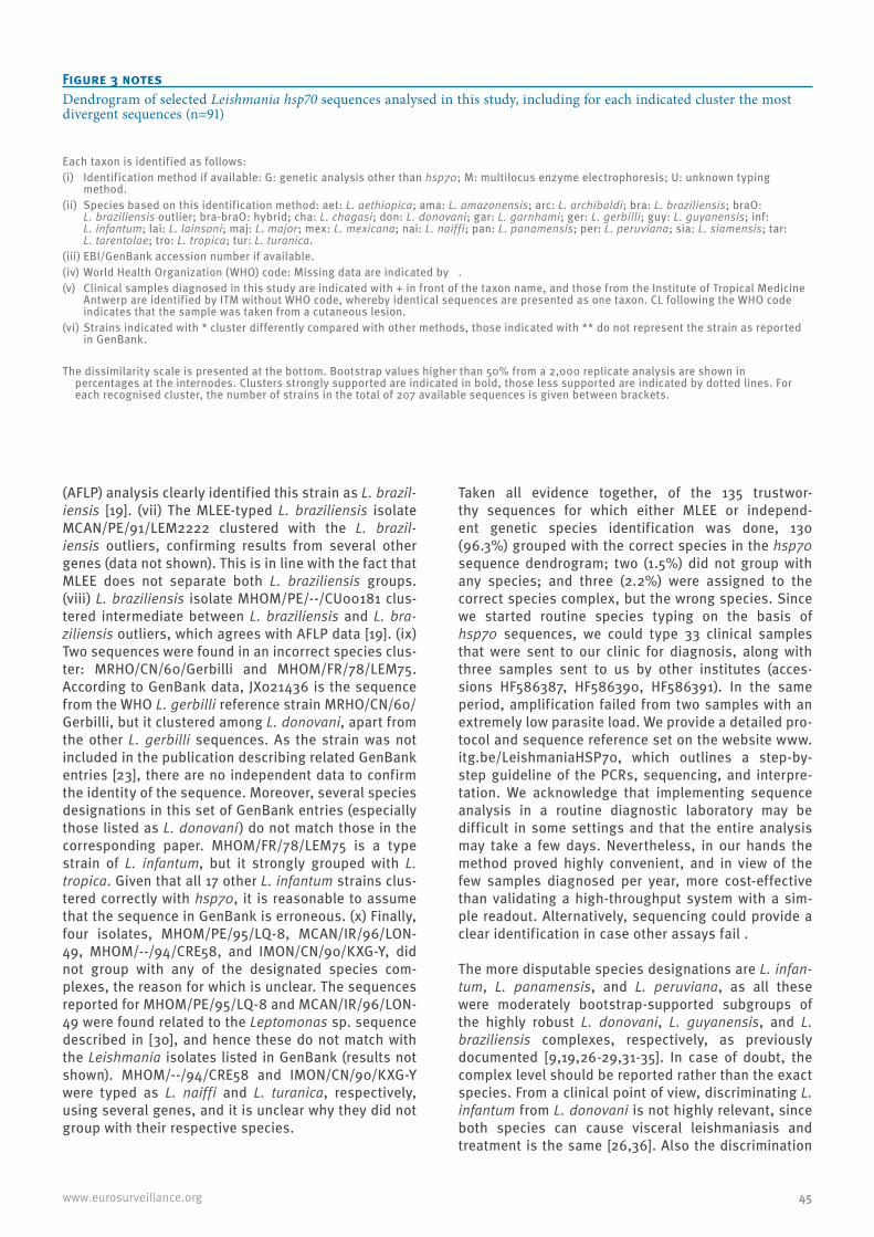
45www.eurosurveillance.org
(AFLP) analysis clearly identified this strain as L. brazil-iensis [19]. (vii) The MLEE-typed L. braziliensis isolate MCAN/PE/91/LEM2222 clustered with the L. brazil-iensis outliers, confirming results from several other genes (data not shown). This is in line with the fact that MLEE does not separate both L. braziliensis groups. (viii) L. braziliensis isolate MHOM/PE/--/CU00181 clus-tered intermediate between L. braziliensis and L. bra-ziliensis outliers, which agrees with AFLP data [19]. (ix) Two sequences were found in an incorrect species clus-ter: MRHO/CN/60/Gerbilli and MHOM/FR/78/LEM75. According to GenBank data, JX021436 is the sequence from the WHO L. gerbilli reference strain MRHO/CN/60/Gerbilli, but it clustered among L. donovani, apart from the other L. gerbilli sequences. As the strain was not included in the publication describing related GenBank entries [23], there are no independent data to confirm the identity of the sequence. Moreover, several species designations in this set of GenBank entries (especially those listed as L. donovani) do not match those in the corresponding paper. MHOM/FR/78/LEM75 is a type strain of L. infantum, but it strongly grouped with L. tropica. Given that all 17 other L. infantum strains clus-tered correctly with hsp70, it is reasonable to assume that the sequence in GenBank is erroneous. (x) Finally, four isolates, MHOM/PE/95/LQ-8, MCAN/IR/96/LON-49, MHOM/--/94/CRE58, and IMON/CN/90/KXG-Y, did not group with any of the designated species com-plexes, the reason for which is unclear. The sequences reported for MHOM/PE/95/LQ-8 and MCAN/IR/96/LON-49 were found related to the Leptomonas sp. sequence described in [30], and hence these do not match with the Leishmania isolates listed in GenBank (results not shown). MHOM/--/94/CRE58 and IMON/CN/90/KXG-Y were typed as L. naiffi and L. turanica, respectively, using several genes, and it is unclear why they did not group with their respective species.
Taken all evidence together, of the 135 trustwor-thy sequences for which either MLEE or independ-ent genetic species identification was done, 130 (96.3%) grouped with the correct species in the hsp70 sequence dendrogram; two (1.5%) did not group with any species; and three (2.2%) were assigned to the correct species complex, but the wrong species. Since we started routine species typing on the basis of hsp70 sequences, we could type 33 clinical samples that were sent to our clinic for diagnosis, along with three samples sent to us by other institutes (acces-sions HF586387, HF586390, HF586391). In the same period, amplification failed from two samples with an extremely low parasite load. We provide a detailed pro-tocol and sequence reference set on the website www.itg.be/LeishmaniaHSP70, which outlines a step-by-step guideline of the PCRs, sequencing, and interpre-tation. We acknowledge that implementing sequence analysis in a routine diagnostic laboratory may be difficult in some settings and that the entire analysis may take a few days. Nevertheless, in our hands the method proved highly convenient, and in view of the few samples diagnosed per year, more cost-effective than validating a high-throughput system with a sim-ple readout. Alternatively, sequencing could provide a clear identification in case other assays fail .
The more disputable species designations are L. infan-tum, L. panamensis, and L. peruviana, as all these were moderately bootstrap-supported subgroups of the highly robust L. donovani, L. guyanensis, and L. braziliensis complexes, respectively, as previously documented [9,19,26-29,31-35]. In case of doubt, the complex level should be reported rather than the exact species. From a clinical point of view, discriminating L. infantum from L. donovani is not highly relevant, since both species can cause visceral leishmaniasis and treatment is the same [26,36]. Also the discrimination
Figure 3 notesDendrogram of selected Leishmania hsp70 sequences analysed in this study, including for each indicated cluster the most divergent sequences (n=91)
Each taxon is identified as follows: (i) Identification method if available: G: genetic analysis other than hsp70; M: multilocus enzyme electrophoresis; U: unknown typing
method. (ii) Species based on this identification method: aet: L. aethiopica; ama: L. amazonensis; arc: L. archibaldi; bra: L. braziliensis; braO:
L. braziliensis outlier; bra-braO: hybrid; cha: L. chagasi; don: L. donovani; gar: L. garnhami; ger: L. gerbilli; guy: L. guyanensis; inf: L. infantum; lai: L. lainsoni; maj: L. major; mex: L. mexicana; nai: L. naiffi; pan: L. panamensis; per: L. peruviana; sia: L. siamensis; tar: L. tarentolae; tro: L. tropica; tur: L. turanica.
(iii) EBI/GenBank accession number if available.(iv) World Health Organization (WHO) code: Missing data are indicated by . (v) Clinical samples diagnosed in this study are indicated with + in front of the taxon name, and those from the Institute of Tropical Medicine
Antwerp are identified by ITM without WHO code, whereby identical sequences are presented as one taxon. CL following the WHO code indicates that the sample was taken from a cutaneous lesion.
(vi) Strains indicated with * cluster differently compared with other methods, those indicated with ** do not represent the strain as reported in GenBank.
The dissimilarity scale is presented at the bottom. Bootstrap values higher than 50% from a 2,000 replicate analysis are shown in percentages at the internodes. Clusters strongly supported are indicated in bold, those less supported are indicated by dotted lines. For each recognised cluster, the number of strains in the total of 207 available sequences is given between brackets.

46 www.eurosurveillance.org
between L. guyanensis and L. panamensis is not a pri-ority in clinical practice [36]. Separating L. braziliensis from L. peruviana is considered more relevant, because L. braziliensis potentially causes mucocutaneous com-plications, while L. peruviana generally does not [34]. As no markers are currently available that discriminate strains that do from those that do not cause mucocuta-neous leishmaniasis, identification at the species level is the only option. Both MLEE and genetic analyses have revealed that L. peruviana is a subcluster in the L. braziliensis complex, but discrimination is impaired by the fact that many parasites of this complex seem to have a composite genotype carrying signatures of both species [19,32-35]. The situation is further complicated by the fact that occasionally, L. peruviana can cause mucocutaneous disease [34]. Isolates belonging to the L. braziliensis outlier group have been isolated from mucous lesions as well (data not shown), but whether these were primary or secondary infections is not known. Two other tightly linked species in the hsp70 dendrogram are L. tropica and L. aethiopica. Although the L. tropica isolates cover the entire endemic region, from Morocco to eastern Africa, the Middle-East, and India, they form a clearly separated recognisable group. The same applies to separating L. mexicana from L. amazonensis, even though the latter could not be distinguished from L. garnhami.
One may wonder why some of the above species in the recognised larger complexes seem less clearly defined by sequencing than by single-nucleotide polymor-phism (SNP) assays such as species-specific PCRs or RFLP analysis. The reason is that these assays use a point mutation in the genome of the parasite, which is either present or absent, thereby allowing a binary dis-crimination. When using sequences, much more infor-mation is provided from many polymorphisms and is sometimes contradictory. Typing based on sequencing can therefore be more difficult, but it is more reliable as it uses more data. An accidental mutation may lead to erroneous conclusions in a SNP-based assay, while this is less likely when analysing entire sequences.
The current complete set of trustworthy sequences that can be used for typing amounts to 203, represent-ing 15 species of human medical importance, and origi-nating from 41 endemic countries. This reference set is updated continuously for further improvement of the geographic and genetic coverage, to ensure an ade-quate representation of the existing inter- and intra-species variability. Some species are over-represented from some regions (such as in Peru), but that does not interfere with the typing outcome. It is of crucial impor-tance to base species typing upon sequences that have been quality-checked. In practice, BLAST searches are often used for identification purposes, on the basis of hsp70 sequences found in public databases such as GenBank. We have found several instances where the species designation reported in these databases was incorrect. For example, in entries JX021425 up to JX021443 and JX970993 up to JX970996, several
erroneous L. donovani sequences are reported, even disagreeing with the species assignment as listed in the related publication [23]. Two entries were here shown related to Leptomonas rather than Leishmania (Figure 3 and supplementaty dendrogram). Two entries were determined from the L. infantum type strain MHOM/FR/78/LEM75: Y08020 and X85798. Both sequences clearly grouped with L. tropica, unlike all genuine L. infantum sequences in our analysis. This illustrates that one should be extremely careful when using sequences that have not been quality-controlled for species typing by comparison with other sequences from the same species, as this could result in incorrect typing outcomes, with potential adverse consequences for the patient.
As with all other assays based on the analysis of a single genomic locus, it is assumed that the relation-ship between the sequences mirror the relationship between the parasites. A first requirement to meet this objective is to avoid the use of paralogous sequences. This poses no problem in the case of hsp70 because, although this gene is part of a gene family [18], the primers used in our protocol specifically amplify only one of the family members. Another problem is pre-sented by the occasional inter-species hybrids that have been reported [19,34,37-40], and that are not necessarily evidenced in all genes. Nevertheless, such hybrids do not necessarily go undetected when looking at single genomic loci. For instance, isolate MHOM/PE/2006/CU00181, by AFLP clearly identified as a hybrid between L. braziliensis and L. braziliensis outliers [19], also holds an intermediate position in hsp70 sequences (Figure 3 and supplementary den-drogram). On the contrary, MHOM/PE/2003/LH2538, also shown to be a hybrid between these two clusters, grouped with L. braziliensis. As this isolate derived a much smaller proportion of its genome from the L. braziliensis outliers, such classification is however not problematic. In natural L. donovani–L. aethiopica hybrids, both species alleles were present in all genes investigated, including hsp70 [38]. In an L. infantum–L. major hybrid, both genomes were present [39], hence enabling to type the parasite based on a single gene assay. Theoretically, the chance of detecting inter-spe-cies recombinants increases as more loci are analysed, such as in multilocus-microsatellite, -sequence and -enzyme electrophoresis assays, but this also raises the cost and time of species typing. Given all currently available evidence on the potential of hsp70 to detect reported inter-species hybrids, and as such hybrids are rare, we consider this a negligible setback of using the hsp70 single-locus assay for routine species typ-ing. Nevertheless, additional more variable genes may be able to perform better in discriminating within the complexes, but this would probably require a separate approach for each complex or subgenus [32,41,42].
Ultimately, the use of single-locus sequencing for spe-cies discrimination could be substituted by whole-genome sequencing [43]. With this method becoming

47www.eurosurveillance.org
cheaper, it may soon be the standard in clinical studies. Comparison of whole-genome information could reveal clinically relevant intra-species differences, and has the highest chance of detecting recombination events. Such typing methodology could even make abstrac-tion of the classical concept of typing at the taxonomic levels of species and species complexes [44]. It could open up a whole new era of relating strains on the basis of a selection of genes relevant for disease progression and treatment options, rather than based upon species definitions that may at times not correlate with clini-cal outcome. Nevertheless, whole-genome sequencing seems at present miles away from being implemented in everyday clinical practice, not only because of the complexity of data analysis, but also because it is complicated by the presence of human DNA contamina-tion in clinical samples and related ethical issues. In the meantime there is a need for standardised meth-ods to identify Leishmania strains. We advocate that hsp70 has this potential: the gene is easily amplified, it can be analysed by sequencing in high-resource set-tings, and by simpler methods such as RFLP in limited-resource endemic areas [12,45]. If a global database of hsp70 sequences from endemic regions were to be established, new sequences found in imported leish-maniasis could immediately be related to documented parasites, with clinical information on the patients from whom they were isolated. Such analysis could even be independent of currently used species boundaries, and provide adequate links based on genetic similarity irre-spective of the species.
ConclusionWe present in this paper a complete validation and globally applicable standardised protocol for the use of hsp70 sequences in Leishmania typing. As this vali-dation includes a detailed comparison with other spe-cies identification methods currently used in various laboratories, we feel that implementation of the here presented typing strategy in a clinical diagnostic labo-ratory should be straight-forward, and could entail the validation of only the sequencing process itself rather than the actual species assignment, in view of this report. We intend to further promote our strategy, to identify additional strains with linked clinical informa-tion, and to establish a global database of circulating Leishmania parasites.
Acknowledgements The authors would like to thank Ingrid Felger (Swiss Tropical and Public Health Institute, Basel, Switzerland), Aldert Bart (Academic Medical Centre, Amsterdam, The Netherlands), Mark Bailey (Army Medical Directorate, Camberley, UK), and Ricardo Lleonart (INDICASAT, Panama City, Panama) for providing DNA from strains and clinical samples. We acknowledge the constructive input from all colleagues of the LeishMan consortium. We appreciate the help of Eva De Clercq (Université Catholique de Louvain, Louvain-la-Neuve, Belgium) for the construction of Figure 2. Salary support of GVDA was provided by the Belgian Directorate General for Development cooperation (third framework agreement with ITM).
References1. Bailey MS, Lockwood DN. Cutaneous leishmaniasis. Clin
Dermatol. 2007;25(2):203-11. http://dx.doi.org/10.1016/j.clindermatol.2006.05.008 PMid:17350500
2. Bailey MS, Green AD, Ellis CJ, O’Dempsey TJ, Beeching NJ, Lockwood DN, et al. Clinical guidelines for the management of cutaneous leishmaniasis in British military personnel. J R Army Med Corps. 2005;151(2):73-80. http://dx.doi.org/10.1136/jramc-151-02-03 PMid:16097110
3. Schwartz E, Hatz C, Blum J. New world cutaneous leishmaniasis in travellers. Lancet Infect Dis. 2006;6(6):342-9. http://dx.doi.org/10.1016/S1473-3099(06)70492-3
4. Blum JA, Hatz CF. Treatment of cutaneous leishmaniasis in travelers 2009. J Travel Med. 2009;16(2):123-31. http://dx.doi.org/10.1111/j.1708-8305.2008.00286.x PMid:19335813
5. Mitropoulos P, Konidas P, Durkin-Konidas M. New World cutaneous leishmaniasis: updated review of current and future diagnosis and treatment. J Am Acad Dermatol. 2010;63(2):309-22. http://dx.doi.org/10.1016/j.jaad.2009.06.088 PMid:20303613
6. Goto H, Lindoso JA. Current diagnosis and treatment of cutaneous and mucocutaneous leishmaniasis. Expert Rev Anti Infect Ther. 2010;8(4):419-33. http://dx.doi.org/10.1586/eri.10.19 PMid:20377337
7. Buffet PA, Rosenthal E, Gangneux JP, Lightburne E, Couppie P, Morizot G, et al. Therapy of leishmaniasis in France: consensus on proposed guidelines. Presse Med. 2011;40(2):173-84. http://dx.doi.org/10.1016/j.lpm.2010.09.023 PMid:21106333
8. Boecken G, Sunderkotter C, Bogdan C, Weitzel T, Fischer M, Muller A, et al. Diagnosis and therapy of cutaneous and mucocutaneous leishmaniasis in Germany. J Dtsch Dermatol Ges. 2011;9 Suppl 8:1-51. http://dx.doi.org/10.1111/j.1610-0379.2011.07820.x PMid:22050890
9. Fraga J, Montalvo AM, De Doncker S, Dujardin JC, Van der Auwera G. Phylogeny of Leishmania species based on the heat-shock protein 70 gene. Infect Genet Evol. 2010;10(2):238-45. http://dx.doi.org/10.1016/j.meegid.2009.11.007 PMid:19913110
10. Fraga J, Veland N, Montalvo AM, Praet N, Boggild AK, Valencia BM, et al. Accurate and rapid species typing from cutaneous and mucocutaneous leishmaniasis lesions of the New World. Diagn Microbiol Infect Dis. 2012;74(2):142-50. http://dx.doi.org/10.1016/j.diagmicrobio.2012.06.010 PMid:22819605
11. Montalvo AM, Fraga J, Monzote L, Montano I, De Doncker S, Dujardin JC, et al. Heat-shock protein 70 PCR-RFLP: a universal simple tool for Leishmania species discrimination in the New and Old World. Parasitology. 2010;137(8):1159-68 http://dx.doi.org/10.1017/S0031182010000089 PMid:20441679
12. Montalvo AM, Fraga J, Maes I, Dujardin JC, Van der Auwera G. Three new sensitive and specific heat-shock protein 70 PCRs for global Leishmania species identification. Eur J Clin Microbiol Infect Dis. 2012;31(7):1453-61. http://dx.doi.org/10.1007/s10096-011-1463-z PMid:22083340
13. Garcia L, Kindt A, Bermudez H, Llanos-Cuentas A, De Doncker S, Arévalo J, et al. Culture-independent species typing of neotropical Leishmania for clinical validation of a PCR-based assay targeting heat shock protein 70 genes. J Clin Microbiol. 2004;42(5):2294-7. http://dx.doi.org/10.1128/JCM.42.5.2294-2297.2004 PMid:15131217 PMCid:PMC404633
14. Marfurt J, Nasereddin A, Niederwieser I, Jaffe CL, Beck HP, Felger I. Identification and differentiation of Leishmania species in clinical samples by PCR amplification of the miniexon sequence and subsequent restriction fragment length polymorphism analysis. J Clin Microbiol. 2003;41(7):3147-53. http://dx.doi.org/10.1128/JCM.41.7.3147-3153.2003 PMid:12843055 PMCid:PMC165364
15. Schönian G, Nasereddin A, Dinse N, Schweynoch C, Schallig HD, Presber W, et al. PCR diagnosis and characterization of Leishmania in local and imported clinical samples. Diagn Microbiol Infect Dis. 2003;47(1):349-58 http://dx.doi.org/10.1016/S0732-8893(03)00093-2
16. Stevenson LG, Fedorko DP, Zelazny AM. An enhanced method for the identification of Leishmania spp. using real-time polymerase chain reaction and sequence analysis of the 7SL

48 www.eurosurveillance.org
RNA gene region. Diagn Microbiol Infect Dis. 2010;66(4):432-5. http://dx.doi.org/10.1016/j.diagmicrobio.2009.11.005 PMid:20226334 PMCid:PMC2856081
17. Roelfsema JH, Nozari N, Herremans T, Kortbeek LM, Pinelli E. Evaluation and improvement of two PCR targets in molecular typing of clinical samples of Leishmania patients. Exp Parasitol. 2011;127(1):36-41. http://dx.doi.org/10.1016/j.exppara.2010.06.024 PMid:20599989
18. Folgueira C, Ca-avate C, Chicharro C, Requena JM. Genomic organization and expression of the HSP70 locus in New and Old World Leishmania species. Parasitology. 2007;134(Pt 3):369-77. http://dx.doi.org/10.1017/S0031182006001570 PMid:17054823
19. Odiwuor S, Veland N, Maes I, Arévalo J, Dujardin JC, Van der Auwera G. Evolution of the Leishmania braziliensis species complex from amplified fragment length polymorphisms, and clinical implications. Infect Genet Evol. 2012;12(8):1994-2002. http://dx.doi.org/10.1016/j.meegid.2012.03.028 PMid:22516226
20. Deborggraeve S, Laurent T, Espinosa D, Van der Auwera G, Mbuchi M, Wasunna M, et al. A simplified and standardized polymerase chain reaction format for the diagnosis of leishmaniasis. J Infect Dis. 2008;198(10):1565-72. http://dx.doi.org/10.1086/592509 PMid:18816188
21. Odiwuor S, Muia A, Magiri C, Maes I, Kirigi G, Dujardin JC, et al. Identification of Leishmania tropica from micro-foci of cutaneous leishmaniasis in the Kenyan Rift Valley. Pathog Glob Health. 2012;106(3):159-65. http://dx.doi.org/10.1179/2047773212Y.0000000015 PMid:23265373
22. DNA Baser Sequence Assembler. Nucleotide ambiguity code. Pitesti: Heracle BioSoft S.R.L. Available from: http://www.dnabaser.com/articles/IUPAC%20ambiguity%20codes.html
23. Zhang CY, Lu XJ, Du XQ, Jian J, Shu L, Ma Y. Phylogenetic and evolutionary analysis of Chinese Leishmania isolates based on multilocus sequence typing. PLoS One. 2013;8(4):e63124. http://dx.doi.org/10.1371/journal.pone.0063124 PMid:23646184 PMCid:PMC3639960
24. Tamura K, Peterson D, Peterson N, Stecher G, Nei M, Kumar S. MEGA5: molecular evolutionary genetics analysis using maximum likelihood, evolutionary distance, and maximum parsimony methods. Mol Biol Evol. 2011;28(10):2731-9. http://dx.doi.org/10.1093/molbev/msr121 PMid:21546353 PMCid:PMC3203626
25. Mauricio IL, Stothard JR, Miles MA. The strange case of Leishmania chagasi. Parasitol Today. 2000;16(5):188-9. http://dx.doi.org/10.1016/S0169-4758(00)01637-9
26. Lukeš J, Mauricio IL, Schönian G, Dujardin JC, Soteriadou K, Dedet JP, et al. Evolutionary and geographical history of the Leishmania donovani complex with a revision of current taxonomy. Proc Natl Acad Sci USA. 2007;104(22):9375-80. http://dx.doi.org/10.1073/pnas.0703678104 PMid:17517634 PMCid:PMC1890502
27. Ba-uls AL, Hide M, Tibayrenc M. Evolutionary genetics and molecular diagnosis of Leishmania species. Trans R Soc Trop Med Hyg. 2002;96 Suppl 1:S9-13. http://dx.doi.org/10.1016/S0035-9203(02)90045-3
28. Bañuls AL, Jonquieres R, Guerrini F, Le Pont F, Barrera C, Espinel I, et al. Genetic analysis of Leishmania parasites in Ecuador: are Leishmania (Viannia) panamensis and Leishmania (V.) guyanensis distinct taxa? Am J Trop Med Hyg. 1999;61(5):838-45.
29. Cupolillo E, Grimaldi G Jr, Momen H. A general classification of New World Leishmania using numerical zymotaxonomy. Am J Trop Med Hyg. 1994;50(3):296-311. PMid:8147488
30. Srivastava P, Prajapati VK, Vanaerschot M, Van der Auwera G, Dujardin JC, Sundar S. Detection of Leptomonas sp. parasites in clinical isolates of Kala-azar patients from India. Infect Genet Evol. 2010;10(7):1145-50. http://dx.doi.org/10.1016/j.meegid.2010.07.009 PMid:20633704 PMCid:PMC2933273
31. Odiwuor S, Vuylsteke M, De Doncker S, Maes I, Mbuchi M, Dujardin JC, et al. Leishmania AFLP: paving the way towards improved molecular assays and markers of diversity. Infect Genet Evol. 2011;11(5):960-7. http://dx.doi.org/10.1016/j.meegid.2011.03.008 PMid:21439405
32. Oddone R, Schweynoch C, Schönian G, de Sousa CS, Cupolillo E, Espinosa D, et al. Development of a multilocus microsatellite typing approach for discriminating strains of Leishmania (Viannia) species. J Clin Microbiol. 2009;47(9):2818-25.
http://dx.doi.org/10.1128/JCM.00645-09 PMid:19587302 PMCid:PMC2738093
33. Veland N, Boggild AK, Valencia C, Valencia BM, Llanos-Cuentas A, Van der Auwera G, et al. Leishmania (Viannia) species identification on clinical samples from cutaneous leishmaniasis patients in Peru: assessment of a molecular stepwise approach. J Clin Microbiol. 2012;50(2):495-8. http://dx.doi.org/10.1128/JCM.05061-11 PMid:22116151 PMCid:PMC3264178
34. Nolder D, Roncal N, Davies CR, Llanos-Cuentas A, Miles MA. Multiple hybrid genotypes of Leishmania (Viannia) in a focus of mucocutaneous leishmaniasis. Am J Trop Med Hyg. 2007;76(3):573-8. PMid:17360886
35. Bañuls AL, Dujardin JC, Guerrini F, De Doncker S, Jacquet D, Arevalo J, et al. Is Leishmania (Viannia) peruviana a distinct species? A MLEE/RAPD evolutionary genetics answer. J Eukaryot Microbiol. 2000;47(3):197-207. http://dx.doi.org/10.1111/j.1550-7408.2000.tb00039.x
36. World Health Organization (WHO). Control of the leishmaniasis: report of a meeting of the WHO Expert Committee on the Control of Leishmaniases, Geneva, 22-26 March 2010. Geneva, Switzerland: WHO; 2010. Available from: http://whqlibdoc.who.int/trs/WHO_TRS_949_eng.pdf
37. Belli AA, Miles MA, Kelly JM. A putative Leishmania panamensis/Leishmania braziliensis hybrid is a causative agent of human cutaneous leishmaniasis in Nicaragua. Parasitology. 1994;109(Pt 4):435-42. http://dx.doi.org/10.1017/S0031182000080689 PMid:7800411
38. Odiwuor S, De Doncker S, Maes I, Dujardin JC, Van der Auwera G. Natural Leishmania donovani/Leishmania aethiopica hybrids identified from Ethiopia. Infect Genet Evol. 2011;11(8):2113-8. http://dx.doi.org/10.1016/j.meegid.2011.04.026 PMid:21558020
39. Ravel C, Cortes S, Pratlong F, Morio F, Dedet JP, Campino L. First report of genetic hybrids between two very divergent Leishmania species: Leishmania infantum and Leishmania major. Int J Parasitol. 2006;36(13):1383-8. http://dx.doi.org/10.1016/j.ijpara.2006.06.019 PMid:16930606
40. Delgado O, Cupolillo E, Bonfante-Garrido R, Silva S, Belfort E, Grimaldi JG, et al. Cutaneous leishmaniasis in Venezuela caused by infection with a new hybrid between Leishmania (Viannia) braziliensis and L. (V.) guyanensis. Mem Inst Oswaldo Cruz. 1997;92(5):581-2. http://dx.doi.org/10.1590/S0074-02761997000500002 PMid:9566221
41. Kuhls K, Keilonat L, Ochsenreither S, Schaar M, Schweynoch C, Presber W, et al. Multilocus microsatellite typing (MLMT) reveals genetically isolated populations between and within the main endemic regions of visceral leishmaniasis. Microbes Infect. 2007;9(3):334-43. http://dx.doi.org/10.1016/j.micinf.2006.12.009 PMid:17307010
42. Zemanová E, Jirku M, Mauricio IL, Horák A, Miles MA, Lukeš J. The Leishmania donovani complex: genotypes of five metabolic enzymes (ICD, ME, MPI, G6PDH, and FH), new targets for multilocus sequence typing. Int J Parasitol. 2007;37(2):149-60. http://dx.doi.org/10.1016/j.ijpara.2006.08.008 PMid:17027989
43. Downing T, Imamura H, Decuypere S, Clark TG, Coombs GH, Cotton JA, et al. Whole genome sequencing of multiple Leishmania donovani clinical isolates provides insights into population structure and mechanisms of drug resistance. Genome Res. 2011;21(12):2143-56. http://dx.doi.org/10.1101/gr.123430.111 PMid:22038251 PMCid:PMC3227103
44. Van der Auwera G, Fraga J, Montalvo AM, Dujardin JC. Leishmania taxonomy up for promotion? Trends Parasitol. 2011;27(2):49-50. http://dx.doi.org/10.1016/j.pt.2010.11.007 PMid:21147036
45. da Silva LA, dos Santos de Sousa C, da Graça GC, Porrozzi R, Cupolillo E. Sequence analysis and PCR-RFLP profiling of the hsp70 gene as a valuable tool for identifying Leishmania species associated with human leishmaniasis in Brazil. Infect Genet Evol. 2010;10(1):77-83 http://dx.doi.org/10.1016/j.meegid.2009.11.001 PMid:19913112
46. Alvar J, Velez ID, Bern C, Herrero M, Desjeux P, Cano J, et al. Leishmaniasis Worldwide and Global Estimates of Its Incidence. PLoS One. 2012;7(5):e35671. http://dx.doi.org/10.1371/journal.pone.0035671 PMid:22693548 PMCid:PMC3365071

49www.eurosurveillance.org
Perspectives
Leishmaniasis in the era of tumor necrosis factor alpha antagonist therapy – a research agenda for Europe
P Zanger ([email protected])1, S Gabrysch2
1. Institut für Tropenmedizin, Eberhard Karls Universität, Tübingen, Germany2. Institut für Public Health, Ruprecht Karls Universität, Heidelberg, Germany
Citation style for this article: Zanger P, Gabrysch S. Leishmaniasis in the era of tumor necrosis factor alpha antagonist therapy – a research agenda for Europe. Euro Surveill. 2013;18(30):pii=20542. Available online: http://www.eurosurveillance.org/ViewArticle.aspx?ArticleId=20542
Article submitted on 26 May 2012 / published on 25 July 2013
A number of published case reports suggest an asso-ciation of tumor necrosis factor (TNF) alpha antagonist use and manifest leishmaniasis. Despite increasing popularity of antagonising TNF alpha for the treatment of autoimmune disorders, systematic research on the risk of opportunistic leishmaniasis in patients receiv-ing these drugs is lacking. This perspective identifies areas of uncertainty regarding the safety profile of TNF alpha antagonist drugs and their clinical use in patients at risk of leishmaniasis. Then, we reflect on how current pharmacovigilance activities in Europe could be enhanced to help reduce these uncertainties. Our aim is to stimulate a debate about this important drug safety issue with potential consequences for patients receiving TNF alpha antagonists living in or travelling to areas endemic for leishmaniasis.
IntroductionSince their introduction about a decade ago, tumour necrosis factor (TNF) alpha antagonist drugs have greatly improved the clinical management of autoim-mune disorders and are now widely used in rheumatol-ogy. The flipside of the coin is an increase in infectious disease risk in patients treated with these drugs. Both reactivation of latent infections and increased suscepti-bility to new infections have been observed in patients receiving TNF alpha antagonist therapy. Depending on their mode of action, the TNF alpha antagonist drugs most widely used can be distinguished in two major groups: monoclonal antibodies, such as infliximab and adalimumab, and the receptor construct etanercept [1].
Leishmaniasis is endemic in large parts of southern Europe [2-5] where studies suggest a focal prevalence of latent infection of up to 53% in the adult popula-tion [5]. Two species, Leishmania infantum and L. trop-ica are transmitted around the Mediterranean basin by the bite of sandfly species (order Diptera, family Psychodidae, subfamily Phlebotominae) [4]. The spec-trum of manifest infection ranges from a well localised, self-healing cutaneous papule or ulceration to rapidly fatal visceral disease. Host immunity is known to be a key determinant of the clinical manifestation and out-come. Immuno¬suppression, in particular conditions
that alter the type 1 helper T-cell-mediated immune response of which TNF alpha is an important compo-nent, is generally regarded as a risk factor for manifest leishmaniasis and has been identified as a major con-tributor to its re-emergence in Europe [4]. A number of published case reports suggest an association of TNF alpha antagonist use and manifest leishmaniasis [6, 7]. Despite this, no systematic research on opportunistic leishmaniasis in patients receiving TNF alpha antago-nists has been conducted.
Based on a brief summary of published information on opportunistic leishmaniasis and use of TNF alpha antagonists, this perspective identifies areas of uncer-tainty regarding their safety profile and their clinical use in patients at risk of leishmaniasis. We then reflect on how current pharmacovigilance activities in Europe could be enhanced to help reduce these uncertainties. Our aim is to stimulate a debate about this important drug safety issue with potential consequences for patients receiving TNF alpha antagonists living in or travelling to areas endemic for leishmaniasis.
Current knowledge on tumor necrosis factor alpha antagonist therapy and leishmaniasisPublished research on therapy with TNF alpha antago-nists and leishmaniasis is scarce and limited to case reports and case series. We recently reviewed the lit-erature [6] and identified 19 descriptions of patients with leishmaniasis while receiving TNF antagonists, published between 2004 and 2011. Nearly all of the identified cases (18/19) occurred in Europe, and more than two thirds were published in 2010-11. The vast majority were reported from endemic regions of southern Europe (14/18) whereas fewer occurred after travel to and migration from endemic areas (4/18). The reported time period from initiation of treatment to onset of leishmaniasis ranged from 0.5 to 48 months, consistent with both reactivation of latent infection and increased susceptibility to new infection.
Only one reported case was treated with etaner-cept while all others received either infliximab or
50 www.eurosurveillance.org
adalimumab. This was surprising, since prescription data from the countries where the cases occurred showed that each of the three drugs was prescribed about equally often. These findings suggest that opportunistic leishmaniasis is more likely to occur in patients receiving TNF alpha monoclonal antibodies than in patients treated with etanercept. This interpre-tation is supported by similar findings for tuberculosis [8, 9] and by studies in mice [10] and in vitro [11].
Interestingly, published case reports describe various approaches including discontinuation as well as con-tinuation of TNF alpha antagonist therapy during and after anti-parasitic treatment while using the same or a different type of drug for ongoing TNF alpha antago-nisation [6]. Their number, however, is too small and
the data and observation period reported too hetero-geneous to decide whether one or the other approach is associated with an increased risk of recurrent leishmaniasis.
Although it provided us with a hypothesis on the pos-sible differences of leishmaniasis risk by type of TNF alpha antagonist used, our analysis of case reports is limited by potential publication bias and confounding underlying the observed associations. Moreover, the number of published cases is likely to represent only a small fraction of patients treated with TNF alpha antagonists with opportunistic leishmaniasis, leaving the magnitude of this drug safety issue unclear. Hence more systematic research is needed to improve our
Box 1Aims and objectives of a coordinated research effort on opportunistic leishmaniasis in patients treated with tumor necrosis factor alpha antagonists in Europe
Aims
• Estimate the impact of TNF alpha use on the occurrence of leishmaniasis in Europe.
• Reduce leishmaniasis risk by tailoring TNF alpha antagonist therapy to individual risk profile.
• Optimise anti-parasitic and anti-rheumatic therapy in patients with opportunistic leishmaniasis.
Objectives
• Define the absolute risk of leishmaniasis in patients treated with TNF alpha antagonists. Define the relative risk of leishmaniasis in patients treated with TNF alpha antagonists
with respect to classic immunosuppressive regimens; and with respect to type of TNF alpha antagonist used.
• Define the proportionate contribution of new infection and reactivation towards the burden of leishmaniasis in patients treated with TNF alpha antagonists
overall; and by type of TNF alpha antagonist used.
• Define the role of screening (serology, intradermal leishmanin) in patients treated with TNF alpha antagonists for clinical decision making by determining their predictive values
with regard to the overall risk of leishmaniasis; and depending on the type of TNF alpha antagonist used.
• Identify groups at risk of and factors associated with developing leishmaniasis during TNF alpha antagonist therapy namely
environmental/behavioural (e.g. companion animals, travel, region of residence) factors; host factors (e.g. co-morbidity); and others (e.g. immunosuppressive co-medication).
• Optimise therapy of leishmaniasis as complication of TNF alpha therapy through either
interruption or continuation of TNF alpha antagonist; and systemic or local treatment of cutaneous leishmaniasis.
• Explore whether TNF alpha antagonists can be restarted after cure of leishmaniasis and if
there is a need for potential modifications of this therapy; treatment can be continued with identical or alternative TNF alpha antagonist; immunosuppressive co-medication needs to be modified; and anti-parasitic maintenance therapy is necessary to prevent relapses (secondary prevention).
TNF: tumor necrosis factor.

51www.eurosurveillance.org
understanding of opportunistic leishmaniasis in these patients.
Unsolved questionsThe main areas of uncertainty pertaining to opportunis-tic leishmaniasis in patients receiving TNF alpha antag-onist therapy, to be addressed by future research, are outlined below and summarised in Box 1.
There is a clear need to estimate the potential impact of TNF alpha antagonist use on the incidence of leish-maniasis. This information is required to clarify to what extent there is a need to further investigate and corroborate this association and to justify funding of research activities that aim at elucidating risk factors for leishmaniasis among those treated with TNF alpha antagonists.
Any study on risk factors should attempt to answer whether there truly is a difference in risk of leishma-niasis depending on the type of TNF alpha antagonist used. Once supported by evidence from prospective studies, physicians (mainly clinical rheumatologists) could directly translate this knowledge into practice by choosing the TNF alpha antagonist with the lowest risk for opportunistic leishmaniasis for patients living in or travelling to endemic areas. Moreover, the role of behavioural and host-associated risk factors for clini-cally manifest leishmaniasis under TNF alpha antago-nist therapy should be evaluated as knowledge gained could be relevant in clinical decision-making or coun-selling of patients.
Once a risk difference for the various types of TNF alpha antagonists has been established, it will be important to clarify if this is due to reactivation of latent leishma-niasis or increased susceptibility to new infection.
It needs to be determined whether screening for latent infection before initiation of TNF alpha antagonist ther-apy can play a role in preventing opportunistic leish-maniasis. Studies evaluating the predictive values of serologic and intradermal leishmanin testing have great potential to inform clinical decision-making and will equally contribute to our understanding of latent versus newly acquired infection for the onset of oppor-tunistic leishmaniasis in patients receiving anti-TNF alpha therapy.
Apart from defining risk factors, there is a need for clinical research to improve medical care for patients suffering from opportunistic leishmaniasis. In par-ticular, better evidence on whether TNF alpha antago-nists can be continued in patients with clinically active leishmaniasis, or after its cure, is of high clinical rel-evance since many of these patients depend on TNF alpha antagonists to adequately control the underly-ing autoimmune disease. Besides, future research has to address whether there is a need for secondary prevention of opportunistic leishmaniasis in patients that require sustained antagonisation of TNF alpha.
For instance, type and duration as well as indicators for the initiation of anti-parasitic maintenance therapy need to be established. Of note, anti-TNF alpha ther-apy has been continued or re-initiated in several cases of opportunistic leishmaniasis without subsequent relapse [6, 12], indicating that a first episode does not justify long-term anti-parasitic treatment.
Finally, we have to learn more about the risk of general-ised infection secondary to localised cutaneous leish-maniasis in patients receiving TNF alpha antagonists. Published case reports indicate that most clinicians fear this complication and opt for systemic anti-para-sitic therapy. This, however, has to be balanced against increased toxicity when compared to local treatment which could be an option in a setting where close moni-toring of the patient is ensured.
Research challenges concerning opportunistic leish-maniasis and tumor necrosis factor alpha antagonist therapy
Traditionally, spontaneous notification of adverse events to national pharmacovigilance systems has been used to define a drug’s safety profile with regard to rare events and long-term effects that may have remained undetected during pre-licensure clinical tri-als. This approach, however, is subject to significant underreporting, does not allow analysing the number of reported events relative to the number of subjects treated, and cannot provide an estimate of the baseline risk.
In response to these shortcomings and with the support of the pharmaceutical industry, national rheumatology societies in several European countries have initiated national drug registers as post-marketing surveillance tools [13]. Many of these were put in place simultane-ously to the licensing of the first TNF alpha antagonists about a decade ago. Their methodologies were recently reviewed in detail by Zink et al. [13]. In brief, selected care providers in rheumatology enrol patients receiv-ing TNF alpha antagonists or other biotherapies into epidemiological cohort studies or into registers, thus overcoming some of the limitations of traditional phar-macovigilance activities.
Although a huge improvement compared to the tradi-tional reporting systems, it has been called into ques-tion whether national drug registers cover a sufficient patient base to detect rare adverse events. Zink et al. estimate that events with an incidence equal or below one in 1,000 patient-years may not be adequately detected by this approach [13]. With regard to leish-maniasis, we observed that the French register only detected half of leishmaniasis cases reported in pub-lications from France over a defined time period [6], implying that underreporting may not be adequately addressed by the national registers. Besides, none of these registers had detected sufficient numbers of cases to allow analysing whether leishmaniasis risk

52 www.eurosurveillance.org
varies by type of TNF alpha antagonist. As pointed out before, this was possible by combining published information from different European countries. Based hereon, it becomes obvious that fragmentation of data at national level hampers a better description of the safety profile of TNF alpha antagonists with regard to rare opportunistic infections. Therefore, evaluation of data across registers and at European level has been suggested to increase the ability to detect such events [13]. Although this approach has potential to contribute to our understanding of opportunistic leishmaniasis, a range of challenges remain.
Firstly, transmission of leishmaniasis occurs geograph-ically in confined foci since it is bound to the presence of the phlebotomine vector. This is reflected in the large variation of sero-prevalence reported in regions around the Mediterranean basin [2, 3, 5]. Hence, to further advance our knowledge on opportunistic leish-maniasis in patients receiving biotherapies, it will be crucial that national drug registers adequately cover the population in areas where transmission occurs. The variation in transmission risk also implies that estimates of the absolute risk of leishmaniasis in TNF alpha antagonist users calculated from register data will vary according to the proportion of subjects live in endemic regions enrolled in the register.
Secondly, European national drug registers on the safety of TNF alpha antagonist drugs and other bio-therapies use different methodology and do not record data in a uniform manner. To facilitate collaboration between national drug registers and to allow supra-national data analyses, harmonisation of European
registers is a prerequisite. A uniform reporting scheme has so far only been adopted by Great Britain, Sweden, and Germany, but not by France and Spain, countries where leishmaniasis is endemic [13].
Thirdly, it is unclear, whether all countries with leish-maniasis endemic regions have an active national drug register or cohort. For instance, from a number of coun-tries in the Balkan region, there are no peer-reviewed publications available on PubMed reporting on such activities.
The Table outlines potential components of a coordi-nated research effort on opportunistic leishmaniasis in Europe and Box 2 the resulting strengths.
ConclusionsAlthough the initiation of national registers with sup-port of the pharmaceutical industry was a major step forward in better defining the safety profile of TNF alpha antagonist drugs and other biotherapies, there are shortcomings of this system with regard to oppor-tunistic leishmaniasis. Crucial for improved case detec-tion and reduced underreporting will be a closer link between existing national drug registers and insti-tutions specialised in the diagnosis and therapy of leishmaniasis, e.g. tropical medicine institutes, der-matology departments, etc. This will likely enable the detection of a considerable number of additional cases of opportunistic leishmaniasis that are either directly seen at these institutions or were suspected elsewhere but laboratory confirmed there. Using existing net-work structures in Europe such as TropNet (www.tro-pnet.net) and EuroTravNet (www.istm.org/eurotravnet/
TableComponents of a coordinated research effort on opportunistic leishmaniasis in Europe
Component Function/role
Coordinating board hosted byEuropean institution including representatives from existing European network structures in clinical parasitology/ tropical medicine, dermatology, rheumatology, infectious diseases and supported by public health experts
• Design of post-authorisation safety studies (cohort studies) and observational clinical studies
• Ensure uniform methodology and data collection• Enhance national drug registers in countries endemic for leishmaniasis (where
necessary)• Host online platform for case registration by non-endemic countries (i.e. travellers,
migrants)• Increase awareness among policy-makers and professional societies
National drug registers hosted by professional societies in rheumatology
• Implementation of cohort studies• Increase awareness/ case reporting among clinicians
Institutions specialised in diagnosis and therapy of leishmaniasis, represented through existing European network structures in clinical parasitology/ tropical medicine, dermatology, infectious diseases
• Implementation of clinical studies• Detection of cases outside endemic countries• Increase awareness / case reporting among clinicians
Pharmaceutical industry• Data on drug sales with sufficient detail allowing geographically high resolution in
leishmaniasis endemic areas• Funding

53www.eurosurveillance.org
main.html) could facilitate the creation of such links. An extended network of clinical specialists in rheu-matology, infectious diseases, tropical medicine, and dermatology will be necessary to answer the research questions pertaining to the clinical management of patients with opportunistic leishmaniasis set out in this paper.
Prospective data from a well-defined population at risk of acquiring or being latently infected with leishma-nia will be needed to obtain meaningful estimates of the incidence of clinical manifest infection that can be attributed to TNF alpha antagonist use and to evaluate the clinical role of screening for latent infection. This can only be achieved through focused post-marketing safety studies that make use of large cohorts in areas endemic for leishmaniasis. To ensure uniform meth-odology and data collection, such activity should be designed and coordinated at European level before being implemented by the existing national registers. In European countries endemic for leishmaniasis but without a well-functioning register or cohort, this may require enhancing or newly creating such structures. Taken together, these activities will need adequate funding and most importantly, substantial collabora-tion across disciplines including input from epidemi-ologists and public health experts.
Conflict of interest None declared.
References1. Scallon B, Cai A, Solowski N, Rosenberg A, Song XY, Shealy
D, et al. Binding and functional comparisons of two types of tumor necrosis factor antagonists. J Pharmacol Exp Ther. 2002;301(2):418-26. http://dx.doi.org/10.1124/jpet.301.2.418
2. Diza E, Kansouzidou A, Gerou S, Vezyri E, Metallidis S, Antoniadis A. Leishmaniases in northern greece: Seroprevalence of the infection and incidence of the disease during the period 2001-2006. Eur J Clin Microbiol Infect Dis. 2008;27(10):997-1003. http://dx.doi.org/10.1007/s10096-008-0538-y PMid:18512088
3. Federico G, Damiano F, Caldarola G, Fantini C, Fiocchi V, Ortona L. A seroepidemiological survey on leishmania infantum infection. Eur J Epidemiol. 1991;7(4):380-3. http://dx.doi.org/10.1007/BF00145003
4. Ready PD. Leishmaniasis emergence in europe. Euro Surveill. 2010;15(10):19505. (10):pii=19505. Available from: http://www.eurosurveillance.org/ViewArticle.aspx?ArticleId=19505
5. Moral L, Rubio EM, Moya M. A leishmanin skin test survey in the human population of l’alacanti region (spain): Implications for the epidemiology of leishmania infantum infection in southern europe. Trans R Soc Trop Med Hyg. 2002;96(2):129-32. http://dx.doi.org/10.1016/S0035-9203(02)90278-6
6. Zanger P, Kötter I, Kremsner PG, Gabrysch S. Tumor necrosis factor alpha antagonist drugs and leishmaniasis in europe. Clin Microbiol Infect. 2012;18(7):670-6. http://dx.doi.org/10.1111/j.1469-0691.2011.03674.x PMid:21985586
7. Xynos ID, Tektonidou MG, Pikazis D, Sipsas NV. Leishmaniasis, autoimmune rheumatic disease, and anti-tumor necrosis factor therapy, europe. Emerg Infect Dis. 2009;15(6):956-9. http://dx.doi.org/10.3201/eid1506.090101 PMid:19523302 PMCid:PMC2727307
8. Wallis RS, Broder MS, Wong JY, Hanson ME, Beenhouwer DO. Granulomatous infectious diseases associated with tumor necrosis factor antagonists. Clin Infect Dis. 2004;38(9):1261-5. http://dx.doi.org/10.1086/383317 PMid:15127338
9. Tubach F, Salmon D, Ravaud P, Allanore Y, Goupille P, Bréban M, et al. Risk of tuberculosis is higher with anti-tumor necrosis factor monoclonal antibody therapy than with soluble tumor necrosis factor receptor therapy: The three-year prospective french research axed on tolerance of biotherapies registry. Arthritis Rheum. 2009;60(7):1884-94. http://dx.doi.org/10.1002/art.24632 PMid:19565495 PMCid:PMC2921546
10. Allenbach C, Launois P, Mueller C, Tacchini-Cottier F. An essential role for transmembrane tnf in the resolution of the inflammatory lesion induced by leishmania major infection. Eur J Immunol. 2008;38(3):720-31. http://dx.doi.org/10.1002/eji.200737662 PMid:18266271
11. Lohmann-Matthes ML, Luttig B, Hockertz S. Involvement of membrane-associated tnf in the killing of leishmania donovani parasites by macrophages. Behring Inst Mitt. 1991;88:125-32. PMid:2049029
12. Zanger P, Kötter I, Raible A, Gelanew T, Schonian G, Kremsner PG. Successful treatment of cutaneous leishmaniasis caused by leishmania aethiopica with liposomal amphothericin b in an immunocompromised traveler returning from eritrea. Am J Trop Med Hyg. 2011;84(5):692-4. http://dx.doi.org/10.4269/ajtmh.2011.10-0712 PMid:21540377 PMCid:PMC3083735
13. Zink A, Askling J, Dixon WG, Klareskog L, Silman AJ, Symmons DP. European biologicals registers: Methodology, selected results and perspectives. Ann Rheum Dis. 2009;68(8):1240-6. http://dx.doi.org/10.1136/ard.2008.091926 PMid:18647854
Box 2Strengths of a coordinated research effort at European level on opportunistic leishmaniasis in patients treated with tumor necrosis factor alpha antagonists
• Impact – a coordinated research effort will allow estimating the public health impact of tumor necrosis factor alpha (TNF) alpha antagonists on leishmaniasis in Europe.
• Statisticalpower–acoordinatedresearcheffortwillincrease statistical power through improved case detection and more comprehensive coverage of populations in endemic regions.
• Defragmentation–acoordinatedresearcheffortwillensure that data available is not restricted to single countries.
• Standardisation–acoordinatedresearcheffortwillimprove database quality for supra-national analysis through uniform methodology, reporting scheme, and common denominators (e.g. data on drug sales).
• Flexibility–acoordinatedresearcheffortwillallowrapidincorporation of newly emerging biotherapies and rare infections other than leishmaniasis.

54 www.eurosurveillance.org
Review articles
The role of indigenous phlebotomine sandflies and mammals in the spreading of leishmaniasis agents in the Mediterranean region
M Antoniou1, M Gramiccia2, R Molina3, V Dvorak ([email protected])4, P Volf4
1. Laboratory of Clinical Bacteriology Parasitology Zoonoses and Geographical Medicine, Faculty of Medicine,University of Crete, Iraklion, Greece
2. Unit of Vector-borne Diseases and International Health, MIPI Department, Istituto Superiore di Sanità, Rome, Italy 3. Medical Entomology Unit, Servicio de Parasitología, Centro Nacional de Microbiología, Instituto de Salud Carlos III,
Majadahonda, Madrid, Spain4. Department of Parasitology, Faculty of Science, Charles University, Prague, Czech Republic
Citation style for this article: Antoniou M, Gramiccia M, Molina R, Dvorak V, Volf P. The role of indigenous phlebotomine sandflies and mammals in the spreading of leishmaniasis agents in the Mediterranean region. Euro Surveill. 2013;18(30):pii=20540. Available online: http://www.eurosurveillance.org/ViewArticle.aspx?ArticleId=20540
Article submitted on 26 June 2012 / published on 25 July 2013
An updated view of the establishment and spread of the leishmaniases in Europe is presented, mostly with respect to newly emerging and re-emerging foci and the incrimination of neglected as well as new reser-voir hosts. At the same time, a concept of specific ver-sus permissive vectors reassesses the potential role of various sandfly species in Leishmania transmis-sion and considers the risk of introduction of exotic Leishmania species in Europe. The leishmaniases are dynamic diseases and the circumstances of transmis-sion are continually changing in relation to environ-mental, demographic and human behavioural factors. Changes in the habitat of the natural hosts and vec-tors, immunosuppressive conditions (like infection with human immunodeficiency virus (HIV) or organ transplantation-associated therapies in humans) and the consequences of war, all contribute to the trans-formation of the epidemiology of leishmaniasis. Such changes should be considered when studying the spread of the disease throughout Europe for targeted control measures to safeguard public health.
Endemic burden of human leishmaniases in Europe The leishmaniases are vector-borne diseases that have been endemic in southern Europe for centuries. They are transmitted by the bite of phlebotomine sandflies belonging to the genus Phlebotomus and most often exhibit one of two endemic clinical entities in humans, zoonotic visceral leishmaniasis (VL) and sporadic cuta-neous leishmaniasis (CL). Four Leishmania species occur in the Mediterranean basin: L. infantum, the most frequent species, causing both VL and CL; L. major, occurring in North Africa and the Middle East, caus-ing CL; L. tropica, found in Greece, Turkey, the Middle East and North Africa, causing CL; L. donovani, recently introduced in Cyprus, causing both VL and CL. These species are able to spread in new geographical areas where suitable sandfly vectors are present in sufficient
numbers and under favourable ecological conditions. The risk is greater when the anthroponotic species, L. tropica and L. donovani, are involved, because reser-voir hosts other than human are not required to com-plete the transmission cycle. It is widely accepted that the leishmaniases are dynamic diseases. As the conditions of transmission change (environmental, demographic, human behaviour and health), epide-miological studies and control measures to safeguard public health should be adapted for the application of successful monitoring measures.
The incidence of zoonotic VL caused by L. infantum in humans is relatively low (0.02–0.49/100,000 in the general population) with an average of about 700 clini-cal cases reported each year in southern Europe [1,2]. However, outbreaks or recrudescence may occur peri-odically in foci like the new focus in Spain where inci-dences increased up to 56 per 100,000 [3]. Incidence rates of sporadic CL, although generally accepted as high (in the range of one to a few 100), are not avail-able because of poor notification.
Visceral leishmaniasis constitutes a problem in immu-nocompromised individuals. Starting from the early 1990s, the impact of co-infections with Leishmania and human immunodeficiency virus (HIV) was recog-nised as an alarming problem by international health authorities. Cases were reported from 35 countries worldwide, mostly in south-western Europe (France, Italy, Portugal and Spain), showing an association between the HIV pandemic and the zoonotic entity of VL caused by L. infantum. The cumulative number of co-infections recorded by a surveillance network from the World Health Organization (WHO) and UNAIDS was 692 by early 1995, 965 by 1998, and 1911 by early 2001. Spanish cases accounted for 57% of all co-infections worldwide, probably reflecting a relatively large area of Leishmania/HIV sympatry in Spain. The demonstration

55www.eurosurveillance.org
of unusual modes of anthroponotic transmission (i.e. by syringe exchange) and the high rate of relapses following anti-leishmanial treatments, were alarm-ing features indicating a trend toward an even higher incidence. By the end of the 1990s, however, several reports indicated that the L. infantum/HIV epidemic peak declined due to the introduction of the highly active antiretroviral therapy (HAART) which not only reduced the number of new cases of co-infection, but also the rate of VL relapses in individuals with restored immunological parameters (i.e. with a CD4+ count>200/µL). Currently, very few HIV-infected individuals with clinical VL are recorded annually in southern Europe, mainly in patients with acquired immunodeficiency syndrome (AIDS) who are unresponsive to HAART. Other countries, such as Germany, Greece, Switzerland and the United Kingdom currently report sporadic imported cases [4].
The spread of leishmaniases may be enhanced by glo-balisation, climatic change and other conditions which allow the parasite and its vectors to spread in space and time. Studies to foresee the effect of such changes have been undertaken by the EDENext EU FP7 (www.edenext.eu) project in order to safeguard unaffected areas by preventing the introduction, establishment and spread of the Leishmania pathogen and its vec-tors. Data on the disease and its spatial distribution in Europe and the Mediterranean basin were composed and made accessible online to researchers and public health officials (www.edenextdata.com) so that knowl-edge-based decisions could be made for monitoring the disease.
Increasing evidence suggests that elevated rates of asymptomatic L. infantum carriers are an indicator of the intense Leishmania circulation in southern Europe. Infection prevalences, as high as 10–47% in particular age groups, were recorded in healthy individuals from endemic foci of France, Greece, Italy and Spain by tra-ditional and molecular methods [5]. On the other hand, CL cases, autochthonous or imported, may not seek treatment, especially in cases with mild clinical forms or older people and illegal immigrants. When the dis-ease is introduced in new areas, physicians who are not familiar with the problem often do not consider CL in their differential diagnosis, and hence appropriate treatment is not given, allowing parasite circulation.
Literature search strategyScientific literature for the purposes of this review was searched in May 2012 by all participating co-authors, sourcing the PubMed and Scopus databases. An elec-tronic search was conducted among articles from 1970 until recently, as well as a few relevant older refer-ences, cross-referencing the following combination of keywords: ‘leishmaniasis’ and ‘Phlebotomus’ and ‘emergence’ and ‘reservoirs’. Titles relevant to the scope of this review (an updated view of the establish-ment and spread of the leishmaniases in Europe with respect to newly emerging and re-emerging foci and
the incrimination of neglected and new reservoir hosts) were obtained in full text and selected for inclusion. Unpublished data and titles from non-peer-reviewed literature were not considered.
Sandfly vectorsThe vectorial status of phlebotomine sandfly vectors of Leishmania in Europe and the Mediterranean area was recently reviewed [6,7] and new species have recently been incriminated [8,9]. Nine proven, or potential, vec-tor species (Phlebotomus ariasi, P. perniciosus, P. per-filiewi, P. neglectus, P. tobbi, P. kandelaki, P. balcanicus, P. papatasi and P. sergenti) are indigenous in Europe. In addition, species of questionable taxonomic status (P. similis, P. syriacus) or of possible but unproven vec-torial capacity (P. mascittii) should be further studied. Traditionally, the limited number of known vectors was explained by the inability of some sandfly species to support the development of infective stages in their gut or because of unidentified ecological contact with reservoir hosts [6]. However, experimental infections, under laboratory conditions, revealed that only two tested sandfly species, P. papatasi and P. sergenti, are ‘specific vectors’; they allow only the maturation of a single Leishmania species they transmit in nature (L. major and L. tropica, respectively) and do not sup-port development of other Leishmania species [10-13]. Nonetheless, most sandfly species tested to date sup-port development of multiple Leishmania spp. allow-ing them to mature in their midguts, thus falling into a category of the permissive vectors [9]. These species are members of the Larroussius and Adlerius subgen-era, namely P. perniciosus, P. arabicus and P. halepen-sis. Although in nature, P. perniciosus is the proven vector of L. infantum in the western Mediterranean, P. αrabicus the proven vector of L. tropica in Israel and P. halepensis the suspected vector of L. infantum in the Caucasus region, all three species supported full development of L. major and L. tropica under experi-mental conditions [14-16].
The broad vectorial competence of permissive sandfly species may have important epidemiological conse-quences and should be taken into account while esti-mating the risk of new leishmaniasis foci. The most important example is the introduction of L. infantum (syn. L. chagasi) from the Iberian Peninsula to Latin America, where it adapted to the local permissive sandfly Lutzomyia longipalpis [9]. Similarly, we can speculate that L. tropica could be transmitted by per-missive vectors in the Mediterranean area, although P. sergenti was for a long time considered to be its sole vector. The vectorial capacity of P. similis, a sis-ter species of P. sergenti, which is widely distributed in the north-eastern Mediterranean, is yet to be tested. P. arabicus, a proven vector in a CL focus in northern Israel [14], demonstrated a clear potential of permissiv-ity to transmit the parasite.
While transcontinental import of new vectors to Europe by human activities appears improbable, due to the

56 www.eurosurveillance.org
fragile nature of sandflies in comparison to the rather robust invasive mosquito species, a shift of sandfly occurrence to northern areas of Europe, traditionally regarded as Leishmania-free, was recently well docu-mented. In northern Italy, an increase in the density and geographical expansion of the Leishmania vectors P. perniciosus and P. neglectus was observed in 2003 and 2004 compared with the situation described in the 1960s and 1970s; this enabled the establishment and transmission of the parasite in the northern part of the country previously regarded as non-endemic [17]. In a similar manner, an increase in the incidence and distri-bution of canine leishmaniasis (CanL) was reported in 2007 from a new VL focus in southern France, a region outside the traditional endemic area of this disease. As no major changes in land use were observed, it was postulated that the increased CanL transmission could be attributed to vector dispersion (P. perniciosus and P. ariasi) due to an increase in the mean summer tem-perature during the two decades preceding the reported increase, a possible effect of global climate change [18]. A similar situation was described in Spain where the current distribution was compared to the predicted spreading of sandfly vectors based on expected cli-mate changes [19]. In Germany, the detection of leish-maniasis cases in humans and animals (dogs, cats, horses) that had never travelled outside the country, has led to the hypothesis of a recent establishment of autochthonous transmission [20], suggesting a north-ward expansion of L. infantum, although entomological surveys have so far not provided solid evidence for the presence of competent vector species in Germany.
ReservoirsDogs, which may suffer from severe disease (CanL), are the primary domestic reservoir hosts of zoonotic VL caused by L. infantum. Canine infections are wide-spread in southern Europe, representing both a pub-lic health threat and a veterinary problem. Infections in cats and horses have also been reported in areas where CanL is present, and cats may suffer from feline leishmaniasis syndrome, which is less severe com-pared to CanL [21]. In Europe, a number of other indig-enous mammal species have been found infected by L. infantum, including Mus spretus (Algerian mouse), Apodemus sylvaticus (European wood mouse), Rattus rattus (black rat), Rattus norvegicus (brown rat), Meles meles (European badger), Martes martes (European pine marten), Mustela nivalis (weasel), Geneta geneta (common genet) and Vulpes vulpes (red fox) [6,21]. In addition to domestic dogs, the ability to transmit infec-tion has been confirmed by xenodiagnosis in black rats and domestic cats [22], suggesting that they may rep-resent a secondary reservoir host for L. infantum. The important question is: can any of these species serve as reservoir host, and can they participate in the estab-lishment and spread of the parasite in new foci?
Zoonotic CL, caused by L. major, is not considered a threat for Europe, not even a very low risk, since its natural reservoir hosts, gerbils of the genera
Rhombomys, Psammomys and Meriones, are not found in European countries [6]. However, the recent finding that voles of the species Microtus guentheri, a common rodent in Balkan countries, were infected by L. major in Israel [5], has challenged this assumption. In con-trast, CL caused by L. tropica is universally believed to be anthroponotic because it is prevalent in urban settings. However, dogs have been reported as possi-ble reservoirs or accidental hosts of L. tropica in some countries [21]. In a broader geographical context of the Mediterranean region, several zoonotic foci have been described, with rock hyraxes (Procavia capensis) as reservoirs in Israel [7,14] and Ctenodactylus gundi rodents found infected and probably serving as reser-voirs in the area of Maghreb [23]. These two examples from neighbouring areas to Europe illustrate that the traditional terms used for the diseases caused by L. tropica and L. major, ‘anthroponotic CL’ and ‘zoonotic CL’, respectively, may not be fully appropriate.
Spread of endemic L. infantum and the risk of introduction of non-endemic Leishmania species Being previously confined to coastal Mediterranean biotopes, autochthonous leishmaniasis caused by L. infantum does not appear to be limited to these hab-itats anymore, suggesting an expansion towards new biotopes at northern latitudes and higher altitudes. During the period from 2002 to 2009, the northward spread of CanL was monitored in northern Italy, in newly endemic regions, with mean seroprevalences increasing from 1.8 to 4.7%, and reported human VL and CL cases [17]. In a region of the French Pyrenees, outside the traditional area endemic for leishmania-sis, CanL seroprevalence rates increased 10-fold over a period of 13 years between 1994 and 2007 [18]. In south-eastern Spain, a progressive increase in CanL seroprevalence rates was reported at elevated alti-tudes in the Alpujarras region, climbing from 9.2% in 1984 to 20.1% in 2006 [24]. Furthermore, a new CanL focus was recently detected in a Pyrenean area of north-western Catalonia [25].
It transpires, from both published and unpublished information that leishmaniasis cases due to Leishmania species that are not indigenous to Europe, are indeed frequent [6,21], occurring in migrants, visiting friends and relatives, or European citizens travelling to endemic countries outside Europe for tourism or work. Some 100 VL, 400 CL and 700 CanL imported cases have been diagnosed and published from traditionally non-endemic countries of Europe north of the regions with natural occurrence of leishmaniases (Germany, the Netherlands and the United Kingdom). Information about non-indigenous Leishmania importation in endemic areas of southern Europe is scarce because it is difficult to discriminate between autochthonous and non-autochthonous cases that share similar clini-cal aspects. For example in Italy, during 2011 and 2012, about 20 cases (16 of them immunocompetent CL patients) from four New World and 10 non-European

57www.eurosurveillance.org
Old World countries were diagnosed, indicative of the intense circulation of parasites at global level. Most imported cases are CL forms caused by L. tropica or L. major, but also by neotropical parasites of the L. bra-ziliensis, L. amazonensis or L. guyanensis complexes. Although adaptation of the latter parasites to Old World phlebotomine vectors and reservoir hosts does not seem probable, phlebotomine species susceptible to full development of L. tropica (P. sergenti) or L. major (P. papatasi) have been recorded in several southern European countries. Notably, the geographical range of P. sergenti extends to Spain, Portugal and Italy (Sicily), where genetic competence of local sandfly populations for anthroponotic L. tropica transmission has been suggested [26]. Because of the zoonotic nature of L. major, the probability of its introduction appears to be low. Nevertheless, hybrid Leishmania from Portugal have been reported, which share genetic traits from L. infantum and L. major, suggesting that adaptation of novel parasites to southern European vectors may take place in the future [27].
New foci in Spain, Crete and Cyprus
SpainRecently, a new L. infantum focus has been described in Spain. Since late 2010, an unusual increase of human leishmaniasis cases (VL and CL) has been observed in the south-western Madrid region, mainly in Fuenlabrada (204,838 inhabitants) and was consid-ered as an outbreak (Figure 1). The incidence rate in this municipality rose from 2.44/100,000 inhabitants
in 2009 to 54.2/100,000 inhabitants in 2013. From July 2009 to December 2012, 449 leishmaniasis cases were diagnosed in Fuenlabrada and three affected neigh-bouring municipalities, Leganés, Getafe, and Humanes de Madrid, of which 158 (35.3%) were VL.This was the first reported outbreak of VL and CL of such magni-tudein Spain.
From 2005 to 2011 a new periurban green park of around 450 hectares with an irrigation system was established very close to the residence of many of the VL and CL cases. This provided abundant food for hares in an area previously used for agriculture, now free of predators and hunters.
A survey revealed a large population of hares (Lepus granatensis) and a small population of rabbits living in this park, therefore the role of hares and rabbits as potential reservoirs of leishmaniasis in this focal area was studied during 2011 and 2012. Seroprevalence for Leishmania, studied during the same period in the same area in 2,070 dogs (by rK39 dipstick), was found to be 1.64% [28]. In addition, Leishmania was detected using a Leishmania-specific nested PCR (Ln-PCR) amplifying three targets (ITS1, ITS2, and hsp70) which proved to have 100% specificity for Leishmania [29]. Original R223 and R333 primers [30] used in this Ln-PCR assay detected Leishmania in four of 55 spleen samples from cats (7.3%) and in one of 66 spleen sam-ples from rabbits (1.5%). However, the most interest-ing results were found in hares, as 43 of 148 animals studied (29%) were positive in Ln-PCR on spleen or
Figure 1Human leishmaniasis outbreak of Fuenlabrada, Spain, 2009–2012 (n=449)
6
97
196
150
3
66
126
93
3
31
70
57
0
20
40
60
80
100
120
140
160
180
200
220
2009 2010 2011 2012
Num
ber o
f cas
es
Year
Leishmaniasis cases
Cutaneous leishmaniasis
Visceral leishmaniasis

58 www.eurosurveillance.org
skin samples collected between December 2011 and July 2012. Xenodiagnosis assay, carried out on seven hares (using a P. perniciosus colony) revealed four pos-itive animals [3], proving for the first time that sandfly vectors acquire L. infantum by feeding on apparently healthy hares. Direct sequencing of the positive ITS1, ITS2, and hsp70 PCR products was performed [31]. Molecular characterisation, based on the ITS1 and ITS2 regions and the hsp70 gene of 30 isolates, 24 from humans and six from hares (six positive sandflies after xenodiagnosis of three hares), were consistent with L. infantum and 100% identical to the sequence of the L. infantum strain isolated in Spain in 1987 from a patient with CL. Between December 2011 and February 2013, about 1,200 hares were captured in the park, representing a high population density of around 265 hares/km2. A preliminary entomological study was con-ducted in September and October 2011, before starting the disease control measures, in order to analyse by PCR the blood feeding preferences of sandflies (based on the vertebrate cytochrome b gene), which showed a clear feeding preference for hares [32]. In the same study, the detection of Leishmania in the wild-caught P. perniciosus (studied by kDNA-PCR and cpb PCR) showed that 58.5% of flies were positive to L. infan-tum. This was the first evidence that hares can play a role as a reservoir of L. infantum in Europe, suggesting the existence of a sylvatic transmission cycle linked to the urban periphery. As noted above, the creation of the park resulted in an increase of hares as the res-ervoir host and sandfly populations, and thus led to the urbanisation of leishmaniasis. The new VL focus in Fuenlabrada is thus an example of leishmaniasis emergence due to environmental changes induced by man. The role of hares, and other possible sylvatic res-ervoirs, in the epidemiology of leishmaniasis deserves special attention in endemic sites.
CreteIn the island of Crete, Greece, CL was so common sixty years ago that it had a local name, ‘Chaniotico spyri’, meaning ‘the skin lesion found in the area of Chania’. Yet, after DDT spraying against malaria vectors dur-ing World War II, sandfly populations were drastically reduced [33,34] and Crete remained a latent focus for CL for over 25 years [35]. Recently however, CL due to L. tropica has re-emerged and spread to all parts of Crete, with an average of five CL cases per year observed in the last three years. The parasite was isolated from relapsed patients, over 60 years-old, who reported that they had ‘Chaniotico spyri’ during childhood, as most people in their village at that time [35]. Of the 19 CL cases known in Crete during the last three years, 15 were over 60 years-old. Possibly age-related changes in the immune system of these patients allowed the para-site to become activated and cause new lesions. Such cases are expected to appear in larger numbers as peo-ple infected at childhood get older. Currently, L. infan-tum and L. tropica are found circulating in the island of Crete, a closed ecosystem of 8,336 km2 and with a population of 601,131 (Greek statistics department
2001). They are involved in zoonotic and anthroponotic cycles, with an increasing number of human cases and a reported mixed infection in a dog. The two prevailing Phlebotomus species in Crete are P. neglectus and P. similis, the first a proven vector of L. infantum and the second a suspected vector of L. tropica. However, 10 Phlebotomus species are found in the island [35], and vectorial capacities of most of them have not yet been investigated. These species, like P. mascittii, may be able to transmit the local parasites but also other para-site species and/or strains that could be introduced to the island, to humans and other hosts, a situation that may complicate the epidemiology of the disease and its implications for public health in the future.
CyprusL. donovani is anthroponotic, causing VL, CL and post-kala-azar dermal leishmaniasis, depending on the geographical area. It is considered more aggressive than L. infantum and often does not respond to treat-ment with first-line drugs. For decades, L. infantum in Cyprus has been causing canine leishmaniasis without causing any human cases [36,37]. CanL was a seri-ous veterinary problem until 1945 [38], but became latent after the mosquito eradication campaign [39] and the vast reduction in dog numbers (from 46,000 to 6,000) as a consequence of the successful anti-echinococcosis campaign between 1970 and 1975 [40,41]. Nevertheless, the reservoir host and vector populations for leishmaniasis gradually increased and CanL re-emerged on the island. In 1996, overall CanL seroprevalence was reported to be 1.7%; by 2006, it had increased six-fold, reaching 33.3% in some areas. Nevertheless, only one infantile VL case was reported during this period, in 1987 [42]. The situation is differ-ent in the northern part of the island not under effective control of the Government of the Republic of Cyprus, where an increasing number of human CL and VL, as well as CanL cases have been reported [43]. Although the population of the two parts of Cyprus has been free to cross the green line since 2003, leishmaniasis cases were not reported in the southern part until 2006, when three CL and two VL human cases were diagnosed.
For the typing of the isolates from Cyprus, a K26-PCR assay, which is specific for the L. donovani complex and discriminates between L. donovani and L. infan-tum [44], was used, together with multilocus enzyme electrophoresis (MLEE), the current reference method for characterising and classifying Leishmania strains [45], as well as microsatellite analysis [46]. All meth-ods incriminated L. donovani MON-37 as the responsi-ble strain. These isolates were found to be genetically very closely related to the Turkish L. donovani MON-37 and differed from the L. donovani MON-37 found in all other countries. This indicates that the parasite may have been introduced to Cyprus recently, probably from mainland Turkey, where human leishmaniasis is widespread, by Turkish immigrants and/or the army following the war in 1974. The fact that this strain was isolated from both human hosts and P. tobbi in Turkey

59www.eurosurveillance.org
[46], strengthens the hypothesis that this vector may be responsible for the transmission of both Leishmania species in southern Cyprus, where no P. neglectus has been recorded so far [37]. However, further stud-ies should investigate the capacity of other species to transmit L. donovani in Cyprus, such as P. galilaeus. Four new human CL cases (Figure 2) and one VL case, caused by L. donovani, have been diagnosed in Cyprus since. L. donovani was also found, as a mixed infec-tion with L. infantum, in a dog (one of 20 dogs exam-ined by K26-PCR) living in the same district as three CL patients [37].
All evidence indicates that two different transmission cycles are taking place on the island, one of L. infantum in dogs and one of L. donovani in humans. However, the mixed infection in the dog suggests that the cycles meet, demonstrating that some of the sandfly species found on the island bite both dogs and humans, con-trary to what was believed [47]. The question remains: why do humans in the southern part of Cyprus not get infected by L. infantum? A seroepidemiolological study conducted in 600 people in two areas on Cyprus defined as high-risk in a seroepidemiological study conducted in dogs and one area defined as low-risk, did not reveal antibodies against the parasite [37]. However, a larger sample should be studied, to investigate the situation in depth before conclusions can be reached. It is also interesting to note that cases of VL have been reported in tourists visiting Cyprus [48,49] and that no Leishmania and HIV co-infections are known on the island. It is probable that genetic differentiation, in the parasite, the vector or the native population, has taken place, and these possibilities should be investigated to explain the Cyprus paradox [37].
ConclusionIt is apparent that the epidemiology of the leishmani-ases in the Mediterranean basin is changing. Historical foci, silent for several decades, re-emerge and the threat of new strain/species introduction is evident. The new focus in Spain, with hares as reservoirs, clearly shows that hosts, neglected in previous epidemiologi-cal considerations, may play a major role in transmis-sion cycles under changing conditions. At the same time, the concept of specific and permissive vectors draws attention to the possibility that a larger number of sandfly species could be incriminated in parasite transmission.Many reports indicate introduction and spread of exotic Leishmania species and zymodeme variants to areas of Europe that are already endemic. In areas where sandfly vectors are well established and circulating the local parasites, such introductions of for example a new L. tropica zymodeme in Crete and L. donovani in Cyprus [36], if able to support and transmit the new invaders, will enhance the possibility of genetic exchange between different species/strains of the parasite. As a result, new hybrids may be gen-erated with different epidemiology, pathogenicity or drug resistance, a situation already shown in Portugal [27].
There is an urgent need to identify both Leishmania species and their vectors in detail. To safeguard pub-lic health, targeted control measures must be under-taken by local and European authorities. At the same time it is of vital importance for doctors in human and veterinary medicine to be well informed on the disease symptoms, therapy, and resistance of Leishmania to drugs. Scientific consortiums such as EDENext could prove appropriate platforms to accumulate, coordi-nate and integrate up-to-date knowledge and assist decision makers in assessing public health problems related to leishmaniases, appropriately.
Acknowledgements The authors would like to thank Prof. Luigi Gradoni for advice and continuous support and Maribel Jiménez for reviewing the paper and providing important information. This study was partially funded by EU grant GOCE-2003-010284 EDENext and is catalogued by the EDENext Steering Committee as EDEN056 (http://www.edenext.eu). The contents of this pub-lication are the sole responsibility of the authors and do not necessarily reflect the views of the European Commission.
Figure 2Cutaneous leishmaniasis due to Leishmania donovani, Cyprus, 2011
Photograph by Maria Antoniou. The patient agreed for the photograph to be published.

60 www.eurosurveillance.org
References1. Alvar J, Vélez ID, Bern C, Herrero M, Desjeux P, Cano J, et al.
Leishmaniasis worldwide and global estimates of its incidence. PLoS ONE. 2012;7(5):e35671. http://dx.doi.org/10.1371/journal.pone.0035671 PMid:22693548 PMCid:PMC3365071
2. Dujardin JC, Campino L, Ca-avate C, Dedet JP, Gradoni L, Soteriadou K, et al. Spread of vector-borne diseases and neglect of leishmaniasis, Europe. Emerg Infect Dis. 2008;14(7):1013-8. http://dx.doi.org/10.3201/eid1407.071589 PMid:18598618 PMCid:PMC2600355
3. Molina R, Jiménez M, Cruz I, Iriso A, Martín-Martín I, Sevillano O, et al. The hare (Lepusgranatensis) as potential sylvatic reservoir of Leishmania infantum in Spain. Vet Parasitol. 2012;190(1-2):268-71. http://dx.doi.org/10.1016/j.vetpar.2012.05.006 PMid:22677135
4. Alvar J, Aparicio P, Aseffa A, Den Boer M, Canãvate C, Dedet JP, et al. The relationship between leishmaniasis and AIDS: the second 10 years. ClinMicrobiol Rev. 2008;21(2):334-59. http://dx.doi.org/10.1128/CMR.00061-07 PMid:18400800 PMCid:PMC2292576
5. Michel G, Pomares C, Ferrua B, Marty P. Importance of worldwide asymptomatic carriers of Leishmania infantum (L. chagasi) in human. Acta Trop. 2011;119(2-3):69-75. http://dx.doi.org/10.1016/j.actatropica.2011.05.012 PMid:21679680
6. Ready P. Leishmaniasis emergence in Europe. Euro Surveill. 2010;15(10):pii=19505. Available from: http://www.eurosurveillance.org/ViewArticle.aspx?ArticleId=19505 PMid:20403308
7. Jacobson R. Leishmaniasis in an era of the conflict in Middle East. Vector Borne Zoonotic Dis. 2011;11(3):247-58. http://dx.doi.org/10.1089/vbz.2010.0068 PMid:20846030
8. Giorgobiani E, Lawyer PG, Babuadze G, Dolidze N, Jochim RC, Tskhvaradze L, et al. Incrimination of Phlebotomus kandelakii and Phlebotomus balcanicus as vectors of Leishmania infantum in Tbilisi, Georgia. PLoSNegl Trop Dis. 2012;6(4):e1609.
9. Volf P, Myskova J. Sand flies and Leishmania: specific versus permissive vectors. Trends Parasitol. 2007;23(3):91-2. http://dx.doi.org/10.1016/j.pt.2006.12.010 PMid:17207663 PMCid:PMC2839922
10. Killick-Kendrick R, Killick-Kendrick M, Tang Y. Anthroponotic cutaneous leishmaniasis in Kabul, Afghanistan: the low susceptibilty of Phlebotomus papatasi to Leishmania tropica. Trans. R. Soc. Trop. Med. Hyg. 1994;88(2):252-3. http://dx.doi.org/10.1016/0035-9203(94)90320-4
11. Pimenta PF, Saraiva EM, Rowton E, Modi GB, Garraway LA, Beverly SM, et al. Evidence that the vectorial competence of phlebotomine sand flies for different species of Leishmania is controlled by structural polymorphisms in the surface lipophosphoglycan. ProcNatlAcadSci U S A. 1994;91(19):9155-9. http://dx.doi.org/10.1073/pnas.91.19.9155 PMid:8090785 PMCid:PMC44766
12. Kamhawi S, Modi GB, Pimenta PF, Rowton E, Sacks DL. The vectorial competence of Phlebotomus sergenti is specific for Leishmania tropica and is controlled by species-specific, lipophosphoglycan-mediated midgut attachment. Parasitology. 2000;121(Pt 1):25-33. http://dx.doi.org/10.1017/S0031182099006125 PMid:11085222
13. Jecna L, Dostalova A, Wilson R, Seblova V, Chang KP, Bates PA, et al. The role of surface glycoconjugates in Leishmania midgut attachment examined by competitive binding assays and experimental development in sand flies.Parasitology. 2013;140(8):1026-3 http://dx.doi.org/10.1017/S0031182013000358 PMid:23611086
14. Svobodova M, Votypka J, Peckova J, Dvorak V, Nasseredin A, Baneth G, et al. Distinct transmission cycles of Leishmania tropica in 2 adjacent foci, Northern Israel. Emerg Infect Dis. 2006;12(12):1860-8. http://dx.doi.org/10.3201/eid1212.060497 PMid:17326936 PMCid:PMC3291354
15. Myskova J, Svobodova M, Beverly SM, Volf P. A lipophosphoglycan-independent development of Leishmania in permissive sand flies. Microbes Infect. 2007;9(3):317-24. http://dx.doi.org/10.1016/j.micinf.2006.12.010 PMid:17307009 PMCid:PMC2839925
16. Svarovska A, Ant T, Seblova V, Jecna L, Beverly S, Volf P. Leishmania major glycosylation mutants require phosphoglycans (lpg2-) but not lipophosphoglycan (lpg1-) for survival in permissive sand fly vectors. PLoSNegl Trop Dis. 2010;4(1):e580.
http://dx.doi.org/10.1371/journal.pntd.0000580 PMid:20084096 PMCid:PMC2797086
17. Maroli M, Rossi L, Baldelli R, Capelli G, Ferroglio E, Genchi C, et al. The northward spread of leishmaniasis in Italy: evidence from retrospective and ongoing studies on the canine reservoir and phlebotomine vectors. Trop Med Int Health. 2008;13(2):256-64. http://dx.doi.org/10.1111/j.1365-3156.2007.01998.x PMid:18304273
18. Dereure J, Vanwambeke SO, Malé P, Martinez S, Pratlong F, Balard Y, et al. The potential effects of global warming on changes in canine leishmaniasis in a focus outside the classical area of the disease in Southern France. Vector Borne Zoonotic Dis. 2009;9(6):687-94. http://dx.doi.org/10.1089/vbz.2008.0126 PMid:19271999
19. Gálvez R, Descalzo MA, Guerrero I, Miró G, Molina R. Mapping the current distribution and predicted spread of the leishmaniosis sand fly vector in the Madrid region (Spain) based on environmental variables and expected climate change. Vector Borne Zoonotic Dis. 2011;11(7):799-806. http://dx.doi.org/10.1089/vbz.2010.0109 PMid:21417927
20. Naucke TJ, Menn B, Massberg D, Lorentz S. Sandflies and leishmaniasis in Germany. Parasitol Res. 2008;103(Suppl1):S65-8. http://dx.doi.org/10.1007/s00436-008-1052-y PMid:19030887
21. Gramiccia M, Gradoni L. The Leishmaniases of Southern Europe. In: Takken W, Knols B, editors. Emerging pests and vector-borne diseases in Europe, Ecology and control of vector-borne diseases, Wageningen: Academic Publishers; 2007. p.75-95.
22. Quinnell RJ, Courtenay O. Transmission, reservoir hosts and control of zoonotic visceral leishmaniasis. Parasitology. 2009;136(14):1915-34. http://dx.doi.org/10.1017/S0031182009991156 PMid:19835643
23. Jaouadi K, Haouas N, Chaara D, Gorcii M, Chargui N, Augot D, et al. First detection of Leishmania killicki (Kinetoplastida, Trypanosomatidae) in Ctenodactylusgundi (Rodentia, Ctenodactylidae), a possible reservoir of human cutaneous leishmaniasis in Tunisia.Parasit Vectors. 2011;4:159. http://dx.doi.org/10.1186/1756-3305-4-159 PMid:21834996 PMCid:PMC3162927
24. Martín-Sánchez J, Morales-Yuste M, Acedo-Sánchez C, Barón S, Díaz V, Morillas-Márquez F. Canine leishmaniasis in southeastern Spain. Emerg Infect Dis. 2009;15(5):795-8. http://dx.doi.org/10.3201/eid1505.080969 PMid:19402973 PMCid:PMC2687031
25. Ballart C, Alcover MM, Portús M, Gállego M. Is leishmaniasis widespread in Spain? First data on canine leishmaniasis in the province of Lleida, Catalonia, northeast Spain.Trans R Soc Trop Med Hyg. 2012;106(2):134-6. http://dx.doi.org/10.1016/j.trstmh.2011.11.001 PMid:22137192
26. Depaquit J, Ferté H, Léger N, Lefranc F, Alves-Pires C, Hanafi H, et al. ITS 2 sequences heterogeneity in Phlebotomus sergenti and Phlebotomus similis (Diptera, Psychodidae): possible consequences in their ability to transmit Leishmania tropica. Int J Parasitol. 2002;32(9):1123-31. http://dx.doi.org/10.1016/S0020-7519(02)00088-7
27. Volf P, Benkova I, Myskova J, Sadlova J, Campino L, Ravel C. Increased transmission potential of Leishmania major/Leishmania infantum hybrids. Int J Parasitol. 2007;37(6):589-93. http://dx.doi.org/10.1016/j.ijpara.2007.02.002 PMid:17376453 PMCid:PMC2839924
28. Vilas F, Carpintero J, Sevilla S, Martínez A, OrdobásGavín M, Bernal J, et al. Brote de leishmaniasis en la zonasuroeste de la Comunidad de Madrid. Medidas de investigación y control medioambiental.[Leishmaniasis outbreak in the south-western area of the Community of Madrid.Investigation measures and environmental control]. Prof Vet. 2012;17(79):6-15. Spanish.
29. Cruz I, Ca-avate C, Rubio JM, Morales MA, Chicharro C, Laguna F, et al. A nested polymerase chain reaction (Ln-PCR) for diagnosing and monitoring Leishmania infantum infection in co-infected patients with human immunodeficiency virus.Trans R Soc Trop Med Hyg. 2002;96(Suppl.1):S185-9. http://dx.doi.org/10.1016/S0035-9203(02)90074-X
30. van Eys GJ, Schoone GJ, Kroon NC, Ebeling SB. Sequence analysis of small subunit ribosomal RNA genes and its use for detection and identification of Leishmania parasites. MolBiochemParasitol. 1992;51(1):133-42. http://dx.doi.org/10.1016/0166-6851(92)90208-2
31. Fraga J, Montalvo AM, De Doncker S, Dujardin JC, Van der Auwera G. Phylogeny of Leishmania species based on the

61www.eurosurveillance.org
heat-shock protein 70 gene. Infec Genet Evol. 2010;10(2):238-45. http://dx.doi.org/10.1016/j.meegid.2009.11.007 PMid:19913110
32. Jiménez M, González E, Iriso A, Marco E, Alegret A, Fúster F, et al. Detection of Leishmania infantum and identification of blood meals in Phlebotomus perniciosus from a focus of human leishmaniasis in Madrid, Spain. Parasitol Res. 2013;112(7):2453-9. http://dx.doi.org/10.1007/s00436-013-3406-3 PMid:23535889
33. Hertig, M. Phlebotomus and residual DDT in Greece and Italy. Am J Trop Med Hyg. 1949;29(5):773-809. PMid:18141397
34. Hadjinicolaou J. Present status of Phlebotomus in certain areas of Greece. Bull World Health Organ. 1958;19(6):967-79. PMid:20604045 PMCid:PMC2537816
35. Christodoulou V, Antoniou M, Ntais P, Messaritakis I, Ivović V, Dedet JP, et al. Re-emergence of visceral and cutaneous leishmaniasis in the Greek island of Crete. Vector Borne Zoonotic Dis 2012;12(3):214-22. http://dx.doi.org/10.1089/vbz.2011.0004 PMid:22217163 PMCid:PMC3300062
36. Antoniou M, Haralambous C, Mazeris A, Pratlong F, Dedet JP, Soteriadou K. Leishmania donovani leishmaniasis in Cyprus. Lancet Infect Dis. 2008;8(1):6-7. http://dx.doi.org/10.1016/S1473-3099(07)70297-9
37. Mazeris A, Soteriadou K, Dedet JP, Haralambous C, Tsatsaris A, Moschandreas J, et al. Leishmaniases and the Cyprus paradox. Am J Trop Med Hyg. 2010;82(3):441-8. http://dx.doi.org/10.4269/ajtmh.2010.09-0282 PMid:20207870 PMCid:PMC2829906
38. Deplazes P, Grimm F, Papaprodromou M, Cavaliero T, Gramiccia M, et al. Canine leishmaniosis in Cyprus due to Leishmania infantum MON 1. Acta Trop. 1998;7(2):169-78. http://dx.doi.org/10.1016/S0001-706X(98)00064-3
39. Tesh RB, Papaevangelou G. Effect of Insectiside spraying for Malaria Control on the incidence of sandfly fever in Athens, Greece. Am J Trop Med Hyg. 1977;26(1):163-6. PMid:190909
40. Polydorou K. A short history of echinococcosis control in Cyprus.Hist Med Vet. 1984;9(3):61-4. PMid:11621074
41. Economides P. Control of Zoonoses in Cyprus. Rev Sci Tech. 2000;19(3):725-34. PMid:11107615
42. Minter DM, Eitrem UR. Sandflies and disease in Cyprus: 1944-1985. In Hart D.T. (Ed) Leishmaniasis: The current status and new strategies for control. Vol 3. NATO ASI series: Series A: Life sciences. New York: Plenum Press; 1989. P.207-16.
43. Howard MK, Ogunkolade W, Bryceson AD, Davidson RN, Moody AH, Miles MA. A DNA probe for human visceral leishmaniasis.Trans R Soc Trop Med Hyg. 1992;86(1):35-6. http://dx.doi.org/10.1016/0035-9203(92)90430-K
44. Haralambous C, Antoniou M, Pratlong F, Dedet JP, Soteriadou K. Development of a molecular assay specific for the Leishmania donovani complex that discriminates L. donovani/Leishmania infantum zymodemes: a useful tool for typing MON-1. DiagnMicrobiol Infect Dis. 2008;60(1):33-42. http://dx.doi.org/10.1016/j.diagmicrobio.2007.07.019 PMid:17889482
45. Rioux JA, Lanotte G, Serres E, Pratlong F, Bastien P, Perieres J. Taxonomy of Leishmania. Use of isoenzymes.Suggestions for a new classification. Ann Parasitol Hum Comp. 1990;65(3):111-25. PMid:2080829
46. Gouzelou E, Haralambous C, Amro A, Mentis A, Pratlong F, Dedet JP, et al. Multilocus microsatellite typing (MLMT) of strains from Turkey and Cyprus reveals a novel monophyletic L. donovani sensulato group. PLoSNegl Trop Dis. 2012;6(2):e1507.
47. Leger N, Depaquit J, Ferte H, Rioux JA, Gantier JC, Gramiccia M, et al. [Phlebotominesandflies (Diptera-Psychodidae) of the isle of Cyprus. II--Isolation and typing of Leishmania (Leishmania infantum) Nicolle, 1908 (zymodeme MON-1) from Phlebotomus (Larroussius) tobbi Adler and Theodor, 1930].Parasite. 2000;7(2):143-6. French. PMid:10887662
48. Valkoun A, Nádvorník V, Kostrhun L. [A case of visceral leishmaniasis imported from the Mediterranean]. CasLekCesk. 1985;124(51):1582-5. Czech. PMid:4084964
49. Wheatley T, Sacks S, Flemans RJ, Rubenstein D. Visceral leishmaniasis: a rare imported disease. J Infect. 1983;7(2):166-7. http://dx.doi.org/10.1016/S0163-4453(83)90716-8

62 www.eurosurveillance.org
News
LeishMan: harmonising diagnostic and clinical management of leishmaniasis in Europe
J Blum ([email protected])1
1. Swiss Tropical and Public Health Institute, Basel, Switzerland
Citation style for this article: Blum J. LeishMan: harmonising diagnostic and clinical management of leishmaniasis in Europe. Euro Surveill. 2013;18(30):pii=20538. Available online: http://www.eurosurveillance.org/ViewArticle.aspx?ArticleId=20538
Article submitted on 02 July 2013 / published on 25 July 2013
In 2012, an international group of experts from 12 insti-tutions in seven European countries set up LeishMan (Leishmaniasis Management) [1], a project aiming to improve treatment of leishmaniasis on the basis of clin-ical presentation and molecular species differentiation.
The group of experts from currently 12 centres in Belgium, France, Germany, the Netherlands, Spain, Switzerland and the United Kingdom, aims to har-monise the diagnostic and clinical management of patients with cutaneous and mucosal leishmaniasis in Europe and has the following objectives:
• to conduct inter-laboratory comparisons and qual-ity controls for diagnosis and parasite collection procedures;
• to establish and validate a consensus on molecular species typing;
• to address taxonomic problems in human-patho-genic species of the Leishmania genus;
• to implement permanent exchange between special-ists and harmonise treatment recommendations in Europe;
• to collect accurate information on the treatment of cutaneous and mucosal leishmaniasis in Europe.
As different genotyping methods are in use in the vari-ous laboratories, a comparative analysis is required to assess whether they produce congruent results. To this end, a comparison of all currently applied species typing techniques is performed on the basis of a well-defined strain reference set. Development of standard-ised molecular tools is a further goal.
Sequence information from various parasite genome targets will be systematically collected from all clini-cal cases, and the outcome will be linked to the clini-cal parameters for final analysis of treatment success. Clusters of genotypes will be analysed with respect to clinical presentation and treatment outcome.
With the ongoing revision of the taxonomy of the genus Leishmania and after discussing difficulties in discrimi-nating closely related species or species hybrids, the participants have agreed to form a working group with the aim to address these shortcomings.
A multicentre, multinational surveillance has started analysing leishmaniasis treatment protocols and treat-ment outcomes with respect to the infecting parasite genotype or species. All patients with parasitologically confirmed cutaneous or mucosal leishmaniasis are included in the participating centres. The clinical data (patient data, country where the lesion was acquired, localisation and description of the lesion, etc.) are assessed in a questionnaire and documented before and after treatment. Each physician applies their rou-tine treatment schedules. However, suggestions for treatment guidance will be offered to all physicians. Patients will be followed at least until the lesion has healed. Follow-up examinations will be done according to the current guidelines.
References1. European Network for Tropical Medicine and Travel Health
(TropNet). LeishMan. Basel: TropNet. [Accessed 2 Jul 2013]. Available from: http://www.tropnet.net/index.php?id=103&fsize=%2Fetc%2Fpasswd%00