dov zipori regulation of hemopoiesis by the mesenchymal ... · the hemopoietic system presents a...
TRANSCRIPT
]240
Tel. 972 8 934 Fax. 972 8 934E-mail:
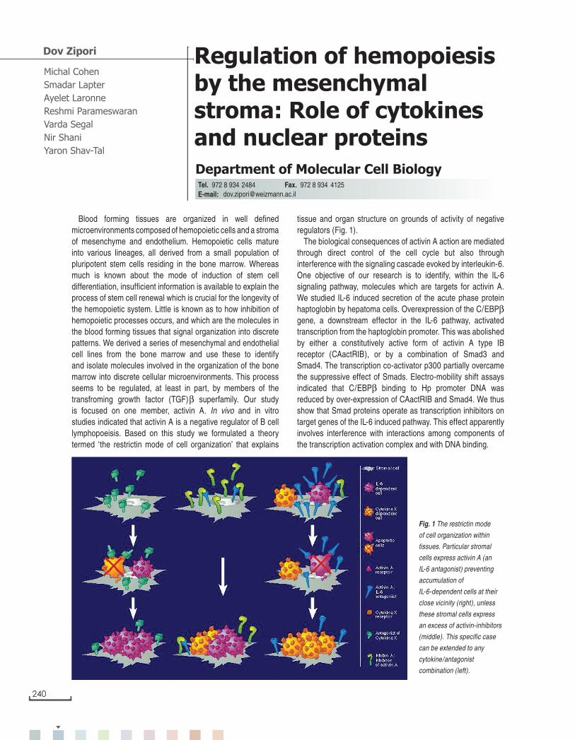
tissue and organ structure on grounds of activity of negative regulators (Fig. 1).
The biological consequences of activin A action are mediated through direct control of the cell cycle but also through interference with the signaling cascade evoked by interleukin-6. One objective of our research is to identify, within the IL-6 signaling pathway, molecules which are targets for activin A. We studied IL-6 induced secretion of the acute phase protein haptoglobin by hepatoma cells. Overexpression of the C/EBPβ gene, a downstream effector in the IL-6 pathway, activated transcription from the haptoglobin promoter. This was abolished by either a constitutively active form of activin A type IB receptor (CAactRIB), or by a combination of Smad3 and Smad4. The transcription co-activator p300 partially overcame the suppressive effect of Smads. Electro-mobility shift assays indicated that C/EBPβ binding to Hp promoter DNA was reduced by over-expression of CAactRIB and Smad4. We thus show that Smad proteins operate as transcription inhibitors on target genes of the IL-6 induced pathway. This effect apparently involves interference with interactions among components of the transcription activation complex and with DNA binding.
Michal Cohen
Smadar Lapter
Ayelet Laronne
Reshmi Parameswaran
Varda Segal
Nir Shani
Yaron Shav-Tal
Regulation of hemopoiesis by the mesenchymal stroma: Role of cytokines and nuclear proteins
Department of Molecular Cell Biology
Dov Zipori
2484 [email protected]
Blood forming tissues are organized in well defined microenvironments composed of hemopoietic cells and a stroma of mesenchyme and endothelium. Hemopoietic cells mature into various lineages, all derived from a small population of pluripotent stem cells residing in the bone marrow. Whereas much is known about the mode of induction of stem cell differentiation, insufficient information is available to explain the process of stem cell renewal which is crucial for the longevity of the hemopoietic system. Little is known as to how inhibition of hemopoietic processes occurs, and which are the molecules in the blood forming tissues that signal organization into discrete patterns. We derived a series of mesenchymal and endothelial cell lines from the bone marrow and use these to identify and isolate molecules involved in the organization of the bone marrow into discrete cellular microenvironments. This process seems to be regulated, at least in part, by members of the transfroming growth factor (TGF)β superfamily. Our study is focused on one member, activin A. In vivo and in vitro studies indicated that activin A is a negative regulator of B cell lymphopoeisis. Based on this study we formulated a theory termed ‘the restrictin mode of cell organization’ that explains
Fig. 1 The restrictin mode
of cell organization within
tissues. Particular stromal
cells express activin A (an
IL-6 antagonist) preventing
accumulation of
IL-6-dependent cells at their
close vicinity (right), unless
these stromal cells express
an excess of activin-inhibitors
(middle). This specific case
can be extended to any
cytokine/antagonist
combination (left).
dov_zipori 27.12.2001, 09:17240
Im
mu
no
lo
gy
a
nd
H
em
at
op
oie
sis
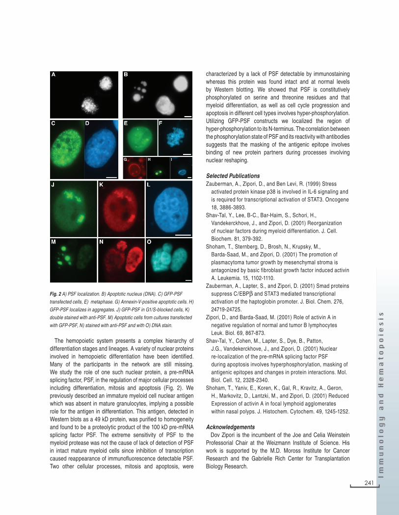
The hemopoietic system presents a complex hierarchy of differentiation stages and lineages. A variety of nuclear proteins involved in hemopoietic differentiation have been identified. Many of the participants in the network are still missing. We study the role of one such nuclear protein, a pre-mRNA splicing factor, PSF, in the regulation of major cellular processes including differentiation, mitosis and apoptosis (Fig. 2). We previously described an immature myeloid cell nuclear antigen which was absent in mature granulocytes, implying a possible role for the antigen in differentiation. This antigen, detected in Western blots as a 49 kD protein, was purified to homogeneity and found to be a proteolytic product of the 100 kD pre-mRNA splicing factor PSF. The extreme sensitivity of PSF to the myeloid protease was not the cause of lack of detection of PSF in intact mature myeloid cells since inhibition of transcription caused reappearance of immunofluorescence detectable PSF. Two other cellular processes, mitosis and apoptosis, were
characterized by a lack of PSF detectable by immunostaining whereas this protein was found intact and at normal levels by Western blotting. We showed that PSF is constitutively phosphorylated on serine and threonine residues and that myeloid differentiation, as well as cell cycle progression and apoptosis in different cell types involves hyper-phosphorylation. Utilizing GFP-PSF constructs we localized the region of hyper-phosphorylation to its N-terminus. The correlation between the phosphorylation state of PSF and its reactivity with antibodies suggests that the masking of the antigenic epitope involves binding of new protein partners during processes involving nuclear reshaping.
Selected PublicationsZauberman, A., Zipori, D., and Ben Levi, R. (1999) Stress
activated protein kinase p38 is involved in IL-6 signaling and is required for transcriptional activation of STAT3. Oncogene 18, 3886-3893.
Shav-Tal, Y., Lee, B-C., Bar-Haim, S., Schori, H., Vandekerckhove, J., and Zipori, D. (2001) Reorganization of nuclear factors during myeloid differentiation. J. Cell. Biochem. 81, 379-392.
Shoham, T., Sternberg, D., Brosh, N., Krupsky, M., Barda-Saad, M., and Zipori, D. (2001) The promotion of plasmacytoma tumor growth by mesenchymal stroma is antagonized by basic fibroblast growth factor induced activin A. Leukemia. 15, 1102-1110.
Zauberman, A., Lapter, S., and Zipori, D. (2001) Smad proteins suppress C/EBPβ and STAT3 mediated transcriptional activation of the haptoglobin promoter. J. Biol. Chem. 276, 24719-24725.
Zipori, D., and Barda-Saad, M. (2001) Role of activin A in negative regulation of normal and tumor B lymphocytes Leuk. Biol. 69, 867-873.
Shav-Tal, Y., Cohen, M., Lapter, S., Dye, B., Patton, J.G., Vandekerckhove, J., and Zipori, D. (2001) Nuclear re-localization of the pre-mRNA splicing factor PSF during apoptosis involves hyperphosphorylation, masking of antigenic epitopes and changes in protein interactions. Mol. Biol. Cell. 12, 2328-2340.
Shoham, T., Yaniv, E., Koren, K., Gal, R., Kravitz, A., Geron, H., Markovitz, D., Lantzki, M., and Zipori, D. (2001) Reduced Expression of activin A in focal lymphoid agglomerates within nasal polyps. J. Histochem. Cytochem. 49, 1245-1252.
AcknowledgementsDov Zipori is the incumbent of the Joe and Celia Weinstein
Professorial Chair at the Weizmann Institute of Science. His work is supported by the M.D. Moross Institute for Cancer Research and the Gabrielle Rich Center for Transplantation Biology Research.
Fig. 2 A) PSF localization. B) Apoptotic nucleus (DNA). C) GFP-PSF
transfected cells, E) metaphase. G) Annexin-V-positive apoptotic cells. H)
GFP-PSF localizes in aggregates. J) GFP-PSF in G1/S-blocked cells, K)
double stained with anti-PSF. M) Apoptotic cells from cultures transfected
with GFP-PSF, N) stained with anti-PSF and with O) DNA stain.
] 241
dov_zipori 27.12.2001, 09:17241