double-contrast upper gastrointestinal radiography: a ... · normal stomach gastric configuration...
TRANSCRIPT

Double-Contrast UpperGastrointestinal Radiography: APattern Approach for Diseases of theStomach1
Stephen E. Rubesin, MDMarc S. Levine, MDIgor Laufer, MD
The double-contrast upper gastrointestinal series is a valu-able diagnostic test for evaluating structural and functionalabnormalities of the stomach. This article will review thenormal radiographic anatomy of the stomach. The princi-ples of analyzing double-contrast images will be discussed.A pattern approach for the diagnosis of gastric abnormali-ties will also be presented, focusing on abnormal mucosalpatterns, depressed lesions, protruded lesions, thickenedfolds, and gastric narrowing.
! RSNA, 2008
1 From the Department of Radiology, Hospital of the Uni-versity of Pennsylvania, 3400 Spruce St, Philadelphia, PA19104. Received July 18, 2006; revision requested Sep-tember 20; revision received October 5; accepted Novem-ber 2; final version accepted January 2, 2007; final re-view by M.S.L. June 19. S.E.R. and M.S.L. are paidconsultants for E-Z-Em, Westbury, NY. Address corre-spondence to M.S.L. (e-mail: [email protected] ).
" RSNA, 2008
REVIEWS
ANDCOM
MENTARY
#REVIEW
FORRESIDENTS
Radiology: Volume 246: Number 1—January 2008 33
Note: This copy is for your personal, non-commercial use only. To order presentation-ready copies for distribution to your colleagues or clients, use the Radiology Reprints form at the end of this article.

This article presents a pattern ap-proach for the diagnosis of dis-eases of the stomach at double-
contrast upper gastrointestinal radi-ography. After describing the normalappearance of the stomach on double-contrast barium studies and the princi-
ples of double-contrast image interpre-tation, we will consider abnormal sur-face patterns of the mucosa, depressedlesions (erosions and ulcers), protrudedlesions (polyps, submucosal masses,and other tumors), thickened folds, andgastric narrowing.
Normal Stomach
Gastric Configuration and Rugal FoldsThe normal stomach is a J-shapedpouch that lies in the left upper quad-rant (Fig 1). The stomach has a fixedconfiguration created by the greaterlength of the longitudinal muscle layeron its greater curvature. The lesser cur-vature of the stomach is suspendedfrom the retroperitoneum by the hepa-togastric ligament, a portion of thelesser omentum. The gastrosplenic liga-ment and gastrocolic ligament (ie, theproximal portion of the greater omen-tum) are attached to the greater curva-ture of the stomach. The gastric cardiais attached to the diaphragm by the sur-rounding phrenoesophageal membrane.
The stomach is arbitrarily dividedinto five segments: the cardia, fundus,body, antrum, and pylorus. The gastriccardia is characterized on barium stud-ies by three or four stellate folds thatradiate to a central point at the gastro-esophageal junction, also known as thecardiac “rosette” (Fig 2) (1,2). The gas-tric fundus is defined as the portion ofthe stomach craniad to the gastric car-dia. The gastric body is defined as theportion of the stomach extending fromthe gastric cardia to the smooth bend inthe mid lesser curvature known as theincisura angularis. The gastric antrum isdefined as the portion of the stomachextending from the incisura angularis tothe pylorus (a structure created by amuscle sphincter shaped like a figureeight).
Rugal folds are most prominent inthe gastric fundus and body, whereasthe gastric antrum is often devoid offolds (Fig 1). Gastric rugae are change-able structures composed of mucosaand submucosa (3,4). The rugal foldsare relatively straight on the lesser cur-vature of the stomach but larger and
more undulating on the greater curva-ture. The thickness of the rugal foldsvaries with the degree of gastric disten-tion (5).
Areae GastricaeThe mucosal surface of the stomachconsists of flat polygonal-shaped tufts ofmucosa, known as areae gastricae, sep-arated by narrow grooves (6,7). Theareae gastricae are recognized on dou-ble-contrast studies as a reticular net-work of barium-coated white lines whenbarium fills the grooves between thesemucosal tufts (Fig 3). Individual muco-sal tufts of areae gastricae normallyhave a diameter of 2–3 mm in the gas-tric antrum and of 3–5 mm in the gastricbody and fundus (Fig 3) (6,8). Areaegastricae are detected on double-con-trast studies in nearly 70% of patientsand are observed with greater fre-quency in the elderly (8,9).
Comparison of Histologic Anatomy withMacroscopic AnatomyA basic understanding of the histologicanatomy of the stomach is helpful forunderstanding peptic ulcer disease, aswell as other gastric abnormalities(5,10). The stomach contains severaltypes of mucosa: cardiac-type mucosa,body/fundic-type mucosa, and antral/pyloric-type mucosa. Gastric foveolae(or pits) are conical depressions in themucosal surface that communicatewith gastric glands (4,10). The glandsare long, straight, and tightly packedstructures. The foveolae in all parts ofthe stomach are lined by surface fove-olar mucous cells. The cardiac-typemucosa comprises a short (1 cm inlength) segment of the gastric mucosaadjacent to the gastroesophageal junc-tion (4). The distinguishing feature ofthe body-type mucosa is the presence
Published online10.1148/radiol.2461061245
Radiology 2008; 246:33–48
Abbreviations:MALT ! mucosa-associated lymphoid tissueNSAID ! nonsteroidal antiinflammatory drug
Authors stated no financial relationship to disclose.
Essentials
# Protruded lesions (eg, polyps andcancers) on the dependent wall ofthe stomach may appear as radi-olucent filling defects in the bar-ium pool, whereas protruded le-sions on the nondependent wallmay appear as barium-coated“ring shadows” due to bariumcoating the edge of these lesions.
# Multiple small, round, uniform nod-ules in the stomach are usuallycaused by lymphoid hyperplasiaassociated with Helicobacter pylorigastritis, whereas irregular nonuni-form nodules may be caused bylow-grade B-cell mucosa-associatedlymphoid tissue lymphoma.
# Aspirin and other nonsteroidalantiinflammatory drugs (NSAIDs)are by far the most commoncause of erosive gastritis, a condi-tion manifested on double-con-trast studies as punctate or slitlikeerosions surrounded by radiolu-cent mounds of edema or, insome patients taking NSAIDs, bylinear or serpiginous erosions onor near the greater curvature ofthe gastric antrum or body.
# H pylori gastritis is by far themost common cause of focally ordiffusely thickened gastric folds,which can be so large and lobu-lated (eg, polypoid gastritis) thatthe radiographic findings resem-ble those of Menetrier disease orlymphoma.
# On barium studies, scirrhous carci-nomas of the stomach may producea linitis plastica appearance withdiffuse narrowing or long- or short-segment narrowing of any portionof the stomach; metastatic breastcancer and lymphoma occasionallymay produce a similar radiographicappearance.
REVIEW FOR RESIDENTS: Double-Contrast Upper Gastrointestinal Radiography Rubesin et al
34 Radiology: Volume 246: Number 1—January 2008

of parietal and chief cells in the glands.The parietal cells produce hydrochlo-ric acid and intrinsic factor, and thechief cells produce proteolytic en-zymes. No parietal or chief cells arefound in antral-type mucosa. The sur-face foveolar mucous cells line both an-tral pits and glands.
Body-type mucosa lines the ana-tomic gastric fundus and the gastricbody and extends into the gastric an-trum along the greater curvature (4).Antral-type mucosa lines the antrumalong the lesser curvature from the py-lorus to the incisura angularis, but onlylines a small amount of antrum along thegreater curvature. Thus, the histologicdivision of the stomach into body- andantral-type mucosa does not correlatewith the anatomic and radiologic divi-sion of the stomach into fundus, body,and antrum (5).
The transition zone between body-and antral-type mucosa is a line thatextends from the incisura angularis tothe distal greater curvature. The transi-tion zone migrates proximally with age,
extending progressively higher on thelesser curvature. Peptic ulcers fre-quently develop on the lesser curva-ture at the transition zone (Fig 4).
Principles of Image Analysis
Appearance of the StomachThe radiologist first examines the over-all position, shape, and size of the stom-ach. The gastric fundus abuts the lefthemidiaphragm. The cardia has a mid-line location, abutting the crus of the lefthemidiaphragm. The stomach curves tothe right across the midline, with thedistal gastric antrum and duodenum ex-tending to the right of the spine. Thereis considerable variation in the size ofthe stomach, depending on the amountof barium and effervescent agent ad-ministered.
Luminal ContourIn the barium pool, the contour is de-marcated by a smooth edge of barium
(Fig 1). With air contrast, the luminalcontour appears as a smooth, continuousbarium-coated white line (Fig 1) (11).
Barium PoolThe pool of high-density barium is thetool the radiologist uses to scrub and
Figure 1
Figure 1: Normal stomach. Double-contrastspot image of stomach with patient supine showsdistal gastric body (B) and antrum (A). Greatercurvature (white arrows) and lesser curvature(black arrows) are coated by barium. Rugal fold onposterior wall of gastric body is depicted as tubu-lar, slightly undulating, radiolucent filling defect(black arrowheads) in shallow barium pool. Densebarium pool outlines contour (white arrowheads)of gastric fundus (F). Mucosal surface and folds infundus are obscured by barium pool, and antrumis devoid of rugal folds.
Figure 2
Figure 2: Double-contrast spot image of gas-tric fundus with patient in right-side-down posi-tion shows normal gastric cardia with smoothfolds radiating to central point (white arrow) atclosed gastroesophageal junction, also known ascardiac rosette. Long, straight fold (arrowheads)extends inferiorly from cardia along lesser curva-ture. Black arrows denote normal extrinsic impres-sion by adjacent spleen.
Figure 3
Figure 3: Double-contrast spot image of stom-ach with patient in left posterior oblique positionshows normal areae gastricae pattern in antrum as2–3-mm polygonally shaped radiolucent tufts ofmucosa outlined by barium in grooves. Areaegastricae are slightly larger in distal gastric bodythan in antrum.
Figure 4
Figure 4: Double-contrast spot image of stom-ach with patient in supine position shows benignlesser curvature gastric ulcer (U) as smooth, ovoidcollection of barium extending outside expectedluminal contour of gastric body. Smooth folds areseen radiating to edge of ulcer crater.
REVIEW FOR RESIDENTS: Double-Contrast Upper Gastrointestinal Radiography Rubesin et al
Radiology: Volume 246: Number 1—January 2008 35

coat the mucosal surface (12–15). Somelesions are best shown in the bariumpool, whereas others are obscured byeven a small pool of high-density bar-ium. Protruded lesions on the depen-
dent wall usually appear as radiolucentfilling defects in the barium pool (Fig 5)(16), whereas protruded lesions on thenondependent wall appear as barium-coated “ring shadows” due to bariumcoating the edge of these lesions (Fig 5).When filled with barium, depressed le-sions appear as barium collections onthe dependent wall (Fig 4). When bar-ium spills out of depressed lesions onthe dependent wall, they may appear asring shadows.
En Face Mucosal DetailWhen viewed en face, the mucosal sur-face either has a smooth appearance(Fig 1) or is manifested as a reticularnetwork of barium-filled grooves be-tween the areae gastricae (Fig 3). Dis-ruption of the normal areae gastricaepattern or the smooth mucosal surfaceof the stomach by barium-coated lines isabnormal (Fig 5).
Pattern Approach for Double-ContrastDiagnosis
Abnormal Mucosal PatternsStriations.—Thin, barium-coated stria-tions perpendicular to the longitudinalaxis of the gastric antrum, also knownas gastric “striae,” are sometimes seenas a transient finding when the antrumis slightly collapsed (Fig 6) (17). Thesestriae are a sign of chronic antral gastri-tis (16).
Conspicuous or enlarged areae gas-tricae.—Visualization of the areae gas-tricae in the stomach depends on the
thickness of barium in the groovesbetween the mucosal tufts in relationto the thickness of barium overlyingthe tufts (8,9). Thus, an increase inthe height of the mucosal tufts or thin-ning of the mucous gel layer in thestomach may cause the areae gastri-cae to become more visible or conspic-uous. When viewed in profile, bariumin the grooves between areae gastri-cae may be manifested as tiny spike-like outpouchings, producing subtlespiculation of the luminal contour thatshould not be mistaken for erosions.In addition, the areae gastricae maybe enlarged by conditions that in-crease the size of the mucosal tuftsbeyond their normal diameter of 2–3mm in the antrum and of 3–5 mm inthe body and fundus. Enlarged areaegastricae have been reported in about50% of patients with Helicobacter py-lori gastritis (Fig 7) (18). In contrast,small or even absent areae gastricaehave been reported in patients withsevere atrophic gastritis and perni-cious anemia (19).
H pylori is a curved or spiral-shaped,gram-negative bacillus (20–22) that in-fects the stomach in more than 50% ofAmericans over 50 years of age and innearly 100% of Japanese adults (23,24).H pylori most frequently involves thegastric antrum (25). H pylori gastritiscan be documented in almost all pa-tients with duodenal ulcers and in about80% of patients with gastric ulcers (26).The mechanism by which H pyloricauses ulceration is not fully under-stood. H pylori gastritis is also a majorcausative factor in the development ofboth gastric carcinoma (27,28) and gas-tric lymphoma (29,30).
Uniform nodules.—Innumerable small(1–2 mm in size), round, uniform nod-ules disrupting the normal polygonalareae gastricae pattern are usuallycaused by lymphoid hyperplasia of thestomach resulting from chronic H pylorigastritis (Fig 8) (31–33). At birth, nolymphoid tissue is present in the stom-ach. When H pylori infects the stomach,the organism colonizes the mucouslayer and attaches to the membranes ofthe surface epithelial cells, resulting inthe development of chronic gastritis
Figure 5
Figure 5: Double-contrast spot image of gas-tric body with patient in supine right posterioroblique position shows multiple hyperplasticpolyps on dependent, or posterior, wall as small(" 1 cm in size), round or ovoid, finely lobulatedradiolucent filling defects in barium pool (arrows).In contrast, polyps on nondependent, or anterior,wall are coated by barium and appear as whitelines (arrowheads). Barium is seen to fill inter-stices between lobules of some polyps.
Figure 6
Figure 6: Double-contrast spot image of an-trum with patient in supine position shows gastricstriae as transient finding due to barium fillingdelicate transverse grooves between thin radiolu-cent folds traversing circumference of slightlycollapsed gastric antrum.
Figure 7
Figure 7: Double-contrast spot image of stom-ach with patient in supine position shows enlargedareae gastricae in patient with H pylori gastritis.Areae gastricae in antrum (white arrow) are largerthan those in distal gastric body (black arrow).
REVIEW FOR RESIDENTS: Double-Contrast Upper Gastrointestinal Radiography Rubesin et al
36 Radiology: Volume 246: Number 1—January 2008
(22). Repeated infections may eventu-ally lead to lymphocytic infiltration ofthe stomach, followed by the formationof lymphoid aggregates and even truelymphoid follicles (34). Thus, when lym-phoid hyperplasia is detected on dou-ble-contrast barium studies, these pa-tients are almost always found to havechronic H pylori gastritis (33).
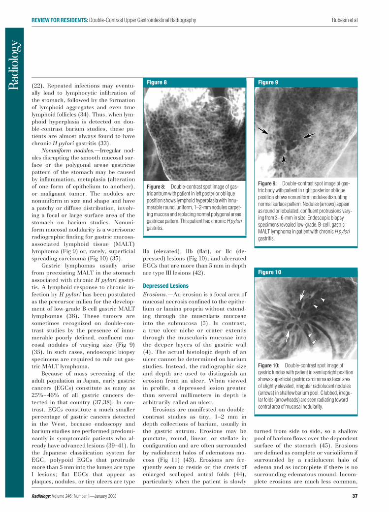
Nonuniform nodules.—Irregular nod-ules disrupting the smooth mucosal sur-face or the polygonal areae gastricaepattern of the stomach may be causedby inflammation, metaplasia (alterationof one form of epithelium to another),or malignant tumor. The nodules arenonuniform in size and shape and havea patchy or diffuse distribution, involv-ing a focal or large surface area of thestomach on barium studies. Nonuni-form mucosal nodularity is a worrisomeradiographic finding for gastric mucosa-associated lymphoid tissue (MALT)lymphoma (Fig 9) or, rarely, superficialspreading carcinoma (Fig 10) (35).
Gastric lymphomas usually arisefrom preexisting MALT in the stomachassociated with chronic H pylori gastri-tis. A lymphoid response to chronic in-fection by H pylori has been postulatedas the precursor milieu for the develop-ment of low-grade B-cell gastric MALTlymphomas (36). These tumors aresometimes recognized on double-con-trast studies by the presence of innu-merable poorly defined, confluent mu-cosal nodules of varying size (Fig 9)(35). In such cases, endoscopic biopsyspecimens are required to rule out gas-tric MALT lymphoma.
Because of mass screening of theadult population in Japan, early gastriccancers (EGCs) constitute as many as25%–46% of all gastric cancers de-tected in that country (37,38). In con-trast, EGCs constitute a much smallerpercentage of gastric cancers detectedin the West, because endoscopy andbarium studies are performed predomi-nantly in symptomatic patients who al-ready have advanced lesions (39–41). Inthe Japanese classification system forEGC, polypoid EGCs that protrudemore than 5 mm into the lumen are typeI lesions; flat EGCs that appear asplaques, nodules, or tiny ulcers are type
IIa (elevated), IIb (flat), or IIc (de-pressed) lesions (Fig 10); and ulceratedEGCs that are more than 5 mm in depthare type III lesions (42).
Depressed LesionsErosions.—An erosion is a focal area ofmucosal necrosis confined to the epithe-lium or lamina propria without extend-ing through the muscularis mucosaeinto the submucosa (5). In contrast,a true ulcer niche or crater extendsthrough the muscularis mucosae intothe deeper layers of the gastric wall(4). The actual histologic depth of anulcer cannot be determined on bariumstudies. Instead, the radiographic sizeand depth are used to distinguish anerosion from an ulcer. When viewedin profile, a depressed lesion greaterthan several millimeters in depth isarbitrarily called an ulcer.
Erosions are manifested on double-contrast studies as tiny, 1–2 mm indepth collections of barium, usually inthe gastric antrum. Erosions may bepunctate, round, linear, or stellate inconfiguration and are often surroundedby radiolucent halos of edematous mu-cosa (Fig 11) (43). Erosions are fre-quently seen to reside on the crests ofenlarged scalloped antral folds (44),particularly when the patient is slowly
turned from side to side, so a shallowpool of barium flows over the dependentsurface of the stomach (45). Erosionsare defined as complete or varioliform ifsurrounded by a radiolucent halo ofedema and as incomplete if there is nosurrounding edematous mound. Incom-plete erosions are much less common,
Figure 8
Figure 8: Double-contrast spot image of gas-tric antrum with patient in left posterior obliqueposition shows lymphoid hyperplasia with innu-merable round, uniform, 1–2-mm nodules carpet-ing mucosa and replacing normal polygonal areaegastricae pattern. This patient had chronic H pylorigastritis.
Figure 9
Figure 9: Double-contrast spot image of gas-tric body with patient in right posterior obliqueposition shows nonuniform nodules disruptingnormal surface pattern. Nodules (arrows) appearas round or lobulated, confluent protrusions vary-ing from 3– 6-mm in size. Endoscopic biopsyspecimens revealed low-grade, B-cell, gastricMALT lymphoma in patient with chronic H pylorigastritis.
Figure 10
Figure 10: Double-contrast spot image ofgastric fundus with patient in semiupright positionshows superficial gastric carcinoma as focal areaof slightly elevated, irregular radiolucent nodules(arrows) in shallow barium pool. Clubbed, irregu-lar folds (arrowheads) are seen radiating towardcentral area of mucosal nodularity.
REVIEW FOR RESIDENTS: Double-Contrast Upper Gastrointestinal Radiography Rubesin et al
Radiology: Volume 246: Number 1—January 2008 37

appearing as punctate or linear collec-tions of barium that may be difficult todifferentiate from barium trapped be-tween areae gastricae or alongside rugalfolds.
Aspirin and other nonsteroidal anti-inflammatory drugs (NSAIDs) are by far
the most common cause of gastric ero-sions (46). NSAID exposure causesbreakdown of the mucosal barrier andmucosal hypoxia, resulting in focal ar-
eas of epithelial necrosis with hemor-rhage, edema, and vascular dilatation inthe lamina propria (47). Because oftenthere is relatively little inflammatory re-sponse, the term NSAID gastropathyrather than NSAID gastritis is favoredby some authors (10,48). NSAID-in-duced erosive gastritis is typically mani-fested as multiple varioliform erosionsin the antrum or antrum and body of thestomach (18). Less frequently, these pa-tients may have incomplete erosionsthat appear as linear or serpiginous bar-ium collections (Fig 12), many of whichare located on or near the greater cur-vature of the gastric body secondary tothe effect of gravity (49).
Other causes of gastric erosions in-clude alcohol, viral infections, Crohndisease, hemorrhagic gastropathy, andiatrogenic trauma (4,50–55). Surpris-ingly, erosions are infrequently seen inpatients with H pylori gastritis (18).
Ulcers.—An ulcer is a focal areaof mucosal disruption that penetratesthrough the muscularis mucosae intothe deeper layers of the gastric wall.When viewed en face, most benign gas-tric ulcers on the dependent wall aremanifested on double-contrast studiesas a smooth round or ovoid collection ofbarium filling the ulcer crater (Fig 13).Some shallow ulcers on the dependentwall and ulcers on the nondependentwall may be manifested as a circular orhemispheric ring due to barium coatingthe rim of the unfilled ulcer crater(Fig 14) (56). Most ulcers are round orovoid, but some may have a linear, ser-pentine, rectangular, flame-shaped, orrod-shaped configuration (56–58).
When viewed in profile, benign gas-tric ulcers may be recognized by a focalbarium collection or barium-coated lineextending outside the expected luminalcontour (Fig 4) (11,59–61). Some ulcershave a smooth radiolucent rim of vari-able thickness directly adjacent to theulcer crater, representing a collar ofedema and inflammation, whereas oth-ers have a thin radiolucent line (alsoknown as a Hampton line) traversingthe base of the crater due to undermin-ing of the submucosa (59). The pres-ence of a Hampton line is diagnostic of abenign gastric ulcer. Chronic inflamma-
Figure 11
Figure 11: Double-contrast spot image ofdistal gastric antrum with patient in left posterioroblique position shows NSAID-induced erosiveantral gastritis. Multiple varioliform erosions areseen as punctate (small white arrow) and linear(large white arrow) collections of barium sur-rounded by radiolucent mounds of edema (blackarrows). This patient was taking aspirin.
Figure 12
Figure 12: Double-contrast spot image ofgastric antrum with patient in left posterior obliqueposition shows NSAID-induced linear and serpig-inous erosions (arrows). This patient was takingaspirin. Surgical clips in right upper quadrant arefrom prior cholecystectomy.
Figure 13
Figure 13: Double-contrast spot image ofgastric body with patient in left posterior obliqueposition shows gastric ulcer (U) as smooth, ovoidcollection of barium on posterior wall. Smooth,straight folds radiate directly to edge of ulcer cra-ter. These are typical findings of a benign gastriculcer.
Figure 14
Figure 14: Double-contrast spot image ofgastric body with patient in supine position showsincompletely filled ulcer on dependent, or poste-rior, wall as hemispheric ring shadow with twocrescent-shaped barium-coated lines (arrows)coating various portions of inferior rim of ulcer.Smooth, straight folds radiate almost to edge ofulcer crater. The findings are characteristic of abenign gastric ulcer with retraction of adjacentgastric wall.
REVIEW FOR RESIDENTS: Double-Contrast Upper Gastrointestinal Radiography Rubesin et al
38 Radiology: Volume 246: Number 1—January 2008

tion and scarring may cause retractionof the adjacent gastric wall with the de-velopment of smooth, straight folds thatradiate directly to the edge of the ulcercrater (Figs 13, 14).
Although giant gastric ulcers are atgreater risk for bleeding and perfora-tion (62), the size of the ulcer crater isnot a useful criterion for differentiatingbenign and malignant gastric ulcers.Most benign gastric ulcers are locatedon the lesser curvature or posterior wallof the stomach at or near the transitionzone between body- and antral-type mu-cosa (Fig 4). Some benign ulcers may belocated on the greater curvature (al-most all of these greater curvature ul-cers are caused by the use of aspirin orother NSAIDs) (14,63) or within hiatalhernias, where the stomach traversesthe diaphragm (64). Thus, ulcer loca-tion also is not a useful criterion fordifferentiating benign and malignantgastric ulcers. Radiologists thereforeshould ignore the size and location ofulcers when assessing the risk of malig-nancy; instead, they should focus on themorphologic features of these lesions.
In general, malignant gastric ulcersproduce radiographic findings diametri-cally opposed to those of benign ulcers.With malignant ulcers, the ulcer craterrepresents a focal area of necrosis andexcavation within a malignant tumor,usually gastric carcinoma or lymphoma.The surface of the ulcer and of the sur-rounding mucosa is therefore composedof nodules, irregular elevations, orirregular depressions of varying sizewithin the tumor (Fig 15) (42). Thefolds adjoining a malignant ulcer mayhave a coarse, lobulated, clubbed, orpenciled shape due to infiltration of thefolds by the tumor (Fig 10) (42).
Radiologists can often differentiatebenign and malignant gastric ulcers onthe basis of the radiographic findings(Fig 16). If an ulcer has a smooth sur-face with smooth, straight folds radiat-ing to the ulcer margin and no sur-rounding mass effect or mucosal nodu-larity (Figs 4, 13, 14), it fulfills theradiographic criteria for a benign gas-tric ulcer. About two-thirds of all gastriculcers diagnosed on double-contrast bar-ium studies have an unequivocally be-
nign radiographic appearance; virtuallyall of these unequivocally benign ulcersare ultimately proved to be benign(56,65).
In contrast, if an ulcer is associatedwith nodularity of the adjacent mucosa,mass effect, or radiating folds that arecoarse, lobulated, or irregular (Figs 10,15), it fulfills the radiographic criteriafor a malignant gastric ulcer, and endos-copy should be performed for a defini-tive diagnosis. Less than 5% of ulcershave an unequivocally malignant radio-graphic appearance; almost all of thesemalignant-appearing ulcers are ulti-mately proved to be malignant.
Finally, one-fourth to one-third ofgastric ulcers have an equivocal or in-determinate appearance that does notallow the radiologist to establish aconfident diagnosis of benignancy ormalignancy. An ulcer is classified asequivocal or indeterminate if there arecoarse areae gastricae or moderatenodularity of the mucosa abutting theulcer (Fig 17), a nodular ulcer collar,or mildly irregular folds radiating tothe ulcer’s edge. In such cases, endos-copy and biopsy are needed to rule outmalignant tumor. Nevertheless, themajority of equivocal or indeterminateulcers are ultimately proved to be be-nign.
Some benign NSAID-induced greater
Figure 15
Figure 15: Double-contrast spot image ofgastric body with patient in supine position showsmalignant gastric ulcer due to lymphoma. Largelobules of tumor (arrows) surround irregular cen-tral ulcer (U) filled with barium, although bariumpool is too dense to clearly delineate margins ofulcer.
Figure 16
Figure 16: List of radiographic features distin-guishing benign and malignant gastric ulcers.
Figure 17
Figure 17: Double-contrast spot image ofgastric body with patient in right posterior obliqueposition shows ulcer (U) on posterior wall fillingwith barium. Small radiolucent nodules (arrow-heads) are seen lateral to ulcer and larger nodules(arrows) are seen just superior to ulcer. This nodu-larity could be secondary to edema, inflammation,metaplasia, dysplasia, or tumor; findings in thiscase do not meet radiographic criteria for a benigngastric ulcer, and lesion should be classified asequivocal. Nevertheless, benign gastric ulcer wasconfirmed at endoscopy and follow-up. Gastricmetaplasia was found at the edge of the ulcer onendoscopic biopsy specimens.
REVIEW FOR RESIDENTS: Double-Contrast Upper Gastrointestinal Radiography Rubesin et al
Radiology: Volume 246: Number 1—January 2008 39

curvature ulcers may have an indeter-minate appearance due to extensivesurrounding mass effect and an appar-ent intraluminal location because ofspasm and inflammatory retraction of
the adjacent greater curvature (66). De-spite a history of NSAID use, thesegreater curvature ulcers therefore mayrequire endoscopy to rule out an ulcer-ated gastric carcinoma. Eventually,large NSAID-induced greater curvatureulcers may penetrate inferiorly via thegastrocolic ligament into the superiorborder of the transverse colon, produc-ing a gastrocolic fistula (67).
Diverticula.—Diverticula are un-commonly found in the stomach. Themajority arise from the posterior wall ofthe gastric fundus (68), presumably be-cause of a gap in the muscular layers ofthe gastric wall in this location. Fundaldiverticula are smoothly contoured,broad-mouthed outpouchings, rangingfrom 1 to 10 cm in size (Fig 18). Rugalfolds are not seen within the diverticula.These diverticula can be distinguishedfrom ulcers by their smooth contour,broad or shallow necks, and lack offolds radiating to their margins.
A variant of a gastric diverticulummay rarely be found on the greater cur-vature of the distal antrum, also knownas a partial antral diverticulum (69).
These tiny sacs are thought to representthe sequela of healed peptic ulcers. Par-tial antral diverticula are differentiatedfrom true ulcers by their variable sizeand shape at fluoroscopy and the ab-sence of associated inflammatory changes.
Protruded LesionsPolyps.—A polyp is a small protrusionfrom the mucosal surface, either sessileor pedunculated. The term polyp doesnot imply an adenomatous histology. Infact, a wide variety of benign and malig-nant polypoid lesions may occur in thestomach. If polyps arise from the mu-cosa, they may have a smooth, nodular,or lobulated surface on double-contraststudies, and when viewed in profile,form acute angles with the adjacent gas-tric wall (Fig 5) (70). In contrast, lesionsarising from the submucosa or muscu-laris propria usually have a very smoothsurface and, when viewed in profile,form right angles or slightly obtuseangles with the adjacent gastric wall(Fig 19). Although large lesions thathave a smooth surface are usually sub-mucosal in origin, it is often difficult todetermine whether protruded lesionsless than 1–1.5 cm in diameter are mu-cosal or submucosal in origin, as smallpolyps originating in the mucosa mayalso have a smooth surface.
Hyperplastic polyps are nonneoplas-tic proliferations of surface foveolarcells, consisting of elongated, distortedpits and numerous branching glands(4). These polyps are typically smoothor finely lobulated sessile lesions lessthan 1 cm in size (Fig 5). Occasionally,however, atypical hyperplastic polypsmay be larger than 1 cm in diameter,pedunculated, and have a coarsely lobu-lated surface (Fig 20) (71–73). At leastone-third of patients with hyperplasticpolyps have multiple polyps, usually inthe gastric body and fundus (Fig 5) (4).Although hyperplastic polyps have nomalignant potential, they usually arise inthe setting of chronic gastritis, the samemilieu that results in gastric metaplasiaand dysplasia. As a result, gastric ade-nomas and carcinomas have been re-ported to occur with increased fre-quency in patients with hyperplasticpolyps (4).
Figure 18
Figure 18: Double-contrast spot image withpatient in right decubitus position shows gastricdiverticulum as smooth barium-coated outpouch-ing (arrow) extending from posterior wall of fun-dus. A small amount of barium is present in lumenof diverticulum.
Figure 19
Figure 19: Double-contrast spot image offundus and upper body of stomach with patient insemiupright position shows large submucosalmass as smooth-surfaced hemispheric lesion(large arrows) forming right angles (small arrows)with adjacent luminal contour. Surgery revealedbenign gastrointestinal stromal tumor.
Figure 20
Figure 20: Double-contrast spot image ofgastric body with patient in supine position shows1-cm in size, sessile, slightly lobulated polyp ongreater curvature as area of increased radiopacitycoated by barium (arrow). Although radiographicfindings are worrisome for an adenomatous polypor even a small polypoid carcinoma, this lesionwas found to be a large hyperplastic polyp on en-doscopic biopsy specimens.
REVIEW FOR RESIDENTS: Double-Contrast Upper Gastrointestinal Radiography Rubesin et al
40 Radiology: Volume 246: Number 1—January 2008

Fundic gland polyps, the secondmost common gastric polyps, are prolif-erations of the deep epithelial compart-ment of body-type mucosa (4). Thesepolyps consist of cystically dilated pitsand glands lined by parietal and chiefcells. Fundic gland polyps are foundboth sporadically and in patients withfamilial adenomatous polyposis syndrome.These polyps typically appear on dou-ble-contrast studies as smooth-sur-faced, sessile protrusions less than 1 cmin size. Fundic gland polyps are usuallylocated in the fundus and upper body ofthe stomach and are often multiple (74).In patients with familial adenomatouspolyposis syndrome, hundreds of small(" 5 mm in size) fundic gland polypsmay be found.
Adenomatous polyps of the stomachare a relatively uncommon macroscopicform of gastric dysplasia. Adenomas areclassified as tubular, tubulovillous, orvillous on the basis of their underlyingarchitecture. Gastric adenomas mayprogress to gastric carcinoma by meansof a polypoid adenoma to carcinoma se-quence similar to that found in the co-lon. In situ carcinoma or invasive carci-noma is found in at least 50% of adeno-matous polyps larger than 2 cm in size(75). Most gastric dysplasias, however,are relatively flat macroscopically. Infact, most gastric carcinomas arise fromflat or slightly elevated or depressedareas of dysplasia, not polypoid adeno-mas (4).
In symptomatic patients, gastric ad-enomas detected on double-contraststudies are usually larger than 1 cm insize (75). These adenomas can be sessile,lobulated, or pedunculated lesions(Fig 21). Although most hyperplasticpolyps are smaller than 1 cm and mostadenomas are larger than 1 cm, it is notalways possible to distinguish a hyper-plastic polyp from an adenomatouspolyp on barium studies. If a polyp is 1cm or larger in size and has a finelynodular or lobulated surface, endoscopyand biopsy therefore should be per-formed to exclude the possibility of anadenoma. Conversely, multiple roundedpolyps 5 mm or smaller in size are al-most always hyperplastic, so that en-doscopy and biopsy are not warranted
in these patients. Atypical hyperplasticpolyps that are unusually large or lobu-lated (Fig 20) are indistinguishable fromadenomatous polyps or even polypoidcarcinomas (71–73).
Retention polyps (juvenile polyps)may occur as solitary lesions or as mul-tiple lesions in Cronkite-Canada syn-drome (76). Xanthelasmas, isolatedhamartomatous polyps, and inflamma-tory fibroid polyps are other benign pol-yps occasionally encountered in thestomach. A focal cluster of polyps mayalso be seen in the gastric antrum orbody in patients with small carcinoidtumors.
Masses.—For gastric masses largerthan 2 cm in size, barium studies areextremely helpful for determining whetherthe lesions arise from the mucosa, sub-mucosa, ormuscularis propria, orwhetherthey are extrinsic to the stomach. Thisdifferentiation enables the radiologist tosuggest a specific diagnosis or differen-tial diagnosis. In general, a mass origi-nating in the mucosa has a nodular orlobulated surface, appearing en face ondouble-contrast studies as a filling de-fect in the barium pool or as an area ofabnormal barium-coated lines, depend-ing on whether it is on the dependent ornondependent walls (Fig 22) (11). Notinfrequently, irregular collections ofbarium are trapped in the interstices ofthe tumor (Fig 21) or in areas of ulcer-ation. Barium thus outlines multipleround or ovoid nodules within the inter-stices of the lesion. For example, a pol-ypoid carcinoma may be manifested as alobulated or fungating mass within theexpected luminal contour (Fig 22).
In contrast, a submucosal mass mayappear en face on double-contrast stud-ies as a round or ovoid, well-circum-scribed, smooth or slightly lobulatedarea of increased radiopacity. Whenviewed in profile, a submucosal massmay be manifested as a hemispheric in-traluminal projection that has a smoothsurface and forms right angles orslightly oblique angles with the adjacentgastric wall (Figs 19, 23). Central ulcer-ation occurs in about 50% of submuco-sal masses due to central ischemia andnecrosis of the tumor or pressure ne-crosis of the overlying epithelium (Fig
23) (4). An ulcerated submucosal massviewed en face produces a characteris-tic “target” or “bull’s-eye” lesion, with acentral ulcer surrounded by a smooth,well-defined mass (Fig 24). Gastrointes-tinal stromal tumors are by far the most
Figure 21
Figure 21: Double-contrast spot image ofgastric body with patient in supine position showsbarium outlining outer contour and interstices of a3-cm sessile, multilobulated lesion (arrows).Surgery revealed tubulovillous adenoma. (Re-printed, with permission, from reference 15.)
Figure 22
Figure 22: Double-contrast spot image ofupper gastric body with patient in supine positionshows multilobulated polypoid mass as confluent,lobulated radiolucent filling defects (black arrows)in barium pool with separate portion of lesionoutlined in white on greater curvature (white ar-row). Surgery revealed polypoid adenocarcinoma.
REVIEW FOR RESIDENTS: Double-Contrast Upper Gastrointestinal Radiography Rubesin et al
Radiology: Volume 246: Number 1—January 2008 41

common solitary submucosal masses inthe stomach (77). Lymphoma and soli-tary metastases are other frequent sub-mucosal tumors. Lipoma is a submuco-sal lesion that may change in size and
shape at fluoroscopy (78) and has fatattenuation at computed tomography(79,80). Granular cell tumors usuallyappear as one or more small submuco-sal lesions. Most other mesenchymal tu-mors (eg, neurofibromas) are indistin-guishable from gastrointestinal stromaltumors.
Ectopic pancreatic rest (ie, myoepi-thelial hamartoma) is an uncommonsubmucosal lesion composed of varyingamounts of pancreatic tissue (includingducts, acini, and islet cells), hypertro-phic smooth muscle fibers, and glandularstructures resembling Brunner glands(81,82). These lesions may be compli-cated by pancreatitis, cysts, islet cell tu-mors, and even pancreatic carcinoma(4). Ectopic pancreatic rests usuallyappear on barium studies as small(1–2 cm), solitary, centrally umbilicatedsubmucosal masses, most often on thegreater curvature of the distal gastricantrum within 1–6 cm from the pylorus,but can occasionally be located else-where in the stomach (Fig 25) (83).
An extrinsic mass that indents butdoes not infiltrate the gastric serosa ismanifested as a smooth, broad-basedinbowing of the gastric wall (Fig 26). Incontrast, an extrinsic inflammatory orneoplastic mass that involves the serosaof the stomach may cause tethering ofthe gastric wall toward the extrinsicprocess, resulting in spiculation of theluminal contour. For example, omentalmetastases invading the greater curva-ture of the stomach through the gastro-colic ligament may cause spiculationand tethering of the greater curvature(84). Extrinsic inflammatory or neoplas-tic processes that involve the gastricwall or occlude gastric lymphatic or ve-nous channels may also result in en-larged gastric folds. For example, pan-creatitis secondarily involving the stom-ach may be manifested as thickenedfolds on the posterior gastric wall.
The precise location of mass lesionsin the stomach may help suggest thediagnosis in a small percentage of cases.As previously discussed, a submucosallesion on the greater curvature of thedistal gastric antrum should suggest anectopic pancreatic rest, whereas a sub-mucosal defect extending from thelesser curvature of the distal antrum tothe pylorus should suggest a hypertro-phied antral-pyloric fold. In contrast, asmooth, undulating submucosal lesionon the medial aspect of the fundus nearthe gastric cardia should suggest a con-glomerate mass of gastric varices (85).
Multiplicity of lesions is another ra-diographic feature that may be helpfulin suggesting a specific diagnosis ordifferential diagnosis. Numerous small(" 1 cm in size), smooth or finely lobu-lated, sessile protrusions are almost al-ways hyperplastic polyps (Fig 5) or, ifconfined to the gastric body or fundus,fundic gland polyps. A cluster of polypselsewhere in the stomach in patientswith chronic H pylori gastritis may rep-resent gastric carcinoid tumors dueto end-stage neuroendocrine hyperpla-sia associated with hypogastrinemia(5,10). Multiple large (# 1 cm in size)polypoid lesions may represent adeno-matous polyps, atypical hyperplasticpolyps, Peutz-Jeghers hamartomas, oreven synchronous polypoid carcinomas.
Figure 23
Figure 23: Double-contrast spot image ofgastric body with patient in supine position shows4-cm smooth-surfaced submucosal mass out-lined in white by barium (arrows). Note centraltriangular-shaped ulcer (arrowheads) in mass.Surgery and clinical follow-up revealed a malig-nant gastrointestinal stromal tumor.
Figure 24
Figure 24: Double-contrast spot image ofgastric body with patient in right posterior obliqueposition shows bull’s-eye or target lesion in stom-ach as a 2-cm ovoid, smooth-surfaced, submuco-sal mass (black arrows) in barium pool containingstellate central ulcer (white arrow). This patienthad breast cancer with hematogenous metastasisto the stomach.
Figure 25
Figure 25: Double-contrast spot image ofgastric antrum with patient in left posterior obliqueposition shows smooth, 1-cm ovoid, radiolucentfilling defect (arrow) in barium pool with shallowcentral umbilication containing trace amount ofbarium (arrowhead). Endoscopic biopsy speci-mens revealed an ectopic pancreatic rest (myoepi-thelial hamartoma). Ectopic pancreatic rests areusually located on greater curvature of distal an-trum, but this lesion was on posterior wall.
REVIEW FOR RESIDENTS: Double-Contrast Upper Gastrointestinal Radiography Rubesin et al
42 Radiology: Volume 246: Number 1—January 2008

Finally, multiple submucosal masses orcentrally ulcerated bull’s-eye lesions mayrepresent hematogenous metastases (suchas metastatic melanoma), disseminatedlymphoma, and, in patients with acquiredimmunodeficiency syndrome, Kaposi sar-coma.
Thickened FoldsNormal rugal folds are thicker in theproximal stomach, have a smooth con-tour in profile, and taper distally (5).Folds also are larger and more undulat-ing on the greater curvature than on thelesser curvature. Rugal folds becomestraight and thinner with increasing gas-tric distention and can even disappearwhen the stomach is fully distended,particularly in the gastric antrum. Be-cause of these normal variations in foldsize, there are no reliable criteria forenlarged folds in the stomach. How-ever, rugal folds are much more likely tobe abnormal when they have an irregu-lar, lobulated, or scalloped contour orwhen they are enlarged or have an an-gled or transverse orientation in a well-distended gastric antrum. Folds that arelarger on the lesser curvature than onthe greater curvature are also consid-ered to be abnormal.
Because rugal folds are composed ofmucosa and submucosa, any processthat infiltrates these layers of the gastricwall can increase the size of the folds.Enlarged folds may be caused by inflam-matory processes such as H pylori gas-tritis (86–89), hyperplastic processessuch as Zollinger-Ellison syndrome (90)and Menetrier disease (91), or malig-nant tumors such as lymphoma and sub-mucosally infiltrating adenocarcinoma.Endoscopic biopsy specimens may berequired to differentiate these variouscauses of enlarged folds, particularlywhen the folds are markedly thickenedand irregular.
Antral gastritis (whether or not it isassociated with H pylori) is usually man-ifested on barium studies as thickened,scalloped folds that have a longitudinalor transverse orientation. Antral gastri-tis may lead to the development of ahypertrophied antral-pyloric fold, seenas a smooth submucosal defect extend-ing from the lesser curvature of the dis-
tal antrum to the pylorus or eventhrough the pylorus into the medial for-nix of the base of the duodenal bulb(Fig 27) (92). In most patients, a hyper-trophied antral-pyloric fold can be dif-ferentiated from a neoplastic lesion byits characteristic appearance and loca-tion (93). Another clue to the pres-ence of a hypertrophied antral-pyloricfold is its variable size and shape atfluoroscopy, with palpation and peri-stalsis. Occasionally, however, a hy-pertrophied fold may be unusuallylarge or lobulated, so it can be mis-taken for a polypoid or plaquelike tu-mor (93).
H pylori gastritis is by far the mostcommon cause of focally or diffuselythickened folds in the stomach. Abnor-mal folds are found in about 75% ofpatients with H pylori gastritis (89).Fold enlargement in H pylori gastritismost commonly involves the gastric an-trum and body but may involve the en-tire stomach or may even be confined tothe gastric fundus. Most patients with Hpylori gastritis have mildly to moder-ately thickened gastric folds withoutsubstantial fold irregularity (Fig 28), sothat the radiographic findings are notworrisome for Menetrier disease orlymphoma. However, some patientswith H pylori gastritis have such en-larged, lobulated folds (ie, polypoid gas-tritis) that the radiographic findings er-roneously suggest a malignant process.Other patients with H pylori gastritismay have focally thickened polypoidfolds confined to the gastric antrum orbody that are mistaken radiographicallyfor a polypoid or infiltrating neoplasm(88). Nevertheless, radiologists cannotassume that all cases of enlarged foldsare caused by this ubiquitous pathogen.If the folds are markedly enlarged, lobu-lated, or irregular (particularly if theyhave a focal or segmental distribution),endoscopic biopsy specimens shouldbe obtained to exclude a malignant tu-mor.
In Menetrier disease, there ismarked hyperplasia of surface foveolarmucous cells (4,10), resulting in amarked increase in the height of thefoveolae and partial atrophy of theglands, with a corresponding loss of vol-
Figure 26
Figure 26: Double-contrast spot image ofgastric antrum and body with patient in supineposition shows smooth indentation on greatercurvature (black arrows) due to extrinsic masslesion compressing stomach. Second barium-coated line (white arrows) indicates that lumen isnarrowed asymmetrically. Area of increased ra-diopacity (D) also results from mass compressinglumen. These findings were caused by massiveretroperitoneal lymphoma.
Figure 27
Figure 27: Double-contrast spot image ofgastric antrum with patient in supine left posterioroblique position shows ovoid, 1.5-cm smooth-surfaced submucosal lesion (arrows) extendingfrom lesser curvature of distal antrum to adjacentpylorus. This lesion changed in size and shape atfluoroscopy with palpation and peristalsis. Find-ings are characteristic of hypertrophied antral-pyloric fold.
REVIEW FOR RESIDENTS: Double-Contrast Upper Gastrointestinal Radiography Rubesin et al
Radiology: Volume 246: Number 1—January 2008 43

ume of parietal and chief cell and subse-quent hypochlorhydria. The rugal foldsmay appear massively enlarged and lob-ulated on barium studies (Fig 29) (91).
Although early reports stated that foldenlargement predominantly involvedthe gastric fundus and body (sparing theantrum) (94), later reports found thatMenetrier disease causes fold enlarge-ment throughout the stomach in at least50% of patients (91), presumably be-
cause surface foveolar cells line the en-tire stomach.
Portal hypertension sometimes maycause mucosal hyperemia with dilatedsubmucosal vessels in the absence oftrue varices, a condition known as por-tal hypertensive gastropathy (95). Thisgastropathy can lead to acute or chronicgastrointestinal bleeding. Thickened,finely nodular folds are seen in the gas-tric fundus on barium studies (96). Gas-tric varices with associated esophagealvarices are usually caused by portal hy-pertension, whereas isolated gastricvarices (in the absence of esophagealvarices) may be caused by portal hyper-tension or, less commonly, by splenicvein obstruction from pancreatic carci-noma, pancreatitis, or pancreatic pseudo-cysts (97,98). Gastric varices can bedistinguished from the abnormal foldsof portal hypertensive gastropathy bytheir undulating and tortuous configura-tion and smooth contour (Fig 30) (97).In some patients, varices may also beseen on double-contrast studies as mul-tiple smooth, round or ovoid nodules,likened to the appearance of a bunch ofgrapes. In others, however, a conglom-erate mass of gastric varices (alsoknown as tumorous varices) may bemanifested as a smooth, undulating sub-mucosal mass on the medial wall of thefundus near the gastric cardia (85).
The normal gastric cardia is usu-ally manifested on double-contraststudies as a stellate collection of thin,smooth folds radiating to a centralpoint at the gastroesophageal junction(Fig 2). Any lesion disrupting or oblit-erating the cardiac rosette with asso-ciated nodularity, mass effect, ulcer-ation, or distorted folds in this regionshould be considered suspicious forcarcinoma of the cardia (Fig 31). Moreadvanced tumors at the cardia mayappear as polypoid, ulcerated, or infil-trating lesions that can easily be visu-alized with a double-contrast tech-nique (Fig 31) (99–101).
Gastric NarrowingNarrowing of the luminal contour of thestomach may be caused by scarring, in-filtrating tumor, or extrinsic diseasessecondarily affecting the stomach. Chronic
Figure 28
Figure 28: Double-contrast spot image of stom-ach with patient in supine position shows moderatelythickened folds in gastric body due to chronic H py-lori gastritis. Folds are considerably less thickenedand lobulated than in patient with Menetrier disease(Fig 29). Note surgical clips from prior vagotomy.
Figure 29
Figure 29: Double-contrast spot image ofgastric body with patient in supine position showsmarkedly thickened, lobulated folds and diffusedistortion of areae gastricae pattern in patient withMenetrier disease.
Figure 30
Figure 30: Double-contrast spot image ofgastric fundus with patient in right-side-downposition shows smooth, undulating submucosalmass (arrows) on posterior wall of fundus extend-ing to cardia. This patient had portal hypertensionwith a conglomerate mass of gastric varices (alsoknown as tumorous varices). Note surgical clipsfrom recent liver transplantation.
Figure 31
Figure 31: Double-contrast spot image ofgastric fundus with patient in right-side-downposition shows polypoid mass (arrows) that hasobliterated and replaced normal cardiac rosette.Arrowheads denote areas of ulceration within tu-mor. This patient had an advanced carcinoma ofcardia.
REVIEW FOR RESIDENTS: Double-Contrast Upper Gastrointestinal Radiography Rubesin et al
44 Radiology: Volume 246: Number 1—January 2008

scarring from peptic ulcer disease mayproduce asymmetric inbowing and re-traction of one wall of the stomach, of-ten associated with smooth, straightfolds that radiate to the site of thehealed ulcer. Other patients with pepticscarring have smooth, tapered narrow-ing of the gastric antrum or, lesscommonly, weblike antral narrowing(Fig 32). Scarring from ingestion ofNSAIDs (ie, chronic NSAID gastropa-thy) can also result in the characteristicflattening of the greater curvature of thedistal antrum (102).
Long-segment narrowing of the stom-ach (either circumferential or confinedto one wall) or diffuse narrowing of thestomach is usually caused by an infiltrat-ing or scirrhous gastric carcinoma (103)or metastatic breast cancer. The nar-rowing with scirrhous carcinoma resultsfrom a desmoplastic reaction to tumorcells infiltrating the submucosa, pro-ducing a linitis plastica appearance.Scirrhous carcinomas may be mani-fested as diffuse, long-segment, oreven short-segment narrowing of anyportion of the stomach (Fig 33) (103).The narrowed lumen is rigid and non-distensible at fluoroscopy, and gastricperistalsis is obliterated in this region.The luminal contour may have a smooth,nodular, or finely ulcerated surface ondouble-contrast studies (Fig 33). Occa-sionally, scirrhous carcinomas of thedistal antrum may be very short, cir-cumferential lesions confined to theprepyloric region of the stomach (104).It is important to recognize that en-doscopy and biopsy have a poor sensi-tivity in depicting scirrhous carcino-mas of the stomach, so that some pa-tients with radiographically diagnosedlesions may require one or more re-peat endoscopic examinations to con-firm the diagnosis.
In contrast, the narrowing withmetastatic breast cancer results fromdense infiltration of the submucosa bytumor. Severe scarring from previouscaustic ingestion may cause diffuse an-tral narrowing indistinguishable fromantral carcinoma on barium studies,but the clinical history should suggestthe correct diagnosis. Tapered nar-rowing of the gastric antrum may also
be caused by antral scarring fromCrohn disease or, rarely, other granu-lomatous diseases such as sarcoidosis,syphilis, and tuberculosis. Occasion-ally, antral narrowing may also resultfrom gastric atrophy related to thepresence of a long-standing gastrojeju-nal anastomosis without an antrec-tomy.
Atrophic gastritis is a condition inwhich body-type mucosal glands are re-placed by metaplastic cells resemblingpyloric- or intestinal-type epithelium(10). Most atrophic gastritis is relatedto chronic inflammation rather than au-toimmune phenomena. This form ofatrophic gastritis is often patchy andmacroscopically flat, so that it is notrecognizable on barium studies. In otherpatients with the autoimmune form ofatrophic gastritis, there is severe loss ofparietal cell mass, resulting in inade-quate secretion of intrinsic factor withthe subsequent development of vitaminB12 deficiency and, eventually, perni-cious anemia. In the later stages of auto-immune atrophic gastritis, decreased pari-etal cell mass is manifested as a dimin-ished mucosal surface volume and loss
of gastric folds. More than 80% of pa-tients with pernicious anemia have adiffusely narrowed stomach with asmooth contour and decreased or ab-sent rugal folds on double-contraststudies (Fig 34).
Figure 32
Figure 32: Double-contrast spot image ofdistal gastric antrum and duodenal bulb with pa-tient in left posterior oblique position shows shortsegment of smooth, circumferential narrowing(arrow) due to antral web related to scarring fromprevious peptic ulcer disease. If mucosal nodular-ity or irregularity of luminal contour had beenpresent, endoscopy and biopsy would have beenrequired to rule out focal gastric cancer.
Figure 33
Figure 33: Double-contrast spot image ofgastric antrum and body with patient in supineposition shows long segment of circumferentialnarrowing extending from mid gastric body to midgastric antrum (arrows denote limits of tumor).Although tumor surface is predominantly smooth,focal areas of mucosal nodularity are seen in prox-imal end of lesion (arrowheads). This patient hadscirrhous gastric carcinoma producing a linitisplastica appearance.
Figure 34
Figure 34: Double-contrast spot image ofgastric fundus and body with patient in right-side-down position shows smooth narrowing offundus and body of stomach with absent rugalfolds and small, barely visible areae gastricae.These findings are characteristic of atrophicgastritis.
REVIEW FOR RESIDENTS: Double-Contrast Upper Gastrointestinal Radiography Rubesin et al
Radiology: Volume 246: Number 1—January 2008 45

References1. Freeny PC. Double-contrast gastrography
of the fundus and cardia: normal landmarksand their pathologic changes. AJR Am JRoentgenol 1979;133:481–487.
2. Herlinger H, Grossman R, Laufer I, KresselHY, Och R. The gastric cardia in double–contrast study: its dynamic image. AJRAm J Roentgenol 1980;135:21–29.
3. Toner PG, Cameron CHS. The gastric mu-cosa. In: Whitehead R, ed. Gastrointestinaland oesophageal pathology. 2nd ed. Edin-burgh, Scotland: Churchill Livingstone,1995; 15–32.
4. Fenoglio-Preiser CM, Noffsinger AE,Stemmermann GN, Lantz PE, Listrom MB.Gastrointestinal pathology: an atlas andtext. Philadelphia, Pa: Lippincott-Raven,1999; 153–236.
5. Rubesin SE, Furth EE, Levine MS. Gastritisfrom NSAIDs to Helicobacter pylori. Ab-dom Imaging 2005;30:142–159.
6. Mackintosh CE, Kreel L. Anatomy and ra-diology of the areae gastricae. Gut 1977;18:855–864.
7. Seaman WB. The areae gastricae. AJRAm J Roentgenol 1978;131:554.
8. Rubesin SE, Herlinger H. The effect of bar-ium suspension viscosity on the delineationof areae gastricae. AJR Am J Roentgenol1986;146:35–38.
9. Charagundla SR, Levine MS, Langlotz CP,Rubesin SE, Laufer I. Visualization of areaegastricae on double-contrast gastrointestinalradiography: relationship to age of patients.AJR Am J Roentgenol 2001;177:61–63.
10. Appelman HD. Gastritis: terminology, eti-ology, and clinicopathologic correlations—another biased view. Hum Pathol 1994;25:1006–1019.
11. Laufer I, Kressel HY. Principles of double-contrast diagnosis. In: Levine MS, RubesinSE, Laufer I, eds. Double contrast gastroin-testinal radiology. 3rd ed. Philadelphia, Pa:Saunders, 2000; 8–46.
12. Laufer I. A simple method for routine dou-ble contrast study of the upper gastrointes-tinal tract. Radiology 1975;117:513–518.
13. Shirakabe H, Kobayashi S, Maruyama M.Principles and application of double con-trast radiography. In: Shirakabe H, Nish-izawa M, Maruyama S. Atlas of x-ray diag-nosis of early gastric cancer. 2nd ed. To-kyo, Japan: Igaku-Shoin, 1982; 19–43.
14. Levine MS, Rubesin SE, Herlinger H,Laufer I. Double contrast upper gastroin-testinal examination: technique and inter-pretation. Radiology 1988;168:593–602.
15. Rubesin SE, Levine MS. Principles for per-forming a double contrast upper gastroin-testinal examination. Westbury, NY: E-Z-EM, 2000; 1–29.
16. Rubesin SE. Gallery of double contrast ter-minology. Gastroenterol Clin North Am1995;24:259–288.
17. Cho KC, Gold BM, Printz DA. Multipletransverse folds in the gastric antrum. Ra-diology 1987;164:339–341.
18. Dheer S, Levine MS, Redfern RO, MetzDC, Rubesin SE, Laufer I. Antral gastritisradiographic findings in patients with andwithout Helicobacter pylori. Br J Radiol2002;75:805–811.
19. Levine MS, Palman CL, Rubesin SE, LauferI, Herlinger H. Atrophic gastritis in perni-cious anemia: diagnosis by double contrastradiography. Gastrointest Radiol 1989;14:215–219.
20. Warren JR. Unidentified curved bacilli ongastric epithelium in chronic active gastri-tis. Lancet 1983;1:1273–1275.
21. Marshall BJ, Warren JR. Unidentifiedcurved bacilli in the stomach of patientswith gastritis. Lancet 1984;1:1311–1315.
22. Yardley JH, Hendrix TR. Gastritis, gas-tropathy, duodenitis, and associated ulcer-ative lesions. In: Yamada Y, Alpers DH,Laine L, Owyang C, Powell DW, eds. Text-book of gastroenterology. 3rd ed. Philadel-phia, Pa: Lippincott Williams & Wilkins,1999; 1463–1499.
23. Dooley CP, Cohen H, Fitzgibbons PL, et al.Prevalence of Helicobacter pylori infectionand histologic gastritis in asymptomaticpersons. N Engl J Med 1989;321:1562–1566.
24. Graham DY, Go MF. Helicobacter pylori:current status. Gastroenterology 1993;105:279–282.
25. Bayerdorffer E, Lehn N, Hatz R, et al. Dif-ference in expression of Helicobacter pylorigastritis in antrum and body. Gastroenter-ology 1992;102:1575–1582.
26. Dixon MF. Helicobacter pylori and pepticulceration: histologic aspects. J Gastroen-terol Hepatol 1991;6:125–130.
27. Parsonnet J, Friedman GD, VandersteenDP, et al. Helicobacter pylori infection andthe risk of gastric carcinoma. N Engl J Med1991;325:1127–1131.
28. Hansson LE, Engstrand L, Nyren O, et al.Helicobacter pylori infection: independentrisk indicator of gastric adenocarcinoma.Gastroenterology 1993;105:1098–1103.
29. Wotherspoon AC, Ortiz-Hidalgo C, Flaxon,Isaccson PG. Helicobacter pylori associated
gastritis and primary B-cell gastric lym-phoma. Lancet 1991;338:1175–1176.
30. Parsonnet J, Hansen S, Rodriguez L, et al.Helicobacter pylori infection and gastriclymphoma. N Engl J Med 1994;330:1267–1271.
31. Bahk YW, Ahn JR, Choi HJ. Lymphoid hy-perplasia of the stomach presenting as um-bilicated polypoid lesions. Radiology 1971;100:277–280.
32. Sbeih F, Abdullach A, Sullican S, Meren-kow Z. Antral nodularity, gastric lymphoidhyperplasia and Helicobacter pylori inadults. J Clin Gastroenterol 1996;22:227–230.
33. Torigian DA, Levine MS, Gill NS, et al.Lymphoid hyperplasia of the stomach: ra-diographic findings in five adult patients.AJR Am J Roentgenol 2001;177:71–75.
34. Genta RM, Hamner HW. The significanceof lymphoid follicles in the interpretation ofgastric biopsy specimens. Arch Pathol LabMed 1994;118:740–743.
35. Yoo CC, Levine MS, Furth EE, et al. Gastricmucosa-associated lymphoid tissue lymphoma:radiographic findings in six patients. Radiology1998;208:239–243.
36. Eidt S, Stolte M. Prevalence of lymphoidfollicles and aggregates in Helicobacter py-lori gastritis in antral and body mucosa.J Clin Pathol 1993;46:832–835.
37. Kaneko E, Nakamura T, Umeda N, FuginoM, Niwa H. Outcome of gastric carcinomadetected by gastric mass survey in Japan.Gut 1977;18:626–630.
38. Kaibara N, Kawaguchi H, Nishidoi H, et al.Significance of mass survey for gastric can-cer from the standpoint of surgery. Am JSurg 1981;142:543–545.
39. Montesi A, Graziani L, Pesaresi A,DeNigris E, Bearzi I, Ranaldi R. Radio-logic diagnosis of early gastric cancer byroutine double-contrast examination.Gastrointest Radiol 1982;7:205–215.
40. Gold RP, Green PH, O’Toole KM, SeamanWB. Early gastric cancer: radiographic ex-perience. Radiology 1984;152:283–290.
41. White RM, Levine MS, Enterline HT,Laufer I. Early gastric cancer: recent expe-rience. Radiology 1985;155:25–27.
42. Shirakabe H, Nishizawa M, Maruyama M,Kobayashi S. Definition and classification ofearly gastric cancer. In: Shirakabe H, Nish-izawa M, Maruyama M, Kobayashi S, eds.Atlas of x-ray diagnosis of early gastric can-cer. 2nd ed. Tokyo, Japan: Igaku-Shoin,1982; 1–18.
43. Laufer I, Hamilton J, Mullens JE. Demon-
REVIEW FOR RESIDENTS: Double-Contrast Upper Gastrointestinal Radiography Rubesin et al
46 Radiology: Volume 246: Number 1—January 2008

stration of superficial gastric erosions bydouble contrast radiography. Gastroenter-ology 1975;68:387–391.
44. Op den Orth JO, Dekker W. Gastricerosions: radiological and endoscopic as-pects. Radiol Clin (Belg) 1976;45:88–89.
45. Kikuchi Y, Levine MS, Laufer I, HerlingerH. Value of flow technique for double-con-trast examination of the stomach. AJRAm J Roentgenol 1986;147:1183–1184.
46. Lanza FL, Nelson RS, Rack MF. A con-trolled endoscopic study comparing thetoxic effects of sulindac, naproxen, aspirin,and placebo on the gastric mucosa ofhealthy volunteers. J Clin Pharmacol 1984;24:89–95.
47. Lanza FL. NSAIDS and the gastrointestinaltract. Abdom Imaging 1997;22:1–4.
48. Grace E. Approach to the patient withgross gastrointestinal bleeding. In: YamadaT, ed. Textbook of gastroenterology. Phila-delphia, Pa: Lippincott Williams & Wilkins,2003; 698–723.
49. Levine MS, Verstandig A, Laufer I. Serpiginousgastric erosions caused by aspirin and othernonsteroidal anti-inflammatory drugs. AJRAm J Roentgenol 1986;146:31–34.
50. Roberts DM. Chronic gastritis, alcohol andnon-ulcer dyspepsia. Gut 1972;13:768–774.
51. Balthazar EJ, Megibow AJ, Hulnick DH. Cy-tomegalovirus esophagitis and gastritis inAIDS. AJR Am J Roentgenol 1985;144:1201–1204.
52. Laufer I, Trueman T. Multiple superficialgastric erosions due to Crohn’s disease ofthe stomach: radiologic and endoscopic di-agnosis. Br J Radiol 1976;49:726–728.
53. Ariyama J, Wehlin L, Lindstrom CG,Wenkert A, Roberts GM. Gastroduodenalerosions in Crohn’s disease. GastrointestRadiol 1980;5:121–125.
54. Sloan JM. Acute haemorrahagic gastritisand acute infective gastritis: gastritiscaused by physical agents and corro-sives—uraemic gastritis. In: WhiteheadR, ed. Gastrointestinal and oesophagealpathology. 2nd ed. Edinburgh, Scotland:Churchill-Livingstone, 1995; 461–470.
55. Rumerman J, Rubesin SE, Levine MS, LongWB, Laufer I. Gastric ulceration caused byheater probe coagulation. Gastrointest Ra-diol 1988;13:200–202.
56. Levine MS, Creteur V, Kressel HY, LauferI, Herlinger H. Benign gastric ulcers: diag-nosis and follow-up with double-contrastradiography. Radiology 1987;164:9–13.
57. Bloom SM, Paul RE JR, Matsue H, Poplack
WE, Goldsmith MR. Improved radio-graphic detection of multiple gastric ulcers.AJR Am J Roentgenol 1977;128:949–952.
58. Braver JM, Paul RE, Philipps E, Bloom S.Roentgen diagnosis of linear ulcers. Radiol-ogy 1979;132:29–32.
59. Nelson SW. The discovery of gastric ulcersand the differential diagnosis between be-nignancy and malignancy. Radiol ClinNorth Am 1969;7:5–25.
60. Wolf BS. Observations on roentgen fea-tures of benign and malignant ulcers. SeminRoentgenol 1971;6:140–150.
61. Ott DJ, Gelfand DW, Wu WC. Detection ofgastric ulcer: comparison of single and dou-ble-contrast examination. AJR Am J Roent-genol 1982;139:93–97.
62. Barragry TP, Blatchford JW, Allen MO.Giant gastric ulcers: a review of 49 cases.Ann Surg 1986;203:255–259.
63. Kottler RE, Tuft RJ. Benign greater curva-ture gastric ulcer: the “sump ulcer.” Br JRadiol 1981;54:651–654.
64. Hocking BV, Alp MH, Grant AK. Gastriculceration with hiatus hernia. Med J Aust1976;2:207–208.
65. Thompson G, Somers S, Stevenson GW.Benign gastric ulcer: a reliable radiologicdiagnosis? AJR Am J Roentgenol 1983;141:331–333.
66. Zboralske FF, Stargardter FL, Harel GS.Profile roentgenographic features of benigngreater curvature ulcers. Radiology 1978;127:63–67.
67. Levine MS, Kelly MR, Laufer I, Rubesin SE,Herlinger H. Gastrocolic fistulas: the in-creasing role of aspirin. Radiology 1993;187:359–361.
68. Eisenberg RL, Levine MS. Miscellaneousabnormalities of the stomach. In: Gore RM,Levine MS, eds. Textbook of gastrointesti-nal radiology. Philadelphia, Pa: Saunders,2000; 659–681.
69. Treichel J, Gerstenberg E, Palme G,Klemm T. Diagnosis of partial gastric diver-ticula. Radiology 1976;119:13–18.
70. Gordon R, Laufer I, Kressel HY. Gastricpolyps found on routine double-contrast ex-amination of the stomach. Radiology 1980;134:27–30.
71. Joffe N, Antonioli DA. Atypical appear-ances of benign hyperplastic polyps. AJRAm J Roentgenol 1978;131:147–152.
72. Smith HJ, Lee EL. Large hyperplastic pol-yps of the stomach. Gastrointest Radiol1983;8:19–23.
73. Cherukuri R, Levine MS, Furth EE, RubesinSE, Laufer I. Giant hyperplastic polyps inthe stomach: radiographic findings in sevenpatients. AJR Am J Roentgenol 2000;175:1445–1448.
74. Hizawa K, Iida M, Matsumoto T, Aoyagi K,Yao T, Fujisima M. Natural history of fundicgland polyps without familial adenomatouscoli: follow-up observations in 31 patients.Radiology 1993;189:429–432.
75. Op den Orth JO, Dekker W. Gastric adeno-mas. Radiology 1981;141:289–293.
76. Daniel ES, Ludwig SL, Lewin KJ, RuprechtRM, Rajacich GM, Schwabe AD. TheCronkhite-Canada syndrome: an analysis ofclinical and pathologic features and therapyin 55 patients. Medicine 1982;61:293–309.
77. Suster S. Gastrointestinal stromal tumors.Semin Diagn Pathol 1996;13:297–313.
78. Maderal F, Hunter F, Fuselier G, Gonzales-Rogue P, Torres O. Gastric lipomas: anupdate of clinical presentation, diagnosisand treatment. Am J Gastroenterol 1984;79:964–967.
79. Megibow AJ, Redmond PE, Bosniak MA,Horowitz L. Diagnosis of gastrointestinallipomas by CT. AJR Am J Roentgenol 1979;133:743–745.
80. Imoto T, Nobe T, Koga M, Miyamoto Y,Nakata H. Computed tomography of gastriclipomas. Gastrointest Radiol 1983;8:129–131.
81. Barbosa JJ, Dockerty MB, Waugh JM.Pancreatic heterotopia: a review of the lit-erature and report of 41 authenticated sur-gical cases of which 25 were clinically signif-icant. Surg Gynecol Obstet 1946;82:527–542.
82. Lai EC, Tompkins RK. Heterotopicpancreas: review of a 26-year experience.Am J Surg 1986;151:697–700.
83. Besemann EF, Auerbach SH, Wolfe WW.The importance of roentgenologic diagnosisof aberrant pancreatic tissue in the gastro-intestinal tract. Am J Roentgenol RadiumTher Nucl Med 1969;107:71–76.
84. Rubesin SE, Levine MS, Glick SN. Gastricinvolvement by omental cakes: radio-graphic findings. Gastrointest Radiol 1986;11:223–228.
85. Carucci LR, Levine MS, Rubesin SE, LauferI. Tumorous gastric varices: radiographicfindings in 10 patients. Radiology 1999;212:861–865.
86. de Lange EE. Radiographic features of gas-tritis using the biphasic contrast technique.Curr Probl Diagn Radiol 1987;16:273–308.
87. Gelfand DW, Ott DI, Chem MY. Radiologic
REVIEW FOR RESIDENTS: Double-Contrast Upper Gastrointestinal Radiography Rubesin et al
Radiology: Volume 246: Number 1—January 2008 47

evaluation of gastritis and duodenitis. AJRAm J Roentgenol 1999;173:357–361.
88. Sohn J, Levine MS, Furth EE, et al. Helico-bacter pylori gastritis: radiographic find-ings. Radiology 1995;195:763–767.
89. Levine MS, Rubesin SE. The Helicobacterpylori revolution: radiologic perspective.Radiology 1995;195:593–596.
90. Ellison EH, Wilson SD. The Zollinger-Ellison syndrome: re-appraisal and evalua-tion of 260 registered cases. Ann Surg1964;160:512–530.
91. Olmsted WW, Cooper PH, Madewell JE.Involvement of the gastric antrum in Men-etrier’s disease. AJR Am J Roentgenol1976;126:524–529.
92. Glick SN, Cavanaugh B, Teplick SK. Thehypertrophied antral-pyloric fold. AJRAm J Roentgenol 1985;145:547–549.
93. Arora R, Levine MS, Harvey RT, Laufer I,Rubesin SE. Hypertrophied antral-pyloricfold reassessment of radiographic findings
in 40 patients. Radiology 1999;213:347–351.
94. Reese DF, Hodgson JR, Dockerty MB. Gi-ant hypertrophy of the gastric mucosa (Me-netrier’s disease): a correlation of theroentgenographic, pathologic, and clinicalfindings. Am J Roentgenol Radium TherNucl Med 1962;88:619–626.
95. Smart HL, Triger DR. Clinical features,pathophysiology, and relevance of portalhypertensive gastropathy. Endoscopy1991;23:224–228.
96. Chang D, Levine MS, Ginsberg GG, RubesinSE, Laufer I. Portal hypertensive gastropathy:radiographic findings in eight patients. AJRAm J Roentgenol 2000;175:1609–1612.
97. Levine MS, Kieu K, Rubesin SE, HerlingerH, Laufer I. Isolated gastric varices: splenicvein obstruction or portal hypertension?Gastrointest Radiol 1990;15:188–192.
98. Muhletaler C. Gerlock J, Goncharenko V,Avant GR, Flexner JM. Gastric varices sec-ondary to splenic vein occlusion: radio-
graphic diagnosis and clinical significance.Radiology 1979;132:593–598.
99. Balthazar EJ, Goldfine S, Davidian NM.Carcinoma of the esophagogastric junction.Am J Gastroenterol 1980;74:237–243.
100. Freeny PC, Marks WM. Adenocarcinomaof the gastroesophageal junction: bariumand CT examination. AJR Am J Roentgenol1982;138:1077–1084.
101. Levine MS, Laufer I, Thompson JJ. Carci-noma of the gastric cardia in young people.AJR Am J Roentgenol 1983;140:69–72.
102. Laveran-Stieber RL, Laufer I, Levine MS.Greater curvature antral flattening: a radio-logic sign of NSAID-related gastropathy.Abdom Imaging 1994;19:295–297.
103. Levine MS, Kong V, Rubesin SE, Laufer I,Herlinger H. Scirrhous carcinoma of thestomach radiographic and endoscopic diag-nosis. Radiology 1990;175:151–154.
104. Balthazar EJ, Rosenberg H, Davidian MM.Scirrhous carcinoma of the pyloric channeland distal gastric antrum. AJR Am J Roent-genol 1980;134:669–673.
REVIEW FOR RESIDENTS: Double-Contrast Upper Gastrointestinal Radiography Rubesin et al
48 Radiology: Volume 246: Number 1—January 2008

Radiology 2007 This is your reprint order form or pro forma invoice
(Please keep a copy of this document for your records.)
Author Name _______________________________________________________________________________________________ Title of Article _______________________________________________________________________________________________ Issue of Journal_______________________________ Reprint # _____________ Publication Date ________________ Number of Pages_______________________________ KB # _____________ Symbol Radiology Color in Article? Yes / No (Please Circle) Please include the journal name and reprint number or manuscript number on your purchase order or other correspondence. Order and Shipping Information Reprint Costs (Please see page 2 of 2 for reprint costs/fees.) ________ Number of reprints ordered $_________ ________ Number of color reprints ordered $_________ ________ Number of covers ordered $_________ Subtotal $_________ Taxes $_________ (Add appropriate sales tax for Virginia, Maryland, Pennsylvania, and the District of Columbia or Canadian GST to the reprints if your order is to be shipped to these locations.) First address included, add $32 for each additional shipping address $_________
TOTAL $_________
Shipping Address (cannot ship to a P.O. Box) Please Print Clearly Name ___________________________________________ Institution _________________________________________ Street ___________________________________________ City ____________________ State _____ Zip ___________ Country ___________________________________________ Quantity___________________ Fax ___________________ Phone: Day _________________ Evening _______________ E-mail Address _____________________________________ Additional Shipping Address* (cannot ship to a P.O. Box) Name ___________________________________________ Institution _________________________________________ Street ___________________________________________ City ________________ State ______ Zip ___________
Country _________________________________________ Quantity __________________ Fax __________________ Phone: Day ________________ Evening ______________ E-mail Address ____________________________________ * Add $32 for each additional shipping address
Payment and Credit Card Details Enclosed: Personal Check ___________ Credit Card Payment Details _________ Checks must be paid in U.S. dollars and drawn on a U.S. Bank. Credit Card: __ VISA __ Am. Exp. __ MasterCard Card Number __________________________________ Expiration Date_________________________________ Signature: _____________________________________ Please send your order form and prepayment made payable to: Cadmus Reprints P.O. Box 751903 Charlotte, NC 28275-1903 Note: Do not send express packages to this location, PO Box.
FEIN #:541274108
Invoice or Credit Card Information Invoice Address Please Print Clearly Please complete Invoice address as it appears on credit card statement Name ____________________________________________ Institution ________________________________________ Department _______________________________________ Street ____________________________________________ City ________________________ State _____ Zip _______ Country ___________________________________________ Phone _____________________ Fax _________________ E-mail Address _____________________________________ Cadmus will process credit cards and Cadmus Journal
Services will appear on the credit card statement. If you don’t mail your order form, you may fax it to 410-820-9765 with
your credit card information. Signature __________________________________________ Date _______________________________________ Signature is required. By signing this form, the author agrees to accept the responsibility for the payment of reprints and/or all charges described in this document.
Reprint order forms and purchase orders or prepayments must be received 72 hours after receipt of form either by mail or by fax at 410-820-9765. It is the policy of Cadmus Reprints to issue one invoice per order.
Please print clearly.
Page 1 of 2 RB-9/22/06

Radiology 2007 Black and White Reprint Prices
Domestic (USA only) # of
Pages 50 100 200 300 400 500
1-4 $213 $228 $260 $278 $295 $313 5-8 $338 $373 $420 $453 $495 $530
9-12 $450 $500 $575 $635 $693 $755 13-16 $555 $623 $728 $805 $888 $965 17-20 $673 $753 $883 $990 $1,085 $1,185 21-24 $785 $880 $1,040 $1,165 $1,285 $1,413 25-28 $895 $1,010 $1,208 $1,350 $1,498 $1,638 29-32 $1,008 $1,143 $1,363 $1,525 $1,698 $1,865
Covers $95 $118 $218 $320 $428 $530
International (includes Canada and Mexico) # of
Pages 50 100 200 300 400 500
1-4 $263 $275 $330 $385 $430 $485 5-8 $415 $443 $555 $650 $753 $850
9-12 $563 $608 $773 $930 $1,070 $1,228 13-16 $698 $760 $988 $1,185 $1,388 $1,585 17-20 $848 $925 $1,203 $1,463 $1,705 $1,950 21-24 $985 $1,080 $1,420 $1,725 $2,025 $2,325 25-28 $1,135 $1,248 $1,640 $1,990 $2,350 $2,698 29-32 $1,273 $1,403 $1,863 $2,265 $2,673 $3,075
Covers $148 $168 $308 $463 $615 $768 Minimum order is 50 copies. For orders larger than 500 copies, please consult Cadmus Reprints at 800-407-9190. Reprint Cover Cover prices are listed above. The cover will include the publication title, article title, and author name in black. Shipping Shipping costs are included in the reprint prices. Domestic orders are shipped via UPS Ground service. Foreign orders are shipped via a proof of delivery air service. Multiple Shipments Orders can be shipped to more than one location. Please be aware that it will cost $32 for each additional location. Delivery Your order will be shipped within 2 weeks of the journal print date. Allow extra time for delivery.
Color Reprint Prices
Domestic (USA only) # of
Pages 50 100 200 300 400 500
1-4 $218 $233 $343 $460 $579 $697 5-8 $343 $388 $584 $825 $1,069 $1,311 9-12 $471 $503 $828 $1,196 $1,563 $1,935 13-16 $601 $633 $1,073 $1,562 $2,058 $2,547 17-20 $738 $767 $1,319 $1,940 $2,550 $3,164 21-24 $872 $899 $1,564 $2,308 $3,045 $3,790 25-28 $1,004 $1,035 $1,820 $2,678 $3,545 $4,403 29-32 $1,140 $1,173 $2,063 $3,048 $4,040 $5,028
Covers $95 $118 $218 $320 $428 $530
International (includes Canada and Mexico)) # of
Pages 50 100 200 300 400 500
1-4 $268 $280 $412 $568 $715 $871 5-8 $419 $457 $720 $1,022 $1,328 $1,633 9-12 $583 $610 $1,025 $1,492 $1,941 $2,407 13-16 $742 $770 $1,333 $1,943 $2,556 $3,167 17-20 $913 $941 $1,641 $2,412 $3,169 $3,929 21-24 $1,072 $1,100 $1,946 $2,867 $3,785 $4,703 25-28 $1,246 $1,274 $2,254 $3,318 $4,398 $5,463 29-32 $1,405 $1,433 $2,561 $3,788 $5,014 $6,237
Covers $148 $168 $308 $463 $615 $768 Tax Due Residents of Virginia, Maryland, Pennsylvania, and the District of Columbia are required to add the appropriate sales tax to each reprint order. For orders shipped to Canada, please add 7% Canadian GST unless exemption is claimed. Ordering Reprint order forms and purchase order or prepayment is required to process your order. Please reference journal name and reprint number or manuscript number on any correspondence. You may use the reverse side of this form as a proforma invoice. Please return your order form and prepayment to: Cadmus Reprints P.O. Box 751903 Charlotte, NC 28275-1903 Note: Do not send express packages to this location, PO Box. FEIN #:541274108 Please direct all inquiries to:
Rose A. Baynard 800-407-9190 (toll free number) 410-819-3966 (direct number) 410-820-9765 (FAX number)
[email protected] (e-mail)
Reprint Order Forms and purchase order or prepayments must be received 72 hours after receipt of form.
Page 2 of 2