dose units and dosimetrydental.ufl.edu/files/2013/03/radiation-biology1-3-compatibility...dose units...
TRANSCRIPT

11
Dose units and dosimetry
Radiation Absorbed Dose
Dose Equivalent
Effective Dose EquivalentEffective Dose Equivalent
Collective Effective Dose Equivalent or Collective
Dose
Dose Rate
Radiation Units
OLD NEWRoentgen (R) Measure of air exposure
RAD Measure of absorbed dose i.e. amount GRAYof energy absorbed from beamof energy absorbed from beam
REM Allows for different biological effects SIEVERTof different types of radiation. Provides
a common unit of DOSE EQUIVALENCE
CONVERSION 100 RAD = 1 GRAY (Gy)100 REM = 1 Sievert (Sv)
Dose Equivalent
Measures the Relative Biological Effectiveness (RBE) of different types of radiation - i.e. the damage caused by different types of radiation;Converts to a common unit
DOSE EQUIVALENT = RADIATION ABSORBED DOSE x QDOSE EQUIVALENT = RADIATION ABSORBED DOSE x Q
Q= Quality factor
X-rays, gamma rays Q = 1Neutrons Q = 10Alpha particles Q = 20
Effective Dose Equivalent or “Dose”
Measure which allows doses from investigations of different parts of the body to be compared, by converting all doses to an equivalent whole body dose
Effective dose equivalent = Dose equivalent x WW = weighting factor, i.e. Testes, ovaries = 0.25
Breast = 0.15Thyroid = 0.03
S.I. unit - Sievert (Sv); subunit - milliSievert mSv
Example of Effective Dose Equivalent or “Dose”
Thyroid examination
Measured Radiation Absorbed Dose = 10 mGyMeasured Radiation Absorbed Dose = 10 mGy
Dose equivalent (Q = 1) = 10 mSv
Effective dose equivalent (W = 0.03) = 10 x .03
DOSE = 0.3 mSv

22
Low Dose EffectsLow Dose Effects
Principle effects are non-lethal changes in cells
Usually occur at doses less than 10 cgy ( rad )
Ionizing Radiation as a Mutagen (Weak)Ionizing Radiation as a Mutagen (Weak)
First shown by H.J. Muller in 1927
-using fruit flies (won Nobel Prize)
Radiation increases spontaneous mutation rate
No evidence for a threshold
Radiation CarcinogenesisRadiation CarcinogenesisDirect Damage: I
DNA or RNA in a chromosome takes direct hit from x-ray photon
disrupting bonds between nucleic acids.
May cause:May cause:
Cell death
Abnormal replication (cancer)
Failure of transfer of information i.e. protein synthesis
Direct Damage: II
Somatic cells (non-reproductive) radiation induced
malignancy
Genetic cells congenital abnormalitiesGenetic cells congenital abnormalities
Indirect Damage
70% of cells is waterRadiation + H2O H2O+ + e-
H2O+ H+ + OH-
H O + e- H O-H2O + e H2OH2O- H + OH-
H and OH are free radicals
H + H H2 (Hydrogen gas) OH + OH H2O2 (Hydrogen peroxide)H2 + H2O2 Tissue damage

33
Indirect Damage (Summary)
Irradiation results in ionization of tissues at the atomic level
IonizationPhysical damage > atoms highly reactive Ch i l h l l h diff h i l Chemical change > molecule now has different chemical structureBiological function > changed or impaired
Biological effect may result in cell damage
Somatic vs Genetic Radiation Effects
Somatic: non-reproductive cells e.g. muscle, bone.
High doses > cataracts, reduced blood supply,
cancer
Genetic: Reproductive cells if damaged by radiation
can produce congenital abnormalities
Radiation Effects at Tissue/ Organ Level
If few cells die – no clinical change
As dose increases, cell deaths increase clinical changes
Severity of changes are proportional to cell loss
Short term: hypoplasia atrophy
Long term: changes related to blood vessel necrosis,
fibrosis, stenosis. O2 transport decreases
Radiation damageRadiation damage
Additive with repeated exposuresAdditive with repeated exposuresRepair processes may not be complete Repair processes may not be complete following initial exposurefollowing initial exposureS b t thS b t thSubsequent exposures thus cause more Subsequent exposures thus cause more damagedamage
Bergonie and TribondeauLaws of Bergonie and TribondeauLaws of Bergonie and Tribondeau
Radiation response of tissues depend on:
Number of cells in active proliferation
Number of cells in differentiationNumber of cells in differentiation
Number of future divisions/mitotic future
Other factors:
Also: Oxygen tension and the capacity of cells to repopulate.
44
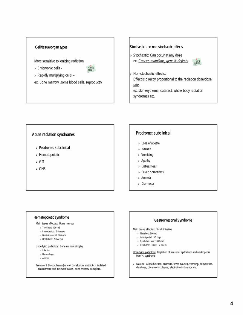
Cell/tissue/organ typesCell/tissue/organ types
More sensitive to ionizing radiation
Embryonic cells -y
Rapidly multiplying cells –
ex. Bone marrow, some blood cells, reproductiv
Stochastic and non-stochastic effectsStochastic and non-stochastic effects
Stochastic: Can occur at any doseex. Cancer, mutations, genetic defects.
Non-stochastic effects: Effect is directly proportional to the radiation dose/dose rate. ex. skin erythema, cataract, whole body radiation syndromes etc.
Acute radiation syndromesAcute radiation syndromes
Prodrome: subclinical
Hematopoietic
GIT
CNS
Prodrome: subclinicalProdrome: subclinical
Loss of apetiteNauseaVomittingApathyListlessnessFever, sometimesAnemiaDiarrhoea
Hematopoietic syndromeHematopoietic syndromeMain tissue affected: Bone marrow
Threshold: 100 radLatent period: 2-3 weeksDeath threshold: 200 radsDeath time: 2-8 weeks
Underlying pathology: Bone marrow atrophy:Infection HemorrhageAnemia
Treatment: Blood/plasma/platelet transfusion; antibiotics; isolated environment and in severe cases, bone marrow transplant.
Gastrointestinal SyndromeGastrointestinal Syndrome
Main tissue affected: Small intestineThreshold: 500 radLatent period: 3-5 daysDeath threshold: 1000 radsDeath time: 3 days 2 weeksDeath time: 3 days - 2 weeks
Underlying pathology: Depletion of intestinal epithelium and neutropenia from H. syndrome
Malaise, GI malfunction, anorexia, fever, nausea, vomiting, dehydration, diarrhoea, circulatory collapse, electrolyte imbalance etc.

55
CNS SyndromeCNS Syndrome
Main tissue affected: BrainThreshold: 2000 radsLatent period: 1/4 - 3 hoursDeath threshold: 5000 radsDeath time: < 3 days : Usually 24-48 hrsDeath time: < 3 days : Usually 24 48 hrs.
Underlying pathology: Vasculitis, encephalitis, meningitis, edema
Symptoms: Lethargy, tremors, convulsions, ataxia, coma
Background Radiation 3.6 mSv (360 mrem) per year
Radon 55%
Cosmic 8%
Terrestrial 8%
Internal 11%
Medical/artificial 18%
Is there a safe dose of x-rays?
No.
Unproven assumption of no threshold dose
but a linear relationship between dose and
probability of damage.
Decision to Use Radiodiagnostic ProceduresDecision to Use Radiodiagnostic Procedures
Numerical Comparisons of RisksNumerical Comparisons of Risks
RISK ESTIMATION, PROTECTION AND SAFETY

66
Basic question
What is the benefit to the patient from the radiographs you prescribe?radiographs you prescribe?
Radiation Safety
Patient benefit is directly linked to the
diagnostic information provided by thediagnostic information provided by the
radiograph
Types of Ionizing Radiation
Electromagnetic and Particulate radiation:
Electromagnetic radiation: pure form of energy with no mass; emitted in wave form.
Particulate radiation: Constituted by sub-atomic particles that have mass, energy, and sometimes, charge;
Eg. Alpha, beta (negative & positive), protons, neutrons etc. as in radioactive decay.
Electromagnetic radiation
XX--rays:rays: Formed from energy transfers involving Formed from energy transfers involving electronselectronsXX--rays:rays: Formed from energy transfers involving Formed from energy transfers involving electronselectrons
ϒϒ--rays:rays: From the nucleus when excess energy is From the nucleus when excess energy is emitted in the unstable state, interactions of nucleus emitted in the unstable state, interactions of nucleus with other particles in the region of the electrostatic with other particles in the region of the electrostatic field of the nucleus. Ex. Radioactive decay.field of the nucleus. Ex. Radioactive decay.
ϒϒ--rays:rays: From the nucleus when excess energy is From the nucleus when excess energy is emitted in the unstable state, interactions of nucleus emitted in the unstable state, interactions of nucleus with other particles in the region of the electrostatic with other particles in the region of the electrostatic field of the nucleus. Ex. Radioactive decay.field of the nucleus. Ex. Radioactive decay.
Stopped by leadStopped by leadStopped by leadStopped by lead found in medical usesfound in medical usesfound in medical usesfound in medical uses
X and gamma radiation are penetrating X and gamma radiation are penetrating radiationradiation
X and gamma radiation are penetrating X and gamma radiation are penetrating radiationradiation
Naturally present in soil and Naturally present in soil and cosmic radiationcosmic radiation
Naturally present in soil and Naturally present in soil and cosmic radiationcosmic radiation
and are EXTERNAL HAZARDSand are EXTERNAL HAZARDS

77
Ionizing Radiationcan deposit energy in neighboring atoms resulting in the removal of electrons.Ionizing Radiationcan deposit energy in neighboring atoms resulting in the removal of electrons. Non-Ionizing RadiationNon-Ionizing Radiation
Does not have enough energy to remove electrons from surrounding atomsDoes not have enough energy to remove electrons from surrounding atomsDoes not have enough energy to remove electrons from surrounding atomsDoes not have enough energy to remove electrons from surrounding atoms
Particulate radiation:
Alpha
Electron ′-
α++
Beta
Proton
Neutron
Electron Positron ′+
p+
n0
Alpha Radiation is only a hazard when inside your body Alpha Radiation is only a hazard when inside your body (internal hazard)(internal hazard)
Alpha Radiation is only a hazard when inside your body Alpha Radiation is only a hazard when inside your body (internal hazard)(internal hazard)
Your skin will stop it internal
hazard
stopped by paper
found in soil, radon and other radioactive
materials
Beta Radiation is a Skin, Eye and Internal HazardBeta Radiation is a Skin, Eye and Internal Hazard
skin, eye and internal hazardskin, eye and internal hazardskin, eye and internal hazardskin, eye and internal hazard
stopped by plasticstopped by plasticstopped by plasticstopped by plastic
found in natural food, air and waterfound in natural food, air and waterNeutrons have no charge & can penetrate deepNeutrons have no charge & can penetrate deep

88
UNSTABLE atoms emit energyUNSTABLE atoms emit energy
RF μwave infrared visible uv x-ray γ-ray cosmic
low energylow energy high energyhigh energy
non-ionizingnon-ionizing ionizing radiationionizing radiation
As
Low
ALARA
As
Reasonably
Achievable
Sources of human exposure to radiationradiation Cosmic 28Cosmic 28
Radon – 200
Background & manufactured radiation in the U.S. contributes 360 mrem per yearBackground & manufactured radiation in the U.S. contributes 360 mrem per year
Cosmic - 28Cosmic - 28
Diet - 40Diet - 40
Terrestrial - 28Terrestrial - 28
Manufactured sources Manufactured sources of radiation contribute an average of radiation contribute an average of 60 mrem/year60 mrem/year
Cigarette smoking Cigarette smoking –– 1300 mrem1300 mremCigarette smoking Cigarette smoking –– 1300 mrem1300 mrem
Round trip US by airRound trip US by air5 mrem per trip5 mrem per trip
Round trip US by airRound trip US by air5 mrem per trip5 mrem per trip
Medical Medical –– 53 mrem53 mremMedical Medical –– 53 mrem53 mrem
Building materials Building materials -- 3.6 mrem3.6 mremFallout < 1 mremFallout < 1 mrem
Smoke detectors Smoke detectors -- 0.0001 mrem0.0001 mrem
Altitude
Higher the altitude, the more the exposure to cosmic radiation
People in Denver, CO exposed to more radiation than Floridians, for example

99
Annual Dose Limits
Region Classified Workers General Public
Whole Body 50 mSv 1 mSv
50mSv per year OR 1mSv per week (with two weeks of annual vacation)
Occupational exposure
Dental Office (1988) 0.2 mSv per year (3 weeks background)1
World data for 1990-1994 show a mean annual occupational dose of only 0.06 mSv for dental workers (UNSCEAR, 2000)Dental personnel are not expected thus, to receive occupational exposures greater than recommended threshold for monitoring of 1 mSv per year.
Ref:1. White & Pharoah.Oral Radiology. 2000. p 48.NCRP: Exposure of the US population from occupational radiation, NCRP Report 101, 1989. (Uses 1980
data)
Instructions Concerning Prenatal Radiation Exposure
NCRP Regulatory Guide June 1999
Pregnant worker should notify dentist in writing, the approx
date of conception
Dentist must counsel worker that because of very low
doses it is safe to continue working with x-rays
Dental rad studies on pregnant patientsYou may expose:
if the benefits outweigh the risksat any part of the pregnancy
However, you should not expose anybody to ROUTINE x-ray exams (i.e every 6, 12 months …) including pregnant womenReassure the patient that there is minimal risk to the baby –use lead apron for reassuranceUnless the primary beam is directed at the abdomen, lead aprons are not necessary.
RADIATION PROTECTIONPATIENT PROTECTION
Radiographic selection criteria – avoids unnecessary exposures
Use fastest film: currently, “F” speed, or better still, direct digital
Use rectangular collimators
Avoid retakes
Do NOT need lead aprons with highest speed film and tight collimation; used only to alleviate patient concerns & to comply with policy
Benefits of Using Selection Criteria
43% reduction in number of intraorals ordered versus use of all FMX
White et al. Oral Surg Oral Med Oral Pathol 1994;77:531-540.

1010
Use Fastest Film Speed (if working with film)
Kodak has “F” speed (20% less radiation than “E” speed)
“E”, in turn, needs 50% less than “D”
e
FILM SPEED AND RELATIVE EXPOSURE
1.0
Rel
ativ
e Ex
posu
re
DIGITALA
B
C
FED0
0.5
Lead Apron
Lead Aprons
Not required by Federal or Florida State Laws since exposure so minimal, and possible cross infection hazard
Can be offered for the patient's reassurancep
Many States still require their use but from tradition not science
Similarly the ADA and UF handbook recommend their use
Reduction of exposure from Primary Beam
Must use a collimator or beam limiting device - exit beam diameter max 2.75”
Use a rectangular collimator to reduce exposure by 50%g p y
Or use a rectangular plug into round collimator to convert to rectangular
BEAM COLLIMATION
COLLIMATION BY PID
COLLIMATION UISNG A FILM HOLDER

1111
Rectangular Collimator & Position Indicating Device (PID)PERIAPICAL EXAMINATION
Image Interpretation
Label the mount – name, dateCheck the dot position on filmMount the films in a dark mountMount the films in a dark mountView with a masked-out view box to preserve visual contrast sensitivityMagnifying glass, darkened roomKnow NORMAL anatomy
Mounting and viewing radiographs
Viewing and interpreting radiographs
RADIATION PROTECTION FOR OPERATOR

1212
Radiation wall barriers & shields
In general 2-3 sheets of plaster-board or 1/16” lead sheet
Lead glass windows to view patientLead glass windows to view patient
Consult health physicist for shield design
Operator must stand outside the room behind protective barrier
90º
Operator position
135º
90º
Inverse square law of distance versus exposure
Inverse Square Law
Radiation is reduced by the square of the distance
Distance (feet) 1 2 3 4 5
Dose units
1 1/4 1/9 1/16 1/25
The operator should never hold the film in The operator should never hold the film in the patient’s mouth when the patient the patient’s mouth when the patient p pp p
cannot hold it steady. cannot hold it steady.
Monitoring radiation exposureMonitoring radiation exposure
Radiation dosimeters measure radiation dose to people.Radiation dosimeters measure radiation dose to people.
Unnecessary for the average office
Not required by law as dosage is low

1313
Government Regulations
FederalOSHA (Occupational Safety Health Administration) –staffFDA (Food and Drug Administration) NEXT surveys ( g ) yof dental radiation exposures NCRP (Nat Council of Radiation Protection)
Florida StateRegulations, equipment registration, inspections
Dental Radiology State Regulations
State of FloridaDepartment of Health,Control of Radiation Hazard,,Chapter 64E-5.506 Intraoral dental radiographic systemsFlorida Administrative Code
http://www9.myflorida.com/environment/radiation/regs/64part5.pdfpages 34, 35
Minimize Dose by Good PracticesMinimize Dose by Good Practices
TIME - reduce time of exposureDISTANCE - increase distance
SHIELDING - use shielding
TIME - reduce time of exposureDISTANCE - increase distance
SHIELDING - use shielding
Radiation equipment sticker
State of FL requiresState of FL requires"WARNING: This x"WARNING: This x--ray unit may be ray unit may be
dangerous to patient and operatordangerous to patient and operatorl f f t d til f f t d tiunless safe exposure factors and operating unless safe exposure factors and operating
instructions are observed."instructions are observed."
Registration of X-ray equipment
State of Florida requires registration of x-ray equipment
when installed with an annual fee of $31 (1997) plus $11 ( ) p
for each additional unit.
Every 5 years the State will inspect equipment