doras.dcu.iedoras.dcu.ie/15673/1/e-copy_paul_fitzparick_phd_thesis.doc · web viewadded to these...
TRANSCRIPT

The Role of LASP-1 in Cardiovascular Disease.
Paul Fitzpatrick, B.Sc, M.Sc
Thesis Submitted to Dublin City University (DCU) for the Degree of Doctor of Philosophy (Ph.D)
Research was carried out in the School of Health and Human Performance, DCU under the supervision of Doctor Ronan P. Murphy.
School of Health and Human Performance,
Dublin City University August 2010
1

Declaration
I hereby certify that this material, which I now submit for assessment on the programme of
study leading to the award of Doctor of Philosophy is entirely my own work, that I have
exercised reasonable care to ensure that the work is original and does not to the best of my
knowledge breach any law of copyright, and has not been taken from the work of others
save and to the extent that such work has been cited and acknowledged within the text of
my work.
Signature:_________________________ I.D._______________________
Date:____________________
2

Acknowledgements
I would like to thank the following colleagues, family and friends for all their support over
the past four years.
My most sincere gratitude and appreciation go to Dr. Ronan P. Murphy for allowing me to
carry out this research in his laboratory. Your guidance and kind words of encouragement
were always gratefully appreciated and will never be forgotten. It was both a pleasure and
an honour to work for you.
I would also like to take this opportunity to express my gratitude to Drs. Gerardene Meade
and Philip M. Cummins for their many useful suggestions, critisms and general all round
helpfulness and advice in many areas. Also thanks to Phil for the many lunches in 1838
over the last four years were very nice
This is also the perfect opportunity to thank the other members of the lab for a very
memorable four years not to mention all your help, support and the odd laugh or two over a
pint or prank. In particular Mishan Britto for his profound ability to listen and to ‘thrash’
ideas out with, our conversations were always interesting and challenging. Also, to the
other members of the lab including Tony Walsh, Anthony Guinan, Shaunta Guha and
Andrew Murphy. To those lab members who have left over the years and who I also owe a
huge thank you: Paul Fitzpatrick, Nick Tobin, Olga Colgan, Wei Gao, Maria Killeen and
Nora Collins.
I spent an unforgettable four years in this lab of which I will never forget. I hope that for
the new lab members, Ciarán McGinn, Brian McDonald, Chun Shan, Keith Rochfort and
Fiona Martin, that your time in the lab will be just as memorable.
I also owe a huge thank you to Jean O’Neil, Jesse Moss, Shane Long, Juliet Burke, Derek
Walsh, Nicolas Perritaz and Rebecca De Burgh.
3

Dedication
To Family and Friends
4

Poster Presentations
North American Vascular Biology Organisation (NAVBO)
– Cape Cod, MA, USA.
Cytoskeletal re-organisation by mechano-transduced LASP-1 regulates cell-matrix and
cell-cell adhesion through activation of rac1
Fitzpatrick, P., Britto, M., Walsh, T.G., Meade, G., Cummins, P.M., Murphy, R.P.
The role of hemodynamic stimuli on endothelial cells miRNA expression signature:
Potential new regulators for actin binding proteins
*Britto, M., *Fitzpatrick, P., Meade, G., Barron, N., O’Gorman, D., Moyna, N., Cummins,
P.M., Murphy, R.P.
Regulation of blood brain barrier function by shear stress: Role of RhoGTPases in tight
junction assembly and function
Walsh, T.G., Fitzpatrick, P., Murphy, R.P., Cummins, P.M.
Irish Society of Gene and Cell Therapy (ISGCT)
- Cork, Ireland
The Role of Haemodynamic stimuli on Endothelial cell microRNA expression signature:
Potential new targets for Cardiovascular gene therapy
*Britto, M., *Fitzpatrick, P., Meade, G., Barron, N., Cummins, P.M., Murphy, R.P.
5

XXI Congress of the International Society of Thromobosis and Haemostasis (ISTH)
- Geneva, Switzerland
Mechano-Regulation of LASP-1 in the pathogenesis of Cardiovascular disease
Fitzpatrick, P., Britto, M., Cahill, P.A., Cummins, P.M., Meade, G., Murphy, R.P.
Molecular and Cellular dynamics of Moesin in vascular cells via mechano-transduction
Britto, M., Fitzpatrick, P., Meade, G., Cahill, P.A., Cummins, P.M., Murphy, R.P.
6

Oral Presentations
12th International symposium on ‘Signalling at the Blood-Brain and Blood-Retinal
Barriers’
– University of London (UCL), UK (Sept ’09)
The role of the actin cytoskeleton adaptor protein LASP-1 in RhoGTPase induced changes
to Endothelial cell adhesion.
Fitzpatrick, P., Britto, M., Walsh, T.G., Meade, G., Cummins, P.M., Murphy, R.P.
XXII Congress of the International Society of Thrombosis and Haemostasis (ISTH) –
Boston, USA (July ’09)
Bio-mechanical Regulation Of microRNA Expression and Endothelial Cell Phenotype In
Vitro.
University of Virginia, Invited Seminar
– Charlottesville, Virginia, USA (March ’09)
Actin-binding proteins, miRNA and Cell adhesion: The LASP Frontier
Dublin City University, School of Biotechnology Research Day
– DCU (Jan ’09)
Cell Adhesion: The LASP Frontier
Fitzpatrick, P., Britto, M., Walsh, T.G., Meade, G., Cummins, P.M., Murphy, R.P.
*Prize awarded for best postgraduate oral presentation
7

Irish Society of Gene and Cell Therapy (ISGCT)
– Galway, Ireland
The Role of the Actin binding proteins Moesin and LASP-1, in the pathogenesis of
Cardiovascular disease
Fitzpatrick, P., Britto, M., Ferguson, G., Cummins, P.M., Cahill, P.A., Meade, G., Murphy,
R.P.
8

List of subsequent Grants awarded through contributions from
this study. Dublin City University Research Award: For the establishment and design of a
customised “Bioimaging System for the Investigation of Cellular Biomechanics and Actin
Dynamics”.
International Visitors Programme: For the establishment of a collaborative link with the
Department of Molecular Biology & Functional Genomics, Universita Vita-Salute San
Raffaele, Milan: “Investigation of Urokinase Receptor-Integrin interaction”.
Health Research Board of Ireland: Additional Capital/Equipment Grant 2005-2006
(RPAdd/2006/60): Chemi-luminescence gel documentation & analysis system.
Science Foundation Ireland: Equipment Grant (2007): ABI 7900HT Fast Real-Time PCR
System with Low Density Gene Array Upgrade.
Programme for Research in Third Level Institutes Cycle IV: “Molecular Therapeutics
T^3– Exploiting Novel Targets for Health”
Health Research Board Project Grant 2009-2012 (HRA/2009/122): “The Role of
microRNA in Modulating Vascular Smooth Muscle Cell Phenotype: Novel Regulators of
Vascular Physiology and Disease”.
Enterprise Ireland, Industrial Research and Commercialisation Committee (IRCC):
Proof of Concept Programme. “Development of a Micro Molecular Index Card of
Cardiovascular Health: Next Generation Prognostic and Diagnostic BioChip”.
9

Future Publications The role of microRNA in Integrin Signalling: Novel players in vascular
inflammation.
Eden, G., Archinti, M., Furlan, F., Fitzpatrick, P., Murphy, R. And Degryse, B.
“The Cell Migration Signalosome” Nova Science Publishers, Inc. New York.
Fitzpatrick, P., Britto, M., Walsh, T.G., Meade, G., Cummins, P.M., Murphy,
R.P. LASP-1 is an important regulator in the molecular complex controlling cell-
cell and cell-matrix adhesion in the vasculature. Manuscript in preparation
10

Table of Contents
DECLARATION..................................................................................................................................... 2
ACKNOWLEDGEMENTS..................................................................................................................... 3
DEDICATION......................................................................................................................................... 4
POSTER PRESENTATIONS.................................................................................................................. 5
ORAL PRESENTATIONS...................................................................................................................... 7
LIST OF SUBSEQUENT GRANTS AWARDED THROUGH CONTRIBUTIONS FROM THIS STUDY..................................................................................................................................................... 9
FUTURE PUBLICATIONS................................................................................................................... 10
TABLE OF CONTENTS....................................................................................................................... 11
ABSTRACT........................................................................................................................................... 16
ABBREVIATIONS................................................................................................................................ 17
UNITS.................................................................................................................................................... 22
INDEX OF TABLES AND FIGURES...................................................................................................24
TABLES........................................................................................................................................................24FIGURES.......................................................................................................................................................25
CHAPTER ONE - INTRODUCTION....................................................................................................40
1.0 INTRODUCTION..................................................................................................................... 41
1.1 LASP-1............................................................................................................................................. 41
1.1.1 LIPOMA PREFERRED PARTNER (LPP)...................................................................................................441.1.2 ZYXIN.................................................................................................................................................451.1.3 VASP.................................................................................................................................................461.1.4 PALLADIN...........................................................................................................................................46
1.2 THE VASCULAR SYSTEM............................................................................................................ 47
1.2.1 CARDIOVASCULAR DISEASE...........................................................................................................481.2.2 THE ENDOTHELIUM.........................................................................................................................49
1.2.2.1 The Endothelium and cell permeability.......................................................................................511.2.2.2 Regulation of Vascular Tone.......................................................................................................531.2.2.3 Maintenance of thrombo-resistance............................................................................................561.2.2.4 Modulation of EC-Leukocyte interactions...................................................................................57
11

1.2.2.5 Endothelial Dysfunction..............................................................................................................58
1.3 VASCULAR MECHANO-REGULATION......................................................................................60
1.3.1 HAEMODYNAMIC FORCES.........................................................................................................................601.3.1.1 Cyclic Strain.................................................................................................................................611.3.1.2 Shear Stress.................................................................................................................................63
1.3.2 MECHANO-TRANSDUCTION.......................................................................................................................671.3.2.1 Mechano-regulated Intracellular Signaling Pathways................................................................69
1.2.2.1.1 Mitogen activated protein kinases (MAPK)...........................................................................................691.2.2.1.2 Mechano-regulation of Endothelial gene expression.............................................................................70
1.3.2.2 Mechano-sensors.........................................................................................................................711.2.2.2.1 Adhesion Receptors...............................................................................................................................711.2.2.2.2 Membrane Structures.............................................................................................................................761.2.2.2.3 The Endothelial Actin Cytoskeleton......................................................................................................81
1.3.3 MECHANO-TRANSDUCTION AND ATHEROSCLEROSIS........................................................................................82
1.4 THE CYTOSKELETON.................................................................................................................. 85
1.4.1 MICROTUBULES AND INTERMEDIATE FILAMENTS...............................................................................861.4.2 ACTIN CYTOSKELETON.......................................................................................................................89
1.4.2.1 Organisation of the actin cytoskeleton........................................................................................901.4.2.2 Actin Organising Proteins...........................................................................................................901.4.2.3 Actin Polymerisation...................................................................................................................941.4.2.4 Actin Based Membrane Protrusions............................................................................................96
1.3.2.4.1 The Lamellipodium...............................................................................................................................961.3.2.4.2 Stress Fibers........................................................................................................................................1001.3.2.4.3 Cortical Actin Cytoskeleton.................................................................................................................1031.3.2.4.4 Nuclear Actin......................................................................................................................................104
1.4.3 REGULATION OF THE ACTIN CYTOSKELETON....................................................................................1061.4.3.1 Haemodynamic Forces..............................................................................................................1071.4.3.2 RhoGTPases..............................................................................................................................1081.4.3.3 Actin Binding/Scaffolding/adaptor Proteins..............................................................................112
1.3.3.3.1 Multi-domain Proteins.........................................................................................................................112
1.5 THE ACTIN CYTOSKELETON AND CELL ADHESION..........................................................118
1.5.1 CELL-MATRIX ADHESION.................................................................................................................1181.5.2 CELL-CELL ADHESION......................................................................................................................123
1.6 STUDY BACKGROUND AND SPECIFIC OBJECTIVES............................................................126
CHAPTER TWO - MATERIALS AND METHODS...........................................................................129
2.1 MATERIALS................................................................................................................................. 130
2.1.1 REAGENTS AND CHEMICALS.............................................................................................................1302.1.2. INSTRUMENTATION..........................................................................................................................1362.1.3 PREPARATION OF STOCK SOLUTIONS AND BUFFERS..........................................................................137
2.1.3.1 Immuno-blotting........................................................................................................................1372.1.3.2 Molecular Biology Buffers and Medium....................................................................................1402.1.3.3 RhoGTPase Pulldown Assays....................................................................................................142
12

2.1.3.3.1 Rap1 Activation Assay Buffers...........................................................................................................1422.1.3.3.2 Rac1 and Cdc42 Activation Assay Buffers..........................................................................................1432.1.3.3.3 RhoA Activation Assay Buffers..........................................................................................................144
2.2 METHODS..................................................................................................................................... 146
2.2.1 CELL CULTURE TECHNIQUES............................................................................................................1462.2.1.1 Bovine Aortic Endothelial Cell (BAEC) Culture.......................................................................1462.2.1.2 Human Aortic Endothelial Cell (HAEC) Culture......................................................................1462.2.1.3 Rat Aortic Smooth Muscle Cell (RAOSMC) Culture.................................................................1472.2.1.4 Trypsinisation of adherent cell lines..........................................................................................1472.2.1.5 Cell Counting............................................................................................................................1482.2.1.6 Cryo-Preservation of Cells........................................................................................................149
2.2.2 MOLECULAR BIOLOGY TECHNIQUES.................................................................................................1502.2.2.1 Reconstituting Plasmid cDNA...................................................................................................1502.2.2.2 Transformation of competent cells............................................................................................1502.2.2.3 Midi-preparation of Plasmid DNA............................................................................................1512.2.2.4 Spectrometric analysis of Plasmid DNA....................................................................................1522.2.2.5 Agarose Gel Electrophoresis.....................................................................................................1522.2.2.6 DNA Precipitation.....................................................................................................................153
2.2.3 TRANSFECTIONS................................................................................................................................1532.2.3.1 Lipofectamine Transfection.......................................................................................................1532.2.3.2 Plasmid DNA Nucleofection......................................................................................................1532.2.3.3 Microporation...........................................................................................................................1552.2.3.4 siRNA........................................................................................................................................156
2.2.4 IMMUNO-DETECTION TECHNIQUES...................................................................................................1572.2.4.1 Immuno (Western)-Blotting.......................................................................................................157
2.2.4.1.1 Preparation of whole cell lysate...........................................................................................................1572.2.4.1.2 Bicinchoninic Acid (B.C.A) Assay......................................................................................................1582.2.4.1.3 Polyacrylamide Gel Electrophoresis (SDS-PAGE)..............................................................................1582.2.4.1.4 Electrotransfer to a PDVF membrane..................................................................................................1612.2.4.1.5 Immuno-blotting and Chemiluminesence band detection....................................................................1632.2.4.1.6 Coomassie Gel Staining.......................................................................................................................1652.2.4.1.7 Ponseau-S Membrane Staining............................................................................................................1652.2.4.1.8 Membrane Stripping............................................................................................................................165
2.2.4.2 Immuno-Precipitation...............................................................................................................1662.2.4.3 Proteo-Cellular Fractionation..................................................................................................167
2.2.4.3.1 Protein Precipitation............................................................................................................................1682.2.4.4 Immuno-Fluorescence Imagining..............................................................................................168
2.2.5 QUANTITATIVE REAL-TIME PCR (QRT-PCR)..................................................................................1702.2.5.1 RNA isolation............................................................................................................................1702.2.5.2 Spectrometric analysis of isolated RNA.....................................................................................1712.2.5.3 cDNA synthesis by Reverse Transcriptase PCR........................................................................1712.2.5.4 Design of PCR Primer Sets.......................................................................................................1722.2.5.5 Polymerase Chain Reaction (PCR)...........................................................................................1722.2.5.6 QRT-PCR..................................................................................................................................174
2.2.6 PHYSIOLOGICAL ASSAYS..................................................................................................................1752.2.6.1 Cyclic Strain..............................................................................................................................175
13

2.2.6.2 Non-Pulsatile Laminar Shear Stress..........................................................................................1752.2.6.3 Cell-Matrix Adhesion................................................................................................................176
2.2.6.3.1 Coating Cell Adhesion Plates..............................................................................................................1762.2.6.3.2 Cell Adhesion Protocol........................................................................................................................176
2.2.6.4 Trans-endothelial Cell Permeability Protocol...........................................................................1772.2.6.5 Endothelial Cell Micro-Particle (EMP) Analysis......................................................................1782.2.6.6 Angiogenesis.............................................................................................................................179
2.2.6.6.1 Tube Formation Assay.........................................................................................................................1792.2.6.6.2 Matrigel Mesh Investigation................................................................................................................179
2.2.6.7 Integrin αVβ3 Activation Assay...................................................................................................1802.2.6.8 Proliferation..............................................................................................................................1812.2.6.9 Quantification of Filamentous (F)-actin to Monomeric Globular (G)-Actin.............................1812.2.6.10 Shear Stress by means of iBidi flow slides...............................................................................1822.2.6.11Triton-soluble/insoluble extraction...........................................................................................183
2.2.7 RHO/RAS GTPASE ACTIVATION ASSAYS..........................................................................................1842.2.7.1 Rap 1 activation assay...............................................................................................................184
2.2.7.1.1 RalGDS-RBD-GST Bead production..................................................................................................1842.2.7.1.2 Quantification of GST-RBD bound to beads.......................................................................................1852.2.7.1.3 Rap 1 Activation Assay.......................................................................................................................186
2.2.7.2 Rac 1/Cdc42 activation assay....................................................................................................1862.2.7.3 Rho A activation assay..............................................................................................................187
2.2.8 MICRORNA (MIRNA) MICRO-REGULATION OF LASP-1...................................................................1872.2.8.1 miRNA Isolation........................................................................................................................1872.2.8.2 Total RNA quality check............................................................................................................1882.2.8.3 Multiplex Reverse Transcription for TaqMan® array analysis..................................................1882.2.8.4 TaqMan® Low Density Array (TLDA) PCR...............................................................................189
2.2.9 FACS ANALYSIS ASSAYS.................................................................................................................1902.2.9.1 G.F.P. Analysis of Transfected cells..........................................................................................190
2.2.10 TREATMENT WITH PHARMACOLOGICAL INHIBITORS.......................................................................1902.2.11 STATISTICAL ANALYSIS..................................................................................................................191
CHAPTER THREE - THE MECHANO-REGULATION OF LASP-1 AND ITS ROLE IN ACTIN POLYMERISATION........................................................................................................................... 192
3.1 INTRODUCTION.......................................................................................................................... 193
3.1.1 STUDY AIMS:............................................................................................................................. 198
3.2 RESULTS....................................................................................................................................... 199
3.3 DISCUSSION................................................................................................................................. 213
CHAPTER FOUR - THE ROLE OF MECHANO-REGULATED PROTEIN LASP-1 IN CELL-MATRIX AND CELL-CELL ADHESION..........................................................................................218
4.1 INTRODUCTION.......................................................................................................................... 219
4.1.1 STUDY AIMS.............................................................................................................................. 225
14

4.2 RESULTS....................................................................................................................................... 226
4.3 DISCUSSION................................................................................................................................. 241
CHAPTER FIVE - THE ROLE OF LASP-1 IN THE REGULATION OF RHO- AND RAS- GTPASES............................................................................................................................................................. 246
5.1 INTRODUCTION.......................................................................................................................... 247
5.1.1 STUDY AIMS.............................................................................................................................. 255
5.2 RESULTS....................................................................................................................................... 256
5.3 DISCUSSION................................................................................................................................. 268
FUTURE PLANS................................................................................................................................. 272
Bibliography......................................................................................................................................... 276
15

AbstractVascular endothelial (VE)-cadherin- cell-cell and integrin-mediated cell-matrix adhesions
coordinate spatial and temporal rearrangements of the actin cytoskeleton. The expression
and dynamics of actin regulating adaptor/scaffold proteins, such as the LIM and SH3
protein (LASP-1), act as mediators for the regulation of vascular homeostasis through their
unique protein-actin and protein-protein interactions. This study aims to elucidate the role
for LASP-1 in actin dynamics of the vasculature.
Models mimicking the haemodynamic forces, of cyclic strain and shear stress, acting on
endothelial cells (ECs) reveal that LASP-1 expression and localisation is mechanically
altered within the vasculature. LASP-1 is seen to localise to multiple sites of dynamic actin
assembly such as; focal contacts; focal adhesions; lamellipodia; membrane ruffles; cell-cell
junctions and the nucleus, where interactions occur with Zyxin, Palladin and VASP.
Cellular fractionation of ECs document the translocation of Lasp-1 from the cytosol to the
membrane under pathogenic strain. This study also reveals its presence in the nucleus,
where LASP-1 may play a role in nuclear actin dynamics and transcriptional regulation.
Upon mechanical stimulation quantification of F- and G-actin ratios illustrates that LASP-1
over-expression promotes F-actin polymerisation and increased cell spreading. A resultant
increase in focal adhesion number ensures an elevated cell-matrix attachment with
significant adhesion to the β3-integrin ligand Vitronectin. At cell-cell junctions LASP-1
over-expression increases levels of active GTP-Rac1 resulting in tighter cell-cell
interactions and barrier formation, as monitored using a transendothelial permeability
assay.
Overall LASP-1 mediates the changes in cell-matrix and cell-cell adhesion orchestrated by
the concerted effort of VE-Cadherin and β3 integrins, possibly through coordination of
global ratios of active Rac1 and Rap1, thus playing a crucial role in the pathogenesis of
vascular disease.
16

Abbreviations
aa Amino Acid
ABP Actin Binding Protein
Abl Abelson Tyrosine Kinase
ACE Angiotensin converting enzyme
ADP Adenosine Di-phosphate
Ang Angiotensin
APS Ammonium Persulphate
Arg Abl – related gene
Arp2/3 Actin related protein 2/3
ATP Adenosine Tri-Phosphate
BAEC Bovine Aortic Endothelial Cell
BCA Bicinchoninic acid
BSA Bovine Serum Albumin
cDNA Complimentary DNA
C-Terminal Carboxyl Terminal
CVD Cardiovascular disease
DMSO Dimethlysulphoxide
DNA Deoxyribonucleic acid
dNTP Deoxy nucleotide tri-phosphate
17

DTT Dithiothreitol
EC Endothelial Cells
ECM Extracellular matrix
EDTA Ethylenediaminetetraacetic acid
EDRF Endothelial derived relaxing factor
EGFR Epidermal Growth Factor
eNOS Endothelial nitric oxide synthase
EPS8 Epidermal growth factor receptor pathway substrate 8
ERK Extracellular signal-regulated kinase
ERM Ezrin/Radixin/Moesin
F-actin Filamentous – Actin
FAK Focal Adhesion Kinase
FBS Foetal Bovine Serum
FD40 FITC-Dextran 40kDa
FITC Fluorescein isothiocyanate
G-Actin Globular - Actin
GDP Guanine Di-Phosphate
GEF Guanine nucleotide exchange factor
GFP Green Fluorescent Protein
GST Gluthione S-Transferase
GTP Guanine Tri-Phosphate
18

HAEC Human Aortic Endothelial Cell
ICAM Intracellular Adhesion Molecule
IGF Insulin like growth factor
IRSp53 Insulin Receptor Tyrosine Kinase substrate p53
LASP LIM And SH3 Protein
LB Luria-Bertani
LIM Lin-II, isl-I, mec-3
LIMK LIM Kinase
LPA Lysophosphatidic Acid
LPP Lipoma Preferred Partner
LSS Laminar Shear Stress
MAPK Mitogen Activated Protein Kinase
mDia Mammalian homologue of Drosophila diaphanous
MLCK Myosin light chain kinase
MgCl2 Magnesium Chloride
Mn Manganese
NaCl Sodium Chloride
NaOH Sodium Hydroxide
NFκB Nuclear factor – κB
NO Nitric oxide
P Significance
19

PAGE Polyacrylamide gel electrophoresis
PAK p21-activated kinase
PBS Phosphate buffered saline
PCR Polymerase chain reaction
PDGF Platelet derived growth factor
PECAM Platelet Endothelial Cell Adhesion Molecule
PI3K Phosphatidylinositol-3-kinase
PKC Protein kinase C
P/S Penicillin/Streptomycin
qRT-PCR Quantitative Real-Time - PCR
Raf Receptor associated factors
RAP Ras like Protein
RAOSMC Rat Aortic Smooth Muscle Cell
Rho Ras Homology gene family
RIAM Rap1-GTP Interacting adaptor Molecule
RNA Ribonucleic acid
ROS Reactive oxygen species
Rpm Rotations per minute
RT-PCR Reverse transcriptase – PCR
SDS Sodium dodecyl sulphate
SEM Standard Error Mean
20

SH3 Src Homology region 3
SSB Sample Solubilisation Buffer
ss Single stranded
siRNA Small interfering RNA
TAE Tris acetate EDTA
TE Tris – EDTA
TEE Trans-Endothelial Exchange
TEMED N,N,N’, N’-Tetramethylethlylenediamine
TF Tissue Factor
TGF-β Transforming growth factor – β
t-PA Tissue Plasminogen activator
VASP Vasodilator Stimulated Phosphoprotein
VE Vascular Endothelial
VEGF Vascular Endothelial growth factor
VEGFR VEGF - Receptor
VSMC Vascular Smooth Muscle Cells
VWF Von Willebrand Factor
WASp Wiskott-Aldrich syndrome protein
WAVE WASP family verpolin-homologous protein
21

Unitsbp Base pairs
cm Centimetre
oC Degree Celsius
kDa KiloDaltons
μg Microgram
μl Microlitre
μM Micromolar
g Grams
hr Hours
kg Kilogram
L Litre
M Molar
mA Milliamperes
mg Milligrams
min Minutes
ml Millilitres
mM Millimolar
ng Nanograms
nm Nanometres
pmol Picomolar22

sec Seconds
w/v Weight per volume
V Volts
23

Index of Tables and Figures
Tables
Chapter Two
Table 2.1 Silencer® select siRNA. Table of siRNA used for the knockdown of
LASP-1 and VE-Cadherin. The table documents the siRNA sequence
and their target site for RNA interference on each gene.
Table 2.2 SDS-PAGE resolving gel composition. The table documents the
individual components of SDS-PAGE resolving gels of different
percentage acrylamide and the individual volumes of each
component necessary to make each gel.
Table 2.3 SDS-PAGE stacking gel composition. The table documents the
individual components of SDS-PAGE stacking gels of 4%
acrylamide and the individual volumes of each component necessary
to make a gel.
Table 2.4 Protein loading volumes. Table documents a list of proteins
investigated by western blot analysis and the quantity of each protein
loaded onto the SDS-PAGE gels to carry out this investigation.
Table 2.5 Antibody dilutions. The table documents the various primary and
secondary antibodies used in this study along with the appropriate
dilution factor of each antibody used.
24

Table 2.6 Table of PCR primers used for the detection of LASP-1 expression
from cells stimulated with haemodynamic forces. The table
documents the sequences of the LASP-1 forward and reverse primers
along with those of the house keeping genes GAPDH and TBP, their
resulting PCR product sizes along with the annealing temperature of
each set of primers.
Figures
Chapter One
Figure 1.1 This illustration displays the three main layers of the blood vessel:
Tunica Intima – This is the innermost layer consisting of a single
layer of endothelial cells with sub-endothelial connective tissue
containing SMCs. Tunica Media – This layer is composed of
helically arranged SMCs and elastic collagen fibers. Tunica
Adventita – The outermost layer contains fibroblasts and elastic
collagen fibers.
Figure 1.2 This schematic representation illustrates the multi-factorial nature of
vascular disease and the central role of the endothelium. Vascular
disease is associated with many systemic risk factors and is
characterised by increased endothelial dysfunction and chronic
inflammation induced by disturbed, non-laminar blood flow. At the
molecular level however the endothelial cell adapts to such injury by
rearranging its actin cytoskeleton and this has a direct impact not
only on cellular morphology and structure but also on cell signalling
and behaviour such as cell adhesion and permeability.
25

Figure 1.3 A schematic representation of the molecular organisation of
endothelial junctions. ZO – 1 and – 2 – Zonula Occludens – 1 and –
2; VE-Cadherin – Vascular endothelial Cadherin.
Figure 1.4 Vascular Endothelial cells are influenced by two distinct
haemodynamic forces and it is the regulated balance between the
frictional shear stress (τ) of blood flowing over endothelial cells and
the cyclic circumferential stretch (ρ) of the vessel in response to the
pumping of blood that keeps our vascular system healthy.
Figure 1.5 In straight regions of arteries the rate, direction and patterns of blood
flow are always laminar (Blue). In regions where arteries diverge or
curve sharply there are regions in which complex flow patterns
develop (Red). Flow in these regions is reduced and can reverse
direction or become oscillatory in nature. ECs in regions of high
laminar shear have a quiescent, anti-inflammatory phenotype and
align in the direction of flow.These regions are protected from
atherosclerosis. However ECs in regions of disturbed flow have an
activated, pro-inflammatory phenotype with poor cell alignment,
oxidative stress and expression of inflammatory genes. These regions
are highly susceptible to atherosclerosis. Cells under laminar flow
(blue) transiently activate various inflammatory signalling pathways,
however these are downregulated over time periods of 1 hr.
Dusturbed flow also activates the same pathways but in a sustained
manner. The changes in gene expression that occurs over hours to
days sustain oxidative stress and the activated phenotype.
Figure 1.6 This image illustrate the three types of filaments found in the
cytoskeleton and their distribution patterns. Actin filaments,
Microtubules and Intermediate filaments.
26

Figure 1.7 The organisation of actin filaments requires the expression of actin-
binding proteins along with signals mediated by the RhoGTPases.
Actin Binding proteins can be broadly grouped into families that
serve to sequester monomeric G-actin into filamentous F-actin, cap
filament ends, stabilise actin into long bundles, cross-link filaments,
server and depolymerise actin or anchor actin to membrane-based
adhesion complexes.
Figure 1.8 This image illustrates the Actin treadmilling effect of polymerisation
and depolymerisation. Actin treadmilling occurs when the association
rate of free ATP-G-actin to the ends of actin filaments is balanced by
the rate of subunit loss and no net growth occurs. Actin treadmilling
is powered by ATP hydroylsis and this energy can be used to perform
work
Chapter Two
Figure 2.1 The Haemocytometer grid consists of four squares of 4x4 boxes at
each corner (S1-S4) along with a centre grid consisting of 5x5 boxes.
Under a 10X objective lens, cells were counted in each of the four
squares: S1, S2, S3 and S4 however while the cells touching the right
and upper lines were counted those touching the left and bottom lines
were not counted.
Figure 2.2 Transfection by nucleofection is a non-viral method involving a
combination of cell-type specific reagents and electrical parameters
to deliver DNA or siRNA directly to the nucleus. The procedure
involves (A) the nucleofector apparatus itself for the delivery of the
electroporation protocol and (B) the nucleofection kit containing the
cuvettes to deliver the electrical parameters to the cells and the
nucelofection solutions. (http://www.amaxa.com)
27

Figure 2.3 Another electroporation based transfection technique which consists
of; (A) The microporation unit that generates the electrical
parameters necessary for the transfection of DNA and siRNA into
cells; (B) The microporation unit consisting of the microporation
tube containing the electroporation buffer and the micriporation
pipette used to deliver the electrophoration parameters to the cells
suspended in the gold-plated pipette tip; (C) The microporartion kit
containing the necessary microporation solution, the tips and the
microporation tubes. (http://www.microporator.com)
Figure 2.4 Figure illustrates the assembly of the transfer cassette used in western
blot analysis to transfer proteins from the SDS-PAGE gel, upon
resolving, to the nitrocellulose membrane. Starting with the black
side of the cassette face down a piece of filter paper is laid over a
fibre sponge or pad and over the filter paper is placed the
nitrocellulose membrane cut to the dimensions of the gel followed by
the SDS-PAGE gel itself. Over the gel is placed another piece of
filter paper followed by a further piece of filter paddling.
Figure 2.5 The tank transfer system was assembled in the following manner.
The assembled compact transfer cassette was placed in the electrode
module with the correct orientation. The module was then placed into
the buffer tank with a bio-ice cooling unit frozen at -200C. The unit
was then closed with the lid in the correct orientation and the transfer
started.
Figure 2.6 The trans-endothelial cell permeability assay. Figure illustrates the
transwell plate inserts into which the trypsinised cells were allowed
to adhere overnight to form a confluent cell monolayer. The
endothelial cell permeability assay is an assay that measures the rate
of diffusion of FD40 across the semi-permeable membrane. The
28

degree and magnitude of cell-cell adhesion can be ascertained from
the ability of the EC monolayer to prevent FD40 diffusion.
Figure 2.7 Ibidi Flow Slides. The figure illustrates the two ibidi flow slides used
to investigate the effect of laminar shear stress on LASP-1 expression
and localisation in (A) a straight part of the vessel (the μ-slide 1) and
(B) in a part of the vessel containing bifurcations or branch points (μ-
Slide Y).
Chapter Three
Figure 3.1 Cells can be exposed to multiple forces in the vaculature including
the shear stress of blood flow over the cell, cytoskeletally generated
contractile forces and tensileforces acting through the ECM. (B) A
close-up of a focal adhesion showing the balance of external and
internal forces (Fext and Fcell respectively) in driving stress at a
mechanosensor. Illustrated are actin stress fibres (red) anchored into
focal adhesions (multi-coloured array of proteins) that bind to the
ECM (blue) through integrins.
Figure 3.2 (A) Western blot analysis of LASP-1 expression (37kDa) in BAECs
and RAOSMCs after 24 hr. cyclic strain. (B) Densitometry
measurements of LASP-1 protein expression in BAECs and (C)
RAOSMCs, following cyclic strain cells were harvested and lysates
resolved by SDS-PAGE. Proteins were transfered to nitrocellulose
and visualised by incubation with anti-LASP-1 followed by HRP-
conjugated anti-mouse secondary antibody. Blots were quantified by
densitometric scanning and values represent mean ± SEM for three
independent experiments. *P≤0.03 , **P≤0.003.
Figure 3.3 (A) BAECs and (B) RAOSMCs were exposed to cyclic strain at 0, 5
and 10% over a 24 hr. period. Cells were harvested, RNA extracted
29

and a two-step qRT-PCR was carried out using specific primer sets
for the measurement of LASP-1 gene expression. Results were
normalised to the housekeeping gene GAPDH and the histgrams
represent the normalised values relative to the unstrained (0%)
control. The results were averaged from three independent
experiments ± SEM. *P≤0.006, **P≤0.0053.
Figure 3.4 Following exposure of BAECs to 10 dynes/cm2 of physiological,
lamnar, shear stress over a 24 hr. time period, cell lysates were either
analysed by either (A) Western blot analysis for LASP-1 protein
expression (37kDa) with (B) the densitometry measurements of the
LASP-1 bands relative to the static control or (C) QRT-PCR for
LASP-1 gene expression with the resultant values normalised to
GAPDH expression. Histogram illustrate the average LASP-1
expression, relative to the unsheared static control, over three
independent experiments ±SEM. *P≤0.05.
Figure 3.5 Following the exposure of BAECs to, pathogenic levels, or 2
dynes/cm2 of shear stress over a 24 hr. time period, LASP-1
expression was quantified using two techniques (A) Western blot
analysis to generate LASP-1 protein bands (37kDa) that could be
assessed by (B) densitometry analysis. (C) LASP-1 gene expression
was quantified by QRT-PCR and normalised to GAPDH. Histograms
document the average LASP-1 expression over three independent
experiments ±SEM. *P≤0.02; **p≤0.03.
Figure 3.6 Following the investigation of the effect of shear stress on LASP-1
expression, phase contrast images of ECs exposed to static and 10
dynes/cm2 laminar shear stress were taken. These images serve as a
control to ensure that the cells had realigned appropriately in the
direction of media flow which serves as a positive indicator of shear
30

stress mediated changes to the EC. Once the cells were shown to be
sheared the appropriate experiments could be undertaken to examine
mechano-regulated signal transduction changes to the cells. The
arrow indicates the direction of the media flow. Both images are
representative images randomly selected.
Figure 3.7 Cultured BAECs were exposed to cyclic circumferential stretch over
a 24 hr. time period. Following this the protein content from four
cellular compartments the: cytosol, membrane, nucleus and
cytoskeleton were fractionated. LASP-1 expression in each cellular
compartment after 0, 5 and 10% cyclic strain is illustrated as a
percentage of total cellular LASP-1 protein. The data is
representative of at least 3 independent experiments ±SEM.
*p≤0.005.
Figure 3.8 Cultured BAECs were exposed to laminar shear stress at 0, 2 and 10
dynes/cm2 over a 24 hr. time period. Following this, four cellular
compartments were fractionated into the cytosol, membrane, nucleus
and cytoskeleton. LASP-1 expression in each compartment was then
analysed by western blotting and illustrated as a percentage of total
cellular LASP-1 expression. Data is representative of at least 3
independent experiments ±SEM. *P≤0.003
Figure 3.9 Following each proteo-cellular fractionation experiment the integrity
of each compartmental fraction was monitored by western blotting
for the nuclear protein Lamin A (74kDa) along with the membrane
and cytoskeletal expressed protein Pan-Cadherin (140kDa).
Figure 3.10 Cellular localisation of LASP-1 was analysed by immunofluorescent
imaging, co-staining for F-actin and, monitoring of LASP-1 (Green),
with F-actin (Red) by standard fluorescent microscopy. Arrows
31

indicate LASP-1 localisation with actin at the lamellipodia leading
edge and sites of membrane ruffling (A), cell-cell junctions (B) focal
adhesions and stress fibres (C).
Figure 3.11 Human endothelial cells overexpressing LASP-1-GFP and its empty
GFP control vector were cultured and following exposure of these
cells to laminar shear stress at 10 dynes/cm2 for 24 hr. the F/G-actin
ratios were calculated. The histograms illustrate both the F- and G-
actin ratios in cells overexpressing either LASP-1 or the GFP control
exposed to either 0 or 10 dynes/cm2 of laminar shear stress. Data is
representative of at least 3 independent experiments ±SEM.
*P≤0.003.
Chapter Four
Figure 4.1 Simplified diagram depicts the active role of actin in modulating EC
permeability. Only major protein constituents illustrated are
occluden, Claudin, Zonula Occluden (ZOs) and junctional adhesion
molecule (JAM) which are presented in this simplified endothelial
cell tight junction complex. The diagrams purpose is to emphasis the
active role of actin filament reorganisation in modulating cell
junctions. [A] Represents the cell under normal conditions with the
intact actin filaments supporting and strengthening the
transmembrane proteins. [B] Illustrates actin filament disruption in
the presence of Latrunculin A.
Figure 4.2 Cell-cell adhesion was measured indirectly by plateing cells on a
semi-perimeable transwell insert for a 6-well plate. The cells were
allowed to form the various adhesion junctions and the flow of a
Fluorescent molecule FITC-Dextran was measured over a specific
period of time. High fluorescence from the abluminal compartment
was indicative of decreased cell-cell adhesion.32

Figure 4.3 Diagrammatic representation of the Lim and SH3 protein (LASP-1)
along with the LASP-1-GFP expression constructs used for in vitro
functional studies. LASP-1 and its functional domains were sub-
cloned into an EGFP vector (Clontech). Constructs were then verified
by sequencing and western blot analysis.
Figure 4.4 Representative LASP-1 morphometry in CHO cells expressing β3
integrins (αIIbβ3) transfected with various LASP-1-GFP expression
constructs. CHO cells stably expressing β3 integrins were transfected
with various LASP-1-GFP constructs and GFP as control. After a 24-
36 hr. cells were harvested and allowed to adhere to immobilised
ligand, e.g. fibrinogen for 30-45 minutes. Cells were then fixed and
permeabilised prior to staining for actin (rhodamine conjugated
phalloidin) and vinculin (anti-vinculin mAB, Sigma). Cells were then
analysed by confocal microscopy. For phenotype assessment >100
cells were scored blindly by 3 individuals. Note the excessive
membrane ruffling and active lamellipodia formation with wild-type
and DC LASP-1 in these adhesion assays. Areas of the images
marked in red denote regions of membrane ruffling.
Figure 4.5 HAECs overexpression [A] and downregulating [B] LASP-1, and
GFP as a control, were allowed to adhere within specific time periods
to a variety of extracellular matrices including vitronectin,
fibronectin, collagen and laminin. Graphs illustrate that LASP-1
increases cell attachment to the ECM vitronectin. Data is
representative of at least 3 independent experiments ±SEM.
*p<0.005; **p<0.025.
Figure 4.6 Following exposure to static and shear stress at 10 dynes/cm2,
HAECs, overexpressing LASP-1-GFP and GFP as a control, were
replated into transwell plate inserts and monitored for permeability to
40kDa FITC-Dextran. Data points represent fluorescence from the
abluminal compartment for a given time point (0-180 min.) expressed 33

as a percentage of total luminal fluorescence at t=0 min. or %
transendotelial exchange (%TEE) of FD40. Results are the average
from three independent experiments ±SEM. *p<0.003 (Two way
ANOVA of unsheared versus sheared data points for LASP-1
HAECs; [B] Below is a graph represent the permeability of LASP-1
overexpressing HAECs after two hours of exposure to FD-40.
Histograms represent the fluorescence for each construct relative to
the GFP static control.
Figure 4.7 Following the exposure of HAECs, treated with LASP-1 and
scrambled siRNA, to static and LSS at 10 dynes/cm2 cells were
replated into transwell plate inserts and monitored for permeability to
40kDa FITC-Dextran. Data points represent fluorescence from the
abluminal compartment for a given time point (0-180 min.) expressed
as a percentage of total luminal fluorescence at t=0 min.or %
transendotelial exchange (%TEE) of FD40. Results are the average
from three independent experiments ±SEM. *p<0.035 (Two way
ANOVA of unsheared versus sheared data points for LASP-1
HAECs); [B] Documents a graph representing the permeability of
HAECs down-regulating LASP-1 after two hours of exposure to FD-
40. Histograms represent the fluorescence of FD40 for cells treated
with siRNA, relative to the scrambled siRNA static control.
Figure 4.8 Following the transfection of HAECs with full length, wild-type,
LASP-1 and its individual domains, as outlined in figure 4.1, cells
were replated into transwell plate inserts and monitored for
permeability to 40kDa FITC-Dextran. Data points represent the
fluorescence of samples taken from the abluminal compartment for a
given time point (0-180 min.) expressed as a percentage of total
luminal fluorescence at t=0 min. or % trans-endothelial exchange
(%TEE) of FD40. Results are the average from three independent
experiments ±SEM. *p<0.03; [B] Documents a graph representing
34

the permeability of HAECs, transfected with either full length LASP-
1 or one of its individual domains, after two hours of exposure to FD-
40. Histograms represent the fluorescence of abluminal samples
taken from each construct relative to the GFP transfection control.
Figure 4.9 [A] Refers to HAECs plated to confluence in transwell plate inserts,
and monitored for permeability to 40kDa FITC-dextran, following
treatment with the actin polymerising drug Jasplakinolide and the
actin depolymerising drug Latrunculin A. Data points represent the
fluorescence of samples taken from the abluminal compartment for a
given time point (0-180 min.) expressed as a percentage of total
luminal fluorescence at t=0 min. or % trans-endothelial exchange
(%TEE) of FITC-dextran. Results are the average from three
independent experiments ±SEM. *p<0.005 (Two way ANOVA of
untreated versus treated data points for HAECs). [B] Documents a
graph representing the permeability of HAECs either treated with the
actin polymerising/depolymerising drug, after two hours exposure to
FD40, relative to the untreated control cells.
Figure 4.10 Graph illustrates the permeability of HAECs transfected with either
LASP-1-GFP or the GFP control construct. Following transfection
recovery, cells were replated into transwell plate inserts and the
following day were treated with the actin depolymerising drug
Latrunculin A before being exposed to FITC-dextran. Untreated cells
were used for control purposes. Data points represent the
fluorescence of samples taken from the abluminal compartment for a
given time point (0-180 min.) expressed as a percentage of total
luminal fluorescence at t=0 min. Results are the average of three
independent experiments ±SEM. *p<0.05 (Two way ANOVA of
untreated versus treated data points for HAECs).
35

Figure 4.11 Graph illustrates the permeability of HAECs transfected with either
LASP-1-GFP or the GFP control construct. Following transfection
recovery, cells were replated into transwell plate inserts and the
following day they were treated with an RGD inhibitor to inhibit
integrin ligation to RGD matrices (e.g. Fibronectin, vitronectin,
fibrinogen etc.) before being exposed to FITC-dextran. Untreated
cells were used for control purposes. Data points represent the
fluorescence of samples taken from the abluminal compartment for a
given time point (0-180 min.) expressed as a percentage of total
luminal fluorescence at t=0 min. Results are the average of three
independent experiments ±SEM. *p<0.044 (Two way ANOVA of
untreated versus treated data points for HAECs).
Chapter Five
Figure 5.1 Signal transduction pathways induced by the RhoGTPases, Rac, Rho
and cdc42 which are thought to contribute to formation of actin
structures including lamellipodia formation, stress fibres and filopodia
respectively. Cdc42 is also crucial for cell polarity while Rac1
promotes actin nucleation and polymerisation events via WAVE and
an unknown mediator.
Figure 5.2 Diagram illustrates an overview of the Rap1 signalling network
whereby DAG – diacylglycerol; E/V – Ena/VASP; GPCR – G-
protein coupled receptors; PIP3 – Phosphoinositol –tri-phosphate;
PLC – Phospholipase C; RTK – Receptor tyrosine kinase. Bos, 2005.
Figure 5.3 To obtain the time point of maximum Rac 1 activation upon onset of
laminar shear stress a time course of shear stress against time was
carried out. Initially serum starved HAECs were stimulated with
laminar shear stress at 10 dynes/cm2 for the times indicated in the
graph above. [A] Levels of Rac 1-GTP were then detected by western 36

blotting (22kDa) and [B] quantified by densitometry measurements
of the Rac 1 bands using image J software (NIH). Histograms
illustrate the average LASP-1 expression, relative to the static
control, over three independent expriments and error bars were
calculated ±Standard Error Mean (SEM). *p<0.05
Figure 5.4 Cells over-expressing LASP-1 or the GFP control were either
exposed to static or laminar shear stress (10 dynes/cm2) conditions.
[A] Levels of Rac 1-GTP were then detected by western blotting
(22kDa) and [B] quantified by densitometry measurements of the Rac
1 bands using image J software (NIH). Histograms illustrate the
average LASP-1 expression, relative to the static control, and error
bars were calculated over three independent expriments ±SEM.
*p<0.05
Figure 5.5 Cells down-regulating LASP-1 following siRNA treatment or the
scrambled control were either exposed to static or laminar shear
stress (10 dynes/cm2) conditions. [A] Levels of Rac 1-GTP were then
detected by western blotting (22kDa) and [B] quantified by
densitometry measurements of the Rac 1 bands using Image J
software (NIH). Histograms illustrate the average LASP-1
expression, relative to the static control, and error bars were
calculated over three independent expriments ±SEM. *p<0.005
Figure 5.6 To obtain the time point of maximum Rap 1 activation upon onset of
laminar shear stress a time course of shear stress against time was
carried out. Initially serum starved HAECs were stimulated with
laminar shear stress at 10 dynes/cm2 for the times indicated in the
graph above. [A] Levels of Rap 1-GTP were then detected by
western blotting (23kDa) and [B] quantified by densitometry
measurements of the Rap 1 bands using image J software (NIH).
Histograms illustrate the average LASP-1 expression, relative to the
37

static control, and error bars were calculated over three independent
experiments ±SEM. *p<0.05
Figure 5.7 Cells over-expressing LASP-1 or the GFP control were either
exposed to static or laminar shear stress (10 dynes/cm2) conditions.
[A] Levels of Rap 1-GTP were then detected by western blotting
(23kDa) and [B] quantified by densitometry measurements of the
Rap 1 bands using image J software (NIH). Histograms illustrate the
average LASP-1 expression, relative to the static control, and error
bars were calculated over three independent expriments ±SEM.
*p<0.005
Figure 5.8 Cells down-regulating LASP-1 following siRNA treatment or the
scrambled control were either exposed to static or laminar shear
stress (10 dynes/cm2) conditions. [A] Levels of Rap 1-GTP were then
detected by western blotting (23kDa) and [B] quantified by
densitometry measurements of the Rap 1 bands using image J
software (NIH). Histograms illustrate the average LASP-1
expression, relative to the static control, and the error bars were
calculated over three independent expression ±SEM. *p<0.03
Figure 5.9 Cells over-expressing LASP-1 or the GFP control were either
exposed to static or laminar shear stress (10 dynes/cm2) conditions.
Levels of RhoA -GTP were then detected and quantified by the
RhoA GTPase detection kit GLISA (Cytoskeleton Inc.). Histograms
illustrate the average LASP-1 expression, relative to the static control
using image J software (NIH), and the error bars were calculated over
three independent expriments ±SEM. *p<0.1
Figure 5.10 Cells over-expressing LASP-1 or the GFP control were either
exposed to static or laminar shear stress (10 dynes/cm2) conditions.
[A] Levels of WOW-1 were then detected by western blotting (28
kDa) and [B] quantified by densitometry measurements of the
38

WOW-1 bands. WOW-1 labelling of the integrin αVβ3 indicating
increased integrin activation through. Histograms illustrate the
average LASP-1 expression, relative to the static control using image
J software. The histogram represents an n=1 experiment due to
difficulties with the activity of the antibody.
Figure 5.11 Illustrated in this diagram is the pathway by which Rap1 induces
integrin activation. In this case integrin αIIbβ3 is activated by means
of; the GEF protein, EPAC, activating Rap1 which in turn binds to
RIAM at the plasma membrane. RIAM assembles the integrin
activation complex by binding to Talin. Modified from Han et al.,
2006.
39

Chapter One
Introduction
40

1.0 Introduction
1.1 LASP-1.
LIM and SH3 protein 1 (LASP-1) was initially identified from a cDNA library of
metastatic auxiliary lymph nodes (MLN) more than a decade ago. It was found to be
overexpressed in human breast and ovarian cancer and became the first member of a newly
defined LIM-protein sub-family of the nebulin group characterised by the combined
presence of LIM and SH3 domains (Tomasetto et al., 1995). LASP-1 is ubiquitously
expressed and is involved in cytoskeletal architecture, especially in the organisation of
focal adhesions. LASP-1 is also important during early embryo – and fetogenesis and is
highly expressed in the cortical nervous system of the adult. However within this LIM-
protein sub-family only LASP-1 seems to participate significantly in neuronal
differentiation and plays an important functional role in migration and proliferation of
certain cancer cells while the role of LASP-2 is more structural (Chew et al., 2002). The
LASP-1 gene was mapped to chromosome 17q11-q21.3, is approximately 4.0kb long and
transcribes a protein 261 amino-acids long and 37kDa molecular weight. The protein
consists of an N-terminal LIM domain and src homology 3 (SH3) domain at its C-terminal
end. LASP-1 also contains two actin binding nebulin sites as well as a linker region or
specific domain containing two phosphorylation sites (Li et al., 2004).
The LIM domain is an arrangement of eight cysteine and histidine residues (C-X2-C-X16/23-
H-X2-C-X2-C-X2-C-X2-C-X16/21-C-X2/3-C/D/H) and is known to mediate protein-protein
interactions as a molecular binding interface. Although no binding for the LIM domain of
LASP-1 has been identified so far, the zinc finger module in the LIM-domain of LASP-1 is
a morphologically and perhaps functionally independent folding unit of this protein
harbouring the possibility of direct binding to DNA (Spence et al., 2006). The N-terminal
domain is followed by two nebulin-like repeats called R1 and R2, each 35 residues long
enabling the protein to bind to F-actin. The actin-binding domains of LASP-1 mediate a
direct interaction between LASP-1 and actin at the cell membrane extensions. The binding
of LASP-1 to actin stress fibres is mediated through its interaction with palladin that binds
to the SH3 domain of LASP-1. The siRNA knockdown of palladin leads to loss of LASP-1 41

at actin stress fibres and redirection to focal contacts without changing actin filaments
(Rachlin et al., 2006). Thus palladin is necessary to recruit LASP-1 to actin stress fibres but
not to focal contacts. It is also via the nebulin-like actin binding repeats that LASP-1 has an
additional interaction with kelch related protein 1 (krp1), a focal adhesion protein involved
in pseudopodia elongation and cell migration. The binding between LASP-1 and krp1
occurs in co-localisation to the membrane-bound receptor CD44 and to the adaptor protein
ezrin – both of which mediates the cellular contact to the extracellular matrix and
intracellular signal transduction (Chewet al., 2000). The exact function of LASP-1 is not
known yet, but the protein has previously been reported to localise within multiple sites of
dynamic actin assembly such as focal contacts, focal adhesions, lamellipodia, membrane
ruffles, and pseudopodia, suggesting that it plays an essential role in actin cytoskeleton
organisation at leading edges of migrating cells (Lin et al., 2004).
The actin – binding motifs are followed by a linker region with several characterised
specific phosphorylation residues at serine/threonine and tyrosine that regulate function and
localisation of the protein. In fact, human LASP-1 is phosphorylated by cAMP- and cGMP-
dependent protein kinases (PKA and PKG) at serine 146 (Terasaki et al., 2004). In rabbit
parietal cells, elevation of intracellular cAMP by forskolin induced a partial translocation of
LASP-1 to the apically directed F-actin rich intracellular canaliculus, which is the site of
active HCL secretion. Lack of gastrin stimulation led to decreased LASP-1 phosphorylation
and subsequent lack of HCL secretion without changing the total amounts of LASP-1
protein (Chew et al., 2000). In PTK2 cell, transfected with LASP-1 mutant S146D, the
pseudo-phosphorylation resulted in a translocation of the protein from the membrane to the
cytosol followed by reduced cell migration. In contrast to human LASP-1, murine LASP-1
is phosphorylated at threonine 156 by PKA and PKG. Nevertheless, exposure of human and
murine mesangial cells to forskolin induced a translocation of both human and murine
LASP-1, from focal contacts to the cell interior without affecting F-actin structure and a
comparison of various murine and human tissues revealed a similar prominent LASP-1
expression, suggesting no functional differences between human and murine LASP-1
(Chew et al., 1998). Additionally human LASP-1 is phosphorylated at tyrosine 171 by
Abelson tyrosine kinase (Abl). Phosphorylation at tyrosine 171 is also associated with loss
42

of LASP-1 from focal adhesions and furthermore with initiation of cell death, but without
changes in the dynamics of migratory processes (Lin et al., 2004).
The C-terminal SH3 motif is a small domain of 60 amino acids. At focal adhesions the C-
terminal SH3 domain of LASP-1 is involved in protein-protein interactions through binding
to proline-rich sequences, specifically with Palladin, Lipoma preferred partner (LPP), Pro-
interleukin-16 (Pro-IL-16), Vasodilator stimulated phosphoprotein (VASP) and Zyxin.
Invasion assays with ΔC, SH3 deletion, mutant of LASP-1 led to the conclusion that
especially its SH3 domain is necessary for pseudopodial formation, extension, and
invasion. LASP-1 is an actin binding scaffolding protein that has been reported to regulate
cell migration, proliferation and to localise at focal adhesions, along stress fibres and
leading edges like lamellipodia, filopodia and pseudopodia. When expressed as a truncated
form (LASP-1-ΔC), LASP-1 remains co-localised with F-actin at the tips of pseudopodia
but pseudopodia elongation is suppressed demonstrating the important role of LASP-1 in
cell motility (Grunewald et al., 2006). Whether this is the result of LASP-1 dysfunction or
the disrupted binding of the SH3 domain interacting partners remains to be elucidated. In
non-motile serum-starved cells, LASP-1 is localised to the peripheral edge of the cell.
Exposure of the cells to growth factors that activate cell migration caused a rapid (1-2 min.)
relocalisation of LASP-1 from the periphery to focal adhesions and later on (>15 min.) to
actin-rich membrane ruffles on the cell surface. Phosphorylation of LASP-1 at tyrosine 171
by Abl-kinase prevents translocation of LASP-1 to focal contacts while phosphorylation at
serine 146 by PKA renders the protein more cytosolic (Nakagawa et al., 2006).
LASP-1 is highly expressed in the central nervous system where it is prominently
expressed in the cortex, hippocampus, cerebellium and densely concentrated at the
postsynaptic membrane of dendritic spines. Interestingly, chromosome 17, where LASP-1
is located, contains many prominent genes linked to autism and in such capacity the region
around the LASP-1 gene is of particular interest to many geneticists studying the spectrum
of autism disorders. In facts one study selected a dense panel of nucleotide polymorphisms
(SNPs) associated to autism and through various linkage techniques significantly identified
either single SNPs or haplotype based associations in 15 genes of which MY01D, ACCN1
and LASP-1 stand out as genes with autism risk alleles (Philips et al., 2004). Another
43

study, in the nervous system, found LASP-1 to be continuously phosphorylated in various
pathologies such as schizophrenia and neurodegeneration, suggesting a role for LASP-1.
Moreover, studies from PC12 cells, a cell line commonly used as a cell model for neurite
outgrowth found LASP-1 localised at the leading sites of growth cones but distributes, in
early differentiation of the cultured hippocampal cells, along the dendritic membranes and
subsequently clusters at postsynaptic densities of dendritic spines, implicating LASP-1 in
neuronal differentiation and development (Sommer et al., 2004).
LASP-1 is differentially up-regulated in several breast cancer cell lines. In particular cells
with E-cadherin mutations demonstrate accelerated migration. E-cadherin interacts with β-
catenin, which in turn interacts with the Wnt (wingless) signalling pathways, affecting a
number of target genes including LASP-1. Anti-Wnt antibodies mediate LASP-1 down-
regulation. A recent study, identified genes associated with insulin-like growth factor-1
receptor (IGR-IR) – mediated cellular transformation (Loughran et al., 2005). In MCF-7
cells over-expressing the IGR-IR, LASP-1 was also found to be over-expressed. MCF-7
breast cancer cells treated with IGF-1 exhibit up-regulated expression of LASP-1.
Expression induction required the activation of the PI3K signalling pathway suggesting that
LASP-1 may mediate IGF-IR function in cancer progression, operating as a signal
transducer.
LASP-1 binds to and interacts with a number of proteins including; LPP; Zyxin; VASP;
and Palladin.
1.1.1 Lipoma preferred partner (LPP).
LASP-1 interacts with LPP, a shuttle protein and transcription factor that transduces signals
from focal contacts to the nucleus. LPP is a known interaction partner of the tumour
suppressor protein Scrib and both proteins co-localise in cell-cell contacts. This interaction
links Scrib to a communication pathway between cell-cell contacts and the nucleus and
implicates LPP in scrib-associated functions. Although the role of this interaction between
LASP-1 and LPP is still unclear (Hansen et al., 2006).
44

1.1.2 Zyxin
Zyxin is localised primarily at focal adhesion plaques and is crucial for actin filament
polymerisation in mammalian cells but also has the ability to shuttle into the nucleus like
LPP. Silencing of zyxin in HeLa cells resulted in a significant reduction of actin stress
fibres, whereas under cyclic stretch, zyxin only dissociated from focal contacts and
accumulated in the nucleus, without affecting vinculin or actin filaments. In genetically
zyxin – deficient fibroblasts, the cells display deficits in actin cytoskeleton remodelling.
LASP-1 silencing in human breast and ovarian cancer cells led to a diffuse cytoplasmic
localisation of zyxin without protein loss and without changes in neither vinculin
distribution nor actin stress fiber organisation, emphasizing the importance of LASP-1 for
binding and recruiting zyxin to focal adhesions (Li et al., 2004). The loss of zyxin at the
sites of focal contacts without changing cellular zyxin protein levels is not restricted to
cancer cells but also observed in human umbilical vein endothelial cells. Interestingly, in
these cells zyxin could still be detected along the actin stress fibres, indicating the potential
existence of another zyxin – recruiting protein (Palladin) along stress fibres. In zyxin
knock-down experiments, neither changes in LASP-1 localisation, actin cytoskeleton nor
vinculin distribution indicating that zyxin alone does not change focal adhesion
morphology. The decreased cell motility after LASP-1 silencing can be explained by the
functional loss of zyxin as a scaffolding protein that facilitates the formation of molecular
complexes to promote site-specific actin assembly required for cell migration (Hoffmann et
al., 2006). This is in agreement with previous findings using a non-genetic approach by
injecting a peptide derived from the N-terminus of zyxin to displace zyxin from its normal
subcellular location and thus leading to reduced cell migration. Zyxin also shuttles through
the nucleus – most likely by association with other LIM-proteins and regulates gene
transcription. During mitosis, a fraction by zyxin associates with the tumour suppressor h-
warts (LATS1) at the mitotic apparatus. H-warts (LATS1) are a key player in mitosis in
mammalian cells and loss of its function disrupts normal cell cycle regulation possibly
leading to tumour development. When located at focal contacts, zyxin enhances cell
migration (Yoshigi et al., 2005). Therefore it is conceivable that tumour cells
overexpressing LASP-1 could recruit more zyxin to focal contacts and thereby contribute to
45

accelerated proliferation and migration of these cells, as higher LASP-1 expression,
correlates with metastatic stage and tumour size of human breast cancer.
1.1.3 VASP.Vasodilator stimulated phosphoprotein (VASP) is a proline-rich protein substrate for both
cAMP and cGMP dependent protein kinases. VASP is associated with focal adhesions,
cell-cell contacts, microfilaments and highly dynamic membrane regions such as the
leading edge. Functional data indicates that VASP is a crucial factor involved in the
enhancement of actin filament formation, linking several proteins to profilin including
Zyxin, and vinculin. VASP is the founding member of the family of proline – rich proteins,
which includes Enabled (Ena) and its mammalian homologue Mena (Castellano et al.,
2001). These proteins all share an identical overall domain organisation comprising highly
homologous N-terminal EVH1 (Ena-VASP homology 1) and C-terminal EVH2 domains
which are separated by a proline rich central segment which is the binding site for LASP-1s
SH3 domain. VASP binding partners include profilin, Zyxin and Vinculin. Recently, VASP
has been implicated in the force strengthening of cell-cell adhesions via β1 integrin
signalling. VASP has also been proven in an in vivo study to be essential for the regulation
of the micro-vascular blood brain barrier (Furman et al., 2007).
1.1.4 Palladin
Palladin is a cytoskeletal protein that is part of the myotilin – myopalladin – paladin family.
In humans, Palladin is alternatively spliced to give rise to seven different isoforms. These
isoforms contain between three to five copies of an Ig – like domain and between one and
two copies of a poly-proline domain (depending on the isoform). Palladin is involved in
cytoskeletal organisation, embryonic development, cell motility, scar formation and nerve
cell development. Recently, palladin was discovered to activate Rac1 GTPase via the GEF
EPS8. Palladin also binds and recruits α-actinin to stress fibers where the 90kDa isoform
binds to LASP-1 via its poly-proline segments (Rachlin et al., 2006).
46

The following sections of the introduction will examine the potential impact of the actin
cytoskeleton and its protein regulators, including LASP-1, on the maintenance of vascular
health and homeostasis.
1.2 The Vascular System.
From the developing embryo to the fully developed adult, the human body depends on the
circulation of blood for the cellular exchange of nutrients and gases in place of wastes and
metabolic by-products. Added to this essential function, the vascular or circulatory system
also maintains a favourable cellular environment through its ability to stabilise body
temperature and pH, while components of the circulating blood are involved in fighting
diseases and infections (Gimbrone, 1987).
As vertebrates, humans have evolved a closed, pressurised, vascular system that consists of
the heart pumping blood along a vast network of vessels (Hahn and Schwartz, 2009). The
classification of these vessels, based on the direction of blood flow, relative to the heart
demonstrates in one network the convergence of several vessels into one vein that returns
blood to the heart. In contrast, arteries are seen to diverge into several vessels carrying
blood away from the heart and the points of such arterial divergence are known as
bifurcations (Gimbrone, 1987; Hahn and Schwartz, 2009; Lusis, 2000). Intermittent to both
arteries and veins are a series of, over 40 billion, capillaries that form networks between
tissues and cells. In large mammals that have high metabolic rates, including humans, the
vascular system is so highly evolved that it is finely regulated throughout life to match
blood flow to demand so that every tissue receives an adequate perfusion at maximal
efficiency under widely varying conditions (Cines et al., 1998; Hahn and Schwartz, 2009).
Mechanical forces within the vasculature are among the varying stimuli which determine
vessel distribution and diameter while mediating regulatory events, at the cellular level,
within the individual structural layers of the vessels themselves (Vita and Keaney, 2002).
In fact, it is through the ability of individual components within the vessel structure, that
are able to sense and respond to such biochemical and biomechanical stimuli, which makes
the regulation of blood flow possible. Arteries and veins are divided into three structural
layers, the tunica intima, tunica media and tunica adventitia (Cines et al., 1998; Vita and
Keaney, 2002). The outer, tunica adventitia, layer attaches the vessel to the surrounding 47

tissue and is composed of a layer of connective tissue with various amounts of elastic and
collagen fibres. Directly beneath this layer lies the tunica media which is mostly composed
of smooth muscle cells that serve to maintain vascular tone and function. Just below the
luminal surface the tunica intima is a simple, squamous, epithelium surrounded by a
connective tissue basement membrane with elastic fibres (Figure 1.1). The entire luminal
surface itself, and the focus of much investigation in this thesis, is completely covered in a
monolayer of cells approximately 0.2-4μm thick, of a general cuboidal morphology and
elongated in the direction of blood flow – this layer is known as the endothelium
(DeCaterina and Libby, 2007).
Figure 1.1: The Human Blood Vessel:This illustration displays the three main layers of the blood vessel: Tunica Intima – This is the innermost layer consisting of a single layer
of endothelial cells with sub-endothelial connective tissue containing SMCs. Tunica Media – This layer is composed of helically
arranged SMCs and elastic collagen fibers. Tunica Adventita – The outermost layer contains fibroblasts and elastic collagen fibers.
http://www.123rf.com/photo_488512.html
1.2.1 Cardiovascular Disease.
Cardiovascular disease (CVD) refers to a class of diseases that involve the heart and blood
vessels. While there are many pathologies associated with the term cardiovascular disease,
atherosclerosis or arterial disease is the most prevalent subset of this disorder and
throughout this thesis it is atherosclerosis only that will discussed. Most countries currently
face high and increasing rates of CVD. It is the number one cause of death and disability in
the USA and most European counties. Studies indicate that CVD begins in adolescence and
48

gradually progresses for decades and by the time it is detected, by way of heart problems,
the underlying cause (atherosclerosis) is usually the advanced stage of the disease. This
emphasises the importance of early detection and new ways of combating the disease. To
advance the detection and treatment of this disease increased understanding at the
molecular level, in the cell, of the many signalling pathways that propagate CVD, is
needed.
In Ireland alone approximately 1 in every 3 people will die from CVD or its complications.
Growing rates of obesity in Ireland along with more sedentary lifestyles involving, little
exercise, lack of a healthy balanced diet and widespread pursuit of smoking and alcohol, is
increasing our vulnerability to CVD. Each year accounts for 25,000 people diagnosed and
this is rapidly increasing.
This thesis will examine one aspect of a largely undocumented area of cell biology – actin
dynamics and in particular the signalling of a scaffold protein LASP-1. The regulation of
actin is crucial to the cells functioning with the vast majority of the cells signalling
pathways involving actin and (associated proteins) the proteins that regulate it. Actin also
accounts for between 40-65% of a cells total protein. Therefore more understanding is
clearly needed to fully appreciate its impact on the propagation of CVD.
1.2.2 The Endothelium
In appearance, the endothelial cell (EC) takes on a cobblestone morphology that elongates
and aligns in the direction of blood flow, in straight linear sections of arteries (DeCaterina
and Libby, 2007(Cines et al., 1998; Gimbrone, 1987). However, in veins and at arterial
bifurcations they appear to exhibit a cuboidal or plump morphology. Molecularly while
there are few, if any protein/mRNA markers that are both specifically and uniformly
expressed in the endothelium, general EC markers include: angiotensin-containing enzyme
(ACE); Von Willibrand factor (VWF); vascular endothelial growth factor receptors
(VEGFR) -1 and -2; the vascular endothelial (VE)-Cadherin; platelet-EC adhesion
molecule -1 (PECAM-1; CD31); P-selectin; the mucin-like molecule CD34; and E-selectin
(DeCaterina and Libby, 2007).
49

The endothelium was once thought of as being an inert monolayer of cells used only as a
means of structurally separating blood flow from the underlying tissues of blood vessels
(Cines et al., 1998). In fact, this endothelial cell layer is a significant metabolically active
paracrine and endocrine organ, that is not only heterogeneous in its morphological
appearance but also in its multi-functional regulation of the vascular blood vessel. With
such a regulatory and multi-functional role, normal endothelial function is pivotal in
vascular homeostasis and cell fate decisions that limit the development of atherosclerosis
(Lusis, 2000). Thus the healthy endothelial cell responds to a variety of biochemical (e.g.
growth factors, cytokines, chemokines, hormones, complement nitric oxide, oxygen, and
reactive oxygen species) and biomechanical forces (e.g. shear stress and cyclic strain) to
regulate the many vital functions of the endothelium. These functions include the regulation
of the endothelial cell barrier and permeability; regulation of vascular tone; maintenance of
thromboresistance; modulation of leukocyte interactions with tissues; regulation of
inflammatory and immune reactions and regulation of vascular growth or new vessel
formation (DeCaterina and Libby, 2007). Aberrant regulation of such functions has direct
consequences for many cell fate decisions and functions including migration, proliferation,
adhesion, differentiation and permeability (Figure 1.2). Such endothelial dysfunction
signals the initiation of Cardiovascular disease in particular atherosclerosis (Lehoux et al.,
2006; Geng and Libby, 2002).
50

Figure 1.2: Vascular Disease and the Endothelium.
This schematic representation illustrates the multi-factorial nature of vascular disease and the central role of the endothelium. Vascular
disease is associated with many systemic risk factors and is characterised by increased endothelial dysfunction and chronic inflammation
induced by disturbed, non-laminar blood flow. At the molecular level however the endothelial cell adapts to such injury by rearranging its
actin cytoskeleton and this has a direct impact not only on cellular morphology and structure but also on cell signalling and behaviour
such as cell adhesion and permeability.
1.2.2.1 The Endothelium and cell permeability.
Endothelial cells carpeting the luminal surface of blood vessels form a selective barrier.
The ability to regulate this protective endothelial cell barrier and its permeability is an
essential function of the endothelium. The endothelial cell barrier itself is semi-permeable
in nature and regulates the transport of fluids and solutes into and out of the blood stream
while preventing leukocytes and certain macromolecules from penetrating the intima
(Alexander and Elrod, 2002). Endothelial permeability is controlled through three
intercellular junctions including tight junctions, gap junctions and adherens junctions. The
number and complexity of these junctions vary according to the EC type whereby ECs of
the blood brain barrier and large arteries contain many of these junctions while capillary
51

ECs, on the other hand, contain little or none (Dejana et al., 1997; Firth, 2002; Lampugnani
and Dejana, 1997; Lum and Malik, 1996). Several molecular elements function to regulate
these junctions, including cell surface-binding proteins (e.g. VE-Cadherin, Claudin,
Occludin), electrostatic charges of endothelial membranes, basement membrane
composition, signalling proteins (e.g. Src, PI3K) and cytoskeletal binding proteins (e.g.
Vinculin, ZO-1; (Dejana et al., 1997)). During endothelial dysfunction, an altered rate of
macromolecular permeability or transport occurs through these three major junctions, in
response to several inflammatory mediators such as thrombin or histamine, the
consequence of such a compromised barrier results in chronic inflammation of the vascular
wall which is the initial step in the cascade of events leading to atherosclerosis (Lum and
Malik, 1996). Mildly oxidised low density lipoproteins (LDL), but not native LDL, may
also act in early atherosclerosis to increase endothelial permeability through the formation
of contractile myosin based - actin stress fibers and intercellular gaps (Dejana et al., 2001;
Dejana et al., 1997; Vita and Keaney, 2002). For further in-depth discussion of the
intercellular junctions forward to section 1.4.2.
52

Figure 1.3: Endothelial cell junctions.
A schematic representation of the molecular organisation of endothelial junctions. ZO – 1 and – 2 – Zonula Occludens – 1 and – 2; VE-
Cadherin – Vascular endothelial Cadherin. DeCaterina and Libby 2007.
1.2.2.2 Regulation of Vascular Tone.
In a similar manner to its control of vascular permeability, the endothelium exercises
complete regulation of vascular tone through the secretion of compounds that influence
vascular structure and haemodynamics (Forstermann and Munzel, 2006). With the
controlled release of a series of vasodilative substances, that relax the vessel and increase
the arterial lumen size, ECs are able to effect a decrease in blood flow along with blood
pressure (Behrendt and Ganz, 2002). Similarly the secretion of mediators of
vasoconstriction to contract the blood vessel results in an increase in blood flow and
pressure. Thus through its ability to sense the magnitude of forces or stimulation acting on
53

it, the endothelium exerts a response by way of vasodilation or vasoconstriction compound
secretion and so demonstrates its vital control and regulation of the local haemodynamic
environment (Aird, 2005; Endemann and Schiffrin, 2004; Topper and Gimbrone, 1999).
The principle endothelium derived vessel relaxing factors include nitric oxide (NO),
prostacyclin (PGI2) and endothelium derived hyperpolarizing factor (EDHF; DeCaterina
and Libby, 2007; (Ignarro et al., 1987)). Of these, the most important regulatory and vaso-
active substance produced by endothelial cells is nitric oxide. NO arises from the oxidation
of L-arginine to L-citrulline in the presence of nicotinamide adenine dinucleotide phosphate
(NADPH), a reaction catalysed by the enzyme nitric oxide synthase (NOS). NOS itself
exists in three isoforms – the constitutively expressed neuronal NOS (nNOS) and
endothelial NOS (eNOS) along with the inducible NOS (iNOS) that is produced by
cytokine stimulation (interleukin (IL)-1 and TNF-α) to produce large amounts of NO
during inflammatory processes (Endemann and Schiffrin, 2004; Loscalzo, 1995; Loscalzo
and Welch, 1995; Palmer et al., 1987). NO functions by permeating the smooth muscle cell
(SMC) plasma membrane where it then interacts with the iron atom of the heme prosthetic
group of guanylate cyclase, resulting in its activation which leads to increased cyclic
guanosine monophosphate (GMP) formation. The ensuing prevention of calcium entry into
SMCs produces vascular relaxation. On the apical side of the EC, NO release into the
bloodstream inhibits leukocyte adhesion, augments fibrinolysis and inhibits platelet
adhesion (Nishida et al., 1992). Interestingly, different stimuli regulates eNOS and
therefore NO levels, of which the best known physiological activators include shear stress
and cyclic circumferential stretch (Govers and Rabelink, 2001; Lehoux et al., 2006).
Another vasodilator, Prostacyclin (PGI2) works synergistically with NO. PGI2 in the
presence of the enzymes cyclo-oxygenase (COX) -1 and -2 mediates the conversion of
Arachidonic acid (AA) to Prostaglandin H2 (PGH2). While PGH2 itself is a potent
vasoconstrictor, further enzymatic conversion by the enzyme PGI synthase to PGI2 leads to
vasodilation and the inhibition of platelet aggregation (Gryglewski et al., 1995). The
observation of hyperpolarisation or the relaxation of smooth muscle cells without the
increased intracellular levels of cyclic nucleotides led to the concept of an endothelial
derived hyperpolarisation factor (EDHF). EDHF appears instead to function by either
directly or indirectly opening K+ channels on SMCs, or through a hyperpolarisation of ECs, 54

facilitating an electrical coupling between ECs and vascular SMCs (Maclouf et al., 1998).
Evidence from numerous studies points to release of an EDHF, under various physiological
conditions, the most significant of which is through pulsatile stretch or receptor-dependent
calcium-elevating agents (Campbell et al., 1996; Triggle et al., 1999). The consequences of
an EDHF release results in the biological effects of a decrease in vascular abnormalities
including those associated with hypercholesterolemia and atherosclerosis. The exact
chemical identity of the EDHF however has not been largely agreed upon due to the
mitigating factors of tissue and species heterogeneity that implies the existence of multiple
possibilities for the EDHF (DeCaterina and Libby, 2007). Four major candidates for its
identity however exist; Epoxyl-eicosatrienoic acids (EET); metabolities of AA through the
p450 epoxygenase pathway; endogenous cannabinoid anandamide; K+ ions themselves
(Feletou and Vanhoutte, 1999; Urakami-Harasawa et al., 1997).
Alongside the secretion of vasodilators, the endothelium also produces a series of
vasoconstrictor substances such as endothelin (ET)-1 and angiotensin II (AngII). AngII is
produced through a series of enzymatic reactions, the first of which documents the
conversion of angiotensinogen to angiotensin I by the enzyme rennin (Yanagisawa et al.,
1988). A second enzyme, angiotensin converting enzyme (ACE) mediates the second
reaction in which AngI is cleaved into AngII. AngII is in fact a potent vasoconstrictor and
its production increases blood pressure and stimulates aldosterone secretion which in turn
promotes sodium retention (Weiss et al., 2009). Specific receptors termed angiotensin II
receptor 1 and angiotensin II receptor 2 mediate the biological effects of AngII. AT1
stimulation on SMC mediates the main physiological effect of AngII vasoconstriction and
SMC proliferation while ECs also contain AT1 and AT2 albeit in smaller amounts. AngII
exerts pro-inflammatory effects on the vascular endothelium by inducing the most effective
pro-inflammatory genes implicated in the inception of atherosclerotic lesions – Intracellular
adhesion molecule (ICAM)-1 and Vascular cell adhesion molecule (VCAM)-1 (Drexler,
1998; Sowers, 2002). The stimulation of AT1 receptors appears to mediate this effect. As a
secondary function AngII in its capacity as a vasoconstrictor is also pro-oxidant and
stimulates the production of endothelin. Described in literature as the most potent
vasoconstrictor known, ET-1 acts in a paracrine manner by binding to two G-protein
55

coupled receptors, ETA and ETB, located on EC (ETB) along with VSMC and fibroblasts
(ETA and ETB) (Kinlay et al., 2001; Pastore et al., 1999; Pueyo and Michel, 1997).
Endothelial ETB receptors can cause an endothelium-dependent relaxation by inducing NO
release however, ETA and ETB receptors triggers vasoconstriction, cell proliferation,
inflammation, and fibrosis. During pathological conditions ET receptor distribution is
modified and the role of ET-1, in atherosclerosis progression, alters to not only involve the
local perturbation of blood flow and increased SMC proliferative effects but also to
stimulate adhesion molecule expression (e.g. ICAM and VCAM) and leukocyte
chemoattraction (Cannon, 1998; Gryglewski et al., 1995; Pueyo et al., 2000).
1.2.2.3 Maintenance of thrombo-resistance.
A common function of the endothelium is to maintain blood in a fluid state and to promote
limited clot formation, when there is a breech in the integrity of the vascular wall. Such
hemostasis is the consequence of a finely tuned balance between procoagulant and
anticoagulant mediators (Aird, 2005; Aird, 2007a; Aird, 2007b). On the anticoagulant side
ECs express tissue factor pathway inhibitor (TFPI), thrombomodulin, endothelial protein C
receptor (EPCR), and tissue type plasminogen activator, ecto-ADPase, prostacyclin and
nitric oxide (Aird, 2007a; Aird, 2007b; Esmon, 2003b; Rosenberg and Aird, 1999). The
endothelium is also involved in the synthesis of the following procoagulant factors
plasminogen activator inhibitor (PAI-1), von Willibrand factor (VWF), protease-activated
receptors and tissue factor (TF) (Esmon, 2003a; Mann, 1997). The differential distribution
of procoagulants and anticoagulants in the endothelium suggests that ECs from different
sites of the vascular tree use site-specific conditions to balance local hemostasis by altering
total procoagulant : anticoagulant compound ratios, an indication of endothelial cell
heterogeneity in response to local environmental changes within the vasculature (Aird,
2005; Aird, 2007b).
Under normal physiological conditions the endothelium displays anti-platelet, anti-
coagulant, and fibrinolytic properties. Due to the release of vaso-dilative substances,
normal, circulating platelets do not interact with the EC surface (Aird, 2007b). Of most
importance are the release of NO and PGI2 along with ecto-ADPase (CD39) which breaks
down the pro-aggregating mediator ADP. Along with the inhibition of platelet adhesion,
56

activation, secretion and aggregation, NO also inhibits p-selectin expression on the platelet
surface and suppresses the calcium sensitive conformational change in the heterodimeric
integrin αIIbβ3 which is required for fibrinogen binding (Gonzalez-Amaro and Sanchez-
Madrid, 1999). Hemostatic clots themselves are formed from the polymerisation of bound
fibrinogen into fibrin. This reaction is catalysed by thrombin, a serine protease that cleaves
fibrinogen and activates several other coagulation enzymes and cofactors. Thrombin is also
involved with the activation of platelets and ECs (DeCaterina and Libby, 2007). The
pivotal step however in the pathogenic transfer of the inner vessel surface from an
anticoagulant to a procoagulant surface involves the induction of Tissue Factor (TF)
(Taubman et al., 1997). TF dramatically accelerates clotting factor VIIa dependent
activation of the aggregation factors X and IX. Therefore, it is not surprising that the
normal endothelium under physiological conditions does not express TF which participates
only in endothelial dysfunction and various pathological states to accumulate in
atherosclerotic plaques (Grover-Paez and Zavalza-Gomez, 2009; Taubman et al., 1997).
Diverse TF agonists include thrombin, endotoxin, and several cytokines such as IL-1α and
β and TNF-α, shear stress and oxidised lipoproteins (Drake et al., 1989; Taubman et al.,
1997).
1.2.2.4 Modulation of EC-Leukocyte interactions.
Normal endothelial function does not support the binding of white blood cells. Instead this
pathogenic characteristic occurs upon initiation of the atherogenic process whereby patches
of arterial endothelial cells begin to express on their surface adhesion molecules that bind to
various classes of leukocytes (Springer, 1994). This interaction between ECs and
leukocytes depends not only on the expression of adhesion molecules but also on the
production of inflammatory cytokines, critical factors in both the initiation and the
maintenance of an inflammatory event (Aird, 2007a). Among the different chemokines, IL-
8 and monocyte chemoattractant protein -1 (MCP-1) are expressed and play a key role in
the recruitment of leukocytes, lymphocytes and monocytes to the vessel wall (Aird, 2007a).
Following loss of the endothelial barrier integrity during inflammation, loosely tethered
leukocytes first roll over the EC surface then arrest, spread and finally emigrate between or
through the EC to reach the underlying tissues. The initial step of leukocyte rolling involves
the expression of the selectin family of adhesion receptors e.g. P-, E- and L- selectin 57

(Subramaniam et al., 1993). Firmer adhesion to the vascular endothelium is encountered
next and involves the EC-Leukocyte adhesion molecules of the immunoglobulin
superfamily i.e. ICAM -1, -2, -3, VCAM-1 and PECAM-1 (Cybulsky et al., 2001;
Newman, 1997; Subramaniam et al., 1993). Interestingly, expression of these adhesion
receptors is augmented by disturbed non-laminar shear stress. Once through the endothelial
monolayer and resident in the vessel wall, the blood derived Leukocytes participate in and
perpetuates a local inflammatory response (Subramaniam et al., 1993). They express
scavenger receptors for modified lipoproteins permitting them to ingest lipid and become
foam cells and this is the initial step in the commencement of an atherosclerotic lesion
(Lusis, 2000).
1.2.2.5 Endothelial Dysfunction.
The endothelium maintains the integrity of the vasculature through its ability to monitor
and sense localised biochemical and biomechanical changes in its haemodynamic
environment and then to respond to these changes by regulating and fine-tuning the various
functions and mechanisms (vascular tone, permeability and blood flow etc) of the
endothelium. Whilst regulating these functions, endothelial cells generate a variety of
active substances to elicit the necessary changes which allow the endothelium to adapt to
changes in its environment. However, a continuously changing or long-term change in
haemodynamic homeostasis generates localised alterations or “endothelial dysfunctions”
that elicits new functional and molecular properties that dramatically alters vascular
function (Aird, 2007a; Endemann and Schiffrin, 2004; Grover-Paez and Zavalza-Gomez,
2009). It is widely accepted in the literature that such endothelial dysfunctions are enough
to trigger the pathological process leading to localised vessel inflammation with the
subsequent consequence of plaque formation. Endothelial dysfunction is ultimately
characterised by a change in the pattern of synthesis and secretion of the different
substances, discussed in previous paragraphs, which regulate the endotheliums many
functions and initiates the pathogenic cascade of events that ultimately leads to aberrant cell
fate decisions and atherosclerosis. The dysfunctional endothelium displays altered anti-
hemostatic properties, vascular tone, heightened leukocyte adhesion and increased
production of cytokines and growth factors (Aird, 2007a; Endemann and Schiffrin, 2004).
58

In short EC dysfunction may also be recognised by an increased permeation of
macromolecules, increased or decreased production of vasoactive factors producing
abnormal vasoconstriction/vasodilation and increased prothrombotic and/or procoagulant
activity (Esmon, 2003a; Esmon, 2003b; Lum and Malik, 1996).
Despite the fact that the entire arterial tree shares the same endothelium, environmental,
systemic and even genetic factors, atherosclerotic lesions only develop in areas
characterised by low shear rate and/or flow disturbances at arterial bifurcations or branch
points (Hahn and Schwartz, 2009). This observation of endothelial cell heterogeneity has
led to the hypothesis that the different behaviour of ECs exposed to various haemodynamic
conditions is regulated by a number of shear stress sensing and responding elements. In
addition, the literature also documents that the shear stress and the local haemodynamics
modulate not only the clinical manifestations of the disease but also enhances the
progression of atherosclerotic lesions (Aird, 2005; Hahn and Schwartz, 2009).
59

1.3 Vascular Mechano-regulation.
1.3.1 Haemodynamic Forces.
The vascular system is uniquely regulated to match blood flow to tissue demand for
adequate perfusion, at maximal efficiency, under various conditions. Along with the
biochemical mediators of such vascular homeostasis, already discussed previously, the
vasculature is also influenced by biomechanical forces that determine vessel distribution
and diameter (Hahn and Schwartz, 2009). Interestingly, the haemodynamic forces that
appear to drive the physical changes in the developing embryo are also transduced in
adulthood to affect cellular signalling, gene expression and cell function (Hahn and
Schwartz, 2009). Blocking or altering such force generated cell signalling alters many basic
cellular functions such as cell morphology, adhesion, proliferation, differentiation,
migration and apoptosis/survival which ultimately shifts the balance of vascular
homeostasis firmly in the pathogenic direction (Davies and Tripathi, 1993).
Such haemodynamic forces include fluid shear stress which is the frictional force per unit
area that blood exerts as it flows over ECs (Hahn and Schwartz, 2009). This force is
parallel to the EC and is proportional to the viscosity and the velocity of the blood and is
inversely proportional to the radius of the vessel. Another haemodynamic force that is a
direct consequence of the blood pressure that drives fluid flow is circumferential stretching
or cyclic strain. This force is normal to the vessel wall on both the ECs and the VSMCs that
surround the endothelium in arteries(Lehoux et al., 2006). Both of these forces are essential
to vascular health and normal cell function as both have a profound impact on endothelial
cell metabolism and both can induce gene expression alterations that lead to profound
changes in many cell fate pathways.
60

Figure 1.4: Haemodynamic Forces.
Vascular Endothelial cells are influenced by two distinct haemodynamic forces and it is the regulated balance between the frictional shear
stress (τ) of blood flowing over endothelial cells and the cyclic circumferential stretch (ρ) of the vessel in response to the pumping of
blood that keeps our vascular system healthy. Hahn and Schwartz, 2009.
1.3.1.1 Cyclic Strain.
Studies elucidating the effects of cyclic strain on cellular signal transduction in endothelial
and vascular smooth muscle cells use vacuum based technologies to deform flexible-
bottom culture plates and thereby simulate the cardiac cycle in vitro (Huang et al., 2004).
Periodic contraction of the heart causes large, pulsatile, changes in blood pressure on the
arterial side of the circulatory network (Figure 1.4). Large arteries such as the aorta expand
at the peak of this blood pressure cycle (systole) and then gradually deflate as pressure from
the heart drops (diastole) to release blood downstream. Normal levels of this cyclic stretch
of artery walls promotes a quiescent (non-proliferating), contractile state in which VSMCs
61

expresses a full range of differentiation markers (Hahn and Schwartz, 2009; Huang et al.,
2004).
The blood pressure which causes this cyclic stretching is determined mainly by the
diameter of smaller resistance arteries that lead into the capillary beds used to perfuse
various tissues. VSMCs in these arteries actively respond to acute changes in blood
pressure through a mechanism known as the ‘myogenic effect’ (Hahn and Schwartz, 2009).
The myogenic effect describes a situation whereby elevated pressure triggers VSMC
contraction. This contraction narrows small resistance arteries to keep blood flow constant
in downstream capillaries. If pressure remains elevated however, over long periods of time,
VSMCs remodel to cause the vascular wall to thicken in order to resist these forces (Geng
and Libby, 2002; Hahn and Schwartz, 2009). In general the relationship between arterial
vessel dimensions and strain may be described in terms of Laplaces law in which the
tension (T) of the wall is proportional to the difference in pressure (P) across the vessel
times the radius (r) of the vessel and is inversely proportional to the thickness (h) of the
wall or (Folkow, 1993). Therefore vessels with high blood pressure and/or a large
radius require thicker walls to be mechanically stable. Under pathological conditions
whereby pressure remains high, this remodelling can eventually compromise vessel
elasticity and decrease the ability of the artery to adjust blood flow to sudden changes in
blood pressure (Folkow, 1993; Hahn and Schwartz, 2009).
Similarly, to the smooth muscle cell, cyclic stretch also affects endothelial cells. Within the
EC, cyclic strain has been shown to not only regulate the expression and/or activation of
numerous signalling molecules associated with mechano-transduction but it has also been
shown to regulate the expression and/or activation of several classes of effector genes and
gene products. Among these effector molecules are those regulating many cell fate
decisions including; Vessel diameter – nitric oxide (NO), nitric oxide synthase (NOS),
cyclooxygenase-2 (Cox II), endothelin-1 (ET-1); Proliferation – platelet derived growth
factor (PDGF) and vascular endothelial growth factor (VEGF) (Sumpio et al., 1998; Zheng
et al., 2001); Migration – urokinase plasminogen activator (uPA), plasminogen activator
inhibitor type-1 (PAI-1), monocyte chemotactic protein type-1 (MCP-1), MMP-2, MMP-9
62
and MT1-MMP (membrane type-1 matrix metalloproteinase, MMP-14) (Ulfhammer et al.,
2005; Wung et al., 1997); Cell-cell communication/Barrier – intracellular adhesion
molecule type-1 (ICAM-1), zonula occludens 1 (ZO-1) and occluding (Lum and Malik,
1996; Pradhan and Sumpio, 2004); Cell-matrix adhesion – vinculin, vimentin, profilin,
talin, integrins; (Bershadsky et al., 2006)and Angiogenesis – MMP-2, MMP-9, MT1-MMP,
uPA and RGD dependent integrins (Hahn and Schwartz, 2009; Huang et al., 2004; Lum
and Malik, 1996; Tzima and Schimmel, 2006).
Visually, cyclic strain has a profound effect on EC morphology with the formation of actin
stress fibres and realignment of cells perpendicular to the force vector. Possible roles for
the actin cytoskeleton and its regulatory proteins and pathways in tethering and fine tuning
the cells response to cyclic strain and their role in the pathogenic effects of this
biomechanical force remains unclear. Adding to this lack of clarity remains the constant
discovery of new proteins, or new functions for proteins, that regulate or influence the
dynamic activity of the actin cytoskeleton (Hahn and Schwartz, 2009).
1.3.1.2 Shear Stress.
In contrast to cyclic strain which affects both VSMCs and ECs, the application of shear
stress induces two general levels of molecular response that are exclusive to endothelial
cells only. Such responses reflect either rapid molecular changes in the cell over short time
periods, the acute response, or delayed long-term changes that are dependent on protein
synthesis, or the chronic response. These molecular responses reveal the cells ability to
adapt to changes in its haemodynamic environment over specific time periods. In a similar
manner to cyclic strain the magnitude, type and location of the shear stress force define and
tether the cells response to this haemodynamic force (Huang et al., 2004; Janmey and
Weitz, 2004).
The first cellular response of shear stress on ECs reflects the regulation and re-organisation
of pre-existing proteins or complexes in response to acute alterations in flow. An example
of this altered regulation is reflected in the enzyme eNOS which catalyzes production of the
vasodilator NO. Acute increases in shear stress leads to a potent and rapid increase in eNOS
production with subsequent VSMC relaxation (Huang et al., 2004; Loscalzo and Welch,
63

1995). Other examples of proteins that are rapidly activated by shear stress in ECs include:
cell surface potassium channels; members of the mitogen-activated and stress-activated
protein kinase cascades; transcription factors such as nuclear factor – κB and subsets of
receptor associated G-proteins (Janmey and Weitz, 2004). In contrast to the rapid acute
shear mediated effects, many of the delayed or chronic shear mediated responses require
protein synthesis which in turn reflect changes in the appropriate gene expression. In fact,
at the transcriptional level, the ability of several shear responsive promoters to respond to
shear stresses is directly linked to a number of specific cis-acting sequence elements or
shear stress response elements (SSREs; (Chien et al., 1998; Resnick and Gimbrone, 1995).
These SSREs are capable of binding transcription factors to induce gene expression.
However SSREs function in a context specific manner whereby an element that induces a
transcriptional response to shear stress in the context of one promoter may not do so in the
context of another promoter (Khachigian et al., 1997). The finding that shear stress can
induce gene expression through SSREs has led to the identification of numerous
pathophysiologically important endothelial genes whose expression is regulated by blood
flow (Resnick et al., 1993; Shyy et al., 1995). Among the mediators of the cells chronic
response to shear stress include those compounds involved in vascular tone; endothelin-1,
prostacyclin, COX-2; thrombosis and haemostasis; thrombomodulin, tissue plasminogen
activator; inflammation and adhesion; VCAM-1, ICAM-1; cytokines; TGF-β and MCP-1.
Thus alteration in shear stress or shear rate plays a profoundly influential role in the
vasculature and vascular disease (Ando et al., 1994; Diamond et al., 1989; Malek et al.,
1993b; Nagel et al., 1994; Topper et al., 1996).
Studies indicate many alterations of shear stress in vivo. In fact, the different forms of shear
stress studied most often include laminar steady flow, laminar pulsatile flow and turbulent
flow (Figure 1.5). These studies indicate that the effects of pulsatile laminar flow are
qualitatively similar to those of steady laminar flow in modulating genes encoding ET-1,
PDGF-B, bFGF, thrombomodulin, c-Fos and c-Jun (Hsieh et al., 1993; Ku et al., 1985;
Topper and Gimbrone, 1999; Traub and Berk, 1998). However, laminar steady flow also
induces a sustained induction of NOS, COX-2 and the antioxidant Mn superoxide
dismutase indicating the protective nature of laminar shear stress on ECs (Inoue et al.,
64

1996). Turbulent or non-laminar disturbed blood flow on the other hand induces the
atherogenic genes ET-1, bFGF and thrombomodulin.
The expression of many shear responsive genes is also modulated by shear rate. In fact the
expression of c-fos, ICAM-1 and c-type naturetic peptide is correlated with the magnitude
of shear stress acting on the EC (Chien et al., 1998). More specifically the expression of
tissue plasminogen activator and thrombomodulin is increased by shear stresses higher than
5dynes/cm2 (Chien et al., 1998; Diamond et al., 1989; Takada et al., 1994). In contrast, the
secretion of atherogenic mediators such as ET-1 is increased by low shear stress less than
5dyns/cm2 but increased by high shear stress levels greater than 70 dynes/cm2 (Kuchan and
Frangos, 1993). Interestingly, the same magnitude of shear stress does not apply across the
entire vascular tree with different parts of the vascular network experiencing different
forms of shear stress. Therefore some genes are regulated differently as a function of
vascular location particularly at positions of arterial bifurcations and points of extreme
curvature where the vessel is exposed to turbulent, oscillatory shear stress, eddy currents
and back flow all of which abrogate the athero-protective effects of laminar shear stress.
65

Figure 1.5: Shear stress at bifurcations.
In straight regions of arteries the rate, direction and patterns of blood flow are always laminar (Blue). In regions where arteries diverge or
curve sharply there are regions in which complex flow patterns develop (Red). Flow in these regions is reduced and can reverse direction
or become oscillatory in nature. ECs in regions of high laminar shear have a quiescent, anti-inflammatory phenotype and align in the
direction of flow.These regions are protected from atherosclerosis. However ECs in regions of disturbed flow have an activated, pro-
inflammatory phenotype with poor cell alignment, oxidative stress and expression of inflammatory genes. These regions are highly
susceptible to atherosclerosis. Cells under laminar flow (blue) transiently activate various inflammatory signalling pathways, however
these are downregulated over time periods of 1 hr. Dusturbed flow also activates the same pathways but in a sustained manner. The
changes in gene expression that occurs over hours to days sustain oxidative stress and the activated phenotype. Hahn and Schwartz, 2009.
66

Overall, the mean shear stress to which the endothelium is subjected to is approximately
10-70dyns/cm2 (Chien et al., 1998). Between 6-8 hours after onset of laminar shear stress,
changes in endothelial morphology occurs in which cells elongate, the actin cytoskeleton
reorientates its structures and consequently the cell elongates in the direction of shear stress
(Osborn et al., 2006). These morphological changes include the partial disassembly and
reassembly of actin filaments at adherens junctions (AJ) and focal adhesions (FA), the
primary sites of mechanical force application (Osborn et al., 2006). These structural
rearrangements occur due to the localisation of several actin binding scaffolding proteins to
AJs and FAs. While there is little investigation of the importance of this localisation, we
speculate that the additional functional domains of these scaffold proteins modulate and
fine tune the AJ and FA response to mechanical force. By contrast ECs exposed to
turbulent flow or shear stress remain polygonal in shape and exhibit an unorganised pattern
of stress fibres (Helmke and Davies, 2002).
1.3.2 Mechano-transduction.
Due to the pulsatile nature of blood pressure, and flow, the cells that comprise blood
vessels are constantly exposed to haemodynamic forces in the form of cyclic stretch and
shear stress (Hahn and Schwartz, 2009). Of particular importance to many vascular
outcomes and pathologies are the cell fate decisions of endothelial cells. The endothelial
monolayer not only comprises the main component of a selective barrier that restricts
macromolecular permeability between the blood flow and the vessel wall but also functions
in many hemostatic functions, as discussed previously. In fact, endothelial cells that are
subjected to the shear stress of blood flow are able to convert this mechanical stimulus into
intracellular signals that affect and alter its cellular functions such as proliferation,
apoptosis, migration, vessel remodelling and gene expression. Interestingly, these
intracellular signals are further tethered or fine tuned through alterations in the mode or the
magnitude of the shear stress signal. In fact, exposure to laminar, disturbed or oscillatory
flow or an altered shear rate can generate a different endothelial response. This ability of
vascular endothelial cells to detect and respond to a mechanical stimulus through the
initiation of complex signal transduction cascades that lead to functional changes in the cell
is known as mechanotransduction (Davies and Tripathi, 1993; Hahn and Schwartz, 2009).67

The endothelial cell uses multiple sensing mechanisms to detect changes in mechanical
forces leading to the activation of signalling networks, however little is known of the basic
mechanisms by which mechanical force is transduced into a biochemical signal or how the
cell changes its behaviour, properties and structural networks in response to these external
inputs. One hypothesis is that forces transmitted via individual proteins either, at sites of
cell adhesion, to their surrounding proteins or to the stress – bearing members of the
cytoskeleton (e.g. Focal adhesion proteins) cause conformational changes that alter or
manipulate the binding affinity to other intracellular molecules (Chen, 2008; Hahn and
Schwartz, 2009). These altered binding affinities can subsequently either initiate a
biochemical cascade or produce more immediate and local structural changes. The actin
cytoskeleton provides the structural framework for the EC to transmit mechanical forces
between its luminal, abluminal and junctional surfaces along with the cells interior
including the cytoplasm and nucleus (Hahn and Schwartz, 2009). However, a further
question points to what influence the actin cytoskeleton plays in altering or fine tuning the
response of these mechanical transducers to shear stress and what role do proteins that
manipulate the actin structural rearrangements have in the cells response to mechanical
stimulation.
The function and role of these actin-binding scaffold proteins in manipulating the cells
response to mechanical stimulus is something not considered in the literature, however
subsets of these proteins that may be mechanically regulated and localised may play crucial
roles in protein and receptor affinity and avidity signalling through their additional protein
interacting domains. Moreover through their actin-binding domains these proteins may
manipulate the resulting actin structures that form upon activation of mechano-sensitive
cell receptors. These structures are of vital importance in absorbing and generating tension
within cells (Galbraith et al., 1998). Many putative mechanotransducers that bind to the
actin cytoskeleton have been proposed to function in sensing flow including ion channels,
integrins, receptor tyrosine kinases, the apical glycocalyx, heterotrimeric G-proteins,
platelet endothelial cell adhesion molecule-1 (PECAM-1) and vascular endothelial (VE)-
Cadherin (Hahn and Schwartz, 2009; Huang et al., 2004; Schwartz and DeSimone, 2008).
68

1.3.2.1 Mechano-regulated Intracellular Signaling Pathways.
1.2.2.1.1 Mitogen activated protein kinases (MAPK).
Mitogen activated protein kinases (MAPK; EC 2.7.11.24) are a group of serine/threonine
kinases that are activated in response to extra-cellular stimuli to regulate various cellular
activities such as gene expression, mitosis, differentiation, proliferation and cell
survival/apoptosis (Cowan and Storey, 2003). The MAPK pathways are activated by bio-
mechanical stimulation via a signalling cascade consisting of MAP kinase itself, MAP
kinase kinase (MAPKK; EC 2.7.12.2), and MAP kinase kinase kinase (MAPKKK; EC
2.7.11.25). Overall a mechanical force activates a MAPKKK which phosphorylates a
MAPKK on its serine and threonine residues that in turn activates a MAPK through dual
phosphorylation at conserved threonine and tyrosine residues (Crespo et al., 1994).
The MAPK super-family is composed of three main signalling pathways in mammals
including the extracellular signal-regulated kinases (ERK 1, 2, 3, 4, 5, 7 and 8), c-jun N-
terminal kinases or stress activated protein kinases (JNK/SAPK) and the p38 family of
kinases (Jo et al., 1997; Wung et al., 1999). MAPKs function to carry out many roles in the
cell and are instrumental in the regulation of these proteins and transcription factors. ERK
and JNK are activated rapidly in ECs in response to shear stress however their activation is
transient. ERK activation itself may lead to c-fos gene expression while JNK activation
induces c-jun phosphorylation (Chen et al., 1999). The dimerisation of c-fos with c-jun
forms the AP-1/TRE transcription factor complex which in endothelial cells leads to
upregulation in transcription of genes involved in cell proliferation, differentiation, cell
growth and apoptosis/survival.
Activation of the MAPK pathway in response to mechanical stimuli may occur by various
means including G-proteins, integrins, receptor tyrosine kinases (RTKs) and cytoskeleton-
associated non-receptor tyrosine kinases. In ECs, increased activation of the integrin αVβ3
leads to subsequent activation of ERK1/2 and JNK under shear stress (Chen et al., 1999).
Additionally, shear stress increases activation of integrin αVβ3 propagating the activation of
MAPKs in ECs. Shear stress has also been found to activate ERK1/2 via a Giα/ras pathway
and JNK via a Gβy/ras tyrosine kinase pathway in endothelial cells, also indicating that
multiple mechano-sensitive receptors may activate the same MAPKs via different pathways 69

in different cellular locations. However, the molecular significance of this is not discussed
in literature and questions arise whether these pathways function to magnify the effect of
these mechano-sensitive kinases or are functionally redundant (Li et al., 1996; Robinson
and Cobb, 1997).
1.2.2.1.2 Mechano-regulation of Endothelial gene expression.
Studies of various EC genes comparing their expression under static and laminar shear
stress (LSS) conditions led to three characteristic patterns of gene expression in response to
flow; (a) the immediate induction of the gene upon onset of flow followed by a decrease to
or below static levels; (b) the immediate induction of the gene with continued up/down
regulation for several hours in cells exposed to flow; (c) a delayed response (induction of or
suppression) occurring several hours after the onset of flow with continuous up/down
regulation in its expression throughout the duration of flow (Davies, 1995). Interestingly,
genes in category (a) or (b) play a role in the activation of endothelial cells and may be
found in regions of inflammation or atherosclerosis. Genes in category (c) however
function as inhibitors of proliferation, adhesion, thrombogenesis and inflammation, and so
may be grouped with other anti-atherogenic genes (Davies, 1995).
This spatial and temporal response of gene expression and signal transduction to shear
stress is of fundamental importance in vascular biology. Shear stress induces the expression
of the immediate-early (IE) genes in various types of cells through the transcription factor
AP-1. AP-1 is a heterodimer of c-jun and c-fos or a homodimer of c-jun (Davies et al.,
1997). Both bio-chemical stimuli (TPA, cytokines, growth factors) and bio-mechanical
stimuli (cyclic strain, shear stress) can induce the transient and rapid activation of AP-1,
which ultimately expresses IE genes such as c-fos, c-jun or MCP-1 (Uematsu et al., 1995).
These IE genes mediate proliferative responses and monocyte attraction in the short term
response to vascular injury. These genes are down-regulated by sustained shear stress
(Chien et al., 1998). Thus in vivo, in the straight linear parts of the aorta where the shear
stress remains high and sustained, these IE genes are down-regulated. However, at branch
points and curvatures the shear stress undergoes considerable temporal and spatial
fluctuations where flow streams may change not only in magnitude but also in direction
(Aird, 2007a). Therefore these are the regions where the IE genes are not downregulated
70

and are more susceptible to activation by mechanical and chemical stimuli (Chien et al.,
1998).
1.3.2.2 Mechano-sensors
1.2.2.2.1 Adhesion Receptors
Integrins
Integrins are obligate heterodimers, that in mammals consist of eighteen α and eight β
subunits that form twenty-four unique integrins of which sixteen are involved in the
vasculature with seven of these expressed on endothelial cells (Hynes, 1999). Typically
receptors inform a cell of the molecules in its environment and the cells evokes a response
however the unique feature of the integrin family of receptors is that they signal in both
directions allowing rapid and flexible responses to changes in the environment, for example
blood coagulation by platelets (Shyy and Chien, 2002). This type of signalling is known as
‘outside-in’ signalling, whereby the nature and composition of extra-cellular matrix (ECM)
proteins along with other extracellular stimuli induce avidity changes or activated integrin
clustering that induce intracellular signalling cascades. Integrins also signal by means of
‘inside-out’ signalling or affinity modulation where intracellular signals may mediate the
activation of an integrin and modulate its affinity for ECM ligands (Giancotti and
Ruoslahti, 1999; Hynes, 1999; Schwartz and Shattil, 2000).
The functions of integrins have been implicated in a remarkable range of mechano-
transduction phenomena including cellular responses to cyclic strain and shear stress. The
main functions of integrins incorporates the receptors involvement in mediating adhesion of
a cell to the tissue surrounding it, which may be other cells or the ECM (Giancotti and
Ruoslahti, 1999). Integrins also play a major role in cell signalling and act as points of
linkage between the ECM and actin cytoskeleton thereby exclusively defining the cells
shape or morphology along with its role in cell motility and regulation of the cell cycle
(Hall, 1998).
The connection between the cell and the ECM proteins of fibronectin, vitronectin, collagen,
laminin, fibrinogen and osteopontin is crucial to the anchorage of the cell and enables it to
71

withstand physical forces along with sensing and transducing signals in response to
mechanical stimulation (Schwartz and DeSimone, 2008; Schwartz and Shattil, 2000). These
attachment sites between the ECM-integrin-actin cytoskeleton are termed focal adhesions
(FA). The connection of the activated integrin to actin filaments is mediated by actin-
binding, scaffold and signalling proteins that transduce signalling pathways to mediate and
alter cellular functions. Shear stress and cyclic strain induce the rapid remodelling of focal
adhesion contacts. This remodelling occurs from increased blood flow that strengthens and
enlarges the focal adhesions, which occurs from the maturation of the initial sites of focal
contact. In fact, increasing tension on integrins leads to rapid recruitment of many
signalling, scaffolding and actin organising proteins such as vinculin, zyxin, VASP, src and
FAK (Giancotti and Ruoslahti, 1999; Keely et al., 1998).
Integrins mediate signal transduction from the ECM to the cell through its short
cytoplasmic tail which are generally devoid of enzymatic activity. As a result integrins
transduce signals via the recruitment of adaptor and scaffolding proteins that act, through
their multi-domain structures to, directly or indirectly connect the integrin to the actin
cytoskeleton, cytoplasmic kinases and transmembrane growth factors (Hynes, 2002).
Through these groups of proteins, integrins recruit and regulate RhoGTPases (Rac, Rho and
cdc42) along with MAPKs (ERK and c-Jun), guanine nucleotide exchange factors (Tiam-1,
Vav2, C3G, SOS and EPS8) as well as other adaptor and scaffolding proteins (p130Cas, shc,
vinculin, talin, VASP and zyxin) (Giancotti and Ruoslahti, 1999; Hall, 1998; Hynes, 2002;
Keely et al., 1998; Schwartz and Shattil, 2000). Interestingly, as different integrins signal in
different ways it is in fact the ECM composition that acts as the principle modulation of
responses to blood flow. Vascular injury or inflammation results in the endothelial
secretion of specific extracellular matrix proteins such as vitronectin, fibronectin and
fibrinogen (Schoenwaelder and Burridge, 1999). These ECM proteins alter cellular
functions and phenotype through the adhesion activation of specific integrins that support
the flow activation of inflammatory pathways such as NF-κB and PAK (Schwartz and
DeSimone, 2008; Schwartz and Shattil, 2000). The activation of these integrins promote
endothelial migration and the breakdown of cell-cell adhesions resulting in increased
endothelial permeability (Fujiwara et al., 2004; Mehta and Malik, 2006). By contrast cells
plated on basement membrane ECM proteins such as collagen or laminin fail to activate 72

such pathways. This demonstrates that integrin responses to mechanical force have been
shown to be dependent on specific integrin-ECM interactions and that localised vascular
homeostasis has a direct impact on EC function through the interaction of specific ECMs
with their corresponding integrins followed by specific signal transduction from the
activated integrin.
Numerous studies have implicated the rapid shear stress stimulation of the integrin αVβ3 by
conformational activation in endothelial cells plated on fibronectin or vitronectin with
subsequent increases in its binding and cell adhesion (del Pozo et al., 2000; Kiosses et al.,
2001; Tzima et al., 2001). Moreover, studies indicate increased activation of integrin αVβ3
with increased blood flow and also show that this increased activation is localised to the
leading edge of cellular lamellipodia. The integrin αVβ3 has also been documented to be
highly active at initial cell-cell junction formations however while literature does not offer
an explanation for this or a mechanism of αVβ3 involvement in junction formation, many of
the proteins involved in junction formation are involved in αVβ3 cortical actin recruitment,
organisation and maintenance and may even recruit VE-cadherin and PECAM-1 to points
of cell-cell content (Kiosses et al., 2001; Newman, 1997). Interestingly, integrin αVβ3 was
found to be elevated in atherosclerotic plaques compared with quiescent regions of the
same vessels. Moreover a peptide antagonist of integrins α5β1 and αVβ3 blocked flow
induced vasodilation in vivo (Chen, 2008).
Platelet Endothelial Cell Adhesion Molecule (PECAM)-1.
Platelet endothelial cell adhesion molecule (CD31; PECAM)-1 is a member of the
immunoglobulin superfamily (IgSF) that is expressed by endothelial cells, platelets,
monocytes, neutrophils and naive T-lymphocytes. PECAM-1, a single chain
transmembrane glycoprotein, is localised to the cell-cell borders and is composed of three
domains: an intracellular domain that interacts with the actin cytoskeleton, a
transmembrane domain and an extracellular domain that interacts either with other
PECAM-1 receptors in homophilic binding or with other members of the CAM family of
cell adhesion molecules in heterophilic binding. The CAM family consists of the cell
73

receptors belong to the Cadherin, selectin, lymphocyte homing receptor and integrin
families (Newman, 1997).
In endothelial cells, CD31 mediates leukocyte extravasation during the inflammatory
response through its participitation in homophilic adhesion at the cell-cell junctions.
However, PECAM-1 is a mechano-sensitive molecule that is rapidly activated by tyrosine
phosphorylation within 30 sec. of exposure to shear stress greater than 5 dynes/cm2. Upon
activation, CD31 activates ERK via the Ras signalling pathway (Fujiwara, 2006). This
occurs when PECAM-1 activates the adaptor protein SHP-2 (SH2 – containing protein
tyrosine phosphatase – 2) where upon binding to each other under shear stress conditions,
ERK 1/2 is activated via the MAPK pathway (Fujiwara, 2006).
A further mechanism of PECAM-1 mechano-signalling was recently discovered in which
PECAM-1 was found in a complex with VE-cadherin and another transmembrane tyrosine
kinase, vascular endothelial growth factor receptor 2 (VEGFR2) was found to mediate
multiple responses to flow (Tzima et al., 2005). Evidence implicates PECAM-1 in the
activation of a src family kinase in direct response to force, which is the earliest known
event in this pathway. VE-cadherin, a classic cadherin that is specific to ECs is required for
mechanotransduction in this system but interestingly does not require binding to other
cadherins on other cells (Tzima et al., 2005). Instead VE-cadherin seems to act as an
adaptor that associates with VEGFR2 and brings it into proximity with PECAM-1 which
facilitates the transactivation of VEGFR2 by active src. Once activated, VEGFR2 recruits
and activates PI3K which mediates crucial downstream signals. One such signal is the Akt-
dependent phosphorylation of endothelial NO synthase, which contributes to the
stimulation of NO release and vessel relaxation. Another important event that is activated
downstream of the junctional complex through PI3K is the conformational activation of
integrins at the basal surface of cell (Tzima et al., 2005). However this study of the
PECAM-1 – VE-cadherin – VEGFR2 signalling complex lacks a specific mechanism and
questions arise as to which integrin is activated, and is this activation ECM specific or shear
rate dependent and for what purpose in vivo does this complex activate integrin signalling
for? Another question arising from this investigation is what role, if any, does integrin
bidirectional signalling play in activating or tethering this mechano-signalling complex? As
74

integrins are members of the CAM family it would be expected that they would function
also in a signalling complex of their own with PECAM-1 through heterophilic binding.
Interestingly, it has already been shown that leukocyte binding via CD31 was inhibited by
αVβ3 integrin specific antibodies (Piali et al., 1995). Studies carried out in vivo indicate that
PECAM-1 can be pulled from endothelial tissue lysate sample that also identified αVβ3 and
VCAM-1 in the same precipitate in complex with CD31 (Piali et al., 1995). Therefore the
exact mechanism and physiological relevance of PECAM-1 heterophilic binding complexes
under shear stress still need to be properly elucidated. However from literature the cell
receptor probably works in tandem with other CAM family members to upregulate specific
cell adhesion functions. Interestingly although αVβ3 exists at cell-cell junctions no function,
ligand or reason for its presence and activation there has been offered by literature to date.
Vascular Endothelial (VE) – Cadherin.
Vascular endothelial (VE) – Cadherin or Cadherin 5 or CD144 is a calcium dependent cell-
cell adhesion glycoprotein composed of five extracellular cadherin repeats, a
transmembrane region and a highly conserved cytoplasmic tail (Hahn and Schwartz, 2009;
Schwartz and DeSimone, 2008; Vincent et al., 2004). Operating as a classic cadherin by
imparting to cells the ability to adhere in a homophilic manner, the protein plays an
important role in endothelial cell biology through its ability to form and organise
intercellular junctions, ensuring endothelial tissue integrity and organisation. VE-Cadherin
also functions in cell migration, proliferation, cell survival, leukocyte transmigration
through the endothelial monolayer and VE-cadherin is unique in its ability to trigger signal
transduction in the cell nucleus (Bazzoni and Dejana, 2004; Hahn and Schwartz, 2009;
Wallez and Huber, 2008).
VE-Cadherin functions are crucially regulated by the shear stress of blood flow (Hahn and
Schwartz, 2009). As the major protein involved in adherens junctions it mediates cell-cell
adhesion in response to shear stress. Through this function it interacts with the actin
cytoskeleton via several anchoring molecules including β-catenin, α-catenin and
plakoglobulin and p120 catenin (Hahn and Schwartz, 2009; Kondapalli et al., 2004).
Previously, it has been shown that VEGFR2, tyrosine phosphorylates VE-cadherin,
75

plakoglobulin and p120 to form a complex with β-catenin and PI3K that leads to
phosphorylation of the survival signal Akt1 and the induction of Bcl2 in vascular ECs
(Tzima et al., 2005; Wallez and Huber, 2008). Also in endothelial cells null for VE-
cadherin, the shear stress mediated signalling events, such as the phosphorylation of Akt
and p38 or the induction of SSRE dependent EC genes are abolished (Hahn and Schwartz,
2009). The onset of shear stress to VE-cadherin results in the localisation of many proteins
to the adherens junctions essential in cell adhesions. In a similar manner to the application
of tension to focal adhesions these cadherin points of connection to the actin cytoskeleton
are enlarged by the localisation of various cytoskeletal adaptor and scaffolding proteins
such as vinculin, α-actinin, talin, paxillin, VASP, PKC and zyxin (Drenckhahn and Franz,
1986; Geiger et al., 1985; Hansen and Beckerle, 2006; Niessen and Gottardi, 2008; Scott et
al., 2006). In vivo studies also indicate that the expression of VE-cadherin at cell-cell
junctions is weaker in atherosclerosis susceptible sites (bifurcations) with intermittent
staining patterns developing under conditions of low net flow – however total cellular VE-
cadherin levels remained unaltered indicating a redistribution within the membrane (Hahn
and Schwartz, 2009). Interestingly, VE-cadherin is not expressed in the intima of normal
healthy blood vessels but appears expressed in high concentrations in the intima of
atherosclerotic plaques (Ballermann et al., 1998; Malek et al., 1993a).
1.2.2.2.2 Membrane Structures
Mechanosensitive ion channels.
Mechanosensitive ion channels are an example of the simplest known channels that open in
response to stretching of the plasma membrane. They are cell surface membrane complexes
that enable transport of ions across biological membranes and in doing so establish and
control voltage gradients. The passage of large fluxes of cations non-specifically also
allows the cell to accommodate to swelling as a result of being stretched. Due to their
diversity of function numerous ion channels are involved in cardiac, skeletal, and smooth
muscle contraction along with epithelial nutrient and ion transport (Hahn and Schwartz,
2009; Lehoux and Tedgui, 2003; Martinac, 2004).
76

Two different channels have been identified in ECs that are responsive to haemodynamic
forces – shear activated potassium and calcium channels. One of the effects of shear stress
on ECs is to open inwardly rectifying K+ channels and outward Cl- ion channels thereby
increasing Ca2+ influx into endothelial cells with consequences for cell signalling and
functions (Broughman et al., 2004; Brown, 2000). This resultant K+ flux has been proposed
to induce vessel dilation by hyperpolarizing and thereby decreasing the contraction of
VSMCs. However such ion channel opening only occurs with shear stress levels around 20
dynes/cm2 and above (Chen and Gambetti, 2002). Studies indicate that cellular Ca2+
regulation has a profound impact on cell adhesion with Ca2+ depletion resulting in
breakdown of the endothelial barrier and increased subendothelial permeability along with
aberrant integrin signalling (Chen and Gambetti, 2002). Influx or restoration of Ca2+
restores the EC barrier function. Furthermore blocking agents to the K+ channels inhibited
the induction of NO by shear stress (Shasby, 2007). Recently, the identification of a sodium
channel that mediates the activation of ERK1/2 in EC by shear stress suggests that multiple
ion channels are involved in mechanotransduction in the endothelium (Resnick et al., 2003;
Traub et al., 1999).
Interestingly activation of these channels is inhibited by high plasma membrane cholesterol
levels. Moreover shear stress and cyclic strain can act synergistically or antagonistically
through differential activation of ion channels under both laminar and perturbed flow
(Resnick et al., 2003; Shasby, 2007; Traub et al., 1999).
G-Proteins.
G-proteins or guanine nucleotide-binding proteins are a group of proteins involved in signal
transduction and act as ‘molecular switches’ alternating between an inactive guanosine
diphosphate (GDP) and an active guanosine triphosphate (GTP) bound state, ultimately
going on to regulate downstream cell processes (Gutkind, 1998; Hamm, 1998). G-proteins
refer to two distinct families of proteins. These families are known as the ‘small’ and the
‘large’ G-proteins. The ‘small’ G-proteins (20-25kDa) belong to the ras superfamily of
small GTPases which will be discussed later in section 1.3.3.2. The ‘large’ G-proteins exist
as heterodimers consisting of the three subunits Gα, Gβ and Gγ and are located at the inner
77

surface of the plasma membrane associated with transmembrane G-protein coupled
receptors (GPCRs). Mammals are known to have genes for 16Gα, 6Gβ, and 12Gγ subunits,
however only a restricted number of combinations have been detected and no one knows
the full range of their physiological combinations (Gutkind, 1998; Hamm, 1998;
Offermanns, 2003).
Receptor activated G-proteins bound to the inside surface of the cell membrane consist of
the Gα tightly associated with the Gβγ subunits. Upon ligand binding the resulting
conformational change in the GPCR facilitates guanosine diphosphate (GDP) dissociation
from the α-subunits and which is then replaced by guanosine triphosphates (GTP) (Hamm,
1998). Binding of GTP results in dissociation of the βγ from the α dimer which in turn
binds to and activates effector molecules (Hamm, 1998; Taylor et al., 1990). There are four
classes of Gα subunits including Gαs, Gαi, Gαq/11 and Gα12/13. These Gα subunits recognise
different effector molecules but each subunit is activate in the same manner. Some effector
proteins and a family of RGS proteins (regulators of G-protein signalling) promote GTP
hydrolysis returning Gα to the inactive GDP state, which reassociates with Gβγ (Clapham
and Neer, 1993; Meigs et al., 2001).
Heterotrimer G-proteins Gq and Gα13 have been shown to be activated by shear stress in
ECs. G-proteins modulate both the activation of c-jun N-terminal kinase (JNK) and the
mitogen-activated protein kinase (MAPK) pathway in response to shear stress (Exton,
1996). Moreover, antisense Gαq oligonucleotides can block shear induced Ras-GTPase
activity whilst treatment of endothelial cells with pertusis toxin (a Gi inhibitor) prevented
shear stress mediated activation of ERK1/2 (Gudi et al., 1996; Yamada and Huzel, 1989).
Gqα-GTP binds to and stimulates phospholipase Cβ which produces inositol 1, 4, 5-
triphosphate (IP3) and diacylglycerol in response to shear stress. Taken together these
results indicate that G-proteins and/or GPCRs play a significant role in
mechanotransduction (Lotersztajn et al., 1992; Pirotton et al., 1987; Redmond et al., 1998).
78
Receptor Tyrosine Kinase (RTKs).
Receptor tyrosine kinases (RTKs) are monomeric transmembrane cell surface receptors and
are the high-affinity cell surface receptors for many polypeptide growth factors, cytokines
and hormones. Some RTKs are single subunits but many exist in multimeric complexes
(Sadoshima and Izumo, 1993). Each RTK has a single hydrophobic transmembrane
spanning domain composed of 25-38 amino-acids, an extracellular N-terminal region and
an intracellular C-terminal region. Ligand binding causes two subunits to form a dimer and
stimulates transphosphorylation of the partner subunit. This activates the receptor kinase
which phosphorylates and activates effector proteins through the creation of binding sites
for src homology 2 (SH2) and phosphotyrosine binding (PTB) domain containing proteins
that control cellular proliferation and differentiation (Ishida et al., 1996; Jalali et al., 1998).
Binding of these proteins results in their phosphorylation and activation with initiation of
downstream signal transduction pathways (e.g. MAPK pathway). Approximately twenty
different classes of RTKs (RTK Class I – XX) have been identified and in many cases both
shear stress and cyclic strain were implicated in their activation, for example exposure of
VSMCs to both haemodynamic forces results in activation and phosphorylation of PDGF
receptor – α (RTK Class III receptor). In another example, shear stress induces a transient
activation of the VEGF receptor, Flk-1 (RTK Class V receptor), with resulting receptor
oligomerisation, tyrosine phosphorylation and the association of the adaptor protein shc
(Linseman et al., 1995; Soldi et al., 1999). These shear induced changes in Flk-1 occurs
independent of its ligand VEGF (Chen et al., 1999). Blocking Shc however attenuates the
resulting shear activation of extracellular signal regulated kinases (ERK) and c-jun N-
terminal kinases (JNK), and the consequent gene transcription mediated by the binding of
the transcription factor activator protein -1 to the 12 – O – tetradecanoylphorbol – 13 –
acetate – responsive element (TRE). This shear activation of Flk-1 in a ligand independent
manner causes the recruitment of phosphoinositide 3-kinase (PI3K) and the consequent
activation of Akt and eNOS (Jin et al., 2003). Interestingly, the shear induced Flk-1
activation is also attenuated by the blockade of integrins (Wang et al., 2002) suggesting that
integrins may be upstream of Flk-1 in a molecular mechano-sensing network in the
endothelial cells. Shear stress also activates VEGFR2 and Tie-1 (RTK Class XI) however
this activation is independent of the receptor kinases ligands VEGF and Angiopoietin 79

respectively (Chen et al., 1999; Shay-Salit et al., 2002; Lee et al., 2003 and Jan et al.,
2003). Mechanical force may also activate RTKs by altering their conformation thus
promoting the binding of tyrosine kinases such as src, that are capable of phosphorylating
the receptors. Phosphorylation of these tyrosine kinase receptors leads to activation of
various protein kinases, including ERK, JNK, PI3-Kinase and Akt, which results in eNOS
activation and the modulation of several cell functions (Eguchi et al., 1998; Zwick et al.,
1997).
Luminal membrane proteins
The apical surface of endothelial cells is covered with a layer of membrane-bound
macromolecules refered to as the glycocalyx (Hahn and Schwartz, 2009). These
macromolecules include heparan sulphate, chondrotin and hyaluronan protein. The
membrane has been considered a possible shear stress sensor because it is located between
the flowing blood and the cells intracellular signalling pathways and unlike focal adhesions
and adherens junctions, the cell membrane is in direct contact with the shear stress of blood
flow. The involvement of the glycocalyx in EC responses to shear stress has been
demonstrated by the finding that degradation of hyaluronic acid and glycosaminoglycans
by hyaluronidase significantly decreases flow-induced NO production and that the
enzymatic removal of heparan sulfate with heparinase completely inhibits NO production
in response to shear stress (Hahn and Schwartz, 2009; Weinbaum et al., 2007).
There are two possible mechanisms that have been proposed to explain how the EC
glycocalyx mediates shear stress mechanotransduction. One is that heparan sulfate
proteoglycan is present as a folded structure under conditions of low-flow but with
increasing shear stress becomes unfolded into a filament structure. This conformational
change is accompanied by an increase in binding sites for Na+ ions and Na+ binding triggers
intracellular signalling pathways. In addition to this glycocalyx-mediated regulation of the
local concentration gradient and transport of ions, amino-acids and growth factors, it is
possible for shear stress to be transmitted to the cell interior through the actin cytoskeleton
(Weinbaum et al., 2007). Molecules of the endothelial cell glycocalyx are anchored to the
cell surface by means of bundled actin filaments connected to the shear stress realigned
80

cortical actin ring that encircles the entire cell just below the membrane. Therefore it is
possible that the drag force of blood flow across the apical surface of the EC can act
directly on the actin cytoskeleton and its further tempting to postulate that the actin
cytoskeleton may then transmit this shear stress generated force to lateral and basal points
of the cell connected to the cytoskeleton (by means of actin binding proteins) such as focal
adhesions and adherens junctions for example as well as the nucleus. Elements on the
luminal side of the cell that directly experiences shear stress may also mediate flow
responses by increasing the fluidity of the plasma membrane. Increased membrane fluidity
has been shown to activate heterotrimeric G-proteins (Weinbaum et al., 2007).
1.2.2.2.3 The Endothelial Actin Cytoskeleton.
Living cells stabilise their structure and shape by means of an interconnected network of
cytoskeletal components that include microtubules, microfilaments and intermediate
filaments. These various actin networks physically connect different regions of the EC to
transmit forces from the apical membrane, where shear stress is applied to the basal or
lateral domains where mechanotransduction events have been observed (Galbraith et al.,
1998; Hahn and Schwartz, 2009).
Imaging of a green fluorescent protein fusion of the intermediate filament protein vimentin
showed that flow induces non-homogeneous displacements of these filaments in the cell;
regions of high stress were usually observed at lateral and basal structures, which suggested
the transmission of forces to these sites. These results therefore support models in which
cell-matrix or cell-cell adhesions mediate mechano-transduction. Consistent with the idea
that force from flow is transmitted through the cytoskeleton, inhibition of actin,
microtubules or intermediate filaments by drugs or genetic methods blocks many EC
responses to flow. The cytoskeleton has also been shown to play an important role in shear
stress induced NO production and ICAM-1 gene expression by ECs (Dewey, 2002).
The growing number of endothelial molecules and structures that are involved in shear
stress transduction and signalling and the fact that some of them are localised to endothelial
compartments, which are not directly exposed to flow raise the question: is there a common
structure that can tie together all the shear stress receptors (Ali and Schumacker, 2002)?
81
The requirements from such a universal mechano-sensor would include the ability to sense
a mechanical force and transduce it to different compartments within the cell. By reasoning
a natural candidate is the endothelial cytoskeleton which undergoes rapid changes in
response to flow and indeed is capable of binding directly or indirectly to all shear stress
receptors discussed so far. The actin cytoskeleton was demonstrated to transduce force
signalling to several endothelial compartments, including cell-matrix focal adhesions,
lateral adherens junctions and the nucleus (Bojanowski et al., 1998, ingber et al., 1998;
maneostis et al., 1997; Pourati et al., 1998). Disruption of the actin cytoskeleton was shown
to inhibit shear stress mediated signalling and changes in gene expression (Imberti et al.,
2000); Kaudsin and Fangis., 1997).
1.3.3 Mechano-transduction and atherosclerosis.
Atherosclerosis is characterised by chronic functional changes to the endothelial cells lining
the arterial wall, including injury. Factors including increased plasma lipids, hypertension,
high glucose, obesity, lack of excerise and elements in cigarette smoke among others show
strong correlation with endothelial dysfunction and the development of atherosclerotic
lesions (Hahn and Schwartz, 2009). Interestingly, atherosclerosis occurs in large and
medium-sized arteries in which lesions form that contain lipid, leukocytes, VSMCs and at
late stages, necrotic cores with cholesterol crystals and calcification. The risk factors
mentioned above have important roles in the incidence and progression of atherosclerosis.
However, these risk factors are relatively uniform throughout the vasculature, whereas
atherosclerosis is initially highly focal and occurs mainly at artery bifurcations, branch
points and regions of high curvature that results in complex blood flow patterns (Hahn and
Schwartz, 2009; Lusis, 2000).
Atherogenic flow patterns include low flow, flow separation, gradient flow reversal and, in
limited locations, turbulence. An inevitable consequence of the branched structure of the
arterial tree is the appearance of irregularities in flow where arteries branch or turn sharply.
These sites show a low level of chronic inflammation that is generally benign but in the
presence of the associated risk factors can become more severe and progress to
atherosclerotic plaques (Lusis, 2000; Orr et al., 2006). Regions of disturbed flow in vivo are
82

associated with high rates of both EC proliferation and apoptosis, higher permeability to
solutes, failure of the cell to structurally re-align in the direction of flow, increased
production of reactive oxygen species (ROS) and increased expression of inflammatory
mediators (Jaalouk and Lammerding, 2009). These plaques can gradually narrow the
arteries, decreasing blood flow and resulting in pain or limited function, as in angina,
congestive heart failure or peripheral vascular disease. Interestingly there is a localisation
of plaques to regions of arterial bifurcations where shear stress is lower on average and
turbulent. This turbulent flow pattern can induce expression of leukocyte adhesion
receptors such as intercellular adhesion molecule (ICAM), vascular cell-adhesion molecule
(VCAM-1) and chemokines such as monocyte chemotactic protein 1 (MCP1). Together
leukocyte adhesion receptors and chemokines recruit leukocytes and thereby initiate and
maintain inflammation within the vessel wall (Nakashima et al., 1998). When additional
systematic risk factors are present, monocytes are retained in the vessel wall and
differentiate into macrophages that take up lipoproteins to become foam cells. These cells
have a more highly activated inflammatory phenotype. As they secrete additional
inflammatory mediators and further promote progression toward atherosclerosis.
Unidirectional laminar or pulsatile blood flow on the other hand which occurs in most of
the vascular system, actively suppresses atherogenesis. Compared with cells under static
conditions, high laminar shear decreases EC turnover, suppresses expression of
inflammatory mediators and activates multiple antioxidant pathways that decrease ROS
levels. The localised appearance therefore of plaques at regions of disturbed flow involves
two spatially complementary sets of responses: activation of pro-atherosclerosis pathways
in regions of disturbed shear and activation of anti-atherosclerosis pathways in regions of
laminar, atheroprotective shear. These two mechanisms give rise to the highly local
appearance of atherosclerotic plaques in regions of disturbed shear (Malek et al., 1999).
The application of laminar shear to an unstimulated endothelial monolayer initiates rapid
signalling events, which include opening of ion channels, release of prostacyclin and NO
activation of integrins, production of ROS and activation of many kinases and GTPases.
Within an hour transcription factors are activated on induced, including NF-κB, activation
protein 1 (AP-1), early growth response 1 (EGR1). Strikingly, in laminar flow, many of
83

these events are transient. Over one to several hours production of ROS and activity of
many of the kinases, GTPases and transcription factors return to baseline levels. The
activity of pro-inflammatory pathways, such as NF-κB, ROS and JNK pathways,
eventually decrease to substantially below the baseline that is defined by cells under static
conditions which is consistent with the atheroprotective effects of laminar flow. In
disturbed flow, many of the same events are stimulated, including the activation of NF-κB,
ROS production and the expression of pro-inflammatory genes (Malek et al., 1999; Orr et
al., 2006). However, in this case the induction is sustained. These pathways and genes are
also generally activated in atheroprone regions of arteries in vitro even in wild-type mice
that do not develop atherosclerosis. These results suggest that the crucial difference
between atheroprotective flow and atheropromoting disturbed flow might be the ability of
cells to adapt and downregulate relevant pathways.
Endothelial cells on the other hand respond to increased arterial blood flow by causing the
relaxation of the surrounding smooth muscle by producing substances, such as NO, PGI2
and AA that induce smooth muscle cell hyperpolarisation. Hyperpolarisation is associated
with relaxation which makes the VSMCs less likely to activate the voltage – dependent
calcium channels that open when the cell depolarises. These channels admit calcium ions to
trigger the activation of myosin and thus cell contraction. VSMC relaxation in response to
blood flow occurs over seconds to minutes widening arterial diameters to restore wall shear
stresses to initial levels. If high pressure persists, flow dependent signals from the ECs lead
to VSMC remodelling of the artery wall over weeks to months to enlarge the artery lumen
(Hahn and Schwartz, 2009). Conversely decreased flow induces vessel narrowing that is
also mediated by signals from the endothelium.
84

1.4 The CytoskeletonThe structure and function of the endothelial cytoskeleton prevents vascular disease by
regulating the structure of the endothelium to act as a molecular barrier to atherogenic
proteins and by becoming an activated layer of migrating cells to repair denuding injuries.
The cytoskeleton is a key regulator in maintaining endothelial integrity and in restoring
integrity following injurious denudation such as those that occur in the pathogenesis of
atherosclerosis. Disruption and dysfunction of the cytoskeleton may result in impairment of
endothelial function, subsequently tipping the balance toward vascular disease. Thus an
understanding of the cellular and molecular biology of the endothelial cytoskeleton is
essential in understanding the pathogenesis of vascular disease, especially atherosclerosis
(Ettenson and Gotlieb, 1995; Lee and Gotlieb, 1999; Lee and Gotlieb, 2003; Vyalov et al.,
1996).
When endothelial structural integrity is disrupted, repair and re-establishment of structural
integrity is accomplished at least in part by a complex meshwork of three major
components of the cytoskeleton – microtubules, intermediate filaments and actin filaments.
These structural filaments are each assembled from monomeric precursors that are
sequestered through the exact spatial and temporal control of multiple cellular processes
(Osborn et al., 2006). Filaments both within each of the three systems are cross-linked as
well as interlinked to one of the other three systems by binding proteins which connect
other proteins, with multiple functional, scaffolding and signalling domains, to the
cytoskeleton. The cytoskeleton is required for achieving and maintaining cellular structure
and polarity as well as being indispensable for cell movement and cell division. Added to
these functions is the signal transduction potential of the cytoskeleton which is mediated
through these multiple domain actin-binding, scaffolding, proteins that are recently being
proven to adapt and modulate cellular signals (Lee and Gotlieb, 2003; Vyalov et al., 1996).
Of the three components of the cytoskeleton, actin microfilaments are the smallest with a
diameter of around 8nm (Figure 1.6). Actin filaments are flexible and are more easily cross-
linked into bundles that absorb external forces acting on the cell along with the internally
exerted contractile myosin based forces (Higgs and Pollard, 2001). Microtubules are the
largest of the three systems with a diameter of approximately 25nm. They act as rigid,
85

hallow reinforcing, rods that sustain both compression and tension. It is the ability of actin
filaments along with microtubules to resist deformation and to transmit forces that allows
the cytoskeleton to determine cell shape and hence the structure of both tissues and
organisms. Intermediate filaments have a diameter between that of actin filaments and
microtubules of approximately 10nm (Desai and Mitchison, 1997). The main function of
intermediate filaments is to prevent excessive stretching of cells by external forces. If
intermediate filaments are defective, tissues are mechanically fragile. To add perspective, if
microtubules were enlarged one million fold to a diameter of 25mm, microtubules would
have mechanical properties similar to a steel pipe: quit stuff locally but flexible over
distances. On the same scale, actin filaments would be similar to 8-mm steel wires and
intermediate filaments would be likened to braided steel cables. Thus when endothelial
structural integrity is disrupted, repair and re-establishment of structural integrity is
accomplished much more easily by microfilaments and microtubules co-ordinated by
intermediate filaments (Figure 1.6; Goldman et al., 1999).
Figure 1.6: The Cytoskeleton.
This image illustrate the three types of filaments found in the cytoskeleton and their distribution patterns. Actin filaments, Microtubules
and Intermediate filaments.
1.4.1 Microtubules and Intermediate filaments.Microtubules are stiff, cylindrical polymers that provide support for a variety of cellular
components and tracks for movements powered by a large number of motor proteins.
Microtubules have a uniform structural orientation in which the wall of a microtubule is
86

composed of a helical array of tubulin heterodimers. Each tubulin heterdimer is made of
one α-tubulin and one β-tubulin molecule (Hu et al., 2006). Each microtubule is comprised
of thirteen linear protofilaments bundled in parallel to form a cylinder, with each
protofilament composed of alternating α- and β-tubulin subunits arranged in a ‘head – to –
tail manner such that a different (α or β) tubulin subunit faces each end of the microtubule.
The thirteen protofilaments are aligned in parallel with the same polarity so that tubulin
preferentially adds onto one end. This fast growing end is known as the ‘plus end’ and
consequently the slow growing end is termed the ‘minus end’ (Lee and Gotlieb, 2003).
Microtubule polymerisation at the plus end requires GTP to be bound to the β-tubulin
subunit of the incoming tubulin heterodimer, whereas microtubule depolymerisation
involves GTP hydrolysis at the β-tubulin subunit. Interaction of microtubules with both
actin filaments and intermediate filaments reinforce the cytoskeleton (Kaverina et al.,
2002). Different motors move toward the plus end and minus end of microtubules. The
minus end-directed dynein and the kinesin family of plus end motors move membrane
bound organelles and other components including RNA, along microtubules. A growing
number of proteins are known to regulate microtubule assembly and architecture. At least a
dozen distinct microtubule – stabilising proteins (MAPs) stabilise microtubules (Drewes et
al., 1998). Most bind along the length of microtubules rather than act by capping the
microtubule end. These proteins share similar tubulin-binding motifs consisting of eighteen
residues arrayed in three or four imperfect tandem repeats. All such proteins are rod shaped
and project from the surface of the microtubule. Phosphorylation of the microtubule
binding motifs by MAPKs inhibits binding and destabilises microtubules. An accessory
protein, mapmodulin, regulates these MAPs by competing with microtubules for the
microtubule – binding motifs (Drewes et al., 1998).
Microtubules are key players in signal transduction pathways, transporting and bringing
signalling proteins together to induce gene transcription. In the MAPK signalling pathway,
mixed lineage kinase (MLK2; a MAPKKK), which strongly activates JNK, interacts with
kinesin and colocalises with JNK on microtubules along with ERK2. Microtubules act as a
means of transporting Rac and Rho within the cell body or to the leading cell edge via their
binding to kinectin, an anchoring protein of kinesin (Best et al., 1996, Vigal et al., 2001). In
smooth muscle cells microtubule disruption induced significant vasoconstriction through
87

modulation of the rho-kinase pathway (Zhang et al., 2001). While in ECs the Rap1 GTPase
activating GEF, EPAC, colocalises to microtubules to increasing the length of microtubules
along with increasing the activation of membrane bound Rap1 GTPase. Increased Rap1
GTPase has been documented to increased endothelial cell barrier function though the
actual mechanism of this remains unknown (Sehrawat et al., 2008). Microtubules also bind
proteins that interact with both the actin and microtubule cytoskeleton such as contraction,
coronin, microtubule cross-linking factor (MACF), CLIP-170 and mDia1. Since mDia1 is
an effector protein of Rho and coordinates actin polymerisation and microtubule
orientation, there is a close association for coordinated signalling between the microtubule
and microfilament cytoskeleton during cell spreading and migration via their binding to
linking proteins that interact with both cytoskeletal systems (Lee and Gothiet, 2002).
Intermediate filaments are flexible but strong polymers that provide mechanical support for
cells and to prevent excessive stretching of cells in tissues. A continuous network of
intermediate filaments stretches from the nuclear envelope to attachments on the plasma
membrane called desmosomes and hemi-desmosomes (Zamansky et al., 1991). This
network transmits mechanical forces from one cell to another cell through desmosomes and
to the extracellular matrix through hemidesmosomes. This network of intermediate
filaments gives tissues their mechanical integrity. In contrast to the highly conserved actins
and tubulins, the protein subunits of intermediate filaments vary considerably in sequence
and size (Martys et al., 1999). Vertebrates have more than fifty genes encoding six different
classes of intermediate filament proteins. Each subunit has a unique amino acid sequence,
but all these proteins share a rod like domain with variable head and tail domains at the two
ends. Intermediate filament protein molecules exist either as homodimers or heterodimers.
One striking difference between intermediate filaments to actin filaments and microtubules
is that they lack polarity and therefore spontaneously self assemble into structures
resembling intermediate filaments within a few minute under physiological conditions
(Green et al., 1987). Subunits add to both of the filament ends along with the sides of the
polymer, in contrast to actin filaments and microtubules, which grow only at their ends.
Once assembled the nucleation mechanism that initiates polymerisation is still under
investigation (Goldman et al., 1999).
88

In general, animal and plant cells express nuclear lamins while the cytoplasmic
intermediate filaments varies greatly in different cell types indicating that each isoform has
unique properties appropriate for the cells that use them. Most cells predominantly express
one class of cytoplasmic intermediate filament, e.g. epithelial cells express keratin, muscle
cells (including vascular smooth muscle cells) express both desmin and vimentin, while
fibroblasts, leukocytes and endothelial cells express vimentin (Goldman et al., 1999). A
number of proteins bind intermediate filaments and link them to other cellular components
including membranes and other cytoskeletal components. In general the following families
of proteins carry out the following functions; plectin – cross-links with microtubules; lamin
– binds to inner nuclear membrane; ankyryin – binds actin to intermediate filaments at the
base of the cell; desmoplakin – binds intermediate filaments at site of desmosome (Fuchs
and Cleveland, 1998). Motor proteins that associate with actin filaments and microtubules
can use intermediate filaments as cargo and move them around in the cytoplasm. Up to now
intermediate filaments were believed to be static within the cell only hardening to increased
haemodynamic flow. Recently, however intermediate filaments in endothelial cells under
shear stress demonstrates the spatial and temporal movement of vimentin intermediate
filaments throughout the cell (Martys et al., 1999). Acute polymerisation or
depolymerisation however was absent.
1.4.2 Actin CytoskeletonThe structure and function of the endothelial cytoskeleton prevents vascular disease
through the direct regulation of such cell functions as cell adhesion, migration and
proliferation. Actin contributes to cell functions through the formation of ‘higher’ cell
structures and through the localisation of actin-binding proteins that have multiple domains
for actin organisation, protein scaffolding and cell signalling transduction (Hu et al., 2006).
Networks of cross-linked actin filaments serves to resist deformation, transmit forces and
restrict the diffusion of organelles. The region of the cell between the cell membrane and
the organelles known as the cortex contains dynamic actin filaments that function to
reinforce the plasma membrane. In fact actin carries out its main functions through cycles
of polymerisation and depolymerisation in the network of cortical actin filaments (Cooper
and Schafer, 2000). These filaments are organised to extend and form structures such as
pseudopods, filopodia and lamellipodia. Secondly, actin filaments bound to the protein 89

myosin, form myosin filaments that in turn forms a stable contractile apparatus. Actin in
combination with various actin binding proteins and myosin form bundles called stress
fibres that apply tension between adhesive structures on the plasma membrane (Bershadsky
et al., 2006). The cell uses these adhesive structures to attach to each other or to the
extracellular matrix and so these adhesive formations can be modulated through various
manipulations of the filaments bound to them.
1.4.2.1 Organisation of the actin cytoskeleton
It has become increasingly clear that most transmembrane protein receptors (e.g. growth
factor receptors, adhesion receptor proteins and ion channels) are either permanently or
transiently associated with the sub-membrane system of actin microfilaments (MF). The
actin cytoskeleton is organised into micro-filaments whose core subunits are actin
monomers. Monomeric or globular (G) – actin associates together to form filamentous (F)-
actin. Humans have six actin isoforms which are divided into three groups: alpha (α), beta
(β) and gamma (γ). α-Actins are expressed mainly in muscle cells (skeletal, cardiac and
smooth muscle) whereas β- and γ-isoforms are found in non-muscle cells (Borisy and
Svitkina, 2000). Strands of F-actin group together to form a simple helix where it becomes
known as a microfilament. Actin microfilaments have a defined polarity with fast growing
ends known as ‘barbed ends’ and a relatively inert slow-growing end known as the ‘pointed
end’. The barbed end usually associates with the plasma membrane due to the fact that all
actin based motors are myosins that pull in a direction away from the barbed end
(Machesky and Insall, 1999). This polarity is key to the mechanism of actin assembly in
cells. The barbed end is favoured for growth and actin filaments in cells are strongly
orientated with respect to the cell surface, barbed ends outward. Actin is folded into two
domains that are stabilised by an adenine nucleotide lying in between (Small, 1994).
1.4.2.2 Actin Organising Proteins.
A remarkable number of protein families regulate the dynamics and organisation of cellular
actin filaments. Broadly, these proteins can be grouped into families that bind monomers,
sever filaments, cap filament ends, cross-link filaments, stabilise filaments or move along
filaments (Figure 1.7). Proteins that bind actin monomers control the pool of
90

unpolymerised actin in cells along with the nucleotide bound to actin. β-Thymosins bind to
ATP-actin with higher affinity than ADP-actin. Their function is to inhibit both actin
polymerisation and nucleotide exchange. Members of the ADF/cofilin family bind ADP-
actin monomers with higher affinity than ATP-actin (Bamburg, 1999; Pollard et al., 1994).
They inhibit nucleotide exchange, but not polymerisation, instead they bind and destabilise
ADP-actin filaments. Profilins on the other hand bind to nucleotide free actin monomers
and stimulate nucleotide exchange; they inhibit nucleation and elongation at the slow
growing pointed end but not growth at the fast-growing barbed ends. This results in profilin
binding to the barbed end of the actin subunits where it blocks most polymerisation
reactions but not association of the actin – profilin complex with the barbed end (Blanchoin
and Pollard, 2002). In general terms unpolymerised actin exchanges rapidly between these
three proteins. ADP/cofilins bind ADP-actin subunits in filaments, as well as ADP-actin
monomers, after they dissociate from filaments. This ADP-actin then binds profilin which
causes a rapid dissociation of ADP and is exchanged for ATP. The profilin-ATP-Actin
complex is then stored until barbed filament ends are available for elongation. After the
profilin-actin complex adds to a barbed end, the profilin dissociates (Borisy and Svitkina,
2000). In cells with high concentrations of thymosin, much of the ATP-actin is stored
bound to thymosin where profilin shuttles ATP-actin from thymosin to growing filaments
(De La Cruz et al., 2000).
91

Figure 1.7: The Functions of Actin-Binding Proteins.
The organisation of actin filaments requires the expression of actin-binding proteins along with signals mediated by the RhoGTPases.
Actin Binding proteins can be broadly grouped into families that serve to sequester monomeric G-actin into filamentous F-actin, cap
filament ends, stabilise actin into long bundles, cross-link filaments, server and depolymerise actin or anchor actin to membrane-based
adhesion complexes.
Capping proteins bind to either the barbed or pointed end of actin filaments, where they
block subunit addition and dissociation. These proteins can stimulate the formation of new
filaments and some may additionally sever actin filaments. The gelsolin family of proteins
bind tightly to the side and barbed end of actin filaments and in doing so trap Ca2+ thus
blocking the dissociation and association of actin subunits (Sun et al., 1999). The fragmin
and severin families function in a similar manner to gelsolin. Another class of capping
proteins are the heterodimeric capping proteins which consist of two subunits that cap
barbed ends with high affinity, independent of Ca2+, and promote nucleation by stabilising
small actin oligomers (Bamburg et al., 1999). Within this class of proteins, the Arp2/3
complex consists of two actin related proteins (Arp2 and Arp3) tightly bound to five other
proteins. The Arp2/3 complex nucleates new actin branches off the sides of existing MFs.
The complex also localises and regulates the assembly of new actin MF networks at the
leading edge by nucleating and capping the slow growing minus ends of MFs (Amann and
92

Pollard, 2001). Therefore, this allows for actin elongation at its plus end whilst being
capped at the minus end. Thus, the Arp2/3 complex may be a control centre for actin
organisation at the leading edge of the cell by regulating the interplay between lamellipodia
and filopodia networks as well as controlling rates of actin assembly (Pantaloni et al.,
2000).
Three classes of proteins already introduced – gelsolon, fragmin/severin and ADF/cofilin
also sever actin filaments into short fragments. These proteins bind to the side of actin
filaments between subunits and disrupt the filament in a Ca2+ dependent manner.
ADF/cofilin however, independent of Ca2+, bind ADP-actin subunits in filaments, change
the helical twist of the filament and promote severing and depolymerisation (Svitkina and
Borisy, 1999).
The possession of two actin-binding sites enables cross-linking proteins to link filaments
and to stabilise higher order assemblies of actin filaments. These proteins promote the
formation of both random networks and regular bundles of filaments. α-Actinin is found in
the cortical actin network, at intervals along stress fibres and on the cytoplasmic side of cell
adhesion plaques. Fimbrin and villin stabilise actin filament bundles in microvilli. Filamin
cross-links filaments in the cell cortex and anchors these filaments to an integrin
(Volkmann et al., 2001). The actin filament cross-linking proteins of the plasma membrane
skeleton, such as spectrin and dystrophin have binding sites for integral membrane proteins
such as integrins and cadherins in addition to actin filaments (Pollard et al., 1994).
Relatively little is known about how cells regulate cross-linking proteins, although Ca2+ can
inhibit the binding of some α-actinins to actin.
In addition to these actin organising protein families: tropomyosin, nebulin and caldesmon
bind along the sides of actin filaments. Tropomyosin increases the tensile strength of actin
filaments (Nyakern-Meazza et al., 2002). Nebulin may determine the length of the actin
filaments in skeletal muscle however very little is known of its function outside these cells.
Less is known about the function of caldesmon although it is speculated that caldesmon
together with tropomyosin, forms a Ca2+ - calmodulin sensitive regulator of actin-myosin
interaction. Caldesmon is also phosphorylated by cell cycle kinases and may play a role in
reorganisation of actin filaments during mitosis (Pollard et al., 1994). Disassociation from 93

actin microfilaments may be induced by thrombin or by the Rac family of small GTPases
both of whom increase cellular levels of polyphosphoinositides. Polyphosphoinositides
directly bind to capping proteins and cause it to dissociate via Rac1 from the actin plus end
allowing microfilament elongation to occur (Qiao et al., 1995).
Figure 1.8: Actin Polymerisation and treadmilling.
This image illustrates the Actin treadmilling effect of polymerisation and depolymerisation. Actin treadmilling occurs when the
association rate of free ATP-G-actin to the ends of actin filaments is balanced by the rate of subunit loss and no net growth occurs. Actin
treadmilling is powered by ATP hydroylsis and this energy can be used to perform work
1.4.2.3 Actin Polymerisation.
Actin polymerisation directly drives several cellular processes including cell migration and
adhesion. It consists of two specific events termed nucleation and elongation. During actin
nucleation three actin monomers come together in a specific geometric configuration to
94

provide a nucleation site for further polymerisation to occur. In elongation, polymerisation
of actin at both ends is accomplished by the addition of actin monomers with bound ATP.
Hydrolysis of the bound ATP promotes actin depolymerisation (Bai et al., 2007).
Actin itself self-assembles by means of a series of biomolecular reactions. Physiological
concentration of monovalent and divalent cations bind to low affinity sites on monomeric
G-actin and promote the assembly of three or more actin monomers into trimers. Stable
actin trimers act as a platform that initiates polymer growth. Reactions required for the
formation of trimers are more favourable compared to reactions required for the elongation
of polymers larger than trimers (Bear et al., 2001). In order to initiate new filament growth,
cells use actin binding proteins to overcome the favourable trimer-forming reactions and to
elongate the actin filament beyond the trimer. Filament growth occurs with the addition of
new subunits at the two ends of the polymer. Monomeric G-actin binds tightly to ATP and
in the presence of Mg and K+ along with actin proteins, filamentous (F)-actin is assembled.
Hydrolysis of the bound ATP and subsequent phosphate dissociation during assembly
modifies the behaviour of actin filaments and determines the affinity of some regulatory
proteins for actin filaments (Vasioukhin et al., 2000). Actin-ATP is hydrolysed to an
intermediate state, actin – ADP - P, the phosphate dissociates slowly over several minutes
where actin – ADP then dissociates from the filament. When the concentration of actin –
ATP, which associates to the actin barbed ends, equals that of actin – ADP dissociation
from the actin pointed ends, this is known as the critical concentration. When the pool of
actin-ATP is above the critical concentration actin polymerisation occurs and when the
concentration of actin-ATP is below the critical concentration depolymerisation occurs
(Castellano et al., 2001). Under steady-state conditions this ‘actin treadmilling’ occurs with
actin-ATP association at the barbed end and actin-ADP dissociation from the pointed end
(Figure 1.8; Cooper and Schafer, 2000).
Actin nucleation may occur upon activation of nucleation promoting factors of specific
signalling pathways. Pivotal to this dynamic are members of the WASp and Scar/WAVE
protein families (Nakagawa et al., 2001). These active nucleation – promoting factors that
stimulate the Arp2/3 complex to initiate a new filament. However the relative contributions
of nucleation-promoting factors and pre-existing filaments to the activation of Arp2/3
complex have not been well defined. However many reports speculate that WASp and 95

WAVE/scar proteins cooperate with Arp2/3 complex and actin filaments to stimulate the
formation of new barbed ends (Westphal et al., 2000). WASp and N-WASp proteins are
intrinsically inactive due to strong auto-inhibition which is overcome by signalling
molecules including Rho-family GTPases, PIP2, profilin and SH3 domain proteins.
Scar/WAVE proteins were proposed to be activated by Rac1 to stimulate the formation of
lamellipodia however there is no direct interaction between Rac1 and WAVE so the
mechanism of this activation is still unknown (Snapper et al., 2001). Rac1 however has
been shown to stimulate p21-activated kinase (PAK) to activate a protein known as LIM
kinase which phosphorylates ADF/cofilin. ADF/cofilin proteins bind ADP-actin filaments
and promote their disassembly. The phosphorylation of ADF/cofilins blocks their
interaction with ADP-actin to inhibit depolymerisation (Machesky and Insall, 1998).
Many questions still remain to be answered, including the identity of the cellular signals
used to stimulate actin assembly during mechanical stimulation. There is still no pathway
identified leading from a migratory chemo-attractant/repellent to actin
polymerisation/depolymerisation stimulation and lamellipodia formation.
1.4.2.4 Actin Based Membrane Protrusions.
1.3.2.4.1 The Lamellipodium
Lamellipodia, filopodia and membrane ruffles are essential for cell motility, the
organisation of membrane domains, phagocytosis and the development of substrate
adhesion. Their function depends on the regulated recruitment of molecular scaffolds to
their tips, to harness and localise actin polymerisation coupled to the co-ordinated
organisation of actin filaments into lamella networks and bundled arrays (Small, 1994).
The lamellipodium is defined as the thin (~0.2μm) membrane protrusion at the front of
spreading and migrating cells or the cells leading edge. The membrane is remodelled and
relocated by cellular contractile forces. When such protrusions are parallel to the
extracellular substrate, they are referred to as the ‘leading’ lamella’ or the lamellipodium
(Small, 1994). When they curl upward they are referred to as ‘membrane ruffles’.
Depending on the cell type and condition, the lamellipodium can vary in breadth from
~1μm to 5μm and can exhibit highly variable numbers of radiating bundles 0.1-0.2μm in
96

diameter and many micrometers long (Small et al., 2002). When contained within the
breadth of the lamellipodium the bundles have often been referred to as ‘ribs’ or
‘microspikes’. Therefore actin bundles that do not project beyond the cell edge are known
as microspikes and ‘filopodia’ when they do. There microspikes are part of the
lamellipodium and can be potential precursors of filopodia (Zhang et al., 2005). Another
term ‘cortical actin’ refers to actin associated complexes of actin-binding, scaffolding
proteins that are associated at the cell membrane and contain such proteins as spectrin,
ezrin, dystrophin, zyxin and VASP (Rottner et al., 1999; Vasioukhin et al., 2000; Zigmond,
1996). Alongside their protrusive activity, lamellipodia serve other important roles such as,
they are involved in the development of adhesion complexes at the leading cell edge and to
the matrix substrate. Many cell functions, such as adhesion and migration, require the
organisation of lamellipodial actin filaments.
Lamellipodia contain a mesh of highly dynamic actin filaments joined to each other at a
precise angle. The lamellipodium is a very dynamic structure in which tread-milling
(polymerisation/depolymerisation cycles) of actin filaments takes place during cell
movement whereby the whole structure pulls the cell across an extracellular substrate. Both
endo- and exo-cytosis also takes place in the lamellipodia (Pantaloni et al., 2000). There are
several actin-binding organisational and scaffolding in the lamellipodium that regulate the
actin processes. One of the most important ABPs involved in the organisation of the actin
network is the Arp2/3 complex. The complex binds to the sides of existing filaments and
initiates growth (nucleation) of new filaments at a distinctive 700 angle, thus creating a
branched actin network (Pollard et al., 2000).
VASP was found to accumulate at the tips of lamellipodia and filopodia corresponding to
the sites where the fast growing ends of the actin filaments contact the cell membrane. The
amount of VASP recruited to lamellipodium tips increases with the protrusion rate,
pointing to a positive role of Ena/VASP proteins for actin assembly (Rottner et al., 1999).
As VASP expression and knock out both increases the rate of cell migration, the
incompatibility of these findings might be expected by changes in their ruffling and
adhesion dynamics or by a combination of event that allow the cell to adhere to an
alternative extracellular matrix and a more efficient net translocation (Krause et al., 2003).
97
In vertebrates, another family of proteins, Scar/WAVE have been implicated in activating
ARP2/3 in lamellipodia formation. Scar/WAVE proteins interact directly with the Abelson
tyrosine kinase, c-Abl, which has previously been implicated in actin dynamics, integrin,
and cell-cell based adhesion complexes (Cestra et al., 2005). However because fibroblasts
deficient in Abl and its relative Arg (Abl-related gene) can still form lamellipodia, c-Abl is
not essential but might serve in the modulation of lamellipodia protrusion (Cestra et al.,
2005).
The assembly of actin-based membrane projections is regulated by small GTPases of the
Rho family. Two members of this family, Rac1 and Cdc42 can be mediated by stimulation
of both growth factor and integrin receptors and requires GDP-GTP exchange factors
(GEFs). RhoGTPases are synthesized as cytosolic proteins but can be targeted to
membranes by a series of post-translational modifications (Barry et al., 1997). General
membrane localisation cannot explain the focal induction of lamellipodia or filopodia at the
cell periphery, and so it is tempting to speculate that Rac1 and Cdc42 might be locally
activated to induce these protrusions. Rac1 in live cells recently revealed an accumulation
of its activated GTP state in membrane ruffles upon cell stimulation (Nobes and Hall,
1995). For example upon ligand binding growth factor receptors can activate
phosphoinositide 3-kinases, a product of which, phosphatidylinositol (3,4,5)-triphosphate,
in turn activates GEFs such as Vav and Sos. Interestingly, lipid products of
phosphoinositide 3-kinase (such as phosphatidylinositol (3,4,5)-triphosphate) accumulates
in a polarised way at the protruding membranes and are thought to contribute to the spatial
activation of RhoGTPases (Ridley et al., 1992). It is therefore as exciting question whether
GEFs are present at the sites of actin assembly or whether RhoGTPases are recruited to
these sites after being activated by GEFs elsewhere in the cell. An indication that the
former is true comes from a recent study demonstrating that Vav-1 and Tiam-1, both GEFs
activate both Rac1 and Cdc42 and are recruited to the tips of filopodia (Liu and Burridge,
2000). Both Vav-1 and Tiam-1 activation have also been implicated in VE-cadherin based
adhesion complexes and Tiam-1 can also activate Rac1 at integrin based adhesion junctions
(Ten Klooster et al., 2006). Of the many effector proteins that interact specifically with
GTP-Cdc42, only Wishott-Aldrich syndrome protein (WASP) and its ubiquitous family
member N-WASp provide a direct link to actin assembly through activation of the 98

nucleating activity of the Arp2/3 complex (Miki et al., 1998). In vitro, phosphatidylinositol
(4,5) – bisphosphate and GTP-Cdc42 can activate N-WASp in a cooperative manner, and it
has therefore been proposed that N-WASp could, upon recruitment to and activation at the
membrane, effect the protrusion of filopodia and or lamellipodia. N-WASp is not however
essential for Cdc42 based filopodia formation. As opposed to the direct interaction of N-
WASp and Cdc42, Scar/WAVE (which transduces Rac-mediated lamellipodium formation
via the Arp2/3 complex) cannot bind to Rac directly and its mode of action is therefore
currently unknown (Nobes and Hall, 1995).
In a migrating lamellipodium the actin meshwork remains essentially constant indicating a
balance between assembly at the front and disassembly at the rear. Protrusion and retraction
rates are regulated at the level of actin assembly, apparently through the recruitment or
dissociation of regulatory scaffolds. Disassembly is thought to be activated by proteins of
the ADF/cofilin family and possibly severing proteins like gelsolin. Depolymerisation of
actin by cofilin is inhibited through phosphorylation by LIM kinase, and enhanced by the
actin interacting protein Aip1 (Okada et al., 2002). Cofilin localises throughout the
lamellipodium suggesting that depolymerisation is not restricted to the rear of the
lamellipodium, consistent with a graded distribution of filament lengths front to rear
(Bamburg, 1999). In order that unproductive actin polymerisation distal from the cell edge
is avoided it has been suggested that any fast growing free filament ends that might be
exposed after severing for example are excluded from the polymerisation pool through
‘capping’ by actin filament capping protein. Capping protein can be substituted by gelsolin,
implying that gelsolin family members might play two complementary roles in lamellipodia
(Nyakern-Meazza et al., 2002).
Actin polymerisation at the lamellipodium tip must be tightly coupled to the establishment
of molecular linkages that constrain the generated actin filaments within a membrane sheet,
through filament-filament and filament – membrane interactions. Emphasis has recently
been placed on the possible role of Arp2/3 complex in initiating and structuring actin
networks. Two of the proteins shown to bind to and activate the Arp 2/3 complex in vitro,
cortactin and Abp1, also co-distribute with Arp2/3 across the lamellipodium. Cortactin can
activate Arp2/3 when bound to F-actin and inhibits debranching in vitro. Arp2/3 actin
99

complexes serve as a stabiliser of the putative actin filament branches in the lamellipodium
(Ren et al., 2009). As a potential receptor linker, cortactin might couple actin flow to
receptors or the surface of the lamellipodium. Abp1 has similar properties to cortactin, and
complementary studies in cells suggest that it might link actin polymerisation with
endocytosis (Glogauer et al., 1998). Other candidates for actin network stabilisation are the
classical actin crosslinking proteins filamin and α-actinin. In addition to binding to F-actin,
filamin can associate with transmembrane proteins through its C-terminal region (Weaver
et al., 2001). Filamin could serve as a linker between the membrane and the cytoskeleton to
recruit signalling proteins to the vicinity of sites of actin polymerisation and remodelling.
The reported association of filamin with the RhoGEF Trio and small GTPases supports an
involvement of filamin as a docking site for the signalling molecules although the
significance is unclear. α-Actinin crosslinks actin filaments into bundles and networks in
vitro and localises throughout the lamellipodium (Bellanger et al., 2000). Different
isoforms are partially segregated between actin compartments, with actinin 1 and actinin 4
in ruffles (Araki et al., 2000). α-Actinin-null cells show no motility deflects except in a null
background of the filamin homology, ABP120, suggesting structural complementation
between these crosslinkers in the lamellipodium. Coronin, an actin binding and crosslinking
protein, is similarly homogeneously distributed in lamellipodium, and its deletion leads to
decreased motility and impaired cytokinesis. Coronin over-production amplifies
lamellipodia formation (Mishima and Nishida, 1999).
1.3.2.4.2 Stress FibersAnother actin based structure is the stress fibre. Stress fibres are composed of straight
bundles of approximately 10-30 actin filaments that transverse the cell and terminate in
dense plaques at the base of the cell. In these structures individual actin filaments are
bundled together through cross-linking proteins (Pellegrin and Mellor, 2007). They have
the ability to contract and, via focal adhesions, transmit the contractile generated forces
they produce to the ECM. In fact, the term ‘stress fibre’ was choosen to fit the idea that
these structures arise from the effects of tension.
Stress fibres have been divided into three classes on the basis of their subcellular location:
ventral stress fibres, dorsal stress fibres and transverse arcs. Ventral stress fibres are the
100

most commonly observed structures and lie along the base of the cell, attached to integrin-
rich focal adhesions at each end. Ventral stress fibres may form by the end to end joining of
two dorsal stress fibres to form a structure that is anchored at both ends by a focal adhesion.
In some cases the two dorsal stress fibre ends find each other and join directly (Pellegrin
and Mellor, 2007). In other cases they meet at a transverse arc which then disassemblies.
Dorsal stress fibres are attached to a focal adhesion at one end only, which tether them to
the base of the cell. The rest of the structure rises towards the dorsal surface, terminating in
a loose matrix of actin filaments. Dorsal stress fibres elongate from focal adhesions to form
short filaments containing α-actinin (Wong et al., 1983). Clusters of myosin are then woven
into these structures, displacing α-actinin to give the classic periodic staining patterns seen
in most stress fibre images. In contrast to this, transverse arcs form by end to end joining of
short bundles of actin to bundles of myosin beneath the dorsal surface of migrating cells
directly behind the protruding lamella. Studies indicate that aggregates of myosin form at
the leading edge of the cell and then travel backwards through the lamella by retrograde
flow, becoming associated with loose network of actin filaments at the base of the
protrusion. Transverse arcs are not anchored to the plasma membrane by focal adhesions
and therefore it is unclear how or if they transmit their myosin generated contractile forces
(Sanger et al., 1983). Dorsal stress fibres, due to their uniform polarity, are not contractile
structures and instead they provide rigidity to the cell and allow the EC to spread out in the
blood vessel thus negating the effects of shear stress. They may also simply represent a
transient stage in the formation of ventral stress fibres. Ventral stress fibres are contractile
in their central region where fibres from opposing ends meet each other leading to tension
across the structure and because they are tethered at both ends by focal adhesions, this
would allow the resultant force to be transmitted to the substratum (Noria et al., 2004).
The structure of stress fibres are not uniform and contain regularly spaced thickenings
called dense bodies. dense bodies are composed of several actin binding proteins,
scaffolding proteins, such as α-actinin that bind and signal to many signalling proteins such
as kinases. There are two distinct ways in which stress fibres are formed (Hotulainen and
Lappalainen (2006)). Dorsal stress fibres are formed by formin (mDia1/DRF1) mediated
actin polymerisation at focal contact sites. Transverse arcs are not directly attached to the
ECM and are generated by endwise annealing of myosin bundles and Arp2/3 nucleated 101

actin bundles at the lamella. Both of these fibre types can be converted to ventral stress
fibres which are connected to focal adhesions at both ends (Li and Higgs, 2003). The
formation of stress fibres also requires physical stress applied to the cells. Under normal
conditions in vivo most cells are protected and reinforced by three dimensional ECM and
when this support matrix is compromised by a wound the cells rapidly react by forming
stress fibres. The molecular mechanism underlying the regulation of stress fibre thickness
in response to mechanical stimuli is unknown (Hotulainen and Lappalainen, 2006).
Actin stress fibres disassemble in response to the clostridium botulinum C3 toxin, for which
the major cellular targets are three members of the Rho family of small GTPases – RhoA,
RhoB and RhoC (Aepfelbacher et al., 1997). Downstream effectors of RhoA, in particular
the ROCK/ROK protein kinases and the diaphanous-related formin, mDia1. ROCK-1 and
ROCK-2 are serine/threonine kinases that are activated by RhoA binding and cause
prominent stress fibre formation. ROCK is localised in part to stress fibres and
phosphorylates at least four targets in the stress fibre pathway – all of which lead to
increased myosin phosphorylation and increased actinomysosin contractility (Watanabe et
al., 1999). ROCK directly phosphorylates myosin light chain 2 (MLC2). ROCK also
phosphorylates MBS (MYPT) – the regulatory myosin binding subunit of myosin light
chain phosphatase. Phosphorylation of MBS by ROCK inhibits phosphatase activity and so
leads to increased MLC2 phosphorylation and increased stress fibre formation (Kawano et
al., 1999).
The activation of ROCK alone does not generate the thick, parallel stress fibres seen after
RhoA activation – the activity of mDia1 is also required. ROCK activation produces thick
stress fibres but these typically form a star-like pattern in the centre of the cell.
Overexpression of mDia1 produces parallel actin filaments, but these are not thickly
bundled. Both activities are required to recapitulate the effects of active RhoA (Watanabe et
al., 1999). The cellular function of mDia1 is to nucleate the polymerisation of actin
filaments from cell membranes. mDia1 action also provides a nucleating activity in the
focal contact to form a short actin bundle that can then be elongated into a stress fibre.
Isolated focal adhesion complexes nucleate actin polymerisation and localise mDia1. In
102

addition, depletion of mDia1 in vivo significantly inhibits the formation of dorsal stress
fibres (Carbajal and Schaeffer, 1999).
Cells of the vasculature, under stress from haemodynamic forces, stimulates the formation
of actin stress fibres, which become aligned in the direction of flow in vessels experiencing
high levels of fluid shear, such as in the aorta or in response to hypertension. Fluid shear
leads to RhoA activation in endothelial cells and the consequent induction of stress fibres
requires the action of RhoA and ROCK. Vascular smooth muscle cells use stress fibres to
resist hydrostatic pressure and regulate vascular tone (Li et al., 1996).
1.3.2.4.3 Cortical Actin Cytoskeleton.Exposure of cells to a variety of external signals causes rapid changes in plasma membrane
morphology. Plasma membrane dynamics, including membrane ruffle and microspike
formation, fusion or fission of intracellular vesicles, and the spatial organisation of
transmembrane proteins is directly controlled by the dynamic reorganisation of the
underlying actin cytoskeleton. Tight actin regulation and actin based signalling through
Cdc42 and Rac ultimately results in activation of the actin-related protein (Arp)2/3 complex
(Rivero et al., 1996). Additionally, the activity of both receptor and non-receptor protein
tyrosine kinases along with numerous actin binding proteins work in concert with Arp2/3 –
mediated actin polymerisation in regulating the formation of dynamic cortical actin –
associated structures.
Dynamic changes in the cortical actin cytoskeleton are central to a wide variety of cellular
events, including cell motility, adhesion, endo- and phagocytosis, cytokinesis, movement of
intracellular particles through the cytoplasm and organisation of transmembrane proteins
(Yamada and Nelson, 2007). The actin cytoskeleton at the periphery consists of a highly
organised meshwork of filamentous (F)-actin closely associated with the overlying plasma
membrane. Cdc42 and Rac play a pivotal role in the transmission of extracellular signals
that induce cortical cytoskeletal rearrangement, eliciting their efforts through a large
number of binding (or ‘effector’) proteins (Gupton et al., 2007). It is now well established
that members of the Wiskitt-Aldrich syndrome protein (WASp) family serve as the effector
proteins for Cdc42 and Rac – mediated cortical actin polymerisation by binding directly to
103

an activating the actin nucleation activity of the actin related protein (Arp) 2/3 complex
(Rodriguez-Viciana et al., 1997).
Activation of Cdc42 and Rac are also closely coordinated with tyrosine kinase signalling
pathways. In the case of receptor tyrosine kinases, such as the EGFR or PDGF, ligand
binding resulting in cortical actin rearrangements responsible for membrane ruffling and
motility are mediated by downstream activation of Cdc42 and Rac (Zwick et al., 1997).
EGFR and PDGF activation also results in activation of cellular src (c-src or src) family
non-receptor tyrosine kinases. The activity of src and related kinases are responsible for the
tyrosine phosphorylation of numerous actin-binding proteins that impact cortical actin
cytoskeleton organisation and cell shape (Resnick et al., 1993). This is highlighted in cells
expressing oncogenic forms of src, which causes dramatic alterations in the actin
cytoskeleton resulting in aberrant cell adhesion and morphology. While Cdc42, Rac and Src
are all activated during integrin-mediated cell adhesion, divergent pathways are utilised in
regulating GTPase and src activity, suggesting that tyrosine phosphorylation of cytoskeletal
src substrates can occur independently of Cdc42 and Rac activation. Src – mediated
tyrosine phosphorylation creates binding sites for proteins with src homology (SH) 2
domains, allowing for phosphotyrosine-based assembly of macromolecular signalling
complexes as well as directly influencing substrate activity (Avizienyte et al., 2004). It is
now clear that proteins involved in both Rho family GTPase and tyrosine kinase signalling
pathways play important cooperative and integrative roles in cortical actin cytoskeletal
signalling. Signalling pathways utilised in the control of plasma membrane dynamics and
organisation require reorganisation of the cortical actin cytoskeleton. Dynamic changes in
the actin skeleton require the coordinated activity of tyrosine kinase and Rho GTPase
signalling pathways. Molecules at the ‘cross-roads’ of these two pathways play an
important role in integrating and mediating cellular responses (Rivero et al., 1996).
1.3.2.4.4 Nuclear Actin.Eukaryotic cells express a plethora of actin-binding proteins (ABP) allowing cells to
control the organisation of the actin cytoskeleton in a flexible manner. In recent years there
has been a steady flow of reports detailing not only the translocation of ABPs into and out
104
of the nucleus but also describing their role in the nuclear compartment (Gettemans et al.,
2005). Evidence that structurally and functionally unrelated cytoplasmic ABPs regulate
transcription by various nuclear receptors is steadily increasing. Furthermore, the recent
finding that actin is a necessary component of the RNA polymerase II-containing pre-
initiation complex opens up new opportunities for nuclear ABPs in gene transcription
regulation (Kukalev et al., 2005). In fact, the identification of actin in several nuclear
complexes implicates it in diverse nuclear activities including transcription, chromatin
remodelling and nucleo-cytoplasmic trafficking.
In eukaryotic cells, the cytoplasm and nucleus are separated by the nuclear envelope.
Trafficking of proteins and RNA molecules across this double membrane occurs via the
nuclear pore complex, a large proteinaceous channel embedded in the nuclear membrane
and composed of approximately 30 different proteins, termed nucleoporins (Hofmann et al.,
2004). Protein transport is mediated by soluble transport receptors known as importins and
exportins. These proteins recognise transport proteins and their cargoes and mediate
translocation through the nuclear pore complex, primarily through interactions with
nucleoporins (Chen and Shen, 2007). Once inside the nucleus, actin can be exported via
two highly conserved and functional nuclear export signals (NESs), which are found in α, β
and γ-actin. Exportin-6, a member of the importin-β family forms a stable export complex
with actin, but only of profilin is present (Stade et al., 1997). As seen in the previous
sections filamentous (F)-actin can form various cellular structures for example actin
bundles, orthogonal networks or actin arcs in the cell cortex. However in the nucleus actin
does not appear to form such structures and may adopt a unique conformation (Percipalle
and Visa, 2006). While the presence of actin in the nucleus is controversial, conventional β-
actin is a highly conserved form of actin whose presence in the nucleus is hotly debated.
The involvement of actin in the regulation of nuclear transcription or chromosome
condensation has already been documented and more recently it has been reported that
actin is a component of preinitiation complexes and stimulates transcription by RNA
polymerase II (Hofmann et al., 2004). Additionally, actin and nuclear myosin associate
with and stimulate RNA polymerase I (Hofman et al and Kukalev et al). Finally it was
demonstrated that actin is required for RNA polymerase III transcription (Hu et al). Thus,
actin plays a direct role in transcription by RNA polymerases I, II and III. Among the many
105
reasons there was for doubting the existence of actin in the nucleus was that fluorescently
tagged phalloidin, which specifically binds (F)-actin uniquely stains the cytoplasm (Hu et
al., 2004). This criticism was alleviated when immunofluorescence studies with a particular
anti-actin monoclonal antibody (2G2) revealed the puntate staining pattern in
formaldehyde-fixed cells that was restricted almost entirely to the nucleus (Vartiainen,
2008).
As early as 1987 the first nuclear anti-binding protein (nABP) was identified and since then
other members have been added to the growing list including profilin, thymosin β4, CapG,
gelsolin, flightless I, myopodin, α-actinins, plastins, supervillin, filamin A, zyxin, VASP
and paladin (Kukalev et al., 2005). While most of these proteins are predominantly
cytoplasmic, they are able to enter the nucleus under certain conditions (mechanical stress,
differentiation or cellular stimulation by hormones or growth factors). Of most interest is
cofilin which may be involved in the nuclear import of actin. Studies indicate that stress
induced F-actin disassembly in cells induces nuclear translocation of actin (Pederson and
Aebi, 2002). Accumulation of nuclear actin in permeabilised cells was blocked by an anti-
cofilin antibody suggesting that cofilin is required for actin import. The glucocorticoid
steroid receptor was recently identified as a nuclear target of cofilin.
1.4.3 Regulation of the actin cytoskeletonThe actin cytoskeleton participates in many fundamental processes including the regulation
of cell shape, motility and adhesion. Endothelial cells of the vascular system are regulated
by and in turn regulate haemodynamic forces such as shear stress and cyclic strain (Osborn
et al., 2006). Upon onset of these mechanostimuli the first alteration to the cells
biochemistry is to the actin cytoskeleton. The cellular and molecular mechanism by which
the actin cytoskeleton is altered by haemodynamic forces have not been completely
elucidated. Protein kinases such as Abelson tyrosine kinase, focal adhesion kinase and src
have been documented to phosphorylate several actin organising and scaffolding/adaptor
proteins (Kain and Klemke, 2001). Many of these multi-domain scaffolding proteins have
cellular signalling roles including the ability to recruit other actin organising proteins to
polymerise, sever, bundle or depolymerise actin; they facilitate gene transcription in the
106

nucleus and mediate the activation of several GTPases possibly through the recruitment of
GTPase activating GEF proteins (Bos et al., 2007).
1.4.3.1 Haemodynamic Forces.
Endothelial cells populating atheroprotective regions are elongated into ‘torpedo’ like
shapes that point into the direction of flow, while those located at atherogenic regions are
polygonal. Previous studies have confirmed that it is the arterial regions with the lowest
shear stress that correlate with aberrant endothelial cell shapes and that are at greatest risk
of atherosclerotic lesion development. Therefore it can be concluded that cell shape,
structure and morphology is a marker for physiological and pathogenic mechanical forces
and by conclusion atherosclerosis (Galbraith et al., 1998). A growing body of evidence
supports a decentralised, integrated signalling network in which cytoskeletal polymers
transmit shear stress forces from the luminal membrane to the abluminal and junctional cell
attachment sites at which conformational changes occur in associated proteins that initiate
signalling at these integrin, and VE-cadherin adhesion sites leading to receptor activation
(Noria et al., 2004). Changes in the number, type and structure of cytoskeletal connections
alter properties (magnitude and direction) of transmitted forces and many modify the
specific endothelial phenotype, depending on the spatial and temporal microstimuli that
each endothelial cell senses (Noria et al., 2004). Of the three main cytoskeletal polymers
that determine endothelial cell shape, actin filaments are the most abundant and locate in
close proximity to the cell membrane. Crosslinking proteins organise F-actin to
interconnect transmembrane proteins and signalling complexes located at adhesion sites
(Zhang et al., 2005). Mechanically recruited actin binding proteins directly connect F-actin
to β-integrin tails by talin, vinculin and filamin as well as to cadherins by vinculin and
catenins. Haemodynamic forces alter and regulate, therefore, the distribution of filament
lengths, the number of free filament ends available for subunit exchange, and the rate of
subunit addition and loss from these ends by altering the expression and localisation of the
proteins involved in their regulation (Hazan et al., 1997).
Force induced remodelling of actin is an important mechanism for the cell to distribute the
stress it experiences by minimising the load and/or by optimising its structure to resist the
surface mechanical load. This phenomenon may take the form of a pure mechanical
107

optimisation or may also induce remodelling and alteration of mechanotransducing
signalling complexes at the actin membrane surface. Depending on surface topography,
neighbouring endothelial cells in a confluent monolayer can have radically different
phenotypes, given their specific force stimulation. Indeed, this phenomenon is observed in
vivo where endothelial cell shape and arterial atherogenicity are markedly different between
cells located just a few cell diameters away from each other. Therefore, changes in the actin
network structure and connections mediated by remodelling events may have a significant
impact on cellular mechanotransduction and the atherogenic process (Davies et al., 1997).
Temporally, the rapid decrease in filamentous actin shortly after flow stimulation is
preceded by an increase in actin filament turnover, revealing that the earliest phase of the
actin cytoskeletal response to shear stress is net cytoskeletal depolymerisation. Motility
eventually returns to pre-shear stress levels but actin remodelling remained highly dynamic
in many cells after alignment, suggesting continual cell shape optimisation. Therefore shear
stress regulates a cytoplasmic actin-remodelling response that is used for endothelial cell
shape change instead of bulk cell translocation (Hahn and Schwartz, 2009).
1.4.3.2 RhoGTPases.
Cytoskeletal linkages enable several receptors, most prominently integrin and cadherins, to
mediate cell adhesion and regulate cell-shape and gene expression (Choquet et al., 1997).
Another regulator of the actin cytoskeleton are the family of RhoGTPases. The
RhoGTPases are regulatory molecules that link surface receptors to organisation of the
actin cytoskeleton and therefore play a major role in fundamental cellular processes. In
particular, Rho signalling pathways of the vasculature are ultimately involved in the
regulation of endothelial barrier function, inflammation, transendothelial leukocyte
migration, platelet activation, thrombosis and oxidative stress as well as smooth muscle
contraction, migration, proliferation and differentiation and so are implicated in many
changes associated with atherogenesis.
Currently 22 genes of the Rho (Ras homologous) family encode for more than 25 proteins
in humans. These proteins have been divided into six groups of which RhoA, Rac1 and
Cdc42 have been the most extensively characterised. However all have highly conserved
108

regions that correspond to the target domain of Ras along with a highly variable region at
the C-terminus of about 16 amino-acids, followed by a highly conserved C-terminal
common cysteine-AA-Leucine region, where A represents an aliphatic amino acid (Arthur
et al., 2002). Subsequent post-translational modifications leads to association with the cell
membrane. Members of the RhoGTPase family were initially identified due to their ability
to regulate the actin cytoskeleton. Cdc42 was shown to regulate filopodia, Rac was
documented to regulate the formation of focal contacts and lamellipodia formation and Rho
induces the formation of stress fibers and focal adhesions.
The RhoGTPases act as molecular switches, cycling between the active GTP-bound state
and inactive GDP-bound state. RhoGTPases have no intrinsic catalytic activity, but exhibit
selective binding to effector proteins when in their GTP-bound form. Their activity is
determined by the ratio of GTP to GDP-bound forms, with cycling between the two states
regulated by a large number of activators and inactivators (Hall, 1998). The guanosine
nucleotide exchange factors (GEFs) augment GTPase activation by facilitating the
exchange of GDP for GTP, while GTPase activating proteins (GAPs) attenuate GTPase
signalling by increasing the rate of GTP hydrolysis. Guanine dissociation inhibitors (GDIs)
appear to sequester GDP-bound RhoGTPases in the cytoplasm and inhibit their
spontaneous GDP-GTP exchange activity. Upon activation, GTPases dissociation from
GDI, thus enabling their interaction with GEFs and exchange of GDP and GTP (Hall,
1998).
RhoGTPases are activated by serum components including phospholipids, cytokines and
growth factors, many of which signal through G-protein coupled receptors (GPCRs).
Agonists reported to activate Rho proteins in vascular cells include thrombin, platelet-
derived growth factor (PDGF), angiotensin II, lysophosphatidic acid from oxidised LDL,
splingosine – 1 – phosphate produced by platelets and endothelin-1 (Hall et al., 1993).
Activity is also regulated by cell-cell adhesion, mechanical stretch and shear stress.
RhoGTPases also act in a higher level of activation whereby Cdc42 is able to activate Rac
which in turn can both inhibit and activate RhoA. Rho, however, does not affect the
activation of Rac or Cdc42 (Chong et al., 1994). Recently there has also been several
reports that several actin binding scaffold proteins have the ability to activate Rac1.
109

The biological effects of Rho are mediated by a number of downstream effector proteins
including the Rho-associated kinases (ROCK and the closely related p160ROCK), protein
kinase N (PKN) – related kinases, citron kinase, rhotekin, mDia and the myosin binding
subunit of myosin light chain (MLC) phosphatase. Active Rho elevates myosin light chain
phosphatase via its downstream effector, ROCK, which phosphorylates and inhibits the
myosin binding subunit of MLC phosphatase, it also directly phosphorylates MLC
(Kawano et al., 1999). The increased myosin contractility and resulting tensin is thought to
drive the formation of stress fibres and focal adhesions. Rho also stimulates actin
polymerisation via another effector mDia which binds to the G-actin binding protein
profilin and induces actin polymerisation and organisation into stress fibers with the aid of
myosin filaments. Downstream targets of Rho-kinase induce adducin which binds to F-
actin and LIM kinase which phosphorylates and inhibits the actin depolymerising protein
ADF/cofilin, thus stabilising actin filament arrays such as stress fibers. Rho also increases
levels of phosphatidylinositol (4,5) bisphosphate (PIP2) which binds to and regulates the
activity of several actin binding proteins (Katoh et al., 2001).
Targets of Rac and Cdc42 include serine/threonine kinases of the PAK family which in turn
phosphorylate LIM kinase. POR1 (partner of Rac) plays a role in Rac-mediated membrane
ruffling and PI-4-P5 kinase signalling. WASp, N-WASp and WAVE, proteins of the
Wiskott-aldrich syndrome protein (WASp) family have also been shown to be downstream
effectors of Cdc42 and Rac (Abraham et al., 1999). WAVE acts downstream of Rac
organising the actin cytoskeleton in the lamellipodium while MENA and N-WASp are
downstream effectors of Cdc42 regulating filopodia formation. Rac1 also recruits high
affinity integrin αVβ3 to the lamellipodium (Kiosses et al., 2001).
RhoGTPases have been implicated for roles in almost every aspect of vascular biology,
influencing the biology of endothelial cells, smooth muscle cells, leukocytes and platelets.
Endothelial barrier function is maintained both by tightly organised adherens junctions and
RhoGTPases. VE-cadherin mediates endothelial cell-cell adhesion and is also involved in
endothelial migration and survival. In this regard there is some controversy concerning the
precise roles of Rho proteins in regulating endothelial permeability (Adamson et al., 2002).
A number of reports suggest that Rho signalling is responsible for maintaining endothelial
110
barrier function while Rac mediates production of reactive oxygen species and tyrosine
phosphorylation of the VE-cadherin complex that leads to barrier dysfunction (Baumer et
al., 2009). However, many of these studies use data derived from cells overexpressing Rac-
GTPase activity and increase the ratio of RacGTP : RacGDP to extreme concentrations. A
high concentration of RacGTP will also inhibit RhoA activity and aberrantly alter the ratio
of RacGTP : RhoAGTP which is critical to physiological cellular activity and long term
endothelial permeability. Other reports demonstrate that Rac inhibition increases
endothelial permeability and conversely the inhibition of Rho kinase enhances endothelial
barrier function. Wojciak-Stothard and Ridley recently suggested that barrier function may
be enhanced by increasing the Rac/Rho ratio and that Rac plays a major role in endothelial
barrier integrity by modulating actin filament polymerisation and regulating the link
between VE-cadherin and the cytoskeleton (Wojciak-Stothard and Ridley, 2002). To
support this argument several very recent reports demonstrate that siRNA knockdown of
specific actin regulating scaffolding proteins such as ZRP-1 and VASP lead to complete
breakdown of the endothelial barrier and this breakdown was restored after exposure of the
cells to reagents that increase Rac1 activation. However, while reduced Rho activation
improves endothelial barrier function, long-term inactivation of Rho leads to a loss of
intercellular junctions and increased permeability. This junctional stability may require a
delicate balance between Rho and Rac activity which may determine which signalling
pathway or network of effectors are activated.
Endothelial migration is regulated by RhoGTPase particularly in wound repair and
angiogenesis. RhoA/Rho kinase signalling mediates endothelial migration and angiogenesis
by vascular endothelial growth factor (VEGF), sphingosine – 1 – phosphate and shear
stress. Rac1 is activated during cell adhesion to fibronectin and laminin and is essential for
integrin-mediated cell spreading and migration on these matrices. Further to this, a recent
study has shown that PECAM-1 promotes co-ordinated endothelial migration in response to
injury via increased Rac1 activation which probably recruits integrin αVβ3 (Piali et al.,
1995).
111

1.4.3.3 Actin Binding/Scaffolding/adaptor Proteins.
Actin binding proteins modulate the properties and functions of the actin cytoskeleton.
More than 60 families of actin-binding proteins regulate the assembly and dynamics of
actin-based structures in the cytoplasm.
1.3.3.3.1 Multi-domain Proteins.The process by which extracellular signals are relayed from the plasma membrane to
specific intracellular sites is an essential facet of cellular regulation. Many signalling
pathways do so by altering the phosphorylation state of tyrosine, serine, threonine residues
of target proteins. Recently, it became apparent that regulatory mechanisms exist to
influence where and when protein kinases and phosphatases are activated in the cell. The
role of scaffold, anchoring and adaptor proteins that contribute to the specificity of signal
transduction events by recruiting active enzymes, protein kinases and RhoGTPases into
signalling networks or by placing enzymes close to their substrates (Pawson, 1995). Many
protein kinases and phosphatases have relatively broad substrate specificities and may be
used in varying combinations to achieve distinct biological responses. Thus, mechanisms
must exist to organise the correct repertoires of enzymes into individual signalling
pathways. This function can be achieved either by recruitment of active signalling
molecules into multiprotein signalling networks or activation of dormant enzymes already
positioned close to their substrates. The assembly of signalling proteins into biochemical
pathways or networks is initiated by the association of auto-phosphorylated receptor
tyrosine kinases such as focal adhesion kinase (FAK) with cytoplasmic proteins that
contain specialised protein modules that mediate formation of signalling complexes or
scaffolds (Zhao et al., 2000).
During the characterisation of signalling pathways, several patterns of amino acid sequence
appeared repeatedly in different proteins. The presence of such compactly folded domains
mediate the interactions of proteins with each other, with lipids, with other proteins and
with actin monomers and filaments. These adaptor protein domains are named from the
proteins they were first discovered in. These adaptor proteins have the following general
characteristics: (1) Adaptor proteins mediate interactions required to assemble proteins into
112
multimolecular functional units that typically carry out a series of reactions. To facilitate
these interactions many signalling proteins have more than one adaptor domain or ligand
for adaptor domains, or both (Schlaepfer and Hunter, 1998). In signal transduction these
physical associations make transmission from receptors to effectors increasingly reliable;
(2) All members of each family of adaptors have similar structures but differ in their range
of similar ligands. For example, all SH2 domains bind peptides with a sequence
phosphotyrosine – X – X – hydrophobic residue where X is any amino acid (Avizienyte et
al., 2004). All require phosphotyrosine but differ in their affinity for peptides depending on
the hydrophobic residue and the intervening residues; (3) Some interactions depend on
reversible covalent modifications of ligands, tyrosine phosphorylation for SH2 and some
PTB domains, serine phosphorylation for some 14-3-3 and ww domains (domain
characterised by two conserved Tryptophan amino acids and a proline) and 3-
phosphorylation of inositol for some PH domains (van der Geer and Pawson, 1995). This
allows networks using these adapters to assemble and disassemble in response to signals
that modulate phosphorylation; (4) Many interactions of adapter domains with their ligands
are tenious so associations are reversible allowing rearrangements in response to signals.
Examples of the most common protein domains and their functions include: the SH2
domains that bind short peptide sequences that begin with a phosphotyrosine. This
phosphotyrosine acts to increase the affinity of a peptide for its partner SH2 domain by
orders of magnitude, allowing reversible phosphorylation to control interactions between
SH2 domains and their ligands. SH2 interactions with their target peptides and proteins are
relatively weak allowing for rapid exchange of partners and dephosphorylation of
phosphotyrosine. However the sequences of the SH2 docking sites on a given receptor
tyrosine kinase dictate which SH2 containing targets associate with the receptor and will
therefore help determine which signalling pathways the receptor can activate. These
modules are coupled directly or indirectly to downstream signalling molecules, including
enzymes that control phospholipid metabolism, ras-like guanosine triphosphates (GTPases),
protein kinases, transcription factors, and polypeptides that regulate cytoskeletal
architecture and cell adhesion.
113

The pTyr-binding (PTB) domains of the SHC and insulin receptor substrate -1 (IRS1)
proteins recognise phosphopeptide motifs in which pTyr are preceded by residues that form
a β turn and usually with the consensus sequence NpXpY (Loughran et al., 2005; van der
Geer and Pawson, 1995). PTB domains may sense a somewhat different purpose from SH2
domains because they are found primarily as components of docking proteins that recruit
additional signalling proteins to the vicinity of an activated receptor PTB domains target
adapter proteins to phosphotyrosines on receptor tyrosine kinases such as the insulin
receptor.
Vertebrates have at least seven genes for 14-3-3 subunits that assemble into homodimers or
heterdimers. Protein ligands with an appropriate sequence and a central phosphoserine bind
each subunit with sub-micromolar affinity. Peptides with two appropriate sequences bind
much tighter (Sommer et al., 2004). 14-3-3 proteins regulate protein kinases including the
Ras-activated kinase Raf and cellular death pathways. During interphase, a 14-3-3 protein
inhibits the cell cycle phosphatase Cdc25, when it is phosphorylated on a serine.
WW domains are small modules of between 35 and 40 residues in length that are found in
more than 100 proteins. They bind certain phosphoserine or phosphothreonine peptides
along with proline-rich motifs commonly with the consensus PPXY or PPLP and WW
domains mostly contribute to recognition. An intriguing feature of the protein Pin1 is the
ability of its catalytic domain to specifically recognise phosphorylated (Ser/Thr) proline
motifs, raising the possibility that ser-thr phosphorylation of cell cycle regulators creates a
recognition site for Pin1 which in turn could motify their conformation and functional
properties (Ingley and Hemmings, 1994).
Several proteins have pleckstrin domains that serve as the major substrate site for protein
kinase C in platelets. Pleckstrin homology (PH) domains binds the charged headgroups of
specific phosphoinositides and may therefore regulate the subcellular targeting of signalling
proteins to specific regions of the plasma membrane. Such interactions target proteins with
PH domains to membrane bilayers rich in PIP2 and PIP3 and make these membrane
interactions response to the activities of phosphoinositide kinases. PH – domain proteins
include kinases (PKB/Akt, PDK1), signalling scaffolding proteins (insulin receptor
114

substrate 1 (IRS1)), enzymes (phospholipase), and guanine nucleotide exchange factors
(Ingley and Hemmings, 1994).
SH3 domains bind proline-rich peptides on the surface of specific target proteins. SH3
domains are also found in tyrosine kinases and cytoskeletal proteins, including myosin I,
spectrin, and cortactin. Ligand peptides form a left handed, type II polyproline helix that
repeats every three residues. Depending on the SH3 domain, the peptide can be orientated
in either direction. Residues flanking the central proline helix contribute to binding
specificity. There are several parallels between SH3 and PDZ domains. Proteins can have
multiple SH3 domains potentially allowing clustering of several distinct ligands and ser or
thr phosphorylation adjacent to the proline-rich ligand may influence SH3 domain
interactions. Serine phosphorylation of a PDZ or SH3 recognition site results in uncoupling
of signalling proteins (Avizienyte et al., 2004) (Ebnet et al., 2000).
EVH1 domains are found in WASp and other proteins that respond to signals and help to
initiate actin polymerisation. EVH1 domains are folded like PH and PTB domains but they
bind type II proline-rich helices in a groove that is occupied by an α helix instrinsic to some
PH domains (Callebaut et al., 1998). The conserved domain EVH1 of profilin binds
proteins such as VASP and MENA through proline-rich peptide sequences such as the
(E/D)FPPPPX(D/E). The EVH1 domain couples cytoskeletal proteins such as zyxin and
vinculin to actin remodelling (Fedorov et al., 1999).
PDZ domains are found in one to seven copies in scaffolding proteins that cluster together
ion channels and signal transduction proteins mainly at synapses and in photoreceptors.
PDZ domains bind tightly to very specific sequence motifs, most commonly ones found at
the very C-terminus of proteins. EH domains are small and comprise a bundle of four α
heices that bind peptides with the sequence asparagines – proline – phenylalanine. Flanking
residues contribute to specificity. The best characteristiced EH-mediated interactions are
involved with endocytosis(Callebaut et al., 1998; Ebnet et al., 2000).
Adaptor proteins
α-Actinin is a rod shaped, anti-parallel homodimer which cross links and bundles actin
filaments. It binds to the cytoplasmic domain of a number of integral membrane proteins 115

and signalling molecules such as PIP2, PI3K and PKN. α-Actinin is localised in structures
to which actin filaments are anchored including the z-disc in skeletal muscle, dense bodies
in smooth muscle, and adherens-type membrane junctions in smooth-, cardiac- and non –
muscle cells. α-Actinin can be divided into three domains, a N-terminal actin binding
domain, four spectrin-like repeats and a C-terminal region containing two EF-hand motifs
(Nieset et al., 1997). Interesting, at low concentrations α-actinin forms networks complexes
with its binding partners; α-catenin, zyxin, PIP2, vinculin. However at high concentrations
α-actinin bundles actin filaments. The EFI domain of α-actinin is responsible for Ca 2+
independent cross-linking of actin filaments. α-Actinin also binds to the cytoplasmic tails of
L-selectin, β1, β2 and β3 integrins, I-CAM-1, I-CAM-2 and various cadherins (via α-catenin)
(Nakashima et al., 1998). Therefore α-actinin serves as an adaptor protein that assembles
protein scaffolds to link a receptor to various signalling pathways via its binding to the
actin cytoskeleton. This function is mediated via its multi-domain structure.
α-Catenin is another such protein that mediates the anchorage of classical cadherins to the
actin cytoskeleton and its impaired expression results in dysfunction in the cadherin cell-
cell adhesion system. α-Catenin is very homologous to the protein vinculin, a protein first
described as a component of focal adhesions and suggested evidence for the existence of a
vinculin-related family of proteins that mediate cytoplasmic anchorage of cell-cell and cell-
substrate adhesion molecules to the cytoskeleton (Yamada et al., 2005). α-Catenin binds to
both β-catenin and p120-catenin along with F-actin pointing to a possible cooperative
and/or regulatory role of the VE-cadherin receptor 1 that is dependent on the recruitment of
α-catenin to F-actin. Therefore the polymerisation/depolymerisation cycles of the actin
cytoskeleton can have a direct impact on the function of cell adhesion receptors and a F-
actin is also responsible for the transmission of haemodynamic forces throughout the cell
this provides a direct link of shear stress to VE-cadherin as such cell-cell adhesion receptors
are not directly exposed to the shear stress of blood flow (Wang et al., 2006). In a number
of cell types, α-catenin has been found to bind directly to the tight junction protein ZO-1
suggesting that a possible connection may exist between tight junction and adherens
junction regulation however the connection between the two proteins has not yet been
studied in endothelial cells. Interestingly α-catenin may either form a complex with β-
catenin or γ-catenin, but not both (Balda and Anderson, 1993). The dynamics and impact of
116

these two distinct protein complexes has not been studied as yet but offers many intriguing
possibilities for the regulation of VE-cadherin. Mutation or impaired expression of α-
catenin for example during many cancers weakens VE-cadherin mediated cell-cell adhesion
leading to eventually break down even when β-catenin or VE-cadherin is overexpressed.
Vinculin is another anchoring protein which is associated with cell-ECM adhesions, in
particular focal contacts, and cell-cell adhesions, in particular adherens junctions. In both
cases vinculin is responsible for the assembly of a submembrane ‘plaque’ or complex of
proteins that are responsible for the attachment of actin filaments to their respective plasma
membrane receptor. Vinculin exists in a head-tail folded structure, the unfolding process
induced by acidic phospholipids leads to the exposure of its many binding sites including
those for α-actinin, talin and VASP, proteins with SH3 domains, paxillin and actin (Hazan
et al., 1997). The general cellular function of vinculin and its role in the formation of
adhesion sites were characterised by modulating the expression levels of the protein in
cultured cells. It was shown that reduction in vinculin levels by antisense transfection
strongly suppresses cell adhesion while overexpression enhances cell-cell adhesion and
focal contact assembly.
Paxillin localises to actin – membrane attachment sites at cell-ECM junctions. Src and FAK
tyrosine phosphorylation of paxillin is stimulated by integrin mediated cell adhesion and in
response to growth factors. In addition, paxillin binds several structural and regulatory
proteins including vinculin, FAK, vCrk and SH3 binding proteins. Due to the presence of
LIM domains paxillin localises to the cytoplasmic face of focal adhesions. The C-terminus
of paxillin contains four LIM domains. LIM domains are histidine and cysteine-rich
stretches of approximately 50 amino acids that chelate two zinc ions, folding each LIM
domain into two zinc fingers (Brown et al., 2002). Paxillin is phosphorylated by tyrosine
kinases in an integrin dependent manner during cell adhesion to the ECM. In fact, paxillin
phosphorylation results from a complex interplay between several tyrosine kinases. This
phosphorylation has been associated with cytoskeletal remodelling and results in the
generation of SH2 binding sites (Y31 and Y118), however the implications of this has not
been fully studied but may involve the binding of such proteins as tensin and csk (Bach,
2000).117

1.5 The Actin Cytoskeleton and Cell Adhesion
1.5.1 Cell-Matrix AdhesionCell-matrix interactions to the ECM as well as to pericytes and vascular smooth muscle
cells are required for many cellular processes including vascular development, cell
morphology, migration, adhesion, vasculagenesis, angiogenesis and actin cytoskeletal
regulation. Correct vascular organisation is dependent on interactions with the basic
vascular unit (endothelium – pericytes – basement membrane) with the surrounding cells.
Integrins are transmembrane glycoproteins that mediate the adhesion of cells to the
extracellular matrix and to one another (Ruoslahti and Pierschbacher, 1987). Integrins
provide a critical functional linkage between the extracellular environment and the
cytoskeleton. The ECM proteins bound by integrins include: fibrinogen, fibronectin,
vitronectin, thrombospondin and von-Willibrand factor. Each of these proteins contains an
RGD sequence and RGD containing peptides therefore may interfere with integrin
interaction with these ECMs (Qiao et al., 1995). All integrins are heterodimers composed of
non-covalently associated α and β subunits. Both α and β subunits are transmembrane
glycoproteins and cell surface expression of each subunit is dependent on heterodimer
formation (Benz et al., 2009). Specific αβ subunit combinations define ligand binding
activities and various signalling functions. The bulk of the integrin heterodimer is exposed
to the extracellular environment, while interactions with the actin cytoskeleton and
signalling machinery generally occur via the relatively short cytoplasmic domains of each
subunit (Schlegel and Waschke, 2009). The adhesion-dependent behaviours of a given cell
typically reflect a combination of receptor-ligand association. Integrin α and β subunits are
distinct proteins with little sequence similarity. At present, 18α subunits and eight β
subunits have been identified in mammals. Integrin α subunits contain seven N-terminal
repeats of about 60 amino acids each, three to four of which include divalent cation binding
EF hand motifs with the consensus sequence DXDXDGXXD (where x represents any
amino acid; Bieche et al., 1996). These metal binding domains are important for integrin
ligand binding function and conformational changes that affect receptor affinity for ligand.
118

As a group, integrins recognise a wide assortment of extracellular ligands and do so with
varied specificities. For example α5β1 binds exclusively to the ECM protein fibronectin,
whereas αVβ3 is reported to bind at least seven different ECM ligands (Blystone, 2004).
Therefore there is a high degree of functional ‘redundancy’ and/or compensatory adhesive
mechanisms. The most obvious determinant of integrin ligand binding specificity is the αβ
composition of each heterodimer. The β1 subfamily for example provides a convincing
demonstration of this fact; each of these 12 receptors share a common β1 subunit but differ
widely in terms of both the number of ligands and types of ligand binding sites recognised.
Thus the α subunit can be thought of as defining the binding specificity of a given receptor,
particularly in the context of the β1 and β2 integrins(Burridge and Chrzanowska-Wodnicka,
1996). A number of integrins were identified on the basis of their ability to recognise RGD
sequences in proteins like fibronectin and vitronectin. However, a receptor that recognises
RGD in fibronectin (e.g.α5β1) may not necessarily recognise the RGD in other ECM
molecules.
Integrins maintain different functional activation states. Activation in this instance may be
defined as a change in the ligand binding affinity of an individual receptor. The process has
been termed ‘affinity’ modulation. A change in the conformation of these receptors is
brought about by an ‘inside-out’ mechanism that modulates extracellular ligand binding
(Burridge and Chrzanowska-Wodnicka, 1996). Therefore, affinity modulation may be
thought of as a rapid way to up-regulate (or down-regulate) the adhesive function of a cell
in response to various physiological insults or stimuli (e.g. vascular injury or
inflammation). Metal ions play a critical role in the ligand binding functions of all integrin
heterodimers as suggested by the presence of both the EF hand and the MIDAS motif.
Therefore chelators such as EDTA, which results in the loss of integrin ligand binding
activity while Mn2+ leads to a demonstrable up-regulation in the affinity of most integrins
activated state while many receptors are stabilised in a low affinity confirmation (resting
state) in the presence of Ca2+.. An increase in integrin ligand binding activity may be
brought about independently of integrin conformational changes (Chen et al., 1994). The
most obvious of these is the increase in cell avidity that could occur upon clustering of the
receptor. In this model, both multivalent extracellular ligands and/or receptor association
119

with the cytoskeleton would limit receptor diffusion in the membrane and thus increase the
apparent strength of an adhesive interaction.
The two most widely documented categories of integrin signalling include ‘outside-in’ and
‘inside-out’. The integrin mediated adhesion of cells to the extracellular matrix results in
the transduction of signals that influence cell cycle progression, cell survival, changes in
gene expression, cell migration and cytoskeletal organisation. Interestingly, the pathways
that lead to these events are not unique to integrin signalling but also may involve the
activities of various growth factors. A second consideration is that integrins are not known
to have enzymatic activity so it is necessary for these receptors to associate with signal
transduction machinery in order to effect various downstream functions (Fukuhara et al.,
2005). Among the physiological changes brought about by integrin ligand binding (and
receptor clustering) are increases in intracellular pH and cAMP levels, changes in cytosolic
Ca2+ levels, increased tyrosine phosphorylation and MAP kinase activation, and changes in
gene expression. Rapid phosphorylation of cytosolic proteins on tyrosine is observed
following cell attachment to fibronectin mediated by the activity of pp125 FAK (FAK). The
molecular details of how integrins regulate the activity of FAK is not well understood but
the clustering of integrins in focal adhesions appears critical and may reflect a requirement
for oligomerisation and transphosphorylation of the kinase in a mechanism similar to some
growth factor receptor dependent phosphorylation events (Wang et al., 2001). This initial
phosphorylation would lead to the recruitment of src family kinases. This in turn leads to
the recruitment of other signalling and cytoskeletal molecules to the focal adhesion
complex for example, FAK phosphorylation leads to subsequent binding of the protein
Grb2 which provides a link to PI3K, the Ras pathway and MAP kinase. Phosphorylation of
the adaptor protein pp130cas by FAK associated src leads to activation of MAP kinase and
also to cell migration. Recent evidence supports a bidirectional link between integrins and
the Rho family of small GTPases, which are involved in regulating actin cytoskeleton
dynamics and adhesive behaviour. Ligation of integrins may lead to the activation of Rho,
Cdc42 and Rac. In turn, activated Rac, Rho and Cdc42 can lead to the assembly of
adherens complexes containing integrins and associated proteins such as paxillin, and FAK
(Nobes et al., 1995). Rho can also stimulate the phosphorylation of FAK. Adhesion
dependent activation of Rho family GTPases provides another example of the probable 120

synergy between integrin/ECM interaction and their signalling pathways (Cadherin).
Integrin ligation promotes the accumulation of both integrin and EGF, PDGF, and bFGF
receptors in focal adhesion complexes. Another signalling control mechanism could be
mediated by adhesion-dependent cell shape changes. A continuum of tension from the
ECM through integrins to the cytoskeleton and ultimately to the nuclear matrix and
chromatin might help mediate transcriptional control (Ettenson et al., 2005).
Modulation of integrin extracellular ligand binding by inside-out signals has been
documented in many systems, but has been especially well studied in platelets. Normal
platelet activation involves changes in the functional state of αIIbβ3 and is supported by the
observation that resting platelets express the receptor at their surfaces yet are unable to bind
fibrinogen until they are activated. Constitutive fibrinogen or ligand-mimetic mAb binding
to αIIbβ3 can be induced by deleting the cytoplasmic tail of αIIb, or by mutating the
conserved α subunit membrane proximal sequence GFFKR on the β subunit. Activation of
integrins by disruption of these sequences is energy and cell type-dependent, suggesting the
involvement of instrinsic changes in the subunit associations of the heterodimer rather than
interactions with other cellular effector (Bazzoni et al., 1998). One model to explain these
data involves a salt bridge that forms between the membrane proximal sequences, this
bridge forms a structural constraint that keeps the integrin in a default low affinity state.
When this bridge is interrupted a conformational change is propagated to the extracellular
ligand binding domains. This intersubunit association has been likened to a ‘hinge’ and
mutations that disrupt this association structure termed hinge mutants. Experiments
employing chimeric integrins in which the cytoplasmic tail of αIIb was replaced by the
cytoplasmic tails of various other α subunits. The cytoplasmic tails of α5, α6A, α6B and α2
conferred a high affinity binding state on αIIbβ3. In contrast to hinge mutants, modulation
of the chimeric receptors required energy and was cell type specific. This suggests that
additional cellular components may interact with the cytoplasmic tails to effect subunit
associates and propagate changes in the extracellular conformation of the receptor
(Coppolino et al., 2000). The intracellular signals responsible for the modification of
αIIbβ3 ligand binding activity are still incompletely understood. Agonists include thrombin,
thromboxane A2, norepinephine, adenosine diphosphate, and collagen. While these
molecules act through different receptors it appears that one point of convergence in their 121

signalling pathways is that they induce phosphoinositide hydrolysis and the production of
IP3 and diacylglycerol. There are many potential cytoplasmic tail binding proteins that may
regulate receptor functions. In particular there are certain proteins, such as β3-endonexin,
that interact specifically with the β3 cytoplasmic tail and this association increases the
affinity state of αIIbβ3. H-Ras and R-Ras proteins are highly homologues GTP binding
proteins that have opposing effects on integrin activation. Activated R-Ras proteins, such as
Rap 1 is reported to, activate integrins whereas H-Ras proteins suppress integrin activation
(Bivona et al., 2004). It would appear that affinity modulation is mostly responsible for
activation-dependent changes in ligand binding, whereas changes in avidity contribute but
are likely more important for subsequent signalling. Interestingly, NPXY is a conserved
sequence found in most β subunit cytoplasmic tails that appears to be involved in regulating
adhesive activity by recruiting integrins to focal adhesions (O’Toole et al., 1995).
Focal adhesions are sites where integrins are both clustered and engaged with their
extracellular ligands. A major consequence of integrin-mediated adhesion is a series of
tyrosine phosphorylation events. The major kinases activated are FAK, members of the src
family, Abl, Syk and many others. For many cells adhesion to ECM stimulates
autophosphorylation of FAK. This provides a binding site for SH2 domains of src family
kinases, thereby recruiting these kinases into a complex. In turn, src family kinases
phosphorylate additional sites within FAK that act to recruit other SH2 domain containing
signalling proteins. The FAK/ src complex also phosphorylates other focal adhesion
proteins such as paxillin and p130cas, which similarly recruit other binding proteins. In the
context of signalling from focal adhesions, most attention has been directed towards the
activation of kinases and their downstream cascades (Chen et al., 1999). However, other
modes of signal transduction may also be initiated from focal adhesions. For example,
zyxin contains a nuclear export signal and this protein has been shown to shuttle between
the nucleus and focal adhesions. The significance of this shuttling has not been established.
Zyxin contains LIM domains that are also prominent in several transcription factors. The
precedent of protein functioning within the nucleus to regulate transcription, as well as at
regions of cell adhesion has been established with β-catenin (Drees et al., 1999). It remains
to be determined whether zyxin regulates transcription, or functions to transfer information
from focal adhesions into the nucleus or from the nucleus to focal adhesions.122

1.5.2 Cell-Cell Adhesion.Endothelial cell-cell adherens junctions are formed by Cadherin-catenin complexes and
regulate vascular permeability and shear induced endothelial shape change. Cadherin
proteins are single chain transmembrane linker proteins that are comprised of a highly
conserved cytoplasmic region and an extracellular domain containing Ca2+ binding motifs
which result in homophilic calcium-dependent cell-cell adhesion. VE-Cadherin is the major
constituent of adherens junctions in essentially all types of endothelium and its
redistribution increases monolayer permeability without cell retraction or gap formation.
Catenins (α-, β-, and γ-) are intracellular attachment proteins that bind with the highly
conserved cadherin cytoplasmic domain and mediate the linkage of the cadherin-catenin
complex with the actin cytoskeleton. - and γ-catenin bind α-catenin which in turn binds
actin directly or through the actin binding proteins α-actinin, zonula occludens -1 and
vinculin (Hewat et al., 2007). Other proteins such as moesin and zyxin are also associated
with adherens junctions. In addition, two proteins from the armadillo gene family termed
p120 and p100 have been found to associate with the cadherin-catenin complex. Since p120
is a substrate for pp60Src and for several receptor tyrosine kinases, it may play a role in the
regulation and assembly of cadherin-catenin cell-cell adhesion and in RhoGTPase-mediated
cell adhesion and migration (Akijama et al., 1994).
The adherens junction (AJ) is a specialised type of membrane cytoskeletal based
intercellular contact required for cell-cell adhesions to the cytoskeleton, adherens junctions
can create a transcellular network that is fundamental for co-ordinating the behaviour of a
population of cells. The adherens junction, as a region of close cell-cell contact where the,
membranes appear perfectly parallel over an intercellular space and extending anywhere
between 0.2-0.5 μm in length (Hartstock et al., 2008). The cytoplasmic side of the adherens
contact is characterised by a plate-like densification or ‘plaque’ into which numerous
microfilaments feed. Three fundamental classes of proteins are required to organise
adherens junctions (1) adhesive components spanning the intercellular space of the junction
and linking adjacent cells; (2) A cytoskeletal network anchoring the adhesive components;
(3) Plaque-associated proteins linking the adhesive components to the cytoskeleton and/or
mediating junctional regulation.
123

Classical cadherins were the first family of adhesion molecules found in the adherens
junctions. These molecules are type I, single pass transmembrane glycoproteins that
mediate Ca2+-dependent adhesion. Specific adhesive binding is conferred by the
ectodomain, which for the classical cadherins contains five 110 amino acid repeats. The
cadherins ectodomain projects into the intercellular cleft of the adherens junction where it
is thought to engage an identical molecule on the adjacent cells surface. The cytoplasmic
domain likely connects with the cytoskeleton through its association with proteins known
as catenins. Thus together these molecules are thought to constitute a multiprotein complex
for adhesion. β-Catenin interacts directly with the cadherin and belongs to the armadillo
family of proteins (Dejana et al., 2001). Plakoglobin has been shown to interact with the C-
terminus of classical cadherins but forms a mutually exclusive cadherin complex to β-
catenin. α-Catenin is associated with the complex through its direct binding with the N-
terminus of β-catenin. α-Catenin is structurally unrelated to β-catenin. Another catenin also
interacts with the cytoplasmic domain of cadherins, p120 catenin. In contrast to β-catenin
and plakoglobin, p120 binds to a region of the cytoplasmic domain that is more proximal to
the transmembrane region of the cadherin and does not interact with α-catenin (Kuroda et
al., 1998). The stoichiometry of cadherin adhesion sites are such that not every cadherin is
occupied by p120 and the affinity of its interactions with the cadherin are low.
Interestingly, it’s been documented that p120 expression in endothelial cells is up-regulated
in response to shear stress and consistently localises to VE-cadherin where its interaction
helps to stabilise VE-cadherin interactions. A number of actin-associated proteins have
been localised to this region including vinculin, α-actinin, ZO-1 and members of the ezrin –
radixin – moesin family of membrane cytoskeletal linker regions. ZO-1 has also been
shown to interact in vitro with both α-catenin and actin. ZO-1 was originally identified as a
peripheral component of tight junctions in polarised epithelial cells (Meigs et al., 2001).
ZO-1 belongs to a family of MAGUK proteins (membrane-associated guanylate kinase
homologues) each of which share a conserved domain organisation consisting of PDZ-type
protein-protein interaction motifs and a region of guanylate kinase homology. Therefore,
there may be distinct ways to link α-catenin to the actin cytoskeleton. In addition, cell
contact has emerged as an important way to regulate cellular behaviour, for example, by
bringing extracellular cell-cell ligand receptor signalling molecules together numerous
124

cytoplasmic scaffolding and signalling proteins are localised to the adherens junctions,
including many growth factor receptor tyrosine kinases such as EGFR and VEGF, Src
family kinases, tyrosine phosphatases; adaptor molecules such as shc, a nucleotide
exchange factor for Rac, Tiam-1 and an effector of both Cdc42 and Rac1, IQGAP1
(Kuroda et al., 1998). Both the EGFR and several protein tyrosine phosphatases (PTP) have
been shown to interact directly with the cadherin core complex via either β-catenin or the
cadherin cytoplasmic domain. There is also evidence that the extracellular segment of
cadherins forms lateral dimers on the same cell and that this dimer is essential for its
adhesive function. Dimerisation may be necessary to form a homophilic binding site which
would allow the formation of a higher order structure. This observation, led to the ‘linear
zipper’ model for cell-cell adhesion, which proposes that lateral dimers from adjacent cells
interdigitate and thus a ‘zippering-up’ effect of two membranes occurs (Katoh et al., 2007).
125

1.6 Study Background and specific objectives.Integrins are signalling receptors that control both binding to the extracellular matrix
(ECM) proteins and the intracellular responses following adhesion, thus, they create a
communication axis between the ECM and the actin cytoskeleton. This linkage is mediated
via adaptor proteins that link them to signalling protein complexes. Integrins signal
bidirectionally through two processes – outside-in and inside-out signalling. While outside-
in signalling is triggered by ligand binding to the ECM, inside-out signalling on the other
hand regulates the rapid changes in integrin affinity and/or avidity that are required for
ligand binding. Integrin αVβ3 is expressed in proliferating endothelial cells, smooth muscle
cells, osteoclasts, platelets and certain subpopulations of leukocytes and tumour cells. In
adults αVβ3 has been implicated in cell migratory processes ranging from wound healing to
tumour angiogenesis, arterial restenosis, osteoporosis and tumour progression. Despite its
biological significance, αVβ3 integrin modulation is poorly understood at the molecular
level but it is thought to be regulated in a similar manner to the much studied αIIbβ3 in
platelets.
Biochemical studies in platelets of recombinant αIIbβ3 have implicated several signalling
molecules in the regulation of agonist mediated activation activation of αIIbβ3.
Interestingly, megakaryocytes derived from mice deficient in the transcription factor NF-E2
displayed a significantly decreased agonist – induced fibrinogen binding despite expressing
αIIbβ3 (Shiraga et al., 1999). As the megakaryocytes derived from NF-E2-/- mice display a
deficiency in ‘inside-out’ integrin signalling therefore the genes regulated by NF-E2 must
be required for inside-out signalling (Shiraga et al., 1999). In a follow up study cDNA
microarrays and subtraction techniques were used to identify mRNAs that were
differentially expressed in NF-E2-/- and NF-E2+/+ megakaryocytes (Eto et al., 2002). This
study provided a rich set of 150 candidate genes as potential mediators of inside-out
signalling including the protein LASP-1 which was downregulated 21 fold in the NF-E2-/-
megakaryocytes (Eto et al., 2002; Han et al., 2006; Shiraga et al., 1999). LASP-1 was
initially identified from a breast cancer derived metastatic lymph node cDNA library and is
present at elevated levels in 8-12% of breast cancers. LASP-1 is present in many cell types
however little is known about the biological function and regulation of LASP-1. It is our
hypothesis therefore that LASP-1 regulates the dynamics of integrin mediated cell adhesion
126

via focal complexes/adhesion. Together LASP-1 via its association with Zyxin, VASP,
Palladin and probably αVβ3 may provide the simultaneous regulation of the dynamic actin
cytoskeleton and cell adhesion via it ability to scaffold and traffic signalling complexes.
Therefore this thesis, via several molecular biology techniques, has the following study
objectives to:
Investigate the mechano-regulation of LASP-1 in vascular endothelial and smooth
muscle cells. As integrins are a major mechano-transducer in the vasculature, cells
will be stimulated via in vitro models of cyclic strain and shear stress. The
quantification and comparison of LASP-1 expression (mRNA and protein levels) in
cells exposed to static and vascular haemodynamic forces will be made by using a
combination of QRT-PCR and western blotting. This examination of the
transcriptional and translational expression levels of LASP-1 will be quantified to
deduce if LASP-1 is mechano-regulated by vascular haemodynamic forces.
Examine the role of LASP-1 in regulating the two forms of endothelial cell
adhesion. Cell-matrix and cell-cell adhesion have both been documented to be
regulated by integrins. Therefore techniques such as cell-matrix adhesion quantifing
cell-attachment assays along with the in vitro based FITC-dextran cell permeability
assays will be employed to determine the degree of cell-cell adhesion.
Examine the role of LASP-1 in the regulation of the actin cytoskeleton by
quantifying the ratios of filamentous (F) actin to monomeric (G) actin. LASP-1 and
several of its binding partners have been implicated in actin filament stabilisation
and have anti-capping roles at the polymerising end of new filaments. Therefore F-
actin pulldown assays will be employed to investigate the impact of haemodynamic
forces on the nature of actin forms in the vasculature.
Investigate the role of LASP-1 in Rac 1 activation. Confocal imaginging of cells
overexpressing LASP-1 display increased lamellipodia formation and localisation of
LASP-1 to regions of dynamic actin remodelling. These regions are also ‘hot spot’
locations for increased Rac1 activity. Using activated Rac1 pulldown assays in
combination with western blotting 127

Elucidate the pathway by which LASP-1 may regulate integrin αVβ3, inside-out,
signalling. LASP-1 came to our attention via a study to determine the mediators of
integrin inside-out signalling and given the evidence that VASP activates integrin
activation via the Rap1-GTP – RIAM – talin pathway the potential role of LASP-1
in integrin activation will be examined. This examination will not only involve a
quantification of Rap1 activity via Rap-GTPase pulldown assays but also by
western blotting using the activated αVβ3 antibody WOW-1.
128

Chapter Two
Materials and Methods
129

2.1 Materials.
2.1.1 Reagents and Chemicals.The following companies supplied the following molecular biology chemicals and reagents:
Abcam, Cambridge, UK:
Primary antibodies: Rabbit polyclonal to Zyxin, Mouse monoclonal to Rac, Chicken
polyclonal to cdc42, Mouse monoclonal to GFP, Mouse monoclonal to GAPDH (HRP),
Rabbit polyclonal to Beta-Actin, Mouse monoclonal to Lamin A, Mouse monoclonal to
Pan-Cadherin.
Secondary antibodies: Rabbit polyclonal to Chicken IgY H&L (HRP), Goat polyclonal to
Chicken IgG H&L (CY5).
Aktiengeselischaft and Company, Numbrecht, Germany:
Micro-tubes (0.5ml; 30x8mm); Micro-tubes (1.5ml; 39x10mm); 6, 24 and 96-well plates; 2,
5, 10, and 25ml serological pipettes; 2ml aspirator pipettes; 15 and 50ml tubes; cell
scrapers; 58cm2, 152cm2 tissue culture dishes.
Amaxa, Cologue, Germany:
Primocin, Basic endothelial nucleofector kit.
Ambion Inc., Tx, USA:
Silencer® pre-designed LASP-1 siRNA; mirVana™ miRNA isolation kit.
130

Applied Biosystems, CA, USA:
96-well optical Plates, Adhesive covers, Power Sybr Green®; TaqMan Low Density Arrays
(TLDAs) v1.0.
Barloworld-Scientific, London, UK:
White flat bottomed 96-well plates.
Becton Dickinson, NJ, USA:
BD Cytoperm/Cytofix kit, FACS sheath fluid, FACS rinse, FACS clean, BD matigel
matrix.
Bio-Rad, Hercules, CA, USA:
10X Tris/Glycine/SDS transfer buffer, Coomassie stain, Goat monoclonal to Mouse IgG
(HRP) secondary antibody, Thick mini trans-blot filter paper, Laemelli sample buffer,
IScript cDNA synthesis kit, Precision plus protein marker.
Cell Signalling Technology Inc, MA, USA:
Rabbit polyclonal to VE-Cadherin antibody.
Cell Applications, CA, U.S.A:
Rat Aortic Smooth Muscle Cells (RAOSMCs).
131

Coriell Cell BioRepository, NJ, U.S.A:
Bovine Aortic Endothelial Cells (BAECs).
Cytoskeleton Inc, CO, USA:
Rac1, RhoA, cdc42 pull-down assay; F-actin G-actin in vivo assay.
DAKO, CA, USA:
Fluorescent mounting media.
Dunn Labortechnik GmBH, Asbach, Germany:
6-well Bioflex plates.
Eppendorf, Cambridge, UK:
UVette - UV disposable cuvettes.
Fermentas, York, UK:
DNA Ladder.
GE-Healthcare, Bucks, UK:
Hyperfilm, Protein-G sepharose beads, Glutathione Sepharose 4B.
132

Invitrogen, CA, USA:
DNase, RNase free distilled water, Phosphate buffered saline (PBS), alexa fluor 488 goat
anti-Mouse secondary antibody, DH5α E-coli competent cells, alexa fluor 488 phalloidin,
Lipofectamine, Lipofectamine-2000, Versene, SyberSafe®.
Integrated BioDiagnostics (ibidi), Martinsried, Germany:
μ-slide I, μ-slide y-shaped.
Merck Bioscience, CA, USA:
Proteoextract subcellular proteome kit, Cyclic RGDFV peptide.
Millipore, MA, USA:
Mouse monoclonal to LASP-1 primary antibody, Mouse polyclonal to phospho-tyrosine
antibody, Rabbit polyclonal to phospho-serine antibody, Mouse monoclonal to IgG1
isotype control (clone DD7) antibody, Mouse monoclonal to Integrin αVβ3 antibody, Mouse
monoclonal to Src antibody, Rabbit polyclonal to FAK antibody, Rabbit polyclonal to Abl
antibody, Mouse monoclonal to β1 Integrin active conformations (HUTS-4) antibody,
Human Plasma fibronectin, Millicell hanging cell culture inserts.
MWG Biotech LTD, Ebersberg, Germany:
LASP-1, TBP and GAPDH oligonucleotide primers.
133

Nunc, Rochester, NY, USA:
Cryovials.
Pall Life Science, NY, USA:
Acrodisc 32mm syringe filter with 0.2µm super membrane, Nitrocellulose transfer
membrane.
Pierce Biotechnology, IL, USA:
Bicinchoninic acid (BCA) assay kit, Restore stripping buffer, Supersignal west pico
chemiluminescent substrate.
Promocell, Heidelberg, Germany:
Detach kit-125, Endothelial cell growth medium MV, Cryo-SFM serum free medium for
cryopreservation, Human Aortic Endothelial Cells (HAECs).
Qiagen, West Sussex, UK:
DNase, HiSpeed Midi-prep kit.
Roche Diagnostics, Basal, Switzerland:
Laminin, Collagen, Protease inhibitor cocktail.
134

R&D Systems, MN, USA:
Recombinant Sonic protein, Human Vitronectin.
Sigma Chemical Company Ltd, Dorset, UK:
2-(N-Morpholino) ethanesulfonic acid, Rabbit polyclonal to β-actin polyclonal antibody,
Kanamycin sulphate, Autoradiography developing and fixer solutions, 0.1% (w/v) Ponceau
S solution, Acetone, 1X Paraformaldehyde, Tris-HCl pH 7.4, Ampicillin (disodium salt),
RPMI, DMEM, Fetal bovine serum, Penicillin/Streptomycin (100X), Trypsin-EDTA
(10X), Human TNF-α, Human histamine, Jasplakinolide, Cytochalasin-D, Latrunculin A,
FITC-Dextran (40kDa), Agarose, Bovine serum albumin, Hydrochloric acid, Sodium
orthovanadate, Sodium Chloride, Potassium Chloride, Potassium Hydroxide, Triton®X-100,
Tween®-20, Copper sulphate, Isopropanol, Acetic Acid, DAPI.
Star Labs, Milton Keynes, UK:
1µL (P2), 10µL (P10), 20µL (P20), 200µL (P200) and 1000µL (P1000) tips.
Thermo Fisher Scientific, Leicestershire, UK:
pH 4.0 Buffer solution (Phthalate), pH7.0 Buffer solution (Phosphate), pH10.0 Buffer
solution (Borate).
Zymed Laboratories, San Francisco, CA, USA:
Mouse monoclonal to phospho-tyrosine antibody.
135

2.1.2. Instrumentation.
Applied Biosystems, CA, USA:
Applied Biosystems 7500 Real Time PCR system with sequence detection software (SDS).
BioTEK, VT, USA:
Microplate Reader.
Corbett Life Science, Sydney, Australia:
qRT-PCR rotor gene real-time PCR machine.
Eppendorf, Cambridge, UK:
Centrifuge, Biophotometer.
Flexcell International Corp, NC, USA:
Flexercell® tension plus™ FX-4000T system.
Global Medical Instrumentation Inc., MN, USA:
Mikro 200R micro-centrifuge.
Integrated BioDiagnostics (ibidi), Martinsried, Germany:
Ibidi pump and flow system, Incubator.
136

Nalgene, Rochester, NY, USA:
Cryo-freezing container.
Olympus Inc., PA, USA:
Phase contrast microscope (Ck30).
Stuart Scientific Ltd, Staffordshire, UK:
Vortex, Orbital Shakers (SSL1, SSM1), See-Saw Rocker, Block Heater, Rotator.
Syngene, Cambridge, UK:
G-Box fluorescence and chemi-luminescence gel documentation and analysis system.
2.1.3 Preparation of stock solutions and buffers.
2.1.3.1 Immuno-blotting.
RIPA Cell Lysis Buffer Stock (1.28X)
HEPES, pH7.5 64mM
NaCl 192mM
Triton X-100 1.28% (v/v)
Sodium Deoxycholate 0.64% (v/v)
SDS 0.128% (w/v)
dH2O to final volume 500ml
137

RIPA Cell Lysis Buffer (1X)
1.28X RIPA Stock 1X
Sodium Fluoride 10mM
EDTA, pH8.0 5mM
Sodium Phosphate 10mM
Sodium Orthovanadate 1mM
Protease Inhibitors 1X
Sample Solubilisation Buffer (4X)
Tris-HCl, pH6.8 250mM
SDS 8% (w/v)
Glycerol 40% (v/v)
β-Mercaptoethanol 4% (v/v)
Bromophenol Blue 0.008% (w/v)
Running Buffer (1X)
Tris-HCl 25mM
Glycine 192mM
SDS 0.1%
138

Transfer Buffer (1X)
Tris-HCl 25mM
Glycine 192mM
SDS 0.1%
Methanol 20%
TBS wash Buffer
Tris-HCl, pH7.4 50mM
NaCl 150mM
Coomassie Stain
Coomassie Brilliant Blue R250 0.25g
Methanol:dH2O (1:1 v/v) 90ml
Glacial Acetic acid 10ml
Filter the Coomassie stain using a 0.25μm filter
Coomassie Destain Solution
Methanol : Acetic acid mixed in a 50:50 ratio
139

Stripping Buffer
Tris-HCl, pH6.8 62.5mM
SDS 2% (w/v)
Β-Mercaptoethanol 100mM
2.1.3.2 Molecular Biology Buffers and Medium.
SOC Media
Tryptone 2% (w/v)
Yeast Extract 0.5% (w/v)
NaCl 10mM
KCl 2.5mM
MgCl2 10mM
MgSO4 10mM
Glucose 20mM
Luria-Bertani (L.B) Media
Tryptone 1% (w/v)
Yeast Extract 0.5% (w/v)
NaCl 171mM
140

Luria-Bertani (L.B) Agar (1L)
Agar 15g
Made up to 1L with L.B. media
Antibiotics
Antibiotic Solvent
Final Concentration
(μg/ml)
Ampicillin dH2O 100
Chloramphenicol EtOH 34
Kanamycin dH2O 50
Tetracycline
Ethanol:dH2O
(50:50) 15
Antibiotics dissolved with dH2O were subsequently sterilised using a 0.25μm filter.
TE Buffer
Tris 10mM
EDTA (pH8.0) 1mM
DNA Loading Buffer
Sucrose 40% (w/v)
Bromophenol Blue 0.25% (w/v)
141

2.1.3.3 RhoGTPase Pulldown Assays.
2.1.3.3.1 Rap1 Activation Assay Buffers.
Sonication Buffer
Tris-HCl, pH8.0 50mM
NaCl 100mM
EDTA 1mM
Β-mercaptoethanol 10mM
Protease Inhibitors 1X
Rap1 Pulldown Cell Lysis Buffer
Tris-HCl, pH7.4 55mM
NaCl 132mM
NaF 22mM
Sodium Pyrophosphate 11mM
MgCl2 10mM
Triton X-100 1% (v/v)
Protease Inhibitors 1X
142

2.1.3.3.2 Rac1 and Cdc42 Activation Assay Buffers.
Bacterial Lysis Buffer
Tris-HCl, pH7.5 50mM
NaCl 150mM
Triton X-100 1% (v/v)
MgCl2 5mM
DTT 1mM
Protease Inhibitors 1X
Bacterial Wash Buffer
Tris-HCl, pH7.5 50mM
NaCl 150mM
Triton X-100 0.5% (v/v)
MgCl2 5mM
DTT 1mM
Protease Inhibitors 1X
Cell Lysis Buffer
Tris-HCl, pH7.2 50mM
NaCl 500mM
MgCl2 10mM
143

Triton X-100 1% (v/v)
Protease Inhibitors 1X
TBS: See above in section 2.1.3.1
Tris Buffer
Tris-HCl, pH7.2 50mM
NaCl 150mM
MgCl2 10mM
Triton X-100 1% (v/v)
Protease Inhibitors 1X
2.1.3.3.3 RhoA Activation Assay Buffers.
Bacterial Lysis Buffer: See above in section 2.1.3.3.2
Bacterial Wash Buffer: See above in section 2.1.3.3.2
Cell Lysis Buffer
Tris-HCl, pH7.2 50mM
NaCl 500mM
MgCl2 5mM
Triton X-100 1% (v/v)
144

SDS 0.1% (w/v)
Protease Inhibitors 1X
TBS: See above in section 2.1.3.1
145

2.2 Methods.
2.2.1 Cell Culture Techniques.Throughout this study the following cell types were used: Vascular endothelial cells (ECs;
Bovine aortic (BAECs) and Human aortic (HAECs)); Vascular smooth muscle cells
(VSMCs; Rat aortic (RAOSMCs)); Microvascular blood brain endothelial cells
(MvBBECs; Bovine (BMvBBECs)). In the culturing of both endothelial and smooth
muscle cells all techniques were undertaken in a clean and sterile environment using a Bio
Air 2000 MAC laminar flow cabinet. All cell types were grown in a 370C humidified
atmosphere containing 5% CO2.
2.2.1.1 Bovine Aortic Endothelial Cell (BAEC) Culture.
BAECs are a strongly adherent cell line forming a confluent contact inhibited monolayer
with a distinct cobblestone morphology. BAECs (Coriell Cell Repository) were derived
from the thoracic aorta of a one-year-old Hereford cow post-mortem. The isolated
endothelial cells were maintained in RPMI-1640 supplemented with 10% (v/v) fetal bovine
serum (FBS), 100U/ml penicillin and 100µg/ml streptomycin. These BAECs were cultured
in 21, 58 and 152cm2 culture dishes along with 6 and 24-well plates. Cells involved in
cyclic strain experiments were grown in Bioflex® series culture plates (Dunn Labortechnik
GmBH) with a flexible, pronectin, bonded growth surface. BAECs between passages 7-15
were used for experimental purposes.
2.2.1.2 Human Aortic Endothelial Cell (HAEC) Culture.
Similar to BAECs, HAECs are also an adherent cell line that forms a confluent contact
inhibited monolayer with a distinct cobblestone morphology. HAECs (Promocell) were
derived from the thoracic aorta of a twenty-one-year-old Human male post-mortem. The
isolated endothelial cells were maintained in Endothelial cell growth medium MV
supplemented with a separate SupplementMix® of growth factors containing ECGS/H 0.4%
(v/v), FCS 5.0% (v/v), EGF 10ng/ml and Hydrocortison 1µg/ml. The antibiotic Primocin
was also added to a final concentration of 100μg/ml. These HAECs were cultured in 21, 58
146

and 152cm2 culture dishes along with 6 and 24-well plates. Cells involved in cyclic strain
experiments were grown in Bioflex® series culture plates (Dunn Labortechnik GmBH) with
a flexible, pronectin, bonded growth surface. In the case of transendothelial permeability
assays HAECs were grown on 24mm round polyester filters (0.4µm pore size; Millipore).
For immunofluorscence analysis involving sheared HAECs, cells were grown on ibidi µ-
slides I, VI and Y-shaped slides. HAECs between passages 5-10 were used for
experimental purposes.
2.2.1.3 Rat Aortic Smooth Muscle Cell (RAOSMC) Culture.
RAOSMCs (Cell applications) were maintained in RPMI-1640 supplemented with 10%
(v/v) fetal bovine serum (FBS), 100U/ml penicillin and 100µg/ml streptomycin.
RAOSMCs were cultured in 21, 58 and 152cm2 culture dishes along with 6 and 24-well
plates. Cells involved in cyclic strain experiments were grown in Bioflex® series culture
plates with a flexible, pronectin, bonded growth surface. RAOSMCs between passages 7-15
were used for experimental purposes.
2.2.1.4 Trypsinisation of adherent cell lines.
The trypsinisation of adherent cells was required for the purpose of sub-culturing. Adherent
cells were passaged at 70-90% confluence. To begin, all necessary solutions including
trypsin, PBS and cell culture medium were allowed to reach room temperature. New
culture dishes were filled with media in a laminar flow cabinet and these dishes were then
pre-incubated in an incubator at 370C with a humidified 5% CO2 (v/v) atmosphere for at
least 30 minutes prior to sub-culturing. Using a sterile aspiration pipette, growth media was
removed from the confluent monolayer and cells were washed with PBS by gently swirling
the culture dish for 30 sec. to remove α-macroglobulin and other trypsin inhibitors present
in the serum of culture medium. Upon removal of this PBS from the cells approximately
100µl (0.25% (v/v)) trypsin/EDTA solution, per cm2 of culture dish surface, was added and
the cells were incubated at room temperature. When approximately 50% of cells appeared
loose the culture dish was gently tapped on the side to detach the cells from the growth
surface. When the cells were completely detached, an equal volume of growth medium
147

containing FBS was added to prevent further trypsinisation. The cells were then removed
from suspension by centrifugation at 700rpm for 5 min. at room temperature. Following
removal by aspiration of the trypsin/ growth media solution, approximately 1ml of growth
media was added and the pellet gently re-suspended by careful pipetting up and down. The
cells were then counted and seeded at an appropriate density based on experimental
requirements or sub-cultured at 3,000 cells per cm2 of culture dish surface.
2.2.1.5 Cell Counting.
For healthy cell growth during experimental procedures cells were seeded at precise
numbers and densities. To do this, cell counts were performed using a Neubauer chamber
haemocytometer following trypan blue staining for cell viability. The haemocytometer was
covered with a precision ground coverslip and this was pressed until newton rings were
visible.
Approximately, 20µl of trypan blue was mixed with 100µl of cell suspension and the
haemocytometer chamber was filled by capillary attraction. Cells were visualised by phase
contrast microscopy where viable cells excluded the trypan blue stain whilst dead cells
stained blue. Under a 10X objective, viable cells in the four squares (S1-S4) in each corner
of the central area were counted. Cells touching the right and upper lines were counted but
not those touching the left and bottom lines. The number of cells was calculated in the
following manner – the total number of cells (S1+S2+S3+S4; Figure 2.1) was divided by four
to give the average count per 1mm2. Since the area is 1mm and the depth is 0.1mm, the
volume underneath the coverslip is 0.1mm3 (1mm x 1mm x 0.1mm = 0.1mm3 = 0.0001cm3
= 0.0001ml or 10-4ml). Hence the total cell number =
Average cell number x 1.2 (dilution factor with trypan blue) x 1.0 x 10⁴ (conversion factor)
= viable cells/ml.
148

Figure 2.1: The Haemocytometer
The Haemocytometer grid consists of four squares of 4x4 boxes at each corner (S1-S4) along with a centre
grid consisting of 5x5 boxes. Under a 10X objective lens, cells were counted in each of the four squares: S1,
S2, S3 and S4 however while the cells touching the right and upper lines were counted those touching the left
and bottom lines were not counted.
2.2.1.6 Cryo-Preservation of Cells.
For long term storage, cells were stored in a liquid nitrogen cryo-freezer unit. Following
trypsinisation, cells were centrifuged at 700rpm for 5 minutes and the supernatant was
removed. In the case of BAECs and RAOSMCs the resultant pellet was resuspended in an
appropriate volume of freezing medium consisting of RPMI media supplemented with 20%
(v/v) FBS, 10% (v/v) dimethyl sulfoxide (DMSO) and 1% (v/v) Penicillin/streptomycin. In
the case of HAECs the pellet was resuspended in Cry-SFM™ serum free medium
(Promocell). The resuspended cells were then transferred in 1ml aliquots to sterile cryo-
vials and frozen in a -800C freezer at a rate of -10C per minute using a Mr Freeze® cryo-
freezing container. Following this procedure cryovials were stored in a liquid nitrogen
containing cryo-freeze unit until required.
149
S1 S2
S3 S4

For recovery of cells, cryo-vials were heated rapidly in a 370C water bath and added to a
58cm2 cell culture dish containing 5ml of pre-warmed (370C) growth medium to dilute the
DMSO. After 24 hours the medium was removed, the cells were washed in 5ml PBS and
fresh growth media was added.
2.2.2 Molecular Biology Techniques.
2.2.2.1 Reconstituting Plasmid cDNA.
DNA based plasmids arriving on air dried filter paper were reconstituted in the following
manner. Using flame sterilised scissors and forceps, a 1cm2 area of filter paper containing
the appropriate plasmid was cut out and added to a sterile 1.5ml micro-centrifuge tube.
Approximately 100μl of sterile, endotoxcin-free, TE-buffer was added to the micro-
centrifuge tube. This mixture was vortexed for 5-10 sec. and subsequently incubated at
room temperature for 5 min. Following this incubation the mixture was again vortexed for
5-10 sec. and the tube was then centrifuged for 15 sec. at 1000rpm. Approximately, 1-10μl
of supernatant was used in subsequent bacterial transformations.
2.2.2.2 Transformation of competent cells.
Plasmids were transformed into DH5α E-Coli bacterial colonies to facilitate rapid
replication and subsequent purification of high quantities of the plasmid DNA. In the case
of RhoGTPase pulldown assays the plasmid containing the GST tagged RhoGTPase
effector binding domain was transformed into BL21 E-Coli bacterial cells to facilitate IPTG
induced recombinant protein synthesis. The transformation protocol was carried out in the
following manner.
Approximately 1-10μl of reconstituted filter paper DNA or approximately 10μg of purified
DNA construct was added to 25μl of competent bacterial cells. This DNA bacteria mix was
then incubated on ice for 30 min. The competent cells were then heat shocked by
incubation at 420C for 30 sec. followed by a subsequent incubation on ice for 2 min.
Following this step, 500μl of room temperature SOC media was added and the cells were
incubated once again for 30 min. at 370C, shaking at 150rpm. Transformants were spread
on two L.B. agar plates containing the appropriate selective antibiotic in volumes of 50μl
150

and 500μl. Spread plates were then inverted and incubated at 37oC overnight and
transformed colonies were selected the next day.
2.2.2.3 Midi-preparation of Plasmid DNA.
Successfully transformed colonies were picked from selective agar plates, the following
day after transformation, and then grown overnight in sterilised baffled culture flasks
containing 150ml of L.B broth supplemented with the appropriate selective antibiotic at
37oC with 150rpm shaking. DNA purification was carried out using a Qiagen plasmid midi-
prep kit as per the manufactures instructions.
In brief, the bacterial cells were pelleted at 6000 x g for 15 min. at 40C and the supernatant
was removed by inverting the tube. The bacterial pellet was resuspended by vortexing in
6ml of chilled buffer P1. As recommended 6ml of buffer P2 was then added to lyse the re
suspended cells and this mixture was vigorously mixed by shaking for 30 sec. and was
subsequently incubated at room temperature for 5 min. A further 6ml of chilled buffer P3
was added to neutralise the lysis activity of P2 and once again the tube was shaken
vigorously for 30 sec. This cell lysate was poured into the barrel of a QIA midi-cartridge
and incubated at room temperature for 10 min. to settle the cell debris. Using the QIA filter,
the cell lysate was filtered into a, previously, equilibrated hispeed midi-prep tip where the
DNA was allowed to bind to the resin column. Bacterial cell proteins were removed by
washing the column with 20ml of QC buffer. The DNA was eluted from the column with
5ml of QF buffer and then precipitated with the addition of 3.5ml room temperature
isopropanol and the tube was gently mixed by inversion a few times. The DNA was
allowed to precipitate at room temperature for 5 min. The isopropanol mixture was then
added to the QIA-precipitator and filtered through. Plasmid bound to the precipitator was
then washed with 2ml, 70% (v/v) ethanol, air dried by flushing air through and eluted into a
1.5ml micro-centrifuge tube by the addition of 700μl sterile TE-buffer. Finally, the eluted
DNA was flushed through the precipitator once again to remove any unbound DNA and
quantified by spectrometric analysis.
151

2.2.2.4 Spectrometric analysis of Plasmid DNA.
The concentration of DNA purified from bacterial culture was quantified by one of two
methods, a basic measurement of absorbance at 260nm or by an automated nano-drop
technique. Firstly, measurement of the absorbance of DNA at 260nm was undertaken
subsequent to a 1:50 dilution in TE buffer. Assuming that 1 absorbance unit was equivalent
to 50μg/ml of double-stranded DNA.
The quality of each DNA extraction was analysed through the measurement of its ratio of
absorbance at 260nm and 280nm. Pure DNA that has no bound protein impurities should
have an A260/A280 ratio of 1.8-1.9.
2.2.2.5 Agarose Gel Electrophoresis.
Agarose gel electrophoresis was used to visualise and estimate the concentration of plasmid
DNA, reverse transcribed cDNA, and oligonucleotide primers. Agarose gel electrophoresis
was also used as a means to quality-control QRT-PCR samples post amplification.
The agarose gel apparatus consists of a horizontal gel rig with electrodes, a buffer chamber,
a gel plate and a comb. The agarose solution was prepared by adding 2g ultra pure agarose
to a conical flask. The flask was brought to a total volume of 200ml (1% w/v) with 1X TBE
buffer. The solution was boiled to dissolve the agarose until a clear and transparent liquid
was obtained. After a slight cooling of the agarose, 20μl (1X) syber safe was added to the
molten agarose for visualisation purposes. The gel plate was sealed tightly with autoclave
tape to prevent leakage of agarose and the gel was poured into this plate. Upon pouring the
comb, with the most appropriate number of wells was carefully placed into the gel. The gel
was allowed to set for approximately 20-30 minutes when the comb and autoclave tape
were carefully removed. The solidified gel was placed into the rig and the chamber was
then filled with 1X TBE until the buffer overlaid the gel.
For primer visualisation, 5μl aliquots of each forward and reverse primer were mixed with
3μl loading dye. For QRT-PCR samples, 10μl of each sample was added to 6μl loading dye
whilst for plasmids approximately 1-2μg was loaded. The gel was electrophoresised for 80
minutes at 40mA or until the loading buffer was seen to run three-quarters of the way down
the gel. DNA was visualised using a G-Box fluorescence gel documentation and analysis
system (Syngene, UK).
152

2.2.2.6 DNA Precipitation.
An ethanol precipitation protocol was used to purify and concentrate DNA. Briefly, sodium
acetate of approximately 1/10th of the sample volume along with 2 volumes of 100%
ethanol were added to the DNA sample and mixed by gentle inversion. The mixture was
then incubated at -80oC for 10 min. and following this the sample tube was centrifuged at
13,000rpm for 20 min. at 4oC. The supernatant was then removed and the pellet was
washed with 70% (v/v) ethanol. Following another centrifugation at 13,000rpm for 10 min.
at 4oC, the pellet was air dried on the bench overnight and re-suspended in 50μl sterile
DNase free distilled water.
2.2.3 Transfections.
2.2.3.1 Lipofectamine Transfection.
On the day before transfection with lipofectamine, a 58cm2 tissue culture dish of HAECs
was passaged and 5 x 10⁵ cells was added to each well of a six well plate and the cells were
allowed to adhere overnight. For each well to be transfected 1.5μg of plasmid DNA was
diluted into 25μl of serum and antibiotic free endothelial cell growth media. In a separate
tube 8μl lipofectamine was diluted into 25μl serum-free Endothelial cell growth media.
Following this the two mixtures were combined, gently mixed and incubated at room
temperature for 45 min. During the incubation, cells were washed twice with 2ml serum
and antibiotic free endothelial cell growth media. Following the incubation, for each
transfection mixture, 750μl of serum and antibiotic-free Endothelial cell growth media was
added to each tube containing the lipid-DNA complexes and gently mixed. The DNA-
lipofectamine mix was then added drop-wise to the washed cells and incubated in a tissue
culture incubator for 5 hrs. Subsequently, 1.5ml endothelial cell growth media with 20%
(v/v) serum but no antibiotics was added without removing the transfection mixture. Fresh
media was added to each well the following morning.
2.2.3.2 Plasmid DNA Nucleofection.
Nucleofection is a novel transfection technique in which the DNA/siRNA is transported
directly into the nucleus to give high transfection efficiency and cell viability. The
technique itself is a non-viral based method that is based on a combination of unique 153

electrical parameters and cell-specific solutions (Figure 2.2). For all nucelofector
transfections the basic endothelial cell kit was used in accordance with the manufacturers
instructions.
Briefly, for each well of a six well plate to be transfected, 2.5ml of culture media was added
and subsequently pre-warmed to 370C in a tissue culture incubator. HAECs and BAECs
were trypsinised, counted and pelleted as documented in sections 2.2.1.4 and 2.2.1.5.
Residual media was removed by washing the cell pellets once with PBS. Following this,
cells were centrifuged at 700rpm for 5 min. and residual PBS was removed by aspiration
and cells were re-suspended at a final concentration of 5 x 10⁵ cells/100μl, in nucleofection
solution. Care was taken not to store the cell suspension in nucleofection solution longer
than 15 min. as this will result in a serious loss of cell viability. For each individual sample,
100μl cell suspension (5 x 10⁵ cells) was mixed with 1.5-3.0μg DNA or 0.5-3.0μg siRNA
and the sample was transferred to a nucleofection cuvette. At this stage any bubbles in the
cuvette were carefully removed and the cuvette was closed. For HAECs the programme
used to nucleofect the cells was M-003 and immediately following nucleofection 500μl of
pre-warmed 370C culture media containing serum but no antibiotic was added. The
nucleofected sample was then carefully transferred to the pre-incubated well of a six well
plate and this plate was incubated overnight in a tissue culture incubator at 370C and culture
media was replaced the following day.
154

Figure 2.2: Nucleofection
Transfection by nucleofection is a non-viral method involving a combination of cell-type specific reagents
and electrical parameters to deliver DNA or siRNA directly to the nucleus. The procedure involves (A) the
nucleofector apparatus itself for the delivery of the electroporation protocol and (B) the nucleofection kit
containing the cuvettes to deliver the electrical parameters to the cells and the nucelofection solutions.
(http://www.amaxa.com)
2.2.3.3 Microporation.
Microporation is a unique transfection technique that uses a gold-plated pipette tip as an
electrophoration device. This allows a more uniform electric field to be generated with
minimal heat generation, metal ion dissolution, pH variation and oxide formation that are
consistent difficulties in conventional electrophoration protocols. Microporation was
carried out using a microporation solution kit in accordance with the manufacturers
instructions.
The required number of cells was trypsinised as documented in section 2.2.1.4. The cells
were then centrifuged at 700rpm for 2 min. at room temperature and the solution aspirated.
The remaining pellet was washed using PBS (Mg2+, Ca2+ free). The cells were then counted,
re-suspended and divided into aliquots containing a final density of 500,000 cells/100μl.
Aliquots of cell suspension were centrifuged at 700rpm for 5 min. at room temperature. The
appropriate number of wells in a six well plate was prepared by pre-incubating 2ml of
culture media, containing serum but without antibiotics, in a humidified 370C/5% CO2
tissue culture incubator. Aliquoted cell pellets were re-suspended in 110μl/transfection of 155
A
B

buffer R. Re-suspended cells were transferred to a 1.5ml micro-centrifuge tube and 1.5μg
plasmid DNA or 1-100nM siRNA/500,000 cells was added. Upon the addition of 3ml
buffer E the microporation tube was inserted into the pipette station to form the
microporation unit (Figure 2.3). The cell/oligo mixture was then pipetted into a 100μl gold-
tip using the microporator pipette before this pipette was inserted into the microporation
unit (Figure 2.3). For HAECs the desired pulse conditions were set; 1000V, 30 pulse width
(ms) and 3 pulse number. After the microporation the 100μl sample was transferred to the
2ml pre-incubated culture media. Cells were allowed to recover overnight and the
following day the culture media was changed.
Figure 2.3: Microporation
Another electroporation based transfection technique which consists of; (A) The microporation unit that
generates the electrical parameters necessary for the transfection of DNA and siRNA into cells; (B) The
microporation unit consisting of the microporation tube containing the electroporation buffer and the
micriporation pipette used to deliver the electrophoration parameters to the cells suspended in the gold-plated
pipette tip; (C) The microporartion kit containing the necessary microporation solution, the tips and the
microporation tubes. (http://www.microporator.com)
2.2.3.4 siRNA.
For the purposes of experiments involving siRNA, the Ambion silencer® select pre-
designed siRNA service was used. This service
(http://www5.appliedbiosystems.com/tools/sirna/) provides pre-designed and validated siRNAs
156
A B C

for knockdown experiments and was utilised to provide siRNAs for LASP-1 and VE-
Cadherin. Three siRNAs were optimised for LASP-1 knockdown along with two options
for VE-Cadherin. The sequences of these siRNAs can be found in Table 2.1.
Table 2.1: Silencer® select siRNA. Table of siRNA used for the knockdown of LASP-1 and VE-Cadherin.
The table documents the siRNA sequence and their target site for RNA interference on each
gene.
Gene siRNA code siRNA Sense siRNA Antisense Target
LASP-1 144174 GGUUUCAGCGUAGUGGCAGtt CUGCCACUACGCUGAAACCttNM_006148:
Exon 4
144175 GGGUGGAAGGUAAGAGGUUtt AACCUCUUACCUUCCACCCtgNM_006148:
Exon 7
17585 GGGAAAUGACUUUAUUGCtt GCAUAAAGUCAUUUUCCCtgNM_006148:
Exon 7VE-
Cadherin s27870 CCACGAAACGUGAAGUUCAtt UGAACUUCACGUUUCGUGGtgNM_016580.2
Exon 1
s27871 GUAUUAUCACAAUAACGAAtt UUCGUUAUUGUGAUAAUACgtNM_016580.2
Exon 1
2.2.4 Immuno-Detection Techniques.
2.2.4.1 Immuno (Western)-Blotting.
2.2.4.1.1 Preparation of whole cell lysate.
For the purposes of western blotting and immuno-precipitation and pulldown assays, cells
were harvested in the following manner. On ice or at 40C, culture media was removed and
cells were washed three times in PBS solution. Following complete aspiration of this PBS,
cells were harvested using a cell scraper into 1X radioimmunoprecipitation assay (RIPA)
lysis buffer. RIPA lysis buffer is more denaturing than other buffers because it contains the
ionic detergents SDS and sodium deoxycholate as active constituents and is particularly
useful for nuclear disruption. RIPA gives low background however it can also denature
kinases. Following harvesting, the cell suspension was rotated for 1 hr. at 40C and
subsequently centrifuged at 10,000rpm for 20 min. at 40C to advance the sedimentation of
triton insoluble material to a compact pellet. After the removal of the triton soluble
supernatant to a fresh tube the cell lysate samples were either stored at -800C for later
157

analysis or used immediately in a BCA assay and western
blot/immuno-precipitation/pulldown experiment.
2.2.4.1.2 Bicinchoninic Acid (B.C.A) Assay.
The BCA or Smith assay is a biochemical assay for quantifying the total amount of protein
in a solution. The assay depends on two reactions, firstly the ability of peptide bonds to
reduce Cooper (Cu2+) ions to Cu1+ and secondly, on the ability of bicinchoninic acid (green)
to chelate with each Cu1+ to form a complex (purple) that strongly absorbs light at 562nm.
The commercially supplied assay reagents (Pierce Biotechnology) that come as two
solutions; (A) an alkaline bicarbonate solution and (B) a cooper sulphate solution were
mixed at a ratio of one part (B) to fifty parts (A). A 200μl aliquot of this mixture was then
added to each of the ten BSA standards (0-2mg/ml) along with each protein lysate. Each
protein lysate sample and BSA standard were assayed in triplicate form on a 96-well plate.
The 96-well plate of samples was then incubated for 30 min. at 370C and thereafter the
absorbance was read at 562nm using a micro-plate reader.
2.2.4.1.3 Polyacrylamide Gel Electrophoresis (SDS-PAGE).
SDS-PAGE resolution of protein lysates was carried out according to the protocol of
Laemmli., 1970. In brief, 10 x 100mm glass plates (one short and one long with 1.0mm
spacer; BioRad) were rinsed with 70% (v/v) ethanol and dried completely. The components
of the Mini-PROTEAN electrophoresis system (BioRad) were assembled according to the
manufacturers instructions. Accordingly, the short plate was placed over the long plate so
that the spacers were separating both plates and the assembly was sealed tight using the
casting frame and stand.
Resolving gels of 7.5% (v/v; Cadherin), 10% (v/v; LASP-1, Rac 1, Rho A, Lamin A, GFP
and active Rap 1) or 12% (v/v; Total Rap 1) polyacrylamide were set up in the manner
outlined in Table 2.2 below. Having added the TEMED last, approximately 7-8ml of the
resolving gel mix (depending on gel thickness) was poured gently between the two plates
avoiding bubbles. A 3ml layer of 70% (v/v) ethanol was then poured on top of the resolving
gel to remove any surface air bubbles and to give an even surface to the gel. The gel was
then allowed to polymerise for 30-45 min. at room temperature.
158

Table 2.2: SDS-PAGE resolving gel composition. The table documents the individual components of
SDS-PAGE resolving gels of different percentage acrylamide and the individual
volumes of each component necessary to make each gel.
7.50% 10% 12%
Distilled water 4.85ml 4.00ml 3.35ml
1.5M Tris-HCl, pH 8.8 2.50ml 2.50ml 2.50ml
Acrylamide/Bis-acrylamide 30% 2.50ml 3.33ml 4.00ml
10% SDS 100μl 100μl 100μl
10% Ammonium persulfate 50μl 50μl 50μl
TEMED 5μl 5μl 5μl
Total Volume 10ml 10ml 10ml
A 4% (v/v) polyacrylamide stacking gel was used throughout all experiments and its
components were assembled in the order documented in Table 2.3.
Table 2.3: SDS-PAGE stacking gel composition. The table documents the individual components of SDS-
PAGE stacking gels of 4% acrylamide and the individual volumes of each component necessary
to make a gel.
4%
Distilled water 6.10ml
0.5M Tris-HCl, pH 6.8 2.50ml
Acrylamide/Bis-acrylamide 30% 1.30ml
10% SDS 100μl
10% Ammonium persulfate 100μl
TEMED 10μl
Total Volume 10ml
Once polymerised the 70% ethanol was poured off the top of the resolving gel and the
space between the plates dried using filter paper. The TEMED was then added to the
stacking gel, the solution mixed quickly before being poured carefully on the top of the
159

polymerised resolving gel (approximately 3ml). Any bubbles were removed with a pipette
tip and the comb gently inserted. The stacking gel was allowed to polymerise for 15 min. at
room temperature.
Upon polymerisation of the stacking gel the comb was removed and the gel was inserted
into the electrophoresis tank. The complete chamber between the two gels along with the
bottom of the tank was filled with approximately 300ml of reservoir buffer. Any
unpolymerised acrylamide in the wells was flushed out using a pipette tip and reservoir
buffer.
In accordance with Table 2.4 the appropriate quantity of protein was mixed with 4X sample
solubilisation (SSB) buffer and lysis buffer to give the optimised concentration of protein in
table 2.3 and 1X SSB in a total volume of 30μl.
Table 2.4: Protein loading volumes. Table documents a list of proteins investigated by western blot analysis
and the quantity of each protein loaded onto the SDS-PAGE gels to carry out this investigation.
Protein Name Protein loaded per lane (μg)
LASP-1 15
Active and Total Rac 1 See protocol 2.2.8.1
Active and Total Rho A See protocol 2.2.8.2
GFP 25
Active Rap See protocol 2.2.8.3
Total Rap 25
VE-Cadherin 40 (non-reducing conditions)
The samples were then boiled on a heating block at 950C for 5 min. before being allowed to
cool on ice for 5 min. and centrifuged briefly at 1000 rpm for 20 sec. to collect any
condensation on the lid of the tube. In the case of VE-Cadherin which required non-
reducing conditions the samples were neither boiled nor placed in SSB containing β-
mercaptoethanol. The samples (30μl) were then loaded into each lane along with 2μl of
precision plus protein molecular weight marker (BioRad). The samples and molecular
weight markers then underwent electrophoresis for approximately 80 min. at 100V.
160

2.2.4.1.4 Electrotransfer to a PDVF membrane.
Electrophoretic elution or electrotransfer is the process of transferring proteins from a gel to
a membrane whilst maintaining their relative position and resolution. The tank or wet
transfer method of Towbin et al., 1970 was the transfer method of choice for all western
blot experiments. Briefly, upon completion of the SDS-PAGE procedure, gels were
removed from the glass plates and the stacking gel was discarded. Whilst preparing the
transfer cassette the resolving gel was allowed to soak for a minimum of ten minutes in
transfer buffer to dilute any SDS complexes bound to the gel that may impede a successful
protein transfer. Subsequently, nitrocellulose (Milipore) membrane was cut to the exact
dimensions of the resolving gel and allowed to soak in transfer buffer for 5-10 min.
Following the gel and membrane pre-soakings the transfer cassette accompanying the Mini-
PROTEAN electrophoresis system (BioRad) was assembled according to Fig. 2.4 below.
Figure 2.4: Wet transfer cassette assembly.
Figure illustrates the assembly of the transfer cassette used in western blot analysis to transfer proteins from
the SDS-PAGE gel, upon resolving, to the nitrocellulose membrane. Starting with the black side of the
161

cassette face down a piece of filter paper is laid over a fibre sponge or pad and over the filter paper is placed
the nitrocellulose membrane cut to the dimensions of the gel followed by the SDS-PAGE gel itself. Over the
gel is placed another piece of filter paper followed by a further piece of filter paddling.
Once completed the cassette was checked for air bubbles, if present these were removed by
rolling a pencil over the cassette whilst submerged in transfer buffer and then the cassette
was sealed tightly. The transfer apparatus was assembled in the manner described in Fig 2.5
below.
Figure 2.5: Tank transfer apparatus assembly.
The tank transfer system was assembled in the following manner. The assembled compact transfer cassette
was placed in the electrode module with the correct orientation. The module was then placed into the buffer
tank with a bio-ice cooling unit frozen at -200C. The unit was then closed with the lid in the correct
orientation and the transfer started.
Once the transfer apparatus was assembled, proteins were transferred at 100V for 2 hr. or
overnight at 40C in the coldroom at 50V.
162
2.2.4.1.5 Immuno-blotting and Chemiluminesence band detection.
Following protein transfer, PDVF membranes were either blocked overnight at 40C or for 1
hr. (Rac 1, Rho A, Rap 1) at room temperature in a 5% (w/v) BSA - 1% Tween-TBS
solution with gentle rocking. Membranes containing active Rac1, RhoA or Rap1 were
blocked in a 5% BSA - 0.05% Tween–TBS solution. Once blocked membranes were rinsed
briefly in TBS buffer to remove any excess blocking solution. Subsequently membranes
were incubated with gentle rocking for 2 hr. at room temperature with the appropriate
primary antibody, in a 5% BSA-1% Tween-TBS solution, at a dilution referenced in Table
2.5. Alternatively for blots of active Rac1, RhoA and Rap1 the primary antibody was
incubated overnight, rocking, at 40C in a 0.05% Tween-TBS solution without blocking
agent.
Table 2.5: Antibody dilutions. The table documents the various primary and secondary antibodies used in
this study along with the appropriate dilution factor of each antibody used.
Primary Antibody
Primary
antibody dilution
Secondary
Antibody
Secondary
Antibody
dilution
LASP-1 (Millipore) 1:2000 Mouse (BioRad) 1:6000
GAPDH (Abcam) 1:3000 Mouse 1:5000
Rac1 (Cytoskeleton) 1:500 Mouse 1:1000
RhoA (Cytoskeleton) 1:500 Mouse 1:1000
Rap1 (Cell Signalling) 1:500
Rabbit (Cell
signalling) 1:1000
VE-Cadherin (BV6; Alexis
Biochemicals) 1:500 Rabbit 1:1000
LaminA (Abcam) 1:500 Mouse 1:1000
GFP (Abcam) 1:500 Mouse 1:1000
Upon completion of the primary antibody incubation step the membranes were washed five
times for 5 min. (30 min. in total) in wash buffer (1% Tween-TBS or 0.05% Tween-TBS
163

for blots of active Rac1, RhoA and Rap1) with vigorous agitation. The blots were then
incubated with an appropriate HRP (Horse Radish Peroxidase)-conjugated secondary
antibody in 5% BSA – 1% Tween-TBS solution for 1 hr. at room temperature with gentle
rocking at a dilution referenced in Table 2.5. Alternatively, for blots of active Rac1, RhoA
and Rap1 the HRP-conjugated secondary antibody was incubated with gentle rocking for
90 min. at room temperature in a 0.05% Tween-TBS solution with no blocking agent.
Membranes were then washed five times for 5 min. in wash buffer with vigorous agitation
prior to detection by chemi-luminescence.
Supersignal® west pico (Pierce) chemi-luminescent substrate was used to detect HRP-
conjugated secondary antibody binding. The substrate was prepared in accordance with the
manufacturers instructions and was incubated with the membranes for 3 min. Excess
substrate was removed and the chemi-luminescent signal was detected by autoradiography
(GE Healthcare). Scanned immuno-blots were exposed to band densitometry analysis using
NIH Image v1.61 software (http://rsb.info.nih.gov/ij/) to obtain a relative comparison
between bands.
2.2.4.1.6 Coomassie Gel Staining.
Coomassie gel staining is used to visualise protein bands on an SDS-PAGE gel. This is a
useful technique to ensure that the transfer method along with the immuno-precipitation
and pulldown assay techniques worked as expected. In brief, the SDS-PAGE gel was
overlaid with filtered (0.25μm) coomassie solution whilst rocking for approximately 4 hr.
The gel was then de-stained, whilst rocking, in a methanol : acetic acid (50:50) solution
until the protein bands appeared clear and distinct with no background staining, changing
the de-stain solution regularly.
2.2.4.1.7 Ponseau-S Membrane Staining.
Ponceau-S-solution (Sigma), along with glyceraldehyde 3-phosphate dehydrogenase
(GAPDH) blotting, was used to ensure equal protein transfer and loading prior to the
blotting steps in all western blot experiments. Upon completion of the transfer, membranes
were soaked in Ponceau-S-solution for approximately 3 min. The membrane was then
164

scanned and the stain was subsequently removed by washing in wash buffer for 5 min. or
until the red stain was completely removed.
2.2.4.1.8 Membrane Stripping.
In order to blot for proteins of similar molecular weights on the same membrane (e.g.
LASP-1 - 37kDa and GAPDH – 40kDa) or for the optimisation of antibody concentrations
without re-running the electrophoresis steps, membranes were stripped using RESTORE®
western blot stripping buffer (Pierce). The stripping procedure was carried out in
accordance with the manufacturers instructions. The membrane was washed in TBST wash
buffer to remove the chemi-luminescent substrate, from the development of the previous
blot. The membrane was then incubated, whilst rocking, in RESTORE® western blot
stripping buffer for 15 min. at 370C. The membrane was then removed from the stripping
buffer and washed in TBST wash buffer and re-blocked using the same procedure as
described in section 2.2.4.1.5. After completion of the blocking step, membranes were
incubated in the appropriate secondary antibody and developed as described in section
2.2.4.1.5 above to test for the removal of the primary and secondary antibodies from the
previous blotting procedures. Once a clear piece of film was obtained the membrane was
washed again in TBST washing buffer to remove the chemi-luminescent substrate,
reblocked as in section 2.2.4.1.5, and re-blotted with the alternative primary antibody.
2.2.4.2 Immuno-Precipitation.
Following RIPA mediated cell lysis as described in section 2.2.4.1.1, immuno-precipitation
was carried out to isolate the LASP-1 protein from a sample of whole cell lysate. Once
isolated the phosphorylation status of LASP-1 under various bio-mechanical stimuli could
be checked. The association of other proteins such as Abl or Src with LASP-1 was also
investigated. Immuno-precipitation of such intact protein complexes is known as co-
immuno-precipitation (Co-ip). Co-IP works by selecting an antibody that targets a known
protein that is believed to be part of a complex of proteins. By targeting this known
complex member it may be possible to pull the entire protein complex out of solution and
thereby identify unknown members of the complex.
165

Immuno-precipitation was carried out where approximately 500μg total protein was
incubated with 2.5μl (1:200 dilution) LASP-1 antibody along with 20μl 10% (w/v) BSA,
20μl of protein G-sepharose beads and 1X protease inhibitor cocktail. Samples were made
up to a total volume of 500μl with ice-cold 1X RIPA lysis buffer and incubated for 1 hr. or
overnight in the 40C coldroom on the rotator. Care was taken to ensure that the protein
lysate sample was kept as cold as possible throughout the experiment. The following
morning beads were centrifuged at 10,000 x g for 1 min. at 40C. The supernatant was
carefully removed and discarded whilst the pellet was washed with 500μl ice-cold lysis
buffer. Following an additional two washes with 1X RIPA lysis buffer the beads/pellet was
washed three more times in lysis buffer containing no triton X-100. After the last wash, the
beads were re-suspended in 30μl of SDS-PAGE sample buffer. Samples were then loaded
onto a 10% SDS-PAGE gel, electrophoresised and transferred to a nitrocellulose membrane
before undergoing the immuno-blot process.
2.2.4.3 Proteo-Cellular Fractionation.
Proteo-cellular fractionation was carried out to investigate the expression of LASP-1 in the
sub-cellular compartments, the cytosolic fraction, the membrane/organelle, nuclear and the
cytoskeletal fractions. The fractionation technique was carried out using the
ProteoExtract® sub-cellular proteome extraction kit (Merck Biosciences) in accordance
with the manufacturers instructions. The kit was used to document the translocation of
LASP-1 from the different sub-cellular compartments under haemodynamic forces and uses
four reagents that take advantage of the different solubilities of certain sub-cellular
compartments. For adherent cells, this protocol was performed directly on the culture plate
and all extraction steps were carried out in the 40C coldroom.
Briefly, the growth media was aspirated and the cells were washed with 800μl ice-cold
wash buffer. The cells were gently agitated on a rocker at 40C for 5 mins. the wash buffer
was removed and the cells washed once more, before 400μl of ice-cold extraction buffer 1
and 1X protease inhibitor cocktail were added to each well of the 6-well plate carefully
noting that all cells were covered with the buffer. The tissue culture plates were then
incubated at 40C with gentle agitation on the rocker for 10 min. Following this incubation,
166
the supernatant (Fraction 1 – Cytosolic fraction) was transferred to a 1.5ml micro-
centrifuge tube and the fraction was maintained on ice or flash frozen in liquid N2 and
stored at -800C until use. Subsequently, 400μl of extraction buffer II with 1X protease
inhibitor cocktail was added to each well of the six-well plate whilst taking care to cover all
the cells. The tissue culture plates were then incubated for 30 min. at 40C with gentle
agitation. Following this incubation the supernatant (Fraction 2 – the membrane/organelle
fractions) was transferred to a micro-centrifuge tube and the fraction stored on ice or flash
frozen in liquid N2 and stored at -800C until use. Following this, 200μl of ice-cold
extraction buffer III was mixed with 1X protease inhibitor cocktail and 0.5μl benzonase and
the mixture was added carefully to cover all cells. After a 10 min. incubation at 40C under
gentle agitation the supernatant (Fraction 3 - the nuclear fraction) was, once again,
transferred to a micro-centrifuge tube and either maintained in ice or flash frozen and stored
at -800C until use. Cells were then covered with 200μl of room temperature extraction
buffer IV and 1X protease inhibitor cocktail and the remaining cell structures – the
cytoskeletal fraction, was collected upon treatment into a micro-centrifuge tube. The
sample was then either stored on ice or flash frozen and stored at -800C.
2.2.4.3.1 Protein Precipitation.
Before further analysis of the protein fractionation samples could be carried out protein
sample were precipitated using the organic solvent acetone to concentrate the samples and
ensure they would fit into the wells of an SDS-PAGE gel for western blot analysis. Briefly,
five volumes of ice-cold acetone was added to one volume of the protein sample. The
mixture was vortexed gently and centrifuged for 15 sec. at 1000rpm and 40C. Samples were
then incubated at -200C for 1 hr. with regular inversion every 10 min. to prevent the
samples freezing. Following this the mixture was centrifuged at 15,000 x g for 5 min. at
40C and subsequently the acetone based supernatant was discarded. The protein pellets
were then allowed to air dry for 15 min. and once all the acetone was removed the protein
samples were re-suspended in 30μl of SSB loading buffer.
The concentrated protein fractionation samples were then analysed for LASP-1 expression
in each of the four cellular fractions by western blot analysis according to the protocols in
167

sections 2.2.4.1.3 – 2.2.4.1.5. To control for fraction integrity the blot was stripped
according to the protocol documented in section 2.2.4.1.8 and the membrane was then
reprobed for the nuclear only expressing protein Lamin A and for Pan-Cadherin which, is
solely expressed in the membrane and cytoskeletal fractions.
2.2.4.4 Immuno-Fluorescence Imagining.
Immuno-fluorescent imaging was carried out on glass coverslips to determine the
localisation of LASP-1 and its association with actin. Coverslips were immersed in ethanol
and flame sterilised before being placed in a sterile well of a six well plate. The coverslip
was then overlaid in approximately 1ml of the extracellular matrix fibronectin (10μg/ml) in
PBS, making sure the entire coverslip was fully covered with the matrix. Coverslips were
incubated overnight at 40C.
The following morning the fibronectin was aspirated and the coverslips were blocked for 1
hr. at room temperature with 5mg/ml of denatured BSA. BSA was denatured by boiling to
600C for 30 min. and then sterilised by filtration (0.25μm). Cells were then collected with
EDTA (1mM), counted, washed with PBS and re-suspended at a density of 1.5 x 10⁵
cells/ml, and 1ml of cell suspension was plated and these cells were allowed to adhere
overnight on the matrix coated coverslips.
The following day, cells were washed with 1ml of warmed (370C) PBS (pH7.4) before
being fixed with 1ml of 3.7% (v/v) para-formaldehyde (pH7.4) at room temperature for 10
min. Coverslips were then washed twice with PBS followed by permeabilisation in a TBS
solution with 0.2% (v/v) triton X-100 for 5 min. Cells were then rinsed three times with
TBS buffer and then blocked with 500μl per well of TBS buffer, supplemented with 1%
(w/v) BSA and 1% (v/v) Goat serum, for 1 hr. while gently rocking. The coverslips were
then incubated in 100μl primary LASP-1 antibody (Millipore) diluted 1:50 (stock
~0.1mg/ml) in the blocking buffer mentioned above for 2 hr. at room temperature while
rocking. Following primary antibody incubation, cells were washed three times with TBS
buffer. Secondary goat anti-mouse alexa fluor 488 (Invitrogen) was prepared in blocking
buffer at a dilution of 1:500 and 100μl was incubated with cells for approximately 1 hr. at
room temperature. Coverslips were then washed three times with TBS buffer and the slips 168

were incubated with Rhodamine Phalloidin (Invitrogen) at a dilution of 1:40 in blocking
buffer for 20 min. at room temperature. Cells were then washed three times and the
coverslips were inverted and mounted on ethanol washed microscope slides using DAKO
fluorescent mounting media. The slides were then covered in thin foil and kept for 1-2 hr.
to allow the DAKO medium to set before placing in the fridge.
2.2.5 Quantitative Real-Time PCR (QRT-PCR).
2.2.5.1 RNA isolation.
RNA was extracted for the purposes of investigating gene expression following
haemodynamic cell stimulus and siRNA optimisation. Total RNA isolation was carried out
using an RNeasy® RNA purification kit (Qiagen). This kit can be used for the purification
of total RNA from animal cells, tissues, yeast or bacteria and provides a fast, simple and
inexpensive method for preparing up to 100μg total RNA from small amounts of starting
material.
Cells grown to confluency on a six well plate and stimulated were then placed on ice and
washed three times with 6ml of ice-cold PBS, aspirating the PBS fully at the end of the
third wash. Briefly, 10μl (14.3M) β-mercaptoethanol was added to 1ml lysis buffer RLT
and 350μl per well of this solution was added to each well ensuring the cells were
completely immersed. The cell lysate was then collected with a cell scraper and
homogenized by vortexing for approximately 1 min. ensuring no cell clumps were visible.
A volume (350μl) of 70% (v/v) ethanol was added to the homogenized lysate and mixed by
pipetting. Up to a maximum of 700μl of this sample was then applied to an RNeasy mini
filter column and the column was then placed into a 2ml collection tube. The tube was
closed and centrifuged at room temperature for 15 sec. at 10,000rpm after which the flow
through was discarded. Approximately, 350μl of wash buffer RW1 was pipetted into the
same RNeasy mini-column, centrifuged for 15 sec. at 10,000rpm and once again the flow
through was discarded. Upon completion of this step 10μl DNase was carefully added to
70μl buffer RDD and this mixture was added directly to the spin column membrane and
incubated for 15 min. at room temperature. After this incubation 350μl of wash buffer RW1
was added into the RNeasy mini-column and centrifuged for 15 sec. at 10,000rpm, and the
flow through was discarded afterwards. The mini-column was then transferred into a new 169

2ml collection tube and 500μl of buffer RPE was added to wash the RNA and following
centrifugation for 15 sec. at 10,000rpm the flow through was discarded. To ensure the
membrane was completely dry of any carryover ethanol a further centrifugation step for 2
min. at 10,000rpm was carried out. The column was then transferred to a 1.5ml collection
tube and the RNA was eluted by the addition of 50μl RNase-free water directly onto the
membrane followed by a 1 min. centrifugation at 10,000rpm.
2.2.5.2 Spectrometric analysis of isolated RNA.
RNA concentrations were determined by measuring the absorbance (using a quartz cuvette)
at 260nm. A 40μg/ml solution of RNA (diluted with TE buffer) has an absorbance reading
of 1.0. Therefore to calculate the concentration of RNA in samples the following
calculation was used:
OD260 X 40μg/ml X dilution factor = Xμg/ml RNA
The purity of RNA samples was noted by also reading the absorbance at 280nm and
establishing the ratio. Pure RNA should have a ratio of 2.0 while lower ratios
indicate the presence of proteins.
2.2.5.3 cDNA synthesis by Reverse Transcriptase PCR.
RT-PCR is a variant of the polymerase chain reaction (PCR), a common technique used to
generate or amplify many copies of a DNA sequence. RT-PCR consists of a step to reverse
transcribe the RNA strand into its DNA complement using an enzyme called reverse
transcriptase (Roth et al., 1985; Sambrook et al., 1989). Quantitative real time PCR (QRT-
PCR) could then be used to monitor the expression of mRNA from a particular gene of
interest. RT-PCR was carried out using the IScript™ cDNA synthesis kit (Bio-Rad). This
IScript™ kit provides a sensitive and easy to use solution for a two-step RT-PCR. IScript is
a modified Moloney Murine Leukemia Virus Reverse transcriptase (M-MLV-RT).
Used in accordance with the manufacturers instructions, using sterile aerosol barrier tips
500μg RNA was added to 4μl 5X iScript™ reaction mix and 1μl reverse transcriptase. The
reaction mix was brought to a final volume of 20μl by the addition of sterile nuclease-free
170

water. This reaction mix then underwent a thermal cycle with incubations including 5 min.
at 250C followed by 30 min. at 420C and 5 min. at 850C and afterwards samples were either
used immediately or stored at -800C for future use.
2.2.5.4 Design of PCR Primer Sets.
For PCR, DNA primers were designed using the online website
http://frodo.wi.mit.edu/primer3/ in accordance with the standard rules of primer design. Each
primer was designed to anneal to exon-exon border regions so as to only amplify reverse
transcribed cDNA instead of genomic DNA. Using the website
http://www.basic.northwestern.edu/biotools/oligocalc.html the designed primers were then
verified by BLAST, to check their sequence homology and to ensure there would be no
primer dimer formation, and mFold, to check for any folding of the primer with itself. The
set of primers used, their optimum temperature and product lengths can be found in Table
2.6. For all QRT-PCRs the housekeeping genes used were GAPDH and TATA binding
protein (TBP).
Table 2.6: Table of PCR primers used for the detection of LASP-1 expression from cells stimulated
with haemodynamic forces. The table documents the sequences of the LASP-1 forward and reverse primers
along with those of the house keeping genes GAPDH and TBP, their resulting PCR product sizes along with
the annealing temperature of each set of primers.
Gene Primer Sequence (5'.......3')
Product Length
(bp)
Annealing Temperature
(oC)LASP-1 Forward CCA CGG AGA AGG TGA ACT GT 312 52 Reverse TGA TCT GGT CCT GGG TCT TC 52 GAPDH Forward AGG TCA TCC ATG ACC ACT T 320 52 Reverse TTG AAG TCG CAG GAG ACA A 52 TBP Forward CAC GAA CCA CGG CAC TGA TT 310 55 Reverse TTT TCT TGC TGC CAG TCT GGA 55
171

2.2.5.5 Polymerase Chain Reaction (PCR).
The standard polymerase chain reaction (PCR) was used as a quick, simple and cost
effective way of checking that newly designed primers worked before committing them to
QRT-PCR analysis. PCR is a technique to amplify a single or a few copies of a strand of
DNA, generating between thousands and millions of copies of a particular DNA sequence.
PCR was used to amplify a specific target sequence of DNA within the LASP-1 and
GAPDH genes. The set of primers used, their optimum temperatures and product lengths
can be found in Table 2.6.
PCR reactions were carried out in a final volume of 25μl, where the following components
were added
4.0μl 5X Reaction Buffer (Promega, USA; 160mM (NH4)2SO4, 670mM Tris-HCL
(pH8.8), 0.1% Tween-20)
2.5μl 10X dNTP mix (Promega, USA; 2mM dATP, dTTP, dCTP and dGTP in sterile
dH2O)
1.5μl 25mM MgCl2 (Promega)
1.0μl 10μM Forward Primer (25pmol/μl)
1.0μl 10μM Reverse Primer (25pmol/μl)
0.5μl Taq Polymerase
1.0μl cDNA
Sterile distilled water to final volume of 25μl
Optimum conditions for each PCR were chosen from a primer and magnesium curve. This
mixture was then placed into a MJ-Mini gradient thermocycler (Bio-Rad) with hot-lid for
thermal cycling. Samples were subjected to an initial denaturation step of 920C for 2 min.
followed by 30 cycles comprised of 920C for 1 min., annealing temperature of 500C for 1
172

min. and 720C for 1 min. before a final extension of 720C for 5 min. PCR products were
visualised by agarose gel electrophoresis.
2.2.5.6 QRT-PCR.
Quantitative real-time polymerase chain reaction (QRT-PCR) is used to amplify and
simultaneously quantify a targeted DNA sequence. This technique follows the general
principle of PCR with amplified DNA being quantified as it accumulates in the reaction in
real-time. This quantification was facilitated using Syber green, a double stranded DNA
binding dye that causes fluorescence. Therefore an increase in DNA product during PCR
leads to a quantifiable increase in fluorescence intensity.
After the extraction of total RNA from the cells of interest and the reverse transcription of
this RNA into its complement DNA, the LASP-1 cDNA of interest was amplified and
quantified by QRT-PCR using gene specific primers. Expression of the house-keeping gene
GAPDH was also monitored in tandem with LASP-1, to serve as a loading control and to
facilitate semi-quantitative analysis of expression of LASP-1 by relative quantification or
the ratio of LASP-1 expression to GAPDH expression.
Quantitative real-time PCR was carried out using an Applied Biosystems 7300 real time
PCR machine. A master mix containing the following for each tube was made up.
Component Volume Final Concentration
cDNA 2.0μl 10ng
Power Syber Green PCR mix 12.5μl 1X
Forward Primer 1.0μl 300nM
Reverse Primer 1.0μl 300nM
DNase free dH2O 8.5μl to the final volume.
Final Volume 25.0μl
173

Samples were then subjected to an initial denaturation step of 950C for 10 min. followed by
40 cycles of 950C for 15 sec. and 500C for 1 min. and 720C for 30 sec. before a final
extension step of 720C for 5 min.
2.2.6 Physiological Assays.
2.2.6.1 Cyclic Strain.
For cyclic strain studies, BAECs were seeded into six well bioflex® plates (Dun
Labortechnik GmBH, Germany) at a density of approximately 6 x 10⁵ cells/well. Bioflex®
plates contain a pronectin-coated silicon membrane bottom which enables precise
deformation of cultured cells by microprocessor-controlled vacuum (Banes et al., 1985).
When cells were two days post confluent, a flexercell® tension plus™ FX-400T™ system
(Flexcell International Corp, NC, USA) was subsequently employed to apply both
physiological and pathogenic levels of cyclic strain to each plate (0, 5 and 10% strain; 60
cycles/min; 0-24 hr.). Control plates with static endothelial cell cultures were placed in the
same incubator.
Following 24 hr. of cyclic strain, cells were either harvested for (I) western blotting (II) for
analysis of mRNA expression by real-time PCR (QRT-PCR).
2.2.6.2 Non-Pulsatile Laminar Shear Stress.
In accordance to published protocols for laminar shear stress studies, vascular endothelial
cells were seeded into a 6-well plate at a density of 1 X 10⁵ cells/well and allowed to come
to confluency (Hendrickson et al., 1999). Following this the cells were serum starved by
reducing the available serum in the culture medium to 0.2 % (v/v) for 24 hr. and the next
day the medium was removed and 4ml of fresh growth medium was added. Cells were then
sheared at 10dynes/cm2 for 24 hr. on an orbital shaker (stuart scientific mini orbital shaker
OS) set to the appropriate speed of 150rpm as determined by the following equation
(Hendrickson et al., 1999; Pearce et al., 1996).
Shear Stress =
Where:
174

α = radius of rotation (cm)
ρ = density of medium (g/L)
n = 7.5 X 10³ (dynes/cm2 at 37oC)
f = rotation per sec
2.2.6.3 Cell-Matrix Adhesion.
Cell adhesion or cell attachment assays were used to study the binding of a cell to a surface
extracellular matrix. Cell-matrix adhesion is crucial to many cellular processes including
endothelial cell migration, shape and permeability. Cell-matrix adhesion is regulated by the
integrin family of cell receptors that bind to specific extracellular matrix ligands. Among
these ligands are the matrices fibronectin, vitronectin, collagen and laminin. In this study
LASP-1 was over-expressed or down-regulated in HAECs and their cell adhesion or
attachment to each of the above matrices was monitored over time. Cell-matrix attachment
assays were carried out as documented in literature (Guan et al., 1991; Clark et al., 2007).
2.2.6.3.1 Coating Cell Adhesion Plates.
Briefly, the appropriate wells of a clean 96-well plate were coated with each extracellular
matrix under investigation. Approximately, 100μl of PBS was pipetted into each well to
wet the surface. Following aspiration each matrix; fibronection; vitronectin; collagen; and
laminin were made up to a concentration of 10μg/ml with Ca2+ and Mg2+ free PBS. Into
each well 100μl of the appropriate matrix was pipetted ensuring the surface of the well was
fully covered and no air bubbles were detected. The plate was then incubated overnight at
40C. The following day 5% BSA was made up using PBS, denatured by boiling at 60oC for
30 min. and filtered (0.25μm) to remove any insoluble material. The matrix was then
carefully aspirated and the wells were washed twice with 200μl PBS before the addition of
500μl of BSA to each well ensuring no air bubbles were present. The wells were then
incubated for 1 hr. at room temperature or overnight at 4oC.
175

2.2.6.3.2 Cell Adhesion Protocol.
Once blocked serum starved HAE cells either over-expressing or down-regulating LASP-1
were washed with PBS. Serum starved cells were then detached using either 1mM EDTA
(pH8.0) or versene (Invitrogen) at 37oC. EDTA/Versene was then removed from the cells
by centrifugation at 700rpm for 5 min. Cells were then counted and resuspended at a
density of 500,000 cells/ml in media. The cells were then allowed to recover by incubation
at 37oC for 30 min. before plating 50,000 cells onto the appropriate matrix. To initiate
adhesion the plates containing the cell suspensions were centrifuged for 30 sec. at 50 x g
and allowed to adhere for the following time points: 0, 15, 30, 60, 90 and 120 min. at 37oC.
At the end of each time point the unattached cells were removed by aspiration and the
adhered cells were washed twice with 500μl PBS and fixed with 4% (v/v) para-
formaldehyde in PBS for 15 min. at room temperature. The fixed cells were then rinsed
twice with PBS and stained with 0.1% (w/v) crystal violet for 30 min. at room temperature.
Once stained, wells were washed four times with 500μl PBS. The degree of cell adhesion
was then quantified by adding 10% (v/v) glacial acetic acid to the crystal violet stained
wells. The acetic acid cell lysis releases the cells releasing the crystal violet stain taken up
by viable adhered cells.
The absorbance of each well was then measured using a spectrophotometer at 600nm. The
number of cells adhered to the plate therefore was proportional to the amount of crystal
violet released after cell lysis. Samples were run in triplicate and background staining was
measured by plating no cells and continuing the protocol as normal.
2.2.6.4 Trans-endothelial Cell Permeability Protocol.
Following laminar shear stress studies in 6-well plates, cells were trypsinized and replated
into transwell® hanging cell culture inserts (Figure 2.6; Millipore) at a cell density of 5.0 x
105 cells per insert. For a standard six-well plate 2ml of culture medium was added to the
apical (inside) compartment and 4ml medium was added to the baso-lateral (outside)
compartment. After 24 hr. when cells had adhered in a confluent monolayer,
transendothelial permeability was monitored (Figure 2.6).
176

At time (t) = 0, fluorescein isothiocyanate (FITC) – labelled dextran (40kDa; Sigma) was
added to the apical chamber at a final concentration of 250μg/ml and this was allowed to
diffuse across the monolayer for a period of three hours. Every 30 min., 28μl media
samples from the baso-lateral compartment were taken and these were monitored in
triplicate by adding 372μl media and then aliquoting into 3 x 100μl samples. Fluorescence
was monitored at excitation and emission wavelengths of 490 and 520nm respectively.
Trans-endothelial exchange (%TEE) of FITC dextran 40kDa was expressed as the total
basolateral fluorescence at a given point expressed as a percentage of total baso-lateral
fluorescence at t=0min.
Figure 2.6: The trans-endothelial cell permeability assay. Figure illustrates the transwell plate inserts
into which the trypsinised cells were allowed to adhere overnight to form a confluent cell monolayer. The
endothelial cell permeability assay is an assay that measures the rate of diffusion of FD40 across the semi-
permeable membrane. The degree and magnitude of cell-cell adhesion can be ascertained from the ability of
the EC monolayer to prevent FD40 diffusion.
2.2.6.5 Endothelial Cell Micro-Particle (EMP) Analysis.
Endothelial micro-particles (EMPs) are small vesicles that are released from endothelial cells
and can be found circulating in the blood. Endothelial micro-particles have been identified
to contain biologically active receptors and proteins and their increased levels within the
blood are indicative of endothelial dysfunction and cardiovascular disease. In this study
177

EMPs were isolated and their content was analysed to investigate if LASP-1 was a
component of endothelial cell micro-particles.
EMPs were isolated according to the method of Menzentsev et al., 2006. Accordingly,
confluent endothelial cells were incubated for 4 hr. in serum-free medium. The culture
medium was then collected into a falcon tube and the tube was centrifuged at 2000 x g for
10 min. to remove any cells and cell debris. The supernatant was then ultra-centrifuged at
100,000 x g at 4oC for 2 hr. and the supernatant was then discarded. The EMP pellets were
then re-susupended in 1X RIPA lysis buffer and the samples were immediately rotated at
4oC for 1 hr. followed by a centrifugation at 10,000rpm for 20mins at 40C to remove triton
insoluble material. The samples were then added to SDS-PAGE sample buffer and the
samples were analysed by western blotting.
2.2.6.6 Angiogenesis.
Angiogenesis is a physiological process involving the growth of new blood vessels from
pre-existing vessels. The angiogenic potential of cells over-expression and down-regulating
LASP-1 was assessed using two techniques; the tube formation assay and the matrigel
angiogenesis assay to examine the ability of the cells to form a mesh network of small
vessels.
2.2.6.6.1 Tube Formation Assay.
Collagen gels were prepared as previously described (Zheng et al., 2001). A neutralised
collagen mixture was prepared by mixing type I rat tail collagen stock (1.5mg/ml) into
acetic acid (10mM) with growth medium and NaOH (1M). Per well of a 24-well plate,
200μl of collagen was mixed with 20μl of culture medium and 16.67μl NaOH.
Approximately 200μl of this mixture was pipetted into each well and allowed to gel in an
incubator containing 5% CO2 – 95% air at 37oC for 1 hr. After polymerisation the gels were
then incubated with 1ml of growth medium overnight at 37oC. BAECs over-expressing and
down-regulating LASP-1 were then washed, trypsinised, counted and approximately 20,000
cells were seeded into each collagen gel. The cells were then incubated overnight and tube
formation was monitored using an Olympus SP-350 digital camera. Four random fields of
178

vision were photographed from each gel and the length of tube formation was quantified by
measuring the length of the network of connected cells in each well using cell F imaging
software (Olympus).
2.2.6.6.2 Matrigel Mesh Investigation.
Matrigel (BD Biosciences) is the trade name for a gelatinous protein mixture secreted by
mouse tumor cells. Dispensing matrigel in small, chilled, volumes and incubating it at 37 0C
allows matrigel proteins to self-assemble, producing a thin film coat. Endothelial cells
grown on this coating create an intricate, spiderweb like, networks that are highly
suggestive of the microvascular capillary systems that suffuse living tissues with blood.
Matrigel itself contains laminin and collagen along with growth factors that promote
differentiation and proliferation, and numerous other proteins in small amounts however its
exact composition is unknown.
Prior to the experiment all equipment including tips, 96-well plates and tubes were pre-
chilled at -800C for at least an hour. The vial of matrigel itself was allowed to thaw
overnight on ice in the coldroom. To each well of a 96-well plate, 70μl of matrigel was
added whilst continuously working on ice to avoid fluctuations in temperature.
Prior to cell seeding the plate was incubated at room temperature for 10 min. To level out
the meniscus and to remove any air bubbles the plate was centrifuged at room temperature
for 10 sec. at 10,000 rpm. The plate was then incubated at 370C in the cell culture incubator
for 30 min. prior to the addition of 120μl of culture medium into the well. Endothelial cells
were then trypsinised, counted and 14,000 cells were plated per well. Following an 18 hr.
incubation at 370C in the cell culture incubator, four random images were taken of each
well using a 10X lens. Experiments were carried out in triplicate with the appropriate
experimental controls.
2.2.6.7 Integrin αVβ3 Activation Assay.
An integrin activation assay was performed to investigate if altered LASP-1 expression
could activate the RGD integrin αVβ3. Briefly, the wells of a six well plate were coated with
179

the matrix, 1μg/ml Fibronectin and 10μg/ml Collagen I. Wells were then blocked with 1%
(w/v) BSA. Upon adhesion, confluent human aortic endothelial cells transfected with
LASP-1-GFP and the EGFP empty vector control were serum starved and exposed to
laminar shear stress at 0 and 10dynes/cm2 for 24 hr.
After shear stress stimulation of integrins, the media was removed and PBS containing
1mM MgCl2 and his-tagged recombinant WOW-1 Fab (30μl/ml; from the laboratory of
Prof. Sandy Shattil) for the detection of active αVβ3 was added to cells. To control wells,
0.5mM Manganese was added to positively activate αVβ3 and 2mM EDTA was added to
prevent activation or to deactive αVβ3. All wells were then incubated for 30 min. at 37oC.
Following this incubation, any unbound marker was removed by two washes in PBS
containing 1mM MgCl2. Cells were then lysed with 1X RIPA lysis buffer in the normal
manner. Lysates with SDS-PAGE sample buffer were boiled for 10 min. in a heat block
and centrifuged at 15,000 rpm for 5 min.
After transfer, of protein samples to nitrocellulose, the membrane was immuno-blotted
using a primary monoclonal anti-his tag (1:1000) antibody along with a HRP conjugated
goat anti-mouse (1:5000) secondary.
2.2.6.8 Proliferation.
HAECs, serum starved for 24 hrs in 0.2% serum, were washed, trypsinised, counted and
50,000 cells were seeded into each well of a six well plate and the cells were incubated at
370C in the cell culture incubator and allowed to proliferate for 0, 12, 24, 36 and 48 hr.
After each time point cell proliferation was monitored by performing cell counts in
triplicate as described in sections 2.2.1.4 – 2.2.1.5. For all counts cell viability was
monitored using trypan blue staining as documented in section 2.2.1.5.
180

2.2.6.9 Quantification of Filamentous (F)-actin to Monomeric Globular (G)-
Actin.
F/G actin ratios were calculated to determine the degree of filamentous (F)-actin
polymerisation in response to a stimulus or over-expression/down-regulation of LASP-1
expression.
All equipment and reagents including the centrifuge and ultracentrifuge were warmed to
37oC or tissue culture temperature. The lysis buffer (LAS2) was prepared by adding 1ml of
LAS1 buffer, for each 6cm plate, to 10μl 100mM ATP per ml of LAS1 and 10μl of
protease inhibitor cocktail per ml of LAS1. LAS2 was then warmed to 37oC. Media from
the cultured cells was then aspirated and the appropriate volume of LAS2 lysis buffer was
added until all of the cells were covered. Once LAS2 buffer was added the timer was
starter. The cells were then scraped from the dish and the cell suspension pipetted into a
labelled centrifuge tube. The cells were then homogenised within 10 min. of the addition of
LAS2 buffer by pipetting up and down 8-10 times with a fine 200μl pipette tip.
Following this 1/100th the sample volume of F-actin enhancing solution (1μM final
concentration) was added to each positive control tube and 1/100 th the sample volume of F-
actin depolymerisation solution (10μM final concentration) was added to each negative
control tube. The lysates were then incubated at culture temperature for a total of 10 min.
beginning after cell suspension in LAS2. If a cell lysis of less than 95% was obtained the
suspension was homogenised more vigorously and incubated for a longer time. A 10μl
aliquot of each sample was retained for determining the total protein concentration. Lysates
were then centrifuged at 2000rpm for 5 min. to pellet unbroken cells and the supernatant
from each tube was pipetted into a corresponding ultra-centrifuge tube. The supernatants
were then ultra-centrifuged at 100,000 x g for 1 hr. at 37oC. Upon completion of the ultra-
centrifugation step the supernatants of each sample were immediately transferred to a
corresponding tube and this sample was maintained on ice. The F-actin enriched pellet was
then re-suspended to the same volume in each tube as the supernatant from the ultra-
centrifugation step using ice cold milli-Q water plus 1/100th the volume of F-actin
depolymerising solution (10μM Cytochalasin D) and samples were incubated on ice for 1
hr. to dissociate F-actin. Every 15 min. the samples were pipetted up and down with a fine
200μl pipette tip. The G-actin containing supernatant and the F-actin pellet suspended
181

solutions were then diluted ten fold into milli-Q water and mixed well. Following this 16μl
of each sample was pipetted into a tube containing 4μl of SDS sample buffer and boiled to
95oC for 2 min. Equivalent volumes of 20μl each were then loaded onto a 12% SDS-PAGE
gel in duplicate and electrophorsised.
2.2.6.10 Shear Stress by means of iBidi flow slides.
In exposing ECs to shear stress using the ibidi flow system ibidi μ-slides – 1 and y were
used (Figure 2.7). Prior to the experiment the slide and media were incubated at 37 0C in a
tissue culture incubator overnight and allowed to equilibrate. The next day cells were
trypsinised and counted as described in sections 2.2.1.4 – 2.2.1.5. Approximately 200μl of a
1.0 X 10⁷ cells/ml cell suspension was used to seed each of the ibidi slides, dispensing the
suspension using a p1000 pipette. After allowing the cells to adhere to the channel, 60μl of
medium was added to each of the reservoirs. Upon adhesion of the cells, the slides were
connected to the pump system via its luer connectors in accordance with the manufacturers
instructions and taking care not to introduce air bubbles. The cells were then exposed to the
appropriate shear stress for the required time and the imaging protocol was then carried out
as described in section 2.2.4.4.
Figure 2.7: Ibidi Flow Slides. The figure illustrates the two ibidi flow slides used to investigate the effect
of laminar shear stress on LASP-1 expression and localisation in (A) a straight part of the vessel (the μ-slide
1) and (B) in a part of the vessel containing bifurcations or branch points (μ-Slide Y).
2.2.6.11Triton-soluble/insoluble extraction.
As described in literature, (Schlegel and Waschke., 2009; Wright et al., 2002) a triton-
soluble/insoluble extraction protocol was followed to examine the interaction of VE-
182
A B

Cadherin with the actin cytoskeleton and to investigate, through the use of LASP-1 siRNA,
if this interaction could be attenuated.
Endothelial monolayers were washed once with PBS and then incubated for 10 min. in
cytoskeletal extraction buffer (50mM 2 – [N-morpholino] ethanesulfonic acid, pH6.8;
25mM EGTA; 5mM MgCl2; 0.5% Triton X-100). Thereafter, cell lysates were collected
and centrifuged at 4oC for 5 min. at 4,000rpm. The supernatant was then collected and
defined as the triton-soluble fraction. The remaining pellet defined the triton insoluble
fraction which contained the cytoskeletal fibers and associated proteins. Both triton-soluble
and triton-insoluble fractions were dissolved in Laemmli sample buffer and subjected to
SDS-PAGE gel electrophoresis followed by western blotting.
2.2.7 Rho/Ras GTPase activation Assays.Pulldown and detection of active Rho/Ras GTPases were made possible by isolating the
active form of the individual GTPases via their specific downstream effector proteins. The
respective binding domain of the downstream effector for each small GTPase was
expressed as a recombinant GST-fusion protein which, when immobilised on a sepharose
resin was used to pull-down the active (i.e. GTP-bound) GTPase. After a number of wash
steps the GTPase was then detected by western blot using the GTPase specific antibody.
2.2.7.1 Rap 1 activation assay.
The Rap 1 pulldown assay was carried out to determine the amount of active GTP bound
Rap 1 in a cell lysate following mechanical insult and/or transfection with LASP-1-GFP
constructs or siRNA to over-express or down-regulate the actin binding protein LASP-1.
2.2.7.1.1 RalGDS-RBD-GST Bead production.
Two starter cultures of 50ml sterile LB broth containing 50μg/ml ampicillin and 35μg/ml
chloramphenicol were inoculated with RalGDS-RBD-GST transformed BL21 E-coli
bacterial cells. These cultures were then incubated overnight at 370C whilst shaking at
150rpm.
183
The next day, two 500ml flasks of sterile LB media containing 50μg/ml ampicillin and
35μg/ml chloramphenicol were inoculated with these starter cultures. These flasks were
then incubated at 37oC on a shaker at 150rpm and bacterial growth was monitored by
measuring the OD600 every hour until an absorbance of 0.6 was obtained. Recombinant
protein synthesis was then induced by treatment with 1mM Isopropyl β-D-1-
thiogalactopyranoside (IPTG) and the bacteria was grown for a further 5 hr.
After this incubation, bacterial cells were centrifuged at 4000 x g for 15 min. at 4oC and
pellets were flash frozen in liquid nitrogen and stored at -80oC overnight. The following
day the bacterial cell pellets were thawed on ice and re-suspended thoroughly in 1ml of ice-
cold sonication buffer per 50ml LB broth culture (i.e. 10ml sonication buffer per 500ml LB
broth culture). Bacterial cell lysates were then pooled into two tubes with a maximum
volume of 10ml per tube and the cells were sonicated on ice in 6 x 30sec bursts with 1 min.
of cooling in between each burst. The cell suspensions were sonicated until all clumps were
removed whilst minimizing bubble/foam formation.
Following sonication, 0.02% (v/v) triton X-100 was added and the sonicated cell
suspension was centrifuged at 13,000 x g for 30 min. at 4oC to remove cell debris. The
cleared supernatant was then decanted into a chilled 15ml tube. During the previous
centrifugation step, approximately 50μl of glutathione sepharose 4B (GE Healthcare, UK)
beads per 100ml of the original bacterial culture (i.e. 250μl beads per 500ml of growth
culture) was centrifuged at 1000rpm for 5 min. in a 1.5ml micro-fuge tube and the ethanol
preservation solution was removed. The remaining bead pellet was then washed three times
with Rap1 pulldown lysis buffer and the beads were centrifuged at 1000rpm for 1 min.
between each wash. After washing, the beads were incubated with the bacterial cell lysates
for 1 hr. rotating on the wheel at 4oC in the coldroom to bind the GST bead to the tagged
GST Rap 1 effector binding domain. Samples were then centrifuged at 2000 x g at 4oC for
2 min. and the supernatant was discarded. The remaining bead pellet was then washed three
times with 600μl sonication buffer containing 0.02% (v/v) triton X-100 and the beads were
centrifuged between each step at 1000 rpm for 1 min. at 4oC. Following the final wash as
much of the supernatant as possible was removed using gel loading tips and the beads were
re-suspended in 100μl of ice-cold Rap 1 pull-down lysis buffer containing 10% (v/v)
184

glycerol per 100ml of the original bacterial culture volume (500μl per 500ml of culture
broth). The beads were then re-suspended thoroughly and aliquoted into volumes of 100μl
before being flash frozen in liquid nitrogen and stored at -80oC until the pulldown assay
was ready to run.
2.2.7.1.2 Quantification of GST-RBD bound to beads.
An estimate of the amount of GST fusion protein bound to the beads can be made by
setting up a standard curve. On a 12% SDS-PAGE gel; 7.0, 14.0 and 21.0μl of RalGDS-
RBD-GST beads in 4XSSB were loaded alongside 0, 0.5, 1.0, 2.5, 5 and 10μg per well of
BSA. After electrophoresis the gel was stained with Coomassie blue. An estimate of the
amount of GST-RBD protein bound to the beads was then made by comparing band
intensities to that of the known amounts of BSA.
2.2.7.1.3 Rap 1 Activation Assay.
HAE cells were grown to confluence in a six-well plate, serum starved for 24 hours with
media containing 0.2% (v/v) serum and then exposed to laminar shear stress for an
appropriate time period. Prior to the start of the pulldown assay the required buffers, tubes
and pipettes were chilled for at least an hour. Using 80μl of ice-cold Rap 1 pulldown lysis
buffer, cells were scraped into a 1.5 ml micro-centrifuge tube and the samples were
incubated for 30 min. on the rotating wheel in the 4oC coldroom. Samples were then
centrifuged at 13,000 rpm for 5 min. at 4oC and the supernatant was retained.
Approximately, 35μl of supernatant from each sample was then aliquoted for protein
quantification using a BCA assay and the remainder of this sample was used for monitoring
total Rap 1 protein as a loading control. Following this 20μl (20μg/ml) RalGDS-RBD-GST
pulldown beads were added to 500μg of protein lysate and this was incubated for 1 hr. at
4oC with rotation. Samples were then centrifuged at 13,000 rpm at 4oC for 1 min. and 90%
of the supernatant was removed. The beads were then washed three times with 500μl of ice-
cold Rap 1 lysis buffer with 1 min. centrifugation at 1000rpm between each step. After the
last wash as much of the supernatant as possible was removed using gel loading tips and the
remaining bead pellet was re-suspended in 30μl 4XSSB and samples were boiled for 10
185

min. at 90oC. The entire sample was then loaded onto a 10% SDS-PAGE gel and
transferred to a nitrocellulose membrane where samples were blotted for Rap 1. Control
pull-down samples of beads incubated with lysis buffer in place of lysate were also run.
2.2.7.2 Rac 1/Cdc42 activation assay.
This assay was carried out in the exact same manner as the Rap1-GTP pulldown assay with
some differences. In place of the RalGDS-RBD-GST construct the cdc42/Rac1 interactive
binding region (CRIB) – GST construct was used to produce the effector domain for
Rac1/Cdc42 – GTP binding. Also the Rac1 lysis, washing and pulldown assay buffers as
described in section 2.1.3.3.2 were used to replace the Rap1 buffers. Upon development of
the Rac1 western blot the membrane was the stripped in accordance with section 2.2.4.1.8
and reprobed for Cdc42.
2.2.7.3 Rho A activation assay.
This assay was carried out in the exact same manner as the Rap1-GTP pulldown assay
discussed in section 2.2.7.1 with some minor differences. Bacterial BL21 E-coli containing
the Rhotekin binding domain (RBD) – GST construct was used to produce the RhoA
effector protein for RhoA – GTP pulldown. The equivalent RhoA lysis, washing and
pulldown assay buffers as described in section 2.1.3.3.3 were used in place of the Rap1
buffers.
2.2.8 MicroRNA (miRNA) micro-regulation of LASP-1.
2.2.8.1 miRNA Isolation.
Following cellular mechano-stimulation RNA was extracted for the purposes of
investigating miRNA expression and its regulation of actin cytoskeletal dynamics. For the
purposes of Low density TaqMan® array analysis, total RNA isolation was carried out using
the mirVana™ miRNA isolation kit. This kit uses an organic extraction method followed by
immobilisation of RNA on glass-fiber filters to purify total RNA.
For adherent human endothelial cells (102-107) the culture medium was aspirated and the
cells washed with ice-cold PBS. The culture plate was then placed on ice and 600μl of
lysis/binding solution was added. The cells were then harvested and collected by scraping
186

the lysate into a sterile RNase-free micro-centrifuge tube. The sample was then vortexed to
completely lyse the cells and to obtain a homogenous lysate.
To the cell lysate a 1/10 volume was added of miRNA homogenate additive and the
solution was well mixed by vortexing the tube several times. This sample was then
incubated on ice for 10 min. before one volume of acid-phenol:chloroform was added,
where one volume was equal to the lysate volume before addition of miRNA homogenate
additive. The sample was again vortexed for 30-60 sec. to mix before being centrifuged at
10,000 x g for 5 min. at room temperature to separate the aqueous and organic phases. At
this stage care was taken to observe a compact interphase, if this was not noted then the
centrifugation step was repeated. The aqueous (upper) phase was then removed carefully
and transferred to a fresh tube ensuring the lower organic phase was not disturbed.
Approximately 1.25 volumes of room temperature ethanol (100%) was then added to the
aqueous phase. The lysate/ethanol sample was then pipetted onto a filter cartridge up to a
maximum of 700μl. The sample was then centrifuged for 15 sec. at 10,000 x g to pass the
mixture through the filter and the flow through was discarded. To the filter cartridge 700μl
of miRNA wash solution 1 was added, the sample was centrifuged for 10 sec. at 10,000 x g
and the flow through was once again discarded. Following this 500μl of wash solution 2/3
was applied to the filter cartridge. The sample was then centrifuged for 15 sec. at 10,000 x
g with the flow through being discarded. This wash step was repeated once more and
following this the filter cartridge was dried with an additional centrifugation step for a
further 1 min. to remove any residual fluid from the filter. The filter cartridge was then
transferred into a fresh collection tube and 100μl of pre-heated (950C) nuclease-free water
was added to the centre of the filter and the cartridge was centrifuged for 30 sec. at 10,000
x g to recover the RNA.
2.2.8.2 Total RNA quality check.
Before proceeding further with experiments RNA quality (A260/A280) was checked using a
nano-drop spectrophotometer at a dilution of 1:50 and the total RNA was quantified as
described in section 2.2.5.2. A ratio (A260/A280) of 1.8 – 2.0 was obtained before committing
the RNA samples to further analyses. 187

The RNA samples were also checked qualitatively using an agilent microfluidi bio-analyser
to calculate ribosomal ratios of the total RNA sample.
2.2.8.3 Multiplex Reverse Transcription for TaqMan® array analysis.
This procedure was carried out using a Taqman microRNA reverse transcription kit
(Applied Biosystems). In an appropriate sized the RT-PCR master mix was prepared in the
following manner per reaction.
100mM dNTPs 0.2μl
Multiscribe RT 50U/μl 2.0μl
10X RT Buffer 1.0μl
RNase inhibitor 20U/μl 0.125μl
dH20 2.675μl
Total RNA 100ng 2.0μl
RT Primer pool 2.0μl
Total 10.0μl
The master mix (8μl) was dispensed into each well of a MicroAmp™ optical reaction plate
(Applied Biosystems) followed by 2.0μl of each total RNA sample template. The plate was
then sealed using MicroAmp™ optical adhesive film and centrifuged briefly before the plate
was incubated on ice for 5 min. The multiplex reverse transcription was carried out using
the following program: one cycle of 160C for 30 min., 420C for 30 min., and finally 850C
for 5 min. The samples were then stored at -200C for analyses by TLDA micro-array.
188

2.2.8.4 TaqMan® Low Density Array (TLDA) PCR.
Following the RT-PCR, each RT reaction mix was diluted 62.5 fold by adding 615μl of
nuclease free water to each of the 10μl reactions. For each RT reaction a 1.5ml micro-
centrifuge tube was labelled and the following components were added to each tube:
Diluted RT reaction 50.0μl
Taqman 2X Universal PCR master mix 50.0μl
Total Volume 100.0μl
The micro-centrifuge tube was then mixed by inversion and briefly centrifuged to collect
the mixture and the tubes were placed on ice. After the TaqMan® low density array (TLDA)
v1.0 was equilibrated to room temperature 100μl of the RT reaction-specific PCR mix was
loaded into the appropriate ports using a 100μl micro-pipette. The taqman array was then
centrifuged using the following centrifuge settings.
Up ramp rate 9
Down ramp rate 9
Rotational speed 1,200 rpm
Time 2 x 1 min.
Once centrifuged the TaqMan array was sealed appropriately and the real-time PCR run
was commenced using the 7900HT real-time PCR machine (Applied Biosystems). Each
TaqMan array arrives with an information CD including the necessary SDS file containing
the thermal cycling parameters for the QRT-PCR run.
Following QRT-PCR the miRNAs up-regulated/down-regulated were analysed using the
Sanger microRNA database (http://microrna.sanger.ac.uk/) to identify their gene specific
location. This location was used to hypothesise their regulatory role in the vasculature.
189

2.2.9 FACS Analysis Assays.
2.2.9.1 G.F.P. Analysis of Transfected cells.
Following transfection of BAECs and HAECs with a GFP encoding construct the cells
were analysed by fluorescence activated cell sorting (FACS) to evaluate the percentage of
cell transfection. Approximately 24 hr. after GFP transfection BAEC and HAEC
monolayers were washed once with PBS and harvested by trypsinisation. Cells were then
pelleted by centrifugation at 700 rpm for 5 min. and the supernatant removed. Cell pellets
were then washed with 1ml ice-cold PBS supplemented with 0.1% (w/v) BSA and
centrifuged once again. Following the removal of the supernatant the cell pellets were
resuspended in ice-cold PBS with 0.1% (w/v) BSA and placed in ice. Samples were then
analysed by flow cytometry using a Becton Dickinson FACSCAN flow cytometer.
2.2.10 Treatment with Pharmacological inhibitors.Where experiments were carried out using pharmacological inhibitors, cells were routinely
cultured for at least two passages prior to treatment with pharmacological inhibitors. For
these experiments HAECs were grown to confluency after which the growth media was
removed and cells were then washed twice in PBS. Inhibitors were diluted in HAEC
medium supplemented with antibiotics. For DMSO-soluble inhibitors, a suitable stock
concentration was prepared so that the final concentration of DMSO in working solutions
was less than 0.5%.
For pharmacological inhibition studies the following the following treatments were used;
Jasplakinolide (10μM; Schlegel and Waschke, 2009) is a potent inducer of actin
polymerisation and stabilisation in vitro; Latrunculin A (10μM; Schlegel and Waschke,
2009) which binds actin monomers near the nucleotide binding cleft with 1:1 stoichiometry
and prevents them from polymerising. Cyclic RGD inhibitor (800μM; Schlegel and
Waschke, 2009) prevents integrin binding to RGD matrices vitronectin and fibronectin.
Inhibitors were applied for at least 1 hr. prior to initiation of the experiment (and
subsequently for the duration of the experiment).
Inhibitor concentrations used in experiments were taken from current literature and based
on manufacturers recommendations in tandem with an inhibitor concentration gradient.
190

2.2.11 Statistical Analysis.Results are expressed as a mean ±SEM of a minimum of three independent experiments
(n=3) unless otherwise stated. Statistical comparisons were performed using unpaired
students t-test or two-way ANOVA with replication where applicable. A value of p≤0.05
was considered significant.
191

Chapter Three
The Mechano-Regulation of LASP-1 and its
role in Actin Polymerisation.
192

3.1 Introduction.The adhesive interactions of a cell with its neighbouring cells and with the extracellular
matrix (ECM) are essential for many cell functions and cell fate outcomes including
cellular morphogenesis, migration, proliferation and differentiation (Hirata et al., 2008).
Adherent endothelial cells form and develop specialised interactive adhesive structures both
at their basal and lateral surfaces. At the basal surface these adhesion sites are known as
focal adhesions (FA) and at the lateral surface, the cell-cell, connections are mediated by
the adherens junctions.
Focal adhesions consist of the primary ECM receptors, the heterodimeric transmembrane,
integrin family of cell receptors (Figure 3.1). Integrins cluster and adhere specifically to
ECM proteins. Intracellularly, the actin cytoskeleton is anchored to this integrin cluster
through a variety of cytoplasmic, actin-binding scaffolding, proteins such as vinculin, talin
and α-actinin that accumulate and are recruited to the focal adhesions (Burridge et al.,
1996; Geiger et al., 2001; Hirata et al., 2008b). These cytoplasmic proteins, therefore,
anchor the actin cytoskeleton to the ECM via the integrin family of cell-matrix adhesion
receptors (Figure 3.1). Along the actin cytoskeleton, actomyosin (stress fibre) based
contractile forces are transmitted from cells to the ECM at FAs (Harris et al., 1980;
Chrzanowska-Wodnicka and Burridge, 1996; Balaban et al., 2001). Stimulation of such
contractile forces drives the development of FAs internally while their inhibition leads to
FA disassembly. Moreover, the size of individual FAs is also dependent on the amount of
force acting on them and the application of external forces to cells induces an enlargement
of FAs (Riveline et al., 2001; Wang et al., 2001; Galbraith et al., 2002; Kaverina et al.,
2002). Thus haemodynamic forces are crucial for the regulation of these actin cytoskeleton
– integrin – ECM contact points.
Haemodynamically, in endothelial cells, the linkage between the integrin and the actin
cytoskeleton at FAs is exposed to mechanical loadings from both forms of biomechanical
forces, cyclic stretch and the shear stress of blood flow which acts as a drag force on the
cell (Figure 3.1; Pavalko et al., 1991; Rajfur et al., 2002). The mechanical stability of the
linkage is crucial therefore for not only maintaining the organisation of FAs and the actin
cytoskeleton but also for maintaining endothelial cell adhesion to the intima and thereby
193

preventing increased permeability through the endothelial monolayer with consequential
inflammation and possible atherosclerosis. The linkage is strengthened through force
dependent FA enlargement which results from the recruitment of more and more
cytoplasmic proteins that, in turn, function to accumulate, reorganise and polymerise actin
filaments that enforce and strengthen the actin cytoskeleton – integrin –ECM linkage
(Glacy et al., 1983; Wang et al., 1993; Choquet et al., 1997; Felsenfield et al., 1999). This
local accumulation of F-actin could be based primarily on the redistribution of pre-existing
actin filaments, simply reorganised and localised to FAs, and/or de novo actin
polymerisation at the FAs themselves (Beckerle et al., 1998; Turnacioglu et al., 1998;
Fradelizi et al., 2001). However, the effect of mechanical forces on the redistribution of
actin filaments and on actin polymerisation at FAs remains largely unknown. This currently
is awaiting further developments in microscopy technology that would enable such
innovative images to be captured for publication; however the characterisation of the
functions of actin-binding and organising proteins with subsequent immuno-fluorescent
imaging would allow some minor insight into the behaviour of the actin filaments bound to
them.
194

Figure 3.1: The application of a force to a cell.
(A) Cells can be exposed to multiple forces in the vaculature including the shear stress of blood flow over the cell, cytoskeletally
generated contractile forces and tensileforces acting through the ECM. (B) A close-up of a focal adhesion showing the balance of external
and internal forces (Fext and Fcell respectively) in driving stress at a mechanosensor. Illustrated are actin stress fibres (red) anchored into
focal adhesions (multi-coloured array of proteins) that bind to the ECM (blue) through integrins. Diagram modified from Chen, 2008.
Recently, it was revealed that mechanical forces induce actin polymerisation by promoting
monomeric G-actin incorporation into F-actin at FA sites themselves (Fradelizi et al.,
2001). In normal ECs the actin-regulatory proteins Arp2/3 complex, mammalian
diaphanous (mDia) – related formins and Ena/VASP proteins have been implicated in the
process of actin polymerisation (Calderwood et al 2000; Nix et al., 2001; Blystone, et al.,
2004). Interestingly, for various reasons it is uncertain whether mDia1, mDia2 or the
Arp2/3 complex plays any role in the force dependent regulation of actin polymerisation at
FAs. This is due to the fact that these proteins are not mechanically localised to FAs where
the force induced accumulation of actin filaments occur and where the bio-mechanical
force itself is applied. In contrast to its partners mDia and Arp2/3 however, the Ena/VASP
family of actin binding adaptor proteins are localised to FAs (Bulter et al., 2006; 195
Hotalainen et al., 2006; Guptor et al., 2007). They are recruited there in a force dependent
manner and bind to another actin bound protein zyxin and its binding partner LASP-1
(Zhuang and Trueb., 2004; Grunewald et al., 2006). While little is known about LASP-1,
the localisation of zyxin to FAs is also dependent on mechanical force as zyxin dissociates
from FAs when mechanical loads are reduced (Cataruzza et al., 2004; Yoshigi et al., 2005;
Hirata et al., 2008). Moreover the C-terminal region of zyxin, which consists of three
tandem LIM domains, is sufficient for the localisation of zyxin to the FA and it is this LIM
domain recruitment that is force dependent (Yoshigi et al., 2005; Hirata et al., 2008b).
Zyxin has four main binding partners including α-actinin, Ena/VASP, Palladin and LASP-1
(Crawford et al., 1992; Boukhelifa et al., 2004; Grunewald et al., 2006; Hoffmanetal et al.,
2006). All four partners have been documented in actin-binding, scaffold and adaptor
protein roles, three of partners (α-actinin, VASP and Palladin; Wei et al., 2003; Jin et al.,
2009) have already been documented in literature to be mechanically regulated and
mechanically localised proteins while two of these (VASP and Palladin) have been
documented to play crucial roles in organising actin filaments in a force dependent manner
(Vasiouklin et al., 2000). Such studies involving LASP-1 have not been undertaken.
However, LASP-1 has been documented to stabilise the crosslinkage of actin bundles in
epithelial cells through its binding of krp1 and ezrin (Spence et al., 2006). Interestingly,
siRNA mediated knockdown of LASP-1 resulted in a significant decrease in zyxin
accumulation in FAs (Grunewald et al., 2006; Grunewald et al., 2007) leading to our
hypothesis that LASP-1 is recruited to FAs in a force dependent manner and
consequentially LASP-1 may bind to zyxin and Ena/VASP to mediate actin organisation.
Further to this, when a mechanical force is applied to a cell in the form of fluid flow both
the orientation and the thickness of the resulting stress fibres are altered (Gavard et al.,
2003; Vasioukhin et al., 2000). Increased levels of zyxin and Ena/VASP are localised not
only to FAs but also to actin polymerising sites distributed in a periodic manner along these
stress fibres. As palladin localises to stress fibres it is postulated that palladin binds to
LASP-1 which in turn binds zyxin and Ena/VASP to facilitate the crucial actin organisation
events required to orientate, and to thicken the actin stress fibres in response to the shear
stress of blood flow (Yoshigi et al., 2005; Hoffman et al., 2006; Furman et al., 2007). Of
196

most interest is the fact that the application of cyclic strain to cells was found to result in
the translocation of zyxin and Ena/VASP from FAs to the nucleus and these membrane
bound stress fibres (Cattarcizza et al., 2004). Further evidence documents zyxins role in
regulating gene transcription of SSREs (Chien et al., 1998). It is tempting to hypothesis that
LASP-1 bound to zyxin and Ena/VASP is involved in the transcription of mechano-
sensitive atherogenic or athero-protective genes.
In a similar manner to FAs, at AJs, VE-cadherin clusters are linked to actin – myosin based
stress fibre cables. The development of AJs is also dependent on actinomyosin based
internal force and the externally applied bio-mechanical forces of cyclic stretch and shear
stress (Shewan et al., 2005; Mijake et al., 2006; Yamada et al., 2007). The AJ is also an
active site for dynamic actin polymerisation which drives both the development and
strengthening of AJs in a mechano-dependent manner (Vasioukhin et al., 2000). Both zyxin
and Ena/VASP alongside other cytoplasmic actin-binding proteins localise to AJs where
Ena/VASP has been documented to be involved in the actin polymerisation and its presence
is therefore essential for the development of AJs, the promotion of cell-cell adhesion and
the enhancement of both the macro-vascular and micro-vascular endothelial cell barrier
(Furman et al., 2007). LASP-1 has not yet been identified to be present at AJs however the
presence of all its binding partners, identified to date, brings us to postulate that LASP-1
should carry out a similar role at AJs as it does at FAs.
Although it is still a relatively unknown protein there are several lines of evidence for
LASP-1 playing such a crucial role in the dual functions of cell signalling as well as the
structural organisation of the actin cytoskeleton. Therefore this chapter looks to examine
the following questions: (1) is LASP-1 similar to its binding partners a mechanically
regulated protein in the vasculature? (2) where does LASP-1 localise to in vascular cells
under mechanical stimulation? and, (3) can altering LASP-1 expression have an effect on
F-actin polymerisation?
197

3.1.1 Study Aims:The work carrier out in this chapter set out to determine if the protein LASP-1 was
regulated by haemodynamic forces at the gene and/or protein level and to determine if
LASP-1 could, like its binding partners play a role in either actin stabilisation or
polymerisation. Therefore the overall aims of this chapter include:
The investigation of the effect of mechanical stimulation by cyclic strain and shear
stress on LASP-1 transcriptional and translational expression and to establish the
mechanical profile of LASP-1 expression in VECs and VSMCs under each
haemodynamic force.
Investigation of the localisation of the actin binding protein LASP-1 in vascular
ECs and examine its translocation following stimulation by cyclic strain and shear
stress.
Examination of the co-localisation of LASP-1 with actin and actin-based structures
such as lamellipodia.
The investigation of the effect of shear stress on actin polymerisation and to
establish if LASP-1 promotes actin polymerisation in vascular ECs.
198

3.2 Results.Actin filaments are actively polymerised at multiple sites including stress fibers (SF), focal
adhesions (FA) and adherens junctions (AJ). These multiple sites of dynamic actin
polymerisation are then further developed and shaped by the bio-mechanical forces of the
vasculature (Hirata et al., 2008b). Therefore we investigated the effect of both forms of
haemodynamic forces; cyclic strain and shear stress on the expression of the actin-binding
Lim and SH3 protein LASP-1.
To determine an expression and regulatory profile for LASP-1 in response to various
haemodynamic forces, Bovine aortic endothelial cells (BAECs) were exposed to in vitro
mechano-transduction models to induce cyclic strain and haemodynamic shear stress. The
impact of these bio-mechanical forces on LASP-1 expression was examined at the 24 hour
time-point to maximise the probability of detecting a response in LASP-1 expression. As
endothelial cell polarisation and morphology change is driven by alterations to the actin
cytoskeleton and due to the fact that endothelial cells retain cytoskeletal changes and their
signalling pathways as a ‘mechanical memory’ for several hours after the absence of
mechanical stimulation there appeared to be little reason to document earlier time-points of
LASP-1 expression other than to document its increase with respect to time. Following
exposure of both BAECs and Rat aortic endothelial cells (RAOSMCs) to cyclic strain at
static (0%), physiological (5%) and pathogenic (10%) levels over a 24 hour time period,
LASP-1 protein expression was monitored by western blotting analysis (Figure 3.2(A)).
From densitometric measurements (Figure 3.2(B)) protein expression of LASP-1 was found
to have decreased by approximately 23% in BAECs at 5% cyclic strain relative to the static
control. However in RAOSMCs, at the same level of strain, LASP-1 was found to have
significantly decreased protein expression with levels approximately 35% lower than the
static control (Figure 3.2 (C)). Reaching pathogenic levels of cyclic strain LASP-1 was
observed to have significantly decreased by about 60% in BAECs and 70% in RAOSMCs
relative to the static control.
Experiments pertaining to cyclic strain in BAECs and RAOSMCs were repeated once more
and LASP-1 expression was monitored by mRNA isolation and QRT-PCR analysis.
Relative to the static control, LASP-1 mRNA expression was slightly increased by about
199

4% in BAECs (Figure 3.3 (A)) post cyclic strain, at 5% for 24 hours and however after
10% cyclic strain, for the same time period, LASP-1 mRNA significantly decreased by
approximately 43%. In RAOSMCs, mRNA expression (Figure 3.3(B)) is decreased by
10%, relative to the static control, at physiological levels of cyclic strain. At 10% cyclic
strain a significant decrease of 55% is documented for LASP-1 mRNA expression in
RAOSMCs.
Following the exposure of BAECs to laminar shear stress (10 dynes/cm2; 24 hr.), LASP-1
protein expression was increased by approximately 40% relative to the static control
(Figure 3.4 (A+B)). At pathogenic low shear stress (2 dynes/cm2; 24 hr.) levels, LASP-1
increases its protein expression by 10% (Figure 3.5 (A+B)).
Messenager RNA levels reflect these protein quantities with LASP-1 increasing its
expression significantly by 40% in BAECs exposed to laminar shear stress at 10 dynes/cm2
for 24 hr. (Figure 3.4 (C)). At 2 dynes/cm2, LASP-1 mRNA expression is upregulated by
approximately 20% relative to the static control (Figure 3.5 (C)). As a positive
experimental control during the investigation of mechano-regulated LASP-1 protein and
mRNA expression, a phase contrast image was taken to ensure the cells had realigned in the
direction of media flow thus ensuring the appropriate shearing of cells prior to continued
analysis (Figure 3.6).
The temporal and spatial expression and regulation of LASP-1 was investigated using
cellular fractionation and imaging protocols. Using a proteo-cellular fractionation technique
followed by a protein enrichment protocol involving acetone precipitation, vascular
endothelial cells (BAECs) were then exposed to static (0%), physiological (5%) and
pathogenic (10%) cyclic strain over a 24 hour time period. LASP-1 expression in each
cellular compartment was then quantified as a percentage of total cellular LASP-1 protein.
From the graphical representation (Figure 3.7), LASP-1 expression is documented to be
highest in the cytosol (74%) under static conditions followed by the membrane (15%),
nucleus (7%) and cytoskeleton (4%). At physiological (5%) levels of cyclic strain LASP-1
expression in the cytosol drops to 52%, while the membrane increases to 20%, the nucleus
also increases to 22% along with the cytoskeletal fraction to 6%. Following exposure of
cells to 10% cyclic strain overnight it can be seen that the majority of LASP-1 protein is 200

now being expressed in the membrane fraction (36%) followed by the cytosol (31%), the
cytoskeleton (19%) and the nuclear fraction expressed the smallest quantity of LASP-1
(14%). During all proteo-cellular fractionation experiements, fractional integrity was
monitored by stripping the membrane of LASP-1 antibody and reprobing for the proteins
Pan-Cadherin which was, as expected, only expressed in the membrane and cytoskeletal
fractions and did not appear to be expressed in the cytosolic or nuclear fractions. Another
fractional control, Lamin A, was also used and this protein was only seen to be expressed in
the nucleus and as expected was not detected in the other compartment fractions (Figure
3.9).
The same technique that was used to examine LASP-1 localisation in cells exposed to
cyclic strain was then used to investigate BAECs exposed to laminar shear stress at static (0
dynes/cm2), pathogenic low level shear stress (2 dynes/cm2) and physiological (10
dynes/cm2) shear stress for 24 hours (Figure 3.8). In static cells, LASP-1 was primarily
localised to the cytosol (61%) followed by the membrane (27%), nucleus (7%) and
cytoskeleton (5%). Following exposure to pathogenic low level laminar shear stress LASP-
1 expression was then quantified in the cytosol (60%), membrane (28%), nucleus (4%) and
cytoskeleton (8%). However after exposure of ECs to physiological 10 dynes/cm2, LASP-1
expression was noted in the cytosol (29%), membrane (56%), nucleus (6%) and the
cytoskeleton (9%). Similar to cyclic strain experiments fractional integrity was monitored
by western blotting for the membrane and cytoskeletal protein P-Cadherin along with the
nuclear protein Lamin A (Figure 3.9).
Following proteo-cellular fractionation of vascular endothelial cells, LASP-1 localisation
was examined by immunofluorescence imaging and F-actin staining as described in section
2.2.4.4. LASP-1 co-localisation with F-actin was examined by staining cells with
rhodamine phalloidin (Figure 3.10(A)). This co-stain with F-actin reveals LASP-1
association with dynamic actin at the leading cell edge in particular in lamellipodial
regions, areas of membrane ruffling (Figure 3.10(A); sites of freshly polymerised actin) and
at sites of cell-cell contact (Figure 3.10(B), focal adhesions and stress fibres (Figure
3.10(C)).
201

As LASP-1 is a protein that binds to actin, it was decided to investigate the possible role
that LASP-1 may play in actin organisation such as filamentous actin
polymerisation/depolymerisation. As such HAECs overexpressing LASP-1 or its GFP
empty vector control were exposed to laminar shear stress (LSS) at static, 0 dynes/cm2, and
physiological, 10 dynes/cm2. Following this the ratio of filamentous (F)-actin to monomeric
globular (G) actin was calculated in accordance with section 2.2.6.9. The graphical
representation of the actin ratios (Figure 3.11) document that under static conditions the
ratio of both F/G-actin was 49% : 51% in cells overexpressing LASP-1. However following
exposure of the cells, overexpressing LASP-1, to LSS the ratio changes to 76% : 24%.
Following the mock transfection with the GFP empty control vector the F/G actin ratio in
static cells was documented to be 56% : 44% respectively and following exposure to LSS
this changed to 62% : 38%. These results indicate that while shear stress is seen to promote
actin polymerisation, LASP-1 overexpression is seen to significantly produce an elevated
filamentous actin ratio in comparison to the sheared GFP control. According to the kit
manufacturers recommendations normal unstimulated cells have an average F/G actin ratio
falling between 45% : 55% and 60% : 40%.
202

Figure 3.2 The effect of cyclic strain on LASP-1 protein expression.
(A) Western blot analysis of LASP-1 expression (37kDa) in BAECs and RAOSMCs after 24 hr. cyclic strain. (B) Densitometry
measurements of LASP-1 protein expression in BAECs and (C) RAOSMCs, following cyclic strain cells were harvested and lysates
resolved by SDS-PAGE. Proteins were transfered to nitrocellulose and visualised by incubation with anti-LASP-1 followed by HRP-
conjugated anti-mouse secondary antibody. Blots were quantified by densitometric scanning and values represent mean ± SEM for three
independent experiments. *P≤0.03 , **P≤0.003.
203
(A)
(B)
(C)
*
*
**
**
BAECs
RAOSMCs

Figure 3.3: The effects of cyclic strain on LASP-1 mRNA expression.
(A) BAECs and (B) RAOSMCs were exposed to cyclic strain at 0, 5 and 10% over a 24 hr. period. Cells were harvested, RNA extracted and a two-step qRT-PCR was carried out using specific primer sets for the measurement of LASP-1 gene expression. Results were normalised to the housekeeping gene GAPDH and the histgrams represent the normalised values relative to the unstrained (0%) control. The results were averaged from three independent experiments ± SEM. *P≤0.006, **P≤0.0053.
204
(A)
(B)
*
**
BAECs
RAOSMCs

Figure 3.4: The effect of shear stress on LASP-1 expression.
Following exposure of BAECs to 10 dynes/cm2 of physiological, lamnar, shear stress over a 24 hr. time period, cell lysates were either
analysed by either (A) Western blot analysis for LASP-1 protein expression (37kDa) with (B) the densitometry measurements of the
LASP-1 bands relative to the static control or (C) QRT-PCR for LASP-1 gene expression with the resultant values normalised to
GAPDH expression. Histogram illustrate the average LASP-1 expression, relative to the unsheared static control, over three independent
experiments ±SEM. *P≤0.05.
205
(A)
(B)
(C)
*

Figure 3.5: The effect of pathogenic low level laminar shear stress LASP-1 expression.
Following the exposure of BAECs to, pathogenic levels, or 2 dynes/cm2 of shear stress over a 24 hr. time period, LASP-1 expression was
quantified using two techniques (A) Western blot analysis to generate LASP-1 protein bands (37kDa) that could be assessed by (B)
densitometry analysis. (C) LASP-1 gene expression was quantified by QRT-PCR and normalised to GAPDH. Histograms document the
average LASP-1 expression over three independent experiments ±SEM. **p≤0.03.
206
(A)
(B)
(C)

Figure 3.6: The effect of shear stress on vascular endothelial cells.
Following the investigation of the effect of shear stress on LASP-1 expression, phase contrast images of ECs exposed to static and 10
dynes/cm2 laminar shear stress were taken. These images serve as a control to ensure that the cells had realigned appropriately in the
direction of media flow which serves as a positive indicator of shear stress mediated changes to the EC. Once the cells were shown to be
sheared the appropriate experiments could be undertaken to examine mechano-regulated signal transduction changes to the cells. The
arrow indicates the direction of the media flow. Both images are representative images randomly selected.
207
10 dynes/cm2

Figure 3.7: The effect of cyclic strain on LASP-1 localisation in vascular endothelial cells by proteo-
cellular fractionation.
Cultured BAECs were exposed to cyclic circumferential stretch over a 24 hr. time period. Following this the protein content from four
cellular compartments the: cytosol, membrane, nucleus and cytoskeleton were fractionated. LASP-1 expression in each cellular
compartment after 0, 5 and 10% cyclic strain is illustrated as a percentage of total cellular LASP-1 protein. The data is representative of
at least 3 independent experiments ±SEM. *p≤0.005.
208
**
*P≤0.005
*P≤0.005

Figure 3.8: The effect of shear stress on LASP-1 localisation in vascular endothelial cells by proteo-
cellular fractionation.
Cultured BAECs were exposed to laminar shear stress at 0, 2 and 10 dynes/cm2 over a 24 hr. time period. Following this, four cellular
compartments were fractionated into the cytosol, membrane, nucleus and cytoskeleton. LASP-1 expression in each compartment was
then analysed by western blotting and illustrated as a percentage of total cellular LASP-1 expression. Data is representative of at least 3
independent experiments ±SEM. *P≤0.003
209
*
p≤0.003

Figure 3.9: Lamin A and Pan-Cadherin western blotting fraction controls.
Following each proteo-cellular fractionation experiment the integrity of each compartmental fraction was monitored by western blotting
for the nuclear protein Lamin A (74kDa) along with the membrane and cytoskeletal expressed protein Pan-Cadherin (140kDa).
210

Figure 3.10: LASP-1 localisation by immunofluorscence microscopy.
Cellular localisation of LASP-1 was analysed by immunofluorescent imaging, co-staining for F-actin and, monitoring of LASP-1
(Green), with F-actin (Red) by standard fluorescent microscopy. Arrows indicate LASP-1 localisation with actin at the lamellipodia
leading edge and sites of membrane ruffling (A), cell-cell junctions (B) focal adhesions and stress fibres (C).
211
(A)
(B) (C)

Figure 3.11: The effect of LASP-1 overexpression on shear stress induced F-actin polymerisation.
Human endothelial cells overexpressing LASP-1-GFP and its empty GFP control vector were cultured and following exposure of these
cells to laminar shear stress at 10 dynes/cm2 for 24 hr. the F/G-actin ratios were calculated. The histograms illustrate both the F- and G-
actin ratios in cells overexpressing either LASP-1 or the GFP control exposed to either 0 or 10 dynes/cm 2 of laminar shear stress. Data is
representative of at least 3 independent experiments ±SEM. *P≤0.003.
212
*
*
p≤0.003
p≤0.003
LSS(Dynes/cm2)

3.3 Discussion.Blood vessels are constantly exposed to haemodynamic forces in the form of cyclic strain
and shear stress due to the pulsatile nature of blood pressure and flow. However, the
development of atherosclerotic lesions occurs at sites in specific regions of the arterial
network with specific geometries such as those in the vicinity of branch points, the outer
wall of bifurcations and the inner wall of curvatures where disturbed flow occurs. The
continuously altering haemodynamics at these sites lead the cell into a continuous state of
adaptation to its continuously changing environment. This process of adaptation leads to
many alterations in the cells functions including adhesion, migration, permeability and
proliferation. However the main adaptation occurs when the cell continuously attempts to
alter its actin cytoskeleton to structurally reinforce itself against the mechanical forces
acting on it. The thickening and rearrangements of actin filaments and stress fibres provides
the structural framework for the EC to absorb and transmit mechanical forces between its
luminal, abluminal and junctional surfaces as well as into its interior; the cytoplasm,
nucleus and focal adhesions. Continuous changes to actin filaments in these mechanically
challenaged regions are mediated by processes of actin polymerisation and reorganisation
particularly at regions of dynamic actin accumulation such as the integrin containing, cell-
matrix, focal adhesion sites and the VE-cadherin containing, cell-cell, adherens junctions.
Results from several in vitro studies indicate that shear stress and/or mechanical strain can
modulate the expression of genes involved in several actin polymerisation functions (Kris
et al., 2008). This study therefore investigated if LASP-1 is a mechanically induced actin-
binding protein essential to the cells adaptation or reinforcement response to the continous
haemodynamic assault.
In this study, therefore, we focused on the effects of various levels of, physiological and
pathogenic, cyclic strain and shear stress on the transcriptional expression of LASP-1. We
found that a comparison between static and physiological (5%) levels of cyclic strain
resulted in no change to LASP-1 mRNA expression in both BAECs and RAOSMCs.
However in a comparison between static and pathogenic (10%) levels of cyclic strain, a
dramatic decrease in LASP-1 mRNA expression was documented in both cell types.
Subsequent to this, western blot analysis of LASP-1 correlated this mRNA, transcriptional,
213

expression profile with its equivalent, translational, protein synthesis profile. To contrast
this, a similar study of LASP-1, mRNA and protein expression profiles from BAECs
exposed to physiological and low level, pathogenic, shear stress was carried out. Results
indicate that at both the transcriptional mRNA and translational protein level LASP-1
expression was significantly upregulated in response to physiological levels of shear stress
(10 dynes/cm2 LSS) and is upregulated also at pathogenic, low level, shear stress (2
dynes/cm2 LSS) albeit to a lesser extent. These results indicate that LASP-1 expression is
mechanically regulated by both cyclic strain and shear stress with LASP-1 expression
down-regulated with increasing cyclic stretch and up-regulated with increasing shear stress.
Despite this contradiction in the mechanical profile of LASP-1, there are two possible
reasons why LASP-1 reacts differently in response to two forms of mechanical stimulus.
Interestingly, LASP-1 was discovered to bind zyxin in focal adhesions however following
cyclic strain zyxin was found to translocate to the nucleus probably in response to a
decrease in LASP-1 expression. However, both types of bio-mechanical forces act on the
cell differently, shear stress is a force that is applied parallel or tangential to the cell while
cyclic strain is a transmural force that stretches and relaxes the vessel wall in a periodic
manner, therefore it is very conceivable that the two forces acting differently on a cell will
produce a different structural response. On the other hand under cyclic strain the cell is
required to contract and decrease in size in response to the transmural force periodically
stretching and relaxing it. In contrast, under shear stress, cell spreading and elongation has
been documented to be the response of this parallel force acting only across its luminal
surface. As cell contraction is a Rho GTPase mediated response and cell spreading is a Rac
GTPase mediated response, the interaction of the cell with one bio-mechanical force may
simply be stimulating a pathway that LASP-1 may not be a member of. The second
possible reason for the two opposing responses of LASP-1 expression to mechanical force
points to the possibility that LASP-1 expression could be a chronic response and therefore
its levels may conceivably drop over a 24 hour period from the onset of cyclic strain only to
recover at a later point or be up-regulated on a continous basis at pathogenic levels of
strain, further analysis would be required to clarify this point.
214

To investigate the localisation of LASP-1 in response to cyclic strain and shear stress a
series of proteo-cellular fractionation experiments and images were undertaken. Following
exposure of vascular endothelial cells to cyclic strain at physiological (5%) levels, a
translocation of LASP-1 from the cytosolic compartment to the nucleus was documented,
interestingly this is a similar translocation to that documented for the LASP-1 binding
partner, Zyxin. This is the first time that LASP-1 was experimentally shown to translocate
to the nucleus. As zyxin localisation to the nucleus plays a scaffolding role in binding
transcription factors to regulate gene expression, a similar role for LASP-1 is tempting to
suggest however, which genes exactly are up/down regulated in response to either LASP-1
or zyxin nuclear translocation is currently unknown. At pathogenic levels (10%) cyclic
strain, a significant translocation of LASP-1 from the cytosol to the membrane occurs. This
event could indicate two possibilities the first of which hypothesises that the pro-migratory
protein, LASP-1, is translocating to the cell membrane, the cellular compartment
responsible for initiating cell migration. Added to this is the fact that cyclic strain is a
known up-regulator of cell migration and the presence of LASP-1 at the membrane could
indicate a process that is not only force dependent but also LASP-1 dependent. Another
explanation points to LASP-1 translocation to ventral stress fibres that reinforce the cell
against higher levels of cyclic strain.
Following exposure of vascular endothelial cells to shear stress at physiological (10
dynes/cm2 LSS) and pathogenic (2 dynes/cm2) levels little or no difference was noted for
LASP-1 localisation between static and 2 dynes/cm2. However, after exposure to LSS
levels of 10 dynes/cm2 a significant translocation of LASP-1 from the cytosol to the
membrane was documented. This membrane localisation may indicate the translocation of
LASP-1 to either adhesive structures FA, AJs or to stress fibres in a similar manner to its
binding partners however further microscopy analysis is required to clarify this point.
LASP-1 also increases its concentration in the cytoskeletal compartment as a consequence
of LSS at 10 dynes/cm2. The immuno-fluorescence images, of figure 3.10, illustrate cellular
localisation of LASP-1. These images are co-stained for F-actin and highlight the fact that
LASP-1 colocalises within multiple sites of dynamic actin assembly such as focal contacts,
lamellipodia, membrane ruffles, cell junctions, stress fibres and focal adhesions. All such
215

sites are mechanically regulated and all are sites that depend on actin polymerisation and
organisation.
Due to LASP-1 involvement in these sites of dynamic actin organisation along with LASP-
1 localisation to the leading edge of lamellipodia, a global quantification of monomeric G-
and filamentous F-actin ratios was determined. Protrusion of a lamellipodia from a cell
membrane occurs by polymerisation and elongation of actin filaments at the tip of this leaf-
like structure thereby pushing the membrane forward. Under steady-state conditions the
network of actin filaments in lamellipodia maintains a constant breadth by coordinated
depolymerisation from the filament ends, towards the rear in a tread-milling regime.
Regulation can take place on several levels, actin filament nucleation, elongation and
depolymerisation, monomeric sequestration and filament end capping. It has been reported
in literature that many of the LASP-1 binding partners are involved in actin polymerisation
such as Ena/VASP which interacts with profilin bound G-actin to prevent the capping of
actin barbed ends by capping proteins CapG and gelsolin and thus allowing the barbed ends
of actin to continue elongating (Scott et al., 2006). Another study implicates zyxin
recruitment of Ena/VASP to FAs in a force dependent manner to promote actin
polymerisation (Hoffman et al., 2006). However the remainder of this pathway is currently
unknown. In this study, LASP-1 along with the GFP empty vector control was
overexpressed in vascular ECs and these cells were then exposed to 10 dynes/cm2 for 24
hours. From figure 3.11, it can be seen that LASP-1 is involved in enhancing actin
polymerisation. Added to this is the fact that LASP-1 enhanced actin polymerisation is
mechano-regulated with shear stress promoting a higher F-actin to G-actin ratio.
While the story of the mechano-regulation of LASP-1 and its involvement in actin
polymerisation is a very compelling one, further work remains to elucidate the full range of
LASP-1 function in the vasculature. Images that capture the LASP-1 polymerisation under
haemodynamic flow along with the activities of the LASP-1 binding partners would reveal
the mechanism, order and time-line for this pathway. The characterisation of the full list of
LASP-1 binding partners and the conditions with which they bind would further enlighten
this pathway and the potential range of LASP-1 function. To date an ensemble of more than
216

sixty different proteins have been identified to orchestrate the organisation and
polymerisation of actin. Moreover there is a growing number of proteins that have the dual
function in serving as a structural and signalling protein. Interestingly there are but a few of
these dual proteins that are mechano-regulated. From results obtained in this chapter the
evidence suggests that LASP-1 could be one such protein.
217

Chapter Four
The Role of Mechano-Regulated protein
LASP-1 in Cell-Matrix and Cell-Cell
Adhesion.
218

4.1 Introduction.Under haemodynamic forces endothelial cells are morphologically polarised in the
direction of blood flow. Many characteristics are associated with this adaptive polarisation
including a more defined and distinct plasma membrane, with a cuboidal cell shape and,
strong attachment to neighbouring cells as well as the underlying basement membranes
(Nelson, 2003). The ability of endothelial cells to polarise in this manner requires
modifications to the cells, cell-matrix and cell-cell, adhesions which are mediated by the
Integrin and Vascular Endothelial (VE) – Cadherin receptor families respectively (Berrier
and Yamada, 2007; Hemler and Rutishauser, 2000). Polarisation, itself, is defined as the
process by which endothelial cells remodel their actin cytoskeleton to acquire the tall,
cuboidal, cell shape that aligns in the direction of blood flow (Figure 4.1; Desai et al., 2008;
Zhang et al., 2005). Polarisation of endothelial cells is a crucial factor involved in
maintaining the endothelial cell barrier that is required to maintain vascular health
(Bogatcheva and Verin, 2008). Up to now much investigation in this area has centred on
specific adhesion receptor signalling (i.e. VE-Cadherin or integrin signalling) and their
pathways concluding with the sudden acquisition of actin filaments. However, the
signalling events that lead to actin polarisation itself are relatively unknown. As actin is
regulated by a series of actin binding proteins and scaffolds, that signal in both integrin and
cadherin based signalling pathways, the natural competition between integrins and
cadherins for these actin organising proteins must manipulate their signalling networks and
receptor functions in particular events such as the cellular decision to adopt a pro-migratory
or a pro-adhesion phenotype (Webb et al., 2002). These competitive pathways have not yet
been elucidated.
Endothelial cell polarisation involves actin bundles that are present as a thick
circumferential ring around each cell, aligned with the cell borders (Owaribe et al., 1981;
Yonemura et al., 1995; Zamansky et al., 1991). In addition to the circumferential
organisation, a less abundant population of actin bundles also terminates at cell-cell
contacts and focal adhesions (Green et al., 1987; Sanger et al., 1983; Vasioukhin et al.,
2000; Yonemura et al., 1995; Zamansky et al., 1991, Zhang et al, 2005). Cell polarisation
occurs in a series of stages including: (1) The breakdown of cell-cell contacts; (2)
Reorganisation and formation of new integrin mediated cell-matrix contacts; (3) Formation
219

of new cell-cell contacts; (4) Maturation of focal contacts into focal adhesions; (5)
Stabilisation of cell-cell contacts; (6) Cell junction maturation and (7) acquisition of a
cuboidal cell morphology (McNeill et al., 1993; Vasioukhin et al., 2000; Zhang et al.,
2005). Most investigations up to now have focused on steps 2, 3, 4 and 5.
Figure 4.1: Actin is critical for endothelial cell junction formation.
Simplified diagram depicts the active role of actin in modulating EC permeability. Only major protein constituents illustrated are
occluden, Claudin, Zonula Occluden (ZOs) and junctional adhesion molecule (JAM) which are presented in this simplified endothelial
cell tight junction complex. The diagrams purpose is to emphasis the active role of actin filament reorganisation in modulating cell
junctions. [A] Represents the cell under normal conditions with the intact actin filaments supporting and strengthening the
transmembrane proteins. [B] Illustrates actin filament disruption in the presence of Latrunculin A.
In steps (1) and (2) the polymerisation and organisation of actin filaments is crucial along
with the formation of lamellipodia and the tight regulation of integrins. During steps (3)
and (4) the formation of new cell-cell contacts is mediated by cadherin receptors (Resink et
al., 2009; Yap et al., 1997). In the presence of Ca2+ ions, cellular cadherin receptors probe
each other in a series of transient, weak contacts mediated by the receptors capacity to form
homophilic interactions with cadherin receptors on the connecting cell (referred to as the
220

‘Zipping’ model). These weak contacts are followed by clustering of cadherins into
structures called ‘puncta’ (Adams et al., 1996; McNeill et al., 1993). ‘Puncta’ appear soon
after cell-cell contact as discrete dots at the contacting membranes and define the points of
actin, and actin binding protein recruitment, in a similar manner to the recruitment of these
protein groups to focal adhesions in integrin signalling. This recruitment of actin is
predicted to restrict the mobility of clustered cadherin receptors on the membrane and also
reinforces cell-cell adhesion by facilitating the progression from transient to stable contacts.
(Yamada, 2003; Adams et al., 1998; Adams et al., 1996). At this point the stabilisation of
newly formed contacts is manifested as a continuous line of cadherin and actin localisation
at the cell-cell borders.
Once cell-cell contacts are stabilised, the junctional maturation process takes place
whereby, two spatial actin populations are at first distinguishable; junctional actin and
peripheral thin bundles (Zhang et al., 2005). Over time the relative position of these two
populations changes and becomes indistinguishable to form a cortical actin ring that is
characteristic of mature, fully polarised, endothelial cells (Ren et al., 2009). The molecular
process behind this remodelling is currently unknown. Thus endothelial morphological
polarisation and actin reorganisation are complex processes that involve the reorganisation
of actin filaments, particularly, during junctional maturation. Integrin mediated, cell-ECM,
adhesion and migration often occur together with cadherin-based, cell-cell adhesion.
Moreover a variety of papers document numerous events where there is a coordinated
regulation of these two different modes of adhesion (Chen et al., 2006). For example, both
of these adhesions are simultaneously involved in cell migration, both individual, and group
migration in cell sheets. This collective migration is also observed during invasions and
metastasis of carcinomas (Friedl and Wolf, 2003). In such events path-finding cells
localised at the leading tips of the collective wound-healing cell sheets, or tissue, typically
possess both the β1 integrin, contained in membrane ruffles, at the leading edges and
cadherin-based, cell-cell, adhesions at the rear and sides of the cells (Hegerfeldt et al.,
2002).
In addition to cell migration, the structure and function of the endothelial cytoskeleton
prevents vascular disease by regulating the structure of the endothelium to act as a resting
221

molecular and cellular barrier to atherogenic proteins, and by becoming an activated layer
of migrating cells to repair denuding injuries. Thus the cytoskeleton is a key regulator in
maintaining endothelial integrity and in restoring homeostasis following injurious
denudation such as those that occur in the pathogenesis of atherosclerosis (Seebach et al.,
2007). In fact, upon contact, two migrating cells use activated β3 integrins to form new cell
contacts with the cooperation of the transmembrane nectin proteins. These contacts mature
by recruiting VE-cadherin to the lamellipodia tip. It can be therefore seen that the integrity
of the endothelial cell monolayer is dependent on the complex interaction between cell-
ECM and cell-cell adhesion sites, along with the dynamic interactions of the endothelial
cytoskeleton and the muti-domain scaffolding protein complexes that bind the activated
adhesion receptors. These complexes in fact depend on receptor and actin conformational
changes to bind, form complex networks that rearrange and treadmill the actin filament, in
order to facilitate intracellular signalling.
The documentation and classification of cell-ECM adhesions, including focal adhesions,
fibrillin adhesions, focal complexes podosomes and invadosomes, are based on their
complex morphology and method of formation (Hynes, 2007). In this capacity, proteins
that regulate actin dynamics and organisation, such as vasodilator-stimulated
phosphoprotein (VASP), wiskott-aldrich syndrome protein (WASp), profilin and the
Arp2/3 complex, localise to the periphery of protruding lamellipodia, focal adhesions and
adherens junctions (Vasioukhin and Fuchs, 2001; Critchley, 2000). Furthermore in HeLa
cells, focal adhesion kinase (FAK) has been shown to mediate integrin signalling and to
inhibit Rac1 activity at the cell periphery, suppressing motility and stabilisation of N-
cadherin at cell-cell contacts (Yano et al., 2004). Integrin-fibronectin ligation of endothelial
cells resulted in the activation of cellular Sarcoma (Src), which resulted in significant
changes to the actin cytoskeleton along with decreased binding of γ-catenin to VE-cadherin
and loss of VE-cadherin/catenin from cell-cell contacts (Wang et al., 2006; Chen and
Gumbiner, 2006). In fact, increasing evidence is suggesting that Src and FAK play critical
roles in the crosstalk between integrins and cadherins: whereby activated Src and/or FAK
would regulate the stability of the cadherin/catenin complexes (Lee et al., 1993; Lee et al.,
1994; Koenig et al., 2006), cell contractility at the cell periphery (Wang, 1985; Avizienyte
et al., 2004) and/or remodelling of the actin cytoskeleton through the phosphorylation of 222

several actin binding proteins that are necessary for the recruitment of Rac GEFs and the
activation of Rac1 GTPase.
Multiple actin binding proteins contribute to these processes and the task of recruiting actin
binding-proteins to specific sub-cellular locations is performed by cytoskeletal scaffolds.
LASP-1 is a multi-domain actin associated protein that possesses the key features of a
cytoskeletal scaffold. LASP-1 contains a variety of conserved domains that function as
protein-interaction sites: at the N-terminus there is a LIM domain, followed by two actin
binding nebulin repeat motifs and a C-terminal SH3 domain. Several lines of evidence
suggest that LASP-1 plays an important role in the cytoskeletal changes that drive cell
migration (Lin et al., 2004; Tomasetto et al., 1995a; Tomasetto et al., 1995). Firstly, LASP-
1 was originally cloned from a cDNA library derived from metastatic breast cancer and is
overexpressed in many breast tumors, suggesting that it may be involved in the abnormal
motility that is characteristic of metastatic cells (Tomasetto et al., 1995; Bieche et al.,
1996). Secondly, LASP-1 binds directly to actin and is concentrated in actin-rich structures
that are required for motility, including focal adhesions and lamellipodia (Schreiber et al.,
1998; Chew et al., 2002; Lin et al., 2004), and in the motile growth cones of cultured
neurons (Philips et al., 2004). Thirdly, a recent study demonstrated that either depletion of
LASP-1 by RNAi or overexpression of LASP-1 by transient transfection disrupts the ability
of cultured cells to migrate in a chemotaxis assay (Lin et al., 2004). Another study
expressing the truncated form of LASP-1 (LASP-1 delta C (SH3 domain deleted)) found
that LASP-1 remains co-localised with F-actin at the tips of pseudopodia however the
pseudopodia could not elongate in this study and also failed to develop into a lamellipoia
(Spence et al., 2006). With the role of LASP-1 in cell migration clearly documented, this
chapter set out to define the role of LASP-1 in cell-matrix and cell-cell adhesion in the
confluent endothelial cell monolayer and also to determine if this role is dependent on actin
polymerisation and mechano-regulation. This chapter also sets out to define the force
mediated strengthening of adherens junctions through the actin cytoskeleton in particular
through LASP-1. Though there are many similarities in the composition of focal adhesions
and adherens junctions, both strengthen similarly with time and while there is ample
evidence to support the focal adhesion strengthening with applied force, there is only
indirect evidence that force-mediated adhesion strengthening occurs in adherens junctions 223

(Kris et al., 2008). There are several lines of evidence to suggest a role for LASP-1 in cell
adhesion, however the main point looks to the fact that LASP-1 binds to VASP and Zyxin
to mediate increased cell migration through dynamic actin polymerisation and stabilisation.
Both VASP and Zyxin have been implicated in force mediated cell-ECM and cell-cell
adhesion processes (Kris et al., 2008; Berkerle, 1997; Hansen and Beckerle, 2006). These
papers implicate the LIM domain proteins (including LPP- another LASP-1 binding
partner) and the actin polymerisation activities of the EHV1 domain of VASP in cell-cell
junction assembly and strengthening via actin reorganisation. Interestingly, the LASP-1
binding partner LPP does not localise to cell-ECM adhesion sites. However it does localise
to cell-cell adhesions where it takes part in a cadherin – nucleus communication pathway.
The question remains unanswered as to whether it binds to LASP-1 at cell-cell contacts or
in the nucleus (Petit et al., 2003).
224

4.1.1 Study Aims.The work undertaken in this chapter set out to determine if the protein LASP-1 mediated
the regulation of cell-ECM and/or cell-cell adhesion and, if so, to determine the dynamics
and interplay between the individual components of this pathway the Integrin – actin
cytoskeleton - VE-cadherin ‘adhesion loop’ in respect to LASP-1 signalling. Therefore the
overall aims of this chapter include:
The investigation of the effect of altered LASP-1 expression on HAEC cell-matrix
adhesion and to determine if LASP-1 expression could influence integrin – ECM
activity through cell-attachment assays to numerous individual extracellular
matrices.
To determine if altered LASP-1 can mediate cell-cell adhesion by indirect
measurement of HAEC permeability (Figure 4.2).
Using the results of the cell-ECM attachment assays, could inhibition of an integrin
or group of integrins (e.g. integrins that bind to RGD containing matrices) alter cell-
cell adhesion via LASP-1 signalling and therefore establish a role for LASP-1 in the
crosstalk between cell-matrix and cell-cell adhesion.
To determine the role of actin filament dynamics in any LASP-1 mediated adhesion
process specifically investigating if actin polymerisation/depolymerisation could
alter adhesion states.
225

4.2 Results.From chapter 3 it can be seen that LASP-1 is a protein that is responsive to mechanical
force both temporally and spatially, at the molecular, biochemical and cellular levels within
endothelial cells. It was additionally demonstrated to promote actin polymerisation both
under static and shear stress conditions. In this capacity, LASP-1 was seen to localise to
sites of dynamic actin activity including; cell-cell junctions, cell-matrix focal adhesions,
stress fibres and the lamellipodia and membrane ruffles of migrating cells. The
concentration of LASP-1 at these centres of cell adhesion, along with recent evidence
pointing towards the involvement of several LASP-1 binding proteins (VASP, Zyxin, and
Palladin) in force mediated adherens junction strengthening and focal adhesion maturation,
lead us to investigate the effects of altered LASP-1 expression, in vascular ECs, on integrin
mediated cell-matrix and VE-cadherin cell-cell adhesion.
Using the constructs, illustrated in figure 4.3, along with siRNA mediated knockdown
(section 2.2.3.4), several experiments were undertaken to evaluate the effect of altered
LASP-1 expression and to determine its involvement in cell-matrix adhesion. The GFP-
tagged full human LASP-1 construct was engineered by PCR cloning the entire open
reading frame into a pEGFP vector (Clontech) with the GFP tag positioned at the C-
terminal end of LASP-1. The constructs were provided to us by Dr. Richard Klemke (The
Scripps Institute, San Diego, USA). Constructs were also produced with the individual
functional domains of LASP-1 associated with this GFP tag – delta (Δ) C LASP-1, LIM
and SH3.
Chinese Hamster ovary (CHO) cells, stably expressing β3 (αIIbβ3) integrins, were
transfected with these various GFP-tagged LASP-1 constructs and after 24 hours. CHO
cells have been used as a standard model for twenty years to look at integrin signalling
mechanisms as they are not only easy and inexpensive to transfect but they also recapitulate
the features of β3 integrin signalling in platelets (Shattil et al., 1998; Borges et al., 2000;
Arias-Salgado et al., 2003; Watanbe et al., 2008). Cells were then harvested and allowed to
adhere to the immobilised ligand, fibrinogen, for 30-45 min. The LASP-1 morphometry in
these CHO cells (A5-CHO cells) is illustrated though representative images in figure 4.4.
The adhesion morphologies illustrate excessive membrane ruffling along with active
226

lamellipodia formation with wild-type and delta C LASP-1 constructs. These images also
illustrate enhanced cell spreading observed for wild-type LASP-1 expression. Wild-type
and delta C LASP-1-GFP was observed to significantly increase active lamellipodia
formation and membrane ruffling in comparison to the GFP control transfected cells. These
images document that LASP-1 co-stains with F-actin at focal adhesions (vinculin) and, in
comparison with GFP, these LASP-1 transfected cells also display higher levels of
membrane ruffling (at sites marked in red alongside the cells). Interestingly, membrane
ruffling appears to occur independently of the SH3 domain and seems to be LIM domain
specific with excessive ruffling documented for the wild-type and ΔC LASP-1 constructs
that have the additional actin binding, nebulin repeat, motifs.
A series of cell attachment assays (Figure 4.5) were then performed in which transfected
ECs, either over-expressing full length LASP-1 GFP or siRNA to down-regulate LASP-1,
were plated on various extracellular matrices consisting of either fibronectin, vitronectin,
collagen or laminin. The cells were then allowed to adhere over specific periods of time
between 0 – 120 min. and the empty GFP vector was used as an experimental control along
with scrambled siRNA. The results indicate, that relative to the GFP empty vector control,
ECs over-expressing LASP-1 (Figure 4.5a) showed increased levels of attachment to the
RGD matrix vitronectin (2.64 ± 0.11 fold, n=4 ; p<0.005) compared to collagen and
laminin (1.17 ± 0.06 and 1.02 ± 0.15 fold, n=4; p<0.005). Cells also showed greater
adhesion to fibronectin in comparison to either collagen or laminin (1.60 ± 0.04 fold
increase relative to GFP control, n=4; p<0.0045). Higher numbers of ECs overexpressing
LASP-1 adhered to vitronectin and fibronectin at each time point evaluated compared with
laminin and collagen. Additionally a greater proportion of cells attached to vitronectin and
fibronectin at each time point than to collagen and laminin. Furthermore, when cultured on
vitronectin, the maximal force of attachment is reached within 30 minutes with no
statistical difference between the number of cells attached for subsequent time points.
Repeating this experiment with vascular ECs whilst down-regulating LASP-1 (Figure
4.5b), by siRNA, we see that LASP-1 is not essential for cell adhesion as both the over-
expression and down-regulation of LASP-1 may upregulate cell adhesion. An overall
transfection efficiency of 90% was achieved in HAECs as determined by FACS analysis of
the LASP-1 GFP tag. This transfection effiency allowed for a 72% knockdown of LASP-1 227

in HAECs transfected with LASP-1 siRNA as determined by both QRT-PCR and western
blot quantification of LASP-1 expression. However, when ECs down-regulating LASP-1
adhere to various cell matrices these cells show greater attachment to fibronectin (1.76 ±
0.16 fold increase relative to scrambled siRNA control cell, n=4; p<0.025) compared with
vitronectin (1.68 ± 0.04 fold increase, n=4, p<0.025), collagen (1.25 ± 0.06 fold increase,
n=4; p<0.02) and laminin (1.05 ± 0.12 fold increase, n=4; p<0.05). Similar to cells
overexpressing LASP-1, knock-out cells also adhered to fibronectin and vitronectin in
increased numbers at each time point evaluated however, at lower time points this was
statistically insignificant.
Due to the presence of LASP-1 at cell-cell junctions, illustrated by the immuno-fluorescent
images in figure 3.11d, along with the finding that LASP-1 induces actin polymerisation
(Figure 3.12), the role of LASP-1 in the regulation of cell-cell adhesion was, indirectly,
investigated. Following transfection of cells with LASP-1-GFP and the GFP empty vector
control these LASP-1, over-expressing and control; cells were exposed to laminar shear
stress (10 dynes/cm2, 24 hours). As trans-endothelial permeability cannot be directly
monitored in 6-well plates post shear, both the static control and shear conditioned HAECs
were re-plated into semi-permeable transwell® plate inserts at a density sufficient to reach
confluence within 24 hr. Following exposure of the cells to FITC-dextran (FD40; 40kDa)
the flow of this fluorescent molecule from the luminal compartment to the abluminal
compartment was measured over a specific period of time (0-3 hr.). Results graphed as a
percentage of total luminal fluorescence or transendothelial exchange (%TEE) indicate,
firstly, that shear stress significantly reduces HAEC permeability to FITC-dextran (Figure
4.6). In fact, by comparison to the unsheared control, the sheared GFP transfected cells
displayed a decrease in permeability from 7.69 ± 0.176 %TEE to 5.09 ± 0.18 %TEE for
sheared GFP cells which amounts to more than a 34% drop in permeability relative to the
unsheared control (Figure 4.6b). Moreover in HAECs, overexpressing LASP-1 protein,
there was significantly less FD40 detected in the abluminal compartment compared with
the static GFP control (4.71 ± 0.27 %TEE). Additionally when cells, over-expressing
LASP-1, were exposed to laminar shear stress there was a much more prominent decrease
in endothelial permeability in comparison with the unsheared and shear exposed GFP
228

controls (3.35 ± 0.145 %TEE). Relative to the unsheared GFP control this represents a 56%
drop in permeability in HAECs over-expressing LASP-1 protein.
In contrast, HAECs LASP-1 knock-out expression, using siRNA mediated technology,
reported the opposite effect in permeability assays (Figure 4.7). Results indicate, once
again, that shear stress exposed cells display reduced permeability to FD40 in HAECs,
transfected with the scrambled control siRNA, compared with the unsheared control (4.54 ±
0.153 %TEE in static cells; 4.32 ± 0.145 %TEE in sheared cells). However HAECs treated
with LASP-1 specific siRNA display increased permeability to FITC-dextran when
exposed to static and, to a greater extent in cells exposed to, laminar shear stress conditions
(5.55 ± 0.176 %TEE in static cells; 6.28 ± 0.145 %TEE in sheared cells). Relative to the
unsheared, scrambled siRNA transfected control cells, this represents a 38% increase in EC
permeability in response to siRNA mediated knockdown in LASP-1 expression.
To further investigate the role of LASP-1 in cell-cell adhesion, the series of LASP-1
domain constructs (Figure 4.3) were transfected into HAECs and exposed to FD40 where
once again the permeability of the transfected cells was monitored (Figure 4.8). The results
illustrates full-length LASP-1-GFP to have the least amount of FITC-dextran in the
abluminal compartment (4.87 ± 0.30 %TEE) compared with the GFP (10.14 ± 0.025
%TEE) followed by deltaC (6.09 ± 0.011 %TEE), LIM (6.98 ± 0.008 %TEE), and the SH3
domain (8.70 ± 0.017 %TEE).
An investigation of the effect of F-actin polymerisation on trans-endothelial permeability
was then investigated (Figure 4.9). Unstimulated HAECs were treated with either 10μM
Jasplakinolide or 10μM Latrunculin A. A comparison with the untreated HAEC control
(6.994 ± 0.24 %TEE), cells treated with the actin polymerising drug Jasplakinolide,
resulted in the detection of 56% less FD40 in the transwell abluminal compartment (3.119
± 0.27 %TEE). While those cells treated with the actin depolymerising drug Latrunculin A
indicate an increase in the endothelial monolayer permeability of approximately 27%
(8.895 ± 0.22%TEE).
To add to this result, an investigation of the LASP-1 dependent decrease in EC
permeability questioned if this increase in barrier function was mediated by actin
229

polymerisation. Therefore HAECs were once again transfected with LASP-1-GFP along
with the GFP empty vector control before being exposed to static and laminar shear stress
conditions. A comparison in the permeability of both static and sheared cells was then
performed after the addition of the actin depolymerising drug Latrunculin A. The results
(Figure 4.10) indicate that in both LASP-1 transfected and in the GFP transfected controls,
treated with Latrunculin A, an increase in permeability was detected (GFP control - 11.16 ±
0.30 %TEE; LASP-1 transfected cells – 12.06 ± 0.42 %TEE) compared with the untreated
cells (GFP control – 9.84 ± 0.05 %TEE; LASP-1 overexpression 7.70 ± 0.03 %TEE).
Moreover, upon exposure of the cells to shear stress, this effect was further exasperated. In
HAECs transfected with either LASP-1 or GFP and then exposed to shear stress prior to
Latrunculin A treatment there was a significant increase in FD40 permeability compared to
the unsheared cells (GFP control – 12.97 ± 0.446 %TEE; LASP-1 transfected cells – 14.15
± 0.232 %TEE) and compared to the equivalent untreated cells (GFP control – 6.36 ± 0.047
%TEE; LASP-1 overexpressing cells 4.9 ± 0.094 %TEE).
To further elucidate the dynamics of LASP-1 mediated barrier function, HAECs
overexpressing LASP-1 were once again exposed to static and laminar shear stress
conditions before being treated with an RGD inhibitor to prevent RGD mediated integrin
activation through their respective matrices (fibronectin and vitronectin). Results appear
similar to cells treated with Latrunculin A (Figure 4.10) whereby unsheared cells treated
with the RGD inhibitor (Figure 4.11) displayed an increase in permeability (GFP control –
11.47 ± 0.12 %TEE; LASP-1 over-expressing cells - 13.48 ± 0.244 %TEE) in comparison
to the untreated cells (GFP control – 9.97 ± 0.083 %TEE; LASP-1 over-expressing cells –
8.37 ± 0.046 %TEE). In HAECs transfected with LASP-1-GFP and the GFP empty vector
control and exposed to shear stress prior to treatment with the RGD inhibitor a more
significant increase in permeability was detected for LASP-1 over-expressing cells exposed
to laminar shear stress at 10 dynes/cm2 (GFP control – 13.24 ± 0.14 %TEE; LASP-1 over-
expressing cells – 15.74 ± 0.09 %TEE) compared with the RGD inhibitor untreated cells
(GFP control – 6.25 ± 0.06 %TEE; LASP-1 over-expressing cells – 4.21 ± 0.022 %TEE).
230

Figure 4.2: Endothelial Cell Permeability Assays:
Cell-cell adhesion was measured indirectly by plateing cells on a semi-perimeable transwell insert for a 6-well plate. The cells were
allowed to form the various adhesion junctions and the flow of a Fluorescent molecule FITC-Dextran was measured over a specific
period of time. High fluorescence from the abluminal compartment was indicative of decreased cell-cell adhesion.
231

Figure 4.3: LASP-1 GFP domain and full length constructs.
Diagrammatic representation of the Lim and SH3 protein (LASP-1) along with the LASP-1-GFP expression constructs used for in vitro
functional studies. LASP-1 and its functional domains were sub-cloned into an EGFP vector (Clontech). Constructs were then verified by
sequencing and western blot analysis. The transfection efficiency of LASP-1 into HAECs was then monitored by FACS analysis and the
overexpression and down-regulated of LASP-1 protein was monitored by western blotting.
232
P P
R1
R2
R3
Figure 4.4: LASP-1 Morphometry imaging.
Representative LASP-1 morphometry in CHO cells expressing β3 integrins (αIIbβ3) transfected with various LASP-1-GFP expression
constructs. CHO cells stably expressing β3 integrins were transfected with various LASP-1-GFP constructs and GFP as control. After a 24-36
hr. cells were harvested and allowed to adhere to immobilised ligand, e.g. fibrinogen for 30-45 minutes. Cells were then fixed and permeabilised
prior to staining for actin (rhodamine conjugated phalloidin) and vinculin (anti-vinculin mAB, Sigma). Images were acquired using a BioRad
1024 MRC laser scanning confocal imaging system. Images were flattened to obtain a composite image using Inovision software on an SGI
workstation (Del Pozo et al., 2002; Kiosses et al., 2001). For phenotype assessment >100 cells were scored blindly by 3 individuals. Areas of
the images marked in red denote regions of membrane ruffling.
233

Figure 4.5: LASP-1 and HAEC – ECM attachment dynamics.
HAECs overexpression [A] and downregulating [B] LASP-1, and GFP as a control, were allowed to adhere within specific time periods
to a variety of extracellular matrices including vitronectin, fibronectin, collagen and laminin. Graphs illustrate that LASP-1 increases cell
attachment to the ECM vitronectin. Data is representative of at least 3 independent experiments ±SEM. *p<0.005; **p<0.025.
234
(A)
(B)
*
**

Figure 4.6: The role of LASP-1 overexpression in cell-cell adhesion.
Following exposure to static and shear stress at 10 dynes/cm2, HAECs, overexpressing LASP-1-GFP and GFP as a control, were replated
into transwell plate inserts and monitored for permeability to 40kDa FITC-Dextran. Data points represent fluorescence from the
abluminal compartment for a given time point (0-180 min.) expressed as a percentage of total luminal fluorescence at t=0 min. or %
transendotelial exchange (%TEE) of FD40. Results are the average from three independent experiments ±SEM. *p<0.003 (Two way
ANOVA of unsheared versus sheared data points for LASP-1 HAECs; [B] Below is a graph represent the permeability of LASP-1
overexpressing HAECs after two hours of exposure to FD-40. Histograms represent the fluorescence for each construct relative to the
GFP static control.
235
(A)
(B)
*

Figure 4.7: The effect of siRNA mediated LASP-1 down-regulation on cell-cell adhesion.
Following the exposure of HAECs, treated with LASP-1 and scrambled siRNA, to static and LSS at 10 dynes/cm2 cells were replated into
transwell plate inserts and monitored for permeability to 40kDa FITC-Dextran. Data points represent fluorescence from the abluminal
compartment for a given time point (0-180 min.) expressed as a percentage of total luminal fluorescence at t=0 min.or % transendotelial
exchange (%TEE) of FD40. Results are the average from three independent experiments ±SEM. *p<0.035 (Two way ANOVA of
unsheared versus sheared data points for LASP-1 HAECs); [B] Documents a graph representing the permeability of HAECs down-
regulating LASP-1 after two hours of exposure to FD-40. Histograms represent the fluorescence of FD40 for cells treated with siRNA,
relative to the scrambled siRNA static control.236
(A)
(B)

Figure 4.8: The effect of the LASP-1 domains on EC permeability.
Following the transfection of HAECs with full length, wild-type, LASP-1 and its individual domains, as outlined in figure 4.1, cells were
replated into transwell plate inserts and monitored for permeability to 40kDa FITC-Dextran. Data points represent the fluorescence of
samples taken from the abluminal compartment for a given time point (0-180 min.) expressed as a percentage of total luminal
fluorescence at t=0 min. or % trans-endothelial exchange (%TEE) of FD40. Results are the average from three independent experiments
±SEM. *p<0.03; [B] Documents a graph representing the permeability of HAECs, transfected with either full length LASP-1 or one of its
individual domains, after two hours of exposure to FD-40. Histograms represent the fluorescence of abluminal samples taken from each
construct relative to the GFP transfection control.
237
(A)
(B)

Figure 4.9: The effect of actin polymerisation on HAE cell permeability.
[A] Refers to HAECs plated to confluence in transwell plate inserts, and monitored for permeability to 40kDa FITC-dextran, following
treatment with the actin polymerising drug Jasplakinolide and the actin depolymerising drug Latrunculin A. Data points represent the
fluorescence of samples taken from the abluminal compartment for a given time point (0-180 min.) expressed as a percentage of total
luminal fluorescence at t=0 min. or % trans-endothelial exchange (%TEE) of FITC-dextran. Results are the average from three
independent experiments ±SEM. *p<0.005 (Two way ANOVA of untreated versus treated data points for HAECs). [B] Documents a
graph representing the permeability of HAECs either treated with the actin polymerising/depolymerising drug, after two hours exposure
to FD40, relative to the untreated control cells.
238
*(A)
(B)

Figure 4.10: The effect of actin depolymerisation on cell permeability in cells over-expressing LASP-1.
Graph illustrates the permeability of HAECs transfected with either LASP-1-GFP or the GFP control construct. Following transfection
recovery, cells were replated into transwell plate inserts and the following day were treated with the actin depolymerising drug
Latrunculin A before being exposed to FITC-dextran. Untreated cells were used for control purposes. Data points represent the
fluorescence of samples taken from the abluminal compartment for a given time point (0-180 min.) expressed as a percentage of total
luminal fluorescence at t=0 min. Results are the average of three independent experiments ±SEM. *p<0.05 (Two way ANOVA of
untreated versus treated data points for HAECs).
239
*

Figure 4.11: The effect of RGD mediated integrin inhibition on the cell permeability of HAECs over-
expressing LASP-1.
Graph illustrates the permeability of HAECs transfected with either LASP-1-GFP or the GFP control construct. Following transfection
recovery, cells were replated into transwell plate inserts and the following day they were treated with an RGD inhibitor to inhibit integrin
ligation to RGD matrices (e.g. Fibronectin, vitronectin, fibrinogen etc.) before being exposed to FITC-dextran. Untreated cells were used
for control purposes. Data points represent the fluorescence of samples taken from the abluminal compartment for a given time point (0-
180 min.) expressed as a percentage of total luminal fluorescence at t=0 min. Results are the average of three independent experiments
±SEM. *p<0.044 (Two way ANOVA of untreated versus treated data points for HAECs).
240
*

4.3 Discussion.It is now well established that besides their structural roles, cadherins and integrins can
provide bi-directional transmission of signals across regions of the plasma membrane
(Hynes, 2002; Wheelock and Johnson, 2003). It has already been established that these two
receptor types finely coordinate major cell-cell and cell-matrix adhesion signals during
many important physiological and pathological processes, including morphogenesis and
cancer (Frame, 2002). At the cellular level vascular ECs are vital to the maintenance of
vascular homeostasis. They prevent atherosclerosis by maintaining an endothelial cell
barrier that prevents macro-molecular migration into the vessel (e.g. Macrophage,
Leukocyte) that would instigate inflammatory pathways and initiate the cascade of events
leading to vessel blockage. Upon physical injury to the vessel, ECs coordinate tissue and
cellular migration by manipulating their cell-cell and cell-matrix signalling pathways to
repair the injury. It must be postulated that a finely tuned molecular crosstalk must be
coordinated both temporally and spatially between these two fundamental adhesion
receptors, integrins and cadherins. Further to this, as actin is the major structural molecule
within the cell, it must be a reasonable hypothesis to suggest that coordination between
cell-cell and cell-matrix signalling pathways must be mediated through the structural
rearrangements of the actin cytoskeleton, and therefore the proteins that regulate and
organise these structural rearrangements must act as the molecular switches for the
adhesion/migration cell fate decisions. However to date no cell-cell and cell-matrix
pathway has been fully elucidated. Recent discoveries have unearthed the involvement of
several LASP-1 binding partners in regulating the endothelial macro- and micro-vascular
barriers as well as their involvement in actin polymerisation and stabilisation along with
cell migration including the proteins VASP, Zyxin and Palladin (Kris et al., 2008; Schlegel
and Waschke, 2008; Hirata et al., 2008; Hirata et al., 2008b; Furman et al., 2007; Hansen
and Beckerle, 2006; Boukhelifa et al., 2004). With several papers already indicating LASP-
1 involvement in cell migration (Lin et al., 2004) and from our discovery of LASP-1
mediated actin polymerisation, this study therefore set out to establish what role, if any,
LASP-1 plays in cell-matrix and cell-cell adhesion and how this might be regulated.
The potential involvement of LASP-1 in cell-matrix adhesion was investigated using a
series of LASP-1-GFP constructs. A series of cell adhesion experiments were performed
241

using an integrin specific model, on β3 integrin ligands. CHO cells, stably expressing
integrin alphaIIb beta3, were transfected with each of the LASP-1-GFP constructs and co-
stained for F-actin and focal adhesions (vinculin). The resultant images illustrate that
LASP-1 in comparison with the GFP control enhances cell spreading which is indicative of
Rac1 GTPase activity. Interestingly wild-type and delta C LASP-1 significantly increased
active lamellipodia formation and membrane ruffling which is actually a further indication
of Rac1 activation. Previous publications demonstrate that the small GTP binding protein
Rac1 plays a crucial role in cell migration and spreading on ECM proteins through
induction of lamellipodia that form the advancing cell edges in both processes. Contact of
lamellipodia with ECM results in the accumulation of integrins, cytoskeletal proteins and
signalling proteins into dynamic cellular moieties known as focal complexes. Formation of
these new adhesions is critical for cell migration and adhesion. Previous findings also
define the lamellipodia as an adhesive structure that concentrates high-affinity integrins at a
location that facilitates their binding to ECM proteins (Kiosses et al., 2001). Therefore from
these images of LASP-1 we can conclude that LASP-1 is involved not only in increasing
cell-ECM adhesion by increasing the number of focal adhesions but may carry out this
function by recruiting active β3 integrins to focal adhesion complexes via Rac1 activation.
Interestingly, all three of the LASP-1 binding proteins have been documented to activate
Rac1 including paladin, zyxin and VASP. Further evidence of LASP-1 involvement in
Rac1 activation points to increased membrane ruffling in cells overexpressing LASP-1.
Interestingly this ‘modus operandi’ appears to be regulated by the LASP-1, LIM and
nebulin, domains. As nebulin domains bind specifically to actin filaments this is a strong
indicator that LASP-1 cell-matrix adhesion may be regulated by Rac1 via actin
polymerisation.
Following this, a series of cell attachment assays were performed in which cells over-
expressing and down-regulating LASP-1 were allowed to adhere over specific periods of
time (0-120 min.) to various ECMs including fibronectin, vitronectin, collagen and laminin.
Results, relative to the GFP control transfection, appear to indicate that cells over-
expressing LASP-1 bind preferentially to the RGD matrices vitronectin and fibronectin.
Although, LASP-1, knockout cells also show increased adhesion to fibronectin and
vitronectin. Interestingly, cells with increased LASP-1 expression demonstrated increased 242

binding to vitronectin while those down-regulating LASP-1 showed preferential adhesion
to fibronectin. Both of these ECMs bind selectively to the integrin αVβ3 however previous
publications demonstrate that vitronectin is secreted in abundant amounts at sites of
vascular endothelial injury and denudation while fibronectin is secreted at sites of
inflammation at vessel bifurcations. The physiological implications of the attachment data
further demonstrate a possible role for LASP-1 in cell adhesion and migration. It is also
interesting to note that cells grown on fibronectin and vitronectin display increased Rac
activation while collagen has been documented to suppress Rac 1 activation.
The involvement of LASP-1 in cell-cell adhesion was measured indirectly. HAECs over-
expressing (Figure 4.6) and down-regulating (Figure 4.7) LASP-1 were either exposed to
laminar shear stress at 0 or 10 dynes/cm2 and replated on a semi-permeable transwell®
insert. After exposure of the cells to FITC-dextran, the permeability of the endothelial
monolayer was measured by taking fluorescent measurements of samples from the
abluminal compartment of such inserts. These samples reveal that cells over-expressing
LASP-1 demonstrated decreased cell permeability and this, indirect, measurement increase
in cell-cell adhesion was further enhanced by the application of shear stress. To back up
this result cells down-regulating LASP-1 expression displayed increased permeability, with
continued break down in the cell-cell adhesions upon the application of shear stress. These
results indicate that not only is LASP-1 involved in cell-cell adhesion and the maintenance
of endothelial cell barriers but it also indicates that LASP-1 involvement in cell-cell
adhesion is force mediated. Such force mediated increases in cell-cell adhesion probably
indicates that LASP-1 increases cell-cell adhesion strength via its role in actin
polymerisation rather than by creation of new cell-cell adherens junctions. A similar role
for VASP has recently been identified (Kris et al., 2008). However VASP involvement in
enhanced cell-cell adhesion and actin polymerisation raises the question of whether LASP-
1 recruits VASP, which then carries out its function to strengthen cell-cell adhesions or is
LASP-1 acting in concert with VASP to regulate actin induced cell-cell adhesion
strengthening. Intense research has focused on characterising the role of actin nucleation in
cell-cell adhesion and the involvement of the RhoGTPases, however far less is known
about the actin regulatory pathways and how they coordinate RhoGTPase activation to
organise actin filaments that contribute to cell adhesion. Moreover, it appears that LASP-1 243
involvement in cell-cell adhesion is mediated through the LIM domain and the actin
binding nebulin domains as documented for the ΔC – LASP-1 contruct (Figure 4.8).
However little is known about the function of the LIM domain however based on evidence
from LIM containing proteins in the nucleus it is tempting to postulate that proteins
containing LIM domains form homodimers with each other that act to enhance the
localisation of LASP-1. The SH3 domain mainly targets proteins to the membrane and
directly binds to proteins containing a poly-proline sequence and therefore while this
domain in itself does not act to increase the EC barrier it may act to localise LASP-1 to the
cell membrane and anchor the protein into complex scaffolds that regulate the nature and
diversity of actin filaments.
To investigate the role of actin filaments in cell-cell adhesion, HAECs, were treated with
the actin polymerising drug Jasplakinolide along with the actin depolymerising drug
Latrunculin A (Figure 4.9a). Interestingly, the results indicate that actin polymerisation
does in fact decrease cell permeability and that the state of polymerisation is crucial for the
maintenance of the endothelial cell barrier. With this in mind HAECs transfected and over-
expressing LASP-1-GFP along with the GFP empty vector control were treated with
Latrunculin A to prevent the LASP-1 induced actin polymerisation. Transfected cells were
also exposed to shear stress at 10 dynes/cm2 along with having the appropriate static
controls. Latrunculin A binds to actin monomers and therefore prevents their incorporation
into filaments. From the subsequent permeability assay which clearly demonstrates
increased endothelial permeability in both static and sheared cells over-expressing LASP-1
and following treatment with Latrunculin A. It can be included therefore that the LASP-1
induced decrease in endothelial monolayer permeability is mediated by increased actin
polymerisation and probably increased cell-cell adherens junction strengthening or
reinforcement.
To further establish the regulatory pathways mediating the LASP-1 induced, force
strengthening of cell-cell adhesions, the first above experiment was repeated. HAECs over-
expressing LASP-1 and exposed to static and laminar shear stress conditions (10
dynes/cm2) were treated with an RGD inhibitor to determine whether impaired integrin-
mediated adhesion could affect LASP-1 induced endothelial barrier strengthening. Figure
244

4.11 illustrates increased permeability to RGD treatment in HAECs over-expressing LASP-
1 both under static and shear stress conditions. This finding concludes that intact integrin
mediated signalling is required for the maintenance of the endothelial cell barrier. The
significant role for focal adhesions in the maintenance of endothelial barrier properties has
also been recognised in previous studies (Qiao et al., 1995; Critchley, 2000; Mehta and
Malik, 2006; Su et al., 2007; Lorenowicz et al., 2008). This finding also concludes that
LASP-1 mediates endothelial cell-cell adhesion via integrin signalling and actin
polymerisation. It has been demonstrated that adhesion weakening in cells of the blood
brain and lung barriers contributes to the loss of polarisation in response to shear stress
(Deritziz et al., 2005).
Clearly however there are still questions left to be answered. Firstly what effect, if any,
does LASP-1 expression and localisation have on VE-cadherin. It has been demonstrated in
VASP knockdown cells that VE-cadherin expression in unaffected but it fails to be
recruited in large amount to the cell-cell adherens junctions. This highlights the need to
elucidate the role of LASP-1 in VE-cadherin spatial cellular dynamics. Also are the
localisation of β-catenin, α-catenin, p120 catenin or ZO-1 expression and/or localisation
affected by either LASP-1 over-expression or down-regulation. Previous studies have
already documented these proteins to be sensitive to shear stress regulation and all are
essential for normal physiological function at the cell-cell adhesion. From the attachment
and adhesion assays, it may be concluded that β3 integrins play a role in LASP-1
signalling. The exact integrin needs to be identified and the role, if any, of β1 integrin
activity needs investigating along with the effect of LASP-1 down-regulation on vinculin,
talin, α-actinin localisation to focal adhesions. While these findings are exciting they are
not exclusive and unique to LASP-1 signalling, therefore the exact role of the LASP-1
binding proteins and their order in a LASP-1 signalling pathway or protein complex
formation needs to be elucidated. Identifying the exact changes in LASP-1 localisation
during the change-over from a migrating cell to an adhered cell in a confluent monolayer,
along with additional changes during wound healing when it changes from an adhered cell
to a pro-migratory phenotype, would shed further light on LASP-1 and its role in
manipulating cell-matrix and cell-cell adhesion signalling.
245

Chapter Five
The Role of LASP-1 in the regulation of Rho- and RasGTPases
246

5.1 Introduction.The coordinate modulation of VE-Cadherin and Integrin adhesion receptor signalling plays
an essential role in many fundamental physiological and pathological cell processes in
particular regulating the cell migration – cell adhesion axis that is of pivotal importance to
vascular health (Hynes, 2002; Wheelock and Johnson, 2003). However, the molecular
mechanisms underlying the functional crosstalk between cadherins and integrins still
remain elusive. It is generally regarded that such a finely tuned molecular crosstalk must be
coordinated both spatially and temporally between integrins and cadherins. As the actin
cytoskeleton is the common molecule that strengthens and stabilises both focal adhesions
and adherens junctions therefore it is our hypothesis that the proteins that regulate the actin
cytoskeleton must form the ‘cornerstone’ of any common pathway between the two
receptors.
The major candidate cross-regulatory molecules are the small GTPases of the Ras and the
Rho families. These small GTPases act as molecular switches by alternating between an
inactive GDP-bound form and an active GTP-bound form which allows interactions with
effectors, thus controlling a wide range of essential biochemical pathways in all eukaryotic
cells (Bar-Sagi and Hall, 2000). They are tightly regulated by guanine nucleotide exchange
factors (GEFs), which stimulate GTP loading, GTPase activating proteins (GAPs), which
catalyze GTP hydrolysis, and guanine nucleotide dissociation inhibitors (GDIs), which
antagonise both GEFs and GAPs (Bar-Sagi and Hall, 2000).
Distinct members of the RhoGTPase family, including RhoA, Rac1 and Cdc42, have been
shown to play a role in the regulation of both integrin and cadherin adhesive functions
(Arthur et al., 2002). Studies of lower eukaryotes and Drosophila, as well as evidence from
recent investigations in mice and mammalian cells, point to Rap1, a member of the Ras
superfamily of GTPases, as a crucial regulator of fundamental cell adhesion-dependent
pathways and events (Bos et al., 2001; Hattori and Minato, 2003). A number of recent
studies have firmly established that Rap1 regulates the inside-out activation of most
integrins by mediating their polarised spatial redistribution and stabilisation in an active
conformation (Bivona et al., 2004; Bos et al., 2003; Caron, 2003; Dustin et al., 2004;
Katagiri et al., 2003) suggesting that Rap1 activation is required for the induction and
247

maintenance of integrin-mediated cell adhesion. At the same time, a study in Drosophila
indicates that Rap1 might also play an important role in regulating morphogenetic
processes through the control of adherens junction positioning during cell division and cell
motility (Knox and Brown, 2002), whereas defective Rap1 activation has been recently
associated with loss of adherens junctions and cell scattering, suggesting that Rap1 may
play a role in adherens junction formation and maintenance (Hogan et al., 2004; Price et al.,
2004; Yajnik et al., 2003). In addition, recent studies have indicated that either defective or
excess Rap1 activation can contribute to malignancy by affecting many integrin and
cadherin based functions (Balzac et al., 2005; Hattori and Minato, 2003; Yajnik et al.,
2003). Nevertheless, current knowledge regarding these vital pathways into cell-matrix and
cell-cell adhesion is still lacking.
One such area of research that is majorly lacking, in the dynamics of cell-matrix and cell-
cell adhesion, is the signalling interplay or crosstalk between the Rho- and the Ras family
of GTPases, in particular Rac 1 and Rap 1. A number of striking features have emerged
from the analysis of the Ras and RhoGTPase families: (1) the diversity of membrane
receptors and upstream regulators that can activate these GTPases; (2) the diversity of
cellular targets that can interact with an individual GTPase, and (3) the extensive cross-talk
and cooperation that exists between GTPase-regulated signal transduction pathways.
Interestingly, a recent study in S. Cerevisiae of a protein Bindip – a protein related to
mammalian Rap1, was found to be essential to define the position of a new bud (still
attached to the parent cell) and worked in tandem with cdc41p – a similar protein to RhoA
which promoted and assembled the components necessary for bud formation at this site.
This is particularly interesting as lamellipodia formation occurs in mammalian cells
similarly to bud formation in yeast (Park et al., 1997). Therefore this study demonstrated
that a RhoGTPase like and RasGTPase like protein can work to provide a cell with a close
temporal and spatial link to carry out specific functions. Previous studies also identify these
two signalling families to have a major function in cellular gene expression, proliferation,
cell migration and cell adhesion however there has been no attempt to elucidate a cross-talk
pathway for either function.
248

Key to Rap/Rho GTPase signalling is the central role played by GEFs and effectors in these
regulatory pathways. The activation of Rap and Rho GTPases in animal cells by
extracellular stimuli is mediated by GEFs. The cdc25 domain, was originally identified in
yeast and subsequently found in at least a dozen mammalian proteins and, defines a family
of GEFs that are active as members of the Ras family, while the DH/PH motif was
originally identified in yeast cdc25p, it defines a family of GEFs active on the Rho family
of GTPases. In both cases the number of GEFs exceeds the number of GTPases in which
case it is likely that this reflects the ability of a single stimulus to activate different subsets
of GTPases depending on the cell type and the biological context.
Within the vasculature, parallel signalling of the Ras/Rho GTPases may be crucial to a pro-
migratory, phenotype through lamellipodia formation (Figure 5.1), which requires tight
spatial and temporal control of cell-cell and cell-matrix regulation. In mammalian cells, Rac
is crucial for generating the actin-rich lamellipodial protrusions that have been
demonstrated to be the driving force for movement (Ridley et al., 1992). While not required
for cell movement, cdc42 is required for polarity signal generation which controls directed
migration (Allen et al., 1998; Nobes and Hall, 1999). Rho on the other hand is required to
generate stress fibres (Figure 5.1) and the retraction forces (actomyosin-dependent) needed
to pull the cell body along (Allen et al., 1998; Bar-Sagi and Hall, 2000). Interestingly in
fibroblasts, partial inhibition of Rho sped up cell migration but complete inhibition results
in cell detachment. Furthermore, Ras proteins also play a role in cell migration, whereby
both Rac 1 and Rap 1 appear to be involved in focal adhesion turnover (Nobes and Hall,
1999). As focal adhesion assembly is controlled by RhoA, an interesting hypothesis is that
Ras controls migration by inhibiting and tethering Rho activity (Bar-Sagi and Hall, 2000).
249

Figure 5.1: Signal Transduction pathways of the RhoGTPases.
Signal transduction pathways induced by the RhoGTPases, Rac, Rho and cdc42 which are thought to contribute to formation of actin
structures including lamellipodia formation, stress fibres and filopodia respectively. Cdc42 is also crucial for cell polarity while Rac1
promotes actin nucleation and polymerisation events via WAVE and an unknown mediator.
Adherent, mammalian, cell function is also dependent on adhesion to the ECM. The
binding of integrins to matrix proteins triggers a variety of intracellular signalling events by
outside-in signalling. Both activated Ras and Rho GTPases regulate this extracellular
binding activity of integrins through inside-out signalling, primarily through their effects on
the actin cytoskeleton (Giancotti and Ruoslahti, 1999; Howe et al., 1998; Schoenwaelder
and Burridge, 1999). Equally, cell attachment through integrins may modulate the
signalling activities of both Ras and Rho GTPases. It has been demonstrated that inhibition
of cell-matrix integrin activation inhibited the ability of Rac to activate its target, PAK1
(del Pozo et al., 2000). An additional study in fibroblasts deficient for C3G, a Rap1-GEF,
displayed impaired adhesion and accelerated migration. Interestingly over-expression of
RIAM, another GEF for Rap 1, promoted active β1 and β2 integrin conformation and
integrin mediated adhesion. Knockdown of RIAM expression displaced Rap1-GTP from
the plasma membrane and prevented Rap1 induced adhesion. Moreover over-expression of
RIAM induced changes in the actin cytoskeleton that were reflected through increased cell
spreading and lamellipodia formation, these changes are also documented to be functions
Rac1 signalling. Moreover RIAM knockdown cells had reduced polymerised actin content.
250

This role in actin polymerisation was later found to be mediated via the interaction of
RIAM with VASP which was shown to activate Rap 1-GTP.
An additional possible function in the vasculature mediated by RhoGTPase / RasGTPase
crosstalk and required for normal physiological functioning, is in the regulation of vascular
endothelial barriers. The vascular endothelium provides a barrier between the blood and
interstitial tissue which controls the passage of water and molecules (Mehta and Malik,
2006). Impaired endothelial barrier function is a hallmark of inflammation and edema
formation, therefore the mechanisms required for maintenance of endothelial barrier
functions under resting conditions along with the signalling pathways leading to the
inflammatory breakdown of barrier properties are highly important to vascular health.
During the last decade, or so, it was established and well documented that the small
RhoGTPase Rac1 as well as the second messenger cyclic adenosine monophosphate
(cAMP) stabilise endothelial barrier properties (Vandenbroucke et al., 2008). Also
documented was the fact that mediators that increase cAMP such as atrial natriuretic
peptide (ANP), prostaglandin E2 and I2 (PGE2 and PGI2) and laminar shear stress stabilise
endothelial barrier functions via activation of Rac 1 (Baumer et al., 2009; Birukova et al.,
2008; Birukova et al., 2007b). Studies in particular point to the involvement of four Rac 1
activating GEFs in regulating barrier function Tiam-1, Vav2, Trio and EPS8. More
recently, an important role for endothelial barrier integrity has been attributed to proper
adhesion of ECs to the ECM via focal adhesions (Critchley, 2000; Qiao et al., 1995; Su et
al., 2007). Of note, it is becoming an increasingly recognised paradigm, that focal
adhesions and focal adhesion-associated proteins modulate signalling pathways that
contribute to endothelial barrier stabilisation (Birukova et al., 2007a; Birukova et al., 2009;
Birukova et al., 2008). In this respect it has been reported that the activity of Rac1 can be
regulated by scaffolding proteins situated at focal adhesion sites (Brown et al., 2002; Webb
et al., 2002; Zhao et al., 2000)
Rap1 GTPase is also activated through various extracellular stimuli, triggering different
guanine nucleotide exchange factors (GEFs), including (C2G, RIAM, EPAC and SOS;
Figure 5.2), which are in turn regulated by common second messengers such as Ca2+,
diacylglycerol and cAMP (all regulators of Rac activation). Recently, Rap1 was found to be
251

involved in regulating endothelial barrier function whereby Rap1 was activated, in response
to cAMP, and in turn activated Rac1 (Cullere et al., 2005; Fukuhara et al., 2005; Wittchen
et al., 2005). This effect was completely abolished by knocking down Epac1 or another
GEF, 007, which inhibits the thrombin induced increase in permeability (Baumer et al.,
2009). Other studies point to the adhesion of cells to the fragment crystallisable region
(FC)of VE-cadherin antibody coated plates being inhibited by the expression of RapGAP
indicating that Rap1 activity is required for VE-cadherin mediated adhesion (Fukuhara et
al., 2005). Furthermore, junction disassembly, including loss of the tight junction protein
ZO-1 in endothelial cells was accomplished by RapGAP over-expression to inhibit Rap 1
GDP-GTP conversion or activation (Hogan et al., 2004; Wittchen et al., 2005). As Rap1
also regulates integrin function these observations confirm the complexity of Rap 1
signalling in cell adhesion.
252

Figure 5.2: RapGTPase Signal Transduction.
Diagram illustrates an overview of the Rap1 signalling network whereby DAG – diacylglycerol; E/V – Ena/VASP; GPCR – G-protein
coupled receptors; PIP3 – Phosphoinositol –tri-phosphate; PLC – Phospholipase C; RTK – Receptor tyrosine kinase. Bos, 2005.
A common theme to both of the Rac and Rap parallel regulation of cell-ECM and cell-cell
adhesion appears to be processes at the plasma membrane that require a link to the actin
cytoskeleton. At these sites both Rac and Rap 1 responds to spatial cues, such as the initial
contact site in adherens junctions or focal adhesions, or to second messengers such as
cAMP. Therefore, due to the association of numerous Rac and Rap GEFs with
adaptor/scaffold proteins at sites of actin-adhesion receptor contact, focal adhesions and
adherens junctions, we chose to investigate the effect of altered LASP-1 expression on
levels of both Rac and Rap1 GTP activation. RhoGTP and Cdc42 activation states were
also examined. The basis of our hypothesis is that adaptor/scaffold proteins such as LASP-1
may offer the signalling point where cell-cell and cell-matrix adhesion signals meet to
253
compete for dominance over actin filaments that strengthen their respective contacts via the
convergence of RhoGTPase and Ras GTPase family signalling. Both of which regulate cell
adhesion and actin dynamics. LASP-1 is an adaptor/scaffold protein consisting of LIM and
SH3 domains along with two actin-binding nebulin motifs. In previous chapters LASP-1
has been proven to have a role in actin dynamics and exerts this role through regulation of
cell permeability and integrin adhesion. LASP-1 localises to sites of dynamic actin turnover
including lamellipodia, focal adhesions and adherens junctions, all regions of Rac and Rap
activation. LASP-1 also binds to VASP, Zyxin and Palladin, all of which have been
documented to activate Rac1, while VASP has been documented to associate with the
Rap1-GEF, RIAM, potentially providing a link to LASP-1s involvement in Rap activation.
Furthermore the Rac 1 and Rap 1 GEFs; Abi1/2 and Sos-1 have been documented to form a
complex with EPS8, which binds to the LASP-1 associated kinase Abl via its PXXP and
SH3 domains. This evidence provides both direct and indirect evidence that outlines a
compelling paradigm for the involvement of LASP-1 in Rap 1/Rac 1 GTPase signalling.
254

5.1.1 Study AimsFollowing results from the previous two results chapters, in which LASP-1 was
documented to localise to regions of dynamic actin activity such as cell-matrix, focal
adhesions, cell-cell junctions and lamellipodia, this chapter therefore had the following
objectives in mind;
By pulldown assay to examine if altered LASP-1 expression could up-regulate the
activation state of the RhoGTPase Rac1 (Rac1-GTP as opposed to Rac1 -GDP).
Given the data documenting VASPs involvement in integrin αIIbβ3 activation to
investigate if its binding partner LASP-1 could activate the integrin αVβ3 in
endothelial cells, by indirectly measuring the activation state of Rap1-GTP.
To backup the Rap1 – GTP study by direct measurement of integrin αVβ3
activation using the selective WOW-1 antibody.
255

5.2 Results.In endothelial cells laminar flow induces distinct patterns of cytoskeletal remodelling
characterised by increased activation of Rac with continual translocation of actin
polymerisation activators to the cell periphery and enhancement of the cortical actin
cytoskeleton and peripheral adhesions (Tzima et al., 2002; Wojciak-Stothard and Ridley,
2003). While Rap1 was found to maintain even circumferential adherens junction
distribution in cells, and thus along with the RhoGTPase family members, share the ability
to regulate the cytoskeleton and cell adhesion (Asha et al., 1999; Knox and Brown, 2002).
Previously chapters have demonstrated that LASP-1 is a mechano-regulated, actin binding
adaptor/scaffolding protein that mediated cell-matrix adhesion and can up-regulate
endothelial cell barrier function in an integrin-actin dependent manner. LASP-1 also binds
to vasodilator-stimulated phosphoprotein, a member of the ENA/VASP family that is
required to maintain the endothelial barrier in vivo and in vitro, in an actin dependent
manner (Furman et al., 2007) as well as in an integrin dependent manner (Schlegel and
Waschke, 2009). This finding that Rac1 activation is reduced in the VASP deficient
endothelium and when integrin – mediated adhesion is impaired lead us to investigate
whether LASP-1 also has the ability to activate Rac1. VASP was found to carry out this
barrier up-regulating effect with the co-operation of active Rap1 GTPase also (Fukuhara et
al., 2005). Moreover, the LASP-1 binding partner Palladin was recently discovered to also
increase Rac 1 activation. Interestingly, LASP-1 was found in chapter one to localise to
regions of dynamic actin formations including the lamellipodia, membrane ruffles, focal
adhesions, adherens junctions and the nucleus, all sites of Rac and Rap activation. In the
nucleus, Rap up-regulates genes of the serum response element promoter (Bar-Sagi and
Hall, 2000).
In this chapter, both Rac1 and Rap1 activation were analysed by a series of pull-down
assays. The Rac and Rap activation switch operates by alternating between an active, GTP-
bound state and an inactive GDP-bound state. The assay itself uses the Cdc42/Rac 1
interactive binding (CRIB) region (also called the p21 binding domain, PBD) of the Cdc
42/Rac effector protein, p21 activated kinase 1 (PAK) or in the case of Rap1 pull-downs,
the RalRBD domain. The CRIB/PBD protein motif has been shown to bind specifically to
the GTP-bound form of Rac and/or Cdc42 proteins while the RBD protein motif binds
256

specifically to the GTP-bound form of Rap1, Ral and Raf. The fact that the PBD region of
PAK has a high affinity for Rac-GTP means that it can be isolated, via affinity purification
technology from cell lysates. Therefore the PAK-PBD-GST was capable of pulling down
GST–PAK–PBD – RacGTP complexes allowing the quantification of Rac–GTP by western
blot. In a similar manner the pull-down of the GST–Ral-RBD – RapGTP complex may also
be quantified by western blotting with a specific antibody to Rap 1.
Firstly, binding of integrins to the ECM proteins stimulates transient activation of Rac 1
(del Pozo et al., 2000). Since shear stress induces new integrin binding to the ECM (Tzima
et al., 2001) we chose to examine if LASP-1 could mediate the regulation of Rac1
activation in the presence and absence of shear stress. In order to determine the period of
maximum shear stress activation of Rac1, a time course was carried out in which serum
starved HAECs were exposed to shear stress for specific periods of time, between 0-24 hr.
as indicated in figure 5.3. Lysates were then prepared and the amount of Rac GTP
precipitated with the GST-PBD was determined by western blotting. The results indicate
that shear stress stimulated a transient increase in Rac 1 activity that peaked at 30 min. and
then returned to basal levels.
Once this optimal time point was determined for shear stress stimulation of Rac1 in HAECs
this time point was used in all subsequent experiments in order to acquire as robust a
response as possible for Rac1 activation. The role of altered LASP-1 levels was then
investigated in which HAECs were transfected with either LASP-1-GFP or the GFP empty
vector control. Following exposure of these HAECs, over-expressing LASP-1, to shear
stress for 30 min. lysates were then prepared and Rac1 was precipitated. Following western
blotting, the result indicates (Figure 5.4) that, relative to the static GFP control, cells over-
expressing LASP-1 increases the activation of Rac1 by 20% under static conditions.
However in cells over-expressing LASP-1 and exposed to 10 dynes/cm2 of laminar shear
stress Rac1 activation increases by approximately 60%.
In a reciprocal experiment, LASP-1 knockout HAECs were maintained under static laminar
shear stress conditions (Figure 5.5) along with the scrambled controls. Cells down-
regulating LASP-1 under both static and shear stress conditions fail to activate Rac1.
257

Similarly, for Rap1 GTPase, we also investigated if LASP-1 levels could affect its
activation state. A time curve was firstly set up to look at Rap1 activation following onset
of laminar shear stress. Following exposure of cells to shear stress for time periods
indicated in figure 5.6, cell lysates were prepared and Rap 1 GTP was pulled down with
GST-Ral-RBD and quantified by western blot analysis. The results (Figure 5.6) indicate
that shear stress stimulates Rap1 activation with peak activity occurring at the 30 min. time
point similar to Rac1. However Rap1 activity did not return to basal levels and instead
remained activated by up to 30% following 24 hours of shear stress.
In a similar experiment, to figure 5.4, once this time point was identified to be the optimal
time point for Rap1 activation with respect to shear stress, exposure to this level of
mechanical stimulation was applied to all subsequent experiments. The role of altered
LASP-1 expression was then investigated in which HAECs were transfected with either
LASP-1-GFP or the GFP empty vector control. Following exposure of these HAECs over-
expressing LASP-1 to laminar shear stress for 30 minutes, Rap-GTP was precipitated from
the subsequent cell lysate. Following western blot analysis (Figure 5.7) LASP-1 over-
expression was determined to increase Rap1 activation under static conditions by
approximately 20% relative to the static GFP control. However in cells, over-expressing
LASP-1, there was a 150% increase in Rap 1 activation in response to shear stress.
In the reciprocal experiment HAECs treated with either LASP-1 or scrambled siRNA were
exposed to static and laminar shear stress conditions. Following Rap1 GTP precipitation
from the cell lysate the result indicates that knockdown of LASP-1 prevented Rap1
activation (Figure 5.8). Moreover under laminar shear stress conditions LASP-1 siRNA
was seen to down-regulate Rap1 activation relative to the scrambled static control.
Following the investigation into Rac and Rap GTPase activation, a similar study was
carried out for the remaining RhoGTPases – Rho A and Cdc42 (Figure 5.9). However there
only appeared to be minor changes to the activation state of these proteins and the results
did not appear to be statistically significant and so were not further investigated.
Recent studies discovered that Rap1 GTPase mediates integrin β3 activation following
RIAM recruitment to the focal adhesion by the adaptor/scaffold protein VASP (Lafuente et
258

al., 2004; Lee et al., 2009), a binding partner of LASP-1. Therefore we investigated if
LASP-1 was a necessary component of this signalling complex. HAECs were transfected
with either LASP-1-GFP or the GFP empty vector control. Cells were then plated on a
collagen and fibronectin mixed ECM. Few reagents are available to directly assess
conformational integrin activation separate from the effects of clustering/avidity. A recently
developed engineered Fab fragment WOW-1 (Pampori et al., 1999) reacts selectively with
activated αVβ3 integrin and can be used to access the activation state of αVβ3. Wow-1
contains an Arg-Gly-Asp (RGD) tract and binds only to unoccupied integrins in a ligand-
minetic fashion. Wow-1 is a monovalent antibody and is therefore insensitive to changes in
integrin clustering and reports mainly changes in integrin affinity. Transfected cells were
therefore subjected to static and shear stress conditions at 0 and 10 dynes/cm2 respectively.
Cells were then fixed and labelled with fragments of Wow-1 which could then be detected
by western blot. The result indicates that LASP-1 under static conditions up-regulates
integrin αVβ3 activation by approximately 10% and by 20% under shear stress conditions
relative to the GFP static control. However weak antibody detection was noted even in the
controls due to batch variability with the wow-1 antibody. As a control, cells were labelled
with wow-1 in the presence of EDTA which blocks ligand specific binding to αVβ3 and
Mn2+ which alters integrin conformation allowing increased WOW-1 labelling.
259

Figure 5.3: The transient activation of Rac 1 in response to laminar shear stress.
To obtain the time point of maximum Rac 1 activation upon onset of laminar shear stress a time course of shear stress against time was
carried out. Initially serum starved HAECs were stimulated with laminar shear stress at 10 dynes/cm 2 for the times indicated in the graph
above. [A] Levels of Rac 1-GTP were then detected by western blotting (22kDa) and [B] quantified by densitometry measurements of the
Rac 1 bands using image J software (NIH). Histograms illustrate the average LASP-1 expression, relative to the static control, over three
independent expriments and error bars were calculated ±Standard Error Mean (SEM). *p<0.05
260
(A)
(B)
*

Figure 5.4: The effect of LASP-1 over-expression on levels of Rac 1 activation.
Cells over-expressing LASP-1 or the GFP control were either exposed to static or laminar shear stress (10 dynes/cm 2) conditions. [A]
Levels of Rac 1-GTP were then detected by western blotting (22kDa) and [B] quantified by densitometry measurements of the Rac 1
bands using image J software (NIH). Histograms illustrate the average LASP-1 expression, relative to the static control, and error bars
were calculated over three independent expriments ±SEM. *p<0.05
261
(B)
(A)
*

Figure 5.5: The effect of LASP-1 down-regulation on levels of Rac 1 activation.
Cells down-regulating LASP-1 following siRNA treatment or the scrambled control were either exposed to static or laminar shear stress
(10 dynes/cm2) conditions. [A] Levels of Rac 1-GTP were then detected by western blotting (22kDa) and [B] quantified by densitometry
measurements of the Rac 1 bands using Image J software (NIH). Histograms illustrate the average LASP-1 expression, relative to the
static control, and error bars were calculated over three independent expriments ±SEM. *p<0.005
262
(B)
(A)
*

Figure 5.6: The activation of Rap 1 in response to laminar shear stress.
To obtain the time point of maximum Rap 1 activation upon onset of laminar shear stress a time course of shear stress against time was
carried out. Initially serum starved HAECs were stimulated with laminar shear stress at 10 dynes/cm 2 for the times indicated in the graph
above. [A] Levels of Rap 1-GTP were then detected by western blotting (23kDa) and [B] quantified by densitometry measurements of the
Rap 1 bands using image J software (NIH). Histograms illustrate the average LASP-1 expression, relative to the static control, and error
bars were calculated over three independent experiments ±SEM. *p<0.05
263
(B)
(A)
*

Figure 5.7: The effect of LASP-1 over-expression on levels of Rap 1 activation.
Cells over-expressing LASP-1 or the GFP control were either exposed to static or laminar shear stress (10 dynes/cm 2) conditions. [A]
Levels of Rap 1-GTP were then detected by western blotting (23kDa) and [B] quantified by densitometry measurements of the Rap 1
bands using image J software (NIH). Histograms illustrate the average LASP-1 expression, relative to the static control, and error bars
were calculated over three independent expriments ±SEM. *p<0.005
264
(B)
(A)
*

Figure 5.8: The effect of LASP-1 down-regulation on levels of Rap 1 activation.
Cells down-regulating LASP-1 following siRNA treatment or the scrambled control were either exposed to static or laminar shear stress
(10 dynes/cm2) conditions. [A] Levels of Rap 1-GTP were then detected by western blotting (23kDa) and [B] quantified by densitometry
measurements of the Rap 1 bands using image J software (NIH). Histograms illustrate the average LASP-1 expression, relative to the
static control, and the error bars were calculated over three independent expression ±SEM. *p<0.03
265
(B)
(A)
*

Figure 5.9: The effect of LASP-1 over-expression on levels of RhoA activation.
Cells over-expressing LASP-1 or the GFP control were either exposed to static or laminar shear stress (10 dynes/cm 2) conditions. Levels
of RhoA -GTP were then detected and quantified by the RhoA GTPase detection kit GLISA (Cytoskeleton Inc.). Histograms illustrate the
average LASP-1 expression, relative to the static control using image J software (NIH), and the error bars were calculated over three
independent expriments ±SEM. *p<0.1
266

Figure 5.10: The effect of LASP-1 over-expression on levels of WOW-1 (αVβ3) activation.
Cells over-expressing LASP-1 or the GFP control were either exposed to static or laminar shear stress (10 dynes/cm 2) conditions. [A]
Levels of WOW-1 were then detected by western blotting (28 kDa) and [B] quantified by densitometry measurements of the WOW-1
bands. WOW-1 labelling of the integrin αVβ3 indicating increased integrin activation through. Histograms illustrate the average LASP-1
expression, relative to the static control using image J software. The histogram represents an n=1 experiment due to difficulties with the
activity of the antibody.
267

5.3 Discussion.It is well established that the Ras and Rho GTPase families can be activated in series, such
that one small GTPase stimulates GTP loading on another. This appears as a common
mechanism of signal divergence within the Rho and Ras sub-families. For example Cdc42
is a strong activator of Rac in many cell types, while Rac has been reported to activate or
inhibit Rho to varying degrees (Nobes and Hall, 1995; Ridley et al., 1992; Sander et al.,
1999). The biochemical events mediating cross-talk between members of the Rho family is
not known. However cross-talk may also occur between the two families, for example Rap
is a potent activator of Rac (Ridley et al., 1992). Another indicator that the two families
may be co-ordinately regulated is that several important GEFs for the activation of Ras and
Rho protein family members, such as Sos-1 and Tiam-1 respectively, contain both a Ras
GTPase activator domain (RBD; cdc25) as well as a Rho GTPase activator domain
(DH/PH). Interestingly Sos-1 may also form a tri-complex with the proteins Abi and EPS8
which activate PI3K and Rac respectively and this complex may also recruit RIAM, a GEF
for Rap 1-GTP.
Membrane ruffling is significantly increased in cells over-expressing LASP-1 which
suggests an important role for LASP-1 in generating and/or regulating these structures.
There are therefore a number of hypothesis that could be formed as to how LASP-1 may
directly or indirectly influence the stages of ruffles formation: (1) the intracellular
signalling pathways after stimulation by integrin activation. (2) actin filament, stability or
polymerisation at the site of protrusion or (3) the structural organisation of actin filaments
leading to the formation of higher-order actin structures that support membrane protrusion.
LASP-1 has been shown in chapter one to support actin polymerisation in endothelial cells
under shear stress and previous publications have demonstrated LASP-1 to bind to a
number of actin associated proteins that may alter nucleation rates, stability and bundling.
Notably, binding interactions have been described between Palladin, ezrin, VASP and
indirectly to α-actinin via Palladin, each of which play a role in actin organisation
(Boukhelifa et al., 2004; Mykkanen et al., 2001; Ronty et al., 2005; Ronty et al., 2004).
Interestingly, Palladin and VASP have been shown to play a role in the signalling pathways
leading to ruffling. In the case of Palladin this is through its interaction with its binding
partner EPS8. Furthermore, EPS8, like all of Palladins binding partners, regulates the actin
268

cytoskeleton and connects Palladin to a pathway that links growth factor stimulation to
dynamic actin changes involved in cell motility. This scaffolding complex of proteins may
also provide LASP-1, through Palladin binding, with the potential not only to influence
actin polymerisation but also to mediate the assembly of existing actin filaments into
bundles and higher order actin structures involved in adhesion and migration. Therefore we
choose to investigate the potential for LASP-1 to influence Rac1 activation.
It can therefore be concluded from this study, in which LASP-1 expression levels were
altered and active Rac-GTP quantified by affinity purification and western blotting
techniques, that LASP-1 mediates Rac1 activation and this affect is further amplified
through onset of laminar shear stress. What mechanism LASP-1 uses to alter activity and
ruffling will require further study but the indirect interaction with EPS8 via Palladin may
prove essential. EPS8 integrates different signalling pathways by participating in (1) actin
remodelling through Rac activation (2) forming a complex with Abi-1 and Sos-1 (3) actin –
based motility processes by capping the barbed ends of actin filaments (Disanza et al.,
2004; Innocenti et al., 2003; Lanzetti et al., 2000). On the other hand LASP-1 may regulate
Rac1 activation via the shear stress regulated GEF Tiam-1, which forms a complex at the
cell-cell junctions. Tiam-1 also contains a RBD (Ras binding domain) which directly
interacts with Rap1 to mediate both cell-cell and cell-matrix adhesion. Rap 1 may up-
regulate cell-cell adhesion through its recruitment of afadin/AF6. One study identifies
Afadin/AF6 adaptor protein as essential for the recruitment of p120 catenin, ZO-1, nectins
and JAM-A to cell junctions (Boettner et al., 2000; Ebnet et al., 2000). The potential for
LASP-1 to be involved in cell-cell junction formation via Rap1 is currently under
investigation.
From the results investigating LASP-1 involvement in Rac1 activation, it was decided to
examine the activation status of Rap1 under conditions of altered LASP-1 expression. This
was decided as it had already been published in a cell-cell junction study that Tiam-1 is the
main GEF regulating Rac1 activation at cell-cell junctions (Mertens et al., 2003).
Therefore, if this was true, both Rac1 and Rap1 GTP levels should be raised if Tiam-1 is
involved. From figures 5.4 – 5.6, in which LASP-1 expression was altered, Rap 1-GTP
affinity purified from lysate, and analysed, it can be concluded that LASP-1 expression
269

regulates Rap1 activation. In a similar manner to Rac1 this activation was amplified further
under conditions of laminar shear stress. This is an interesting result which requires further
investigation, as the binding partner of LASP-1, VASP, binds the protein – RIAM and
RIAM has been demonstrated to not only activate Rap1 but it also links Rap1 – GTP to
talin with subsequent integrin activation (Lafuente et al., 2004). The binding of RIAM and
VASP occurs on the interaction of RIAMs poly-proline site with VASPs, EVH1 domain;
however RIAMs poly-proline site (FPPPP) may also bind to the LASP-1 SH3 domain.
Upon closer examination of the activation of integrin αVβ3 with altered LASP-1 expression
(Figure 5.10) it is illustrated that shear stress significantly increases αVβ3 activation in cells
over-expressing LASP-1. Interestingly, Rap1 regulates an increase in cell-matrix adhesion
by regulating integrin avidity modulation (inside-out signalling) rather than affinity
modulation (outside-in signalling). Through their interaction Rap1 links the actin
cytoskeletal protein, LASP-1, to an integrin avidity modulating pathway that alters cell-
matrix and cell-cell adhesion. Moreover this pathway appears to connect these cell changes
to cell migration. Furthermore RIAM also interacts with Profilin, a key regulator of actin
dynamics. As VASP also interacts with Profilin, it is possible that RIAM interacts either
indirectly with Profilin via VASP or directly with Profillin itself or both. Moreover over-
expression of RIAM promotes lamellipodia formation and cell spreading processes that
require actin reorganisation (Lafuente et al., 2004). Cells over-expressing RIAM also have
a higher F-actin content indicating that RIAM is also involved in the actin polymerisation
possesses (Lafuente et al., 2004). Continued work looking at a possible interaction between
RIAM and LASP-1 is currently underway.
270

Figure 5.11: Rap1 binds to a RIAM complex to induce integrin activation via inside-out signalling.
Illustrated in this diagram is the pathway by which Rap1 induces integrin activation. In this case integrin αIIbβ3 is activated by means of;
the GEF protein, EPAC, activating Rap1 which in turn binds to RIAM at the plasma membrane. RIAM assembles the integrin activation
complex by binding to Talin. Modified from Han et al., 2006.
271

Future Plans
272

This thesis examined the role of the actin cytoskeletal adaptor protein LASP-1 and its
impact on endothelial cell-matrix and cell-cell adhesion within the vasculature.
Interestingly LASP-1 transcriptional and translational expression is altered by the
haemodynamic forces of the vascular system that include cyclic strain and laminar
shear stress. It is documented that LASP-1 is required for the maintenance of
endothelial barrier function in vitro. Furthermore it is reported that LASP-1 acts as an
activator of both Rac1- and Rap1-GTPase. Through these actin filament modulating
proteins this scaffolding protein acts to influence not only the nature of cellular actin
filament structures but also their diversity. The nature and integrity of the cortical actin
band that surrounds the endothelial cell serves as the cornerstone for endothelial barrier
function and its anchorage to the cell-cell adhesion receptors that make up cell adherens
and tight junctions is of critical importance in the tightening of actin filaments involved
in this function. This link anchoring the cell-cell receptors with the actin cytoskeleton is
comprised of adaptor proteins including LASP-1 and many of its binding partners. A
loosening of this anchorage has been implicated in the disruption of this actin structure
and the consequential loss of barrier function. This thesis also documents that LASP-1
is influential in tethering the cell-cell receptor - actin cytoskeleton anchorage to the
haemodynamic environment and may be even be involved in mediating the crosstalk
with integrin signalling. Therefore LASP-1 and its binding partners may act at the
crossroads of integrin based cell-matrix and VE-cadherin based cell-cell adhesion.
To this end future studies to carry on the work completed in this thesis will focus on
exploring the nature of this hypothesis further. As little is known about LASP-1, its
function or its signalling a study that identifies the full cohort of LASP-1 binding
partners would be extremely informative and expose the remaining parts of the pathway
and/or scaffold involving LASP-1 to further investigation. Such studies would involve
GFP pulldown by immunoprecipitation with subsequential identification of the binding
partners by mass spectrometry (either QTOF-MS; LC-MS; LC-MALDI-TOF-MS). It
would be of particular interest to discover the proteins that bind to the LIM domain of
273

LASP-1 while known binding partners of LASP-1 including VASP, Zyxin, Palladin and
LPP etc. would act as positive controls in such a study.
The exploration of the mechano-regulation of LASP-1 could also be undertaken. In
such a study the ibidi flow system would be used to determine expression data for
LASP-1 at very low rates of shear stress (<2 dynes/cm2) and at very high rates (>40
dynes/cm2) over specific time periods. The role of the different ECMs on LASP-1
localisation would be determined. Promoter and site directed mutagenesis studies to
determine the nature of the shear stress sensing element involved in LASP-1 mechano-
regulation would also be of novel interest. Important to the mechano-regulation and
localisation of LASP-1 the role of the phosphorylation sites on LASP-1 translocation
and function would also be investigated. Site directed mutagenesis could be used as a
means to inhibit the newly found shear sensing element and also upon repetition of
these experiments would reinforce this study. The acquisition of detailed confocal
images illustrating the diversity of actin organisation and structure with variable
expression and phosphorylation states of LASP-1. Furthermore images detailing the
effect of the different ECMs along with the cyclic-RGD inhibitor, Latrunculin A and
Jasplakinolide on LASP-1 localisation with actin.
Questions also surround the role of LASP-1 in modulating endothelial cell migration.
The role of LASP-1 expression in cell migration could be further explored using a
series of wound healing and chemotaxsis experiments. In wound healing assays the
acquisition of live images featuring LASP-1 localisation with respect to actin and
vinculin along with VE-Cadherin all initially in separate experiments would extend the
documented evidence of LASP-1 involvement in cell migration. This study could be
reinforced with integrin inhibition studies and of particular interest would be the
inhibition of integrins αVβ3 and α5β1. Repeating these studies while also disrupting
actin polymerisation and Rac1 activation would be also worth investigating as this
would help document the chain of events in the LASP-1 signalling pathway or by
staining for LASP-1s binding partners such studies could bring to light any redundancy
pathways functioning between these binding partners and other adaptor/scaffolding
proteins.
274

With regards to the Rac1 and Rap1 activation studies the next set of experiments would
involve the investigation of the GEFs involved in activating both GTPases and their
relationship to LASP-1 signalling. Furthermore once these GEFs have been identified
the timetable of these signalling events could be worked out to determine the upstream
and downstream points in this pathway. Through the use of laminar shear stress time
points the initial activation of either GTPase may be determined. Once completed the
effector pathways including the cofilin/PAK and MLCK effectors of Rac1 may be
investigated alongside integrin activation studies and actin organisation. The influence
of LASP-1s binding partners in tethering these events must also be taken into
consideration. The inhibition of β3 and β1 integrin signalling pathways along with
blocking VE-cadherin function can be used to reinforce these pathway studies. Of most
interest an investigation of the role of LASP-1 with Rap1, RIAM and VASP in the
inside-out activation of integrin function and its impact on barrier function should also
form a part of any future studies looking at LASP-1 signalling.
275

Bibliography
276

Abraham, V. C., Krishnamurthi, V., Taylor, D. L. and Lanni, F. (1999). The actin-
based nanomachine at the leading edge of migrating cells. Biophys J 77, 1721-32.
Adams, C. L., Chen, Y. T., Smith, S. J. and Nelson, W. J. (1998). Mechanisms of
epithelial cell-cell adhesion and cell compaction revealed by high-resolution tracking of E-
cadherin-green fluorescent protein. J Cell Biol 142, 1105-19.
Adams, C. L., Nelson, W. J. and Smith, S. J. (1996). Quantitative analysis of cadherin-
catenin-actin reorganization during development of cell-cell adhesion. J Cell Biol 135,
1899-911.
Adamson, R. H., Curry, F. E., Adamson, G., Liu, B., Jiang, Y., Aktories, K., Barth, H.,
Daigeler, A., Golenhofen, N., Ness, W. et al. (2002). Rho and rho kinase modulation of
barrier properties: cultured endothelial cells and intact microvessels of rats and mice. J
Physiol 539, 295-308.
Aepfelbacher, M., Essler, M., Huber, E., Sugai, M. and Weber, P. C. (1997). Bacterial
toxins block endothelial wound repair. Evidence that Rho GTPases control cytoskeletal
rearrangements in migrating endothelial cells. Arterioscler Thromb Vasc Biol 17, 1623-9.
Aird, W. C. (2005). Spatial and temporal dynamics of the endothelium. J Thromb Haemost
3, 1392-406.
Aird, W. C. (2007). Phenotypic heterogeneity of the endothelium: I. Structure, function,
and mechanisms. Circ Res 100, 158-73.
Aird, W. C. (2007). Vascular bed-specific thrombosis. J Thromb Haemost 5 Suppl 1, 283-
91.
277

Akiyama, S. K., Yamada, S. S., Yamada, K. M. and LaFlamme, S. E. (1994).
Transmembrane signal transduction by integrin cytoplasmic domains expressed in single-
subunit chimeras. J Biol Chem 269, 15961-4.
Alexander, J. S. and Elrod, J. W. (2002). Extracellular matrix, junctional integrity and
matrix metalloproteinase interactions in endothelial permeability regulation. J Anat 200,
561-74.
Ali, M. H. and Schumacker, P. T. (2002). Endothelial responses to mechanical stress:
where is the mechanosensor? Crit Care Med 30, S198-206.
Allen, W. E., Zicha, D., Ridley, A. J. and Jones, G. E. (1998). A role for Cdc42 in
macrophage chemotaxis. J Cell Biol 141, 1147-57.
Amann, K. J. and Pollard, T. D. (2001). The Arp2/3 complex nucleates actin filament
branches from the sides of pre-existing filaments. Nat Cell Biol 3, 306-10.
Anderson, D., Koch, C. A., Grey, L., Ellis, C., Moran, M. F. and Pawson, T. (1990).
Binding of SH2 domains of phospholipase C gamma 1, GAP, and Src to activated growth
factor receptors. Science 250, 979-82.
Ando, J., Tsuboi, H., Korenaga, R., Takada, Y., Toyama-Sorimachi, N., Miyasaka, M.
and Kamiya, A. (1994). Shear stress inhibits adhesion of cultured mouse endothelial cells
to lymphocytes by downregulating VCAM-1 expression. Am J Physiol 267, C679-87.
Aplin, A. E., Howe, A., Alahari, S. K. and Juliano, R. L. (1998). Signal transduction and
signal modulation by cell adhesion receptors: the role of integrins, cadherins,
immunoglobulin-cell adhesion molecules, and selectins. Pharmacol Rev 50, 197-263.
278

Araki, N., Hatae, T., Yamada, T. and Hirohashi, S. (2000). Actinin-4 is preferentially
involved in circular ruffling and macropinocytosis in mouse macrophages: analysis by
fluorescence ratio imaging. J Cell Sci 113 ( Pt 18), 3329-40.
Arias-Salgado, E. G., Lizano, S., Sarkar, S., Brugge, J. S., Ginsberg, M. H. and
Shattil, S. J. (2003). Src kinase activation by direct interaction with the integrin β
cytoplasmic domain. PNAS 100(23), 13298-13302.
Arthur, W. T., Noren, N. K. and Burridge, K. (2002). Regulation of Rho family
GTPases by cell-cell and cell-matrix adhesion. Biol Res 35, 239-46.
Asha, H., de Ruiter, N. D., Wang, M. G. and Hariharan, I. K. (1999). The Rap1 GTPase
functions as a regulator of morphogenesis in vivo. EMBO J 18, 605-15.
Avizienyte, E., Fincham, V. J., Brunton, V. G. and Frame, M. C. (2004). Src SH3/2
domain-mediated peripheral accumulation of Src and phospho-myosin is linked to
deregulation of E-cadherin and the epithelial-mesenchymal transition. Mol Biol Cell 15,
2794-803.
Bach, I. (2000). The LIM domain: regulation by association. Mech Dev 91, 5-17.
Bai, C. Y., Ohsugi, M., Abe, Y. and Yamamoto, T. (2007). ZRP-1 controls Rho GTPase-
mediated actin reorganization by localizing at cell-matrix and cell-cell adhesions. J Cell Sci
120, 2828-37.
Balaban, N. Q., Schwarz, U. S., Riveline, D., Goichberg, P., Tzur, G., Sabanay, I.,
Mahalu, D., Safran, S., Bershadsky, A., Addadi, L. et al. (2001). Force and focal
adhesion assembly: a close relationship studied using elastic micropatterned substrates. Nat
Cell Biol 3, 466-72.
279

Balda, M. S. and Anderson, J. M. (1993). Two classes of tight junctions are revealed by
ZO-1 isoforms. Am J Physiol 264, C918-24.
Ballermann, B. J., Dardik, A., Eng, E. and Liu, A. (1998). Shear stress and the
endothelium. Kidney Int Suppl 67, S100-8.
Balzac, F., Avolio, M., Degani, S., Kaverina, I., Torti, M., Silengo, L., Small, J. V. and
Retta, S. F. (2005). E-cadherin endocytosis regulates the activity of Rap1: a traffic light
GTPase at the crossroads between cadherin and integrin function. J Cell Sci 118, 4765-83.
Bamburg, J. R. (1999). Proteins of the ADF/cofilin family: essential regulators of actin
dynamics. Annu Rev Cell Dev Biol 15, 185-230.
Bamburg, J. R., McGough, A. and Ono, S. (1999). Putting a new twist on actin:
ADF/cofilins modulate actin dynamics. Trends Cell Biol 9, 364-70.
Banes, A. J., Gilbert, J., Taylor, D. and Monbureau, O. (1985). A new vacuum-operated
stress-providing instrument that applies static or variable duration cyclic tension or
compression to cells in vitro. J Cell Sci 75, 35-42.
Banno, A. and Ginsberg, M. H. (2008). Integrin activation. Biochem Soc Trans 36, 229-
34.
Barry, S. T., Flinn, H. M., Humphries, M. J., Critchley, D. R. and Ridley, A. J. (1997).
Requirement for Rho in integrin signalling. Cell Adhes Commun 4, 387-98.
Bar-Sagi, D. and Hall, A. (2000). Ras and Rho GTPases: a family reunion. Cell 103, 227-
38.
Bartles, J. R. (2000). Parallel actin bundles and their multiple actin-bundling proteins.
Curr Opin Cell Biol 12, 72-8.280

Baumer, Y., Spindler, V., Werthmann, R. C., Bunemann, M. and Waschke, J. (2009).
Role of Rac 1 and cAMP in endothelial barrier stabilization and thrombin-induced barrier
breakdown. J Cell Physiol 220, 716-26.
Bazzoni, G. and Dejana, E. (2004). Endothelial cell-to-cell junctions: molecular
organization and role in vascular homeostasis. Physiol Rev 84, 869-901.
Bazzoni, G. and Hemler, M. E. (1998). Are changes in integrin affinity and conformation
overemphasized? Trends Biochem Sci 23, 30-4.
Bear, J. E., Krause, M. and Gertler, F. B. (2001). Regulating cellular actin assembly.
Curr Opin Cell Biol 13, 158-66.
Beckerle, M. C. (1997). Zyxin: zinc fingers at sites of cell adhesion. Bioessays 19, 949-57.
Beckerle, M. C. (1998). Spatial control of actin filament assembly: lessons from Listeria.
Cell 95, 741-8.
Behrendt, D. and Ganz, P. (2002). Endothelial function. From vascular biology to clinical
applications. Am J Cardiol 90, 40L-48L.
Bellanger, J. M., Astier, C., Sardet, C., Ohta, Y., Stossel, T. P. and Debant, A. (2000).
The Rac1- and RhoG-specific GEF domain of Trio targets filamin to remodel cytoskeletal
actin. Nat Cell Biol 2, 888-92.
Benz, P. M., Blume, C., Seifert, S., Wilhelm, S., Waschke, J., Schuh, K., Gertler, F.,
Munzel, T. and Renne, T. (2009). Differential VASP phosphorylation controls remodeling
of the actin cytoskeleton. J Cell Sci 122, 3954-65.
Berrier, A. L. and Yamada, K. M. (2007). Cell-matrix adhesion. J Cell Physiol 213, 565-
73.281

Bershadsky, A. D., Ballestrem, C., Carramusa, L., Zilberman, Y., Gilquin, B.,
Khochbin, S., Alexandrova, A. Y., Verkhovsky, A. B., Shemesh, T. and Kozlov, M. M.
(2006). Assembly and mechanosensory function of focal adhesions: experiments and
models. Eur J Cell Biol 85, 165-73.
Bieche, I., Tomasetto, C., Regnier, C. H., Moog-Lutz, C., Rio, M. C. and Lidereau, R.
(1996). Two distinct amplified regions at 17q11-q21 involved in human primary breast
cancer. Cancer Res 56, 3886-90.
Birukova, A. A., Alekseeva, E., Cokic, I., Turner, C. E. and Birukov, K. G. (2008).
Cross talk between paxillin and Rac is critical for mediation of barrier-protective effects by
oxidized phospholipids. Am J Physiol Lung Cell Mol Physiol 295, L593-602.
Birukova, A. A., Chatchavalvanich, S., Oskolkova, O., Bochkov, V. N. and Birukov,
K. G. (2007). Signaling pathways involved in OxPAPC-induced pulmonary endothelial
barrier protection. Microvasc Res 73, 173-81.
Birukova, A. A., Cokic, I., Moldobaeva, N. and Birukov, K. G. (2009). Paxillin is
involved in the differential regulation of endothelial barrier by HGF and VEGF. Am J
Respir Cell Mol Biol 40, 99-107.
Birukova, A. A., Zagranichnaya, T., Alekseeva, E., Bokoch, G. M. and Birukov, K. G.
(2008). Epac/Rap and PKA are novel mechanisms of ANP-induced Rac-mediated
pulmonary endothelial barrier protection. J Cell Physiol 215, 715-24.
Birukova, A. A., Zagranichnaya, T., Fu, P., Alekseeva, E., Chen, W., Jacobson, J. R.
and Birukov, K. G. (2007). Prostaglandins PGE(2) and PGI(2) promote endothelial barrier
enhancement via PKA- and Epac1/Rap1-dependent Rac activation. Exp Cell Res 313,
2504-20.
282

Bivona, T. G., Wiener, H. H., Ahearn, I. M., Silletti, J., Chiu, V. K. and Philips, M. R.
(2004). Rap1 up-regulation and activation on plasma membrane regulates T cell adhesion. J
Cell Biol 164, 461-70.
Blaikie, P., Immanuel, D., Wu, J., Li, N., Yajnik, V. and Margolis, B. (1994). A region
in Shc distinct from the SH2 domain can bind tyrosine-phosphorylated growth factor
receptors. J Biol Chem 269, 32031-4.
Blanchoin, L. and Pollard, T. D. (2002). Hydrolysis of ATP by polymerized actin
depends on the bound divalent cation but not profilin. Biochemistry 41, 597-602.
Blanchoin, L., Pollard, T. D. and Mullins, R. D. (2000). Interactions of ADF/cofilin,
Arp2/3 complex, capping protein and profilin in remodeling of branched actin filament
networks. Curr Biol 10, 1273-82.
Blystone, S. D. (2004). Integrating an integrin: a direct route to actin. Biochim Biophys
Acta 1692, 47-54.
Boettner, B., Govek, E. E., Cross, J. and Van Aelst, L. (2000). The junctional
multidomain protein AF-6 is a binding partner of the Rap1A GTPase and associates with
the actin cytoskeletal regulator profilin. Proc Natl Acad Sci U S A 97, 9064-9.
Bogatcheva, N. V. and Verin, A. D. (2008). The role of cytoskeleton in the regulation of
vascular endothelial barrier function. Microvasc Res 76, 202-7.
Bojanowski, K. and Ingber, D. E. (1998). Ionic control of chromosome architecture in
living and permeabilized cells. Exp Cell Res 244, 286-94.
Borges, E., Jan, Y., Ruoslahti, E. (2000). Platelet-derived growth factor receptor β and
vascular endothelial growth factor receptor 2 bind to the β3 integrin through its excellular
domain. J Biol Chem 275, 39867-39873.283

Borisy, G. G. and Svitkina, T. M. (2000). Actin machinery: pushing the envelope. Curr
Opin Cell Biol 12, 104-12.
Bos, J. L., de Bruyn, K., Enserink, J., Kuiperij, B., Rangarajan, S., Rehmann, H.,
Riedl, J., de Rooij, J., van Mansfeld, F. and Zwartkruis, F. (2003). The role of Rap1 in
integrin-mediated cell adhesion. Biochem Soc Trans 31, 83-6.
Bos, J. L., de Rooij, J. and Reedquist, K. A. (2001). Rap1 signalling: adhering to new
models. Nat Rev Mol Cell Biol 2, 369-77.
Bos, J. L., Rehmann, H. and Wittinghofer, A. (2007). GEFs and GAPs: critical elements
in the control of small G proteins. Cell 129, 865-77.
Boukhelifa, M., Parast, M. M., Bear, J. E., Gertler, F. B. and Otey, C. A. (2004).
Palladin is a novel binding partner for Ena/VASP family members. Cell Motil Cytoskeleton
58, 17-29.
Braga, V. M. (2002). Cell-cell adhesion and signalling. Curr Opin Cell Biol 14, 546-56.
Brakemeier, S., Eichler, I., Hopp, H., Kohler, R. and Hoyer, J. (2002). Up-regulation of
endothelial stretch-activated cation channels by fluid shear stress. Cardiovasc Res 53, 209-
18.
Broughman, J. R., Brandt, R. M., Hastings, C., Iwamoto, T., Tomich, J. M. and
Schultz, B. D. (2004). Channel-forming peptide modulates transepithelial electrical
conductance and solute permeability. Am J Physiol Cell Physiol 286, C1312-23.
Brown, D. A. (2000). Neurobiology: the acid test for resting potassium channels. Curr Biol
10, R456-9.
284

Brown, M. C., West, K. A. and Turner, C. E. (2002). Paxillin-dependent paxillin kinase
linker and p21-activated kinase localization to focal adhesions involves a multistep
activation pathway. Mol Biol Cell 13, 1550-65.
Brown, M. T. and Cooper, J. A. (1996). Regulation, substrates and functions of src.
Biochim Biophys Acta 1287, 121-49.
Buck, C. A. and Horwitz, A. F. (1987). Cell surface receptors for extracellular matrix
molecules. Annu Rev Cell Biol 3, 179-205.
Burridge, K. and Chrzanowska-Wodnicka, M. (1996). Focal adhesions, contractility,
and signaling. Annu Rev Cell Dev Biol 12, 463-518.
Butler, B., Gao, C., Mersich, A. T. and Blystone, S. D. (2006). Purified integrin adhesion
complexes exhibit actin-polymerization activity. Curr Biol 16, 242-51.
Butler, B., Gao, C., Mersich, A. T. and Blystone, S. D. (2006). Purified integrin adhesion
complexes exhibit actin-polymerization activity. Curr Biol 16, 242-51.
Butt, E., Gambaryan, S., Gottfert, N., Galler, A., Marcus, K. and Meyer, H. E. (2003).
Actin binding of human LIM and SH3 protein is regulated by cGMP- and cAMP-dependent
protein kinase phosphorylation on serine 146. J Biol Chem 278, 15601-7.
Byers, H. R., White, G. E. and Fujiwara, K. (1984). Organization and function of stress
fibers in cells in vitro and in situ. A review. Cell Muscle Motil 5, 83-137.
Calderwood, D. A., Shattil, S. J. and Ginsberg, M. H. (2000). Integrins and actin
filaments: reciprocal regulation of cell adhesion and signaling. J Biol Chem 275, 22607-10.
285

Callebaut, I., Cossart, P. and Dehoux, P. (1998). EVH1/WH1 domains of VASP and
WASP proteins belong to a large family including Ran-binding domains of the RanBP1
family. FEBS Lett 441, 181-5.
Campbell, W. B., Gebremedhin, D., Pratt, P. F. and Harder, D. R. (1996).
Identification of epoxyeicosatrienoic acids as endothelium-derived hyperpolarizing factors.
Circ Res 78, 415-23.
Cannon, R. O., 3rd. (1998). Role of nitric oxide in cardiovascular disease: focus on the
endothelium. Clin Chem 44, 1809-19.
Cantley, L. C. and Songyang, Z. (1994). Specificity in recognition of phosphopeptides by
src-homology 2 domains. J Cell Sci Suppl 18, 121-6.
Carbajal, J. M. and Schaeffer, R. C., Jr. (1999). RhoA inactivation enhances endothelial
barrier function. Am J Physiol 277, C955-64.
Carlier, M. F. (1998). Control of actin dynamics. Curr Opin Cell Biol 10, 45-51.
Carlier, M. F. and Pantaloni, D. (1986). Direct evidence for ADP-Pi-F-actin as the major
intermediate in ATP-actin polymerization. Rate of dissociation of Pi from actin filaments.
Biochemistry 25, 7789-92.
Caron, E. (2003). Cellular functions of the Rap1 GTP-binding protein: a pattern emerges. J
Cell Sci 116, 435-40.
Castellano, F., Le Clainche, C., Patin, D., Carlier, M. F. and Chavrier, P. (2001). A
WASp-VASP complex regulates actin polymerization at the plasma membrane. EMBO J
20, 5603-14.
286

Castellano, F., Le Clainche, C., Patin, D., Carlier, M. F. and Chavrier, P. (2001). A
WASp-VASP complex regulates actin polymerization at the plasma membrane. EMBO J
20, 5603-14.
Cattaruzza, M., Lattrich, C. and Hecker, M. (2004). Focal adhesion protein zyxin is a
mechanosensitive modulator of gene expression in vascular smooth muscle cells.
Hypertension 43, 726-30.
Cestra, G., Toomre, D., Chang, S. and De Camilli, P. (2005). The Abl/Arg substrate
ArgBP2/nArgBP2 coordinates the function of multiple regulatory mechanisms converging
on the actin cytoskeleton. Proc Natl Acad Sci U S A 102, 1731-6.
Chardin, P., Boquet, P., Madaule, P., Popoff, M. R., Rubin, E. J. and Gill, D. M.
(1989). The mammalian G protein rhoC is ADP-ribosylated by Clostridium botulinum
exoenzyme C3 and affects actin microfilaments in Vero cells. EMBO J 8, 1087-92.
Chen, C. S. (2008). Mechanotransduction - a field pulling together? J Cell Sci 121, 3285-
92.
Chen, K. D., Li, Y. S., Kim, M., Li, S., Yuan, S., Chien, S. and Shyy, J. Y. (1999).
Mechanotransduction in response to shear stress. Roles of receptor tyrosine kinases,
integrins, and Shc. J Biol Chem 274, 18393-400.
Chen, K. D., Li, Y. S., Kim, M., Li, S., Yuan, S., Chien, S. and Shyy, J. Y. (1999).
Mechanotransduction in response to shear stress. Roles of receptor tyrosine kinases,
integrins, and Shc. J Biol Chem 274, 18393-400.
Chen, M. and Shen, X. (2007). Nuclear actin and actin-related proteins in chromatin
dynamics. Curr Opin Cell Biol 19, 326-30.
287

Chen, S. G. and Gambetti, P. (2002). A journey through the species barrier. Neuron 34,
854-6.
Chen, X. and Gumbiner, B. M. (2006). Crosstalk between different adhesion molecules.
Curr Opin Cell Biol 18, 572-8.
Chen, Y. P., O'Toole, T. E., Shipley, T., Forsyth, J., LaFlamme, S. E., Yamada, K. M.,
Shattil, S. J. and Ginsberg, M. H. (1994). "Inside-out" signal transduction inhibited by
isolated integrin cytoplasmic domains. J Biol Chem 269, 18307-10.
Chew, C. S., Chen, X., Parente, J. A., Jr., Tarrer, S., Okamoto, C. and Qin, H. Y.
(2002). Lasp-1 binds to non-muscle F-actin in vitro and is localized within multiple sites of
dynamic actin assembly in vivo. J Cell Sci 115, 4787-99.
Chew, C. S., Parente, J. A., Jr., Chen, X., Chaponnier, C. and Cameron, R. S. (2000).
The LIM and SH3 domain-containing protein, lasp-1, may link the cAMP signaling
pathway with dynamic membrane restructuring activities in ion transporting epithelia. J
Cell Sci 113 ( Pt 11), 2035-45.
Chew, C. S., Parente, J. A., Jr., Zhou, C., Baranco, E. and Chen, X. (1998). Lasp-1 is a
regulated phosphoprotein within the cAMP signaling pathway in the gastric parietal cell.
Am J Physiol 275, C56-67.
Chien, S., Li, S. and Shyy, Y. J. (1998). Effects of mechanical forces on signal
transduction and gene expression in endothelial cells. Hypertension 31, 162-9.
Chong, L. D., Traynor-Kaplan, A., Bokoch, G. M. and Schwartz, M. A. (1994). The
small GTP-binding protein Rho regulates a phosphatidylinositol 4-phosphate 5-kinase in
mammalian cells. Cell 79, 507-13.
288
Choquet, D., Felsenfeld, D. P. and Sheetz, M. P. (1997). Extracellular matrix rigidity
causes strengthening of integrin-cytoskeleton linkages. Cell 88, 39-48.
Chrzanowska-Wodnicka, M. and Burridge, K. (1996). Rho-stimulated contractility
drives the formation of stress fibers and focal adhesions. J Cell Biol 133, 1403-15.
Cines, D. B., Pollak, E. S., Buck, C. A., Loscalzo, J., Zimmerman, G. A., McEver, R.
P., Pober, J. S., Wick, T. M., Konkle, B. A., Schwartz, B. S. et al. (1998). Endothelial
cells in physiology and in the pathophysiology of vascular disorders. Blood 91, 3527-61.
Clapham, D. E. and Neer, E. J. (1993). New roles for G-protein beta gamma-dimers in
transmembrane signalling. Nature 365, 403-6.
Clark, E. A., King, W. G., Brugge, J. S., Symons, M. and Hynes, R. O. (1998). Integrin-
mediated signals regulated by members of the rho family of GTPases. J Cell Biol 142, 573-
86.
Clark, E. S., Whigham, A. S., Yarbrough, W. G. and Weaver, A. M. (2007). Cortactin
is an essential regulator of matrix metalloproteinase secretion and extracellular matrix
degradation in invadopodia. Cancer Res 67, 4227-35.
Condeelis, J. (1993). Life at the leading edge: the formation of cell protrusions. Annu Rev
Cell Biol 9, 411-44.
Cooper, J. A. and Schafer, D. A. (2000). Control of actin assembly and disassembly at
filament ends. Curr Opin Cell Biol 12, 97-103.
Coppolino, M. G. and Dedhar, S. (2000). Bi-directional signal transduction by integrin
receptors. Int J Biochem Cell Biol 32, 171-88.
289

Cowan, K. J. and Storey, K. B. (2003). Mitogen-activated protein kinases: new signaling
pathways functioning in cellular responses to environmental stress. J Exp Biol 206, 1107-
15.
Cox, E. A., Sastry, S. K. and Huttenlocher, A. (2001). Integrin-mediated adhesion
regulates cell polarity and membrane protrusion through the Rho family of GTPases. Mol
Biol Cell 12, 265-77.
Crawford, A. W., Michelsen, J. W. and Beckerle, M. C. (1992). An interaction between
zyxin and alpha-actinin. J Cell Biol 116, 1381-93.
Crespo, P., Xu, N., Simonds, W. F. and Gutkind, J. S. (1994). Ras-dependent activation
of MAP kinase pathway mediated by G-protein beta gamma subunits. Nature 369, 418-20.
Critchley, D. R. (2000). Focal adhesions - the cytoskeletal connection. Curr Opin Cell Biol
12, 133-9.
Cucina, A., Sapienza, P., Borrelli, V., Corvino, V., Foresi, G., Randone, B., Cavallaro,
A. and Santoro-D'Angelo, L. (2000). Nicotine reorganizes cytoskeleton of vascular
endothelial cell through platelet-derived growth factor BB. J Surg Res 92, 233-8.
Cucina, A., Sapienza, P., Corvino, V., Borrelli, V., Randone, B., Santoro-D'Angelo, L.
and Cavallaro, A. (2000). Nicotine induces platelet-derived growth factor release and
cytoskeletal alteration in aortic smooth muscle cells. Surgery 127, 72-8.
Cullere, X., Shaw, S. K., Andersson, L., Hirahashi, J., Luscinskas, F. W. and
Mayadas, T. N. (2005). Regulation of vascular endothelial barrier function by Epac, a
cAMP-activated exchange factor for Rap GTPase. Blood 105, 1950-5.
290

Cybulsky, M. I., Iiyama, K., Li, H., Zhu, S., Chen, M., Iiyama, M., Davis, V.,
Gutierrez-Ramos, J. C., Connelly, P. W. and Milstone, D. S. (2001). A major role for
VCAM-1, but not ICAM-1, in early atherosclerosis. J Clin Invest 107, 1255-62.
Davies, P. F. (1995). Flow-mediated endothelial mechanotransduction. Physiol Rev 75,
519-60.
Davies, P. F., Barbee, K. A., Volin, M. V., Robotewskyj, A., Chen, J., Joseph, L.,
Griem, M. L., Wernick, M. N., Jacobs, E., Polacek, D. C. et al. (1997). Spatial
relationships in early signaling events of flow-mediated endothelial mechanotransduction.
Annu Rev Physiol 59, 527-49.
Davies, P. F. and Tripathi, S. C. (1993). Mechanical stress mechanisms and the cell. An
endothelial paradigm. Circ Res 72, 239-45.
Dawid, I. B., Breen, J. J. and Toyama, R. (1998). LIM domains: multiple roles as
adapters and functional modifiers in protein interactions. Trends Genet 14, 156-62.
De La Cruz, E. M., Ostap, E. M., Brundage, R. A., Reddy, K. S., Sweeney, H. L. and
Safer, D. (2000). Thymosin-beta(4) changes the conformation and dynamics of actin
monomers. Biophys J 78, 2516-27.
Del Pozo, M. A., Kiosses, W. B., Alderson, N. B., Meller, N., Haln, K. M. and
Schwartz, M. A. (2002). Integrins regulate GTP-Rac localised effector interactions through
dissociation of Rho-GDI. Nat Cell Biol 4(3), 232-9.
Dejana, E., Corada, M. and Lampugnani, M. G. (1995). Endothelial cell-to-cell
junctions. FASEB J 9, 910-8.
Dejana, E. and Del Maschio, A. (1995). Molecular organization and functional regulation
of cell to cell junctions in the endothelium. Thromb Haemost 74, 309-12.291

Dejana, E., Spagnuolo, R. and Bazzoni, G. (2001). Interendothelial junctions and their
role in the control of angiogenesis, vascular permeability and leukocyte transmigration.
Thromb Haemost 86, 308-15.
Dejana, E., Valiron, O., Navarro, P. and Lampugnani, M. G. (1997). Intercellular
junctions in the endothelium and the control of vascular permeability. Ann N Y Acad Sci
811, 36-43; discussion 43-4.
Del Maschio, A., Zanetti, A., Corada, M., Rival, Y., Ruco, L., Lampugnani, M. G. and
Dejana, E. (1996). Polymorphonuclear leukocyte adhesion triggers the disorganization of
endothelial cell-to-cell adherens junctions. J Cell Biol 135, 497-510.
del Pozo, M. A., Price, L. S., Alderson, N. B., Ren, X. D. and Schwartz, M. A. (2000).
Adhesion to the extracellular matrix regulates the coupling of the small GTPase Rac to its
effector PAK. EMBO J 19, 2008-14.
DeLisser, H. M., Christofidou-Solomidou, M., Strieter, R. M., Burdick, M. D.,
Robinson, C. S., Wexler, R. S., Kerr, J. S., Garlanda, C., Merwin, J. R., Madri, J. A. et
al. (1997). Involvement of endothelial PECAM-1/CD31 in angiogenesis. Am J Pathol 151,
671-7.
Dertsiz, L., Ozbilim, G., Kayisli, Y., Gokhan, G. A., Demircan, A. and Kayisli, U. A.
(2005). Differential expression of VASP in normal lung tissue and lung adenocarcinomas.
Thorax 60, 576-81.
Desai, A. and Mitchison, T. J. (1997). Microtubule polymerization dynamics. Annu Rev
Cell Dev Biol 13, 83-117.
Desai, R. A., Gao, L., Raghavan, S., Liu, W. F. and Chen, C. S. (2009). Cell polarity
triggered by cell-cell adhesion via E-cadherin. J Cell Sci 122, 905-11.
292

Dewey, C. F., Jr. (2002). Haemodynamic flow: symmetry and synthesis. Biorheology 39,
541-9.
Dewey, C. F., Jr., Bussolari, S. R., Gimbrone, M. A., Jr. and Davies, P. F. (1981). The
dynamic response of vascular endothelial cells to fluid shear stress. J Biomech Eng 103,
177-85.
Dewey, C. F., Jr., Bussolari, S. R., Gimbrone, M. A., Jr. and Davies, P. F. (1981). The
dynamic response of vascular endothelial cells to fluid shear stress. J Biomech Eng 103,
177-85.
Diamond, S. L., Eskin, S. G. and McIntire, L. V. (1989). Fluid flow stimulates tissue
plasminogen activator secretion by cultured human endothelial cells. Science 243, 1483-5.
Disanza, A., Carlier, M. F., Stradal, T. E., Didry, D., Frittoli, E., Confalonieri, S.,
Croce, A., Wehland, J., Di Fiore, P. P. and Scita, G. (2004). Eps8 controls actin-based
motility by capping the barbed ends of actin filaments. Nat Cell Biol 6, 1180-8.
Doyle, D. A., Lee, A., Lewis, J., Kim, E., Sheng, M. and MacKinnon, R. (1996). Crystal
structures of a complexed and peptide-free membrane protein-binding domain: molecular
basis of peptide recognition by PDZ. Cell 85, 1067-76.
Drake, T. A., Morrissey, J. H. and Edgington, T. S. (1989). Selective cellular expression
of tissue factor in human tissues. Implications for disorders of hemostasis and thrombosis.
Am J Pathol 134, 1087-97.
Drees, B. E., Andrews, K. M. and Beckerle, M. C. (1999). Molecular dissection of zyxin
function reveals its involvement in cell motility. J Cell Biol 147, 1549-60.
293

Drenckhahn, D. and Franz, H. (1986). Identification of actin-, alpha-actinin-, and
vinculin-containing plaques at the lateral membrane of epithelial cells. J Cell Biol 102,
1843-52.
Drewes, G., Ebneth, A. and Mandelkow, E. M. (1998). MAPs, MARKs and microtubule
dynamics. Trends Biochem Sci 23, 307-11.
Drexler, H. (1998). Factors involved in the maintenance of endothelial function. Am J
Cardiol 82, 3S-4S.
Dustin, M. L., Bivona, T. G. and Philips, M. R. (2004). Membranes as messengers in T
cell adhesion signaling. Nat Immunol 5, 363-72.
Ebnet, K., Schulz, C. U., Meyer Zu Brickwedde, M. K., Pendl, G. G. and Vestweber,
D. (2000). Junctional adhesion molecule interacts with the PDZ domain-containing proteins
AF-6 and ZO-1. J Biol Chem 275, 27979-88.
Eguchi, S., Numaguchi, K., Iwasaki, H., Matsumoto, T., Yamakawa, T., Utsunomiya,
H., Motley, E. D., Kawakatsu, H., Owada, K. M., Hirata, Y. et al. (1998). Calcium-
dependent epidermal growth factor receptor transactivation mediates the angiotensin II-
induced mitogen-activated protein kinase activation in vascular smooth muscle cells. J Biol
Chem 273, 8890-6.
Endemann, D. H. and Schiffrin, E. L. (2004). Endothelial dysfunction. J Am Soc Nephrol
15, 1983-92.
Esmon, C. T. (2003). Coagulation and inflammation. J Endotoxin Res 9, 192-8.
Esmon, C. T. (2003). Inflammation and thrombosis. J Thromb Haemost 1, 1343-8.
294

Eto, K., Murphy, R., Kerrigan, S. W., Bertoni, A., Stuhlmann, H., Nakano, T., Leavitt,
A. D. and Shattil, S. J. (2002). Megakaryocytes derived from embryonic stem cells
implicate CalDAG-GEFI in integrin signaling. Proc Natl Acad Sci U S A 99, 12819-24.
Ettenson, D. S. and Gotlieb, A. I. (1995). Basic fibroblast growth factor is a signal for the
initiation of centrosome redistribution to the front of migrating endothelial cells at the edge
of an in vitro wound. Arterioscler Thromb Vasc Biol 15, 515-21.
Exton, J. H. (1996). Regulation of phosphoinositide phospholipases by hormones,
neurotransmitters, and other agonists linked to G proteins. Annu Rev Pharmacol Toxicol 36,
481-509.
Fagotto, F. and Gumbiner, B. M. (1996). Cell contact-dependent signaling. Dev Biol 180,
445-54.
Faux, M. C. and Scott, J. D. (1996). Molecular glue: kinase anchoring and scaffold
proteins. Cell 85, 9-12.
Faux, M. C. and Scott, J. D. (1996). More on target with protein phosphorylation:
conferring specificity by location. Trends Biochem Sci 21, 312-5.
Fedorov, A. A., Fedorov, E., Gertler, F. and Almo, S. C. (1999). Structure of EVH1, a
novel proline-rich ligand-binding module involved in cytoskeletal dynamics and neural
function. Nat Struct Biol 6, 661-5.
Feletou, M. and Vanhoutte, P. M. (1999). The alternative: EDHF. J Mol Cell Cardiol 31,
15-22.
Felsenfeld, D. P., Schwartzberg, P. L., Venegas, A., Tse, R. and Sheetz, M. P. (1999).
Selective regulation of integrin--cytoskeleton interactions by the tyrosine kinase Src. Nat
Cell Biol 1, 200-6.295

Firth, J. (2002). Endothelial barriers: from hypothetical pores to membrane proteins. J
Anat 200, 524-525.
Folkow, B. (1993). Early structural changes in hypertension: pathophysiology and clinical
consequences. J Cardiovasc Pharmacol 22 Suppl 1, S1-6.
Forstermann, U. and Munzel, T. (2006). Endothelial nitric oxide synthase in vascular
disease: from marvel to menace. Circulation 113, 1708-14.
Fradelizi, J., Noireaux, V., Plastino, J., Menichi, B., Louvard, D., Sykes, C., Golsteyn,
R. M. and Friederich, E. (2001). ActA and human zyxin harbour Arp2/3-independent
actin-polymerization activity. Nat Cell Biol 3, 699-707.
Frame, M. C. (2002). Src in cancer: deregulation and consequences for cell behaviour.
Biochim Biophys Acta 1602, 114-30.
Friedl, P. and Wolf, K. (2003). Tumour-cell invasion and migration: diversity and escape
mechanisms. Nat Rev Cancer 3, 362-74.
Fuchs, E. and Cleveland, D. W. (1998). A structural scaffolding of intermediate filaments
in health and disease. Science 279, 514-9.
Fujiwara, H., Gu, J. and Sekiguchi, K. (2004). Rac regulates integrin-mediated
endothelial cell adhesion and migration on laminin-8. Exp Cell Res 292, 67-77.
Fujiwara, I., Takahashi, S., Tadakuma, H., Funatsu, T. and Ishiwata, S. (2002).
Microscopic analysis of polymerization dynamics with individual actin filaments. Nat Cell
Biol 4, 666-73.
Fujiwara, K. (2006). Platelet endothelial cell adhesion molecule-1 and
mechanotransduction in vascular endothelial cells. J Intern Med 259, 373-80.296

Fukuhara, S., Sakurai, A., Sano, H., Yamagishi, A., Somekawa, S., Takakura, N.,
Saito, Y., Kangawa, K. and Mochizuki, N. (2005). Cyclic AMP potentiates vascular
endothelial cadherin-mediated cell-cell contact to enhance endothelial barrier function
through an Epac-Rap1 signaling pathway. Mol Cell Biol 25, 136-46.
Furman, C., Sieminski, A. L., Kwiatkowski, A. V., Rubinson, D. A., Vasile, E.,
Bronson, R. T., Fassler, R. and Gertler, F. B. (2007). Ena/VASP is required for
endothelial barrier function in vivo. J Cell Biol 179, 761-75.
Galbraith, C. G., Skalak, R. and Chien, S. (1998). Shear stress induces spatial
reorganization of the endothelial cell cytoskeleton. Cell Motil Cytoskeleton 40, 317-30.
Galbraith, C. G., Yamada, K. M. and Sheetz, M. P. (2002). The relationship between
force and focal complex development. J Cell Biol 159, 695-705.
Garcia-Cardena, G., Comander, J., Anderson, K. R., Blackman, B. R. and Gimbrone,
M. A., Jr. (2001). Biomechanical activation of vascular endothelium as a determinant of its
functional phenotype. Proc Natl Acad Sci U S A 98, 4478-85.
Garcia-Mata, R. and Burridge, K. (2007). Catching a GEF by its tail. Trends Cell Biol
17, 36-43.
Gavard, J., Lambert, M., Grosheva, I., Marthiens, V., Irinopoulou, T., Riou, J. F.,
Bershadsky, A. and Mege, R. M. (2004). Lamellipodium extension and cadherin
adhesion: two cell responses to cadherin activation relying on distinct signalling pathways.
J Cell Sci 117, 257-70.
Geiger, B., Bershadsky, A., Pankov, R. and Yamada, K. M. (2001). Transmembrane
crosstalk between the extracellular matrix--cytoskeleton crosstalk. Nat Rev Mol Cell Biol 2,
793-805.
297

Geiger, B., Volk, T. and Volberg, T. (1985). Molecular heterogeneity of adherens
junctions. J Cell Biol 101, 1523-31.
Geng, Y. J. and Libby, P. (2002). Progression of atheroma: a struggle between death and
procreation. Arterioscler Thromb Vasc Biol 22, 1370-80.
Gettemans, J., Van Impe, K., Delanote, V., Hubert, T., Vandekerckhove, J. and De
Corte, V. (2005). Nuclear actin-binding proteins as modulators of gene transcription.
Traffic 6, 847-57.
Giancotti, F. G. and Ruoslahti, E. (1999). Integrin signaling. Science 285, 1028-32.
Giancotti, F. G. and Ruoslahti, E. (1999). Integrin signaling. Science 285, 1028-32.
Gimbrone, M. A., Jr. (1987). Vascular endothelium: nature's blood-compatible container.
Ann N Y Acad Sci 516, 5-11.
Glacy, S. D. (1983). Subcellular distribution of rhodamine-actin microinjected into living
fibroblastic cells. J Cell Biol 97, 1207-13.
Glogauer, M., Arora, P., Chou, D., Janmey, P. A., Downey, G. P. and McCulloch, C.
A. (1998). The role of actin-binding protein 280 in integrin-dependent mechanoprotection.
J Biol Chem 273, 1689-98.
Goicoechea, S., Arneman, D., Disanza, A., Garcia-Mata, R., Scita, G. and Otey, C. A.
(2006). Palladin binds to Eps8 and enhances the formation of dorsal ruffles and podosomes
in vascular smooth muscle cells. J Cell Sci 119, 3316-24.
Goldman, R. D., Chou, Y. H., Prahlad, V. and Yoon, M. (1999). Intermediate filaments:
dynamic processes regulating their assembly, motility, and interactions with other
cytoskeletal systems. FASEB J 13 Suppl 2, S261-5.298

Gonzalez-Amaro, R. and Sanchez-Madrid, F. (1999). Cell adhesion molecules: selectins
and integrins. Crit Rev Immunol 19, 389-429.
Govers, R. and Rabelink, T. J. (2001). Cellular regulation of endothelial nitric oxide
synthase. Am J Physiol Renal Physiol 280, F193-206.
Green, K. J., Geiger, B., Jones, J. C., Talian, J. C. and Goldman, R. D. (1987). The
relationship between intermediate filaments and microfilaments before and during the
formation of desmosomes and adherens-type junctions in mouse epidermal keratinocytes. J
Cell Biol 104, 1389-402.
Grover-Paez, F. and Zavalza-Gomez, A. B. (2009). Endothelial dysfunction and
cardiovascular risk factors. Diabetes Res Clin Pract 84, 1-10.
Grunewald, T. G., Kammerer, U., Schulze, E., Schindler, D., Honig, A., Zimmer, M.
and Butt, E. (2006). Silencing of LASP-1 influences zyxin localization, inhibits
proliferation and reduces migration in breast cancer cells. Exp Cell Res 312, 974-82.
Grunewald, T. G., Kammerer, U., Winkler, C., Schindler, D., Sickmann, A., Honig, A.
and Butt, E. (2007). Overexpression of LASP-1 mediates migration and proliferation of
human ovarian cancer cells and influences zyxin localisation. Br J Cancer 96, 296-305.
Gryglewski, R. J., Chlopicki, S., Swies, J. and Niezabitowski, P. (1995). Prostacyclin,
nitric oxide, and atherosclerosis. Ann N Y Acad Sci 748, 194-206; discussion 206-7.
Guan, J. L., Trevithick, J. E. and Hynes, R. O. (1991). Fibronectin/integrin interaction
induces tyrosine phosphorylation of a 120-kDa protein. Cell Regul 2, 951-64.
Gudi, S. R., Clark, C. B. and Frangos, J. A. (1996). Fluid flow rapidly activates G
proteins in human endothelial cells. Involvement of G proteins in mechanochemical signal
transduction. Circ Res 79, 834-9.299

Gupton, S. L., Eisenmann, K., Alberts, A. S. and Waterman-Storer, C. M. (2007).
mDia2 regulates actin and focal adhesion dynamics and organization in the lamella for
efficient epithelial cell migration. J Cell Sci 120, 3475-87.
Gutkind, J. S. (1998). The pathways connecting G protein-coupled receptors to the
nucleus through divergent mitogen-activated protein kinase cascades. J Biol Chem 273,
1839-42.
Hahn, C. and Schwartz, M. A. (2009). Mechanotransduction in vascular physiology and
atherogenesis. Nat Rev Mol Cell Biol 10, 53-62.
Hahne, P., Sechi, A., Benesch, S. and Small, J. V. (2001). Scar/WAVE is localised at the
tips of protruding lamellipodia in living cells. FEBS Lett 492, 215-20.
Hall, A. (1998). Rho GTPases and the actin cytoskeleton. Science 279, 509-14.
Hall, A. (1998). Rho GTPases and the actin cytoskeleton. Science 279, 509-14.
Hall, A., Paterson, H. F., Adamson, P. and Ridley, A. J. (1993). Cellular responses
regulated by rho-related small GTP-binding proteins. Philos Trans R Soc Lond B Biol Sci
340, 267-71.
Hamm, H. E. (1998). The many faces of G protein signaling. J Biol Chem 273, 669-72.
Han, J., Lim, C. J., Watanabe, N., Soriani, A., Ratnikov, B., Calderwood, D. A.,
Puzon-McLaughlin, W., Lafuente, E. M., Boussiotis, V. A., Shattil, S. J. et al. (2006).
Reconstructing and deconstructing agonist-induced activation of integrin alphaIIbbeta3.
Curr Biol 16, 1796-806.
300

Hannigan, G. E., Leung-Hagesteijn, C., Fitz-Gibbon, L., Coppolino, M. G., Radeva,
G., Filmus, J., Bell, J. C. and Dedhar, S. (1996). Regulation of cell adhesion and
anchorage-dependent growth by a new beta 1-integrin-linked protein kinase. Nature 379,
91-6.
Hansen, M. D. and Beckerle, M. C. (2006). Opposing roles of zyxin/LPP ACTA repeats
and the LIM domain region in cell-cell adhesion. J Biol Chem 281, 16178-88.
Hansen, M. D. and Beckerle, M. C. (2008). alpha-Actinin links LPP, but not zyxin, to
cadherin-based junctions. Biochem Biophys Res Commun 371, 144-8.
Harris, A. K., Wild, P. and Stopak, D. (1980). Silicone rubber substrata: a new wrinkle in
the study of cell locomotion. Science 208, 177-9.
Hartsock, A. and Nelson, W. J. (2008). Adherens and tight junctions: structure, function
and connections to the actin cytoskeleton. Biochim Biophys Acta 1778, 660-9.
Hato, T., Pampori, N. and Shattil, S. J. (1998). Complementary roles for receptor
clustering and conformational change in the adhesive and signaling functions of integrin
alphaIIb beta3. J Cell Biol 141, 1685-95.
Hattori, M. and Minato, N. (2003). Rap1 GTPase: functions, regulation, and malignancy.
J Biochem 134, 479-84.
Hazan, R. B., Kang, L., Roe, S., Borgen, P. I. and Rimm, D. L. (1997). Vinculin is
associated with the E-cadherin adhesion complex. J Biol Chem 272, 32448-53.
Hegerfeldt, Y., Tusch, M., Brocker, E. B. and Friedl, P. (2002). Collective cell
movement in primary melanoma explants: plasticity of cell-cell interaction, beta1-integrin
function, and migration strategies. Cancer Res 62, 2125-30.
301

Helmke, B. P. and Davies, P. F. (2002). The cytoskeleton under external fluid mechanical
forces: hemodynamic forces acting on the endothelium. Ann Biomed Eng 30, 284-96.
Hemler, M. E. (1998). Integrin associated proteins. Curr Opin Cell Biol 10, 578-85.
Hendrickson, R. J., Cahill, P. A., Sitzmann, J. V. and Redmond, E. M. (1999). Ethanol
enhances basal and flow-stimulated nitric oxide synthase activity in vitro by activating an
inhibitory guanine nucleotide binding protein. J Pharmacol Exp Ther 289, 1293-300.
Hewat, E. A., Durmort, C., Jacquamet, L., Concord, E. and Gulino-Debrac, D. (2007).
Architecture of the VE-cadherin hexamer. J Mol Biol 365, 744-51.
Higgs, H. N. and Pollard, T. D. (2001). Regulation of actin filament network formation
through ARP2/3 complex: activation by a diverse array of proteins. Annu Rev Biochem 70,
649-76.
Hinck, L., Nathke, I. S., Papkoff, J. and Nelson, W. J. (1994). Dynamics of
cadherin/catenin complex formation: novel protein interactions and pathways of complex
assembly. J Cell Biol 125, 1327-40.
Hirata, H., Tatsumi, H. and Sokabe, M. (2008). Mechanical forces facilitate actin
polymerization at focal adhesions in a zyxin-dependent manner. J Cell Sci 121, 2795-804.
Hirata, H., Tatsumi, H. and Sokabe, M. (2008). Zyxin emerges as a key player in the
mechanotransduction at cell adhesive structures. Commun Integr Biol 1, 192-195.
Hoffman, L. M., Jensen, C. C., Kloeker, S., Wang, C. L., Yoshigi, M. and Beckerle, M.
C. (2006). Genetic ablation of zyxin causes Mena/VASP mislocalization, increased
motility, and deficits in actin remodeling. J Cell Biol 172, 771-82.
302

Hofmann, W. A., Stojiljkovic, L., Fuchsova, B., Vargas, G. M., Mavrommatis, E.,
Philimonenko, V., Kysela, K., Goodrich, J. A., Lessard, J. L., Hope, T. J. et al. (2004).
Actin is part of pre-initiation complexes and is necessary for transcription by RNA
polymerase II. Nat Cell Biol 6, 1094-101.
Hogan, C., Serpente, N., Cogram, P., Hosking, C. R., Bialucha, C. U., Feller, S. M.,
Braga, V. M., Birchmeier, W. and Fujita, Y. (2004). Rap1 regulates the formation of E-
cadherin-based cell-cell contacts. Mol Cell Biol 24, 6690-700.
Holt, M. R. and Koffer, A. (2001). Cell motility: proline-rich proteins promote
protrusions. Trends Cell Biol 11, 38-46.
Hordijk, P. L., ten Klooster, J. P., van der Kammen, R. A., Michiels, F., Oomen, L. C.
and Collard, J. G. (1997). Inhibition of invasion of epithelial cells by Tiam1-Rac
signaling. Science 278, 1464-6.
Hotulainen, P. and Lappalainen, P. (2006). Stress fibers are generated by two distinct
actin assembly mechanisms in motile cells. J Cell Biol 173, 383-94.
Houseweart, M. K. and Cleveland, D. W. (1999). Cytoskeletal linkers: new MAPs for old
destinations. Curr Biol 9, R864-6.
Howe, A., Aplin, A. E., Alahari, S. K. and Juliano, R. L. (1998). Integrin signaling and
cell growth control. Curr Opin Cell Biol 10, 220-31.
Hsieh, H. J., Li, N. Q. and Frangos, J. A. (1993). Pulsatile and steady flow induces c-fos
expression in human endothelial cells. J Cell Physiol 154, 143-51.
Hu, P., Wu, S. and Hernandez, N. (2004). A role for beta-actin in RNA polymerase III
transcription. Genes Dev 18, 3010-5.
303
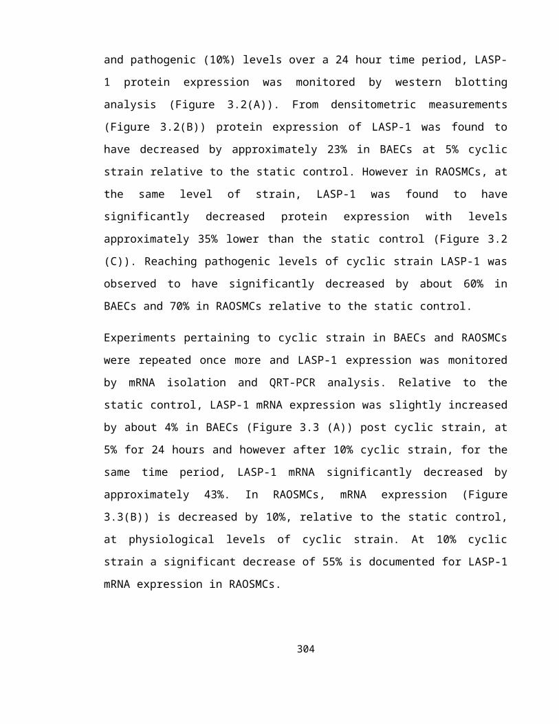
Hu, Y. L., Haga, J. H., Miao, H., Wang, Y., Li, Y. S. and Chien, S. (2006). Roles of
microfilaments and microtubules in paxillin dynamics. Biochem Biophys Res Commun 348,
1463-71.
Huang, H., Kamm, R. D. and Lee, R. T. (2004). Cell mechanics and
mechanotransduction: pathways, probes, and physiology. Am J Physiol Cell Physiol 287,
C1-11.
Hubbard, M. J. and Cohen, P. (1993). On target with a new mechanism for the regulation
of protein phosphorylation. Trends Biochem Sci 18, 172-7.
Hughes, P. E., Diaz-Gonzalez, F., Leong, L., Wu, C., McDonald, J. A., Shattil, S. J.
and Ginsberg, M. H. (1996). Breaking the integrin hinge. A defined structural constraint
regulates integrin signaling. J Biol Chem 271, 6571-4.
Hynes, R. O. (1987). Integrins: a family of cell surface receptors. Cell 48, 549-54.
Hynes, R. O. (1992). Integrins: versatility, modulation, and signaling in cell adhesion. Cell
69, 11-25.
Hynes, R. O. (1999). Cell adhesion: old and new questions. Trends Cell Biol 9, M33-7.
Hynes, R. O. (2002). Integrins: bidirectional, allosteric signaling machines. Cell 110, 673-
87.
Hynes, R. O. (2007). Cell-matrix adhesion in vascular development. J Thromb Haemost 5
Suppl 1, 32-40.
Ignarro, L. J., Byrns, R. E., Buga, G. M. and Wood, K. S. (1987). Endothelium-derived
relaxing factor from pulmonary artery and vein possesses pharmacologic and chemical
properties identical to those of nitric oxide radical. Circ Res 61, 866-79.304

Ihrcke, N. S., Wrenshall, L. E., Lindman, B. J. and Platt, J. L. (1993). Role of heparan
sulfate in immune system-blood vessel interactions. Immunol Today 14, 500-5.
Imamura, Y., Itoh, M., Maeno, Y., Tsukita, S. and Nagafuchi, A. (1999). Functional
domains of alpha-catenin required for the strong state of cadherin-based cell adhesion. J
Cell Biol 144, 1311-22.
Imamura, Y., Itoh, M., Maeno, Y., Tsukita, S. and Nagafuchi, A. (1999). Functional
domains of alpha-catenin required for the strong state of cadherin-based cell adhesion. J
Cell Biol 144, 1311-22.
Imberti, B., Morigi, M., Zoja, C., Angioletti, S., Abbate, M., Remuzzi, A. and
Remuzzi, G. (2000). Shear stress-induced cytoskeleton rearrangement mediates NF-
kappaB-dependent endothelial expression of ICAM-1. Microvasc Res 60, 182-8.
Ingber, D. E. (1998). Cellular basis of mechanotransduction. Biol Bull 194, 323-5;
discussion 325-7.
Ingley, E. and Hemmings, B. A. (1994). Pleckstrin homology (PH) domains in signal
transduction. J Cell Biochem 56, 436-43.
Innocenti, M., Frittoli, E., Ponzanelli, I., Falck, J. R., Brachmann, S. M., Di Fiore, P.
P. and Scita, G. (2003). Phosphoinositide 3-kinase activates Rac by entering in a complex
with Eps8, Abi1, and Sos-1. J Cell Biol 160, 17-23.
Inoue, N., Ramasamy, S., Fukai, T., Nerem, R. M. and Harrison, D. G. (1996). Shear
stress modulates expression of Cu/Zn superoxide dismutase in human aortic endothelial
cells. Circ Res 79, 32-7.
305

Ishida, T., Peterson, T. E., Kovach, N. L. and Berk, B. C. (1996). MAP kinase activation
by flow in endothelial cells. Role of beta 1 integrins and tyrosine kinases. Circ Res 79, 310-
6.
Jaalouk, D. E. and Lammerding, J. (2009). Mechanotransduction gone awry. Nat Rev
Mol Cell Biol 10, 63-73.
Jalali, S., Li, Y. S., Sotoudeh, M., Yuan, S., Li, S., Chien, S. and Shyy, J. Y. (1998).
Shear stress activates p60src-Ras-MAPK signaling pathways in vascular endothelial cells.
Arterioscler Thromb Vasc Biol 18, 227-34.
Janmey, P. A. and Weitz, D. A. (2004). Dealing with mechanics: mechanisms of force
transduction in cells. Trends Biochem Sci 29, 364-70.
Jin, L., Hastings, N. E., Blackman, B. R. and Somlyo, A. V. (2009). Mechanical
properties of the extracellular matrix alter expression of smooth muscle protein LPP and its
partner palladin; relationship to early atherosclerosis and vascular injury. J Muscle Res Cell
Motil 30, 41-55.
Jin, T. G., Satoh, T., Liao, Y., Song, C., Gao, X., Kariya, K., Hu, C. D. and Kataoka,
T. (2001). Role of the CDC25 homology domain of phospholipase Cepsilon in
amplification of Rap1-dependent signaling. J Biol Chem 276, 30301-7.
Jo, H., Sipos, K., Go, Y. M., Law, R., Rong, J. and McDonald, J. M. (1997). Differential
effect of shear stress on extracellular signal-regulated kinase and N-terminal Jun kinase in
endothelial cells. Gi2- and Gbeta/gamma-dependent signaling pathways. J Biol Chem 272,
1395-401.
Jovceva, E., Larsen, M. R., Waterfield, M. D., Baum, B. and Timms, J. F. (2007).
Dynamic cofilin phosphorylation in the control of lamellipodial actin homeostasis. J Cell
Sci 120, 1888-97.306

Kaibuchi, K., Kuroda, S. and Amano, M. (1999). Regulation of the cytoskeleton and cell
adhesion by the Rho family GTPases in mammalian cells. Annu Rev Biochem 68, 459-86.
Kain, K. H. and Klemke, R. L. (2001). Inhibition of cell migration by Abl family tyrosine
kinases through uncoupling of Crk-CAS complexes. J Biol Chem 276, 16185-92.
Kakisis, J. D., Liapis, C. D. and Sumpio, B. E. (2004). Effects of cyclic strain on vascular
cells. Endothelium 11, 17-28.
Kaksonen, M., Peng, H. B. and Rauvala, H. (2000). Association of cortactin with
dynamic actin in lamellipodia and on endosomal vesicles. J Cell Sci 113 Pt 24, 4421-6.
Kano, Y., Katoh, K. and Fujiwara, K. (2000). Lateral zone of cell-cell adhesion as the
major fluid shear stress-related signal transduction site. Circ Res 86, 425-33.
Katagiri, K., Maeda, A., Shimonaka, M. and Kinashi, T. (2003). RAPL, a Rap1-binding
molecule that mediates Rap1-induced adhesion through spatial regulation of LFA-1. Nat
Immunol 4, 741-8.
Katoh, K., Kano, Y., Amano, M., Onishi, H., Kaibuchi, K. and Fujiwara, K. (2001).
Rho-kinase--mediated contraction of isolated stress fibers. J Cell Biol 153, 569-84.
Katoh, M. (2007). WNT signaling pathway and stem cell signaling network. Clin Cancer
Res 13, 4042-5.
Kavanaugh, W. M., Turck, C. W. and Williams, L. T. (1995). PTB domain binding to
signaling proteins through a sequence motif containing phosphotyrosine. Science 268,
1177-9.
307

Kaverina, I., Krylyshkina, O., Beningo, K., Anderson, K., Wang, Y. L. and Small, J.
V. (2002). Tensile stress stimulates microtubule outgrowth in living cells. J Cell Sci 115,
2283-91.
Kawano, Y., Fukata, Y., Oshiro, N., Amano, M., Nakamura, T., Ito, M., Matsumura,
F., Inagaki, M. and Kaibuchi, K. (1999). Phosphorylation of myosin-binding subunit
(MBS) of myosin phosphatase by Rho-kinase in vivo. J Cell Biol 147, 1023-38.
Keely, P., Parise, L. and Juliano, R. (1998). Integrins and GTPases in tumour cell growth,
motility and invasion. Trends Cell Biol 8, 101-6.
Keicher, C., Gambaryan, S., Schulze, E., Marcus, K., Meyer, H. E. and Butt, E.
(2004). Phosphorylation of mouse LASP-1 on threonine 156 by cAMP- and cGMP-
dependent protein kinase. Biochem Biophys Res Commun 324, 308-16.
Khachigian, L. M., Anderson, K. R., Halnon, N. J., Gimbrone, M. A., Jr., Resnick, N.
and Collins, T. (1997). Egr-1 is activated in endothelial cells exposed to fluid shear stress
and interacts with a novel shear-stress-response element in the PDGF A-chain promoter.
Arterioscler Thromb Vasc Biol 17, 2280-6.
Kimura, K., Fukata, Y., Matsuoka, Y., Bennett, V., Matsuura, Y., Okawa, K.,
Iwamatsu, A. and Kaibuchi, K. (1998). Regulation of the association of adducin with
actin filaments by Rho-associated kinase (Rho-kinase) and myosin phosphatase. J Biol
Chem 273, 5542-8.
Kimura, K., Ito, M., Amano, M., Chihara, K., Fukata, Y., Nakafuku, M., Yamamori,
B., Feng, J., Nakano, T., Okawa, K. et al. (1996). Regulation of myosin phosphatase by
Rho and Rho-associated kinase (Rho-kinase). Science 273, 245-8.
308

Kinlay, S., Behrendt, D., Wainstein, M., Beltrame, J., Fang, J. C., Creager, M. A.,
Selwyn, A. P. and Ganz, P. (2001). Role of endothelin-1 in the active constriction of
human atherosclerotic coronary arteries. Circulation 104, 1114-8.
Kiosses, W. B., Shattil, S. J., Pampori, N. and Schwartz, M. A. (2001). Rac recruits
high-affinity integrin alphavbeta3 to lamellipodia in endothelial cell migration. Nat Cell
Biol 3, 316-20.
Knox, A. L. and Brown, N. H. (2002). Rap1 GTPase regulation of adherens junction
positioning and cell adhesion. Science 295, 1285-8.
Koenig, A., Mueller, C., Hasel, C., Adler, G. and Menke, A. (2006). Collagen type I
induces disruption of E-cadherin-mediated cell-cell contacts and promotes proliferation of
pancreatic carcinoma cells. Cancer Res 66, 4662-71.
Kondapalli, J., Flozak, A. S. and Albuquerque, M. L. (2004). Laminar shear stress
differentially modulates gene expression of p120 catenin, Kaiso transcription factor, and
vascular endothelial cadherin in human coronary artery endothelial cells. J Biol Chem 279,
11417-24.
Kooistra, M. R., Dube, N. and Bos, J. L. (2007). Rap1: a key regulator in cell-cell
junction formation. J Cell Sci 120, 17-22.
Kornberg, L., Earp, H. S., Parsons, J. T., Schaller, M. and Juliano, R. L. (1992). Cell
adhesion or integrin clustering increases phosphorylation of a focal adhesion-associated
tyrosine kinase. J Biol Chem 267, 23439-42.
Kranewitter, W. J., Danninger, C. and Gimona, M. (2001). GEF at work: Vav in
protruding filopodia. Cell Motil Cytoskeleton 49, 154-60.
309

Krause, M., Dent, E. W., Bear, J. E., Loureiro, J. J. and Gertler, F. B. (2003).
Ena/VASP proteins: regulators of the actin cytoskeleton and cell migration. Annu Rev Cell
Dev Biol 19, 541-64.
Kraynov, V. S., Chamberlain, C., Bokoch, G. M., Schwartz, M. A., Slabaugh, S. and
Hahn, K. M. (2000). Localized Rac activation dynamics visualized in living cells. Science
290, 333-7.
Kris, A. S., Kamm, R. D. and Sieminski, A. L. (2008). VASP involvement in force-
mediated adherens junction strengthening. Biochem Biophys Res Commun 375, 134-8.
Ku, D. N., Giddens, D. P., Zarins, C. K. and Glagov, S. (1985). Pulsatile flow and
atherosclerosis in the human carotid bifurcation. Positive correlation between plaque
location and low oscillating shear stress. Arteriosclerosis 5, 293-302.
Kuchan, M. J. and Frangos, J. A. (1993). Shear stress regulates endothelin-1 release via
protein kinase C and cGMP in cultured endothelial cells. Am J Physiol 264, H150-6.
Kukalev, A., Nord, Y., Palmberg, C., Bergman, T. and Percipalle, P. (2005). Actin and
hnRNP U cooperate for productive transcription by RNA polymerase II. Nat Struct Mol
Biol 12, 238-44.
Kuroda, S., Fukata, M., Nakagawa, M., Fujii, K., Nakamura, T., Ookubo, T., Izawa,
I., Nagase, T., Nomura, N., Tani, H. et al. (1998). Role of IQGAP1, a target of the small
GTPases Cdc42 and Rac1, in regulation of E-cadherin- mediated cell-cell adhesion. Science
281, 832-5.
Kwiatkowski, A. V., Gertler, F. B. and Loureiro, J. J. (2003). Function and regulation of
Ena/VASP proteins. Trends Cell Biol 13, 386-92.
310

Lafuente, E. M., van Puijenbroek, A. A., Krause, M., Carman, C. V., Freeman, G. J.,
Berezovskaya, A., Constantine, E., Springer, T. A., Gertler, F. B. and Boussiotis, V. A.
(2004). RIAM, an Ena/VASP and Profilin ligand, interacts with Rap1-GTP and mediates
Rap1-induced adhesion. Dev Cell 7, 585-95.
Lai, C. H., Kuo, K. H. and Leo, J. M. (2005). Critical role of actin in modulating BBB
permeability. Brain Res Brain Res Rev 50, 7-13.
Lampugnani, M. G., Corada, M., Caveda, L., Breviario, F., Ayalon, O., Geiger, B. and
Dejana, E. (1995). The molecular organization of endothelial cell to cell junctions:
differential association of plakoglobin, beta-catenin, and alpha-catenin with vascular
endothelial cadherin (VE-cadherin). J Cell Biol 129, 203-17.
Lampugnani, M. G. and Dejana, E. (1997). Interendothelial junctions: structure,
signalling and functional roles. Curr Opin Cell Biol 9, 674-82.
Lanzetti, L., Rybin, V., Malabarba, M. G., Christoforidis, S., Scita, G., Zerial, M. and
Di Fiore, P. P. (2000). The Eps8 protein coordinates EGF receptor signalling through Rac
and trafficking through Rab5. Nature 408, 374-7.
Lee, H. S., Lim, C. J., Puzon-McLaughlin, W., Shattil, S. J. and Ginsberg, M. H.
(2009). RIAM activates integrins by linking talin to ras GTPase membrane-targeting
sequences. J Biol Chem 284, 5119-27.
Lee, J., Ishihara, A., Theriot, J. A. and Jacobson, K. (1993). Principles of locomotion
for simple-shaped cells. Nature 362, 167-71.
Lee, J., Leonard, M., Oliver, T., Ishihara, A. and Jacobson, K. (1994). Traction forces
generated by locomoting keratocytes. J Cell Biol 127, 1957-64.
311

Lee, T. Y. and Gotlieb, A. I. (1999). Early stages of endothelial wound repair: conversion
of quiescent to migrating endothelial cells involves tyrosine phosphorylation and actin
microfilament reorganization. Cell Tissue Res 297, 435-50.
Lee, T. Y. and Gotlieb, A. I. (2003). Microfilaments and microtubules maintain
endothelial integrity. Microsc Res Tech 60, 115-27.
Lehoux, S., Castier, Y. and Tedgui, A. (2006). Molecular mechanisms of the vascular
responses to haemodynamic forces. J Intern Med 259, 381-92.
Lehoux, S. and Tedgui, A. (2003). Cellular mechanics and gene expression in blood
vessels. J Biomech 36, 631-43.
Li, B., Zhuang, L. and Trueb, B. (2004). Zyxin interacts with the SH3 domains of the
cytoskeletal proteins LIM-nebulette and Lasp-1. J Biol Chem 279, 20401-10.
Li, F. and Higgs, H. N. (2003). The mouse Formin mDia1 is a potent actin nucleation
factor regulated by autoinhibition. Curr Biol 13, 1335-40.
Li, Y. S., Shyy, J. Y., Li, S., Lee, J., Su, B., Karin, M. and Chien, S. (1996). The Ras-
JNK pathway is involved in shear-induced gene expression. Mol Cell Biol 16, 5947-54.
Lin, T. H., Chen, Q., Howe, A. and Juliano, R. L. (1997). Cell anchorage permits
efficient signal transduction between ras and its downstream kinases. J Biol Chem 272,
8849-52.
Lin, Y. H., Park, Z. Y., Lin, D., Brahmbhatt, A. A., Rio, M. C., Yates, J. R., 3rd and
Klemke, R. L. (2004). Regulation of cell migration and survival by focal adhesion
targeting of Lasp-1. J Cell Biol 165, 421-32.
312

Linseman, D. A., Benjamin, C. W. and Jones, D. A. (1995). Convergence of angiotensin
II and platelet-derived growth factor receptor signaling cascades in vascular smooth muscle
cells. J Biol Chem 270, 12563-8.
Liu, B. P. and Burridge, K. (2000). Vav2 activates Rac1, Cdc42, and RhoA downstream
from growth factor receptors but not beta1 integrins. Mol Cell Biol 20, 7160-9.
Liu, S., Calderwood, D. A. and Ginsberg, M. H. (2000). Integrin cytoplasmic domain-
binding proteins. J Cell Sci 113 ( Pt 20), 3563-71.
Lorenowicz, M. J., Fernandez-Borja, M., Kooistra, M. R., Bos, J. L. and Hordijk, P.
L. (2008). PKA and Epac1 regulate endothelial integrity and migration through parallel and
independent pathways. Eur J Cell Biol 87, 779-92.
Loscalzo, J. (1995). Nitric oxide and vascular disease. N Engl J Med 333, 251-3.
Loscalzo, J. and Welch, G. (1995). Nitric oxide and its role in the cardiovascular system.
Prog Cardiovasc Dis 38, 87-104.
Lotersztajn, S., Pavoine, C., Deterre, P., Capeau, J., Mallat, A., LeNguyen, D., Dufour,
M., Rouot, B., Bataille, D. and Pecker, F. (1992). Role of G protein beta gamma subunits
in the regulation of the plasma membrane Ca2+ pump. J Biol Chem 267, 2375-9.
Loughran, G., Huigsloot, M., Kiely, P. A., Smith, L. M., Floyd, S., Ayllon, V. and
O'Connor, R. (2005). Gene expression profiles in cells transformed by overexpression of
the IGF-I receptor. Oncogene 24, 6185-93.
Lozano, E., Betson, M. and Braga, V. M. (2003). Tumor progression: Small GTPases and
loss of cell-cell adhesion. Bioessays 25, 452-63.
313

Lukashev, M. E., Sheppard, D. and Pytela, R. (1994). Disruption of integrin function
and induction of tyrosine phosphorylation by the autonomously expressed beta 1 integrin
cytoplasmic domain. J Biol Chem 269, 18311-4.
Lum, H. and Malik, A. B. (1994). Regulation of vascular endothelial barrier function. Am
J Physiol 267, L223-41.
Lum, H. and Malik, A. B. (1996). Mechanisms of increased endothelial permeability. Can
J Physiol Pharmacol 74, 787-800.
Lusis, A. J. (2000). Atherosclerosis. Nature 407, 233-41.
Machesky, L. M. and Insall, R. H. (1998). Scar1 and the related Wiskott-Aldrich
syndrome protein, WASP, regulate the actin cytoskeleton through the Arp2/3 complex.
Curr Biol 8, 1347-56.
Machesky, L. M. and Insall, R. H. (1999). Signaling to actin dynamics. J Cell Biol 146,
267-72.
Maclouf, J., Folco, G. and Patrono, C. (1998). Eicosanoids and iso-eicosanoids:
constitutive, inducible and transcellular biosynthesis in vascular disease. Thromb Haemost
79, 691-705.
Maekawa, M., Ishizaki, T., Boku, S., Watanabe, N., Fujita, A., Iwamatsu, A., Obinata,
T., Ohashi, K., Mizuno, K. and Narumiya, S. (1999). Signaling from Rho to the actin
cytoskeleton through protein kinases ROCK and LIM-kinase. Science 285, 895-8.
Malek, A. M., Alper, S. L. and Izumo, S. (1999). Hemodynamic shear stress and its role
in atherosclerosis. JAMA 282, 2035-42.
314

Malek, A. M., Gibbons, G. H., Dzau, V. J. and Izumo, S. (1993). Fluid shear stress
differentially modulates expression of genes encoding basic fibroblast growth factor and
platelet-derived growth factor B chain in vascular endothelium. J Clin Invest 92, 2013-21.
Malek, A. M., Greene, A. L. and Izumo, S. (1993). Regulation of endothelin 1 gene by
fluid shear stress is transcriptionally mediated and independent of protein kinase C and
cAMP. Proc Natl Acad Sci U S A 90, 5999-6003.
Mallavarapu, A. and Mitchison, T. (1999). Regulated actin cytoskeleton assembly at
filopodium tips controls their extension and retraction. J Cell Biol 146, 1097-106.
Mann, K. G. (1997). Thrombosis: theoretical considerations. Am J Clin Nutr 65, 1657S-
1664S.
Martinac, B. (2004). Mechanosensitive ion channels: molecules of mechanotransduction. J
Cell Sci 117, 2449-60.
Martys, J. L., Ho, C. L., Liem, R. K. and Gundersen, G. G. (1999). Intermediate
filaments in motion: observations of intermediate filaments in cells using green fluorescent
protein-vimentin. Mol Biol Cell 10, 1289-95.
McCue, S., Noria, S. and Langille, B. L. (2004). Shear-induced reorganization of
endothelial cell cytoskeleton and adhesion complexes. Trends Cardiovasc Med 14, 143-51.
McLaughlin, P. J. and Weeds, A. G. (1995). Actin-binding protein complexes at atomic
resolution. Annu Rev Biophys Biomol Struct 24, 643-75.
McNeill, H., Ryan, T. A., Smith, S. J. and Nelson, W. J. (1993). Spatial and temporal
dissection of immediate and early events following cadherin-mediated epithelial cell
adhesion. J Cell Biol 120, 1217-26.
315

Mehta, D. and Malik, A. B. (2006). Signaling mechanisms regulating endothelial
permeability. Physiol Rev 86, 279-367.
Meigs, T. E., Fields, T. A., McKee, D. D. and Casey, P. J. (2001). Interaction of Galpha
12 and Galpha 13 with the cytoplasmic domain of cadherin provides a mechanism for beta -
catenin release. Proc Natl Acad Sci U S A 98, 519-24.
Mezentsev, A., Merks, R. M., O'Riordan, E., Chen, J., Mendelev, N., Goligorsky, M.
S. and Brodsky, S. V. (2005). Endothelial microparticles affect angiogenesis in vitro: role
of oxidative stress. Am J Physiol Heart Circ Physiol 289, H1106-14.
Michaelson, D., Silletti, J., Murphy, G., D'Eustachio, P., Rush, M. and Philips, M. R.
(2001). Differential localization of Rho GTPases in live cells: regulation by hypervariable
regions and RhoGDI binding. J Cell Biol 152, 111-26.
Miki, H., Suetsugu, S. and Takenawa, T. (1998). WAVE, a novel WASP-family protein
involved in actin reorganization induced by Rac. EMBO J 17, 6932-41.
Miralles, F. and Visa, N. (2006). Actin in transcription and transcription regulation. Curr
Opin Cell Biol 18, 261-6.
Mishima, M. and Nishida, E. (1999). Coronin localizes to leading edges and is involved
in cell spreading and lamellipodium extension in vertebrate cells. J Cell Sci 112 (Pt 17),
2833-42.
Miyake, Y., Inoue, N., Nishimura, K., Kinoshita, N., Hosoya, H. and Yonemura, S.
(2006). Actomyosin tension is required for correct recruitment of adherens junction
components and zonula occludens formation. Exp Cell Res 312, 1637-50.
316

Miyamoto, S., Teramoto, H., Coso, O. A., Gutkind, J. S., Burbelo, P. D., Akiyama, S.
K. and Yamada, K. M. (1995). Integrin function: molecular hierarchies of cytoskeletal
and signaling molecules. J Cell Biol 131, 791-805.
Mykkanen, O. M., Gronholm, M., Ronty, M., Lalowski, M., Salmikangas, P., Suila, H.
and Carpen, O. (2001). Characterization of human palladin, a microfilament-associated
protein. Mol Biol Cell 12, 3060-73.
Nagafuchi, A. and Takeichi, M. (1988). Cell binding function of E-cadherin is regulated
by the cytoplasmic domain. EMBO J 7, 3679-84.
Nagel, T., Resnick, N., Atkinson, W. J., Dewey, C. F., Jr. and Gimbrone, M. A., Jr.
(1994). Shear stress selectively upregulates intercellular adhesion molecule-1 expression in
cultured human vascular endothelial cells. J Clin Invest 94, 885-91.
Nakagawa, H., Miki, H., Ito, M., Ohashi, K., Takenawa, T. and Miyamoto, S. (2001).
N-WASP, WAVE and Mena play different roles in the organization of actin cytoskeleton in
lamellipodia. J Cell Sci 114, 1555-65.
Nakagawa, H., Terasaki, A. G., Suzuki, H., Ohashi, K. and Miyamoto, S. (2006).
Short-term retention of actin filament binding proteins on lamellipodial actin bundles.
FEBS Lett 580, 3223-8.
Nakashima, Y., Raines, E. W., Plump, A. S., Breslow, J. L. and Ross, R. (1998).
Upregulation of VCAM-1 and ICAM-1 at atherosclerosis-prone sites on the endothelium in
the ApoE-deficient mouse. Arterioscler Thromb Vasc Biol 18, 842-51.
Nelson, W. J. (2003). Adaptation of core mechanisms to generate cell polarity. Nature 422,
766-74.
Newman, P. J. (1997). The biology of PECAM-1. J Clin Invest 100, S25-9.317

Nieset, J. E., Redfield, A. R., Jin, F., Knudsen, K. A., Johnson, K. R. and Wheelock,
M. J. (1997). Characterization of the interactions of alpha-catenin with alpha-actinin and
beta-catenin/plakoglobin. J Cell Sci 110 ( Pt 8), 1013-22.
Niessen, C. M. and Gottardi, C. J. (2008). Molecular components of the adherens
junction. Biochim Biophys Acta 1778, 562-71.
Nishida, K., Harrison, D. G., Navas, J. P., Fisher, A. A., Dockery, S. P., Uematsu, M.,
Nerem, R. M., Alexander, R. W. and Murphy, T. J. (1992). Molecular cloning and
characterization of the constitutive bovine aortic endothelial cell nitric oxide synthase. J
Clin Invest 90, 2092-6.
Nix, D. A., Fradelizi, J., Bockholt, S., Menichi, B., Louvard, D., Friederich, E. and
Beckerle, M. C. (2001). Targeting of zyxin to sites of actin membrane interaction and to
the nucleus. J Biol Chem 276, 34759-67.
Nobes, C. D. and Hall, A. (1995). Rho, rac, and cdc42 GTPases regulate the assembly of
multimolecular focal complexes associated with actin stress fibers, lamellipodia, and
filopodia. Cell 81, 53-62.
Nobes, C. D. and Hall, A. (1999). Rho GTPases control polarity, protrusion, and adhesion
during cell movement. J Cell Biol 144, 1235-44.
Noria, S., Xu, F., McCue, S., Jones, M., Gotlieb, A. I. and Langille, B. L. (2004).
Assembly and reorientation of stress fibers drives morphological changes to endothelial
cells exposed to shear stress. Am J Pathol 164, 1211-23.
Noria, S., Xu, F., McCue, S., Jones, M., Gotlieb, A. I. and Langille, B. L. (2004).
Assembly and reorientation of stress fibers drives morphological changes to endothelial
cells exposed to shear stress. Am J Pathol 164, 1211-23.
318

Numaguchi, K., Eguchi, S., Yamakawa, T., Motley, E. D. and Inagami, T. (1999).
Mechanotransduction of rat aortic vascular smooth muscle cells requires RhoA and intact
actin filaments. Circ Res 85, 5-11.
Nyakern-Meazza, M., Narayan, K., Schutt, C. E. and Lindberg, U. (2002).
Tropomyosin and gelsolin cooperate in controlling the microfilament system. J Biol Chem
277, 28774-9.
Offermanns, S. (2003). G-proteins as transducers in transmembrane signalling. Prog
Biophys Mol Biol 83, 101-30.
Okada, K., Blanchoin, L., Abe, H., Chen, H., Pollard, T. D. and Bamburg, J. R.
(2002). Xenopus actin-interacting protein 1 (XAip1) enhances cofilin fragmentation of
filaments by capping filament ends. J Biol Chem 277, 43011-6.
Orr, A. W., Helmke, B. P., Blackman, B. R. and Schwartz, M. A. (2006). Mechanisms
of mechanotransduction. Dev Cell 10, 11-20.
Osborn, E. A., Rabodzey, A., Dewey, C. F., Jr. and Hartwig, J. H. (2006). Endothelial
actin cytoskeleton remodeling during mechanostimulation with fluid shear stress. Am J
Physiol Cell Physiol 290, C444-52.
O'Toole, T. E., Katagiri, Y., Faull, R. J., Peter, K., Tamura, R., Quaranta, V., Loftus,
J. C., Shattil, S. J. and Ginsberg, M. H. (1994). Integrin cytoplasmic domains mediate
inside-out signal transduction. J Cell Biol 124, 1047-59.
O'Toole, T. E., Mandelman, D., Forsyth, J., Shattil, S. J., Plow, E. F. and Ginsberg, M.
H. (1991). Modulation of the affinity of integrin alpha IIb beta 3 (GPIIb-IIIa) by the
cytoplasmic domain of alpha IIb. Science 254, 845-7.
319

O'Toole, T. E., Ylanne, J. and Culley, B. M. (1995). Regulation of integrin affinity states
through an NPXY motif in the beta subunit cytoplasmic domain. J Biol Chem 270, 8553-8.
Owaribe, K., Kodama, R. and Eguchi, G. (1981). Demonstration of contractility of
circumferential actin bundles and its morphogenetic significance in pigmented epithelium
in vitro and in vivo. J Cell Biol 90, 507-14.
Palmer, R. M., Ferrige, A. G. and Moncada, S. (1987). Nitric oxide release accounts for
the biological activity of endothelium-derived relaxing factor. Nature 327, 524-6.
Pampori, N., Hato, T., Stupack, D. G., Aidoudi, S., Cheresh, D. A., Nemerow, G. R.
and Shattil, S. J. (1999). Mechanisms and consequences of affinity modulation of integrin
alpha(V)beta(3) detected with a novel patch-engineered monovalent ligand. J Biol Chem
274, 21609-16.
Pantaloni, D., Boujemaa, R., Didry, D., Gounon, P. and Carlier, M. F. (2000). The
Arp2/3 complex branches filament barbed ends: functional antagonism with capping
proteins. Nat Cell Biol 2, 385-91.
Park, H. O., Bi, E., Pringle, J. R. and Herskowitz, I. (1997). Two active states of the
Ras-related Bud1/Rsr1 protein bind to different effectors to determine yeast cell polarity.
Proc Natl Acad Sci U S A 94, 4463-8.
Parsons, J. T. and Parsons, S. J. (1997). Src family protein tyrosine kinases: cooperating
with growth factor and adhesion signaling pathways. Curr Opin Cell Biol 9, 187-92.
Pastore, L., Tessitore, A., Martinotti, S., Toniato, E., Alesse, E., Bravi, M. C., Ferri,
C., Desideri, G., Gulino, A. and Santucci, A. (1999). Angiotensin II stimulates
intercellular adhesion molecule-1 (ICAM-1) expression by human vascular endothelial
cells and increases soluble ICAM-1 release in vivo. Circulation 100, 1646-52.
320

Pavalko, F. M. and Burridge, K. (1991). Disruption of the actin cytoskeleton after
microinjection of proteolytic fragments of alpha-actinin. J Cell Biol 114, 481-91.
Pawson, T. (1995). Protein modules and signalling networks. Nature 373, 573-80.
Pawson, T. and Scott, J. D. (1997). Signaling through scaffold, anchoring, and adaptor
proteins. Science 278, 2075-80.
Pearce, M. J., McIntyre, T. M., Prescott, S. M., Zimmerman, G. A. and Whatley, R. E.
(1996). Shear stress activates cytosolic phospholipase A2 (cPLA2) and MAP kinase in
human endothelial cells. Biochem Biophys Res Commun 218, 500-4.
Pederson, T. and Aebi, U. (2002). Actin in the nucleus: what form and what for? J Struct
Biol 140, 3-9.
Pellegrin, S. and Mellor, H. (2007). Actin stress fibres. J Cell Sci 120, 3491-9.
Percipalle, P. (2007). Genetic connections of the actin cytoskeleton and beyond. Bioessays
29, 407-11.
Percipalle, P. and Visa, N. (2006). Molecular functions of nuclear actin in transcription. J
Cell Biol 172, 967-71.
Petit, M. M., Crombez, K. R., Vervenne, H. B., Weyns, N. and Van de Ven, W. J.
(2005). The tumor suppressor Scrib selectively interacts with specific members of the zyxin
family of proteins. FEBS Lett 579, 5061-8.
Petit, M. M., Meulemans, S. M. and Van de Ven, W. J. (2003). The focal adhesion and
nuclear targeting capacity of the LIM-containing lipoma-preferred partner (LPP) protein. J
Biol Chem 278, 2157-68.
321

Phillips, G. R., Anderson, T. R., Florens, L., Gudas, C., Magda, G., Yates, J. R., 3rd
and Colman, D. R. (2004). Actin-binding proteins in a postsynaptic preparation: Lasp-1 is
a component of central nervous system synapses and dendritic spines. J Neurosci Res 78,
38-48.
Piali, L., Hammel, P., Uherek, C., Bachmann, F., Gisler, R. H., Dunon, D. and Imhof,
B. A. (1995). CD31/PECAM-1 is a ligand for alpha v beta 3 integrin involved in adhesion
of leukocytes to endothelium. J Cell Biol 130, 451-60.
Pirotton, S., Erneux, C. and Boeynaems, J. M. (1987). Dual role of GTP-binding
proteins in the control of endothelial prostacyclin. Biochem Biophys Res Commun 147,
1113-20.
Pollard, T. D., Almo, S., Quirk, S., Vinson, V. and Lattman, E. E. (1994). Structure of
actin binding proteins: insights about function at atomic resolution. Annu Rev Cell Biol 10,
207-49.
Pollard, T. D., Blanchoin, L. and Mullins, R. D. (2000). Molecular mechanisms
controlling actin filament dynamics in nonmuscle cells. Annu Rev Biophys Biomol Struct
29, 545-76.
Pollard, T. D. and Borisy, G. G. (2003). Cellular motility driven by assembly and
disassembly of actin filaments. Cell 112, 453-65.
Pradhan, S. and Sumpio, B. (2004). Molecular and biological effects of hemodynamics
on vascular cells. Front Biosci 9, 3276-85.
Price, L. S., Hajdo-Milasinovic, A., Zhao, J., Zwartkruis, F. J., Collard, J. G. and Bos,
J. L. (2004). Rap1 regulates E-cadherin-mediated cell-cell adhesion. J Biol Chem 279,
35127-32.
322

Pueyo, M. E., Gonzalez, W., Nicoletti, A., Savoie, F., Arnal, J. F. and Michel, J. B.
(2000). Angiotensin II stimulates endothelial vascular cell adhesion molecule-1 via nuclear
factor-kappaB activation induced by intracellular oxidative stress. Arterioscler Thromb
Vasc Biol 20, 645-51.
Pueyo, M. E. and Michel, J. B. (1997). Angiotensin II receptors in endothelial cells. Gen
Pharmacol 29, 691-6.
Qiao, R. L., Yan, W., Lum, H. and Malik, A. B. (1995). Arg-Gly-Asp peptide increases
endothelial hydraulic conductivity: comparison with thrombin response. Am J Physiol 269,
C110-7.
Rachlin, A. S. and Otey, C. A. (2006). Identification of palladin isoforms and
characterization of an isoform-specific interaction between Lasp-1 and palladin. J Cell Sci
119, 995-1004.
Rajfur, Z., Roy, P., Otey, C., Romer, L. and Jacobson, K. (2002). Dissecting the link
between stress fibres and focal adhesions by CALI with EGFP fusion proteins. Nat Cell
Biol 4, 286-93.
Redmond, E. M., Cahill, P. A. and Sitzmann, J. V. (1998). Flow-mediated regulation of
G-protein expression in cocultured vascular smooth muscle and endothelial cells.
Arterioscler Thromb Vasc Biol 18, 75-83.
Ren, G., Crampton, M. S. and Yap, A. S. (2009). Cortactin: Coordinating adhesion and
the actin cytoskeleton at cellular protrusions. Cell Motil Cytoskeleton 66, 865-73.
Renshaw, M. W., Price, L. S. and Schwartz, M. A. (1999). Focal adhesion kinase
mediates the integrin signaling requirement for growth factor activation of MAP kinase. J
Cell Biol 147, 611-8.
323

Resink, T. J., Philippova, M., Joshi, M. B., Kyriakakis, E. and Erne, P. (2009).
Cadherins and cardiovascular disease. Swiss Med Wkly 139, 122-34.
Resnick, N., Collins, T., Atkinson, W., Bonthron, D. T., Dewey, C. F., Jr. and
Gimbrone, M. A., Jr. (1993). Platelet-derived growth factor B chain promoter contains a
cis-acting fluid shear-stress-responsive element. Proc Natl Acad Sci U S A 90, 4591-5.
Resnick, N. and Gimbrone, M. A., Jr. (1995). Hemodynamic forces are complex
regulators of endothelial gene expression. FASEB J 9, 874-82.
Resnick, N., Yahav, H., Shay-Salit, A., Shushy, M., Schubert, S., Zilberman, L. C. and
Wofovitz, E. (2003). Fluid shear stress and the vascular endothelium: for better and for
worse. Prog Biophys Mol Biol 81, 177-99.
Ridley, A. J., Paterson, H. F., Johnston, C. L., Diekmann, D. and Hall, A. (1992). The
small GTP-binding protein rac regulates growth factor-induced membrane ruffling. Cell 70,
401-10.
Riveline, D., Zamir, E., Balaban, N. Q., Schwarz, U. S., Ishizaki, T., Narumiya, S.,
Kam, Z., Geiger, B. and Bershadsky, A. D. (2001). Focal contacts as mechanosensors:
externally applied local mechanical force induces growth of focal contacts by an mDia1-
dependent and ROCK-independent mechanism. J Cell Biol 153, 1175-86.
Rivero, F., Koppel, B., Peracino, B., Bozzaro, S., Siegert, F., Weijer, C. J., Schleicher,
M., Albrecht, R. and Noegel, A. A. (1996). The role of the cortical cytoskeleton: F-actin
crosslinking proteins protect against osmotic stress, ensure cell size, cell shape and motility,
and contribute to phagocytosis and development. J Cell Sci 109 ( Pt 11), 2679-91.
Robinson, M. J. and Cobb, M. H. (1997). Mitogen-activated protein kinase pathways.
Curr Opin Cell Biol 9, 180-6.
324

Rodriguez-Viciana, P., Warne, P. H., Khwaja, A., Marte, B. M., Pappin, D., Das, P.,
Waterfield, M. D., Ridley, A. and Downward, J. (1997). Role of phosphoinositide 3-OH
kinase in cell transformation and control of the actin cytoskeleton by Ras. Cell 89, 457-67.
Rolfe, B. E., Worth, N. F., World, C. J., Campbell, J. H. and Campbell, G. R. (2005).
Rho and vascular disease. Atherosclerosis 183, 1-16.
Ronty, M., Taivainen, A., Moza, M., Kruh, G. D., Ehler, E. and Carpen, O. (2005).
Involvement of palladin and alpha-actinin in targeting of the Abl/Arg kinase adaptor
ArgBP2 to the actin cytoskeleton. Exp Cell Res 310, 88-98.
Ronty, M., Taivainen, A., Moza, M., Otey, C. A. and Carpen, O. (2004). Molecular
analysis of the interaction between palladin and alpha-actinin. FEBS Lett 566, 30-4.
Rosenberg, R. D. and Aird, W. C. (1999). Vascular-bed--specific hemostasis and
hypercoagulable states. N Engl J Med 340, 1555-64.
Rottner, K., Behrendt, B., Small, J. V. and Wehland, J. (1999). VASP dynamics during
lamellipodia protrusion. Nat Cell Biol 1, 321-2.
Ruoslahti, E. and Pierschbacher, M. D. (1987). New perspectives in cell adhesion: RGD
and integrins. Science 238, 491-7.
Sadoshima, J. and Izumo, S. (1993). Mechanical stretch rapidly activates multiple signal
transduction pathways in cardiac myocytes: potential involvement of an autocrine/paracrine
mechanism. EMBO J 12, 1681-92.
Salazar, R., Bell, S. E. and Davis, G. E. (1999). Coordinate induction of the actin
cytoskeletal regulatory proteins gelsolin, vasodilator-stimulated phosphoprotein, and
profilin during capillary morphogenesis in vitro. Exp Cell Res 249, 22-32.
325

Sambrook, P. N., Cohen, M. L., Eisman, J. A., Pocock, N. A., Champion, G. D. and
Yeates, M. G. (1989). Effects of low dose corticosteroids on bone mass in rheumatoid
arthritis: a longitudinal study. Ann Rheum Dis 48, 535-8.
Sanchez-Garcia, I. and Rabbitts, T. H. (1994). The LIM domain: a new structural motif
found in zinc-finger-like proteins. Trends Genet 10, 315-20.
Sander, E. E., ten Klooster, J. P., van Delft, S., van der Kammen, R. A. and Collard, J.
G. (1999). Rac downregulates Rho activity: reciprocal balance between both GTPases
determines cellular morphology and migratory behavior. J Cell Biol 147, 1009-22.
Sander, E. E., ten Klooster, J. P., van Delft, S., van der Kammen, R. A. and Collard, J.
G. (1999). Rac downregulates Rho activity: reciprocal balance between both GTPases
determines cellular morphology and migratory behavior. J Cell Biol 147, 1009-22.
Sanger, J. W., Sanger, J. M. and Jockusch, B. M. (1983). Differences in the stress fibers
between fibroblasts and epithelial cells. J Cell Biol 96, 961-9.
Sauzeau, V., Le Jeune, H., Cario-Toumaniantz, C., Smolenski, A., Lohmann, S. M.,
Bertoglio, J., Chardin, P., Pacaud, P. and Loirand, G. (2000). Cyclic GMP-dependent
protein kinase signaling pathway inhibits RhoA-induced Ca2+ sensitization of contraction
in vascular smooth muscle. J Biol Chem 275, 21722-9.
Schafer, D. A. and Schroer, T. A. (1999). Actin-related proteins. Annu Rev Cell Dev Biol
15, 341-63.
Schaller, M. D., Hildebrand, J. D., Shannon, J. D., Fox, J. W., Vines, R. R. and
Parsons, J. T. (1994). Autophosphorylation of the focal adhesion kinase, pp125FAK,
directs SH2-dependent binding of pp60src. Mol Cell Biol 14, 1680-8.
326
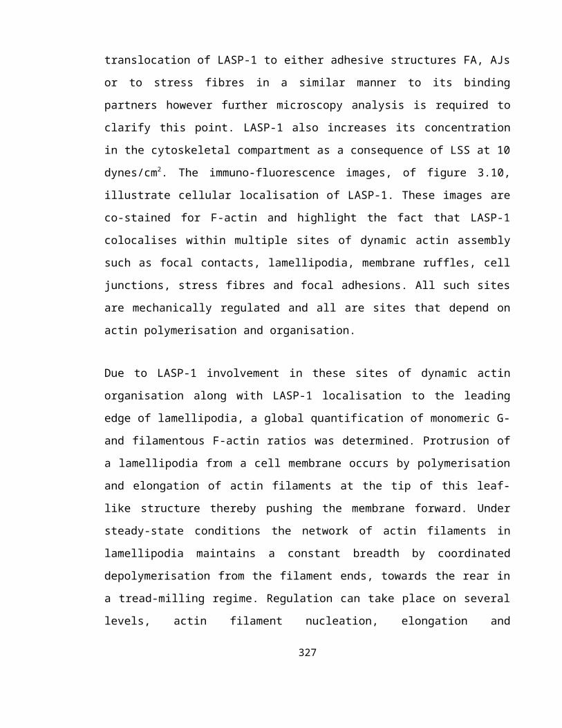
Schaller, M. D., Otey, C. A., Hildebrand, J. D. and Parsons, J. T. (1995). Focal
adhesion kinase and paxillin bind to peptides mimicking beta integrin cytoplasmic domains.
J Cell Biol 130, 1181-7.
Schlaepfer, D. D. and Hunter, T. (1998). Integrin signalling and tyrosine
phosphorylation: just the FAKs? Trends Cell Biol 8, 151-7.
Schlegel, N. and Waschke, J. (2009). Impaired integrin-mediated adhesion contributes to
reduced barrier properties in VASP-deficient microvascular endothelium. J Cell Physiol
220, 357-66.
Schlegel, N. and Waschke, J. (2009). VASP is involved in cAMP-mediated Rac 1
activation in microvascular endothelial cells. Am J Physiol Cell Physiol 296, C453-62.
Schmidt, A. and Hall, M. N. (1998). Signaling to the actin cytoskeleton. Annu Rev Cell
Dev Biol 14, 305-38.
Schoenwaelder, S. M. and Burridge, K. (1999). Bidirectional signaling between the
cytoskeleton and integrins. Curr Opin Cell Biol 11, 274-86.
Schreiber, V., Moog-Lutz, C., Regnier, C. H., Chenard, M. P., Boeuf, H., Vonesch, J.
L., Tomasetto, C. and Rio, M. C. (1998). Lasp-1, a novel type of actin-binding protein
accumulating in cell membrane extensions. Mol Med 4, 675-87.
Schwartz, M. A. and DeSimone, D. W. (2008). Cell adhesion receptors in
mechanotransduction. Curr Opin Cell Biol 20, 551-6.
Schwartz, M. A., Schaller, M. D. and Ginsberg, M. H. (1995). Integrins: emerging
paradigms of signal transduction. Annu Rev Cell Dev Biol 11, 549-99.
327

Schwartz, M. A. and Shattil, S. J. (2000). Signaling networks linking integrins and rho
family GTPases. Trends Biochem Sci 25, 388-91.
Scott, J. A., Shewan, A. M., den Elzen, N. R., Loureiro, J. J., Gertler, F. B. and Yap,
A. S. (2006). Ena/VASP proteins can regulate distinct modes of actin organization at
cadherin-adhesive contacts. Mol Biol Cell 17, 1085-95.
Seebach, J., Donnert, G., Kronstein, R., Werth, S., Wojciak-Stothard, B., Falzarano,
D., Mrowietz, C., Hell, S. W. and Schnittler, H. J. (2007). Regulation of endothelial
barrier function during flow-induced conversion to an arterial phenotype. Cardiovasc Res
75, 596-607.
Shapiro, L., Fannon, A. M., Kwong, P. D., Thompson, A., Lehmann, M. S., Grubel,
G., Legrand, J. F., Als-Nielsen, J., Colman, D. R. and Hendrickson, W. A. (1995).
Structural basis of cell-cell adhesion by cadherins. Nature 374, 327-37.
Shasby, D. M. (2007). Cell-cell adhesion in lung endothelium. Am J Physiol Lung Cell Mol
Physiol 292, L593-607.
Shattil, S. J. and Newman, P. J. (2004). Integrins: dynamic scaffolds for adhesion and
signaling in platelets. Blood 104, 1606-15.
Shattil, S. J., Kashiwagi, H. and Pampori, N. (1998). Integrin signalling: The platelet
paradigm. Blood 91(8), 2645-57.
Shewan, A. M., Maddugoda, M., Kraemer, A., Stehbens, S. J., Verma, S., Kovacs, E.
M. and Yap, A. S. (2005). Myosin 2 is a key Rho kinase target necessary for the local
concentration of E-cadherin at cell-cell contacts. Mol Biol Cell 16, 4531-42.
328

Shiraga, M., Ritchie, A., Aidoudi, S., Baron, V., Wilcox, D., White, G., Ybarrondo, B.,
Murphy, G., Leavitt, A. and Shattil, S. (1999). Primary megakaryocytes reveal a role for
transcription factor NF-E2 in integrin alpha IIb beta 3 signaling. J Cell Biol 147, 1419-30.
Shyy, J. Y. and Chien, S. (2002). Role of integrins in endothelial mechanosensing of shear
stress. Circ Res 91, 769-75.
Shyy, J. Y., Lin, M. C., Han, J., Lu, Y., Petrime, M. and Chien, S. (1995). The cis-
acting phorbol ester "12-O-tetradecanoylphorbol 13-acetate"-responsive element is
involved in shear stress-induced monocyte chemotactic protein 1 gene expression. Proc
Natl Acad Sci U S A 92, 8069-73.
Small, J. V. (1994). Lamellipodia architecture: actin filament turnover and the lateral flow
of actin filaments during motility. Semin Cell Biol 5, 157-63.
Small, J. V., Rottner, K., Kaverina, I. and Anderson, K. I. (1998). Assembling an actin
cytoskeleton for cell attachment and movement. Biochim Biophys Acta 1404, 271-81.
Small, J. V., Stradal, T., Vignal, E. and Rottner, K. (2002). The lamellipodium: where
motility begins. Trends Cell Biol 12, 112-20.
Snapper, S. B., Takeshima, F., Anton, I., Liu, C. H., Thomas, S. M., Nguyen, D.,
Dudley, D., Fraser, H., Purich, D., Lopez-Ilasaca, M. et al. (2001). N-WASP deficiency
reveals distinct pathways for cell surface projections and microbial actin-based motility.
Nat Cell Biol 3, 897-904.
Soldi, R., Mitola, S., Strasly, M., Defilippi, P., Tarone, G. and Bussolino, F. (1999).
Role of alphavbeta3 integrin in the activation of vascular endothelial growth factor
receptor-2. EMBO J 18, 882-92.
329

Sommer, S., Hunzinger, C., Schillo, S., Klemm, M., Biefang-Arndt, K., Schwall, G.,
Putter, S., Hoelzer, K., Schroer, K., Stegmann, W. et al. (2004). Molecular analysis of
homocysteic acid-induced neuronal stress. J Proteome Res 3, 572-81.
Songyang, Z. and Cantley, L. C. (1995). Recognition and specificity in protein tyrosine
kinase-mediated signalling. Trends Biochem Sci 20, 470-5.
Songyang, Z., Shoelson, S. E., McGlade, J., Olivier, P., Pawson, T., Bustelo, X. R.,
Barbacid, M., Sabe, H., Hanafusa, H., Yi, T. et al. (1994). Specific motifs recognized by
the SH2 domains of Csk, 3BP2, fps/fes, GRB-2, HCP, SHC, Syk, and Vav. Mol Cell Biol
14, 2777-85.
Sowers, J. R. (2002). Hypertension, angiotensin II, and oxidative stress. N Engl J Med 346,
1999-2001.
Spence, H. J., McGarry, L., Chew, C. S., Carragher, N. O., Scott-Carragher, L. A.,
Yuan, Z., Croft, D. R., Olson, M. F., Frame, M. and Ozanne, B. W. (2006). AP-1
differentially expressed proteins Krp1 and fibronectin cooperatively enhance Rho-ROCK-
independent mesenchymal invasion by altering the function, localization, and activity of
nondifferentially expressed proteins. Mol Cell Biol 26, 1480-95.
Springer, T. A. (1994). Traffic signals for lymphocyte recirculation and leukocyte
emigration: the multistep paradigm. Cell 76, 301-14.
Stade, K., Ford, C. S., Guthrie, C. and Weis, K. (1997). Exportin 1 (Crm1p) is an
essential nuclear export factor. Cell 90, 1041-50.
Stone, J. L., Merriman, B., Cantor, R. M., Geschwind, D. H. and Nelson, S. F. (2007).
High density SNP association study of a major autism linkage region on chromosome 17.
Hum Mol Genet 16, 704-15.
330

Stossel, T. P., Condeelis, J., Cooley, L., Hartwig, J. H., Noegel, A., Schleicher, M. and
Shapiro, S. S. (2001). Filamins as integrators of cell mechanics and signalling. Nat Rev
Mol Cell Biol 2, 138-45.
Stradal, T., Courtney, K. D., Rottner, K., Hahne, P., Small, J. V. and Pendergast, A.
M. (2001). The Abl interactor proteins localize to sites of actin polymerization at the tips of
lamellipodia and filopodia. Curr Biol 11, 891-5.
Su, A. I., Cooke, M. P., Ching, K. A., Hakak, Y., Walker, J. R., Wiltshire, T., Orth, A.
P., Vega, R. G., Sapinoso, L. M., Moqrich, A. et al. (2002). Large-scale analysis of the
human and mouse transcriptomes. Proc Natl Acad Sci U S A 99, 4465-70.
Su, G., Hodnett, M., Wu, N., Atakilit, A., Kosinski, C., Godzich, M., Huang, X. Z.,
Kim, J. K., Frank, J. A., Matthay, M. A. et al. (2007). Integrin alphavbeta5 regulates
lung vascular permeability and pulmonary endothelial barrier function. Am J Respir Cell
Mol Biol 36, 377-86.
Subramaniam, M., Koedam, J. A. and Wagner, D. D. (1993). Divergent fates of P- and
E-selectins after their expression on the plasma membrane. Mol Biol Cell 4, 791-801.
Sumpio, B. E., Du, W., Galagher, G., Wang, X., Khachigian, L. M., Collins, T.,
Gimbrone, M. A., Jr. and Resnick, N. (1998). Regulation of PDGF-B in endothelial cells
exposed to cyclic strain. Arterioscler Thromb Vasc Biol 18, 349-55.
Sun, H. Q., Yamamoto, M., Mejillano, M. and Yin, H. L. (1999). Gelsolin, a
multifunctional actin regulatory protein. J Biol Chem 274, 33179-82.
Svitkina, T. M. and Borisy, G. G. (1999). Arp2/3 complex and actin depolymerizing
factor/cofilin in dendritic organization and treadmilling of actin filament array in
lamellipodia. J Cell Biol 145, 1009-26.
331

Svitkina, T. M. and Borisy, G. G. (1999). Progress in protrusion: the tell-tale scar. Trends
Biochem Sci 24, 432-6.
Takada, Y., Shinkai, F., Kondo, S., Yamamoto, S., Tsuboi, H., Korenaga, R. and
Ando, J. (1994). Fluid shear stress increases the expression of thrombomodulin by cultured
human endothelial cells. Biochem Biophys Res Commun 205, 1345-52.
Tamkun, J. W., DeSimone, D. W., Fonda, D., Patel, R. S., Buck, C., Horwitz, A. F. and
Hynes, R. O. (1986). Structure of integrin, a glycoprotein involved in the transmembrane
linkage between fibronectin and actin. Cell 46, 271-82.
Taubman, M. B., Fallon, J. T., Schecter, A. D., Giesen, P., Mendlowitz, M., Fyfe, B. S.,
Marmur, J. D. and Nemerson, Y. (1997). Tissue factor in the pathogenesis of
atherosclerosis. Thromb Haemost 78, 200-4.
Taylor, S. S., Buechler, J. A. and Yonemoto, W. (1990). cAMP-dependent protein
kinase: framework for a diverse family of regulatory enzymes. Annu Rev Biochem 59, 971-
1005.
Ten Klooster, J. P., Evers, E. E., Janssen, L., Machesky, L. M., Michiels, F., Hordijk,
P. and Collard, J. G. (2006). Interaction between Tiam1 and the Arp2/3 complex links
activation of Rac to actin polymerization. Biochem J 397, 39-45.
Terasaki, A. G., Suzuki, H., Nishioka, T., Matsuzawa, E., Katsuki, M., Nakagawa, H.,
Miyamoto, S. and Ohashi, K. (2004). A novel LIM and SH3 protein (lasp-2) highly
expressing in chicken brain. Biochem Biophys Res Commun 313, 48-54.
Tomasetto, C., Moog-Lutz, C., Regnier, C. H., Schreiber, V., Basset, P. and Rio, M. C.
(1995). Lasp-1 (MLN 50) defines a new LIM protein subfamily characterized by the
association of LIM and SH3 domains. FEBS Lett 373, 245-9.
332

Tomasetto, C., Regnier, C., Moog-Lutz, C., Mattei, M. G., Chenard, M. P., Lidereau,
R., Basset, P. and Rio, M. C. (1995). Identification of four novel human genes amplified
and overexpressed in breast carcinoma and localized to the q11-q21.3 region of
chromosome 17. Genomics 28, 367-76.
Topper, J. N., Cai, J., Falb, D. and Gimbrone, M. A., Jr. (1996). Identification of
vascular endothelial genes differentially responsive to fluid mechanical stimuli:
cyclooxygenase-2, manganese superoxide dismutase, and endothelial cell nitric oxide
synthase are selectively up-regulated by steady laminar shear stress. Proc Natl Acad Sci U
S A 93, 10417-22.
Topper, J. N. and Gimbrone, M. A., Jr. (1999). Blood flow and vascular gene
expression: fluid shear stress as a modulator of endothelial phenotype. Mol Med Today 5,
40-6.
Towbin, A. (1970). Central nervous system damage in the human fetus and newborn
infant. Mechanical and hypoxic injury incurred in the fetal-neonatal period. Am J Dis Child
119, 529-42.
Traub, O. and Berk, B. C. (1998). Laminar shear stress: mechanisms by which
endothelial cells transduce an atheroprotective force. Arterioscler Thromb Vasc Biol 18,
677-85.
Traub, O., Ishida, T., Ishida, M., Tupper, J. C. and Berk, B. C. (1999). Shear stress-
mediated extracellular signal-regulated kinase activation is regulated by sodium in
endothelial cells. Potential role for a voltage-dependent sodium channel. J Biol Chem 274,
20144-50.
Triggle, C. R., Dong, H., Waldron, G. J. and Cole, W. C. (1999). Endothelium-derived
hyperpolarizing factor(s): species and tissue heterogeneity. Clin Exp Pharmacol Physiol 26,
176-9.
333

Turnacioglu, K. K., Sanger, J. W. and Sanger, J. M. (1998). Sites of monomeric actin
incorporation in living PtK2 and REF-52 cells. Cell Motil Cytoskeleton 40, 59-70.
Tzima, E., Del Pozo, M. A., Kiosses, W. B., Mohamed, S. A., Li, S., Chien, S. and
Schwartz, M. A. (2002). Activation of Rac1 by shear stress in endothelial cells mediates
both cytoskeletal reorganization and effects on gene expression. EMBO J 21, 6791-800.
Tzima, E., del Pozo, M. A., Shattil, S. J., Chien, S. and Schwartz, M. A. (2001).
Activation of integrins in endothelial cells by fluid shear stress mediates Rho-dependent
cytoskeletal alignment. EMBO J 20, 4639-47.
Tzima, E., Irani-Tehrani, M., Kiosses, W. B., Dejana, E., Schultz, D. A., Engelhardt,
B., Cao, G., DeLisser, H. and Schwartz, M. A. (2005). A mechanosensory complex that
mediates the endothelial cell response to fluid shear stress. Nature 437, 426-31.
Tzima, E. and Schimmel, P. (2006). Inhibition of tumor angiogenesis by a natural
fragment of a tRNA synthetase. Trends Biochem Sci 31, 7-10.
Uematsu, M., Ohara, Y., Navas, J. P., Nishida, K., Murphy, T. J., Alexander, R. W.,
Nerem, R. M. and Harrison, D. G. (1995). Regulation of endothelial cell nitric oxide
synthase mRNA expression by shear stress. Am J Physiol 269, C1371-8.
Ulfhammer, E., Ridderstrale, W., Andersson, M., Karlsson, L., Hrafnkelsdottir, T.
and Jern, S. (2005). Prolonged cyclic strain impairs the fibrinolytic system in cultured
vascular endothelial cells. J Hypertens 23, 1551-7.
Urakami-Harasawa, L., Shimokawa, H., Nakashima, M., Egashira, K. and Takeshita,
A. (1997). Importance of endothelium-derived hyperpolarizing factor in human arteries. J
Clin Invest 100, 2793-9.
334

Van Aelst, L. and D'Souza-Schorey, C. (1997). Rho GTPases and signaling networks.
Genes Dev 11, 2295-322.
van den Ent, F., Amos, L. A. and Lowe, J. (2001). Prokaryotic origin of the actin
cytoskeleton. Nature 413, 39-44.
van der Geer, P. and Pawson, T. (1995). The PTB domain: a new protein module
implicated in signal transduction. Trends Biochem Sci 20, 277-80.
Vandenbroucke, E., Mehta, D., Minshall, R. and Malik, A. B. (2008). Regulation of
endothelial junctional permeability. Ann N Y Acad Sci 1123, 134-45.
Vartiainen, M. K. (2008). Nuclear actin dynamics--from form to function. FEBS Lett 582,
2033-40.
Vasioukhin, V., Bauer, C., Yin, M. and Fuchs, E. (2000). Directed actin polymerization
is the driving force for epithelial cell-cell adhesion. Cell 100, 209-19.
Vasioukhin, V. and Fuchs, E. (2001). Actin dynamics and cell-cell adhesion in epithelia.
Curr Opin Cell Biol 13, 76-84.
Venkiteswaran, K., Xiao, K., Summers, S., Calkins, C. C., Vincent, P. A., Pumiglia, K.
and Kowalczyk, A. P. (2002). Regulation of endothelial barrier function and growth by
VE-cadherin, plakoglobin, and beta-catenin. Am J Physiol Cell Physiol 283, C811-21.
Vincent, P. A., Xiao, K., Buckley, K. M. and Kowalczyk, A. P. (2004). VE-cadherin:
adhesion at arm's length. Am J Physiol Cell Physiol 286, C987-97.
Vita, J. A. and Keaney, J. F., Jr. (2002). Endothelial function: a barometer for
cardiovascular risk? Circulation 106, 640-2.
335

Volkmann, N., Amann, K. J., Stoilova-McPhie, S., Egile, C., Winter, D. C.,
Hazelwood, L., Heuser, J. E., Li, R., Pollard, T. D. and Hanein, D. (2001). Structure of
Arp2/3 complex in its activated state and in actin filament branch junctions. Science 293,
2456-9.
Vyalov, S., Langille, B. L. and Gotlieb, A. I. (1996). Decreased blood flow rate disrupts
endothelial repair in vivo. Am J Pathol 149, 2107-18.
Wallez, Y. and Huber, P. (2008). Endothelial adherens and tight junctions in vascular
homeostasis, inflammation and angiogenesis. Biochim Biophys Acta 1778, 794-809.
Wang, H. B., Dembo, M., Hanks, S. K. and Wang, Y. (2001). Focal adhesion kinase is
involved in mechanosensing during fibroblast migration. Proc Natl Acad Sci U S A 98,
11295-300.
Wang, N., Butler, J. P. and Ingber, D. E. (1993). Mechanotransduction across the cell
surface and through the cytoskeleton. Science 260, 1124-7.
Wang, Y., Jin, G., Miao, H., Li, J. Y., Usami, S. and Chien, S. (2006). Integrins regulate
VE-cadherin and catenins: dependence of this regulation on Src, but not on Ras. Proc Natl
Acad Sci U S A 103, 1774-9.
Wang, Y. L. (1985). Exchange of actin subunits at the leading edge of living fibroblasts:
possible role of treadmilling. J Cell Biol 101, 597-602.
Watabe-Uchida, M., Uchida, N., Imamura, Y., Nagafuchi, A., Fujimoto, K., Uemura,
T., Vermeulen, S., van Roy, F., Adamson, E. D. and Takeichi, M. (1998). alpha-
Catenin-vinculin interaction functions to organize the apical junctional complex in
epithelial cells. J Cell Biol 142, 847-57.
336

Watabe-Uchida, M., Uchida, N., Imamura, Y., Nagafuchi, A., Fujimoto, K., Uemura,
T., Vermeulen, S., van Roy, F., Adamson, E. D. and Takeichi, M. (1998). alpha-
Catenin-vinculin interaction functions to organize the apical junctional complex in
epithelial cells. J Cell Biol 142, 847-57.
Watanabe, N., Kato, T., Fujita, A., Ishizaki, T. and Narumiya, S. (1999). Cooperation
between mDia1 and ROCK in Rho-induced actin reorganization. Nat Cell Biol 1, 136-43.
Watanabe, N., Bodin, L., Pandey, M., Krause, M., Coughlin, S., Boussiotis, V.A.,
Ginsberg, M.H. and Shattil, S.J. (2008). Mechanisms and consequences of agonist-
induced talin recruitment to platelet integrin αIIβ3. J Cell Biol 181(7), 1211-1222
Weaver, A. M., Karginov, A. V., Kinley, A. W., Weed, S. A., Li, Y., Parsons, J. T. and
Cooper, J. A. (2001). Cortactin promotes and stabilizes Arp2/3-induced actin filament
network formation. Curr Biol 11, 370-4.
Webb, D. J., Parsons, J. T. and Horwitz, A. F. (2002). Adhesion assembly, disassembly
and turnover in migrating cells -- over and over and over again. Nat Cell Biol 4, E97-100.
Weed, S. A. and Parsons, J. T. (2001). Cortactin: coupling membrane dynamics to
cortical actin assembly. Oncogene 20, 6418-34.
Wei, L., Muller, S., Ouyang, J., Stoltz, J. F. and Wang, X. (2003). Changes of
vasodilator-stimulated phosphoprotein (VASP) and its phosphorylation in endothelial cells
exposed to laminar flow. Clin Hemorheol Microcirc 28, 113-20.
Weinbaum, S., Tarbell, J. M. and Damiano, E. R. (2007). The structure and function of
the endothelial glycocalyx layer. Annu Rev Biomed Eng 9, 121-67.
Weiss, D., Bernstein, K. E., Fuchs, S., Adams, J., Synetos, A. and Taylor, W. R. (2009).
Vascular wall ACE is not required for atherogenesis in ApoE(-/-) mice. Atherosclerosis.337

Westphal, R. S., Soderling, S. H., Alto, N. M., Langeberg, L. K. and Scott, J. D. (2000).
Scar/WAVE-1, a Wiskott-Aldrich syndrome protein, assembles an actin-associated multi-
kinase scaffold. EMBO J 19, 4589-600.
Wheelock, M. J. and Johnson, K. R. (2003). Cadherins as modulators of cellular
phenotype. Annu Rev Cell Dev Biol 19, 207-35.
Willott, E., Balda, M. S., Heintzelman, M., Jameson, B. and Anderson, J. M. (1992).
Localization and differential expression of two isoforms of the tight junction protein ZO-1.
Am J Physiol 262, C1119-24.
Wittchen, E. S., Worthylake, R. A., Kelly, P., Casey, P. J., Quilliam, L. A. and
Burridge, K. (2005). Rap1 GTPase inhibits leukocyte transmigration by promoting
endothelial barrier function. J Biol Chem 280, 11675-82.
Wojciak-Stothard, B., Potempa, S., Eichholtz, T. and Ridley, A. J. (2001). Rho and Rac
but not Cdc42 regulate endothelial cell permeability. J Cell Sci 114, 1343-55.
Wojciak-Stothard, B. and Ridley, A. J. (2002). Rho GTPases and the regulation of
endothelial permeability. Vascul Pharmacol 39, 187-99.
Wojciak-Stothard, B. and Ridley, A. J. (2003). Shear stress-induced endothelial cell
polarization is mediated by Rho and Rac but not Cdc42 or PI 3-kinases. J Cell Biol 161,
429-39.
Wong, A. J., Pollard, T. D. and Herman, I. M. (1983). Actin filament stress fibers in
vascular endothelial cells in vivo. Science 219, 867-9.
Woods, A. and Couchman, J. R. (2000). Integrin modulation by lateral association. J Biol
Chem 275, 24233-6.
338

Wung, B. S., Cheng, J. J., Chao, Y. J., Hsieh, H. J. and Wang, D. L. (1999). Modulation
of Ras/Raf/extracellular signal-regulated kinase pathway by reactive oxygen species is
involved in cyclic strain-induced early growth response-1 gene expression in endothelial
cells. Circ Res 84, 804-12.
Wung, B. S., Cheng, J. J., Hsieh, H. J., Shyy, Y. J. and Wang, D. L. (1997). Cyclic
strain-induced monocyte chemotactic protein-1 gene expression in endothelial cells
involves reactive oxygen species activation of activator protein 1. Circ Res 81, 1-7.
Yajnik, V., Paulding, C., Sordella, R., McClatchey, A. I., Saito, M., Wahrer, D. C.,
Reynolds, P., Bell, D. W., Lake, R., van den Heuvel, S. et al. (2003). DOCK4, a GTPase
activator, is disrupted during tumorigenesis. Cell 112, 673-84.
Yamada, E. W. and Huzel, N. J. (1989). Calcium-binding ATPase inhibitor protein of
bovine heart mitochondria. Role in ATP synthesis and effect of Ca2+. Biochemistry 28,
9714-8.
Yamada, S. and Nelson, W. J. (2007). Localized zones of Rho and Rac activities drive
initiation and expansion of epithelial cell-cell adhesion. J Cell Biol 178, 517-27.
Yamada, S., Pokutta, S., Drees, F., Weis, W. I. and Nelson, W. J. (2005).
Deconstructing the cadherin-catenin-actin complex. Cell 123, 889-901.
Yanagisawa, M., Kurihara, H., Kimura, S., Goto, K. and Masaki, T. (1988). A novel
peptide vasoconstrictor, endothelin, is produced by vascular endothelium and modulates
smooth muscle Ca2+ channels. J Hypertens Suppl 6, S188-91.
Yano, H., Uchida, H., Iwasaki, T., Mukai, M., Akedo, H., Nakamura, K., Hashimoto,
S. and Sabe, H. (2000). Paxillin alpha and Crk-associated substrate exert opposing effects
on cell migration and contact inhibition of growth through tyrosine phosphorylation. Proc
Natl Acad Sci U S A 97, 9076-81.339

Yap, A. S., Brieher, W. M. and Gumbiner, B. M. (1997). Molecular and functional
analysis of cadherin-based adherens junctions. Annu Rev Cell Dev Biol 13, 119-46.
Yonemura, S., Itoh, M., Nagafuchi, A. and Tsukita, S. (1995). Cell-to-cell adherens
junction formation and actin filament organization: similarities and differences between
non-polarized fibroblasts and polarized epithelial cells. J Cell Sci 108 ( Pt 1), 127-42.
Yoshigi, M., Hoffman, L. M., Jensen, C. C., Yost, H. J. and Beckerle, M. C. (2005).
Mechanical force mobilizes zyxin from focal adhesions to actin filaments and regulates
cytoskeletal reinforcement. J Cell Biol 171, 209-15.
Zamansky, G. B., Nguyen, U. and Chou, I. N. (1991). An immunofluorescence study of
the calcium-induced coordinated reorganization of microfilaments, keratin intermediate
filaments, and microtubules in cultured human epidermal keratinocytes. J Invest Dermatol
97, 985-94.
Zhang, J., Betson, M., Erasmus, J., Zeikos, K., Bailly, M., Cramer, L. P. and Braga, V.
M. (2005). Actin at cell-cell junctions is composed of two dynamic and functional
populations. J Cell Sci 118, 5549-62.
Zhao, Z. S., Manser, E., Loo, T. H. and Lim, L. (2000). Coupling of PAK-interacting
exchange factor PIX to GIT1 promotes focal complex disassembly. Mol Cell Biol 20, 6354-
63.
Zheng, W., Seftor, E. A., Meininger, C. J., Hendrix, M. J. and Tomanek, R. J. (2001).
Mechanisms of coronary angiogenesis in response to stretch: role of VEGF and TGF-beta.
Am J Physiol Heart Circ Physiol 280, H909-17.
Zheng, W., Seftor, E. A., Meininger, C. J., Hendrix, M. J. and Tomanek, R. J. (2001).
Mechanisms of coronary angiogenesis in response to stretch: role of VEGF and TGF-beta.
Am J Physiol Heart Circ Physiol 280, H909-17.340

Zhu, X. and Assoian, R. K. (1995). Integrin-dependent activation of MAP kinase: a link to
shape-dependent cell proliferation. Mol Biol Cell 6, 273-82.
Zigmond, S. H. (1996). Signal transduction and actin filament organization. Curr Opin
Cell Biol 8, 66-73.
Zwick, E., Daub, H., Aoki, N., Yamaguchi-Aoki, Y., Tinhofer, I., Maly, K. and Ullrich,
A. (1997). Critical role of calcium- dependent epidermal growth factor receptor
transactivation in PC12 cell membrane depolarization and bradykinin signaling. J Biol
Chem 272, 24767-70.
341