dopamine d3 receptor specifically modulates motor and sensory
TRANSCRIPT

Neurobiology of Disease
Dopamine D3 Receptor Specifically Modulates Motorand Sensory Symptoms in Iron-Deficient Mice
Pascal Dowling,1* Florian Klinker,1* Christine Stadelmann,2 Kenan Hasan,1 Walter Paulus,1 and David Liebetanz1
Departments of 1Clinical Neurophysiology and 2Neuropathology, University Medical Centre Gottingen, 37075 Gottingen, Germany
Restless legs syndrome (RLS) is a common neurological disorder whose exact pathophysiological mechanism remains unclear despite thesuccessful use of dopaminergic treatment and recent discovery of predisposing genetic factors. As iron deficiency has been associatedwith RLS for some patients and there is evidence for decreased spinal dopamine D3-receptor (D3R) signaling in RLS, we aimed atestablishing whether D3R activity and iron deficiency share common pathways within the pathophysiology of RLS sensory and motorsymptoms.
Using a combined mouse model of iron deficiency and dopamine D3-receptor deficiency (D3R�/�), circadian motor symptoms wereevaluated by continuous recording of spontaneous wheel running activity. Testing the acute and persistent pain responses with thehot-plate test and formalin test, respectively, assessed sensory symptoms.
A 15 week iron-deficient (ID) diet alone increased acute and persistent pain responses as compared to control diet. As compared toC57BL/6 (WT), homozygous D3R�/� mice already exhibited elevated responses to acute and persistent pain stimuli, where the latterwas further elevated by concurrent iron deficiency. ID changed the circadian activity pattern toward an increased running wheel usagebefore the resting period, which resembled the RLS symptom of restlessness before sleep. Interestingly, D3R�/� shifted this effect ofiron deficiency to a time point 3– 4 h earlier.
The results confirm the ability of iron deficiency and D3R�/� to evoke sensory and motor symptoms in mice resembling thoseobserved in RLS patients. Furthermore this study suggests an increase of ID-related sensory symptoms and modification of ID-relatedmotor symptoms by D3R�/�.
IntroductionRestless legs syndrome (RLS) is a common disorder with a prev-alence of 2.5–15% (Satija and Ondo, 2008). It is characterized byan urge to move the legs, which is often accompanied by unpleas-ant sensory symptoms. These symptoms are usually worse in theevening and during rest, but they are relieved by movement.The additional symptoms of a sensory perception deficit(Schattschneider et al., 2004; Stiasny-Kolster et al., 2004) andhyperalgesia (Stiasny-Kolster et al., 2004) have been described inboth primary and secondary forms of RLS. Despite the successfuluse of dopaminergic drugs in treating RLS, no evidence of a pri-mary dopaminergic deficit has been identified (Earley et al.,2009). In contrast to this, a worsening of RLS symptoms mayoccur, observed by increased intensity of the symptoms and 2– 4h earlier onset, thus affecting patients already at daytime as op-posed to an evening prevalence (García-Borreguero et al., 2007).This usually occurs after initial success using dopamine therapy
and has been clinically defined as augmentation. This suggeststhat the underlying pathophysiological mechanisms are morecomplex and involve a dopaminergic dysregulation rather than aprimary deficit. Clemens et al. (2006) suggested a decreased D3R-mediated inhibition of spinal sensory and reflex circuits as apathogenetic concept in RLS (Clemens et al., 2006). This is sup-ported by the switch from inhibitory to excitatory dopamine ef-fects on spinal reflexes in D3R�/� mice (Clemens andHochman, 2004) that resembles the increased excitability of theflexor reflex detected in RLS patients (Bara-Jimenez et al., 2000).Additionally, the most commonly and successfully used dopa-mine agonists have a relevant if not predominant D3 agonism.Along with primary RLS, several secondary forms of RLS exist.The association between RLS and iron deficiency is well established(Nordlander, 1953; Ekbom, 1960) where decreased levels of the ironstorage protein ferritin have been described in patients experiencingaugmentation (Trenkwalder et al., 2008). The pathogenetic impor-tance of iron deficiency is further corroborated by the effective treat-ment of iron-deficient (ID) RLS patients’ symptoms by using ironsupplements (Earley et al., 2004; Grote et al., 2009).
The aim of this study was to investigate the behavioral effectsand interactions of absent D3R signaling and iron deficiency ofRLS by assessing sensory and motor symptoms, thus allowingfurther insight into the pathogenesis of both sensory and motormodalities present in human RLS. Experimentally, this wasachieved by using a combined mouse model of iron deficiencyand dopamine D3-receptor knock-out (D3R�/�).
Received Feb. 23, 2010; revised Sept. 2, 2010; accepted Oct. 15, 2010.This work was supported by the Deutsche Forschungsgemeinschaft (DFG) (GK 632) and in part by the DFG
Center for Molecular Physiology of the Brain. We thank M. Schedensack and Angela Dettmar for excellenttechnical assistance.
*P.D. and F.K. contributed equally to this work.Correspondence should be addressed to David Liebetanz, Department of Clinical Neurophysiology, University
Medical Centre Gottingen, Robert-Koch-Strasse 40, 37075 Gottingen, Germany. E-mail: [email protected].
DOI:10.1523/JNEUROSCI.0959-10.2011Copyright © 2011 the authors 0270-6474/11/310070-08$15.00/0
70 • The Journal of Neuroscience, January 5, 2011 • 31(1):70 –77

Continuous recording of spontaneous wheel running wasused to detect changes in the circadian activity pattern that re-semble the increased restlessness of the legs prevailing in RLSpatients predominantly during the final hours before sleep. Thesensory component was determined by measuring the acute andpersistent pain responses using the hot-plate and formalin tests,respectively (Dowling et al., 2009).
Materials and MethodsAnimals. Thirty male C57BL/6 mice were purchased (Charles River Lab-oratories) at postnatal day 28 (P28). In addition, 30 male mice homozy-gous deficient for the dopamine D3 receptor (D3R�/�) were bred fromparents (Drd3tm1Dac) backcrossed with a C57BL/6 background. The ab-sence of the D3R encoding region was confirmed in homozygous parentsby genotyping tail DNA as originally described (Accili et al., 1996).
All mice were housed in separate cages within the same room on a 12 hlight/dark cycle (lights on/off, 7:00 A.M./7:00 P.M.). Experiments wereconducted under the German animal protection laws and protocols ap-proved by the Government of Lower Saxony.
Diets. To test the effects of iron deficiency in mice of WT and D3R�/�strains, mice were given a special diet with a reduced iron content (�8mg/kg iron), as described earlier (Dowling et al., 2009). These formed theID group, whereas control mice were maintained on a diet with normaliron content (179 mg/kg iron). Diets (ssniff Spezialdiaten) were startedfrom age P28. Both food and water were available ad libitum.
Blood iron analysis. Venous blood was extracted from the right ventri-cle of the anesthetized mouse. Subsequently, blood was centrifuged andthe plasma isolated. Spectrophotometric analysis of plasma samples wasperformed on samples from both mouse strains using a Roche/HitachiModular Analytics P800 (Roche Diagnostics) to determine the plasmairon concentration from both ID and control groups for both WT andD3R�/� strains.
Voluntary wheel running. From P42, all mice had continuous and in-dividual access to a running wheel placed inside each cage. The axis ofeach running wheel was connected to a rotation sensor with a resolutionof 16 counts/revolution, one revolution corresponding to 35.5 cm. Usinga customized recording device and software (Boenig & Kallenbach oHG),running wheel revolutions were recorded continuously at a sampling rateof 1/0.48 s. Based on these accumulative data, several parameters werecalculated for time intervals of 1 h using a custom designed MatLabprogram (The MathWorks). We hypothesized an increased wheel usageand activity as a possible correlate for the human RLS motor symptoms,which are characterized by the recurrent urge to move the legs. The timeinterval corresponding best to the preponderance of human RLS symp-toms during the evening would be the final stage of the dark phase beforethe resting phase.
This behavior was assessed by measuring the number of runs (Nrun%)and the time spent running (Trun%) each hour in relation to the cumu-lative value obtained during the dark phase.
To assess physical fitness and endurance of the mouse throughout theexperiment, additional parameters, i.e., distance in meters (Dist), maxi-mum velocity (Vmax), and the mean distance per run (Distmean) weremeasured. Distmean proved to be a suitable measure of endurance in anearlier study (Liebetanz et al., 2007). Data were averaged for each mouseacross 5 consecutive days.
Hot-plate test. An increased static hyperalgesia via A�-fibers has beendescribed in human RLS patients (Stiasny-Kolster et al., 2004). The hot-plate test was used to investigate differences in acute pain response to athermal stimulus. The A� high-threshold heat nociceptors activate underhigh thermal stimulation and mediate fast reflex responses through agreater conduction velocity compared to C-fibers (Leem et al., 1993;Yeomans et al., 1996). Moreover, action potentials via A� fibers reach thespinal cord before those conducted by C-fiber nociceptors, correspond-ing to the recorded initial reflex to the noxious heat stimulus (Leem et al.,1993; Yeomans et al., 1996). A high temperature, as used in our study,results in preferential A�-fiber activation, whereby temperatures be-low a 45°C heat stimulation tend to involve a higher C-fiber activity(Adriaensen et al., 1983). The hot-plate test was performed according to
a previous study (Dowling et al., 2009). After a 15 week period of controlor ID diet starting from P28, mice naive to the heat stimulus were placeddirectly onto a 50°C (�0.5°C) hot plate (MEDAX Nagel). The reactiontime (in seconds) until the first signs of a painful response (hindpawlicking or escape) was recorded under single-blinded conditions betweenthe hours of 7:00 A.M. and 2:00 P.M.
Formalin test. The formalin test assesses mixed A�- and C-fiber func-tion during the initial (phase I) stage of the test (Heapy et al., 1987;Ishizaki et al., 1999), where at the latter stage (phase II), formalin-induced tissue injury and inflammation causes sensitization of primaryafferents, leading to the phenomenon of windup (Mendell, 1966; Li et al.,1999), which itself dominates during this phase and only occurs uponsufficient C-fiber activation (Mendell, 1966; Dickenson and Sullivan,1987; Yamamoto and Yaksh, 1992; McCall et al., 1996). As patientssuffering from secondary RLS present impaired C-fiber function(Schattschneider et al., 2004), the formalin test was used to experimen-tally evaluate any possible change of murine C-fiber function, moreover,to assess the differences in the persistent pain response compared to theshort acute pain measurement described by the hot-plate test.
The formalin test protocol was identical to a former study (Dowling etal., 2009). Twenty-four hours after completing the hot-plate test, thesame control and ID mice from WT and D3R�/� strains were subjectedto this test. This time period was chosen between the two tests, as theimmediate early gene c-Jun is expressed for �16 h (Presley et al., 1990;Herdegen et al., 1991). Therefore, hot-plate-induced c-Jun expressionwould not interfere with the level of formalin-induced c-Jun.
Formalin-naive mice were injected with 20 �l of 4% formalin subcu-taneously into the dorsal left hindpaw. The time spent licking/lifting/biting the injected hindpaw was recorded over 45 min. Afterward, thelocal inflammatory response induced by the formalin injection wasquantified as a control between groups, by measuring the injected (ipsi-lateral) and noninjected (contralateral) hindpaw thicknesses using a cal-iper. The value obtained was denoted the “inflammation score,” asdescribed previously (Ko et al., 2005). The entire procedure was per-formed under single-blinded conditions where, similarly to the hot-platetest a predetermined order was followed.
c-Jun immunohistochemistry. c-Jun immunoreactivity in neurons,which were labeled using a neuronal nuclei (NeuN) marker, was mea-sured to evaluate the level of neuronal activity at dorsal laminae I/II.c-Jun immunoreactivity was measured here to determine whether anychange in the formalin-induced pain response correlates with a change inneuronal activity at the level of the spinal cord. The dorsal spinal laminaeI/II are known to receive primary afferent A�- and C-fibers from noci-ceptors and thermoreceptors (Shepherd, 1994). Identification of theappropriate region for quantification was achieved by general morpho-logical analysis of the dorsal horn as performed in a previous study(Dowling et al., 2009). In addition, the use of a supplementary markerNeuN helped illustrate the segments more clearly.
After postfixation, the tissue was prepared for paraffin embedding andsubsequently sectioning at 2 �m thickness. The immunohistochemistrymethod for investigating c-Jun expression was performed according to aprevious study (Stadelmann et al., 1998). A rabbit anti-c-Jun polyclonalIgG primary antibody (Santa Cruz Biotechnology, H-79: sc-1694) wasincubated at 1:1500 in 10% FCS � PBS for 24 h at 4°C. The secondarybiotinylated anti-rabbit antibody (GE Healthcare) was diluted 1:200 in10% FCS � PBS and incubated for 1 h at room temperature. A 2%3,3�-diaminobenzidine tetra-hydrochloride (DAB) (Sigma-Aldrich) solu-tion was applied to the tissues sections for 3 min. Identifyingcolocalization of c-Jun with laminae I/II neurons involved an immuno-fluorescence method, whereby c-Jun and mouse anti-neuronal nuclei(NeuN) monoclonal antibody (Millipore, MAB377) were incubated for24 h at 4°C. The following secondary antibodies were used: Cy3-conjugated goat anti-rabbit IgG (Dianova) and Alexa Fluor488 rat anti-mouse (Morphosys AbD), diluted in 10% FCS � PBS to 1:200 and 1:250,respectively. This step was followed by a 1 h incubation.
An Olympus BX51 microscope equipped with Olympus DP71 cam-era was used at �10 magnification for immunostained sections underlight level for quantification and under fluorescence for photoimag-ing. Thirteen transverse spinal sections per mouse were selected in a
Dowling et al. • D3 Modulates Iron Deficiency Sensorimotor Symptoms J. Neurosci., January 5, 2011 • 31(1):70 –77 • 71

random manner for quantification. The per-centage number of c-Jun-immunoreactive (c-Jun-IR) nuclei within laminae I and II wasquantified from both ipsilateral and corre-sponding contralateral dorsal horns undersingle-blinded conditions.
Statistics. Separate two-factorial ANOVAswere performed to test whether the diet and/orstrain had an effect upon the plasma iron con-centration or the hot-plate reaction time. Athree-factorial ANOVA with repeated mea-surements was performed to test whetherstrain, diet, and time point had an effect uponthe formalin response score. A three-factorialANOVA was performed to test whether strain,diet, and/or the dorsal horn side (ipsilateral vscontralateral) had an effect upon the numberof c-Jun-IR neurons. Separate three-factorialANOVAs with repeated measurements werealso performed on wheel running data to testwhether strain, diet, and/or time of day, had aneffect on Dist, Distmean, Vmax, Nrun%, and/orTrun%. The statistics of the activity parameterswere performed for the running activity of thedark phase only (7:00 P.M. to 7:00 A.M.). Posthoc tests (Student’s paired t test) were per-formed to compare the values of different dietand strain groups for different time bins. TheANOVAs and Student’s t tests were performedusing SPSS 17.0.0 (SPSS Software). The level ofsignificance for rejecting the null hypothesiswas set at p � 0.05 for all statistical calculations.All data are presented as mean � SEM.
ResultsAll statistical values associated withANOVA interactions, including degrees of freedom and F and pvalues, can be found in supplemental Tables 1 and 2 (available atwww.jneurosci.org as supplemental material).
Blood plasma iron analysisThe ID diet had an effect upon the plasma iron concentrationafter 15 weeks of diet starting from P28 ( p � 0.001). Comparedto the control diet, the ID diet decreases the plasma iron concen-tration in WT (29.4 �mol/L � 2.41 vs 19.6 �mol/L � 1.68) andD3R�/� (32.6 �mol/L � 2.10 vs 20.4 �mol/L � 1.70) strains( p � 0.05). Strain (WT vs D3R�/�) had no effect upon theplasma iron concentration. Also, diet and strain did not interac-tively change this parameter.
Iron deficiency elevates and D3R�/� shifts increasedvoluntary wheel running activity in miceTo assess the impact of the different diets and strains upon thegeneral physical performance, the variables Dist, Distmean, andVmax were compared (Fig. 1). Diet and also strain had no maineffect upon Dist, Distmean, or Vmax. Since the running activityfollows a circadian rhythm, time of day had an effect upon allthree parameters ( p � 0.001). In addition, diet and time of day( p � 0.001), as well as strain and time of day ( p � 0.05), inter-actively influenced them. Moreover, the interaction of all threefactors (time, diet, and strain) had an effect on Vmax ( p � 0.034).
According to post hoc analysis, iron deficiency decreased Distin ID WT and ID D3R�/� groups during the early period of theactivity phase (Fig. 1) and increased Dist for a single time point at4:00 A.M. in the ID D3R�/� group as compared to the respec-
tive control groups ( p � 0.05) (Fig. 1B). Iron deficiency alsodecreased Distmean compared to the corresponding control groupduring the early period of the activity phase (Fig. 1D), however,only in D3R�/� mice. In contrast, iron deficiency increasedDistmean and Vmax between 3:00 and 4:00 A.M. ( p � 0.05) (Fig.1D,F) in D3R�/� mice compared to the D3R�/� group re-ceiving a control diet.
To investigate for possible changes in motor behavior thatmay correspond to the restlessness of human RLS patients, anincreased use of the running wheel was assessed by the variablesnumber of runs (Nrun%) and time spent running (Trun%), ex-pressed in percentage of the cumulative value obtained duringthe dark phase. The results of the three-factorial ANOVAshowed that strain had an effect upon Nrun% ( p � 0.038) andtime of day had an effect upon both Nrun% and Trun% ( p �0.001). Moreover, the interaction of diet and time of day ( p �0.05) as well as strain and time of day ( p � 0.001) had an effecton both activity parameters. In contrast, the interaction of dietand strain as well as of all three factors (time, diet, and strain)had no effect.
As affirmed by the post hoc analysis, iron deficiency increasedNrun% and Trun% in WT mice during the final hours before thestart of the rest phase (Fig. 2A,C). Iron deficiency also increasedthese activity parameters in D3R�/� mice. However, as com-pared to the WT mice, this heightened activity was shifted �2– 4h earlier in the ID D3R�/� group ( p � 0.05) (Fig. 2B,D). In thegroups fed control diet, D3R�/� led to a slightly increased ac-tivity; however, this effect was not significant ( p � 0.085). Addi-tionally, the ID WT groups decreased in activity at single timepoints (8:00 and 11:00 P.M.; p � 0.05).
Figure 1. Overview of voluntary wheel running performance. A–F, The parameters Dist (A, B), Distmean (C, D), and Vmax (E, F )represent the average values achieved by either the WT (A, C, E) or D3R�/� (B, D, F ) groups during a 1 h time bin within thedifferent time periods (in hours) of the circadian cycle. Both light and dark phases are shown, the latter represented by the grayboxed region. Values are means � SEM, n � 15. *Groups differ at that time during the dark phase, p � 0.05.
72 • J. Neurosci., January 5, 2011 • 31(1):70 –77 Dowling et al. • D3 Modulates Iron Deficiency Sensorimotor Symptoms

Shortened hot-plate reaction time in ID WT and both ID andcontrol D3R�/� groupsThe hot-plate test helped determine whether the iron deficiency-increased sensitivity to the thermal stimulus depends on the pres-ence of D3R (Fig. 3A). According to the two-factorial ANOVA, diet( p � 0.006) and strain of mouse ( p � 0.001) had main effects on thehot-plate reaction time, where the interaction was not significant. Ascompared to the corresponding WT groups, D3R�/� led to a fasterreaction time to the thermal stimulus ( p � 0.05) in both ID andcontrol diet groups. Confirming our previous study (Dowling et al.,2009), iron deficiency shortened the response time in WT micewhen compared to the control diet group ( p � 0.05). The reactiontime was not further decreased in D3R�/� on ID diet.
Iron deficiency further elevates the inflammatory painresponse in D3R�/� miceThe formalin test was implemented to determine whether theiron deficiency-enhanced persistent pain response in WT micedepends upon the presence of D3R (Fig. 3B), or whether a fur-ther, “additive” increase in pain response occurs. According tothe three-factorial repeated-measures ANOVA, diet ( p � 0.014),strain ( p � 0.001), and time point ( p � 0.001) had individualmain effects upon the formalin-induced response score. Diet andtime point, as well as strain and time point, interactively influ-enced the pain response ( p � 0.001). Diet and strain, as well as allthree factors (time, diet, and strain), did not interactively changethe pain response score.
During phase II of the formalin test, iron deficiency increasedthe pain response in ID WT and ID D3R�/� groups when com-pared to controls (30 – 45 min, p � 0.05). Although D3R�/�mice had a higher pain response compared to WT mice, irondeficiency increased this further in the ID D3R�/� group (Fig.3B). As a control, the diet or strain did not change the local tissueinflammation score after the formalin injection (Fig. 3C).
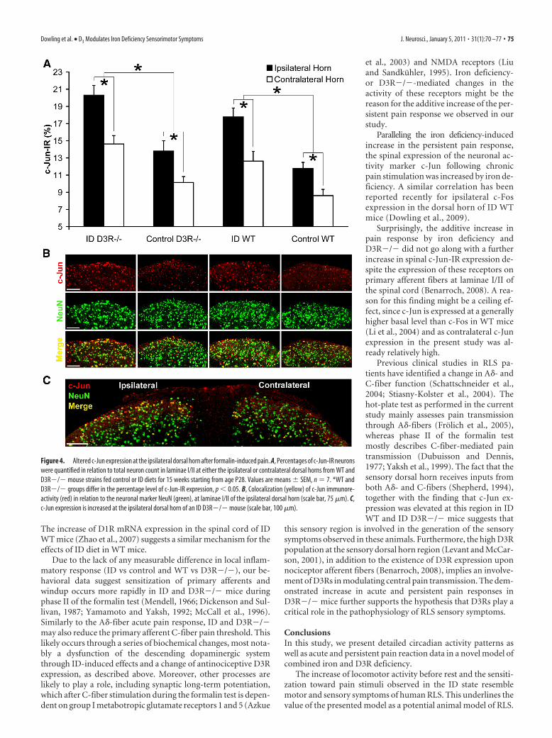
Heightened central neuronal activity detected by ipsilateralc-Jun expression in ID WT and ID D3R�/� groupsAfter completion of the formalin test, the number of c-Jun-IRneurons was quantified at superficial laminae I/II of the ipsilat-
eral and contralateral dorsal horns (Fig.4A). This evaluated whether the irondeficiency-increased neuronal activityafter the formalin test depends on thepresence of D3R. According to the three-factorial ANOVA, diet ( p � 0.001), strain( p � 0.004), and dorsal horn side (ipsilat-eral vs contralateral, p � 0.001) had indi-vidual main effects upon the number ofc-Jun-IR neurons. The interactions be-tween diet and strain, diet and dorsal hornside, strain and dorsal horn side, or allthree factors (diet, strain, and dorsal hornside) were not significant. Post hoc testsrevealed that the number of c-Jun-IR neu-rons at the ipsilateral horn was increasedin all four groups (ID WT, control WT, IDD3R�/�, and control D3R�/�), com-pared to the level at the contralateral horn( p � 0.05) (Fig. 4A). Furthermore, ascompared to control groups, iron defi-ciency increased the expression ofc-Jun-IR neurons at the ipsilateral horn inID WT and ID D3R�/� mice ( p � 0.05)(Fig. 4A). Neuronal c-Jun expression was
confirmed by double immunofluorescence labeling (Fig. 4B,C).
DiscussionIn the current study, we investigated the changes in the circadianpattern of locomotor activity and of acute and persistent painresponses in a combined model of iron deficiency and D3R�/�.The aim was to acquire further insight into the interaction ofthese two influences that are thought to be associated with hu-man RLS.
The increased running wheel usage before the resting periodevoked by iron deficiency resembled RLS motor symptoms. Fur-thermore, iron deficiency resulted in a sensitization to acute andpersistent pain resembling RLS sensory symptoms. AdditionalD3R deficiency modified the time course of the iron deficiency-evoked symptoms and resulted in an additive increase of thepersistent pain response.
D3R�/� and iron deficiency alter circadian motor activityIn the current study, we were able to assess changes in the circa-dian motor activity induced by D3R�/� in combination withiron deficiency in mice by an increased wheel usage, as demon-strated by the parameters Nrun% and Trun%. These parametersproved more sensitive in displaying circadian changes than didabsolute running distance or time, both of which depend moreon the overall running performance.
Iron deficiency increased Nrun% and Trun% in WT mice dur-ing the final hours of the dark phase (6:00 –7:00 A.M.). This is inline with the findings of Dean et al. (2006), who, using polysom-nographic recordings, described an increased awake time duringthe late active period in ID WT mice, probably due to the in-creased dopaminergic activation of neurons in the periaqueduc-tal gray matter. Interestingly, in D3R�/� mice the irondeficiency-induced phase of increased running wheel usage wasshifted to a time point 3 h earlier, whereas no change was seen atthe period from 6:00 to 7:00 A.M. in these mice, suggesting amodulating effect of the D3R on the timing of the iron deficiency-induced activity. A comparable increase of physical activity hasbeen seen after lesioning the diencephalic-spinal A11 region, al-
Figure 2. Differences in voluntary wheel running activity. A–D, The parameters Nrun% (A, B) and Trun% (C, D) represent theaverage values obtained by either the WT (A, C) or D3R�/� (B, D) groups during a 1 h time bin in relation to the cumulative valueachieved within the different time periods (in hours) of the dark phase as represented by the gray boxed region. Values aremeans � SEM, n � 15. *Groups differ at that time during the dark phase, p � 0.05.
Dowling et al. • D3 Modulates Iron Deficiency Sensorimotor Symptoms J. Neurosci., January 5, 2011 • 31(1):70 –77 • 73

though that study did not investigate circadian patterns (Qu etal., 2007). Therefore in light of this study, spinal dopaminergiccircuits may be involved in the observed changes.
The performance parameters Dist, Distmean, and Vmax repre-sent physical fitness and running motivation. Iron deficiency ledto a decrease in Dist, especially during the early night period inboth strains, this being consistent with previous reports (Youdimet al., 1981; Hunt et al., 1994). Youdim et al. (1981) suggested thatthis is caused by an iron deficiency-mediated reduction of dopa-mine D2 receptors (D2Rs). In contrast to our performanceparameters, Youdim et al. (1981) reported an increase of sponta-neous motor activity of ID animals in the morning. This diver-gence is probably due to the different methods used, and mightcorrespond to the increase in activity parameters (Nrun%, Trun%)in the morning phase of our study. Since no anemia was seen in
our ID animals (unpublished data) and iron deficiency reducedDistmean only in D3R�/� mice, reduced physical endurance orfitness is an unlikely cause for a change in the performance pa-rameters. This is also supported by the fact that Vmax remainedunaffected by iron deficiency.
On the other hand, D3R�/� mice did show an increasedrunning performance (Dist and Distmean) during the active pe-riod as described earlier (Accili et al., 1996). Contrary to thecongruent effects on circadian activity, Dist is influenced contrar-ily by iron deficiency and D3R�/�. D3R activation decreasesoverall locomotor activity (Menalled et al., 1999), possibly due todopaminergic inhibition in the Calleja islands (Guitart-Masip etal., 2008), which suggests that the lack of D3R in D3R�/� mightlead to the increase in Dist.
ID mice in our study had increased wheel usage in the phasebefore the resting period. Both the induction of symptoms byiron deficiency and the time point of restlessness as representedby increased wheel usage resemble the symptoms of RLS in hu-mans, which are also characterized by the urge to move duringthe last hours before the resting phase (Nordlander, 1953;Ekbom, 1960). The increased urge to move and restlessness thatRLS patients suffer from is believed to involve the dopaminergicsystem, because dopamine agonists alleviate these symptoms(Akpinar, 1982; Earley and Allen, 1996). Iron deficiency influ-ences the dopaminergic system on different levels. D2R and do-pamine transporter (DAT) are downregulated in ID rats(Ashkenazi et al., 1982; Erikson et al., 2000). A decrease of inhib-itory D2R activation leads to a preponderance of excitatory do-pamine D1 receptor (D1R) activation, which is associated withincreased locomotor activity in rats (Shieh and Walters, 1996;Heijtz et al., 2002). This effect may further be enhanced by an irondeficiency-induced decrease of DAT expression as described in mice(Bianco et al., 2008, 2009) that would increase dopamine levels in thesynaptic cleft. Paulus and Trenkwalder (2006) proposed that themisbalance between D1-like and D2-like activation might fur-ther be increased by treatment-related downregulation of D2-like receptors leading to the phenomenon of augmentation. Adisproportionately extensive downregulation of D2-like recep-tors compared to D1-like receptors due to specific stimulation hasbeen previously reported (Chen et al., 1993). This implicates D2-like receptor (especially D2R and D3R) dysfunction as one patho-genetic agent leading to augmentation. Interestingly, combiningiron deficiency—which is associated with decreased D2R-expres-sion—and D3R deficiency led to a time shift of the irondeficiency-induced increased motor activity in our study. Sinceaugmentation in human RLS is associated with a comparabletime shift of circadian symptoms (Allen et al., 2003; García-Borreguero et al., 2007), a similar mechanism of decreased D2-like dopaminergic signaling might be speculated.
Partially additive alteration of pain responses by D3R�/�and iron deficiencyConfirming our previous findings (Dowling et al., 2009), irondeficiency increased the response to acute and persistent pain inthe WT mouse group. Moreover, we also demonstrated that theD3R�/� status alone is associated with an elevated response toacute and persistent pain. Interestingly, only the persistent painresponse was additively increased by iron deficiency.
The increased acute and persistent pain responses in D3R�/�compared to WT mice are presumably caused by the lack ofD3R-mediated regulation of pronociceptive postsynaptic D1Ractivity (Fleetwood-Walker et al., 1988; Xu et al., 1997), whichincreases in activity upon deletion of D3R (Mizuo et al., 2004).
Figure 3. Response differences to acute and persistent pain. A, The acute pain responsescores (in seconds) until the initial hindpaw licking or escape were recorded by the hot-platetest. Values are means � SEM, n � 7. *WT and D3R�/� groups differ at a diet duration of 15weeks beginning at age P28. B, The persistent pain response to the formalin injection over time(in minutes) in WT and D3R�/� mice fed control or ID diets for 15 weeks starting from ageP28. Values are means � SEM, n � 7. *WT and �D3R�/� groups differ at certain timepoints, p � 0.05. C, After the formalin injection, the injected (ipsilateral) and noninjected(contralateral) hindpaw thicknesses were measured (in millimeters), and the value denoted thelevel of inflammation. Values are means � SEM, n � 7.
74 • J. Neurosci., January 5, 2011 • 31(1):70 –77 Dowling et al. • D3 Modulates Iron Deficiency Sensorimotor Symptoms
The increase of D1R mRNA expression in the spinal cord of IDWT mice (Zhao et al., 2007) suggests a similar mechanism for theeffects of ID diet in WT mice.
Due to the lack of any measurable difference in local inflam-matory response (ID vs control and WT vs D3R�/�), our be-havioral data suggest sensitization of primary afferents andwindup occurs more rapidly in ID and D3R�/� mice duringphase II of the formalin test (Mendell, 1966; Dickenson and Sul-livan, 1987; Yamamoto and Yaksh, 1992; McCall et al., 1996).Similarly to the A�-fiber acute pain response, ID and D3R�/�may also reduce the primary afferent C-fiber pain threshold. Thislikely occurs through a series of biochemical changes, most nota-bly a dysfunction of the descending dopaminergic systemthrough ID-induced effects and a change of antinociceptive D3Rexpression, as described above. Moreover, other processes arelikely to play a role, including synaptic long-term potentiation,which after C-fiber stimulation during the formalin test is depen-dent on group I metabotropic glutamate receptors 1 and 5 (Azkue
et al., 2003) and NMDA receptors (Liuand Sandkuhler, 1995). Iron deficiency-or D3R�/�-mediated changes in theactivity of these receptors might be thereason for the additive increase of the per-sistent pain response we observed in ourstudy.
Paralleling the iron deficiency-inducedincrease in the persistent pain response,the spinal expression of the neuronal ac-tivity marker c-Jun following chronicpain stimulation was increased by iron de-ficiency. A similar correlation has beenreported recently for ipsilateral c-Fosexpression in the dorsal horn of ID WTmice (Dowling et al., 2009).
Surprisingly, the additive increase inpain response by iron deficiency andD3R�/� did not go along with a furtherincrease in spinal c-Jun-IR expression de-spite the expression of these receptors onprimary afferent fibers at laminae I/II ofthe spinal cord (Benarroch, 2008). A rea-son for this finding might be a ceiling ef-fect, since c-Jun is expressed at a generallyhigher basal level than c-Fos in WT mice(Li et al., 2004) and as contralateral c-Junexpression in the present study was al-ready relatively high.
Previous clinical studies in RLS pa-tients have identified a change in A�- andC-fiber function (Schattschneider et al.,2004; Stiasny-Kolster et al., 2004). Thehot-plate test as performed in the currentstudy mainly assesses pain transmissionthrough A�-fibers (Frolich et al., 2005),whereas phase II of the formalin testmostly describes C-fiber-mediated paintransmission (Dubuisson and Dennis,1977; Yaksh et al., 1999). The fact that thesensory dorsal horn receives inputs fromboth A�- and C-fibers (Shepherd, 1994),together with the finding that c-Jun ex-pression was elevated at this region in IDWT and ID D3R�/� mice suggests that
this sensory region is involved in the generation of the sensorysymptoms observed in these animals. Furthermore, the high D3Rpopulation at the sensory dorsal horn region (Levant and McCar-son, 2001), in addition to the existence of D3R expression uponnociceptor afferent fibers (Benarroch, 2008), implies an involve-ment of D3Rs in modulating central pain transmission. The dem-onstrated increase in acute and persistent pain responses inD3R�/� mice further supports the hypothesis that D3Rs play acritical role in the pathophysiology of RLS sensory symptoms.
ConclusionsIn this study, we present detailed circadian activity patterns aswell as acute and persistent pain reaction data in a novel model ofcombined iron and D3R deficiency.
The increase of locomotor activity before rest and the sensiti-zation toward pain stimuli observed in the ID state resemblemotor and sensory symptoms of human RLS. This underlines thevalue of the presented model as a potential animal model of RLS.
Figure 4. Altered c-Jun expression at the ipsilateral dorsal horn after formalin-induced pain. A, Percentages of c-Jun-IR neuronswere quantified in relation to total neuron count in laminae I/II at either the ipsilateral or contralateral dorsal horns from WT andD3R�/� mouse strains fed control or ID diets for 15 weeks starting from age P28. Values are means � SEM, n � 7. *WT andD3R�/� groups differ in the percentage level of c-Jun-IR expression, p � 0.05. B, Colocalization (yellow) of c-Jun immunore-activity (red) in relation to the neuronal marker NeuN (green), at laminae I/II of the ipsilateral dorsal horn (scale bar, 75 �m). C,c-Jun expression is increased at the ipsilateral dorsal horn of an ID D3R�/� mouse (scale bar, 100 �m).
Dowling et al. • D3 Modulates Iron Deficiency Sensorimotor Symptoms J. Neurosci., January 5, 2011 • 31(1):70 –77 • 75

A major advantage of our continuous running wheel recording ishaving the possibility of obtaining a detailed assessment of circa-dian patterns lacking in many other approaches (Qu et al., 2007),where to date only punctual activity levels were recorded. In ad-dition, our model requires a minimum of animal handling, sinceneither surgery nor pharmacologic interventions were imple-mented [unlike, for example, the A11-lesion model (Zhao et al.,2007) or the pharmacologic nafadotride model (Sautel et al.,1995)], thus preventing a disturbance of the very sensitive day–night rhythm. Our model is, however, limited by the use of anonconditional knock-out, since ontogenetic compensatorymechanisms are probable. Furthermore, in human RLS a dys-function of D3 receptors is more likely than the complete absenceof D3 activity in our model.
In furtherance to this, pharmacologic studies assessing thereaction toward dopamine agonists would be suited when furthervalidating the presented model.
Our results also affirm the important role of iron deficiency asa pathogenetic factor in RLS. The increased pain response inD3R�/� indicates an influence of D3R upon pain regulationpossibly at the spinal level. Our results suggest that a dysfunc-tion of either D2R—as present in ID animals— or D3R—as inD3R�/� animals— evoke symptoms comparable to those inRLS patients. Furthermore, our results implicate different sensi-tivity of motor and sensory symptoms toward these influences:the circadian profile of running activity was not affected byD3R�/� alone and ID-induced motor changes were modified,but not increased by D3R�/�. On the other hand, D3R�/� aswell as ID increased acute and persistent pain responses, whereD3R�/� combined with ID had additive effects upon the persis-tent pain response. In light of these findings different pathoge-netic pathways could be considered as a reason for individualdifferences in the preponderance of sensory or motor symptomsin human RLS patients.
ReferencesAccili D, Fishburn CS, Drago J, Steiner H, Lachowicz JE, Park BH, Gauda EB,
Lee EJ, Cool MH, Sibley DR, Gerfen CR, Westphal H, Fuchs S (1996) Atargeted mutation of the D3 dopamine receptor gene is associated withhyperactivity in mice. Proc Natl Acad Sci U S A 93:1945–1949.
Adriaensen H, Gybels J, Handwerker HO, Van Hees J (1983) Responseproperties of thin myelinated (A-delta) fibers in human skin nerves.J Neurophysiol 49:111–122.
Akpinar S (1982) Treatment of restless legs syndrome with levodopa plusbenserazide. Arch Neurol 39:739.
Allen RP, Picchietti D, Hening WA, Trenkwalder C, Walters AS, Montplaisi J(2003) Restless legs syndrome: diagnostic criteria, special considerations,and epidemiology. A report from the restless legs syndrome diagnosis andepidemiology workshop at the National Institutes of Health. Sleep Med4:101–119.
Ashkenazi R, Ben-Shachar D, Youdim MB (1982) Nutritional iron and do-pamine binding sites in the rat brain. Pharmacol Biochem Behav 17[Suppl 1]:43– 47.
Azkue JJ, Liu XG, Zimmermann M, Sandkuhler J (2003) Induction of long-term potentiation of C fibre-evoked spinal field potentials requires re-cruitment of group I, but not group II/III metabotropic glutamatereceptors. Pain 106:373–379.
Bara-Jimenez W, Aksu M, Graham B, Sato S, Hallett M (2000) Periodic limbmovements in sleep: state-dependent excitability of the spinal flexor re-flex. Neurology 54:1609 –1616.
Benarroch EE (2008) Descending monoaminergic pain modulation: bidi-rectional control and clinical relevance. Neurology 71:217–221.
Bianco LE, Wiesinger J, Earley CJ, Jones BC, Beard JL (2008) Iron deficiencyalters dopamine uptake and response to L-DOPA injection in Sprague-Dawley rats. J Neurochem 106:205–215.
Bianco LE, Unger EL, Earley CJ, Beard JL (2009) Iron deficiency alters the
day-night variation in monoamine levels in mice. Chronobiol Int26:447– 463.
Chen JF, Aloyo VJ, Weiss B (1993) Continuous treatment with the D2 do-pamine receptor agonist quinpirole decreases D2 dopamine receptors, D2dopamine receptor messenger RNA and proenkephalin messenger RNA,and increases mu opioid receptors in mouse striatum. Neuroscience54:669 – 680.
Clemens S, Hochman S (2004) Conversion of the modulatory actions ofdopamine on spinal reflexes from depression to facilitation in D3 receptorknock-out mice. J Neurosci 24:11337–11345.
Clemens S, Rye D, Hochman S (2006) Restless legs syndrome: revisiting thedopamine hypothesis from the spinal cord perspective. Neurology67:125–130.
Dean T Jr, Allen RP, O’Donnell CP, Earley CJ (2006) The effects of dietaryiron deprivation on murine circadian sleep architecture. Sleep Med7:634 – 640.
Dickenson AH, Sullivan AF (1987) Evidence for a role of the NMDA recep-tor in the frequency dependent potentiation of deep rat dorsal horn no-ciceptive neurones following C fibre stimulation. Neuropharmacology26:1235–1238.
Dowling P, Klinker F, Amaya F, Paulus W, Liebetanz D (2009) Iron-deficiency sensitizes mice to acute pain stimuli and formalin-inducednociception. J Nutr 139:2087–2092.
Dubuisson D, Dennis SG (1977) The formalin test: a quantitative study ofthe analgesic effects of morphine, meperidine, and brain stem stimulationin rats and cats. Pain 4:161–174.
Earley CJ, Allen RP (1996) Pergolide and carbidopa/levodopa treatment ofthe restless legs syndrome and periodic leg movements in sleep in a con-secutive series of patients. Sleep 19:801– 810.
Earley CJ, Heckler D, Allen RP (2004) The treatment of restless legs syn-drome with intravenous iron dextran. Sleep Med 5:231–235.
Earley CJ, Allen RP, Connor JR, Ferrucci L, Troncoso J (2009) The dopami-nergic neurons of the A11 system in RLS autopsy brains appear normal.Sleep Med 10:1155–1157.
Ekbom KA (1960) Restless legs syndrome. Neurology 10:868 – 873.Erikson KM, Jones BC, Beard JL (2000) Iron deficiency alters dopamine
transporter functioning in rat striatum. J Nutr 130:2831–2837.Fleetwood-Walker SM, Hope PJ, Mitchell R (1988) Antinociceptive actions
of descending dopaminergic tracts on cat and rat dorsal horn somatosen-sory neurones. J Physiol 399:335–348.
Frolich MA, Price DD, Robinson ME, Shuster JJ, Theriaque DW, Heft MW(2005) The effect of propofol on thermal pain perception. Anesth Analg100:481– 486.
García-Borreguero D, Allen RP, Kohnen R, Hogl B, Trenkwalder C, Oertel W,Hening WA, Paulus W, Rye D, Walters A, Winkelmann J, Earley CJ(2007) Diagnostic standards for dopaminergic augmentation of restlesslegs syndrome: report from a World Association of Sleep Medicine-International Restless Legs Syndrome Study Group consensus conferenceat the Max Planck Institute. Sleep Med 8:520 –530.
Grote L, Leissner L, Hedner J, Ulfberg J (2009) A randomized, double-blind,placebo controlled, multi-center study of intravenous iron sucrose andplacebo in the treatment of restless legs syndrome. Mov Disord24:1445–1452.
Guitart-Masip M, Johansson B, Fernandez-Teruel A, Tobena A, Gimenez-Llort L (2008) Divergent effect of the selective D3 receptor agonist pd-128,907 on locomotor activity in Roman high- and low-avoidance rats:relationship to NGFI-A gene expression in the Calleja islands. Psychop-harmacology (Berl) 196:39 – 49.
Heapy CG, Jamieson A, Russel NJW (1987) Afferent C-fiber and A-deltaactivity in models of inflammation. Br J Pharmacol 90:164p.
Heijtz RD, Beraki S, Scott L, Aperia A, Forssberg H (2002) Sex differences inthe motor inhibitory and stimulatory role of dopamine D1 receptors inrats. Eur J Pharmacol 445:97–104.
Herdegen T, Kovary K, Leah J, Bravo R (1991) Specific temporal and spatialdistribution of JUN, FOS, and KROX-24 proteins in spinal neurons fol-lowing noxious transsynaptic stimulation. J Comp Neurol 313:178 –191.
Hunt JR, Zito CA, Erjavec J, Johnson LK (1994) Severe or marginal irondeficiency affects spontaneous physical activity in rats. Am J Clin Nutr59:413– 418.
Ishizaki K, Sasaki M, Karasawa S, Obata H, Nara T, Goto F (1999) The effectof intrathecal magnesium sulphate on nociception in rat acute pain mod-els. Anaesthesia 54:241–246.
76 • J. Neurosci., January 5, 2011 • 31(1):70 –77 Dowling et al. • D3 Modulates Iron Deficiency Sensorimotor Symptoms

Ko S, Zhao MG, Toyoda H, Qiu CS, Zhuo M (2005) Altered behavioralresponses to noxious stimuli and fear in glutamate receptor 5 (GluR5)- orGluR6-deficient mice. J Neurosci 25:977–984.
Leem JW, Willis WD, Chung JM (1993) Cutaneous sensory receptors in therat foot. J Neurophysiol 69:1684 –1699.
Levant B, McCarson KE (2001) D(3) dopamine receptors in rat spinal cord:implications for sensory and motor function. Neurosci Lett 303:9 –12.
Li J, Simone DA, Larson AA (1999) Windup leads to characteristics of cen-tral sensitization. Pain 79:75– 82.
Li X, Lighthall G, Liang DY, Clark JD (2004) Alterations in spinal cord geneexpression after hindpaw formalin injection. J Neurosci Res 78:533–541.
Liebetanz D, Baier PC, Paulus W, Meuer K, Bahr M, Weishaupt JH (2007) Ahighly sensitive automated complex running wheel test to detect latentmotor deficits in the mouse MPTP model of Parkinson’s disease. ExpNeurol 205:207–213.
Liu XG, Sandkuhler J (1995) Long-term potentiation of C-fiber-evoked po-tentials in the rat spinal dorsal horn is prevented by spinal N-methyl-D-aspartic acid receptor blockage. Neurosci Lett 191:43– 46.
McCall WD, Tanner KD, Levine JD (1996) Formalin induces biphasic ac-tivity in C-fibers in the rat. Neurosci Lett 208:45– 48.
Menalled LB, Dziewczapolski G, Garcia MC, Rubinstein M, Gershanik OS(1999) D3 receptor knockdown through antisense oligonucleotide ad-ministration supports its inhibitory role in locomotion. Neuroreport10:3131–3136.
Mendell LM (1966) Physiological properties of unmyelinated fiber projec-tion to the spinal cord. Exp Neurol 16:316 –332.
Mizuo K, Narita M, Miyatake M, Suzuki T (2004) Enhancement ofdopamine-induced signaling responses in the forebrain of mice lackingdopamine D3 receptor. Neurosci Lett 358:13–16.
Nordlander NB (1953) Therapy in restless legs. Acta Med Scand 145:453– 457.
Paulus W, Trenkwalder C (2006) Less is more: pathophysiology ofdopaminergic-therapy-related augmentation in restless legs syndrome.Lancet Neurol 5:878 – 886.
Presley RW, Menetrey D, Levine JD, Basbaum AI (1990) Systemic mor-phine suppresses noxious stimulus-evoked Fos protein-like immunore-activity in the rat spinal cord. J Neurosci 10:323–335.
Qu S, Le W, Zhang X, Xie W, Zhang A, Ondo WG (2007) Locomotion isincreased in a11-lesioned mice with iron deprivation: a possible animalmodel for restless legs syndrome. J Neuropathol Exp Neurol 66:383–388.
Satija P, Ondo WG (2008) Restless legs syndrome: pathophysiology, diag-nosis and treatment. CNS Drugs 22:497–518.
Sautel F, Griffon N, Sokoloff P, Schwartz JC, Launay C, Simon P, Costentin J,Schoenfelder A, Garrido F, Mann A, Wermuth CG (1995) Nafadotride,a potent preferential dopamine D3 receptor antagonist, activates locomo-tion in rodents. J Pharmacol Exp Ther 275:1239 –1246.
Schattschneider J, Bode A, Wasner G, Binder A, Deuschl G, Baron R (2004)Idiopathic restless legs syndrome: abnormalities in central somatosensoryprocessing. J Neurol 251:977–982.
Shepherd GM (1994) Neurobiology. New York: Oxford UP.Shieh GJ, Walters DE (1996) Stimulating dopamine D1 receptors increases
the locomotor activity of developing rats. Eur J Pharmacol 311:103–107.Stadelmann C, Bruck W, Bancher C, Jellinger K, Lassmann H (1998) Alz-
heimer disease: DNA fragmentation indicates increased neuronal vulner-ability, but not apoptosis. J Neuropathol Exp Neurol 57:456 – 464.
Stiasny-Kolster K, Magerl W, Oertel WH, Moller JC, Treede RD (2004)Static mechanical hyperalgesia without dynamic tactile allodynia in pa-tients with restless legs syndrome. Brain 127:773–782.
Trenkwalder C, Hogl B, Benes H, Kohnen R (2008) Augmentation in rest-less legs syndrome is associated with low ferritin. Sleep Med 9:572–574.
Xu M, Koeltzow TE, Santiago GT, Moratalla R, Cooper DC, Hu XT, WhiteNM, Graybiel AM, White FJ, Tonegawa S (1997) Dopamine D3 receptormutant mice exhibit increased behavioral sensitivity to concurrent stim-ulation of D1 and D2 receptors. Neuron 19:837– 848.
Yaksh TL, Hua XY, Kalcheva I, Nozaki-Taguchi N, Marsala M (1999) Thespinal biology in humans and animals of pain states generated by persis-tent small afferent input. Proc Natl Acad Sci U S A 96:7680 –7686.
Yamamoto T, Yaksh TL (1992) Comparison of the antinociceptive effects ofpre- and posttreatment with intrathecal morphine and MK801, anNMDA antagonist, on the formalin test in the rat. Anesthesiology77:757–763.
Yeomans DC, Pirec V, Proudfit HK (1996) Nociceptive responses to highand low rates of noxious cutaneous heating are mediated by differentnociceptors in the rat: behavioral evidence. Pain 68:133–140.
Youdim MB, Yehuda S, Ben-Uriah Y (1981) Iron deficiency-induced circa-dian rhythm reversal of dopaminergic-mediated behaviours and thermo-regulation in rats. Eur J Pharmacol 74:295–301.
Zhao H, Zhu W, Pan T, Xie W, Zhang A, Ondo WG, Le W (2007) Spinalcord dopamine receptor expression and function in mice with 6-OHDAlesion of the A11 nucleus and dietary iron deprivation. J Neurosci Res85:1065–1076.
Dowling et al. • D3 Modulates Iron Deficiency Sensorimotor Symptoms J. Neurosci., January 5, 2011 • 31(1):70 –77 • 77