doi: 10.1126/science.1252785 promotes intestinal … crosstalk between macrophages and ilc3 promotes...
TRANSCRIPT
Microbiota-Dependent Crosstalk Between Macrophages and ILC3 Promotes Intestinal Homeostasis Arthur Mortha, Aleksey Chudnovskiy, Daigo Hashimoto, Milena Bogunovic,
Sean P. Spencer, Yasmine Belkaid, Miriam Merad*
Introduction: The gastrointestinal tract is colonized by an extraordi-narily large number of commensal microbes and is constantly exposed to ingested antigens and potential pathogens. Regulation of intestinal toler-ance thus represents the main task of the immune system of the gut mucosa. Accumulated evidence suggests that gut commensals contribute to the maintenance of intestinal homeostasis, partly through their ability to control the differentiation of effector T lymphocytes in the mucosa and to modu-late infl ammatory responses through the induction of regulatory T cells (Tregs) and interleukin-10 (IL-10) production. Tissue-resident mononuclear phagocytes (MNPs), including macrophages (MPs) and dendritic cells (DCs), are specifi cally equipped to detect a wide range of microbial signals and to capture, process, and present extracellular antigenic material to T lymphocytes. MNPs have been shown to contribute to the maintenance of intestinal immune tolerance through the induction or expansion of Tregs in the intestine. Despite their key role in microbial sensing and immune tolerance, the cellular and molecular cues that translate microbial signals into immuno-regulatory MNPs in the intestine are not completely understood.
Rationale: The cytokine granulocyte-macrophage colony-stimulating factor (GM-CSF), recently renamed Csf2, is a key determinant of myeloid lineage differentiation and is required for the optimal function of tissue MNPs. Recent results from our laboratory revealed that although Csf2-defi cient mice have normal numbers of lymphoid tissue-resident DCs, they display signifi cantly reduced numbers of steady-state nonlymphoid tissue-resident DCs in the small intestine, including the lamina propria CD103+CD11b+ DC subset implicated in the induction of lamina pro-pria Tregs. These results prompted us to further explore the contribution of Csf2 to intestinal immune homeostasis in vivo. We used detailed profi ling
studies and functional immune assays of the MNP and lymphocyte compart-ment in the gut, as well as genetically engineered mice that lack Csf2 or the transducer Myd88 specifi cally in MNPs or lymphocytes, to explore the role of MNPs in the maintenance of immune homeostasis in the gut.
Results: Our results revealed a crosstalk between IL-1β–secreting MPs and Csf2-producing RORγt+ type 3 innate lymphoid cells (ILC3) in the intestinal mucosa. Microbiota-driven IL-1β production by MPs promoted the release of Csf2 by ILC3, which in turn controlled DCs and MPs to maintain colonic Treg homeostasis. Ablation of Csf2 reduced DC and MP numbers and impaired
their ability to produce regulatory factors such as retinoic acid (RA) and IL-10, leading to disrupted Treg homeostasis in the large intestine. Conversely, administration of Csf2 cytokine increased Treg frequency in the gut. Most notably, cell type–specifi c ablation of IL-1 receptor (IL-1R)–dependent signaling in RORγt+ ILC3 abrogated oral tolerance to dietary antigens and compromised intestinal immune homeostasis in vivo. Although the reduction in Treg numbers was mostly observed in the large intestine, adoptive trans-fer studies in Csf2–/– mice revealed impaired Treg differentiation both in the small and large intestine, sug-gesting that Csf2-dependent MNP immunoregulatory functions con-trol Treg induction in both tissues.
Conclusion: This study established the commensal-driven MNP-ILC-Csf2 axis as a key regulator of intestinal T cell homeostasis in the mouse intestine. Disturbance of this axis radically altered MNP
effector function, resulting in impaired oral tolerance to dietary antigens. These results represent an important advance in our understanding of how commensal microbes can regulate host intestinal immunity and may inform the design of novel immunotherapies for patients with infl ammatory intestinal diseases with impaired GM-CSF function.
The list of author affi liations is available in the full article online.*Corresponding author. E-mail: [email protected] this article as A. Mortha et al., Science 343, 1249288 (2014). DOI: 10.1126/science.1249288
READ THE FULL ARTICLE ONLINE
http://dx.doi.org/10.1126/science.1249288
Myeloid cells, innate lymphoid cells, and the
cytokines they secrete cooperate to maintain
immune tolerance in the gut.
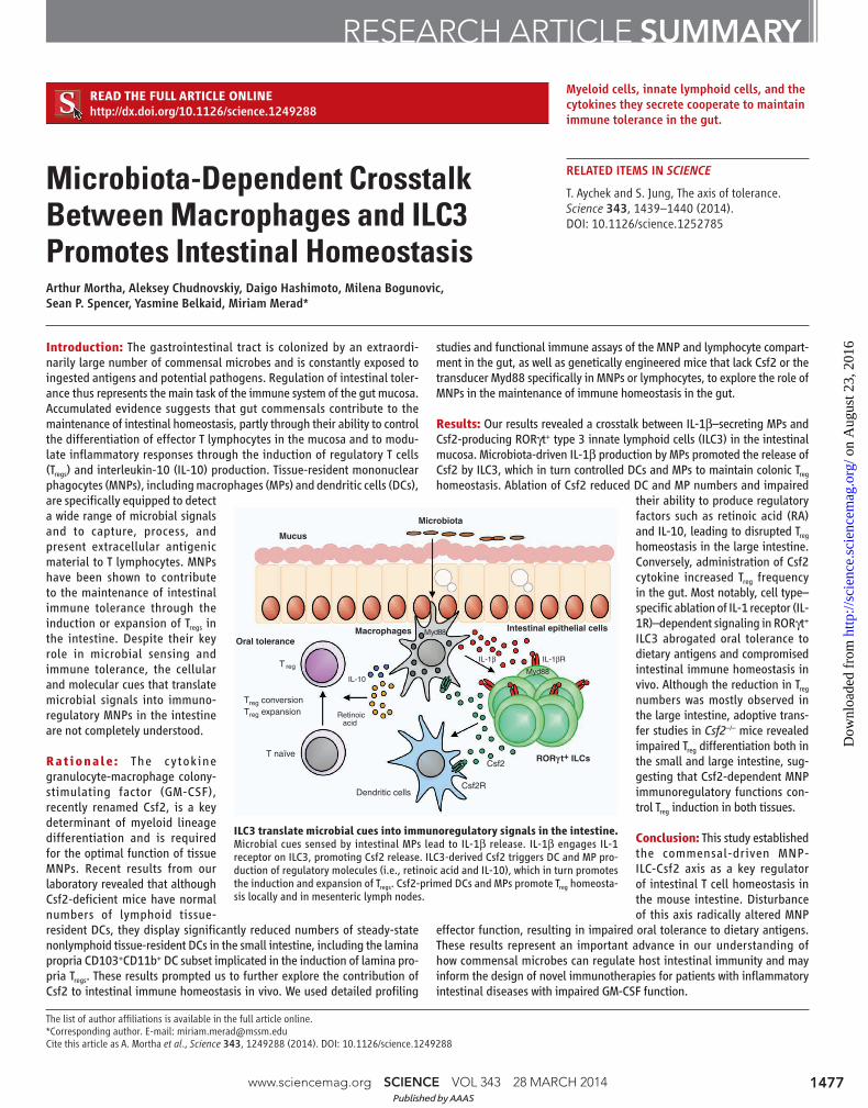
ILC3 translate microbial cues into immunoregulatory signals in the intestine. Microbial cues sensed by intestinal MPs lead to IL-1β release. IL-1β engages IL-1 receptor on ILC3, promoting Csf2 release. ILC3-derived Csf2 triggers DC and MP pro-duction of regulatory molecules (i.e., retinoic acid and IL-10), which in turn promotes the induction and expansion of Tregs. Csf2-primed DCs and MPs promote Treg homeosta-sis locally and in mesenteric lymph nodes.
Csf2
Csf2RDendritic cells
T naïve
Treg conversion
T reg
Treg expansion Retinoicacid
Oral toleranceMacrophages
Mucus
Microbiota
Intestinal epithelial cells
IL-10
IL-1β
Myd88
Myd88
IL-1βR
RORγ t+ ILCs
www.sciencemag.org SCIENCE VOL 343 28 MARCH 2014 1477
RESEARCH ARTICLE SUMMARY
RELATED ITEMS IN SCIENCE
T. Aychek and S. Jung, The axis of tolerance. Science 343, 1439–1440 (2014). DOI: 10.1126/science.1252785
Published by AAAS
on
Aug
ust 2
3, 2
016
http
://sc
ienc
e.sc
ienc
emag
.org
/D
ownl
oade
d fr
om
Microbiota-Dependent CrosstalkBetween Macrophages and ILC3Promotes Intestinal HomeostasisArthur Mortha,1,2,3 Aleksey Chudnovskiy,1,2,3 Daigo Hashimoto,1,2,3* Milena Bogunovic,1,2,3†Sean P. Spencer,4 Yasmine Belkaid,4 Miriam Merad1,2,3‡
The intestinal microbiota and tissue-resident myeloid cells promote immune responses that maintainintestinal homeostasis in the host. However, the cellular cues that translate microbial signals intointestinal homeostasis remain unclear. Here, we show that deficient granulocyte-macrophagecolony-stimulating factor (GM-CSF) production altered mononuclear phagocyte effector functions andled to reduced regulatory T cell (Treg) numbers and impaired oral tolerance. We observed that RORgt
+
innate lymphoid cells (ILCs) are the primary source of GM-CSF in the gut and that ILC-driven GM-CSFproduction was dependent on the ability of macrophages to sense microbial signals and produceinterleukin-1b. Our findings reveal that commensal microbes promote a crosstalk between innatemyeloid and lymphoid cells that leads to immune homeostasis in the intestine.
The gastrointestinal tract is colonized by anextraordinarily large number of commen-sal microbes and is constantly exposed to
ingested antigens and potential pathogens. Reg-ulation of intestinal tolerance thus represents themain task of the immune system of the gut mu-cosa. Defective immune tolerance in the gut isassociated with the onset of inflammatory boweldiseases (IBD), a severe intestinal pathology thatresults from a dysregulated immune response tocommensal microbes leading to chronic intestinalinflammation (1, 2). Accumulated evidence sug-gests that gut commensals contribute to the main-tenance of intestinal homeostasis, partly throughtheir ability to control the differentiation of effectorT lymphocytes in the mucosa (3, 4) and to modu-late inflammatory responses through the inductionof Tregs and interleukin-10 (IL-10) production (4–6).
Tissue-residentmononuclear phagocytes (MNPs)are equipped to detect a wide range of microbialsignals and to capture and process extracellularantigens, including commensal microbial antigensin the form of peptide–major histocompatibil-ity complexes (MHCs) that can be recognizedby T lymphocytes (7). Mucosal tissue-residentMNPs consist of two main cell populations, mac-rophages (MPs) and dendritic cells (DCs) (8).Tissue-resident macrophages are characterized as
MHCII+CD11c+CD103–CD11b+CX3CR1+F4/80+CD64+ cells, whereas tissue-resident DCsare characterized as MHCII+CD11c+CX3CR1int/–
F4/80–CD64– (fig. S1). DCs can further be sub-divided into CD103+CD11b– (CD103+ DCs),CD103+CD11b+ (double-positive or DP DCs),CD103–CD11b+ (CD11b+ DCs), and CD103–
CD11b+CD64+F4/80+ (MP) subsets (9–12) (fig.S1). BothDCs andmacrophages have been shownto contribute to the maintenance of intestinal im-mune tolerance through the induction or expan-sion of Tregs in the intestine (13–19). Despite theirkey role in microbial sensing and immune tol-erance, the cellular and molecular cues that trans-late microbial signals into immunoregulatoryMNPs in the intestine remain poorly understood.
The cytokine granulocyte-macrophage colony-stimulating factor (GM-CSF), recently renamedcolony-stimulating factor 2 (Csf2), is a key deter-minant of myeloid lineage differentiation and isrequired for the optimal function of tissue MNPs,includingmacrophages andDCs, thereby promot-ing host protection against environmental patho-gens and vaccine responses (20, 21). Despite thekey role of Csf2 in promoting MNP survival, dif-ferentiation, and function, previous studies reportedthat mice lacking Csf2 or its receptor displayedonly minor impairment in the development ofspleen and lymph node DCs (22). Subsequentstudies showing that Csf2 expression is increasedin inflamed mice and that adoptively transferredmonocytes generate DCs in the inflamed spleenbut not in the steady-state spleen suggested thatCsf2 is a major proinflammatory cytokine thatcontrols the differentiation of inflammatory butnot steady-state DCs in vivo (23, 24). These re-sults are consistent with the contribution of Csf2to the pathophysiology of numerous inflamma-tory and autoimmune diseases (25–27).
In contrast, we recently observed that althoughCsf2-deficient mice have normal numbers of lymph-oid tissue-resident DCs, they display a significant
reduction in steady-state nonlymphoid tissue-resident DCs, including the CD103+CD11b+ DCsubset found in the small intestine lamina propia(11,28),which have been implicated in the inductionof lamina propria Tregs (14, 15). These resultsprompted us to further explore the contribution ofCsf2 to intestinal immune homeostasis in vivo.
Regulation of Gut DC, Macrophage,and Treg Cell Homeostasis by Csf2We characterized the mucosal Tcell compartmentin Csf2-deficient mice (Csf2−/−) in the steady state.Surprisingly, we observed a significant reductionin the frequency, number, and proliferation ofCD45+TCRb+CD4+Foxp3+ Tregs in the colon ofCsf2−/− mice compared to littermate controls(Fig. 1A and fig. S2A). The reduced Treg numberwas specific to the colon and was not observed inthe small intestine ofCsf2−/−mice. The reductionin the number of colonic Tregs was associatedwith a significant reduction in the frequency andnumber of IL-10– and IL-2–producing T cells,along with a significant increase in the number ofcolonic interferon-g (IFN-g)–producing T cells,whereas IL-17–producing Tcells were unaffectedin 6-week-old Csf2−/− mice compared to wild-type mice (Fig. 1B and fig. S2B). Histologicalanalysis of colonic sections from Csf2-deficientanimals did not reveal overt inflammatory infil-trates in the lamina propria (fig. S2C).
Because Csf2 plays a critical role in the dif-ferentiation and function of tissue MNPs, we hy-pothesized that the alterations in T helper cellsubsets observed in the colon of Csf2−/− animalsmight be due to defects in mucosal MNPs. Ac-cordingly, we found reduced numbers of colonicDCs and macrophages in the absence of Csf2(Fig. 1, C and D), thus establishing an importantrole for Csf2 in the homeostasis of the colonicMNP pool. DCs and macrophages have been re-ported to generate Foxp3+ Tregs, via the produc-tion of the regulatorymediators retinoic acid (RA)and IL-10 in the presence of transforming growthfactor–b (TGF-b). Thus, we analyzed the capacityof DCs and macrophages to produce these reg-ulatory mediators in the absence of Csf2. Weobserved a significant reduction in the activity ofthe RA-generating enzyme retinaldehyde dehy-drogenase (ALDH) throughout all colonic DCsubsets and macrophages inCsf2−/−mice (Fig. 1Eand fig. S2D) associated with reduced expressionof Aldh1 transcripts (fig. S2, E and F). Absenceof Csf2 was also associated with a significantreduction in the release of TGF-b by colonicCD103+CD11b+ DCs and with reduced IL-10secretion bymacrophages (Fig. 1, F andG), whichextends previous observations showing that Csf2controls IL-10 and TGF-b release by peritoneal mac-rophages upon uptake of apoptotic cells (29). No-tably, expression ofAldh1a2 and Il10, and releaseof IL-10were restored inCsf2−/−macrophages uponaddition of exogenous Csf2 (fig. S2, G and H).
These findings suggest that the absence of Csf2results in a reduction in the number, frequency,
RESEARCHARTICLE
1Department of Oncological Sciences, 1470 Madison Avenue,New York, NY 10029, USA. 2The Tisch Cancer Institute, 1470Madison Avenue, New York, NY 10029, USA. 3The ImmunologyInstitute Mount Sinai School of Medicine, 1470 Madison Ave-nue, New York, NY 10029, USA. 4Program in Barrier Immunityand Repair, Mucosal Immunology Section, Laboratory of Par-asitic Diseases, National Institute of Allergy and InfectiousDiseases, Bethesda, MD 20892, USA.
*Present address: Department of Hematology, HokkaidoUniversity Graduate School of Medicine, N15 W7, Kita-Ku,Sapporo 060-8638, Japan.†Present address: Department of Microbiology and Immunol-ogy, Penn State College of Medicine and Milton S. HersheyMedical Center, 500 University Drive, Hershey, PA, USA.‡Corresponding author. E-mail: [email protected]
www.sciencemag.org SCIENCE VOL 343 28 MARCH 2014 1249288-1
on
Aug
ust 2
3, 2
016
http
://sc
ienc
e.sc
ienc
emag
.org
/D
ownl
oade
d fr
om
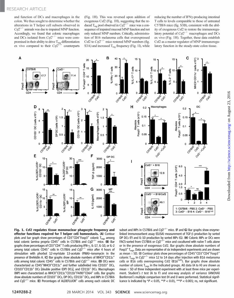
and function of DCs and macrophages in thecolon.We thus sought to determine whether thealterations in T helper cell subsets observed inCsf2−/− animalswas due to impairedMNP function.Accordingly, we found that colonic macrophagesand DCs isolated from Csf2−/− mice were com-promised in their ability to drive Treg differentiationex vivo compared to their Csf2+/+ counterparts
(Fig. 1H). This was reversed upon addition ofexogenous Csf2 (Fig. 1H), suggesting that the re-duced Treg pool observed inCsf2
−/−micewas a con-sequenceof impairedmucosalMNPfunction andnotonly reduced MNP numbers. Critically, administra-tion of B16 melanoma cells that overexpressedCsf2 to Csf2−/−mice restored MNP numbers (fig.S3A) and increased Treg frequency (Fig. 1I), while
reducing the number of IFNg-producing intestinalT cells to levels comparable to those of untreatedC57Bl/6 mice (fig. S3B), consistent with the abil-ity of exogenous Csf2 to restore the immunoregu-latory potential of Csf2−/− macrophages and DCsex vivo (Fig. 1H). Together, these data establishCsf2 as a master regulator of MNP immunoregu-latory function in the steady-state colon tissue.
Fig. 1. Csf2 regulates tissue mononuclear phagocyte frequency andeffector functions required for T helper cell homeostasis. (A) Contourplots and bar graph show percentages of CD3+CD4+Foxp3+ colonic Tregs amongtotal colonic lamina propria CD45+ cells in C57Bl/6 and Csf2−/− mice. (B) Bargraphs showpercentages of CD3+CD4+ T cells producing IFN-g, IL-17, IL-10, or IL-2among total colonic CD45+ cells in C57Bl/6 and Csf2−/− mice after 4 hours ofstimulation with phorbol 12-myristate 13-acetate (PMA)–ionomycin in thepresence of Brefeldin A. (C) Bar graphs show absolute numbers of MHCII+CD11c+
cells among total colonic CD45+ cells in C57Bl/6 and Csf2−/− mice. (D) DCs werecharacterized as CD45+MHCII+CD11c+ and further subdivided into CD103+ DCs,CD103+CD11b+ DCs [double positive (DP) DCs], and CD11b+ DCs. Macrophages(MP) were characterized as MHCII+CD11c+CD11b+F4/80+CD64+ cells. Bar graphsshow absolute numbers of CD103+ DCs, DP DCs, CD11b+ DCs, andMPs in C57Bl/6and Csf2−/− mice. (E) Percentages of ALDEFLUOR+ cells among each colonic DC
subset and MPs in C57Bl/6 and Csf2−/−mice. (F and G) Bar graphs show enzyme-linked immunosorbent assay (ELISA) measurement of TGF-b production by sortedDP DCs (F) and IL-10 production by sorted MPs (G). (H) Colonic MPs or DCs wereFACS-sorted from C57Bl/6 or Csf2−/− mice and cocultured with naïve T cells aloneor in the presence of exogenous Csf2. Bar graphs show absolute numbers ofFoxp3+ Tregs. Data are representative of six independent experiments and are shownas mean T SD. (I) Contour plots show percentages of CD45+CD3+CD4+Foxp3+
colonic Tregs in Csf2−/− mice 12 to 14 days after injection with B16 melanoma
cells or B16 cells overexpressing Csf2 (B16Csf2). Bar graphs show absolutenumber of colonic Tregs in the indicated groups. All data (A to H) are shown asmean T SD of three independent experiment with at least three mice per experi-ment. Student’s t test (A to F) and one-way analysis of variance (ANOVA)Bonferroni’s multiple comparison test (H and I) were performed. Statistical signif-icance is indicated by *P < 0.05, **P < 0.01, ***P < 0.001; ns, not significant.
28 MARCH 2014 VOL 343 SCIENCE www.sciencemag.org1249288-2
RESEARCH ARTICLE
on
Aug
ust 2
3, 2
016
http
://sc
ienc
e.sc
ienc
emag
.org
/D
ownl
oade
d fr
om
Notably, blockade of RA production [with4-diethly-aminobenzaldehyde (DEAB)] and/orblockade of IL-10 [withmonoclonal antibody (mAb)against IL-10] abrogated the ability of Csf2 torescue Treg induction in vitro by Csf2−/− MNPs,whereas addition of RA rescued Treg induction inthese cultures (fig. S3, C and D). Consistent withthese findings, injection of the RA receptor antag-onist LE540 compromised Csf2-mediated rescueof Tregs inCsf2
−/−mice in vivo (fig. S3E), and con-versely, injection of RA but not IL-10 restored Tregfrequency in Csf2−/− mice in vivo (fig. S3F).
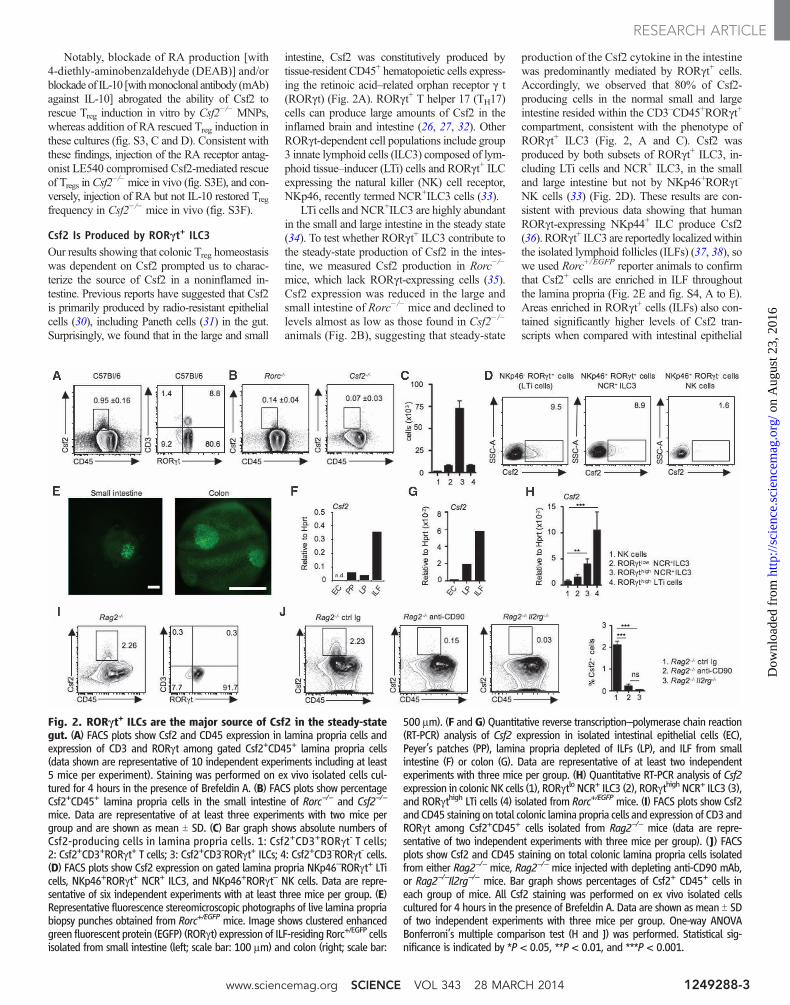
Csf2 Is Produced by RORgt+ ILC3Our results showing that colonic Treg homeostasiswas dependent on Csf2 prompted us to charac-terize the source of Csf2 in a noninflamed in-testine. Previous reports have suggested that Csf2is primarily produced by radio-resistant epithelialcells (30), including Paneth cells (31) in the gut.Surprisingly, we found that in the large and small
intestine, Csf2 was constitutively produced bytissue-resident CD45+ hematopoietic cells express-ing the retinoic acid–related orphan receptor g t(RORgt) (Fig. 2A). RORgt+ T helper 17 (TH17)cells can produce large amounts of Csf2 in theinflamed brain and intestine (26, 27, 32). OtherRORgt-dependent cell populations include group3 innate lymphoid cells (ILC3) composed of lym-phoid tissue–inducer (LTi) cells and RORgt+ ILCexpressing the natural killer (NK) cell receptor,NKp46, recently termed NCR+ILC3 cells (33).
LTi cells and NCR+ILC3 are highly abundantin the small and large intestine in the steady state(34). To test whether RORgt+ ILC3 contribute tothe steady-state production of Csf2 in the intes-tine, we measured Csf2 production in Rorc−/−
mice, which lack RORgt-expressing cells (35).Csf2 expression was reduced in the large andsmall intestine of Rorc−/− mice and declined tolevels almost as low as those found in Csf2−/−
animals (Fig. 2B), suggesting that steady-state
production of the Csf2 cytokine in the intestinewas predominantly mediated by RORgt+ cells.Accordingly, we observed that 80% of Csf2-producing cells in the normal small and largeintestine resided within the CD3–CD45+RORgt+
compartment, consistent with the phenotype ofRORgt+ ILC3 (Fig. 2, A and C). Csf2 wasproduced by both subsets of RORgt+ ILC3, in-cluding LTi cells and NCR+ ILC3, in the smalland large intestine but not by NKp46+RORgt–
NK cells (33) (Fig. 2D). These results are con-sistent with previous data showing that humanRORgt-expressing NKp44+ ILC produce Csf2(36). RORgt+ ILC3 are reportedly localizedwithinthe isolated lymphoid follicles (ILFs) (37, 38), sowe used Rorc+/EGFP reporter animals to confirmthat Csf2+ cells are enriched in ILF throughoutthe lamina propria (Fig. 2E and fig. S4, A to E).Areas enriched in RORgt+ cells (ILFs) also con-tained significantly higher levels of Csf2 tran-scripts when compared with intestinal epithelial
Fig. 2. RORgt+ ILCs are the major source of Csf2 in the steady-stategut. (A) FACS plots show Csf2 and CD45 expression in lamina propria cells andexpression of CD3 and RORgt among gated Csf2+CD45+ lamina propria cells(data shown are representative of 10 independent experiments including at least5 mice per experiment). Staining was performed on ex vivo isolated cells cul-tured for 4 hours in the presence of Brefeldin A. (B) FACS plots show percentageCsf2+CD45+ lamina propria cells in the small intestine of Rorc−/− and Csf2−/−
mice. Data are representative of at least three experiments with two mice pergroup and are shown as mean T SD. (C) Bar graph shows absolute numbers ofCsf2-producing cells in lamina propria cells. 1: Csf2+CD3+RORgt- T cells;2: Csf2+CD3+RORgt+ T cells; 3: Csf2+CD3-RORgt+ ILCs; 4: Csf2+CD3-RORgt- cells.(D) FACS plots show Csf2 expression on gated lamina propria NKp46–RORgt+ LTicells, NKp46+RORgt+ NCR+ ILC3, and NKp46+RORgt– NK cells. Data are repre-sentative of six independent experiments with at least three mice per group. (E)Representative fluorescence stereomicroscopic photographs of live lamina propriabiopsy punches obtained from Rorc+/EGFP mice. Image shows clustered enhancedgreen fluorescent protein (EGFP) (RORgt) expression of ILF-residing Rorc+/EGFP cellsisolated from small intestine (left; scale bar: 100 mm) and colon (right; scale bar:
500 mm). (F and G) Quantitative reverse transcription–polymerase chain reaction(RT-PCR) analysis of Csf2 expression in isolated intestinal epithelial cells (EC),Peyer’s patches (PP), lamina propria depleted of ILFs (LP), and ILF from smallintestine (F) or colon (G). Data are representative of at least two independentexperiments with three mice per group. (H) Quantitative RT-PCR analysis of Csf2expression in colonic NK cells (1), RORgtlo NCR+ ILC3 (2), RORgthigh NCR+ ILC3 (3),and RORgthigh LTi cells (4) isolated from Rorc+/EGFPmice. (I) FACS plots show Csf2and CD45 staining on total colonic lamina propria cells and expression of CD3 andRORgt among Csf2+CD45+ cells isolated from Rag2−/− mice (data are repre-sentative of two independent experiments with three mice per group). (J) FACSplots show Csf2 and CD45 staining on total colonic lamina propria cells isolatedfrom either Rag2−/− mice, Rag2−/− mice injected with depleting anti-CD90 mAb,or Rag2−/−Il2rg−/− mice. Bar graph shows percentages of Csf2+ CD45+ cells ineach group of mice. All Csf2 staining was performed on ex vivo isolated cellscultured for 4 hours in the presence of Brefeldin A. Data are shown as mean T SDof two independent experiments with three mice per group. One-way ANOVABonferroni’s multiple comparison test (H and J) was performed. Statistical sig-nificance is indicated by *P < 0.05, **P < 0.01, and ***P < 0.001.
www.sciencemag.org SCIENCE VOL 343 28 MARCH 2014 1249288-3
RESEARCH ARTICLE
on
Aug
ust 2
3, 2
016
http
://sc
ienc
e.sc
ienc
emag
.org
/D
ownl
oade
d fr
om
cells, Peyer’s patches, and lamina propria depletedof ILF (Fig. 2, F andG, and fig. S4, A to E). Thesedata identify RORgt+ ILC3 in ILFs as the mainproducers of intestinal Csf2 in the steady state.
Analysis of bone marrow chimeric mice thatwere lethally irradiated and then reconstitutedwithcongenic hematopoietic progenitors revealed thathost-derived RORgt+ ILC3 remained resident inthe recipient intestine for several months afterlethal body irradiation, consistent with previ-ously published data (39).We observed high levelsof Csf2 production in host-derived RORgt+ ILC3even 3 months after lethal body irradiation (fig.
S5), suggesting that RORgt+ ILC3 likelycontribute to the steady-state radio-resistantsource of Csf2 reported by other investigators(30). Fluorescence-activated cell sorting (FACS)purification of ILC subsets from Rorc+/EGFP re-porter animals further confirmed RORgt+ ILC3as major producers of Csf2 (Fig. 2H). According-ly, although Csf2-producing cells were detectablein high numbers in the small and large intestine ofRag2−/− mice, which lack RORgt-expressing Tlymphocytes but not ILCs (Fig. 2I), they werereduced in ILC-deficient Rag2−/−Il2rg−/− miceand in Rag2−/− mice depleted of ILCs with mAb
against CD90 (34) (Fig. 2J). ILC depletion inRag2−/− mice led to impaired RA-generating en-zyme activity in colonic DCs and macrophages(fig. S6). Together, these results establish ILCs as akey producer of the myeloid regulatory cytokineCsf2 in the intestine.
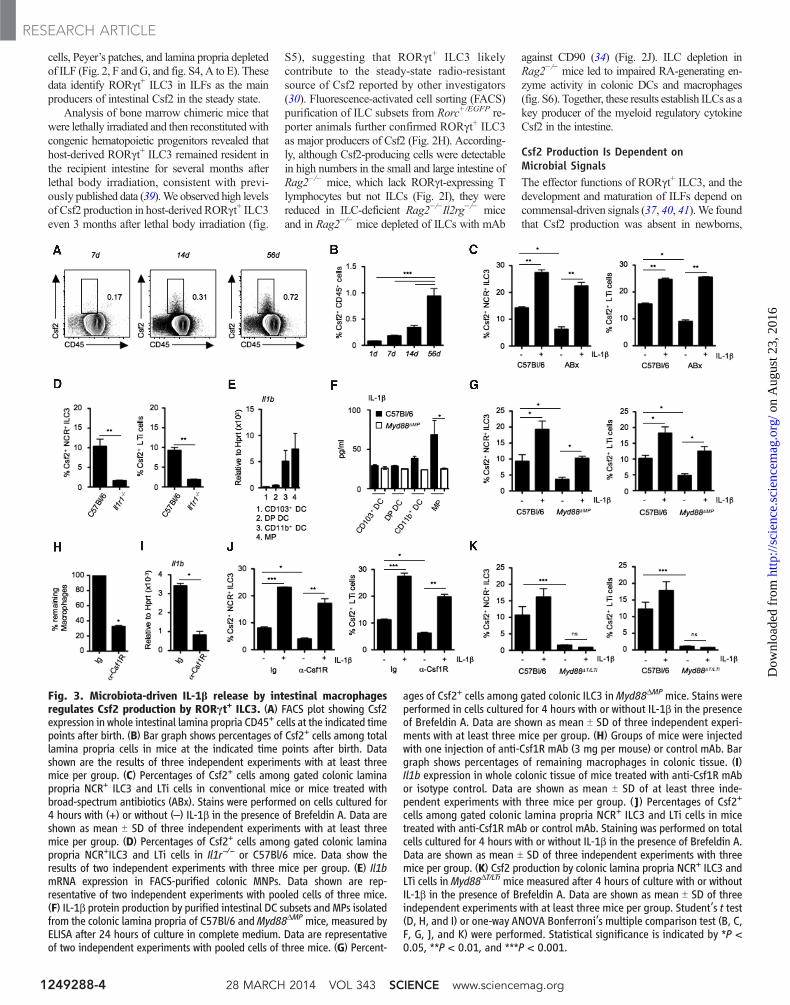
Csf2 Production Is Dependent onMicrobial SignalsThe effector functions of RORgt+ ILC3, and thedevelopment and maturation of ILFs depend oncommensal-driven signals (37, 40, 41).We foundthat Csf2 production was absent in newborns,
Fig. 3. Microbiota-driven IL-1b release by intestinal macrophagesregulates Csf2 production by RORgt+ ILC3. (A) FACS plot showing Csf2expression in whole intestinal lamina propria CD45+ cells at the indicated timepoints after birth. (B) Bar graph shows percentages of Csf2+ cells among totallamina propria cells in mice at the indicated time points after birth. Datashown are the results of three independent experiments with at least threemice per group. (C) Percentages of Csf2+ cells among gated colonic laminapropria NCR+ ILC3 and LTi cells in conventional mice or mice treated withbroad-spectrum antibiotics (ABx). Stains were performed on cells cultured for4 hours with (+) or without (–) IL-1b in the presence of Brefeldin A. Data areshown as mean T SD of three independent experiments with at least threemice per group. (D) Percentages of Csf2+ cells among gated colonic laminapropria NCR+ILC3 and LTi cells in Il1r−/− or C57Bl/6 mice. Data show theresults of two independent experiments with three mice per group. (E) Il1bmRNA expression in FACS-purified colonic MNPs. Data shown are rep-resentative of two independent experiments with pooled cells of three mice.(F) IL-1b protein production by purified intestinal DC subsets and MPs isolatedfrom the colonic lamina propria of C57Bl/6 andMyd88DMPmice, measured byELISA after 24 hours of culture in complete medium. Data are representativeof two independent experiments with pooled cells of three mice. (G) Percent-
ages of Csf2+ cells among gated colonic ILC3 inMyd88DMPmice. Stains wereperformed in cells cultured for 4 hours with or without IL-1b in the presenceof Brefeldin A. Data are shown as mean T SD of three independent experi-ments with at least three mice per group. (H) Groups of mice were injectedwith one injection of anti-Csf1R mAb (3 mg per mouse) or control mAb. Bargraph shows percentages of remaining macrophages in colonic tissue. (I)Il1b expression in whole colonic tissue of mice treated with anti-Csf1R mAbor isotype control. Data are shown as mean T SD of at least three inde-pendent experiments with three mice per group. (J) Percentages of Csf2+
cells among gated colonic lamina propria NCR+ ILC3 and LTi cells in micetreated with anti-Csf1R mAb or control mAb. Staining was performed on totalcells cultured for 4 hours with or without IL-1b in the presence of Brefeldin A.Data are shown as mean T SD of three independent experiments with threemice per group. (K) Csf2 production by colonic lamina propria NCR+ ILC3 andLTi cells in Myd88DT/LTi mice measured after 4 hours of culture with or withoutIL-1b in the presence of Brefeldin A. Data are shown as mean T SD of threeindependent experiments with at least three mice per group. Student’s t test(D, H, and I) or one-way ANOVA Bonferroni’s multiple comparison test (B, C,F, G, J, and K) were performed. Statistical significance is indicated by *P <0.05, **P < 0.01, and ***P < 0.001.
28 MARCH 2014 VOL 343 SCIENCE www.sciencemag.org1249288-4
RESEARCH ARTICLE
on
Aug
ust 2
3, 2
016
http
://sc
ienc
e.sc
ienc
emag
.org
/D
ownl
oade
d fr
om
slightly increased in 7-day-oldmice, and increasedsubstantially from day 14 after birth, concurrentwith the increase in numbers and complexity ofthe intestinal microbial flora at these developmen-tal stages (Fig. 3, A and B). To further investigatethe influence of the commensal flora on Csf2production in the intestine, we treated adult micewith broad-spectrum antibiotics known to strong-ly reduce the gut microbiota. In accordance withour findings in newborn animals, adultmice treatedwith broad-spectrum antibiotics displayed reducedCsf2 production in the small and large intestine(Fig. 3C). These results suggest that commensal-driven signals control the steady-state productionof Csf2 by RORgt+ ILC3 in the mouse intestine.
Murine RORgt+ ILC3 lack Toll-like receptors(TLRs) and cannot directly sense microbial sig-nals in the gut; hence, these cells must rely onother cellular sensors to translate cues from com-mensal bacteria into effector functions (42). Wetherefore explored whether cytokines derived frommyeloid cells could drive Csf2 production byRORgt+ ILC3 ex vivo. Among several cytokines
tested, we observed that IL-1b was a particularlypotent inducer of Csf2 production by RORgt+
ILC3 (Fig. 3C), consistent with the reported roleof IL-1b as a potent driver of ILC function (38).Because the myeloid cytokine IL-23 promotesthe production of the cytokine IL-22 by RORgt+
ILC3, we examined whether IL-23 also promotedCsf2 production by these cells (43). IL-23 wasunable to promote Csf2 production by RORgt+
ILC3 (fig. S7A), whereas it stimulated the releaseof IL-22 (fig. S7B), as previously reported (43).Furthermore, IL-22 production by RORgt+ ILC3was unaffected in Csf2−/− mice (fig. S7C). Expo-sure to IL-1b rescued Csf2 production by RORgt+
ILC3 isolated from antibiotic-treatedmice (Fig. 3C).Accordingly, LTi and NCR+ ILC3 isolated frommice lacking IL-1 receptor 1 (Il1r1−/−) failed toproduce Csf2 (Fig. 3D), thereby implicating IL-1b and IL-1R signaling as key drivers of Csf2production in the intestine. In contrast, absence ofthe other IL-1 superfamily member, IL-18, didnot compromise intestinal Csf2 production (fig.S7D). Together, these data indicate that IL-1b–
producing cells that respond to microbial signalscontrol the steady-state production of Csf2 byRORgt+ ILC3 in the intestine.
Sensing the commensal microflora by theTLR and the activation of the adapter proteinMyd88 is critical for maintaining intestinal ho-meostasis (44) and leads to steady-state IL-1b pro-duction by tissue MNPs (45). Tissue-residentmacrophages, CD103+ DCs, and CD103– DCsarise from different developmental pathwaysand express distinct pattern recognition recep-tors (46). We found that intestinal macrophageswere thehighest producers of Il1b and IL-1b protein,as previously reported (45, 47), suggesting thatthis population is a key regulator of Csf2 pro-duction in the gut (Fig. 3, E and F).
Because microbial signals were required todrive Csf2 production in the intestine, we nextexamined whether deletion of the TLR-adapterprotein Myd88 in phagocytes influenced Csf2 pro-duction by RORgt+ ILC3. Lysozyme M (LysM)is expressed at high levels in macrophages rela-tive toDCs (48), and notably,mice that lackMyd88
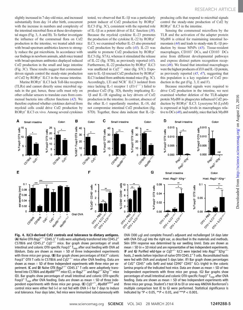
Fig. 4. ILC3-derived Csf2 controls oral tolerance to dietary antigens.(A) Naïve OTII Rag2−/− CD45.1+ T cells were adoptively transferred into CD45.2+
C57Bl/6 and CD45.2+ Csf2−/− mice. Bar graph shows percentages of smallintestinal and colonic OTII-specific Foxp3+ Tregs after oral feeding with OVA adlibidum. Data are shown as mean T SD of three independent experimentswith three mice per group. (B) Bar graph shows percentages of Ki67+ colonicFoxp3+ OTII T cells in C57Bl/6 and Csf2−/− mice after OVA feeding. Data areshown as mean T SD of three independent experiments with three mice per ex-periment. (C and D) Naïve OTII Rag2−/− CD45.1+ T cells were adoptively trans-ferred into C57Bl/6 andMyd88DT/LTimice (C), or Rag2−/− and Rag2−/−Il2rg−/−mice(D). Bar graphs show percentages of small intestinal and colonic OTII-specificFoxp3+ Tregs after OVA feeding. Data are shown as mean T SD of three inde-pendent experiments with three mice per group. (E) Csf2−/−, Myd88DT/LTi andcontrol mice were either fed (+) or not fed with OVA (–) for 7 days to induceoral tolerance. Four days later, fed mice were immunized subcutaneously with
OVA (300 mg) and complete Freund’s adjuvant and rechallenged 14 days laterwith OVA (50 mg) into the right ear, as described in the materials and methods.Skin DTH response was determined by ear swelling (mm). Data are shown asmean T SD (n= 10mice) and are representative of two independent experiments.(F and G) Purified wild-type or Csf2−/− ILC3 were injected into Rag2−/−Il2rg−/−
hosts, 2 weeks before injection of naïve OTII CD45.1+ T cells. Reconstituted hostswere fed with OVA and analyzed 5 days later. (F) Bar graph shows percentagesof Csf2+ CD45+ cells (left) and total CD90+ CD45+ ILCs (right) in the coloniclamina propria of the indicated host mice. Data are shown as mean T SD of twoindependent experiments with three mice per group. (G) Bar graphs showpercentages of small intestinal and colonic OTII-specific Foxp3+ Tregs after OVAfeeding. Data are shown as mean T SD of two independent experiments withthree mice per group. Student’s t test (A to D) or one-way ANOVA Bonferroni’smultiple comparison test (E to G) were performed. Statistical significance isindicated by *P < 0.05, **P < 0.01, and ***P < 0.001.
www.sciencemag.org SCIENCE VOL 343 28 MARCH 2014 1249288-5
RESEARCH ARTICLE
on
Aug
ust 2
3, 2
016
http
://sc
ienc
e.sc
ienc
emag
.org
/D
ownl
oade
d fr
om

specifically in LysM+ cells [LysMCrexMyd88 flox/flox
(Myd88DMP) mice] exhibited a significant reduc-tion in IL-1b production in intestinal macrophages,whereas IL-1b production by intestinal DCs wasunaffected (Fig. 3F). Thedisruptionof IL-1b releaseby intestinal macrophages in Myd88DMP miceabrogated the production of Csf2 by RORgt+ LTiand NCR+ ILC3 in these animals (Fig. 3G),whereas addition of exogenous IL-1b cytokinerescued Csf2 production by RORgt+ ILC3 (Fig.3G). Consistent with a central role for macro-phages in intestinal IL-1b production, adminis-tration of depleting anti-Csf1 receptor monoclonalantibody (anti-Csf1R mAb) depleted tissue mac-rophages (Fig. 3H), aswe previously showed (49),and reduced total Il1b expression (Fig. 3I). Ac-cordingly, Csf2 production by intestinal RORgt+
ILC3 was significantly reduced after depletion ofcolonic macrophages (Fig. 3J). IL-1b cytokinewas capable of rescuing Csf2 production byRORgt+ ILC3 isolated frommice treated with theanti-Csf1R mAb (Fig. 3J), thus confirming thatmacrophage-derived IL-1b is a key driver of Csf2production by RORgt+ ILC3. Myd88 is an es-sential signal transducer in both the TLR andIL-1R pathway (50). To establishwhether IL-1b andIL-1R signaling are required to promote Csf2production by RORgt+ ILC3, we crossed RorcCre
mice with Myd88flox/flox mice to achieve deletionofMyd88 specifically in RORgt+ ILC3 andTcells(Myd88DT/LTi) (51). As expected, we observed areduction in Csf2 production by RORgt+ ILC3fromMyd88DT/LTimice (Fig. 3K). In this case, ad-ministration of IL-1b cytokine failed to rescueCsf2 expression by RORgt+ ILC3, suggesting thatMyd88 functions downstream of IL-1R in RORgt+
ILC3 (Fig. 3K). Taken together, our results suggestthat Myd88-dependent sensing of the commen-sal microflora by intestinal macrophages elicitsproduction of IL-1b, which in turn activates theIL-1R–Myd88 pathway in RORgt+ ILC3 to drivethe steady-state production of Csf2. Alteration ofthe commensal flora using broad-spectrum anti-biotics or deletion of macrophages using anti-Csf1RmAb treatment led to impairedRAproduction in allDC subsets (fig. S8A) and to reduced Treg numbersand proliferation (fig. S8, B andC), confirming therole of tissue macrophages in translating micro-bial cues into immunoregulatory signals that helppromote Treg homeostasis in the steady-state colon.
Csf2 Promotes Oral Tolerance to Fed AntigensBecause one of the key functions of intestinalTregs is the maintenance of oral tolerance to fedantigens, we asked whether deficiency in Csf2affects de novo generation of intestinal Tregs uponoral administration of ovalbumin (OVA). Conver-sion and expansion of OVA-specific Tregs in thesmall and large intestine were impaired in Csf2−/−
mice compared to wild-type mice (Fig. 4, A andB). Similar results were obtained when Treg con-version was analyzed in mice selectively lackingCsf2 in RORgt+ ILC3 (Myd88DT/LTi) or in ILC-deficient mice (Rag2−/−Il2rg−/−) (Fig. 4, C andD). Consistent with Csf2’s key role in oral toler-
ance, OVA feeding of Csf2−/−mice andMyd88DT/LTi
mice failed to protect the mice from delayed-typehypersensitivity (DTH) reaction upon OVA chal-lenge, whereas control mice were protected (Fig.4E). Together, these results establish that alteredCsf2 production by ILCs impairs the induction oforal tolerance to dietary antigens. The defect inTreg conversion observed in the small intestine ofCsf2−/− mice contrasts with the apparent normaltotal Treg numbers observed in the small bowel ofthese mice. These results suggest that compen-satory mechanisms specific to the small bowelmay help restore the number but likely not therepertoire of small intestinal Tregs in Csf2
−/−mice.To establish the direct contribution of Csf2
produced by ILC3 to Treg conversion in vivo, wereconstituted ILC-deficientRag2−/−Il2rg−/−micewith Csf2−/− or Csf2+/+ ILC3. Two weeks later,Rag2−/−Il2rg−/−mice reconstituted with ILCs wereadoptively transferred with OVA-specific T cellreceptor transgenic OTII cells and fed with OVAfor 5 days. Csf2−/− and Csf2+/+ ILC3 engraftedwith the same efficiency in Rag2−/−Il2rg−/−mice(Fig. 4F), and reconstitution ofRag2−/−Il2rg−/−micewith Csf2+/+ ILC3 led to partial recovery of Csf2production in the intestine (Fig. 4F). Although therate ofTreg conversionwas low in allRag2−/−Il2rg−/−
mice due to a defect in lymphoid organ developmentin these mice, OVA-specific Treg conversion wasnonetheless significantly higher in Rag2−/−Il2rg−/−
mice reconstitutedwithCsf2+/+ compared to micereconstituted with Csf2−/− ILC3 (Fig. 4G), furtheremphasizing the contribution of ILC3-derived Csf2to the control of oral tolerance to dietary antigens.
DiscussionPrevious studies have established the role of mi-crobial commensals that colonize the large bowelto promote the induction of Foxp3+ Treg differen-tiation (5). However the cellular cues that promoteTreg accumulation in response to gut commensalshave only recently started to be unraveled (52–54).Our data identify a mechanism by which the gutmicrobiota promotes intestinal homeostasis bysupporting a crosstalk between IL-1b–secretingmacrophages and Csf2-producing RORgt+ ILC3 inthe intestinalmucosa.Microbiota-driven IL-1b pro-duction by macrophages promoted the release ofCsf2 by ILC3, which in turn acted on DCs andmacrophages, allowing for the maintenance ofcolonic Treg homeostasis (fig. S9). Ablation ofCsf2 altered DC and macrophage numbers andimpaired their ability to produce regulatory fac-tors such as RA and IL-10, which led to disruptedTreg homeostasis in the large intestine. Converse-ly, administration of Csf2 cytokine increased Tregfrequency in the gut. Most notably, cell type–specific ablation of IL-1–dependent signaling inRORgt+ ILC3 abrogated oral tolerance to dietaryantigens and compromised intestinal Treg ho-meostasis in vivo. Although the reduction in totalTreg numbers was mostly observed in the largeintestine, adoptive transfer studies in Csf2−/− micerevealed impaired Treg differentiation both in thesmall and large intestine, suggesting that Csf2-
dependentMNP immunoregulatory functions con-trol Treg induction in both tissues
Establishing intestinal tolerance is critical forthe prevention of intestinal diseases such as IBD.IBD includes two broad disease classificationsknown as ulcerative colitis and Crohn’s disease,but there is substantial variation in IBD clinico-pathology in individual patients; hence, it is likelythat numerous subtypes of IBD exist in this group.In a study of more than 300 patients with Crohn’sdisease, the presence of neutralizing antibodies toCsf2 in the serum correlatedwith ileal involvementand the development of penetrating pathology,whereas a more recent study identified reducedlevels of Csf2 receptor (Csf2R) and impaired recep-tor activity in amixedgroupof IBDpatients (55, 56).Previous clinical trials of recombinant Csf2 in IBDhave established patient benefit in terms of reduceddisease severity and lower burden of corticosteroiduse (57). Unpublished results of a larger trial ofCsf2 in IBD has since failed to achieve primaryclinical end points, but it remains likely that a sub-set of IBD patients with defective Csf2 produc-tion or function could benefit from this therapy.
The uncovered key role for Csf2 in the main-tenance of intestinal tolerance is consistent withprevious studies showing that absence of Csf2 canalso contribute to lupus-like disease, insulitis, andage-related glucose intolerance (29, 58) and fur-ther emphasizes the critical role of tissue-residentphagocytes in the maintenance of tissue integrity.
Our data reveal a mechanism by which the gutcommensal flora promotes immune homeostasisin the host. We have identified the commensal-driven MNP-ILC-Csf2 axis as a key regulator ofintestinal Tcell homeostasis in themouse intestine.Disturbance of this axis radically altered MNP ef-fector function, resulting in impaired oral toleranceto dietary antigens. These results represent an impor-tant advance in our understanding of how commen-sal microbes can regulate host intestinal immunityandmay inform the design of new immunotherapiesfor the use in patients with subtypes of IBD.
References and Notes1. K. J. Maloy, F. Powrie, Intestinal homeostasis and its breakdown
in inflammatory bowel disease. Nature 474, 298–306(2011). doi: 10.1038/nature10208; pmid: 21677746
2. B. Khor, A. Gardet, R. J. Xavier, Genetics and pathogenesisof inflammatory bowel disease. Nature 474, 307–317(2011). doi: 10.1038/nature10209; pmid: 21677747
3. I. I. Ivanov et al., Induction of intestinal Th17 cells bysegmented filamentous bacteria. Cell 139, 485–498(2009). doi: 10.1016/j.cell.2009.09.033; pmid: 19836068
4. L. V. Hooper, D. R. Littman, A. J. Macpherson, Interactionsbetween the microbiota and the immune system. Science336, 1268–1273 (2012). doi: 10.1126/science.1223490;pmid: 22674334
5. K. Atarashi et al., Induction of colonic regulatory T cellsby indigenous Clostridium species. Science 331, 337–341(2011). doi: 10.1126/science.1198469; pmid: 21205640
6. J. L. Round et al., The Toll-like receptor 2 pathwayestablishes colonization by a commensal of the humanmicrobiota. Science 332, 974–977 (2011). doi: 10.1126/science.1206095; pmid: 21512004
7. M. Merad, P. Sathe, J. Helft, J. Miller, A. Mortha, The dendriticcell lineage: Ontogeny and function of dendritic cells andtheir subsets in the steady state and the inflamed setting.Annu. Rev. Immunol. 31, 563–604 (2013). doi: 10.1146/annurev-immunol-020711-074950; pmid: 23516985
28 MARCH 2014 VOL 343 SCIENCE www.sciencemag.org1249288-6
RESEARCH ARTICLE
on
Aug
ust 2
3, 2
016
http
://sc
ienc
e.sc
ienc
emag
.org
/D
ownl
oade
d fr
om
8. M. Bogunovic, A. Mortha, P. A. Muller, M. Merad,Mononuclear phagocyte diversity in the intestine.Immunol. Res. 54, 37–49 (2012). doi: 10.1007/s12026-012-8323-5; pmid: 22562804
9. V. Cerovic et al., Intestinal CD103(-) dendritic cells migrate inlymph and prime effector T cells.Mucosal Immunol. 6, 104–113(2013). doi: 10.1038/mi.2012.53; pmid: 22718260
10. S. Tamoutounour et al., CD64 distinguishes macrophagesfrom dendritic cells in the gut and reveals theTh1-inducing role of mesenteric lymph node macrophagesduring colitis. Eur. J. Immunol. 42, 3150–3166 (2012).doi: 10.1002/eji.201242847; pmid: 22936024
11. M. Bogunovic et al., Origin of the lamina propriadendritic cell network. Immunity 31, 513–525 (2009).doi: 10.1016/j.immuni.2009.08.010; pmid: 19733489
12. C. Varol et al., Intestinal lamina propria dendritic cellsubsets have different origin and functions. Immunity 31,502–512 (2009). doi: 10.1016/j.immuni.2009.06.025;pmid: 19733097
13. K. Takeda et al., Enhanced Th1 activity and developmentof chronic enterocolitis in mice devoid of Stat3 in macrophagesand neutrophils. Immunity 10, 39–49 (1999). doi: 10.1016/S1074-7613(00)80005-9; pmid: 10023769
14. C. M. Sun et al., Small intestine lamina propria dendriticcells promote de novo generation of Foxp3 T reg cellsvia retinoic acid. J. Exp. Med. 204, 1775–1785 (2007).doi: 10.1084/jem.20070602; pmid: 17620362
15. J. L. Coombes et al., A functionally specialized populationof mucosal CD103+ DCs induces Foxp3+ regulatoryT cells via a TGF-beta and retinoic acid-dependentmechanism. J. Exp. Med. 204, 1757–1764 (2007).doi: 10.1084/jem.20070590; pmid: 17620361
16. T. L. Denning, Y. C. Wang, S. R. Patel, I. R. Williams,B. Pulendran, Lamina propria macrophages and dendriticcells differentially induce regulatory and interleukin17-producing T cell responses. Nat. Immunol. 8, 1086–1094(2007). doi: 10.1038/ni1511; pmid: 17873879
17. S. Manicassamy et al., Activation of beta-catenin indendritic cells regulates immunity versus tolerance in theintestine. Science 329, 849–853 (2010). doi: 10.1126/science.1188510; pmid: 20705860
18. U. Hadis et al., Intestinal tolerance requires gut homingand expansion of FoxP3+ regulatory T cells in the laminapropria. Immunity 34, 237–246 (2011). doi: 10.1016/j.immuni.2011.01.016; pmid: 21333554
19. T. L. Denning et al., Functional specializations ofintestinal dendritic cell and macrophage subsets thatcontrol Th17 and regulatory T cell responses are dependenton the T cell/APC ratio, source of mouse strain, andregional localization. J. Immunol. 187, 733–747 (2011).doi: 10.4049/jimmunol.1002701; pmid: 21666057
20. M. Jinushi, F. S. Hodi, G. Dranoff, Enhancing the clinicalactivity of granulocyte-macrophage colony-stimulatingfactor-secreting tumor cell vaccines. Immunol. Rev. 222,287–298 (2008). doi: 10.1111/j.1600-065X.2008.00618.x;pmid: 18364009
21. Y. Zhan, Y. Xu, A. M. Lew, The regulation of thedevelopment and function of dendritic cell subsets byGM-CSF: More than a hematopoietic growth factor.Mol. Immunol. 52, 30–37 (2012). doi: 10.1016/j.molimm.2012.04.009; pmid: 22580403
22. D. Vremec et al., The influence of granulocyte/macrophagecolony-stimulating factor on dendritic cell levels in mouselymphoid organs. Eur. J. Immunol. 27, 40–44 (1997).doi: 10.1002/eji.1830270107; pmid: 9021996
23. S. H. Naik et al., Intrasplenic steady-state dendritic cellprecursors that are distinct from monocytes. Nat. Immunol.7, 663–671 (2006). doi: 10.1038/ni1340; pmid: 16680143
24. K. Shortman, S. H. Naik, Steady-state and inflammatorydendritic-cell development. Nat. Rev. Immunol. 7, 19–30(2007). doi: 10.1038/nri1996; pmid: 17170756
25. J. A. Hamilton, Colony-stimulating factors in inflammationand autoimmunity. Nat. Rev. Immunol. 8, 533–544(2008). doi: 10.1038/nri2356; pmid: 18551128
26. L. Codarri et al., RORgt drives production of the cytokineGM-CSF in helper T cells, which is essential for the effectorphase of autoimmune neuroinflammation. Nat. Immunol.12, 560–567 (2011). doi: 10.1038/ni.2027; pmid: 21516112
27. M. El-Behi et al., The encephalitogenicity of T(H)17 cellsis dependent on IL-1- and IL-23-induced production of
the cytokine GM-CSF. Nat. Immunol. 12, 568–575(2011). doi: 10.1038/ni.2031; pmid: 21516111
28. M. Greter et al., GM-CSF controls nonlymphoid tissuedendritic cell homeostasis but is dispensable for thedifferentiation of inflammatory dendritic cells.Immunity 36, 1031–1046 (2012). doi: 10.1016/j.immuni.2012.03.027; pmid: 22749353
29. M. Jinushi et al., MFG-E8-mediated uptake of apoptoticcells by APCs links the pro- and antiinflammatoryactivities of GM-CSF. J. Clin. Invest. 117, 1902–1913(2007). doi: 10.1172/JCI30966; pmid: 17557120
30. L. Egea et al., GM-CSF produced by nonhematopoieticcells is required for early epithelial cell proliferation andrepair of injured colonic mucosa. J. Immunol. 190,1702–1713 (2013). doi: 10.4049/jimmunol.1202368;pmid: 23325885
31. H. Fukuzawa et al., Identification of GM-CSF in Panethcells using single-cell RT-PCR. Biochem. Biophys.Res. Commun. 312, 897–902 (2003). doi: 10.1016/j.bbrc.2003.11.009; pmid: 14651956
32. T. Griseri, B. S. McKenzie, C. Schiering, F. Powrie,Dysregulated hematopoietic stem and progenitor cellactivity promotes interleukin-23-driven chronic intestinalinflammation. Immunity 37, 1116–1129 (2012).doi: 10.1016/j.immuni.2012.08.025; pmid: 23200826
33. H. Spits et al., Innate lymphoid cells—a proposal foruniform nomenclature. Nat. Rev. Immunol. 13, 145–149(2013). doi: 10.1038/nri3365; pmid: 23348417
34. C. Vonarbourg et al., Regulated expression of nuclearreceptor RORgt confers distinct functional fates to NK cellreceptor-expressing RORgt(+) innate lymphocytes.Immunity 33, 736–751 (2010). doi: 10.1016/j.immuni.2010.10.017; pmid: 21093318
35. G. Eberl et al., An essential function for the nuclearreceptor RORgamma(t) in the generation of fetallymphoid tissue inducer cells. Nat. Immunol. 5, 64–73(2004). doi: 10.1038/ni1022; pmid: 14691482
36. M. Cella et al., A human natural killer cell subset provides aninnate source of IL-22 for mucosal immunity. Nature 457,722–725 (2009). doi: 10.1038/nature07537; pmid: 18978771
37. S. L. Sanos et al., RORgammat and commensal microfloraare required for the differentiation of mucosal interleukin22-producing NKp46+ cells. Nat. Immunol. 10, 83–91(2009). doi: 10.1038/ni.1684; pmid: 19029903
38. A. Reynders et al., Identity, regulation and in vivofunction of gut NKp46+RORgt+ and NKp46+RORgt-
lymphoid cells. EMBO J. 30, 2934–2947 (2011).doi: 10.1038/emboj.2011.201; pmid: 21685873
39. A. M. Hanash et al., Interleukin-22 protects intestinalstem cells from immune-mediated tissue damage andregulates sensitivity to graft versus host disease.Immunity 37, 339–350 (2012). doi: 10.1016/j.immuni.2012.05.028; pmid: 22921121
40. D. Bouskra et al., Lymphoid tissue genesis induced bycommensals through NOD1 regulates intestinalhomeostasis. Nature 456, 507–510 (2008).doi: 10.1038/nature07450; pmid: 18987631
41. N. Satoh-Takayama et al., Microbial flora drives interleukin22 production in intestinal NKp46+ cells that provide innatemucosal immune defense. Immunity 29, 958–970 (2008).doi: 10.1016/j.immuni.2008.11.001; pmid: 19084435
42. N. K. Crellin et al., Regulation of cytokine secretion inhuman CD127(+) LTi-like innate lymphoid cells byToll-like receptor 2. Immunity 33, 752–764 (2010).doi: 10.1016/j.immuni.2010.10.012; pmid: 21055975
43. H. Takatori et al., Lymphoid tissue inducer-like cells are aninnate source of IL-17 and IL-22. J. Exp. Med. 206, 35–41(2009). doi: 10.1084/jem.20072713; pmid: 19114665
44. S. Rakoff-Nahoum, J. Paglino, F. Eslami-Varzaneh,S. Edberg, R. Medzhitov, Recognition of commensalmicroflora by toll-like receptors is required for intestinalhomeostasis. Cell 118, 229–241 (2004). doi: 10.1016/j.cell.2004.07.002; pmid: 15260992
45. M. H. Shaw, N. Kamada, Y. G. Kim, G. Núñez,Microbiota-induced IL-1b, but not IL-6, is critical for thedevelopment of steady-state TH17 cells in the intestine.J. Exp. Med. 209, 251–258 (2012). doi: 10.1084/jem.20111703; pmid: 22291094
46. D. Hashimoto, J. Miller, M. Merad, Dendritic cell andmacrophage heterogeneity in vivo. Immunity 35,
323–335 (2011). doi: 10.1016/j.immuni.2011.09.007;pmid: 21943488
47. N. Hoshi et al., MyD88 signalling in colonic mononuclearphagocytes drives colitis in IL-10-deficient mice.Nat. Commun. 3, 1120 (2012). doi: 10.1038/ncomms2113; pmid: 23047678
48. C. Jakubzick et al., Lymph-migrating, tissue-deriveddendritic cells are minor constituents within steady-statelymph nodes. J. Exp. Med. 205, 2839–2850 (2008).doi: 10.1084/jem.20081430; pmid: 18981237
49. D. Hashimoto et al., Pretransplant CSF-1 therapy expandsrecipient macrophages and ameliorates GVHD afterallogeneic hematopoietic cell transplantation. J. Exp. Med.208, 1069–1082 (2011). doi: 10.1084/jem.20101709;pmid: 21536742
50. L. A. O’Neill, A. G. Bowie, The family of five:TIR-domain-containing adaptors in Toll-like receptorsignalling. Nat. Rev. Immunol. 7, 353–364 (2007).doi: 10.1038/nri2079; pmid: 17457343
51. G. Eberl, D. R. Littman, Thymic origin of intestinalalphabeta T cells revealed by fate mapping ofRORgammat+ cells. Science 305, 248–251 (2004).doi: 10.1126/science.1096472; pmid: 15247480
52. P. M. Smith et al., The microbial metabolites, short-chainfatty acids, regulate colonic Treg cell homeostasis.Science 341, 569–573 (2013). doi: 10.1126/science.1241165; pmid: 23828891
53. N. Arpaia et al., Metabolites produced by commensalbacteria promote peripheral regulatory T-cell generation.Nature 504, 451–455 (2013). doi: 10.1038/nature12726; pmid: 24226773
54. Y. Furusawa et al., Commensal microbe-derived butyrateinduces the differentiation of colonic regulatory T cells.Nature 504, 446–450 (2013). doi: 10.1038/nature12721; pmid: 24226770
55. X. Han et al., Granulocyte-macrophage colony-stimulatingfactor autoantibodies in murine ileitis and progressiveileal Crohn’s disease. Gastroenterology 136, 1261–1271,e1–e3 (2009). doi: 10.1053/j.gastro.2008.12.046;pmid: 19230854
56. J. I. Goldstein et al., Defective leukocyte GM-CSF receptor(CD116) expression and function in inflammatory boweldisease. Gastroenterology 141, 208–216 (2011).doi: 10.1053/j.gastro.2011.03.060; pmid: 21557945
57. J. R. Korzenik, B. K. Dieckgraefe, J. F. Valentine,D. F. Hausman, M. J. Gilbert; Sargramostim in Crohn’sDisease Study Group, Sargramostim for active Crohn’sdisease. N. Engl. J. Med. 352, 2193–2201 (2005).doi: 10.1056/NEJMoa041109; pmid: 15917384
58. T. Enzler et al., Functional deficiencies of granulocyte-macrophage colony stimulating factor and interleukin-3contribute to insulitis and destruction of beta cells.Blood 110, 954–961 (2007). doi: 10.1182/blood-2006-08-043786; pmid: 17483299
Acknowledgments: We thank the Merad laboratory forhelpful discussions and input. We are grateful to W.-H. Kwanand W. van der Touw for assistance with the Treg inductionassay. We thank R. Huq and L. O’Rourke at the Icahn School ofMedicine at Mount Sinai Microscopy Shared Resource Facilityfor their training, helpful advice, and support with microscopyimaging. We are grateful to C. Berin and A. Belén Blázquezfor support with the oral tolerance and DTH models. We thankJ. Ochando and the Flow Cytometry facility for technical supportand assistance with cell sorting. The data presented in this paperare tabulated in the main paper and in the supporting materials.M.M. is funded by NIH grants R01 CA154947A, R01 CA173861,and U01 AI095611. A.M. is funded by the German ResearchFoundation (DFG) grant MO2380/1-1. Y.B. was supported by theDivision of Intramural Research of the National Institute ofAllergy and Infectious Diseases.
Supplementary Materialswww.sciencemag.org/content/343/6178/1249288/suppl/DC1Materials and MethodsFigs. S1 to S9References (59, 60)
3 December 2013; accepted 26 February 2014Published online 13 March 2014;10.1126/science.1249288
www.sciencemag.org SCIENCE VOL 343 28 MARCH 2014 1249288-7
RESEARCH ARTICLE
on
Aug
ust 2
3, 2
016
http
://sc
ienc
e.sc
ienc
emag
.org
/D
ownl
oade
d fr
om
published online March 13, 2014 (6178), . [doi: 10.1126/science.1249288] originally343Science
(March 13, 2014) Bogunovic, Sean P. Spencer, Yasmine Belkaid and Miriam Merad Arthur Mortha, Aleksey Chudnovskiy, Daigo Hashimoto, MilenaILC3 Promotes Intestinal HomeostasisMicrobiota-Dependent Crosstalk Between Macrophages and
Editor's Summary
population of cells known to be critical for maintaining immune tolerance in the gut.retinoic acid and interleukin-10, which support the conversion and expansion of regulatory T cells, ainduces myeloid cells (including dendritic cells and macrophages) to produce regulatory factors like the intestine, which then produce the cytokine, colony-stimulating factor 2 (Csf2). Csf-2, in turn,(IL-1) in response to signals derived from the microbiota. IL-1 acts on type 3 innate lymphoid cells in
) found that gut macrophages produce the cytokine interleukin-1Aychek and JungPerspective by (10.1126/science.1249288, published online 13 March; see theet al.Mortha genetically modified mice,
important site of immune tolerance. By studying specific intestinal immune cell populations in With the constant assault of food antigens and its billions of resident microbes, the gut is an
Gut Immune Tolerance
This copy is for your personal, non-commercial use only.
Article Tools
http://science.sciencemag.org/content/343/6178/1249288article tools: Visit the online version of this article to access the personalization and
Permissionshttp://www.sciencemag.org/about/permissions.dtlObtain information about reproducing this article:
is a registered trademark of AAAS. ScienceAdvancement of Science; all rights reserved. The title Avenue NW, Washington, DC 20005. Copyright 2016 by the American Association for thein December, by the American Association for the Advancement of Science, 1200 New York
(print ISSN 0036-8075; online ISSN 1095-9203) is published weekly, except the last weekScience
on
Aug
ust 2
3, 2
016
http
://sc
ienc
e.sc
ienc
emag
.org
/D
ownl
oade
d fr
om