fertility and pregnancy outcome after fetoscopic surgery

1
596 PLATELET-RICH PLASMA TO PREVENT AMNIOTIC FLUID LEAKAGE FOLLOWING FETOSCOPIC INTERVENTIONS: AN IN VITRO STUDY LIESBETH LEWI 1 , MARC HOYLAERTS 2 , LIEVE VERBIST 1 , ELKE BEU- TELS 1 , JAN DEPREST 1 , 1 KU Leuven, University Hospitals, Leuven, Belgium 2 KU Leuven, Department of Molecular and Vascular Biology, Leuven, Belgium OBJECTIVE: To examine (1) the influence of a platelet-rich plasma (PRP) plug on fetal membrane repair, (2) its adherence to damaged fetal membranes and the use of procoagulants to accelerate plug formation, (3) ability to seal a fetoscopic membrane defect, and (4) persistence in amniotic fluid (AF). STUDY DESIGN: (1) A microsurgical defect was created in confluent primary amniocyte cultures from elective c-section at term (n = 5). DMEM (control) or PRP diluted in DMEM at platelet concentrations of 30,000/lL and 3000/lL were added. The defect area was measured at 0, 12, 24, and 48 hrs. (2) Amnion-chorion patches (n = 5) were suspended in AF. PRP with tissue factor, ADP, or thromboxane A 2 was injected around the patches. Site of and time to plug formation were observed. (3) An amnion-chorion patch (n = 7) was attached to the bottom of a cylinder filled with AF. The membranes were traumatized by a 10 Fr trocar and supported by parafilm. PRP was injected around the defect. After 10 min, parafilm was removed and leakage was measured. Plug adhesion was confirmed microscopically. (4) PRP was added to AF in a dilution of 1:4 in a cylinder with a patch of hemostatic collagen at the bottom (n = 6). Time to plug disappearance was observed. RESULTS: (1) In the PRP group most defects were closed after 48 hrs, whereas none were closed in controls. (2) Plug formation was localized around the amnion-chorion patches, and median time to plug formation was shorter with tissue factor (90 sec) compared with ADP (495 sec) or thromboxane A 2 (510 sec) (P = 0.04). (3) After 10 min, no leakage occurred. Microscopy confirmed plug adherence to connective tissue of amnion and chorion. (4) Median plug persistence in an AF environment was 67 days (range: 21-133). CONCLUSION: A PRP plug enhances fetal membrane repair, adheres to connective tissue of amnion and chorion, and persists long enough in AF to allow cell ingrowth. Tissue factor accelerates plug formation, which in a surgical setting may help to keep the plug localized at the defect. 597 FERTILITY AND PREGNANCY OUTCOME AFTER FETOSCOPIC SURGERY LIESBETH LEWI 1 , GRIET VANDENBERGHE 1 , JAN DEPREST 1 , 1 KU Leuven, University Hospitals, Leuven, Belgium OBJECTIVE: To evaluate impact of fetoscopic surgery on subsequent fertility and pregnancy outcome. STUDY DESIGN: Data from consecutive series of 85 women undergoing fetoscopy between 1/1995 and 12/2000. Procedures included laser coagulation of anastomoses for TTTS and cord occlusion in complicated monochorionic (MC) multiplets. Data were obtained by means of postal questionnaire and medical records including (1) in relation to index pregnancy: maternal age, parity, history of infertility or obstetrical problems, mode of delivery and outcome; (2) after fetoscopy: reasons for not attempting, or interval to attempt and time to conception, infertility problems, number of subsequent pregnan- cies, complications and outcome. RESULTS: 65 responded (76%). Median age at fetoscopy was 31 yrs (range 20-39); 54% were nulliparous. Delivery was by CS in 45%. For TTTS overall survival was 63%, 77% with $1 survivor. Survival in the cord occlusion group was 78%. 35 women (54%) attempted and achieved ongoing pregnancies without de novo infertility problems. Median time to subsequent pregnancy attempt was 8 months (range: 2-22) if there were no survivors compared with 21 months (7-48) if at least one infant survived (P = 0.002). Median time to conception was 3 months (0-24). All ongoing pregnancies were uncomplicated, apart from one placenta previa with elective CS at 36 weeks and two spontaneous recurrent twin pregnancies resulting in preterm delivery at 34 and 35 wks. Mean GA at delivery was 39.5 wks (34-41) with mean birthweight = 3460 g (2200-4300). 30 women (46%) did not pursue further pregnancies, all having already at least one child. Nine (30%) indicated the previous experience of a complicated pregnancy as the main reason for not attempting further pregnancies. CONCLUSION: Fetoscopic surgery does not seem to affect subsequent fertility and pregnancy outcome. However, the experience of a complicated pregnancy may influence the choice to pursue further pregnancies. 598 CENTRAL FETAL GENE EXPRESSION OF APPETITE REGULATORY SIGNALS RON BELOOSESKY 1 , DAVE GAYLE 1 , FATANEH AMIDI 1 , SURE- SHBABU AHANYA 1 , MICHAEL ROSS 1 , 1 Harbor-UCLA Medical Center, Department of Obstetrics and Gynecology, Torrance, CA OBJECTIVE: Neural orexic (appetite) mechanisms develop in utero to facilitate newborn feeding necessary for accelerated growth. To explore the development of appetite function, we determined the gene expression of orexigenic (ORI) and anorexigenic (ANOR) factors in the fetal rat brain and adult rat hypothalamus. STUDY DESIGN: Pregnant Sprague-Dawley rats at gestation day 16 and adult rats were provided normal diets. Animals were sacrificed and whole fetal brains and adult hypothalami were harvested and homogenized for mRNA determinations. Levels of ORI (neuropeptide Y, NPY; agouti-related peptide, AgRP) and ANOR (cocaine and amphetamine regulated transcript, CART; proopiomelanocortin, POMC; leptin receptor, OB-Rb) mRNA were determined using real-time RT-PCR. RESULTS: The fetal and adult expression patterns of feeding regulatory signals were markedly different. Fetal NPY expression exceeded all ANOR factors, whereas adult brain demonstrated ANOR (CART, POMC) greater than ORI (AGRP, NPY) gene expression. CONCLUSION: The altered gene expression profile indicates that continued maturation of fetal ORI and ANOR mechanisms continue in late gestation and/or post birth. Fetal exposure to an altered nutrient environment may program developing neural systems, leading to altered feeding behavior in postnatal life. Expression of mRNA in fetal brain versus adult hypothalamus December 2003 Am J Obstet Gynecol S222 SMFM Abstracts

Upload: liesbeth-lewi

Post on 02-Sep-2016

213 views

Category:

Documents


1 download

TRANSCRIPT

596 PLATELET-RICH PLASMA TO PREVENT AMNIOTIC FLUID LEAKAGEFOLLOWING FETOSCOPIC INTERVENTIONS: AN IN VITRO STUDYLIESBETH LEWI1, MARC HOYLAERTS2, LIEVE VERBIST1, ELKE BEU-TELS1, JAN DEPREST1, 1KU Leuven, University Hospitals, Leuven, Belgium2KU Leuven, Department of Molecular and Vascular Biology, Leuven,Belgium

OBJECTIVE: To examine (1) the influence of a platelet-rich plasma (PRP)plug on fetal membrane repair, (2) its adherence to damaged fetal membranesand the use of procoagulants to accelerate plug formation, (3) ability to seala fetoscopic membrane defect, and (4) persistence in amniotic fluid (AF).

STUDY DESIGN: (1) A microsurgical defect was created in confluentprimary amniocyte cultures from elective c-section at term (n = 5). DMEM(control) or PRP diluted in DMEM at platelet concentrations of 30,000/lL and3000/lL were added. The defect area was measured at 0, 12, 24, and 48 hrs. (2)Amnion-chorion patches (n = 5) were suspended in AF. PRP with tissue factor,ADP, or thromboxane A2 was injected around the patches. Site of and time toplug formation were observed. (3) An amnion-chorion patch (n = 7) wasattached to the bottom of a cylinder filled with AF. The membranes weretraumatized by a 10 Fr trocar and supported by parafilm. PRP was injectedaround the defect. After 10 min, parafilm was removed and leakage wasmeasured. Plug adhesion was confirmed microscopically. (4) PRP was added toAF in a dilution of 1:4 in a cylinder with a patch of hemostatic collagen at thebottom (n = 6). Time to plug disappearance was observed.

RESULTS: (1) In the PRP group most defects were closed after 48 hrs,whereas none were closed in controls. (2) Plug formation was localized aroundthe amnion-chorion patches, and median time to plug formation was shorterwith tissue factor (90 sec) compared with ADP (495 sec) or thromboxane A2

(510 sec) (P = 0.04). (3) After 10 min, no leakage occurred. Microscopyconfirmed plug adherence to connective tissue of amnion and chorion. (4)Median plug persistence in an AF environment was 67 days (range: 21-133).

CONCLUSION: A PRP plug enhances fetal membrane repair, adheres toconnective tissue of amnion and chorion, and persists long enough in AF toallow cell ingrowth. Tissue factor accelerates plug formation, which in a surgicalsetting may help to keep the plug localized at the defect.

597

598 CENTRAL FETAL GENE EXPRESSION OF APPETITE REGULATORYSIGNALS RON BELOOSESKY1, DAVE GAYLE1, FATANEH AMIDI1, SURE-SHBABU AHANYA1, MICHAEL ROSS1, 1Harbor-UCLA Medical Center,Department of Obstetrics and Gynecology, Torrance, CA

OBJECTIVE: Neural orexic (appetite) mechanisms develop in utero tofacilitate newborn feeding necessary for accelerated growth. To explore thedevelopment of appetite function, we determined the gene expression oforexigenic (ORI) and anorexigenic (ANOR) factors in the fetal rat brain andadult rat hypothalamus.

STUDY DESIGN: Pregnant Sprague-Dawley rats at gestation day 16 andadult rats were provided normal diets. Animals were sacrificed and whole fetalbrains and adult hypothalami were harvested and homogenized for mRNAdeterminations. Levels of ORI (neuropeptide Y, NPY; agouti-related peptide,AgRP) and ANOR (cocaine and amphetamine regulated transcript, CART;proopiomelanocortin, POMC; leptin receptor, OB-Rb) mRNA were determinedusing real-time RT-PCR.

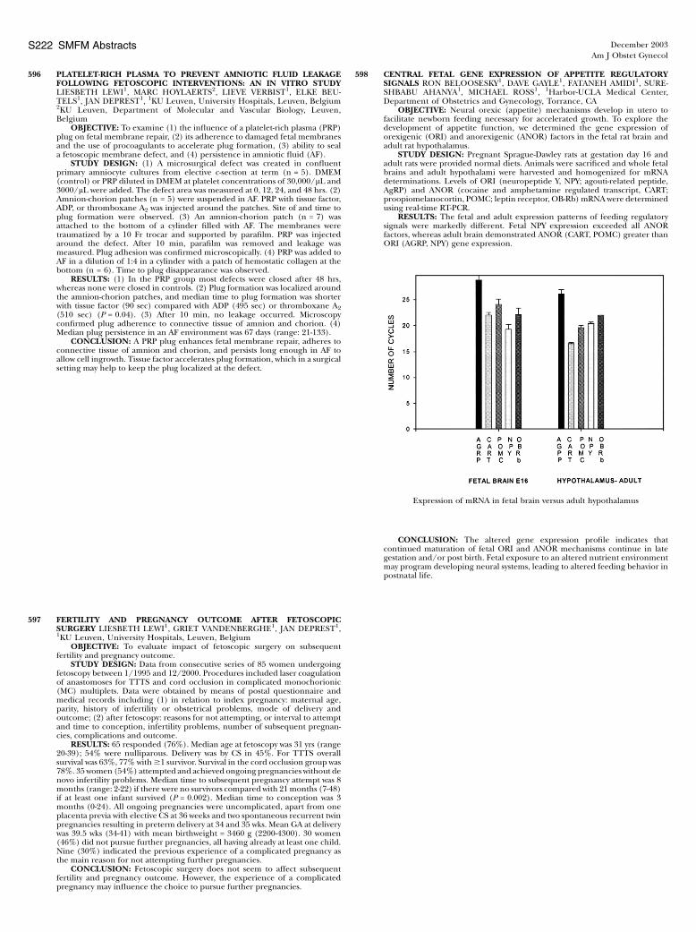
RESULTS: The fetal and adult expression patterns of feeding regulatorysignals were markedly different. Fetal NPY expression exceeded all ANORfactors, whereas adult brain demonstrated ANOR (CART, POMC) greater thanORI (AGRP, NPY) gene expression.

CONCLUSION: The altered gene expression profile indicates thatcontinued maturation of fetal ORI and ANOR mechanisms continue in lategestation and/or post birth. Fetal exposure to an altered nutrient environmentmay program developing neural systems, leading to altered feeding behavior inpostnatal life.

Expression of mRNA in fetal brain versus adult hypothalamus

December 2003Am J Obstet Gynecol

S222 SMFM Abstracts

FERTILITY AND PREGNANCY OUTCOME AFTER FETOSCOPICSURGERY LIESBETH LEWI1, GRIET VANDENBERGHE1, JAN DEPREST1,1KU Leuven, University Hospitals, Leuven, Belgium

OBJECTIVE: To evaluate impact of fetoscopic surgery on subsequentfertility and pregnancy outcome.

STUDY DESIGN: Data from consecutive series of 85 women undergoingfetoscopy between 1/1995 and 12/2000. Procedures included laser coagulationof anastomoses for TTTS and cord occlusion in complicated monochorionic(MC) multiplets. Data were obtained by means of postal questionnaire andmedical records including (1) in relation to index pregnancy: maternal age,parity, history of infertility or obstetrical problems, mode of delivery andoutcome; (2) after fetoscopy: reasons for not attempting, or interval to attemptand time to conception, infertility problems, number of subsequent pregnan-cies, complications and outcome.

RESULTS: 65 responded (76%). Median age at fetoscopy was 31 yrs (range20-39); 54% were nulliparous. Delivery was by CS in 45%. For TTTS overallsurvival was 63%, 77% with$1 survivor. Survival in the cord occlusion group was78%. 35women (54%) attempted and achieved ongoing pregnancies without denovo infertility problems. Median time to subsequent pregnancy attempt was 8months (range: 2-22) if there were no survivors compared with 21months (7-48)if at least one infant survived (P = 0.002). Median time to conception was 3months (0-24). All ongoing pregnancies were uncomplicated, apart from oneplacenta previa with elective CS at 36 weeks and two spontaneous recurrent twinpregnancies resulting in preterm delivery at 34 and 35 wks. Mean GA at deliverywas 39.5 wks (34-41) with mean birthweight = 3460 g (2200-4300). 30 women(46%) did not pursue further pregnancies, all having already at least one child.Nine (30%) indicated the previous experience of a complicated pregnancy asthe main reason for not attempting further pregnancies.

CONCLUSION: Fetoscopic surgery does not seem to affect subsequentfertility and pregnancy outcome. However, the experience of a complicatedpregnancy may influence the choice to pursue further pregnancies.