doctor of philosophy...dr. yashwant singh parmar university of horticulture and forestry, nauni,...
TRANSCRIPT

1985
STUDIES ON EPIDEMIOLOGY AND MANAGEMENT OF PINK CANKER (Corticium salmonicolor Berk. & Br.) IN APPLE
ThesisThesisThesisThesis
by
DURGA PRASHAD
Submitted in partial fulfilment of the requirements for the degree of
DOCTOR OF PHILOSOPHY
in
MYCOLOGY AND PLANT PATHOLOGY
COLLEGE OF HORTICULTURE Dr Yashwant Singh Parmar University of Horticulture and Forestry, Nauni,
Solan - 173230 (H.P.), INDIA
2013

CERTIFICATE - I
This is to certify that the thesis entitled, “Studies on epidemiology and
management of pink canker (Corticium salmonicolor Berk. & Br.) in apple”,
submitted in partial fulfilment of the requirements for the award of degree of
DOCTOR OF PHILOSOPHY in MYCOLOGY AND PLANT PATHOLOGY to
Dr. Yashwant Singh Parmar University of Horticulture and Forestry, Nauni, Solan (H.P.)
is a record of bonafide research work carried out by Mr. Durga Prashad (H-2009-14-D)
under my guidance and supervision. No part of this thesis has been submitted for any
other degree or diploma.
The assistance and help received during the course of investigation have been
fully acknowledged.
Place : Nauni, Solan Dr. Ved Ram
Dated: September, 2013 Chairman
Advisory Committee
Dr. Ved Ram
Senior Plant Pathologist
Department of Plant Pathology
College of Horticulture
Dr. Y. S. Parmar University of Horticulture and
Forestry, Nauni, Solan (HP) – 173 230, India

CERTIFICATE - II
This is to certify that the thesis entitled, “Studies on epidemiology and
management of pink canker (Corticium salmonicolor Berk. & Br.) in apple”,
submitted by Mr. Durga Prashad (H-2009-14-D) to Dr Y. S. Parmar University of
Horticulture and Forestry, Nauni, Solan (H.P.), in partial fulfilment of the requirements
for the award of degree of DOCTOR OF PHILOSOPHY in Mycology and Plant
Pathology has been approved by the Student’s Advisory Committee after an oral
examination of the same in collaboration with the external examiner.
___________________ ____________________
Dr. Ved Ram
Chairman
Advisory Committee
Dr. L. N. Bhardwaj
External Examiner
Members, Advisory Committee
_____________________ ______________________
Dr. I.M. Sharma Dr. J.S. Chandel
Sr. Scientist
(Plant Pathology) Sr. Scientist
(Fruit Science)
_______________________
Dr. Divender Gupta
Sr. Entomologist
(Entomology and Apiculture)
_____________________
Professor and Head
Department of Plant Pathology
__________________________
Dean’s Nominee
______________________________________________________________
Dean
College of Horticulture
Dr. Y.S.P.U.H.F. Nauni, Solan (H.P.)

CERTIFICATE - III
This is to certify that all the mistakes and errors pointed out by the external
examiner have been incorporated in the thesis entitled, “Studies on epidemiology
and management of pink canker (Corticium salmonicolor Berk. & Br. ) in apple”,
submitted to Dr. Y. S. Parmar University of Horticulture and Forestry, Nauni,
Solan (H.P.) by Mr. Durga Prashad (H-2009-14-D) in partial fulfilment of the
requirements for the award of degree of DOCTOR OF PHILOSOPHY in
MYCOLOGY AND PLANT PATHOLOGY.
________________________________
Dr. Ved Ram
Chairman
Advisory Committee
________________________________
Dr. B.C. Suman
Professor and Head
Department of Plant pathology Dr Y S Parmar UHF, Nauni, Solan (H.P.), India

AKNOWLEDGEMENT
It is a great pleasure for me to acknowledge the assistance and contributions of many
individuals in making this manuscript a success. Special mention goes to my enthusiastic
supervisor and chairman of my advisory committee, Dr. Ved Ram, Senior Plant Pathologist,
Department of Plant Pathology for his assistance, ideas, and feedbacks during the process in
doing this manuscript. Without his guidance and support, this dissertation cannot be completed
on time.
Similar, profound gratitude goes to Dr. I. M. Sharma, Senior Scientist, Department of
Plant Pathology who has been a truly dedicated mentor, the worthy members of my advisory
committee and offered so much advice and always guiding me in the right direction. I have
learned a lot from him, without his help I could not have finished my manuscript successfully.
I also appreciate the advice of the committee members, Dr J.S. Chandel, Senior
Scientist, Department of Fruit Science and Dr. Divender Gupta, Senior Entomologist,
Department of Entomology and Apiculture, for their useful suggestions and valuable advice to
carry out present investigations and help at various stages. I avail this rare opportunity to
express my ecstatic thanks to Dr. Vijay Kumar Stokes, Former Head, Department of Mechanical
Engineering, Indian Institute of Technology Kanpur, who not only been a kind but enthusiastic
person as well and helped me a lot in all possible manner. I am also hugely appreciative to Dr.
S.K. Gupta, Sr. Scientist cum Joint Director of Research Extension, who has always inspired
and been very kind to me. I appreciate for his valuable guidance and constant encouragement.
I am also thankful to all the teachers, Department of Plant Pathology especially Dr.
Monica Sharma, Assistant Scientist for their valuable guidance and kind help as and when
needed. I am highly thankful to all the office, field and laboratory staff especially Sh. Shashi
Bhardwaj, Anil Handa, Dinesh, Sohan Lal and my lovable brother Sh. SP Kaul for their kind
help as and when required.
The words are by no means enough to express my feelings toward my best friends
Bhavya, Hoshiyar, Avaneesh, Rahul, Alok, Dr. Manoj, Dr. Pradeep, V. Bhargav, Somnath,
Anupam, D. Dastagiri, Swamy Sekhar, Kiran Mehta, Savtantar Singh, Kishor, Praneet, Manish,
Hans Raj, Neeraj, Ashok, Ajay, Meethu, Neelam, Gurvinder, Sanam, Adikshita, Aditi, Viney,
Anil, Suresh for their encouragement and help during whole of my research work.
Finally, but by no means least, thanks go to my parents for almost unbelievable support.
They are the most important people in my world and I dedicate this thesis to them. I thank my
family, without whom this thesis would not have been started or completed! Your
encouragement and support has never faltered; thank you.
Dated……..
Place: Nauni, Solan (Durga Prashad)

CCOONNTTEENNTTSS
CHAPTER TITLE PAGE (S)
1. INTRODUCTION 1-3
2. REVIEW OF LITERATURE 4-24
3. MATERIALS AND METHODS 25-44
4. EXPERIMENTAL RESULTS 45-90
5. DISCUSSION 91-108
6. SUMMARY AND CONCLUSION 109-112
7. REFERENCES 113-129
ABSTRACT 130
APPENDICES I-II

LLIISSTT OOFF TTAABBLLEESS
TABLE TITLE PAGE
3.1 Plants used to extract essential oil and crude extracts in present work
with their medicinal use
44
4.1 Prevalence of pink canker at different apple growing areas of H.P.
during 2011-12 crop seasons
46
4.2 Pathogenicity of C. salmonicolor isolates on Royal Delicious variety
of apple
49
4.3 Conidial germination of different isolates of C. salmonicolor in
distilled water
50
4.4 Germ tube length of conidia of C. salmonicolor isolates in distilled
water
51
4.5 Disease severity on leaves and lesion size (cm) on fruits of apple after
inoculation with conidia of C. salmonicolor
52
4.6 Effect of different meteorological factors on disease development
during 2011
53
4.7.a Effect of different meteorological factors on disease development
during 2012
54
4..7.b Simple and partial correlation coefficients between disease index and
environmental factors
55
4.8 Multiple correlation coefficients between disease index and
meteorological factors
55
4.9 Screening of apple cultivars against C. salmonicolor under field
conditions
56
4.10 Apple cultivars screening against C. salmonicolor (Mandi isolate) by
Twig inoculation method
57
4.11 Screening of apple cultivars against C. salmonicolor (Mandi isolate)
under in vitro conditions (flask condition)
59
4.12 Screening of apple cultivars against C. salmonicolor (Mandi isolate)
under in vitro conditions (Petri plate conditions)
60
4.13 Screening of apple cultivars against C. salmonicolor (Haripurdhar
isolate) under in vitro conditions
61
4.14 Screening of apple cultivars against C. salmonicolor (Kotgarh isolate) under in vitro conditions
62
4. 15 Screening of apple cultivars against C. salmonicolor (Kullu isolate)
under in vitro conditions
63
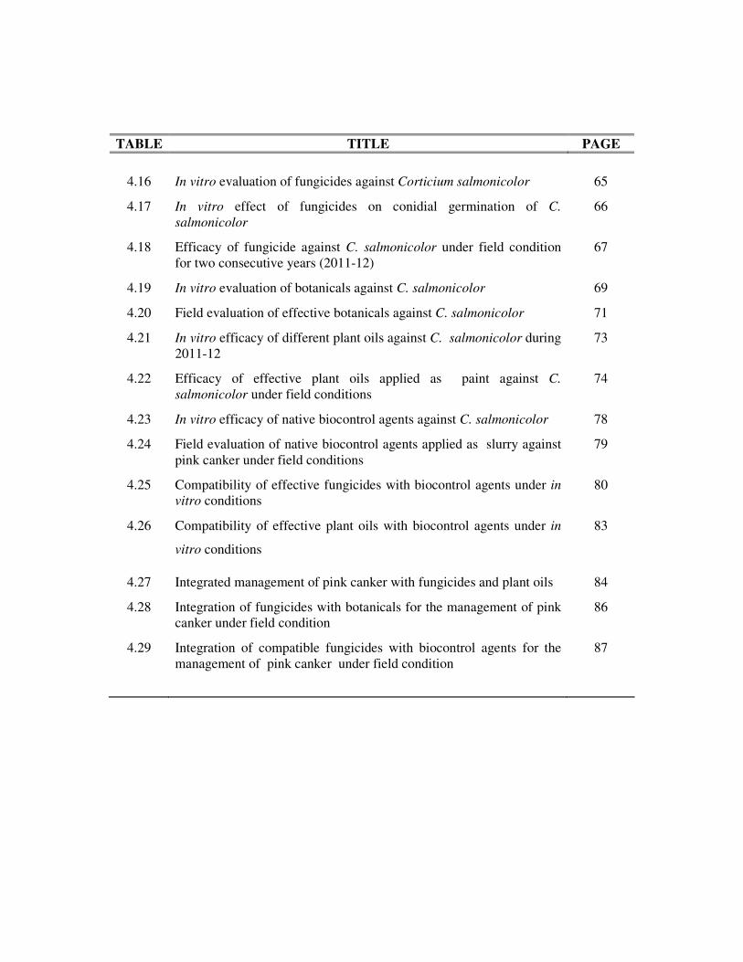
TABLE TITLE PAGE
4.16 In vitro evaluation of fungicides against Corticium salmonicolor 65
4.17 In vitro effect of fungicides on conidial germination of C.
salmonicolor
66
4.18 Efficacy of fungicide against C. salmonicolor under field condition
for two consecutive years (2011-12)
67
4.19 In vitro evaluation of botanicals against C. salmonicolor 69
4.20 Field evaluation of effective botanicals against C. salmonicolor 71
4.21 In vitro efficacy of different plant oils against C. salmonicolor during
2011-12
73
4.22 Efficacy of effective plant oils applied as paint against C.
salmonicolor under field conditions
74
4.23 In vitro efficacy of native biocontrol agents against C. salmonicolor 78
4.24 Field evaluation of native biocontrol agents applied as slurry against
pink canker under field conditions
79
4.25 Compatibility of effective fungicides with biocontrol agents under in
vitro conditions
80
4.26 Compatibility of effective plant oils with biocontrol agents under in
vitro conditions
83
4.27 Integrated management of pink canker with fungicides and plant oils 84
4.28 Integration of fungicides with botanicals for the management of pink
canker under field condition
86
4.29 Integration of compatible fungicides with biocontrol agents for the
management of pink canker under field condition
87

LIST OF PLATES
PLATES TITLE BETWEEN
PAGE(S)
1. Different stages of C. salmonicolor under field conditions 46-47
2. Cultural characters of different isolates of C. salmonicolor 48-49
3. Pathogenicity of C. salmonicolor isolates on Royal Delicious
and other cultivars
48-49
4. Germ tube length of Kotgarh and Mandi isolates after 24 and
48 hrs
50-51
5.a. Induction of Necator stage under in vitro conditions 52-53
5.b. Per cent disease severity and lesion size on leaves and fruits
of Royal Delicious
52-53
6.a. Mycelial growth of C. salmonicolor after 7 days of
inoculation on apple cultivars
60-61
6.b. Disease severity (%) and pustule formation on apple cvs.
after one month of inoculation
60-61
7.a. Varietal susceptibility of apple cultivars against Haripurdhar
isolates
62-63
7.b. Varietal susceptibility of apple cultivars against Kotgarh
isolates
62-63
8. In vitro evaluation of chemicals against C. salmonicolor by
poisoned food method
64-65
9. Effect of fungicides on germ tube deformation of C.
salmonicolor
64-65
10. Efficacy of effective fungicides against test pathogen under
field conditions for 2011-12
66-67
11a. In vitro evaluation of botanicals at different concentrations
by poisoned food method.
68-69
11b. Efficacy of cow urine based formulation against C.
salmonicolor
68-69
12.a. Field efficacy of effective botanicals against C. salmonicolor 70-71
12.b Field efficacy of effective plant oils against C. salmonicolor 70-71
12.c. Field efficacy of effective biocontrol agents against C.
salmonicolor
70-71
13. Efficacy of plant oils against C. salmonicolor under in vitro
conditions
72-73
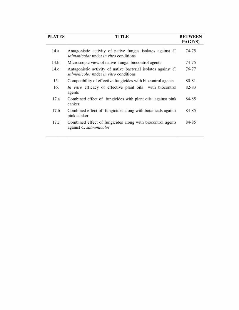
PLATES TITLE BETWEEN
PAGE(S)
14.a. Antagonistic activity of native fungus isolates against C.
salmonicolor under in vitro conditions
74-75
14.b. Microscopic view of native fungal biocontrol agents 74-75
14.c. Antagonistic activity of native bacterial isolates against C.
salmonicolor under in vitro conditions
76-77
15. Compatibility of effective fungicides with biocontrol agents 80-81
16. In vitro efficacy of effective plant oils with biocontrol
agents
82-83
17.a Combined effect of fungicides with plant oils against pink
canker
84-85
17.b Combined effect of fungicides along with botanicals against
pink canker
84-85
17.c Combined effect of fungicides along with biocontrol agents
against C. salmonicolor
84-85

LIST OF FIGURES
FIGURES TITLE BETWEEN
PAGE(S)
4.1 Prevalence of pink canker at different apple growing areas
of H.P. during 2011-12.
47
4.2.a Effect of different meteorological factors on disease
development during 2011 54-55
4.2.b Effect of different meteorological factors on disease
development during 2012
54-55
4.8.6.1.a Phylogenetic Tree of Bacillus subtilis made using
Neighbour Joining method
76
4.8.6.1.b Phylogenetic Tree of Pseudomonas fluorescens made
using Neighbour Joining method
77
4.11.a. Phylogenetic Tree of Corticium salmonicolor made using
Neighbour Joining method
89

Chapter-1
INTRODUCTION
Apple (Malus x domestica Borkh.), one of the most important fruit crop
belongs to the subfamily Pomoideae, family Rosaceae and is generally grown in the
temperate regions of the world. Apple, the premier table fruit of the world, has been
under cultivation since time immemorial. It is a typical temperate fruit, more than 80
per cent of the world’s supply being produced in Europe. Apple has long been the
staple fresh fruit in the temperate parts of the world. Eating apple is believed to
reduce the incidence of dental caries, helps to control obesity and supply extra energy
for heavy exercise. Apple is believed to be the most widely grown fruit tree produced
in all the continents of the world.
Apple was introduced in America after it has been cultivated for more than
2000 years in Europe. In India, apple is cultivated in North Western Himalayan
region which comprising states of Jammu and Kashmir, Himachal Pradesh and
Uttrakhand. However, in recent years its cultivation has been extended to some extent
to North Eastern states, where mild temperate climate conditions prevail (Chadha and
Awasthi, 2005). Himachal Pradesh is well known as an “apple state” of the country
because its cultivation has revolutionized the socio-economic condition of farmers
and plays a pivotal role in the economy of the growers. In the state, area under its
cultivation has reached up to 1, 03640 hectares with an annual production of 4, 12,
360 metric tonnes with productivity of 3.97 metric tonnes per hectare (Anonymous,
2013).
Apple was first introduced in Himachal Pradesh by an American missionary
Satya Nand Stokes who planted the delicious group under the ideal climatic
conditions in Kotgarh as early as 1916. Thereafter it has extended to different apple
zones in many districts of the State. Apple cultivation is a flourishing industry in the
temperate zones of Himachal Pradesh ranging from 1, 850 to 2,770 m.a.m.s.l.
However, its persuit for higher returns, the growers have extended its cultivation to
marginal and sub-marginal areas in lower elevations. Apple orchards are
subsequently threatened by the attack of numerous pathological problems that not
2
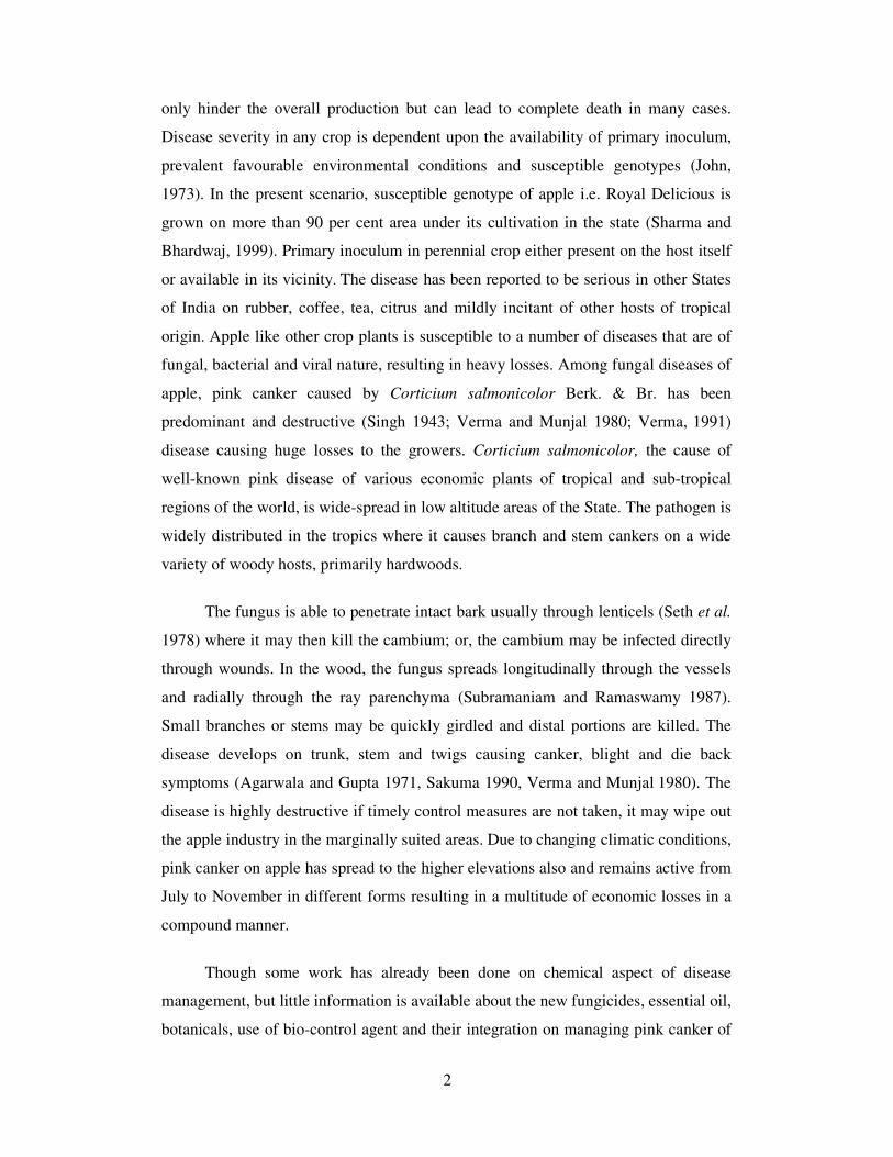
only hinder the overall production but can lead to complete death in many cases.
Disease severity in any crop is dependent upon the availability of primary inoculum,
prevalent favourable environmental conditions and susceptible genotypes (John,
1973). In the present scenario, susceptible genotype of apple i.e. Royal Delicious is
grown on more than 90 per cent area under its cultivation in the state (Sharma and
Bhardwaj, 1999). Primary inoculum in perennial crop either present on the host itself
or available in its vicinity. The disease has been reported to be serious in other States
of India on rubber, coffee, tea, citrus and mildly incitant of other hosts of tropical
origin. Apple like other crop plants is susceptible to a number of diseases that are of
fungal, bacterial and viral nature, resulting in heavy losses. Among fungal diseases of
apple, pink canker caused by Corticium salmonicolor Berk. & Br. has been
predominant and destructive (Singh 1943; Verma and Munjal 1980; Verma, 1991)
disease causing huge losses to the growers. Corticium salmonicolor, the cause of
well-known pink disease of various economic plants of tropical and sub-tropical
regions of the world, is wide-spread in low altitude areas of the State. The pathogen is
widely distributed in the tropics where it causes branch and stem cankers on a wide
variety of woody hosts, primarily hardwoods.
The fungus is able to penetrate intact bark usually through lenticels (Seth et al.
1978) where it may then kill the cambium; or, the cambium may be infected directly
through wounds. In the wood, the fungus spreads longitudinally through the vessels
and radially through the ray parenchyma (Subramaniam and Ramaswamy 1987).
Small branches or stems may be quickly girdled and distal portions are killed. The
disease develops on trunk, stem and twigs causing canker, blight and die back
symptoms (Agarwala and Gupta 1971, Sakuma 1990, Verma and Munjal 1980). The
disease is highly destructive if timely control measures are not taken, it may wipe out
the apple industry in the marginally suited areas. Due to changing climatic conditions,
pink canker on apple has spread to the higher elevations also and remains active from
July to November in different forms resulting in a multitude of economic losses in a
compound manner.
Though some work has already been done on chemical aspect of disease
management, but little information is available about the new fungicides, essential oil,
botanicals, use of bio-control agent and their integration on managing pink canker of
3
apple under Himachal Pradesh conditions. Therefore, keeping in view the importance
of the crop and damage caused by the disease, the present investigations have been
proposed with the following objectives:
i) To record the prevalence of pink canker in different apple growing areas of
Himachal Pradesh.
ii) To study the epidemiological parameters on the development and spread of
the disease.
iii) Screening of available apple cultivars against canker pathogen.
iv) Management of disease through chemicals, botanicals and biocontrol agents.

Chapter-2
REVIEW OF LITERATURE
Apple is the highly remunerative deciduous fruit generally grown in the
temperate regions of the world. The large scale expansion of area under apple since its
early cultivation and wild travel of plant materials exposed it to various pathological
problems. Among these, canker diseases occurring worldwide wherever apple is
grown (Sharma and Bhardwaj, 1999) have become most important and major limiting
factors for its successful cultivation and cost-effective production. Canker diseases are
virulent, which invade the bark and cambium of twigs, branches or main trunks,
forming lesions that ultimately girdle these structures and cause death of all distal
parts, thus producing blight, die-back or cankerous symptoms on twigs, or branches or
lead to death of the entire tree (Thakur, 1970).
OCCURRENCE AND DISTRIBUTION
According to Petch (1911), pink disease was firstly observed in Ceylon by
Thwaites as early as 1873; the causal organism of which later in 1873 was identified
by Berkeley and Broome as Corticium salmonicolor and that was known by different
specific names on different host plants in the tropics and subtropics. The Necator
decretus originally identified by Massee (1898) on coffee was later on described as
the conidial stage of C. salmonicolor by Rant (1912).The pink canker, earlier known
as limb blight (Tims, 1963) having wide host range of 141 wild and cultivated plant
species (Rant, 1912) mostly belonging to gymnosperm and dicotyledonous groups
(Sharples, 1936).
Sakuma (1990) reported C. salmonicolor to cause limb blight in southern
states of America and Japan. Corticium salmonicolor attacks various woody plants
throughout the tropical and subtropical regions. In Asia, it has been recorded in
Andaman Is., Brunei, Burma, Cambodia, China, India, Indonesia, Japan, Malaysia
including Saba and Sarawak, Sri Lanka, Taiwan, Thailand and Vietnam (Bilgrami et
al., 1979; Chandrasrikul, 1962; Liu, 1977; Mordue & Gibson, 1976; Peregrine &
Ahmad, 1982; Singh, 1980; Tai, 1979; Triharso et al., 1975; Turner, 1971; Williams
5
& Liu, 1976). Verma and Munjal (1980) reported 80-90 per cent losses due to this
disease in apple under Himachal Pradesh conditions. Mclzer and Berton (1988) made
an investigation of wood attacking fungi in apple in Santa Catarina and firstly
recorded Corticium salmonicolor in this region. Pink disease being one of the most
important canker diseases in the tropics. The disease spreads throughout the tropical
and subtropical regions and causes severe damage on various useful trees, especially
rubber, coffee, cacao, citrus and eucalypt (Brooks & Sharples, 1914; Mordue &
Gibson, 1976). In the Philippines, it has been reported on Citrus spp., Gliricidia
sepium and Albizia falcataria (Eusebio, et al., 1979; Kobayashi, 1978; Teodoro,
1937). In Mindanao, many plantations of Albizia falcataria are being destroyed by the
outbreak of C. salmonicolor. Pink disease has been recorded on 25 year old
Carrington apple tree at Glenorie in 1934, and two Allsop apple trees at Laughtondale
in 1936 (Birmingham, 1936).
In India, disease was reported by Singh (1943) from Chaubattia of Uttrakhand
on apple, pear and apricot. The pink canker, earlier known as limb blight or twig
blight (Tims, 1963).The literature pertaining to canker diseases in India has been
reviewed by Thakur (1970) and Shandilya (1971), on pink canker by Verma (1978)
and smoky blight by Singh (1985). Their studies were mainly focused on
pathological, epidemiological, physiological, biochemical and chemical control
aspects of the canker diseases.
In Himachal Pradesh, pink canker of apple was first observed by Gupta and
Agarwala (1970) from Kullu valley, thriving in orchards located at lower elevations
(900-1100 m.a.m.s.l). The disease causes economical losses in apple through girdling
of main limb and trunk which ultimately results in death of the tree. Corticium
salmonicolor, the incitant of pink canker disease of apple is one of the most
destructive pathogen causing up to 80-90 per cent disease in Kullu and Rajgarh areas
(Gupta and Agarwala, 1973; Verma and Munjal 1980). In Himachal Pradesh, the
studies on canker disease of apple was initiated by Gupta and Agarwala (1973) when
they described five canker diseases on apple from Kullu and Rajgarh apple belts.
Shandilya et al. (1973) reported thirteen cankers from Kullu valley and five from
Rajgarh apple growing areas including Corticium salmonicolor (Berk. and Br.) with
its necator stage as Necator decretus (Mass.). Thakur (1970) reported moderate to
6
severe intensity of different cankers on apple trees aged between 7-12 years at
different altitude ranges from 1000 to 2000 m. a.m.s.l. Most of the cankers causing
fungi are more severe at bearing age of trees at an elevation ranging between 900-
1200 m a.m.s.l (Shandilya and Kaul, 1973). Kim et al. (1970) recorded about 30 per
cent incidence of cankers on apple.
PATHOGENICITY TEST
Pathogenic nature of canker fungi isolated from apple trees was proved by
various workers (Thakur, 1970; Shandilya, 1971; Verma, 1978 and Singh, 1985) both
in laboratory as a excised twig method as well as under pot culture and field
conditions on injured and uninjured twigs of young and grown up apple trees by
inoculating culture bits from two weeks' old pathogen culture and covering with moist
cotton pads to provide suitable conditions for growth of the pathogen.
SYMTOMATOLOGY
Zimmermann (1901) described the pustuler form of C. salmonicolor as
superficially developed, white, round bodies consisting of thin walled cells, while
Rant (1910, 1911) reported that the part of branch affected by C. salmonicolor was
either covered with fine silvery white film named, ‘spinnengewebe’ or was studied
with white or pink coloured, raised pustules of the size of a pin-head or was covered
with pinkish coloured pock marks caused by the flaking off of scales of the bark
giving this part of branch general pinkish appearance. Brooks and Sharples (1915)
observed that pink disease frequently assumes the form of white or pale pink pustules
arranged more or less in lines parallel with the branches, conidial pustules (Necator
decretus) being orange red. Butler (1918) observed pink disease affecting the twigs or
smaller branches of rubber trees producing first symptom of discolouration and
withering of leaves often without being shed.
Lee and Yates (1919) observed that the sterile dirty white to pink coloured
pustules which pushed through the hardened bark of citrus trees and Schwarf (1925)
studied that very young branches of teak were rapidly killed. The fungus manifests in
four forms viz. cob-web, pustule form, necator form and pink incrustation. However,
Staner (1931) observed that the disease area of the bark extended up to a height of 60
cm from ground level and necrosis of bark phloem and cambium occurred. Pink
7
disease (Corticium salmonicolor) attacks the thick branches of apples, pears and
apricots. Altona (1926) described C. salmonicolor on teak forming large, deep
fissures sometimes saturated with water whereas Bondar (1925) observed C.
salmonicolor on cacoa causing bark disease which resulted into destruction of cortex
by the mycelium. Kalshoven (1928) also recorded similar observations on branches or
sapling mahagoni (Swietenia sp.) trees associated with cankerous growth which
extend to wood and girdled the branches. Bally (1929) described thread blight and
pink disease of coffee in Java and differentiated them as asexual and sexual stages
respectively. Simmonds (1931) observed the well known salmon pink incrustation on
citrus trees during the rainy season and Necator pustules erupt out through the bark
producing irregular spore like cells which served to spread the disease through rain
washed or blown on to healthy branches.
Subha Rao (1936) observed irregular hymenial surface of C. salmonicolor on
tea with sterile basidial fructification and noticed regular basidial hymenia bearing
fertile fructifications on Grevillea robusta, mango and eucalyptus. Dastur (1941)
observed the four subsequent stages of C. salmonicolor on citrus, viz., spider’s web,
sterile pustuler, Necator and basidial stage. The silvery white, later pink and finally
dirty drab mycelium of the spider’s web stage was mainly superficial and thin walled,
sparsely septate,7-15µ thick hypahe formed loose aggregates of cells over the
lenticels, the sterile pustules, either white or pink, developed both on the exterior of
the cortex and within the bark tissues or in the sub-epidermal layers and Necator
pustules produced unicellular, hyaline (pink in mass), angular or rounded spores of 6-
20 x 5-10 µ size while regular or scattered hymenia sparingly produced basidia
bearing basidiospores of 16.6-33.2 x 5-8 µm size. The commonest seat of infection is
the fork of the branches, but the disease sometimes starts from cut surfaces also
(Singh, 1943).
Bakshi et al. (1972) reported that the pink disease manifests during the
monsoon period attacking and girdled the main stem and branches and assumes the
epidemic level from third year onwards. He described Necator stage was observed
only on Populus and Casuarina montana abundantly producing hyaline, orange mass,
ovoid to variously shaped, slightly thick walled, 8-20 x 8-11 µm size conidia. The
basidia were observed to be subclavate to clavate, 8.5-12.2 µm broad with 2-4 stout,6-

8
9 µm long sterigmata bearing hyaline, smooth, thin walled elliptical to obovate, 8-11
x 8.0-9.8 µm basidiospores. The mycelium was thin walled, simple, septate, branched,
smooth 4-9 µm broad hyphae. The bark of stems and branches is affected with lesions
becoming brown and spreading rapidly. The upper part of the lesions becomes
girdled, causing the shoots to wilt and leaves to yellow. Pinkish mycelial mats and
fruit bodies develop on the bark below and above the lesions. In seriously affected
plantations, the crown of the infected trees is destroyed and the death of many trees
can cause significant reduction in production (Kobayashi, 1978 and Eusebio et al.,
1979).
The cobwebby stage appears as a thin layer of vegetative mycelium soon after
infection on the surface of the bark during periods of rainy weather. This is followed
quickly by the formation of pustules, which are pink to salmon coloured sterile
cellular structures. The necator and pink incrustation stages are formed later when the
infected branches and stems are in the process of dying. Sporulation and germination
of the spores are favoured by moist conditions. The necator stage (Necator decretus)
consists of orange fruiting bodies (sporodochia), which produce conidia. The pink
incrustation stage is the perfect state of the fungus and produces basidiospores (Seth
et al., 1978).
The disease appears on trunk and twigs causing canker and kills the affected
branches. The mycelium of the fungus lives in affected tissues but is chiefly seen as a
superficial covering on the smooth surface of the bark with cob-web like growth. This
growth of the fungus gradually turns into a more or less pinkish incrustation. Rarely it
produces basidia on the shaded side of the bark. The cankerous lesions are most
commonly present on the side of the limbs and branches exposed to bright sun-rays,
usually develop globular orange red pustules bursting through the bark. This is
Necator stage of the fungus. Later on the growth of the mycelium is checked and thus
typical cankers are formed. The cankered area extends several inches upward and
downwards in somewhat conical fashion and results in cracking of the bark,
(Shandilya and Agarwala, 1975; Kondal, 1986)
TAXONOMY
A comparative study by Dastur (1946) made on citrus infected by C.
salmonicolor from India and on Rubber, citrus and Acacia arabica in Herb. Crypt.

9
Indiae Orient., New Delhi revealed the complete identity of all the materials. The
substitution of the name Pellicularia salmonicolor (Berk. & Br.) n. comb. for
Corticium salmonicolor Berk.& Br. was therefore proposed. Venkatarayan (1950)
made further changes from Pellicularia to Botryobasidium giving a new combination
B. (Corticium) salmonicolor.
CULTURAL STUDIES
Butler (1918) mentioned that artificial cultures of all the four forms, the sterile
creeping mycelium, nodular form, the fertile Corticium and the Necator stages, quite
agreed in their characters giving a copious mycelia growth which usually turned pale-
rose when exposed to light due to synthesis of carotene pigment. In addition to light
as the chief factor influencing the production of pigment, author mentioned that the
composition of the culture medium and the position of the hypahe on its surface or
immersed in its might also affect the production of pigment. He described that no
form of fructification could be obtained in the culture, the fungus giving only a
mycelial growth with some times sterile nodulation of the hyphae which might
resemble the Necator stage before spore production.
Subha Rao (1936) observed good growth of C. salmonicolor with profuse
colouration in the culture on peptone, canesugar, potato dextrose and Lemco agar. He
also observed the capability of organism to destroy lignin due to development of a
brown halo in onion agar medium containing 2 per cent gall tannin. Resplandy and
Resplandy (1959) noticed that the isolate of Corticium salmonicolor from quince
synthesized alcohol extractable alkaloids when cultured on Lilly and Barnett’s semi-
synthetic medium supplemented with phenylalanine and tyrosine. Rao (1972) could
produce the basidiospores of C. salmonicolor in vitro conditions and stated an
optimum temperature range for their germination as 18-30°C. Rajlakshmi and Pillai
(1975) observed red mycelia aggregates of Corticium salmonicolor near the cotton
plugs in the culture tubes and considered them as structureless formations. Later they
could get cottony white mycelia aggregations at the bottom of culture tubes
containing potato dextrose agar 120 days after inoculation with the test pathogen,
incubated both at room temperature (28±2°C) and in the air conditioned room
(22±2°C). These pseudoparenchymatous masses with a waxy consistency and light
pink coloration were observed to contain hyaline, irregularly shaped conidial cells

10
measuring 30.8x15.8µm. Conidia germinate both singly as well as in mass in sterile
distilled water after 12 hours producing one or two germ tubes.
HOST RANGE
Corticium salmonicolor, the cause of pink disease has been reported in mild to
severe form by several workers occurring on a variety of hosts belonging to the most
diverse families. According to Butler (1918), the disease is known to occur in Burma,
Ceylon, South India, the Malay Peninsula, Java, Borneo, Formosa, Samoa, West
Indies, West Africa and reported to occur on tea, coffee, orange, jack fruit, camphor,
mango, Crotolaria, cacoa, rubber, cinchona, indigo, rhea, nutmeg, pepper, custard
apple, loquat, plum, apple, cinnamon, teak, thuja, eucalyptus, jasmine, rose and many
other wild and cultivated plants.
C. salmonicolor was reported by Brooks and Sharples (1915) from the eastern
and western tropics of Malaya on rubber, tea, coffee, cacoa, cinchona, citrus and other
plantations. Van Hall (1921) from Dutch East Industries on rubber, coffee and
cinchona; Tempany (1922) from Mauritius for the first time on Fiale
blame;Thompson (1924) from Ceylon on stem of tuba root (Dorris elliptica);
Sharples (1927) from Malaya on branches of gutta taban trees; Mc Donald (1924,
1928, 1929) from Kenya on coffee, loquat, litchi, guava. A severe account of pink
disease was reported by Lee (1922) from Philippines on highly cultivated citrus
groves. Bateson (1923) observed pink disease to be mild in 1922 on rubber in North
Borneo, while Mitchell (1923) reported it to be uneconomic in Ceylon. Tunstall
(1925) reported a serious damage caused by Corticium salmonicolor on tea in North
East India and also observed that the disease causes cankerous lesions but without
fructifications. Holland (1925) from Peradeniya and Burger (1924) from Florida State
on citrus plants. Mitra (1928) from Theria on orange crop and Dastur (1941) from
four districts of the Central Provinces on oranges. Bally (1929) described thread
blight and pink disease of coffee in Java and differentiated them as asexual and sexual
stages of Corticium salmonicolor.
Park (1932) from Ceylon noticed pink disease on mangoes, whereas Leach
(1946) from Mausica area on Cacoa. Van Der Goot (1934) reported BL-1 clone of
Hevea rubber being more severely attacked in West Borneo whereas Rao (1972) from

11
Malaya also emphasized the increased susceptibility to pink disease among the high
yielding clones of rubber. Park (1935) from Ceylon on Anona muricata; Subha Rao
(1936) from South India on Brevillea robusta, coffee, Eucalyptus robusta, E.
globulus, mango, jasmine sp., and tea. Singh (1943) from Kumaon on apple, pear and
apricot; Wiltshire (1956) from Malaya on calabash (Crescentia guites); Bugnicourt
(1956) from New Calendonia on custard apple. pigeon pea, citrus, quince, and apple;
Agnihothrudu (1963) from Assam in tea estate for the first time on Albissia falcata
and Tims (1963) from south East U.S.A. on fig, apple, and pear causing pink disease.
Hopkins (1937) from South Rhodesia and Brien and Dingley (1957) from
New Zealand have reported Corticium salmonicolor on apple causing pink disease
while Bitancourt (1937) stated that apples were attacked by C. salmonicolor and C.
koleroga in Brazil. In addition to this, Wallace (1944) considered pink disease to be
associated with apple die back in Tanganyika. Neveling (1956) identified Necator
decretus as the imperfect state of C. salmonicolor on apple branches at Pondoland. A
severe attack of C. salmonicolor was reported by Schwarf (1925) from several places
in Java and Sumatra on teak (Tectona grandis) of any age group.
It was reported to occur on a wide range of woody perennials in the tropics.
These include such important commodity crops as Hevea brasiliensis (rubber), Coffea
spp. (coffee), and Theobroma cacao (cocoa); fruits such as Citrus spp., Malus spp.,
and Litchi chinensis; woody ornamentals such as Cercis canadensis, Gardenia spp.,
and commercial forest plantation species such as Eucalyptus sp, and Acacia sp.
MODE OF PENETRATION
Vincens (1921) emphasized that Corticium salmonicolor could penetrate the
outer layers of wood. Gaumann (1922) stated that C. salmonicolor like other parasites
could invade the tissues at the places in the bark where Septobasidium bagorience
formerly occurred. Tunstall (1925) reported serious damage caused by a Corticium sp.
on tea in India similar in appearance to that responsible for pink disease which
resulted into the whitening and softening of the bark at first and later the formation of
pinkish fruiting patches on twigs, the tissues being penetrated only on the less
vigorous twigs. Schwarf (1925) stated that under favourable conditions for infection,
the fungus could enter the healthy branches through lenticels as well as through
12
wounds. Narasimhan (1933) observed that hyphae emerging from the compact masses
of pseudoparenchymatous calls of C. koleroga, the cause of black rot of coffee leaves
entered the leaf tissues through the stomata and penetrated the spongy parenchyma
often reaching the palisade cells while Bally (1929) expressed the uncertainty whether
the mycelium of C. salmonicolor penetrated the plants through the epidermal cells
and did not find the hyphae within the plant tissues.
EPIDEMIOLOGICAL STUDIES
Brooks and Sharples (1914) observed that shady sides of branches favoured
the production of basidia on the pink incrustation whereas bright light induced
formation of Necator pustules. Butler (1918) correlated the epidemic form of pink
disease on one to three year old Hevea rubber with a humid atmosphere, shade
possibly having more direct influence on the disease development and further noticed
that light was the chief factor for the synthesis of carotene pigment responsible for
the pale colour of the fungus. South (1921) reported the natural check on the pink
disease of rubber due to dry weather while Petch (1923) recorded the prevalence of C.
salmonicolor throughout the tropics on tea bushes. Van Hall (1924, 26) also registered
the occurrence of pink disease of rubber only at the foot of mountains and observe
more severe on 5-6 years old rubber plantations at places having rainfall above
3000mm.
Singh, (1943) reported that the pink canker pathogen of apple overwinters as
mycelium and fruiting structures in cankers. The pathogen spreads by means of
necator stage and disease progresses through the rainy season. Shandilya and
Agarwala (1975) reported that pink canker of apple is specific to low elevations
where the atmospheric temperature rises quite high for along span of time during the
year. Percentage incidence of the disease varies in plants of different age group,
however, the maximum incidence is found in older trees. They reported maximum
incidence of pink canker in orchards where the drainage is not good and trees were
overcrowded resulting increase in humidity. Warm and humid weather conditions are
the main contributing factors for the spread of the disease.
The initial infection by the fungus takes place through lenticels, (Verma and
Munjal, 1983) or through wounds/injuries, (Singh, 1943). Sharma, (1988) revealed
13
that pink disease (Corticium salmonicolor) of apple is generally more serious at low
elevations and in older trees (20-30 yr). Both conidia and basidiospores of Corticium
salmonicolor are spread by wind and are capable of causing infection through intact
bark tissues, but basidiospores are believed to be the most important (Almeida and
Luz, 1986). Serious damage from the disease usually occurs only in areas with rainfall
above 2000 mm (80 inches) per year (Seth et al., 1978). There is no evidence of
pathogenic specialization in the fungus.
Verma, (1988) observed the incitant of pink disease of apple survives and
spread by means of necator spores and by deep seated mycelium. Maximum
germination of conidia and basidiospores takes place at a temperature of 250 C and
rain water also helped in their germination. Verma, (1991) considered overcrowding,
bad drainage, high humidity and warm weather favourable for the development of
pink canker in apple. The canker mostly develops in the crotches of scaffold branches
where rain water stays for a longer duration, thereby raising humidity at micro level,
do increasing the intensity of pink canker of apple. Temperature of 28 0C coupled
with 90 per cent relative humidity during July and August favoured the development
of cobweb, basidial and sterile nodular stages of the disease in apple, while 240
C
coupled with 75% RH during Sept. and Oct. lead to the formation of imperfect stage
(Necator decretus). Infection was most severe on branches facing NE, SE and SW
directions. Apple orchards located >1900 m a.s.l. were completely free from the
disease. The pathogen attacked apple trees only when they had entered into the
reproductive phase, (Verma, 1991).
VARIETAL SUSCEPTIBILITY
Ramakrishnan and Pillay (1962) and RRIM (1992) observed that clones of
Hevea viz., PB 217, PB 311, and RRII 105 are highly susceptible to the pink disease.
Among the other cultivated clones, Tjir 1, LCB 1320, RRIM 501, RRIM 701, etc.,
were also found affected, while clones viz., PB 86, RRIM 513, Gl 1, PR 107, GT 1,
and PB 260 were found less susceptibility to the disease. Verma (1978) observed
Boycon, Early red Bird, early Shanbury, early Victory, Irish Peach, Tydeman Early
Worcester, Walthy Double Red varieties of apple were resistant to pink canker, while
Beauty of Bath, Gelia Beauty, Great Alexander, King David, Lady Early Golden, Mal
Rose, Rose Marine, varieties were found to be moderately resistant. Whereas

14
Baldwin, Neomy, Rus Pippin, Tropical Beauty, Sharping Early, Vered, Winter
Banana were reported to be susceptible. Royal Delicious, Golden Delicious, Red
Delicious, Red June, Rich-A-Red, Mc-Intosh were observed highly susceptible to
pink canker (Corticium salmonicolor). All the commercial cultivars of apple were
susceptible but maximum severity was noticed on Black Ben Davis (Kalidevi), Red
Delicious and Royal Delicious in Kullu valley of Himachal Pradesh, (Shandilya and
Agarwala, 1975).
DISEASE MANAGEMENT
A. Treatment of wounds
Birmingham (1936) suggested all wounds and cuts should be painted with
white lead or tar or application of Bordeaux mixture (6:4:40) or lime sulphur (1:14)
ever year where a regular spraying programme is not carried out. Singh, (1943) and
Kondal, (1986) mentioned different fungicidal paints like Chaubattia (lead oxide,
copper carbonate and raw linseed oil in 1:1:1.25), Bordeaux (copper sulphate, lime
and linseed oil in 1:2:3), Blitox-50 and Benomyl paints effective in healing different
kinds of canker wounds. In addition to it, Calixin paint was also found to be best
against pink canker of apple (Verma and Munjal, 1980). Singh, (1943) suggested,
painting the forks of apple branches before the onset of monsoon with red lead and
copper carbonate paste in raw linseed oil (4:4:5) and painting the pruned surfaces with
the same compounds in lanoline (4:4:5) paste. The excision of diseased material at
least 2 ft. below the last site of invasion and burning it after immersion in 50 per cent
copper sulphate reduces pink disease considerably.
Shandilya and Agarwala (1975) effectively control pink canker of apple with
ferbam (0.25%) in combination with Chaubattia paste (1:1:1:1/4) after scarification
and pruning of dead ends. They also found Benlate (0.05%) and Chaubattia paste
treatment to be equally effective. Gupta and Sharma (1979) established that addition
of kinetin and gibberellic acid in the paints enhances the bark growth, thus healing of
wound is faster. In addition to it, Calixin paint was also found to be best against pink
canker of apple (Verma and Munjal, 1980). Eusebio et al. (1979, 1980, 1981) studied
the pink disease of Albizia falcataria in Mindanao and they came to the conclusion
that selecting planting sites with good soil conditions is most important in avoiding
the disease development. Also, to recover from the infection, the application of

15
Bordeaux mixture on infected trees was an effective control. Verma and Munjal
(1980) obtained maximum (96.71%) wound recovery of apple tree with calixin paint
followed by Chaubattia and blitox paint giving 91.48 and 88.20 per cent recovery
respectively. Leng et al. (1982) obtained effective control of pink canker of apple by
applying a dilute mixture of lime and Tuzet [unspecified] to the trunks of the trees.
Jollands (1983) showed only phenylmercury acetate and cycloheximide to be
effective against Corticium salmonicolor Berk. & Br. below 10 ìg a.i./ml inhibiting
radial growth in culture.
Kondal (1986) recommended the cutting back of affected branches of apple
tree or excising lesions beyond the extent of infection and painting the cut surfaces
with Chaubattia paste (copper carbonate + lead oxide + raw linseed oil, 1:1:1:25) or
Bordeaux paint (copper sulphate + lime + linseed oil, 1:2:2) and fungicide sprays at
infective periods (leaf fall, bud swell, and after fruit set) effectively controlled pink
disease. The disease lesions should be scrapped with sharp knife and dressed with
Chaubattia paste or Bordeaux paint. Similarly, Sharma and Ram (2010) revealed
maximum healing (31%) of pink canker in white paint containing copper carbonate,
red lead and kinetin. Kondal (1986) showed the efficacy of carbendazim or copper
oxychloride paint prepared by using linseed oil in controlling pink canker of apple.
B. Fungicidal sprays
Verma and Munjal (1980) found that spraying of benomyl plus carbendazim
(0.3%) or ferbam (0.2%) plus Calixin (0.05%) completely controlled the growth of
pink canker fungus in apple. Further it was also reported that spray of fungicides
(ferbam 0.25% or Benlate 0.05% or Ziram 0.25%) in combination with applications
of Chaubattia paste or Bordeaux paint on cankered lesions of Corticium salmonicolor
provided 80 per cent control of pink canker disease in apple, (Shandilya and
Agarwala, 1975; Kondal and Agarwala, 1975). Ram et al. (1982) obtained best results
in the field conditions against natural infection by C. salmonicolor infecting Cocoa
through application of Peprosan (copper oxychloride 30% + maneb 10%+ zineb 10%)
at 1% a. i. sprayed at15-30 days intervals, followed by Bayleton (triadimefon) and
Plantvax (oxycarboxin).

16
Kondal (1986) suggested three sprays of orchocide, ziram, ferbam, zineb (200
g in 100 liters water), Bordeaux mixture (prepared by mixing 800 g copper sulphate
and 800 g hydrated lime in 100 liters water) and copper oxychloride (300 g in 100
liters water) at leaf fall, bud swell and after fruit-set stage in summer effectively
control pink canker in apple. Jansen (2005) observed good cultural practices like,
good circulation of wind and a shade management are a preventive method and
suggested plant debris on the trees should be removed and infested branches cut out
and burned. Whereas spay of copper oxide, cuprous oxide on coffee could provide
satisfactory results.
C. Botanicals:
Most of the fungicides, commonly used to minimize losses caused by fungal
diseases, enter the food chain (Majumdar, 1972), resulting in several harmful effects.
Therefore, there is evergrowing need for an intensive research for new, effective and
harmless fungicides. The fungicidal/ bactericidal activity of Ocimum sanctum against
soil borne pathogens can only be partially attributed to the volatile compounds it
contains (Grover and Rao, 1977). The plants belonging to different families, groups,
genera and species were screened in the past for their antimicrobial activity against
different plant pathogens by various workers (Upadhyay and Gupta, 1990; Dubey and
Dwivedi, 1991; Shivpuri et al., 1997; Sindhan et al., 1999; Parimelazhagan and
Francis, 1999; Pandey et al, 2002; Sood and Dohroo, 2003 and Sharma et al, 2003).
Antimicrobial activity of Brassica juncea var. cunefolia and Cymbopogon
citratus against many foliar and soil borne pathogens could be due to the presence of
alkaloids i.e. allyl isothiocyanate and citral respectively. The residual activity may be
attributed to the ajoenes that are thought to be one of several pharmacologically active
compounds in Allium sativum (Singh et al., 1990; Singh et al., 1992). Devi (1998)
evaluated the various plant extracts using different methods and different parts of
plant for their antifungal activity against the fungi i.e. Pythium ultimum, Rhizoctonia
solani and Sclerotium rolfsii causing diseases in the vegetable nurseries. She reported
the effectiveness of extracts taken form plants belonging to family Ranunculacae as
well as other plants such as Lantana camera, Eucalyptus longifolia. Artemisia
tridenta and Cedrus deodara against these fungal pathogens.

17
Mycelial growth of various species of Fusarium was inhibited by the
plant extracts of Convolvulus alsinoides and C. pluricutis (Furgal, 1984); Allium
cepa (El. Shami et al.,1986) Adhatoda vasica, Azadirachta indica, Cinnamomum
camphora and Ocimum sanctum (Prasad and Ojha, 1986); Agave americana, Cassia
nodosa (Reddy and Reddy, 1987); Azadirachta indica (Eswaramoorthy et al; 1989);
Allium cepa (Patel, 1989). Sharma (2005) obtained maximum (88.33%) mycelium
inhibition of Corticium salmonicolor infecting apple with the extract of Emblica
officinalis followed by Dodonia viscosa (71.67%) and Murraya koningi (64.72%)
whereas, minimum inhibition has been observed with Allium sativum (49.72%). Plant
derivatives possessing antimicrobial properties contained an array of chemicals which
induce various types of influences on the pathogen as well as on the host plants
(Doubrava et al., 1998 and Amaresh and Nargund, 2003).
D. Plant oils:
Singh et al. (1980) observed inhibitory effects of essential oils of C. martinii,
C. oliveri, and Trachysperumm ammi on Helminthosporium oryzae, as well as
inhibitory effects of the essential oils from rhizomes and leaves of Zingiber
chrysanthum on plant pathogens such as Alternaria sp. and Fusarium sp. Wilson et
al. (1997) tested 49 essential oils from various plants and found that the oil
and clove buds of Eugenia caryophyllata was effective to control Botrytis
cinerea. A commercial product based on the formulation of plant extracts and
essential oils from pepper, mustard, cassia, and clove extracts was effective in
reducing the population density of F. oxysporum f. sp. chrysanthemi, (Bowers and
Locke, 2000).
Gupta (2001), Sharma (2002) and Kumar (2004) evaluated different plant oils
viz, ovis, mentha, ginger, eucalyptus and nemicidine against Phytophthora fragariae,
causing red stele disease of strawberry; basil, castor, clove, eucalyptus, ginger,
mentha, mustard and neem against soil borne apple nursery pathogens (Dematophora
necatrix, Phytophthora cactorum, Sclerotium rolfsii) and mentha, eucalyptus, castor,
wild marigold and rosemary against Alternaria alternata causing apple leaf spots,
respectively. They reported the comparatively higher per cent inhibition of mycelial
growth of the test pathogens with ovis, ginger mentha, clove and basil oils. The
growth of pathogens decreased with increase in concentrations and increased with

18
increase in incubation period. Sharma (2002) also observed the mycelial distortion of
Dematophora necatrix (white root rot pathogen with eucalyptus and ginger oils.
Pandey et al. (2002) revealed the superiority of Matricaria oil and Mangiferin
at 1000 and 800 ppm respectively, to commercial fungicides with respect to mycelial
inhibition, restricting spore germination and formation of inhibition zone against
Helminthosporium sativum. Sharma et al. (2003) also reported higher antifungal
activity in aloe, dhatura, mentha, neem, neem extract, mentha oil and mustard oils
against various seed borne pathogens. Antifungal and antibacterial properties of
Brassica juncea var. cunefolia can probably be due to the presence of allyl
isothiocyanate and diallyl trisulfide (Jimmy et al., 2003).
Sharma (2005) revealed maximum mycelium inhibition of Corticium
salmonicolor with the oils of Cymbopogon citratus (73.47%) followed by Tagetes
minuta (71.46%) and Eucalyptus hybrida (67.08%), while the least inhibition has
been observed with Azadirachta indica (48.89%). Alkaloids like oxygenated
monoterpenes, a-citral or geranial and b-citral or neral, a major constituent of
Cymbopogon citratus, has strong toxic properties against several bacterial and fungal
pathogens (Tyagi and Malik, 2010).
E. Biological control:
Biological control of plant diseases is increasingly receiving attention, not
only to reduce the dependence on chemical crop protectant having hazardous effect in
ecosystem (Upadhyay and Rai, 1983) but to adapt it to the conceptual scheme of
integrated pest management as an acceptable ecosystem approach (Papavizas and
Lumsden, 1980). Plant surface as a natural habitat representing a heterogeneous
population of microbes comprises of both pathogens and non-pathogens interacting
constantly (Mukerji, 1983). Fungi constituted an important component of the
microbial population inhabiting aerial plant surfaces (Dickinson and Bottomley,
1980), constantly interacting with other saprobic and pathogenic organisms.
Biological control appeared to be a promising strategy for managing foliar and fruit
diseases in number of crops (Sutton and Peng, 1993).
Jollands (1983) found that Trichoderma spp. were antagonistic to C.
salmonicolor infecting rubber and oil palm. Jansen (2005) noticed parasitic fungi
19
(Gliocladium spp., Trichoderma spp., Verticillium spp.) showed antagonistic
properties against C. salmonicolor affecting coffee production. Jollands (1983) also
reported the in vitro antagonistic activity of various micro-organisms viz., Alternaria
sp., Bacillus subtilis, Gliocladium roseum, Trichoderma harzianum and Trichoderma
viride against Corticium salmonicolor (from rubber) and observed various responses
i.e. no antagonism where pathogen overgrown the antagonists, antagonism in which
antagonist overgrown pathogen and mutual antagonism that leads to formation of
inhibition zone at the point of their contact.
Biocontrol has been most successful against diseases of woody plants
(Campbell, 1989) because traditionally little breeding work has been done for
resistance in trees and that very few pesticides have developed specifically for tree
diseases. Moreover, woody stems were more suitable for inoculation with antagonist
because of little competition with other microorganisms. Amongst the natural
enemies, antagonistic fungi have received maximum attention and were commonly
used (Muthusamy, 1999). Of the various fungal microorganisms used as antagonists,
Trichoderma species were extensively exploited by pathologists due to wide
distribution (Domsch et al., 1980) and their higher efficacy, broad spectrum
activity and ease in isolation and cultivation (Mukhopadhyay and Mukherjee,
1996).
Gupta et al. (1999) also reported various antagonistic responses viz., hyphal
contact, lysis or inhibition zone formation with various species/isolates of
Trichoderma and Laetisaria arvelis against canker pathogen (Botryodiplodia
theobromae) in dual culture. In hyphal interactions, antagonists coiled around the
pathogen hyphae or penetrating its hyphal cells by forming hooks, haustoria and
appressorial structures. In lysis, they produced antifungal substances that resulted in
wrinkling, brusting and collapsing of the pathogen mycelium. Besides, Trichoderma
species, Streptoverticillium sp., Laetisaria arvalis, Coniothyrium minitans.
Aspergillus spp., Penicillium spp., and non pathogenic species of Fusarium could be
successfully exploited for biocontrol of plant disease (Muthusamy, 1999). Amongst
the bacterial antagonists, Bacillus subtilis, Pseudomonas fluorescens and
Agrobacterium radiobacter were widely and commonly practiced.
20
Liyanage, (1983) reported soil amendments with sulphur increased the acidity
and consequently the abundance of Trichoderma and Penicillium spp. that showed
antagonistic activity against pink disease of rubber caused by Corticium salmonicolor.
Similarly, Kochuthresiamma et al. (1991) found antagonistic effect of soil
actinomycetes in controlling pink disease of rubber. Treatment of infected twigs with
the actinomycetes broth culture also prevented growth of Corticium salmonicolor
under field conditions.
Aranguren et al. (1994) and Agarwal and Tripathi (1999) reported higher
antagonistic activity of Trichoderma spp. against canker pathogen as well as other
foliar plant pathogens. Anggroeni and Suharti (1994) observed Trichoderma highly
antagonistic to Corticium salmonicolor isolated from forest trees in Java and
elsewhere, Kochuthresiamma et al. (1996) reported the antagonistic activity of
actinomycetes isolated from rubber rhizosphere against Corticium salmonicolor both
in laboratory as well as in field where these on twigs prevented the growth of
pathogen effectively.
Whereas, Sharma (2005) reported maximum (92.50%) wound healing of apple
trees when treated with Trichoderma viride followed by 87.50 and 85.00 per cent with
T. longibrachiatum and T. harzianum respectively, whereas, 12.50 per cent was
noticed in control. Similarly, T. viride resulted in more than 10.00 mm of callus
formation in both year (2003 and 2004). Jansen (2005) observed parasitic fungi
(Gliocladium spp., Trichoderma spp., Verticillium spp.) that showed antagonistic
activity against pink canker of coffee, mango and eucalyptus.
MICROFLORA FROM PHYLLOPLANE
The phylloplane microflora represented a heterogeneous population of
microbes comprising both pathogens and non pathogens which were always engaged
in constant interaction. The various attempts have been made in the past to study the
occurrence and distribution of antagonists in their natural habitats and to study
population dynamics (Papavizas, 1981). Trichoderma spp. have found in various
habitats including decaying bark especially when damaged by other fungi (Danielson
and Davey, 1973). Despite many efforts, scarcity of information did exist on the

21
survival of the antagonists in their natural or new habitats probably due to lack of
precise technique and appropriate culture media for isolation and enumeration.
Andrews et al. (1980) studied the microbial communities on aerial plant
surfaces and positional variation in phylloplane microbial population with the apple
tree canopy. Payghami (1984) isolated 13 different fungi from the washings of
flowers, stems and fruits of apricot, of which Trichoderma viride exhibited
antagonism to foliar pathogens. Alternaria and Cladosporium were most encountered
genera in temperate regions and constituted an important component of both the
phylloplane and litter decomposition problems and were well adapted for survival,
growth and dispersal on aerial plant surfaces (O'Donnell and Dickinson, 1980).
Taniwaki et al. (1989) could isolate various microorganisms including Cladosporium
sp., Phoma sp., Phomopsis sp., Fusarium sp., Trichoderma spp., Alternaria sp.
Penicillium sp., yeasts and other moulds from apple plant surfaces. Grabowski (1994)
and Grabowski and Les (1996) observed the seasonal changes in the microflora
associated with the canker wounds on apple in chemically unprotected orchards and
observed inhibitory activity of majority of saprophytes against Nectria galligena
causing European canker. Kalenich and Padalko (1996) also noticed the seasonal
dynamism based on presence or absence of pesticide applications in the apple
phylloplane micro-organisms. Razdan and Puttoo (2002) isolated 19 fungi belonging
to 17 genera from the phylloplane of stone and nut fruits at three different
phenological stages viz., petal fall, pre and post-harvest stages.
COMPATIBILITY OF FUNGICIDES WITH ANTAGONISTS
Since pesticides have become indispensable in plant disease control, therefore,
these must be used in combination with other management practices. Indigenous or
introduced Trichoderma have higher tolerance for broad spectrum biocides compared
to many other microorganisms (Munnecke, 1972). Yokomizo et al. (1980) observed
inhibitory effects of PCNB, carboxin and ethazol on the growth of various isolates of
Trichoderma sp. Jollands (1983) reported the in vitro compatibility of biocontrol
agents with methfuroxan and fenpropemorph at all and lower concentrations,
respectively. Papavizas (1985) observed the dominance of Trichoderma in fungicide
treated sites due to its inherent resistance to most biocides and its ability to rapidly
colonize various substrates in the absence of significant competition from other

22
microbes. Tronsmo (1989) studied the effects of commonly used pesticides for
controlling insect-pests and diseases on the growth and sporulation of antagonistic
fungi i.e. Trichoderma spp. and revealed tolerance in these fungi to some pesticides
that he recommended for integrated control programme.
Mondal et al. (1995) reported compatible reactions of Trichoderma spp. viz.,
T. koningi, T. harzianum and T. lignorum with Vitavax, neem oil and other neem
formulations. Figueras et al. (1996) revealed the significant differences in the
sensitivity of various isolates of Trichoderma and observed mutant isolates more
tolerant fungicides and recommended their use for integrated management of plant
disease. Karunanithi and Usman (1999) reported that copper oxychloride supported
the survival and competitive saprophytic ability of T. viride while captan and
carbendazim caused marked reduction. Agarwal and Tripathi (1999) also observed the
compatible reactions of T. viride with various fungicides viz., Ronilan, Thiram,
Mancozeb, Captafol, Vitavax, Ridomil even at higher concentrations of 1500 ppm and
2500 ppm.
Sharma et al. (1999) while investigating the compatibility of pesticides and
fertilizers with biocontrol agents, observed compatible reactions with mancozeb,
carbofuran, sebuphos, neem seed cake, urea and other NPK fertilizers at all
concentrations tested from 100 to 2000 ppm except mancozeb at lower concentration.
Bhat and Srivastava (2003) reported complete inhibition of Trichoderma spp. with
Emisan, Bavistin, Benlate, Saaf, Tilt; little mycelial growth with Calixin, Contaf,
Topaz, RIL-Foo4 at lower concentration of 250 ppm and variable growth with Blitox,
Captaf, Indofil M-45, Roko at different concentrations of 250, 500 and 1000 ppm and
also observed increased per cent inhibition with increase in concentration. Gupta
(2004) also revealed T. harzianum exhibited compatible, moderately compatible and
incompatible reactions with Captan, Vitavax and Bavistin, respectively.
Anand et al. (2009) reported Pseudomonas fluorescens (Pf1) to be compatible
with azoxystrobin at different concentrations viz., 100, 150, 200, 250 and 300 ppm
revealed that it was compatible with all the concentrations of azoxystrobin tested and
the growth of the bacterium was unaffected even at the maximum concentration of
300 ppm and reduced downy mildew severity to greater extent. Bagwan
(2010) reported that thiram (0.2%), copper oxychloride (0.2%) and mancozeb (0.2%)
23
was compatible with Trichoderma harzianum and Trichoderma viride under in vitro
conditions. Basha et al. (2010) isolated bacteria from phylloplane of mango (PB28)
was more compatible with thiophanate-methyl (96.07%) at 50 ppm followed by
mancozeb, carbendazim, copper oxychloride and propioconazole. The compatibility
was less (16.09%) with hexaconazole at 25 ppm compared to other fungicides.
Whereas Trichoderma sp. showed varying degree of compatibility with propiconazole
and hexaconazole at 25 ppm respectively. Higher compatibility of PB28 was recorded
with propiconazole (88.05%) at 25 ppm followed by copper oxychloride,
carbendazim, thiophanate-methyl and mancozeb and the least compatible with
hexaconazole (01.07%) at 25 ppm.
Pallavi et al. (2012) proved that Pseudomonas and Bacillus sp., that showed
higher antagonistic activity against grey blight pathogen were tolerant with selected
fungicides (hexaconazole and carbendazim) under in vitro condition. The same result
was previously reported by Malathi et al. (2002). Bacterial strains showed tolerance in
hexaconazole and carbendazim. Tapwal et al. (2012) suggested that Trichoderma
viride was not compatible with Dithane, Bavistin and Ridomil in any level of selected
concentration. However, Trichoderma viride was found to be most sensitive to captan,
tebuconazole, vitavax, propiconazole and chlorothalonil, thus can be integrated to
manage soil borne disease. Deepthi (2013) isolated antagonists from groundnut
rhizosphere were found effective against Sclerotium rolfsii. The bacterial isolate GRE
9 was more compatible with mancozeb followed by carbendazim, Copper
oxychloride. Similar observations were made by Vidyasekaran and Muthamilan
(1995) and reported that carbendazim was not inhibitory to P. fluorescens.
INTEGRATED MANAGEMENT
Jollands (1983) suggested the integrated control of pink disease of rubber
using fungicides and Trichoderma spp., where fungicides provide initial protection by
preventing the growth of pathogens as well as other microorganisms as saprophytes
but competitor of antagonists on the plant surface and antagonists later established in
the wood. Integration of effective antagonists with fungicides is important for the
successful management of diseases (Mondal et al. 1995). Carter and Price (1975) used
Fusarium lateritium Nees ex Fr. in combination with benomyl against Eutypa
armeniacae infection on apricot.

24
Papavizas (1985) demonstrated the use of Trichoderma spp., as a potential
biocontrol agents in the integrated biological control of plant pathogens. They pointed
out that Trichoderma dominated in the habitates treated with sublethal doses of
pesticides, proliferated easily to produce antibiotics, competed for nutrients and aced
as mycoparasite. He also demonstrated the practical significance of Trichoderma and
its role in integrated disease management. Upadhyay and Rai (1983) and Tronsmo
(1991) revealed that fungicide tolerant species of Trichoderma were more effective in
integrated disease management programme. In recent times, much emphasis has been
put over integrated control of plant diseases using different useful components.
Karpagavalli (1997) found copper oxychloride least inhibitory to the radial growth of
T. harzianum and T. viride. However, benzimidazoles provided deleterious
effects on Trichoderma and Penicillium growth (Papavizas et al., 1982;
Vozenilkova, 1996 and Agarwal and Tripathi, 1999) but in contrast to these
fungicides, captan, chlorothalonil, chloroneb, PCNB and some other new fungicides
have shown positive correlation (Abd El-Moity et al., 1982) and can be used in
integrated programme for effective and eco-friendly control of the diseases. Agarwal
and Tripathi (1999) and Ramarethinam et al. (2001) also recommended the
integration of fungicides with compatible biocontrol agents for more effective control
of foliar plant diseases. Tapwal et al. (2012) reported compatibility of aqueous
extracts of Parthenium, Adiantum, Urtica, Polystichum and Cannabis sp. with T.
viride and found that leaf extract of Parthenium, Urtica and Adiantum to be effective
against A. solani, A. zinnia, R. solani, F. oxysporum and C. lunata.

Chapter-3
MATERIALS AND METHODS
The present investigations on pink canker of apple caused by Corticium
salmonicolor were conducted both under laboratory as well as field conditions in the
Department of Plant Pathology, Dr. Y.S. Parmar University of Horticulture and
Forestry, Nauni, Solan during 2011 to 2012. The methodologies adopted during the
course of study are elaborated below with the following heads:
3.1. Prevalence of pink canker of apple
3.2. Isolation of the pathogen
3.3. Identification of the pathogen
3.4. Pathogenicity tests
3.5. Cultural studies
3.6. Epidemiology
3.7. Varietal susceptibility
3.8 Disease management
3.9. Compatibility studies
3.10 Integrated management
3.11 Molecular characterization
3.12. Statistical analysis
3.1. PREVALENCE OF PINK CANKER
General surveys were carried out in different apple growing areas viz., Kullu,
Shimla, Mandi and Sirmour districts, located at different altitudes ranging between
900 -2500 m above mean sea level, in order to record the prevalence i.e. incidence
and/or severity of pink canker in apple orchards consisting trees in different age
groups between 15 to 30 years or more in the month of July-September, 2011 and
2012. Canker samples were collected from different growing areas to isolate the
pathogen as well as other microorganisms (such as fungi, bacteria and actinomycetes
to be used as biocontrol agents against canker diseases) associated within and around
canker affected parts on apple trees.
26
3.1.1 Incidence and severity of the canker
In order to record the prevalence of the disease, the incidence and severity
were recorded in the apple growing areas in different localities surveyed. The canker
incidence was recorded as follows:
Number of diseased plants
Canker incidence (%) = -------------------------------------- x 100
Total number of plants
The data regarding the severity of pink canker was recorded with the slight
modification in scale devised by Verma (1991).
0 = No lesion formation
1 = 0.1-5 cm of lesion size
2 = 5.1- 10 cm of lesion size
3 = 10.1-30 cm of lesion size
4 = > 30 cm of lesion size
The per cent disease severity will be calculated as per Mc Kinney (1923).
Sum of all numerical ratings
PDI = --------------------------------------------------- ------- x 100
Number of twigs observed x Maximum rating
3.2. ISOLATION OF PATHOGEN
The fungus associated with pink canker (Corticium salmonicolor) was isolated
into pure culture from infected twigs and branches partially or completely girdled
with cankerous growth after being treated with Mercuric chloride (0.1%) for one
minute. The isolated canker pathogen was purified by hyphal tip method on potato
dextrose agar (PDA) slants and maintained in pure form. For maintaining purity and
virulence of the isolated pathogens, the sub-culturing was done after every 20 days.
The axenic culture was maintained at 4±l°C in refrigerator on PDA slants.
3.3. IDENTIFICATION OF PATHOGEN
Canker fungus was isolated from infected twigs, branches and stems into pure
culture and identified on the basis of morphological as well as cultural characters as
27
described by many workers (Thakur 1970, Shandilya, 1971 and Verma, 1978). The
measurement of the spores or conidia was done with ocular micrometer under
compound microscope. First calibration of the ocular was done with stage micrometer
and calibration factor was calculated by taking the average of 20 different readings.
The average of minimum and maximum spore size was worked out and was then
multiplied by the calibration factor to obtain the exact size of the spores.
3.4. PATHOGENICITY TEST
Pathogenicity of canker fungus was conducted under laboratory condition
following twig inoculation method.
Under laboratory conditions: Twigs measuring 6-8 cm long, 1-1.5 cm thick,
artificially wounded, surface sterilized were inoculated with test fungus and kept in
sterilized flask containing moist cotton and incubated at 25 ±1oC.Fifteen twigs for
each isolate were kept and recorded for incubation period (days) and lesion size (mm)
after 30 days of inoculation.
3.5. CULTURAL STUDIES
The canker fungus thus obtained from infected plant parts of apple tree was
identified and cultural studies regarding formation of imperfect stage i.e. Necator
decretus as well as production of pink colour under in vitro conditions was observed
as described by various workers (Verma, 1978 and Singh, 1985). Conidial
germination (%) and germ tube length (µm) after 24 and 48 hr was recorded using
slide germination technique adopted by American Phytopathological Society
(Anonymous, 1943).
3.6. EPIDEMIOLOGICAL STUDIES
a. Progress of disease in relation to meteorological factors
To study the role of meteorological factors (temperature, relative humidity,
cumulative rainfall and sunshine hours) on disease development were studied under
field conditions for two crop seasons (2011and 2012). Data regarding disease severity
was recorded by adopting the 0-4 scale disease rating scale devised by Verma (1991).
Disease severity was recorded at 7 days intervals from June to September during both

28
the seasons at hot spot of disease occurrence at Kotgarh in district Shimla and per cent
disease index (PDI) was calculated by following the formula (Sharma et al., 1984).
∑ of all disease ratings
Disease index (%) = ----------------------------------------------------------- x 100
Total no. of observations x Max. disease grade
Meteorological data on weekly temperature, average relative humidity,
cumulative rainfall and sunshine hours were obtained from meteorological
observatory laboratory installed at SN Stokes’ Harmony Hall Orchards at Thanedhar,
District Shimla for the two consecutive crop seasons. The data was subjected to
statistical analysis to find out simple, partial and multiple correlations by using
statistical analysis procedures (Gomez and Gomez, 1986).
3.7. Varietal susceptibility
3.7.1. Under field conditions. In all nine available cultivars of apple at Kotgarh
district Shimla, were screened against Corticium salmonicolor under natural
epiphytotic conditions for two consecutive seasons (2011-12). Data on disease
severity was calculated after every fortnight interval. On the basis of disease severity,
apparent infection rate as well area under disease progress curve was calculated and
the cultivars were categorized into different reaction classes i.e. resistant, moderately
resistant, moderately susceptible and susceptible.
Apparent infection rate (r) and area under disease progress curve (AUDPC)
were calculated by using following formula as mentioned below:
2.3 x2 x1
r = --------- {log--------- - log --------}
(t2-t1) 1- x2 1-x1
Where, r = Apparent rate of infection at log phase of epidemic development, and t1
and t2 for time intervals when disease severities are x1 and x2. For converting log to
natural logarithm, the values are to be multiplied by a factor of 2.3. Whereas (1-x) =
correction factor.
k
AUDPC = ∑ 1/2 (y1 +yi-1) x d
i=1

29
Where, y1 = disease incidence at i th day. K = number of successive evaluation and d
= interval between i and i-1 evaluation of disease.
3.7.2. Under laboratory conditions
A) Flask condition: All seventeen apple cultivars available in the department of
Fruit Science of Dr. Y.S. Parmar University of Horticulture and Forestry, Nauni,
Solan were screened against C. salmonicolor during 2011-12 crop season. Cultivars
were collected and twigs measuring 6-8 cm long, 1-1.5 cm thick, artificially wounded,
surface sterilized were inoculated with test fungus and kept in sterilized flask
containing moist cotton and incubated at 25 ±1oC to observe the incubation period
(days) and lesion size (cm). Moisture was maintained by spraying the twigs with
distilled water. Based on disease severity, apparent infection rate and area under
disease progress curve (AUDPC), all cultivars were screened and categorized in to
different reaction classes (0-5) by using the scale as mentioned below.
B) Petri plate condition: Available cultivars of apple were screened against C.
salmonicolor under lab conditions during 2011-12 by placing twigs of apple cultivars
in Petri plates measuring 6-8 cm long with 1-1.5 cm thickness, surface sterilized with
rectified spirit and then inoculated with test pathogen on injured bark and incubated at
25 ±1oC. Relative humidity was maintained by spraying distilled water in the Petri
plates. On the basis of severity, the cultivars were categorized into different classes’
i.e., highly resistant, resistant, moderately resistant, moderately susceptible,
Rating /
Grade
Area covered
with lesions
(%)
Category Symptoms
0 0 Highly resistant No infection
1 0.01 – 1.0 Resistant Very few lesions on stem and
branches.
2 1.1 - 10.0 Moderately
resistant
Few lesions on stem/ branches up to
10 % necrotic area covered.
3 10.1 – 25.0 Moderately
susceptible
Lesions covering up to 25 % of
stem/branches area covered
4 25.1 – 50.0 Susceptible Necrotic lesions covering up to 50 %
area
5 >50 Highly
susceptible
More than 50 % of twig and branches
covered under necrotic lesions.

30
susceptible and highly susceptible. On the basis of disease severity, apparent infection
rate and AUDPC was calculated.
Disease severity (%) was calculated by slight modification in the scale adopted by
Verma (1991):
0 = No lesion formation, 1 = 0.1-2.1 cm of lesion size, 2 = 2.2- 3.2 cm of lesion
size, 3 = 3.3-4.3 cm of lesion size, 4 = 4.4-5.5 cm of lesion size, 5 = > 6cm of
lesion size.
3.8 DISEASE MANAGEMENT
3.8.1 Evaluation of fungicides
3.8.1a. In vitro evaluation of chemicals on mycelial growth inhibition. Six
different systemic fungicides viz., carbendazim (Bavistin 50WP), difenoconazole
(Score 25EC), propiconazole (Tilt 25EC), hexaconazole (Contaf 5EC), flusilazol
(Governor 40 EC) and azoxystrobin (Amistar 250 SC) were evaluated at 50, 100, 150,
200 µl l-1
and six non-systemic including combi-products namely; carbendazim (12%)
+ mancozeb (63%) (Saaf 75WP), captan (70%) + hexaconazole (5%) (Taqat 75WP),
carbendazim (25%) + iprodione (25%) (Quintal 50WP), copper oxychloride (Blitox-
50WP), zineb (75% WP) + hexaconazole (5 EC) (Avtar15 %WP) and metiram (55%)
+ pyraclostrobin (5%) (Cabrio Top 60 WG) were evaluated at 250, 500, 750 and 1000
µl l-1
under in vitro conditions against C. salmonicolor by poisoned food technique
(Falck, 1907). Data on mycelial growth were recorded after 7 days of incubation at
25±1oC and per cent growth inhibition (PGI) for each treatment was calculated.
3.8.1b. In vitro evaluation of chemicals on conidial germination. All above listed
systemic and non systemic fungicides including combi-products were tested at 50,
100, 150, 200 µl l-1
and 250, 500,750 and 1000 µl l-1
, respectively under in vitro
conditions against C. salmonicolor by the slide germination technique adopted by
American Phytopathological Society (Anonymous, 1943). Required concentrations of
the above fungicides were applied to sterilized cavity slides with the help of a pipette.
Subsequently, conidia from freshly sporulating mycelium were gently dusted on the
cavity slides containing required concentrations of fungicides (Thind et al., 1996).
The slides were in turn placed on glass rod triangles in moist chambers and incubated

31
at 25±1oC. Slides sprayed with sterilized distilled water and subsequently dusted with
conidia served as check. Each treatment was replicated thrice. Observations on
conidial germination were recorded after 48 h of incubation and per cent inhibition of
conidial germination was calculated (Vincent 1947).
Per cent growth inhibition will be calculated by the formula as below
C - T
I = ------------ X 100
C
Where, I = Per cent inhibition, C = Linear growth in control (mm), T = Linear
growth in treatment (mm).
3.8.2. Efficacy of fungicides under field conditions
A field experiment was laid out in Randomized Block Design in the farmer’s
field at Kotgarh, district Shimla, H.P. for two consecutive years (2011-12) to evaluate
in vitro effective fungicide applied as paint against the target disease under field
condition. Plants of susceptible cultivar “Royal Delicious” having characteristic
symptoms were first recorded for initial lesion size in the month of Nov-Dec. and was
scarified with sterilized scalpel or knife to remove dead portion upto the cambium
region and were then applied with recommended fungicidal dose added in white paint.
Subsequently wound recovery was calculated by measuring the lesion size i.e. width
(cm) before treatment and after one year of treatment while callus formation was
calculated as per the scale i.e. - = No callusing, + = 0.1-5.0 mm callusing, ++ =
5.1-10.00 mm callusing and +++ = > 10.00 mm callusing devised by Verma and
Munjal (1980). Each treatment was replicated thrice. Separate treatment of white paint
alone was also kept for comparison for wound recovery and callus formation in
untreated check.
3.8.3 In vitro evaluation of botanicals
3.8.3. a. Preparation of plant extracts
The efficacy of twenty plant origin biopesticides (Table 3.1) viz. Allium
sativum (Cloves), Artemisia roxburghiana (Leaves), Mentha pipereta (Leaves),
Roylea elegans (Leaves), Eucalyptus globulus (Leaves), Vitex negundo (Leaves),
Cymbopogon citratus (Leaves), Melia azedarach (Seed), Adhatoda vasica (Leaves),
32
Emblica officinalis (Leaves), Prunus persica (Leaves), Juglans regia (Leaves),
Lantana camera (Leaves), Carya illieonsis (Shell), Brassica juncea var. cunefolia
(Leaves), Ocimum sanctum (Leaves) and a cow urine based combinations i.e. Cow
urine + Darek + Bana + Walnut, Cow urine + Darek + Eucalyptus + Bougainvellia,
Cow urine + Darek + Bana + Artimisia sp.+ Walnut + Kadu and Cow urine + Darek
+ Bana + Artimisia +Walnut were evaluated at 10, 20, 50 and 100 per cent
concentrations by poisoned food technique (Falck, 1907).
Fresh leaves/ plant materials (100 g) were washed in tap water and then
ground for 5 minutes in warring blender by adding small quantity of distilled water.
After grinding, required quantity of distilled water (1:1W/V) was added and then
homogenized to give 100 per cent plant extract. The extracts were then filtered
through double layered muslin cloth or Whatman No.1 filter paper and stored in an
airtight container at 4°C for further use after tyndallization. All above said bio-
pesticides (plant extract) were evaluated at four concentrations (10, 20, 50 and 100%)
for their inhibitory activity against Corticium salmonicolor. Whereas, all Cow urine
based plant extracts were used in 1:1 (v/v) ratio of 100 per cent extracts of each
component added, which was further diluted to different concentrations by adding
sterilized distilled water. Double strength concentrations of each plant extracts either
alone or in combination at different concentrations were then mixed in double
strength PDA (Peeled potato -250 gm, Dextrose-20 gm, Agar-agar-20gm, Distilled
water-1000ml) media and poured into sterilized Petri plates. After solidification, bit of
test pathogen (4 mm) was kept in the centre of Petri plate and incubated at 25 ±1oC
for a period of 7 days until the mycelial growth in the control fully covered the
medium in the Petri plate. Each treatment was replicated thrice.
3.8.3b. Efficacy of botanicals under field conditions
Efficacy of in vitro effective ten botanicals viz., Mentha pipereta, Roylea
elegans, Melia azedarach, Adhatoda vasica, Ocimum sanctum, Brassica juncea var.
cunefolia and cow urine based formulations like Cow urine + Melia azedarach +
Vitex negundo + Juglans regia (1:1:1:1), Cow urine + Melia azedarach + Vitex
negundo + Artimisia roxburghiana + Juglans regia + Roylea elegans (1:1:1:1:1:1),
Cow urine + Melia azedarach + Eucalyptus lobules + Bougainvellia spectabilis ,
Cow urine + Melia azedarach + Vitex negundo + Artimisia roxburghiana + Juglans
33
regia (1:1:1:1:1) were evaluated in farmers field at Kotgarh and Haripurdhar of
Shimla and Sirmour districts respectively, during 2011-12 crop season. All these
botanicals were added in white paint @ 0.10 per cent concentrations and applied on
scarified lesions up to the cambium region and recorded per cent wound recovery and
callus formation after one year. White paint alone was kept for comparison and
untreated check serve as control.
3.8.4. In vitro evaluation of plant oils
3.8.4a. Extraction and evaluation of plant oils
Thirteen different plant oils viz., Tagetes minuta (Leaves), Artemisia
roxburghiana (Leaves), Eugenia caryophyllata (Flower bud), Olea cuspidata (Fruit),
Cymbopogon citratus (Leaves), Ocimum sanctum (Leaves), Azadirachta indica
(Seed), Brassica juncea var. cunefolia (Seed), Eucalyptus globulus (Leaves), Prunus
armeniaca (Seed), Ricinus communis (Seed), Juniperus virginiana (Wood) and
Juglans regia (Kernels) were evaluated against C. salmonicolor by “Poisoned food
technique”. These plant oils were either procured from local market or extracted from
healthy leaves using commercial Clevenger-type apparatus installed in the
Department of Forest Product, University of Horticulture and Forestry, Nauni, Solan,
Himachal Pradesh. The oil samples obtained from hydro-distillation were freed from
moisture by adding anhydrous sodium sulfate and absolute oil samples were obtained.
All above said plant oils were evaluated at four concentrations (100, 250, 500
and 750 ppm) for their antimicrobial activity against Corticium salmonicolor. In order
to make dilution of plant oils, organic solvent like ethyl alcohol (1: 10 ml) was
pipetted and added in sterilized molten agar media along with the required
concentrations of plant oils which were then mixed thoroughly and poured into
sterilized Petri plates. After solidification a bit of 4 mm of test pathogen was kept on
the centre of Petri plates whereas, control was kept without oil. Each concentration of
plant oil was replicated thrice and incubated at 25 ±1oC for a period of 7 days until the
mycelial growth in the control fully covered the medium in the Petri plate. The data
on per cent growth inhibition was recorded after 7 days of incubation at 25+1o C in
each treatment and were subjected to statistical analysis in completely randomized
design (CRD) factorial using square root transformation as described by Gomez and
Gomez (1983). Critical differences (CD 0.05) to determine the effectiveness of
34
different treatment at 5 % level of significance were also calculated by multiplying
critical‘t’ value with standard error.
3.8.4b. Evaluation of plant oils under field condition
Nine in vitro effective plant oils viz., Eugenia caryophyllata, Cymbopogon
citrates, Olea europea, Ocimum sanctum, Azadirachta indica, Brassica juncea var.
cunefolia, Ricinus communis, Juniperus virginiana and Juglans regia along with
white paint were evaluated at 5 per cent concentration under field condition in
farmer’s orchard located at Kotgarh and Haripurdhar of Shimla and Sirmour districts
respectively during 2011-12 crop season. Separate treatment of white paint alone was
kept for comparison and subsequent wound recovery and callus formation were
recorded in each treatment after one year.
3.8.5. Isolation and Identification of antagonists
The microflora (fungi, bacteria and actinomycetes) that were isolated from the
infected as well as uninfected branches and twigs of apple were subjected to
identification keys devised from time to time based on the cultural and morphological
characters by various workers or taxonomists i.e. A manual of soil fungi (Gilman,
1957), A revision of genus Trichoderma (Rifai, 1969), The genus Fusarium (Booth,
1971) and Fungi of India (Sarbhoy et al., 1975). Bacterial and actinomycetes were
identified based on morphological and cultural characters (Schaad, 1980). Fungal
cultures were send to National Centre for Fungal Taxonomy (NCFT) New Delhi for
identification. However effective bacterial antagonists were identified by their
molecular characterization.
3.8.5.1 In vitro evaluation of biocontrol agents
Antagonistic activity of isolated microorganisms i.e. fungi, bacteria and
actinomycetes was evaluated in vitro against Corticium salmonicolor. Of the
seventeen antagonists including fungi viz., Tv1 (Mandi) and Tv2 (Kotgarh) isolates of
Trichoderma viride; TH1 (Kotgarh) and TH2 (Mandi) of Trichoderma harzianum,
Trichoderma koningi, Th1 (Kullu) and Th2 (Haripurdhar) isolates of Trichoderma
hamatum, Trichoderma viride + Aspergillus sp., Verticillium sp. P1 (Kotgarh) of
Penicillium sp., and Aspergillus versicolor and five bacteria i.e. BS1 (Kotgarh), BS2
(Kullu) and BS3 (Mandi) of Bacillus subtilis, Pseudomonas fluorescens and

35
Pseudomonas aeruginosa and an actinomycetes were isolated and tested under in
vitro conditions by dual culture technique and streak plate method (Huang and Hoes,
1976; Utkhede and Rahe, 1983).
Culture bit of 5 mm size of each of the antagonist and pathogen taken from the
margin of vigorously growing cultures were transferred aseptically to solidified PDA
in Petri plates (90 mm) on opposite sides facing each other. The distance between the
inoculation point of test pathogen and the antagonist was kept 5 cm apart for
formation of inhibition zone, if any. The petriplates containing culture bits of
pathogen alone served as control. Each treatment was replicated four times and
incubated at 25±1°C. The colony diameter of the pathogen was recorded after 7 days
of incubation period when plates were filled with fungal growth and expressed as per
cent inhibition after comparison with control which was calculated according to the
formula given by Vincent (1947).
In case of bacteria and actinomycetes, the antagonistic activity was studied by
streak plate method (Utkhede and Rahe, 1983). The petriplates containing sterilized
solidified NA (bacteria) and actinomycetes were streaked on both sides of the 5 mm
size culture bit of the pathogen placed in the centre of the plates with 48 hours old
colonies of bacteria as well as actinomycetes. In control, only pathogen was
inoculated and replications remained the same in each case as discussed above. Data
on zone of inhibition as well as the per cent inhibition of the radial growth of fungus,
if any was calculated with the same formula as described above. The microorganisms
with higher antagonistic activity against canker pathogens were selected and further
investigated for the management of pink canker under field conditions.
3.8.5.1a. Field evaluation of antagonists
In order to ascertain the efficacy of in vitro effective antagonists against
Corticium salmonicolor, the field trial was carried out with eight biocontrol agents
viz., Trichoderma harzianum, Trichoderma viride, Trichoderma koningi,
Trichoderma hamatum, Aspergillus versicolor, Pseudomonas fluorescens, Bacillus
subtilis (BS1), Actinomycetes sp. under natural epiphytotic conditions by slurry
method.

36
Slurry method: Slurry of biocontrol agents was prepared in malt extract. In this
potato dextrose broth culture of each antagonist was prepared by inoculating
aseptically 500 ml PD broth in 1 litre flask with 3 bits of 5 mm size of antagonist and
was incubated at 25±1°C for 15 days, this broth culture was thoroughly homogenized
on digital shaker for 30 minutes and were mixed in malt-extract in 1:2 ratio (v/w).
Bacterial isolates were mass cultured in nutrient broth (NB) and mixed with malt
extract in the same ratio.
The biocontrol trial against C. salmonicolor was carried out with variety
’Royal Delicious’ in apple orchard of farmer’s field at Kotgarh, district Shimla during
2011 and 2012. The antagonists were applied by slurry method at two stages namely
immediately after pruning in the month of late December and at bud swell stage in the
month of March. The antagonists were applied after scraping off the canker lesions
with sharp edge knife. In each case 20 canker lesions were treated with biocontrol
agents. The control was kept in each case without biocontrol application. The
observations in each treatment were recorded for callus formation and per cent wound
recovery after a year of trial layout.
3.9. COMPATIBILITY STUDIES
3.9.1. In vitro compatibility of effective fungicides with biocontrol agents
Compatibility of in vitro effective fungicides viz., Amistar (azoxystrobin),
Score (difenaconazole), Contaf (hexaconazole), Tilt (propiconazole), Punch
(flusilazole), Bavistin (carbendazim), Saaf (carbendazim + mancozeb), Quintal
(carbendazim+ iprodione), Taqat (contaf + captan), Cabrio Top (pyraclostrobin+
metiram) and Avtar (zineb +hexaconazole) were evaluated at their recommended dose
for their compatibility with effective biocontrol agents under in vitro conditions
following poisoned food technique. A double strength potato dextrose agar (PDA)
medium was prepared by doubling the amount of constituents except water. PDA was
sterilized at15 lb pressure psi for 20 minutes in an autoclave. An equal amount of the
test double strength fungicide was added separately in double strength medium in
different flasks, shaken well, aseptically poured in Petri plates separately and were
allowed to solidify. A small culture bit of 5 mm size of antagonists was cut with a
sterile cork borer and picked up with the help of a sterile inoculating needle and was
placed in the centre of each Petri plates under aseptic conditions in a laminar air flow
37
chamber. Petriplates without fungicide in the medium served as control. Whereas,
bacterial antagonists were evaluated for their compatibility with antagonists by
streaking them on nutrient agar amended with fungicides. A bacterial antagonist that
grows on the medium considered compatible. Each concentration of fungicides was
replicated three times and the Petri plates were incubated at 25+l°C for the period of 7
days until the mycelial growth in control fully covered the medium in Petri plates.
3.9.2. In vitro compatibility of effective plant oils with biocontrol agents
Compatibility of in vitro effective plant oils viz., Brassica juncea var.
cunefolia, Ocimum sanctum, Azadirachta indica, Cymbopogon citratus, Eugenia
caryophyllata and Olea europea were evaluated at 5 per cent concentration to know
their compatibility with effective biocontrol agents using poisoned food technique. A
double strength potato dextrose agar (PDA) medium was prepared by doubling the
amount of constituents except water. PDA was sterilized at15 lb pressure psi for 20
minutes in an autoclave. An equal amount of the test double strength plant oil was
added separately in double strength medium in different flasks, shaken well,
aseptically poured in Petri plate separately and were allowed to solidify. In order to
dissolve plant oil in medium, small amount of solvent i.e. ethyl alcohol (10 ml) was
added in the medium. Small culture bit of 5 mm size of antagonists was cut with a
sterile cork borer and picked up with the help of a sterile inoculating needle and were
placed in the centre of each Petri plates under aseptic conditions in a laminar air flow
chamber. Petri plates without plant oils in the medium served as control. Whereas,
bacterial antagonists were evaluated for their compatibility with antagonists by
streaking them on nutrient agar amended with plant oils. Each concentration of plant
oils was replicated thrice and the Petri plates were incubated at 25+l°C for the period
of 7 days until the mycelial growth in control fully covered the medium in petriplates.
3.10 INTEGRATED MANAGEMENT OF PINK CANKER
3.10.1. Integrated management of pink canker with fungicides and plant oils
A field experiment was laid out in Randomized Block Design in the farmer’s
field at Kotgarh, district Shimla, H.P. for two consecutive years (2011-12). Field
evaluation of most effective compatibility fungicides viz., Score (difenaconazole),
Contaf (hexaconazole), Punch (flusilazole), Saaf (carbendazim + mancozeb), Taqat
(contaf + captan), Avtar (zineb +hexaconazole) and plant oils viz., Cymbopogon
38
citratus, Brassica juncea var. cunefolia and Olea europea were evaluated at different
concentrations against C. salmonicolor. Plant oils were evaluated at 5 per cent
concentration while fungicides were used at their recommended dose in the field
conditions. Both fungicides and plant oils at their respective doses were mixed in
white paint and applied on scarified canker lesion. Separate treatment of white paint
alone was kept for comparison. However control was kept without treatment. Data on
per cent wound recovery and callus formation was recorded after one year of
application.
3.10.2. Integrated management of pink canker with fungicides and botanicals
Field evaluation of in vitro effective fungicides viz., Score (difenaconazole),
Contaf (hexaconazole), Punch (flusilazole), Saaf (carbendazim + mancozeb), Taqat
(contaf + captan), Avtar (zineb +hexaconazole) and plant extracts i.e. combination of
cow urine based formulations like, Cow urine + Melia azedarach + Vitex negundo +
Artimisia roxburghiana + Juglans regia + Roylea elegans (1:1:1:1:1:1), Cow urine +
Melia azedarach + Vitex negundo + Artimisia roxburghiana + Juglans regia
(1:1:1:1:1) and Adhatoda vasica were mixed in white paint and applied on scarified
canker lesion at farmers orchard for two crop years. Fungicides were evaluated at
their recommended field dose whereas, plant extract were evaluated at 10 per cent
concentration. Application of white paint alone was kept for comparison besides the
control without any treatment serve as check.
3.10.3. Combined efficacy of fungicides and biocontrol agents against pink
canker
Combined efficacy of most effective fungicides viz., Score (difenaconazole),
Contaf (hexaconazole), Punch (flusilazole), Saaf (carbendazim + mancozeb), Taqat
(contaf + captan), Avtar (zineb + hexaconazole) and antagonists viz., Trichoderma
harzianum, Trichoderma viride, Pseudomonas fluorescens, Bacillus subtilis (BS1) and
Actinomycetes sp. were evaluated at their recommended dose, while biocontrol agents
were mixed with compatible fungicides using slurry method (3.8.5.1.a.) in the field
laid out in Randomized Block Design in farmers orchard at Kotgarh as well
Haripurdhar of Sirmour district for two years. Data on per cent wound recovery and
callus formation were recorded after one year of treatment.

39
3.11 MOLECULAR CHARACTERIZATION
3.11.1. Genotypic characterization of fungal antagonists and bacterial was done by
A. 18S rRNA gene sequencing
B. 16S rRNA gene sequencing
3.11.1. A. Isolation of fungal genomic DNA
1. Fungal genomic DNA was isolated from the plate, by using the Fungal
Genomic DNA isolation kit (616112500011730).
2. Using consensus primers, the ~1200bp; 18S rRNA, ITS1, 5.8S rRNA, ITS2
and 28S rRNA gene fragment was amplified using high-fidelity PCR
Polymerase.
3. The PCR product was sequenced using the forward, reverse & internal
primers.
4. Sequence data was aligned and analyzed for finding the closest homologous
microbe
3.11.1 a. Amplification of fungal genomic DNA
Amplification of fungal genomic DNA was carried out using universal primer
group specific oligonucleotide primer sequences: according to Widmer et al. (2001).
Reagents
Genomic DNA : ~20ng
dNTP mix (2.5mM each) : 1.0ìl
Forward Primer : 100ng
Reverse Primer : 100ng
Taq Buffer A (10X) : 1X
Taq Polymerase enzyme : 3U
Glass distilled water : to make up the volume 50ìl
PCR conditions:
94°C 94°C 56°C 72°C 72°C
5 min 30sec 30 sec 1min 10 min
Total 35 cycles
40
The PCR products were loaded on 1.0% agarose gel along with StepUpTM
500bp DNA ladder and rest of the steps were similar as discussed below for bacterial
antagonists.
3.11.1b. Isolation of bacterial genomic DNA
Genomic DNA’s from the selected bacterial isolates was isolated according to
manufacturer’s (B.GeNei) specifications using DNA isolation kit. Following steps
involved in it:
1. 1 ml of 24h old culture of each bacterial isolates was grown in nutrient agar
and centrifuged at 6000 rpm for 10 minutes.
2. Each bacterial pellet was re-suspended in 180 µl of lysis buffer1.
3. 20µl of proteinase K was then added to each mixed thoroughly by vortexing
and incubated at 550C for 3 h.
4. 4 µl of RNAse (100mg/ml) was added to each, mixed by vortexing and
incubated at room temperature for 5 minutes.
5. 200 µl of lysis buffer 2 was added to each supernatant, mixed thoroughly by
vortexing and incubated at 700C for 20 minutes.
6. Then 200 µl of absolute ethanol (99.9%) was added to each and mixed
thoroughly by vortexing.
7. The GeNei PureTM
columns were kept in 2ml collection tubes and then the
sample-ethanol mixtures were added to each respective column.
8. They were centrifuged at 11,000 rpm for 5 minutes. Collection tubes with flow
through were then discarded.
9. One volume of wash buffer 1 was diluted with three volumes of absolute
ethanol just before use.
10. The GeNei PureTM
columns were then kept in fresh 2ml collection tubes and
500µl of diluted wash buffer 1 was added to each column. Then they were
spinned at 11,000 rpm for 1 minute. Collection tubes with wash sample were
discarded.
11. One volume of wash buffer 2 was diluted with three volumes of absolute
ethanol just before use.
12. The GeNei PureTM
columns were then kept in fresh 2ml collection tubes and
500µl of diluted wash buffer 2 was added to each column. Then they were

41
spinned at 11,000 rpm for 3 minutes. Wash fractions were discarded and
collection tubes were retained for the next step.
13. The empty GeNei PureTM
columns were spinned at 11,000 rpm for 2 minutes.
Collection tubes were then discarded.
14. The GeNei PureTM
columns were opened and placed in new 1.5 ml vials
(sterile), incubated for 2 minutes at 700C dry bath to ensure complete removal
of ethanol.
15. Required amount of elution buffer was taken in a 1.5 ml vial (sterile) and
prewarmed in a dry bath set at 700C for 5 minutes. 200 µl of elution buffer
was used for one preparation.
16. The GeNei PureTM
columns were placed in fresh 1.5 ml vials and 200 µl of
prewarmed elution buffer was added to each column, incubated at room
temperature for 5 minutes and centrifuged for 1-2 minutes to elute the DNA.
17. Eluted DNA samples in buffer were stored at -200C.
3.11.1a. Amplification of bacterial genomic DNA
Amplification of genomic DNA was carried out using universal primer group
specific oligonucleotide primer sequences: according to Widmer et al. (2001).
Reagents
1. Taq DNA polymerase (5U/µl) (GeNeiTM
)
2. Taq buffer A (10X) containing 15 mM MgCl2 (GeNeiTM
)
3. Deoxynucleotide triphosphate (dNTPs) mixture: 100 mM of each dNTP
(GeNeiTM
)
4. Genomic DNA (25 ng/µl)
5. Random primers (GeNeiTM
)
Procedure
The PCR reaction mixture of 25 µl contained:
1. 18.8 µl sterilised distilled water
2. 0.20 µl Taq DNA polymerase
3. 2.50 µl Taq buffer A
4. 1.50 µl dNTPs mixture
42
5. 0.50 µl forward specific primer (100 pM/µl)
6. 0.50 µl reverse specific primer (110 pM/µl), and
7. 1.0 µl genomic DNA
The PCR amplification was carried out in MJ Mini BIO-RAD personal
thermal cycler-100 (PTC-100) with a total of 35 cycles. Each cycle consisted of:
• Five minute denaturation at 940 C
• Two minutes annealing at 550
C and
• Two minute extension at 720
C
All the PCR samples were given 5 minutes pre-amplification at 950C and 10
minutes post-amplification at 720 C.
3.11.1b. Electrophoresis of amplified DNA
The electrophoresis of the amplified DNA product was carried out in 1.2%
agarose gel stained with ethidium bromide (0.5 mg/ml), under submerged conditions
using 1×TAE buffer (tris HCl 10mM pH 7.4 and EDTA 1mM pH 8.0) as tray buffer
(Felsenstein, 1993). To each PCR amplified sample, 3 µl of 6 X loading dye was
added. 100-10000 bp DNA ladder marker was used as standard and the gel was run at
110 V until the loading dye reached the gel front. The amplified DNA was viewed
under the UV trans-illuminator and the image was taken through BIO-RAD gel
documentation system using Quantity one software and saved in computer.
3.11.1c. Gel elution
The desired amplified bands of C. salmonicolor isolate were cut down with
the help of sterile cutter and their amplified bands were kept in sterile eppendorf tubes
at 40 C and were sent directly for 16S r RNA sequencing.
3.11.1d. DNA sequencing and sequence analysis
For DNA sequencing, purified and amplified DNA products or bands selected
isolate were sent to Bioserve Private limited, India under refrigerated conditions
using gel packs and thermocol box. DNA sequencing was performed at the scientific
solutions. For sequence determination of the Ps-PCR products, a generally applicable
sequencing strategy was developed.

43
3.11.1e. NCBI Blast
Similarity searches of the NCBI Genbank database were performed with
BLAST (Altschul et al., 1997). The sequences were compared with those from
GenBank and analyzed with BioEdit Gene Doc software.
For phylogenetic analysis, sequences were manually aligned to the alignment
retrieved from RDP by us the multiple alignment routine clustering was determined
by unweighed pair group with mathematical averages (UPGMA) analysis of pairwise
genetic distance values. Phylogenetic trees were deduced.
3.12. STATISTICAL ANALYSIS
The data obtained from laboratory as well as field experiments were subjected
to appropriate statistical analysis wherever necessary using standard procedure, as
described by Gomez and Gomez (1986).
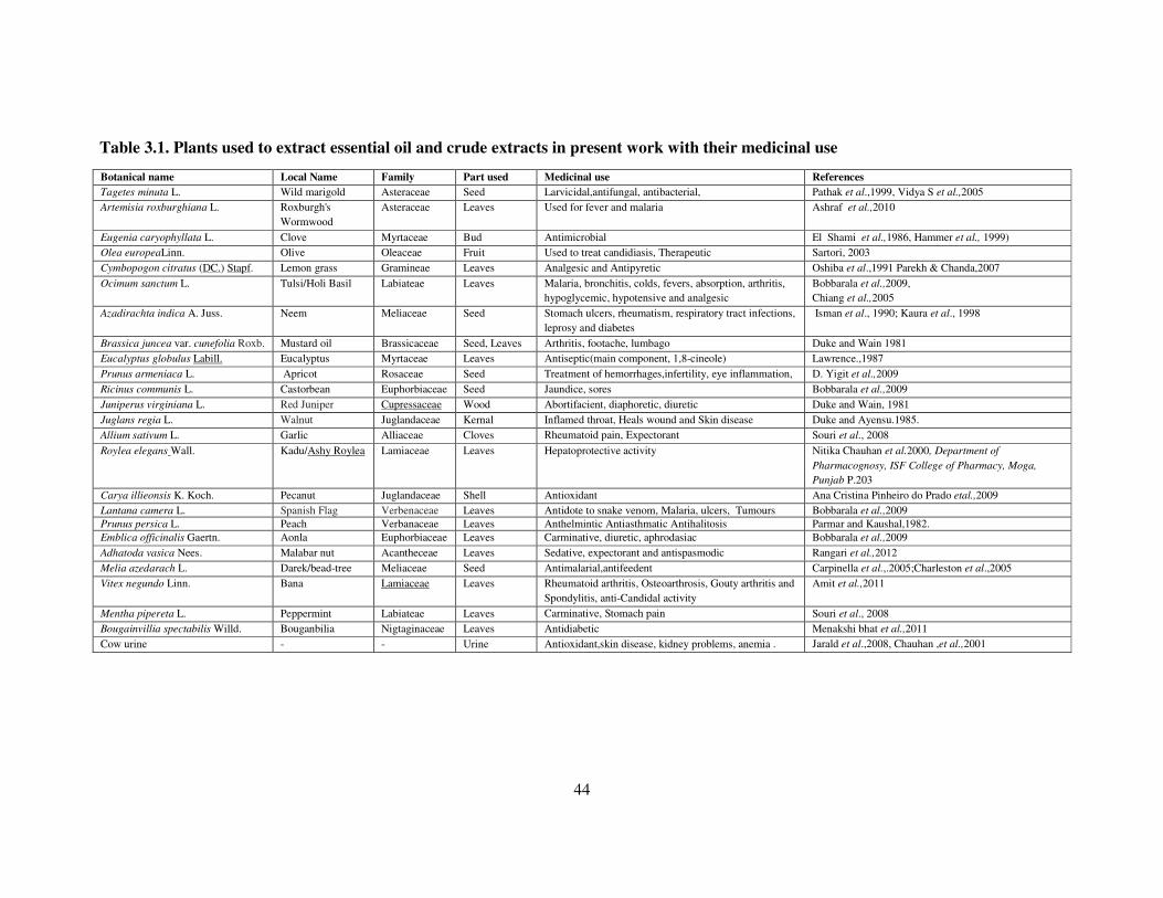
44
Table 3.1. Plants used to extract essential oil and crude extracts in present work with their medicinal use
Botanical name Local Name Family Part used Medicinal use References
Tagetes minuta L. Wild marigold Asteraceae Seed Larvicidal,antifungal, antibacterial, Pathak et al.,1999, Vidya S et al.,2005
Artemisia roxburghiana L. Roxburgh's
Wormwood
Asteraceae Leaves
Used for fever and malaria Ashraf et al.,2010
Eugenia caryophyllata L. Clove Myrtaceae Bud Antimicrobial El Shami et al.,1986, Hammer et al., 1999)
Olea europeaLinn. Olive Oleaceae Fruit Used to treat candidiasis, Therapeutic Sartori, 2003
Cymbopogon citratus (DC.) Stapf. Lemon grass Gramineae Leaves Analgesic and Antipyretic Oshiba et al.,1991 Parekh & Chanda,2007
Ocimum sanctum L. Tulsi/Holi Basil Labiateae Leaves Malaria, bronchitis, colds, fevers, absorption, arthritis,
hypoglycemic, hypotensive and analgesic
Bobbarala et al.,2009,
Chiang et al.,2005
Azadirachta indica A. Juss. Neem Meliaceae Seed Stomach ulcers, rheumatism, respiratory tract infections,
leprosy and diabetes
Isman et al., 1990; Kaura et al., 1998
Brassica juncea var. cunefolia Roxb. Mustard oil Brassicaceae Seed, Leaves Arthritis, footache, lumbago Duke and Wain 1981
Eucalyptus globulus Labill. Eucalyptus Myrtaceae Leaves Antiseptic(main component, 1,8-cineole) Lawrence.,1987
Prunus armeniaca L. Apricot Rosaceae Seed Treatment of hemorrhages,infertility, eye inflammation, D. Yigit et al.,2009
Ricinus communis L. Castorbean Euphorbiaceae Seed Jaundice, sores Bobbarala et al.,2009
Juniperus virginiana L. Red Juniper Cupressaceae Wood Abortifacient, diaphoretic, diuretic Duke and Wain, 1981
Juglans regia L. Walnut Juglandaceae Kernal Inflamed throat, Heals wound and Skin disease Duke and Ayensu.1985.
Allium sativum L. Garlic Alliaceae Cloves Rheumatoid pain, Expectorant Souri et al., 2008
Roylea elegans Wall. Kadu/Ashy Roylea Lamiaceae Leaves Hepatoprotective activity Nitika Chauhan et al.2000, Department of
Pharmacognosy, ISF College of Pharmacy, Moga,
Punjab P.203
Carya illieonsis K. Koch. Pecanut Juglandaceae Shell Antioxidant Ana Cristina Pinheiro do Prado etal.,2009
Lantana camera L. Spanish Flag Verbenaceae Leaves Antidote to snake venom, Malaria, ulcers, Tumours Bobbarala et al.,2009
Prunus persica L. Peach Verbanaceae Leaves Anthelmintic Antiasthmatic Antihalitosis Parmar and Kaushal,1982.
Emblica officinalis Gaertn. Aonla Euphorbiaceae Leaves Carminative, diuretic, aphrodasiac Bobbarala et al.,2009
Adhatoda vasica Nees. Malabar nut Acantheceae Leaves Sedative, expectorant and antispasmodic Rangari et al.,2012
Melia azedarach L. Darek/bead-tree Meliaceae Seed Antimalarial,antifeedent Carpinella et al.,.2005;Charleston et al.,2005
Vitex negundo Linn. Bana Lamiaceae Leaves Rheumatoid arthritis, Osteoarthrosis, Gouty arthritis and
Spondylitis, anti-Candidal activity
Amit et al.,2011
Mentha pipereta L. Peppermint Labiateae Leaves Carminative, Stomach pain Souri et al., 2008
Bougainvillia spectabilis Willd. Bouganbilia Nigtaginaceae Leaves Antidiabetic Menakshi bhat et al.,2011
Cow urine - - Urine Antioxidant,skin disease, kidney problems, anemia . Jarald et al.,2008, Chauhan ,et al.,2001
Chapter-4
EXPERIMENTAL RESULTS
The results obtained during the course of present investigation are being
presented here under:
4.1. PREVALENCE OF PINK CANKER
To assess the magnitude of pink canker in apple at different locations (Fig. 1)
i.e. Sirmour, Kullu, Mandi and Shimla district of Himachal Pradesh, located at
different altitudes ranging between 900 -2500 m above mean sea level (a.m.s.l),
were carried out during normal canker development period i.e. July-September during
2011-12. The data on prevalence of disease was recorded (3.1) as incidence and
severity in the apple orchards comprising trees in different age groups between 15-30
years and data is presented in Table 4.1.
The perusal of data presented in Table 4.1 and Fig.4.1 it is evident that the
disease was widespr ead in Sirmour, Kullu, Mandi and Shimla district of Himachal
Pradesh and was more prevalent and occurring frequently in almost all apple growing
areas surveyed. The incidence and severity of pink canker was found to decrease with
the increase in altitude and increased with the increase in age of the trees i.e. the
disease is more frequent and severe on the aged trees at lower elevations. The
incidence of pink canker was maximum (68.14 %) at Kotgarh of Shimla district
followed by Janjhehli of Mandi district (62.66%),whereas it was minimum at
Narkanda (19.06%) followed by Seobagh (20.49%) in district Kullu. It was found that
age group ranging between 25-30 years was more attacked by the canker fungus than
age group between15-25 years. Severity of pink canker was maximum at Haripurdhar
(51.84 %) followed by Nauhradhar (50.31 %) of Sirmour district, while minimum
severity was at Kotkhai (9.88 %) followed by Narkanda (11.31 %). The severity of
pink canker ranged from 9.88 to 51.84 per cent during 2011-12 crop seasons.

46
Table 4.1. Prevalence of pink canker at different apple growing areas of H.P.
during 2011-12 crop seasons
Disease incidence
(%)
Disease severity
(%)
District/
Location
Age
Group
2011 2012
Mean
Disease
incidence
(%) 2011 2012
Mean
Disease
severity
(%)
Shimla
Kotgarh 15-25 56.97 62.58 59.77 25.11 37.47 31.29
25-30 61.18 75.10 68.14 13.82 53.65 33.73
Matyana 15-25 25.89 40.00 32.94 14.01 19.92 16.96
25-30 30.18 39.22 34.70 11.54 25.50 37.04
Narkanda 15-25 21.87 16.25 19.06 13.57 9.05 11.31
25-30 38.08 20.52 29.3 18.46 12.41 15.43
Thanadhar 15-25 25.00 34.75 29.87 10.58 15.99 13.28
25-30 48.95 56.25 52.60 22.00 21.61 21.80
Kumarsain 15-25 21.87 29.16 25.51 20.14 25.10 22.62
25-30 55.21 45.25 50.23 26.85 22.14 24.49
Kotkhai 15-25 34.37 14.28 24.32 11.89 7.88 9.88
25-30 53.12 19.67 36.39 28.57 10.60 19.58
Kullu
Nagar 15-25 27.67 25.00 26.33 17.18 19.96 18.57
25-30 45.65 41.88 43.76 20.96 23.00 21.98
Raisen 15-25 32.4 36.95 34.67 18.76 25.69 22.22
25-30 40.48 42.6 41.54 26.58 32.50 29.54
Seobagh 15-25 22.23 18.76 20.49 12.45 15.82 14.13
25-30 28.56 25.66 27.11 24.96 22.00 23.48
Mandi
Janjhehli 15-25 41.17 36.74 38.95 22.52 22.24 22.38
25-30 65.84 59.48 62.66 29.66 23.62 26.64
Keolidhar 15-25 36.12 42.10 39.11 25.51 31.20 28.35
25-30 37.5 51.25 44.37 28.99 35.22 32.10
Sirmour
Haripurdhar 15-25 33.75 52.10 42.92 21.29 24.13 22.71
25-30 56.58 61.25 58.91 51.25 52.44 51.84
Nauhradhar 15-25 43.75 36.41 40.08 17.30 37.90 27.60
25-30 65.81 41.01 53.41 51.08 49.55 50.31
Habbon 15-25 28.69 22.51 25.60 22.62 20.89 21.75
25-30 36.65 28.66 32.65 31.45 25.65 28.55
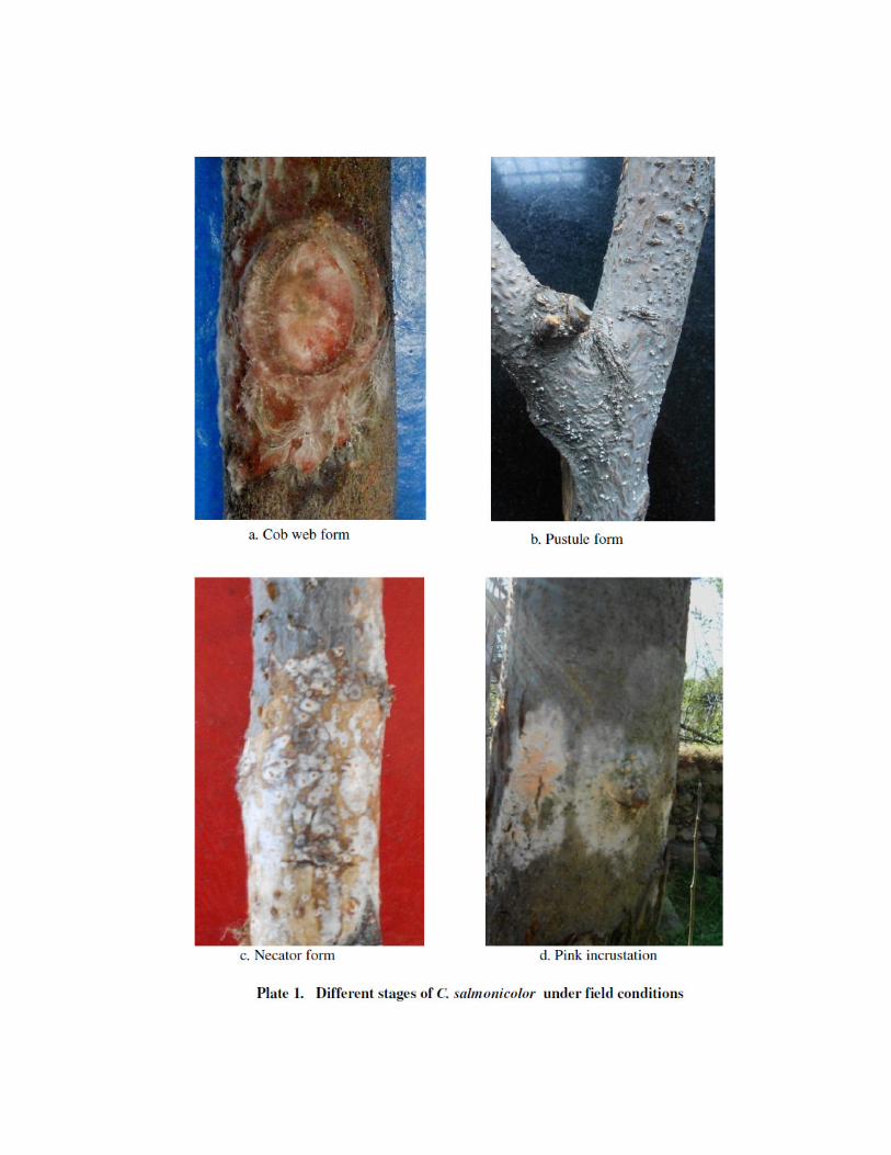

47
Fig 4.1. Prevalence of pink canker at different apple growing areas of
H.P. during 2011-12 crop seasons
4.2 SYMPTOMATOLOGY
During the course of surveys, the disease was found to appear on all above
ground plant parts viz., trunk, branches and twigs. The characteristic symptoms on
these plant parts are described as follows:
The disease manifests in four forms (Plate 1) namely, a). Cob-web stage b).
Pustuler stage c). Necator stage and d). Pink incrustation stage. Cob-web and pustuler
stage occurs immediately after monsoon rains. Cob-web stage consists of silky white
mycelium that grows over the surface of bark covering large area and is the first sign
of infection. The pustuler stage is readily identifiable when pink pustules consisting of
sterile mycelia produced on the affected area that erupts out by breaking lenticels
appearing white in colour. The necator stage is characterized by appearance of
orange-red necator (conidial) stage being produced late in the season and usually
develops on the upper side of infected branches. Whereas pink incrustation stage
consisting of basidial stage or Corticium stage usually developed on underside of the
branches facing less sunshine hours and characterized by development of salmon-
pink colour on the dying infected branch in the winters. Infected branches often die,
resulting in progressive crown dieback. Where infection is localized, death and
shedding of limited areas of bark may lead to the development of open wounds or
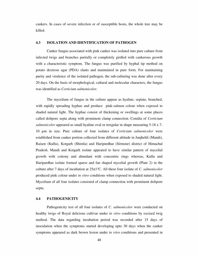
48
cankers. In cases of severe infection or of susceptible hosts, the whole tree may be
killed.
4.3 ISOLATION AND IDENTIFICATION OF PATHOGEN
Canker fungus associated with pink canker was isolated into pure culture from
infected twigs and branches partially or completely girdled with cankerous growth
with a characteristic symptom. The fungus was purified by hyphal tip method on
potato dextrose agar (PDA) slants and maintained in pure form. For maintaining
purity and virulence of the isolated pathogen, the sub-culturing was done after every
20 days. On the basis of morphological, cultural and molecular characters, the fungus
was identified as Corticium salmonicolor.
The mycelium of fungus in the culture appear as hyaline, septate, branched,
with rapidly spreading hyphae and produce pink-salmon colour when exposed to
shaded natural light. The hyphae consist of thickening or swellings at some places
called dolipore septa along with prominent clamp connection. Conidia of Corticium
salmonicolor appeared as small hyaline oval or irregular in shape measuring 5-18 x 7-
10 µm in size. Pure culture of four isolates of Corticium salmonicolor were
established from canker portion collected from different altitude in Janjhehli (Mandi),
Raisen (Kullu), Kotgarh (Shimla) and Haripurdhar (Sirmour) district of Himachal
Pradesh. Mandi and Kotgarh isolate appeared to have similar pattern of mycelial
growth with cottony and abundant with concentric rings whereas, Kullu and
Haripurdhar isolate formed sparse and fan shaped mycelial growth (Plate 2) in the
culture after 7 days of incubation at 25±1°C. All these four isolate of C. salmonicolor
produced pink colour under in vitro conditions when exposed to shaded natural light.
Mycelium of all four isolates consisted of clamp connection with prominent dolipore
septa.
4.4 PATHOGENICITY
Pathogenicity test of all four isolates of C. salmonicolor were conducted on
healthy twigs of Royal delicious cultivar under in vitro conditions by excised twig
method. The data regarding incubation period was recorded after 15 days of
inoculation when the symptoms started developing upto 30 days when the canker
symptoms appeared as dark brown lesion under in vitro conditions and presented in
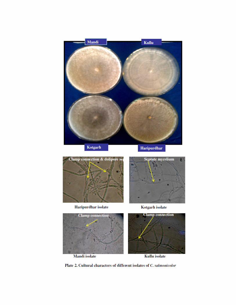


49
Tables 4.2. The per cent infection and average lesion size (mm) was recorded after 30
days of inoculation.
Table 4.2. Pathogenicity of C. salmonicolor isolates on Royal Delicious variety
of apple
Number of infected twigs
(DAI)
Corticium
salmonicolor
isolates 15 days 20 days 25 days 30 days
Per cent
Infection
(30 DAI)
Av. Lesion size
in mm (30
DAI*)
Mandi 5.0 8.0 12.0 14.0 93.33 65.2
Kotgarh 2.0 5.0 7.0 11.0 73.33 52.0
Kullu 2.0 4.0 6.0 10.0 66.67 46.7
Haripurdhar 1.0 3.0 5.0 8.0 46.67 40.4
Control 0.0 0.00 0.00 0.00 0.00 0.00
* Average lesion size was calculated from number of lesions formed
DAI* = Days after inoculation
The perusal of data (Table 4.2 & Plate 3) showed the numbers of twigs
infected by Mandi isolate of Corticium salmonicolor was 5 after 15 days of
inoculation, whereas it was 2, 2 and 1 in Kotgarh, Kullu and Haripurdhar isolates
respectively which increased to 14, 11, 10 and 8 respectively after 30 days of
inoculation. Per cent infection and average lesion size (mm) were more in Mandi
isolates of Corticium salmonicolor (93.33 %) with average lesion size of 65.2 mm
followed by Kotgarh, Kullu and Haripurdhar with 73.33, 66.67 and 46.67 per cent
infection and 52.0, 46.7 and 40.4mm lesion size respectively. While in control, there
were no infected twigs even after 30 days of inoculation.
4.5 CULTURAL STUDIES
4.5.1 Conidial germination of C. salmonicolor isolates
In order to find out the per cent conidial germination of C. salmonicolor
isolates in distilled water after 24 and 48 hrs, conidia of all isolates were separately
placed in distilled water in cavity slide and per cent germination was observed and
mentioned in Table 4.3.

50
Table 4.3. Per cent conidial germination of different isolates of C.
salmonicolor in distilled water
Duration (hrs) Corticium salmonicolor
isolates 24 48
Mean
Mandi 80.34 (63.94) 96.44 (81.74) 88.39 (72.84)
Kotgarh 77.77 (62.16) 87.99 (70.33) 82.88 (66.25)
Kullu 67.61 (55.45) 80.57 (63.98) 74.09 (59.72)
Haripurdhar 60.11 (50.88) 70.49 (57.20) 65.30 (54.04)
CD 0.05
Treatment 5.16
Duration 3.65
It is evident from the table that maximum per cent conidial germination was
observed in Mandi isolate (88.39 %) followed by Kotgarh, Kullu and Haripurdhar
with 82.88, 74.09 and 65.30 per cent germination respectively which were statistically
at par with each other. Per cent conidial germination was maximum after 48 hrs of
incubation in Mandi isolate and was least in Haripurdhar isolate (70.49 %) after 48 hr
of inoculation.
4.5.2 Germ tube length of different isolates of C. salmonicolor
In order to ascertain the germ tube length (µm) of C. salmonicolor isolates in
distilled water after 24 and 48 hrs, conidia of all isolates were separately placed in
distilled water in cavity slide and per cent germ tube length were observed and
mentioned in Table 4.4.
It is clear from the data (Table 4.4 & Plate 4) that Mandi isolate gave
significantly high germ tube length after 48 hrs of incubation (102.78µm) than at 24
hrs (60.91µm) whereas, Kotgarh isolate was next best in order with germ tube length
of 48.70µm and 88.64µm after 24 and 48 hrs of incubation. While Haripurdhar
isolate gave least germ tube length of 39.98µm and 63.99µm after 24 and 48 hrs of
incubation. Based on Pathogenicity, conidial germination and per cent germ tube
length Mandi isolate was found to be more virulent and used for further research
purpose.


51
Table 4.4. Germ tube length of C. salmonicolor isolates in distilled water
Germ tube length (µm) Corticium salmonicolor
isolates 24 48
Mean
Mandi 60.91 102.78 81.85
Kullu 39.70 83.53 61.61
Kotgarh 48.70 88.64 68.67
Haripurdhar 39.98 63.99 51.98
CD 0.05
Treatment 8.04
Duration 5.69
4.5.3. Induction of Necator stage
Imperfect stage of C. salmonicolor i.e. Necator decretus was successfully
induced on potato dextrose agar after incubation of 28-30 days at 25 ±1°C. However,
temperature seems to exert major influence on the formation of Necator stage as it
was found that fungus failed to sporulate above 30°C. Sporulation was observed in
varying degrees within 10 days in the culture in petriplates kept exposed to room
temperature (24-28 °C). It may be considered that diurnal temperature was essential
for induction of anamorph of C. salmonicolor in culture. Necator decretus is
characterized by formation of mycelial aggregate at certain points in the culture
(stroma / sporodochia) which are gelatinous in consistency turns hard in texture later
on (Plate 5a). Transverse section of stroma of fungus consists of
pseudoparenchymatous cells consisting large number of oval to irregular spores
measuring 45-52 µm, which were orange-red in colour. Necator spores and deep
seated mycelium in the bark considered as a primary source of infection.
4.5.4. Mode of disease spread
Pink canker is initiated with the onset of monsoon rains either from deep
seated dormant mycelium of the fungus or from necator spores that may be carried
through wind or rain splashes and lodge on branches, stem and crotches. The infection
starts in the month of June-July when there is no sign of basidial stage, thereby
indicating necator spores to be the source of primary infection which remains dormant
till the first showers of rain. After hydration spores produce germ tube which enter
either through lenticels or wound causing infection through intracellular mycelium.
Infested twig, branches and stem showed browning of cambium region indicating

52
maceration of tissues due to production of hydrolytic and pectinolytic enzymes that
blocked the flow of nutrient to aerial portion, hence produced die-back symptoms.
After it gets established, mycelium produced enormous amount of conidia from
conidiogenous cells that spread through rain or wind to the adjacent tree causing
typical cow-web and other stages later on, depending up on the weather conditions.
4.5.5. Leaves and fruit infection of Royal Delicious
C. salmonicolor, the incitant of pink canker in apple spreads through wind
borne conidia. In order to ascertain the role of conidia in leaf infection, leaves of
Royal Delicious cultivar were dusted with conidia whereas fruits were inoculated with
fresh culture bit of pathogen and data on per cent infection and lesion size (cm) was
recorded.
It is evident from the data (Table 4.5 & Plate 5.b) that leaves were infected to
25 per cent after 7 days and attained cent per cent infection after 14 days of incubation
when placed in petriplates containing 100 per cent relative humidity at 25±1°C. While
lesion size was observed to be 3.6 cm after 10 days and attained lesion size of 4.8 cm
after 14 days of incubation at 25±1°C. Conidia were found to infect leaves with
disease severity of 66.67 per cent and average lesion size of 3.28 cm on fruits of
Royal delicious cultivar.
Table 4.5. Disease severity on leaves and lesion size (cm) on fruits of apple
after inoculation with conidia of C. salmonicolor
Conidia of C. salmonicolor Disease severity (%) in leaves and Lesion
size (cm) on fruits
Royal Delicious After 7 day 10 days 14 days
Mean
Leaves 25.00 75.00 100.00 66.67
Fruits 1.45 cm 3.6 cm 4.8cm 3.28 cm
4.6 EPIDEMIOLOGICAL STUDIES
4.6.1 Progress of disease in relation to meteorological factors under field conditions.
In order to ascertain the role of environmental factors particularly;
temperature, relative humidity, cumulative rainfall and sunshine hours in disease
development under field conditions, experiment was conducted at the farmer’s field
during 2011 and 2012.The observations on daily mean temperature, average relative


53
humidity, cumulative rainfall and average sunshine hours were recorded with effect
from June to September in each year. The data on disease severity was recorded at 7
days interval and the daily observations on meteorological factors were expressed as
mean value for the period intervening the two data recording dates (7 days) and are
presented in Table (4.6.a and b; Fig.4.2. a and b)
The data (Fig.4.2.a & Fig. 4.2.b) on meteorological factors and disease
development studies revealed that the disease first appeared in second and third week
of June with the prevalence of temperature, relative humidity, cumulative rainfall and
weekly sunshine hours up to the extent of 19.86 and 20.85 oC, 95.89 and 50.04 per
cent, 73.00 and 15.24 mm, 41.76 and 90.43 hrs during 2011 and 2012, respectively.
The pink canker infection increased at slow pace up to fourth week of July and
thereafter, a rapid progress was recorded in subsequent weeks of August and obtained
peak severity in the month of September with occurrence of temperature (16.39 and
14.92 o C), per cent relative humidity (85.89 and 80.79), cumulative rainfall (53.00
and 69.57 mm) and sunshine hours (36.05 and 72.35) during both the years.
Table 4.6a. Effect of different meteorological factors on disease development
during 2011
Date of
observation
Weekly
mean
temperature
Mean
relative
humidity
(%)
Cumulative
rainfall
(mm)
Average
sunshine
hours
Disease
severity
(%)
07.6.11 20.24 36.54 14.52 98.33 0.00
14.6.11 19.86 95.89 73.00 41.76 3.86
21.6.11 18.38 46.98 66.40 97.23 7.64
28.6.11 18.35 63.96 40.40 64.37 12.24
05.7.11 17.72 77.76 8.95 88.52 18.95
12.7.11 17.33 75.92 34.85 74.98 28.81
19.7.11 18.95 76.79 45.60 56.77 33.64
26.7.11 17.74 98.83 66.48 53.81 41.06
02.8.11 17.78 81.61 42.20 61.75 46.05
09.8.11 17.91 83.39 64.97 23.45 53.33
16.8.11 17.92 85.48 83.78 33.43 62.24
23.8.11 17.89 87.08 88.64 29.99 68.22
30.8.11 17.28 75.66 142.54 62.06 76.60
06.9.11 17.92 87.44 94.68 68.24 82.41
13.9.11 16.39 85.89 53.00 36.05 87.1

54
Table 4.6.b. Effect of different meteorological factors on disease development
during 2012
4.6.2 Correlation of disease severity with meteorological factors
The correlation analysis (Table 4.7) indicated that simple correlation between
per cent disease severity and relative humidity was positive and highly significant
during both the years, exhibiting its effect on the disease development. Simple
correlation coefficient between disease severity and temperature was negative but
highly significant in both crop seasons. Pooled data also showed similar results
between meteorological factors and disease severity. Further the partial correlation
coefficient between disease severity and temperature was highly significant but
negative in 2011 and 2012; however relative humidity was found positive and highly
significant. Simple correlation of pooled data regarding cumulative rainfall was found
to be positive and highly significant during both the seasons 2011-12. However
sunshine hours were found to be significant but negatively correlated. It also disclosed
the fact that pink incrustation or salmon-coloured crustose phase, consisting of the
sexual Corticium stage usually develops on shaded underside of infected branch or
limbs of many plantation crops.
Date of
observation
Weekly
mean
temperature
Mean
relative
humidity
(%)
Cumulative
rainfall
(mm)
Average
sunshine
hours
Disease
severity
(%)
10.6.12 20.67 42.73 0.25 63.42 0.00
17.6.12 20.85 50.04 15.24 90.43 1.54
24.6.12 20.12 51.71 26.00 87.26 4.06
01.7.12 18.98 66.13 60.94 24.99 11.24
08.7.12 17.55 72.01 57.88 67.33 15.26
15.7.12 18.90 71.15 41.52 71.38 18.64
22.7.12 18.86 76.83 65.15 29.97 22.51
29.7.12 17.09 79.76 45.20 49.56 29.10
05.8.12 17.31 79.33 35.26 69.31 34.14
12.8.12 17.86 83.05 44.69 57.27 40.00
19.8.12 16.61 85.11 65.25 56.71 43.32
26.8.12 15.42 44.70 15.22 73.50 47.76
02.9.12 17.11 83.14 31.21 70.11 52.04
09.9.12 17.42 84.12 55.82 32.90 54.12
16.9.12 14.92 80.79 69.57 72.35 62.80

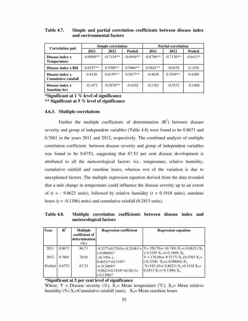
55
Table 4.7. Simple and partial correlation coefficients between disease index
and environmental factors
Simple correlation Partial correlation Correlation pair
2011 2012 Pooled 2011 2012 Pooled
Disease index x
Temperature
-0.8899** -0.7154** -0.6944** -0.8796** -0.7130** -0.6413*
Disease index x RH 0.6257** 0.5300** 0.5866** 0.5842** -0.0478 0.1456
Disease index x
Cumulative rainfall
0.4126 0.6159** 0.5877** -0.4629 0.5549** 0.4200
Disease index x
Sunshine hrs
-0.1471 -0.5678** -0.4102 -0.1762 -0.3533 -0.1468
*Significant at 1 % level of significance
** Significant at 5 % level of significance
4.6.3. Multiple correlations
Further the multiple coefficients of determination (R2) between disease
severity and group of independent variables (Table 4.8) were found to be 0.8671 and
0.7601 in the years 2011 and 2012, respectively. The combined analysis of multiple
correlation coefficient between disease severity and group of independent variables
was found to be 0.6753, suggesting that 67.53 per cent disease development is
attributed to all the meteorological factors viz., temperature, relative humidity,
cumulative rainfall and sunshine hours, whereas rest of the variation is due to
unexplained factors. The multiple regression equation derived from the data revealed
that a unit change in temperature could influence the disease severity up to an extent
of (r = - 9.8623 units), followed by relative humidity (r = 0.1918 units), sunshine
hours (r = -0.1306) units) and cumulative rainfall (0.2813 units).
Table 4.8. Multiple correlation coefficients between disease index and
meteorological factors
Year R2 Multiple
coefficient of
determination
(%)
Regression coefficient Regression equation
2011
2012
Pooled
0.8671
0.7601
0.6753
86.71
76.01
67.53
-9.5277+0.5765+(-0.3248)*+
(-0.08604)*
-16.749+ (-
0.0631)*+0.3195*
+(-0.3469)*
-9.8623+0.1918*+0.2813+
(-0.1306)*
Y= 350.70+(-16.749) X1+(-0.0631) X2
+ 0.3195 X3 +(-0.3469) X4
Y = 178.90+(-9.5277) X1+0.5765 X2+
(-0.3248) X3+(-0.08604) X4
Y=192.45+(-9.8623) X1+0.1918 X2+
0.2813 X3+(-0.1306) X4
*Significant at 5 per cent level of significance
Where, Y = Disease severity (%), X1= Mean temperature (oC), X2= Mean relative
humidity (%) X3=Cumulative rainfall (mm), X4= Mean sunshine hours
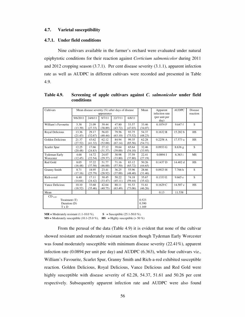
56
4.7. Varietal susceptibility
4.7.1. Under field conditions
Nine cultivars available in the farmer’s orchard were evaluated under natural
epiphytotic conditions for their reaction against Corticium salmonicolor during 2011
and 2012 cropping season (3.7.1). Per cent disease severity (3.1.1), apparent infection
rate as well as AUDPC in different cultivars were recorded and presented in Table
4.9.
Table 4.9. Screening of apple cultivars against C. salmonicolor under field
conditions
Mean disease severity (%) after days of disease
appearance
Cultivars
9/6/2011 24/6/11 9/7/11 22/7/11 6/8/11
Mean Apparent
infection rate
(per unit per day)
AUDPC Disease
reaction
William’s Favourite 5.38
(13.39)
21.09
(27.33)
39.44
(38.89)
47.80
(43.72)
53.57
(47.03)
33.46
(34.07)
0.1074 F 9.647 f S
Royal Delicious 13.36
(21.43)
29.17
(32.67)
56.03
(48.44)
79.56
(63.10)
93.75
(75.52)
54.37
(48.23)
0.1632 B 15.282 b HS
Golden Delicious 21.37 (27.52)
43.62 (41.32)
62.12 (52.00)
84.94 (67.14)
99.35 (85.56)
62.28 (54.71)
0.2258 A 17.573 a HS
Scarlet Spur 12.25
(20.48)
17.66
(24.83)
27.12
(31.37)
39.64
(39.00)
65.64
(54.10)
32.46
(33.95)
0.0933 G 8.636 g S
Tydeman Early
Worcester
4.68
(12.45)
14.72
(22.54)
24.07
(29.37)
30.98
(33.80)
37.59
(37.80)
22.41
(27.19)
0.0894 I 6.363 i MS
Red Gold 8.05 (16.48)
37.22 (37.58)
51.77 (46.00)
71.16 (57.50)
83.12 (65.72)
50.26 (44.65)
0.1437 D 14.402 d HS
Granny Smith 8.71
(17.16)
18.95
(25.79)
23.41
(28.92)
36.25
(37.00)
55.96
(48.40)
28.66
(31.46)
0.0923 H 7.766 h S
Rich-a-red 6.40
(14.64)
17.11
(24.42)
30.45
(33.47)
50.22
(45.11)
74.18
(59.44)
35.67
(35.42)
0.1333 E 9.665 e S
Vance Delicious 10.10
(18.52)
33.68
(35.46)
42.64
(40.75)
80.11
(63.49)
91.53
(73.06)
51.61
(46.26)
0.1629 C 14.507 c HS
Mean 0.13 11.538
CD 0.05
Treatment (T)
Duration (D)
T x D
0.523
0.390
1.169
MR = Moderately resistant (1.1-10.0 %), S = Susceptible (25.1-50.0 %),
MS = Moderately susceptible (10.1-25.0 %), HS = Highly susceptible (> 50 %)
From the perusal of the data (Table 4.9) it is evident that none of the cultivar
showed resistant and moderately resistant reaction though Tydeman Early Worcester
was found moderately susceptible with minimum disease severity (22.41%), apparent
infection rate (0.0894 per unit per day) and AUDPC (6.363), while four cultivars viz.,
William’s Favourite, Scarlet Spur, Granny Smith and Rich-a-red exhibited susceptible
reaction. Golden Delicious, Royal Delicious, Vance Delicious and Red Gold were
highly susceptible with disease severity of 62.28, 54.37, 51.61 and 50.26 per cent
respectively. Subsequently apparent infection rate and AUDPC were also found
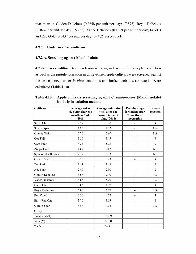
57
maximum in Golden Delicious (0.2258 per unit per day; 17.573), Royal Delicious
(0.1632 per unit per day; 15.282), Vance Delicious (0.1629 per unit per day; 14.507)
and Red Gold (0.1437 per unit per day; 14.402) respectively.
4.7.2 Under in vitro conditions
4.7.2 A. Screening against Mandi Isolate
4.7.2a. Flask condition: Based on lesion size (cm) in flask and in Petri plate condition
as well as the pustule formation in all seventeen apple cultivars were screened against
the test pathogen under in vitro conditions and further their disease reaction were
calculated (Table 4.10).
Table 4.10. Apple cultivars screening against C. salmonicolor (Mandi isolate)
by Twig inoculation method
Cultivars Average lesion
size(cm) after one
month in flask
(2011)
Average lesion size
(cm) after one
month in Petri
plate (2011)
Pustuler stage
formation after
2 months of
inoculation
Disease
reaction
Super Chief 2.27 3.50 - S
Scarlet Spur 1.90 2.32 - MS
Granny Smith 2.70 2.80 - MS
Coe Fuji 3.30 3.45 + S
Cam Spur 4.23 5.05 + S
Zinger Gold 1.67 2.12 - MS
Spur Winter Banana 2.17 3.03 - MS
Oregan Spur 3.30 3.93 + S
Top Red 3.53 3.48 - S
Ace Spur 2.40 2.50 - S
Golden Delicious 5.67 7.40 + HS
Vance Delicious 4.63 5.70 + HS
Gale Gala 3.83 4.07 + S
Royal Delicious 5.00 6.27 + HS
Red Chief 3.20 4.52 + S
Early Red One 3.70 3.95 - S
Golden Spur 4.87 4.98 + HS
CD0.05
Treatment (T) 0.389
Year (Y) 0.100
T x Y 0.411

58
It is evident from the data (Table 4.10 ) that Spur Winter Banana, Zinger Gold,
Granny Smith and Scarlet Spur showed moderately susceptible reaction with
minimum lesion size whereas nine others cultivars viz., Super Chief, Coe Fuji, Cam
Spur, Oregan Spur, Top Red, Ace Spur, Gale Gala, Red Chief and Early Red One
were found to exhibit susceptible reaction while Golden Delicious, Royal Delicious,
Vance Delicious and Golden Spur showed highly susceptible reaction with maximum
lesion size. Maximum lesion size was produced in Petri plate condition than flask
condition and the pustules were also formed in Golden Delicious, Royal Delicious,
Vance Delicious and Golden Spur after one month of incubation. Though pustules
were formed in Coe Fuji, Cam Spur, Oregan Spur, Gale Gala and Red Chief but with
varying degree of lesion size.
4.7.2a. Flask condition: Based on disease severity (%), apparent infection rate, area
under disease progress curve (AUDPC) all seventeen cultivars were screened against
the test pathogen and their disease reaction were calculated and presented in Table
4.11.
It is clear from the data presented (Table 4.11) that none of the cultivar
exhibited resistant reaction though three cultivars viz., Ace Spur, Zinger Gold and
Granny Smith showed moderately resistant reaction with less disease severity (9.88,
9.36, 9.48 %), minimum apparent infection rate (0.044, 0.037, 0.037 per unit per day)
and minimum AUDPC (3.357, 3.201, 3.308) respectively, while rest of the cultivars
behaved moderately susceptible reaction under flask condition. Though it was found
that Golden Delicious having maximum per cent disease severity, more apparent
infection rate per unit per day with high area under disease progress curve followed
by Royal Delicious, Vance Delicious, Golden Spur and Cam Spur, respectively.
4.7.2b. Petri plate condition
All seventeen cultivars were screened against the test pathogen and based on
disease severity (%), apparent infection rate, area under disease progress curve
(AUDPC) and their disease reaction were calculated and presented in Table 4.12.

59
Table 4.11. Screening of apple cultivars against C. salmonicolor (Mandi isolate)
under in vitro conditions
Weekly mean disease severity (%)
after days of disease appearance Cultivars
2/2/2011 9/2/11 16/2/11 23/2/11 2/3/115 9/3/11
Mean Apparent
infection rate
(Per unit per
day)
AUDPC Disease
Reaction
Coe Fuji 5.60
(13.69)
7.14
(15.50)
9.10
(17.55)
12.05
(20.30)
18.80
(25.69)
23.08
(28.70)
12.63
(20.24)
0.046 M 4.300 m MS
Golden Spur 5.27
(13.27)
11.11
(19.46)
15.56
(23.22)
22.22
(28.11)
26.67
(31.08)
31.50
(34.13)
18.72
(24.88)
0.060 D 6.576 d MS
Oregan Spur 5.02
(12.94)
6.67
(14.96)
12.73
(20.90)
19.47
(26.17)
25.56
(30.35)
24.97
(29.97)
15.74
(22.55)
0.053 H 5.560 h MS
Super Chief 5.80
(13.93)
6.74
(15.04)
10.39
18.80)
14.08
(22.03)
22.22
(28.11)
24.11
(29.39)
13.89
(21.22)
0.047 L 4.787 l MS
Spur Winter Banana 5.44
(13.48)
7.11
(15.45)
8.81
(17.26)
12.97
(21.10)
16.31
(23.81)
21.90
(27.89)
12.09
(19.83)
0.045 N 4.120 n MS
Vance Delicious 4.88
(12.76)
13.16
(21.26)
17.07
(24.39)
22.10
(28.03)
27.19
(31.41)
30.62
(33.58)
19.17
(25.24)
0.061 C 6.809 c MS
Cam Spur 5.43
(13.47)
9.52
(17.96)
14.27
(22.18)
23.03
(28.66)
28.24
(32.09)
31.00
(33.82)
18.58
(24.70)
0.059 E 6.529 e MS
Ace Spur 4.49
(12.23)
5.74
(13.85)
6.55
(14.82)
8.65
(17.10)
15.72
(23.35)
18.10
(25.17)
9.88
(17.75)
0.044 O 3.357 o MR
Top Red 5.36
(13.38)
8.37
(16.81)
13.30
(21.38)
16.88
(24.25)
22.22
(28.11)
26.17
(30.76)
15.39
(22.45)
0.052 I 5.358 i MS
Golden Delicious 3.21
(10.32)
12.03
(20.28)
18.15
(25.20)
26.27
(30.82)
28.76
(32.42)
32.00
(34.44)
20.07
(25.58)
0.076 A 7.197 a MS
Royal Delicious 3.41
(10.64)
10.83
(19.20)
16.86
(24.23)
24.30
(29.53)
27.55
(31.65)
32.73
(34.88)
19.28
(25.02)
0.075 B 6.833 b MS
Scarlet Spur 5.92
(14.07)
7.04
(15.38)
11.27
(19.60)
17.49
(24.71)
23.72
(29.14)
26.71
(31.11)
15.36
(22.33)
0.050 J 5.308 j MS
Zinger Gold 4.95
(12.85)
5.99
(14.17)
6.90
(15.22)
9.31
(17.76)
13.07
(21.18)
15.96
(23.54)
9.36
(17.45)
0.037 Q 3.201 q MR
Granny Smith 4.47
(12.20)
6.10
(14.30)
7.57
(15.96)
10.82
(19.20)
13.17
(21.27)
14.72
(22.55)
9.48
(17.58)
0.037 P 3.308 p MR
Gale Gala 5.03
(12.96)
10.01
(18.44)
14.29
(22.20)
21.47
(27.59)
26.27
(30.82)
28.97
(32.55)
17.67
(24.09)
0.058 F 6.232 f MS
Red Chief 5.74
(13.86)
7.36
(15.73)
11.53
(19.84)
17.60
(24.79)
22.22
(28.11)
24.82
(29.87)
14.88
(22.04)
0.048 K 5.180 k MS
Early Red One 5.07
(13.00)
7.80
(16.21)
14.77
(22.59)
19.36
(26.09)
21.57
(27.66)
27.40
(31.55)
15.99
(22.85)
0.056 G 5.581 g MS
CD 0.05 Treatment (T) 0.472
Duration (D) 0.280
T x D 1.155
The perusal of data presented (Table 4.12 & Plate 6.a & b) revealed that only
Zinger Gold showed moderately susceptible reaction against C. salmonicolor with
minimum disease severity (24.90 %) and subsequently low apparent infection rate
(0.0662 per unit per day) as well as area under disease progress curve (8.46). Whereas
ten cultivars viz., Coe Fuji, Oregan Spur, Super Chief, Spur Winter Banana, Ace
Spur, Scarlet Spur, Red Chief, Granny Smith, Top Red and Early Red One behaved
susceptible reaction against the test pathogen while Golden Spur, Vance Delicious,
Gale Gala, Royal Delicious, Golden Delicious and Cam Spur exhibited highly
susceptible reaction. Golden Delicious was found to be most preferred host by
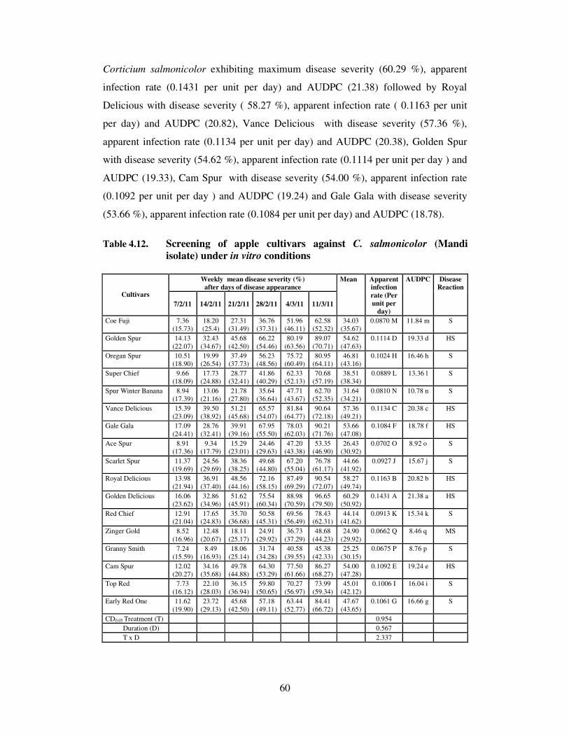
60
Corticium salmonicolor exhibiting maximum disease severity (60.29 %), apparent
infection rate (0.1431 per unit per day) and AUDPC (21.38) followed by Royal
Delicious with disease severity ( 58.27 %), apparent infection rate ( 0.1163 per unit
per day) and AUDPC (20.82), Vance Delicious with disease severity (57.36 %),
apparent infection rate (0.1134 per unit per day) and AUDPC (20.38), Golden Spur
with disease severity (54.62 %), apparent infection rate (0.1114 per unit per day ) and
AUDPC (19.33), Cam Spur with disease severity (54.00 %), apparent infection rate
(0.1092 per unit per day ) and AUDPC (19.24) and Gale Gala with disease severity
(53.66 %), apparent infection rate (0.1084 per unit per day) and AUDPC (18.78).
Table 4.12. Screening of apple cultivars against C. salmonicolor (Mandi
isolate) under in vitro conditions
Weekly mean disease severity (%)
after days of disease appearance Cultivars
7/2/11 14/2/11 21/2/11 28/2/11 4/3/11 11/3/11
Mean Apparent
infection
rate (Per
unit per
day)
AUDPC Disease
Reaction
Coe Fuji 7.36
(15.73)
18.20
(25.4)
27.31
(31.49)
36.76
(37.31)
51.96
(46.11)
62.58
(52.32)
34.03
(35.67)
0.0870 M 11.84 m S
Golden Spur 14.13
(22.07)
32.43
(34.67)
45.68
(42.50)
66.22
(54.46)
80.19
(63.56)
89.07
(70.71)
54.62
(47.63)
0.1114 D 19.33 d HS
Oregan Spur 10.51
(18.90)
19.99
(26.54)
37.49
(37.73)
56.23
(48.56)
75.72
(60.49)
80.95
(64.11)
46.81
(43.16)
0.1024 H 16.46 h S
Super Chief 9.66
(18.09)
17.73
(24.88)
28.77
(32.41)
41.86
(40.29)
62.33
(52.13)
70.68
(57.19)
38.51
(38.34)
0.0889 L 13.36 l S
Spur Winter Banana 8.94
(17.39)
13.06
(21.16)
21.78
(27.80)
35.64
(36.64)
47.71
(43.67)
62.70
(52.35)
31.64
(34.21)
0.0810 N 10.78 n S
Vance Delicious 15.39
(23.09)
39.50
(38.92)
51.21
(45.68)
65.57
(54.07)
81.84
(64.77)
90.64
(72.18)
57.36
(49.21)
0.1134 C 20.38 c HS
Gale Gala 17.09
(24.41)
28.76
(32.41)
39.91
(39.16)
67.95
(55.50)
78.03
(62.03)
90.21
(71.76)
53.66
(47.08)
0.1084 F 18.78 f HS
Ace Spur 8.91
(17.36)
9.34
(17.79)
15.29
(23.01)
24.46
(29.63)
47.20
(43.38)
53.35
(46.90)
26.43
(30.92)
0.0702 O 8.92 o S
Scarlet Spur 11.37
(19.69)
24.56
(29.69)
38.36
(38.25)
49.68
(44.80)
67.20
(55.04)
76.78
(61.17)
44.66
(41.92)
0.0927 J 15.67 j S
Royal Delicious 13.98
(21.94)
36.91
(37.40)
48.56
(44.16)
72.16
(58.15)
87.49
(69.29)
90.54
(72.07)
58.27
(49.74)
0.1163 B 20.82 b HS
Golden Delicious 16.06
(23.62)
32.86
(34.96)
51.62
(45.91)
75.54
(60.34)
88.98
(70.59)
96.65
(79.50)
60.29
(50.92)
0.1431 A 21.38 a HS
Red Chief 12.91 (21.04)
17.65 (24.83)
35.70 (36.68)
50.58 (45.31)
69.56 (56.49)
78.43 (62.31)
44.14 (41.62)
0.0913 K 15.34 k S
Zinger Gold 8.52
(16.96)
12.48
(20.67)
18.11
(25.17)
24.91
(29.92)
36.73
(37.29)
48.68
(44.23)
24.90
(29.92)
0.0662 Q 8.46 q MS
Granny Smith 7.24
(15.59)
8.49
(16.93)
18.06
(25.14)
31.74
(34.28)
40.58
(39.55)
45.38
(42.33)
25.25
(30.15)
0.0675 P 8.76 p S
Cam Spur 12.02 (20.27)
34.16 (35.68)
49.78 (44.88)
64.30 (53.29)
77.50 (61.66)
86.27 (68.27)
54.00 (47.28)
0.1092 E 19.24 e HS
Top Red 7.73
(16.12)
22.10
(28.03)
36.15
(36.94)
59.80
(50.65)
70.27
(56.97)
73.99
(59.34)
45.01
(42.12)
0.1006 I 16.04 i S
Early Red One 11.62
(19.90)
23.72
(29.13)
45.68
(42.50)
57.18
(49.11)
63.44
(52.77)
84.41
(66.72)
47.67
(43.65)
0.1061 G 16.66 g S
CD0.05 Treatment (T) 0.954
Duration (D) 0.567
T x D 2.337
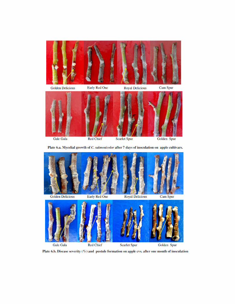
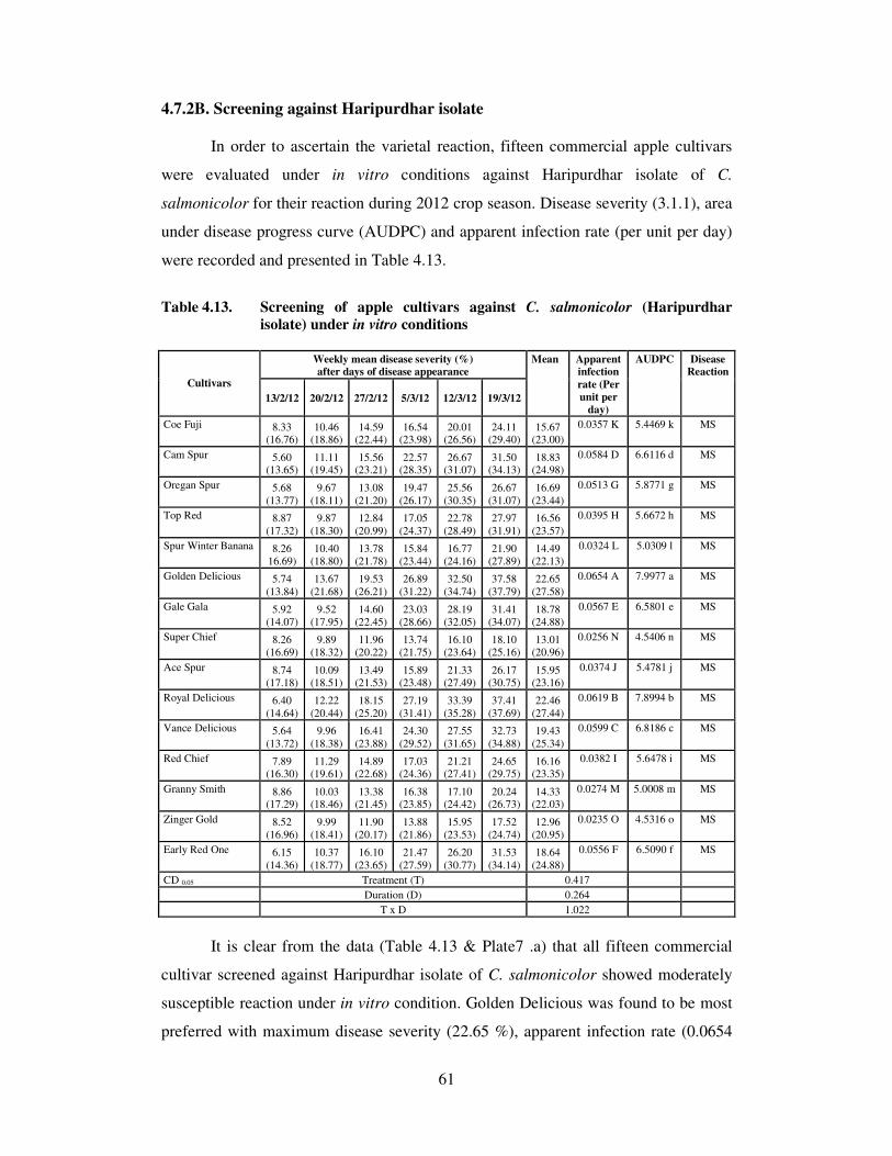
61
4.7.2B. Screening against Haripurdhar isolate
In order to ascertain the varietal reaction, fifteen commercial apple cultivars
were evaluated under in vitro conditions against Haripurdhar isolate of C.
salmonicolor for their reaction during 2012 crop season. Disease severity (3.1.1), area
under disease progress curve (AUDPC) and apparent infection rate (per unit per day)
were recorded and presented in Table 4.13.
Table 4.13. Screening of apple cultivars against C. salmonicolor (Haripurdhar
isolate) under in vitro conditions
Weekly mean disease severity (%)
after days of disease appearance Cultivars
13/2/12 20/2/12 27/2/12 5/3/12 12/3/12 19/3/12
Mean Apparent
infection
rate (Per
unit per
day)
AUDPC Disease
Reaction
Coe Fuji 8.33 (16.76)
10.46 (18.86)
14.59 (22.44)
16.54 (23.98)
20.01 (26.56)
24.11 (29.40)
15.67 (23.00)
0.0357 K 5.4469 k MS
Cam Spur 5.60 (13.65)
11.11 (19.45)
15.56 (23.21)
22.57 (28.35)
26.67 (31.07)
31.50 (34.13)
18.83 (24.98)
0.0584 D 6.6116 d MS
Oregan Spur 5.68
(13.77)
9.67
(18.11)
13.08
(21.20)
19.47
(26.17)
25.56
(30.35)
26.67
(31.07)
16.69
(23.44)
0.0513 G 5.8771 g MS
Top Red 8.87
(17.32)
9.87
(18.30)
12.84
(20.99)
17.05
(24.37)
22.78
(28.49)
27.97
(31.91)
16.56
(23.57)
0.0395 H 5.6672 h MS
Spur Winter Banana 8.26
16.69)
10.40
(18.80)
13.78
(21.78)
15.84
(23.44)
16.77
(24.16)
21.90
(27.89)
14.49
(22.13)
0.0324 L 5.0309 l MS
Golden Delicious 5.74
(13.84)
13.67
(21.68)
19.53
(26.21)
26.89
(31.22)
32.50
(34.74)
37.58
(37.79)
22.65
(27.58)
0.0654 A 7.9977 a MS
Gale Gala 5.92
(14.07)
9.52
(17.95)
14.60
(22.45)
23.03
(28.66)
28.19
(32.05)
31.41
(34.07)
18.78
(24.88)
0.0567 E 6.5801 e MS
Super Chief 8.26
(16.69)
9.89
(18.32)
11.96
(20.22)
13.74
(21.75)
16.10
(23.64)
18.10
(25.16)
13.01
(20.96)
0.0256 N 4.5406 n MS
Ace Spur 8.74
(17.18)
10.09
(18.51)
13.49
(21.53)
15.89
(23.48)
21.33
(27.49)
26.17
(30.75)
15.95
(23.16)
0.0374 J 5.4781 j MS
Royal Delicious 6.40
(14.64)
12.22
(20.44)
18.15
(25.20)
27.19
(31.41)
33.39
(35.28)
37.41
(37.69)
22.46
(27.44)
0.0619 B 7.8994 b MS
Vance Delicious 5.64
(13.72)
9.96
(18.38)
16.41
(23.88)
24.30
(29.52)
27.55
(31.65)
32.73
(34.88)
19.43
(25.34)
0.0599 C 6.8186 c MS
Red Chief 7.89
(16.30)
11.29
(19.61)
14.89
(22.68)
17.03
(24.36)
21.21
(27.41)
24.65
(29.75)
16.16
(23.35)
0.0382 I 5.6478 i MS
Granny Smith 8.86 (17.29)
10.03 (18.46)
13.38 (21.45)
16.38 (23.85)
17.10 (24.42)
20.24 (26.73)
14.33 (22.03)
0.0274 M 5.0008 m MS
Zinger Gold 8.52
(16.96)
9.99
(18.41)
11.90
(20.17)
13.88
(21.86)
15.95
(23.53)
17.52
(24.74)
12.96
(20.95)
0.0235 O 4.5316 o MS
Early Red One 6.15
(14.36)
10.37
(18.77)
16.10
(23.65)
21.47
(27.59)
26.20
(30.77)
31.53
(34.14)
18.64
(24.88)
0.0556 F 6.5090 f MS
CD 0.05 Treatment (T) 0.417
Duration (D) 0.264
T x D 1.022
It is clear from the data (Table 4.13 & Plate7 .a) that all fifteen commercial
cultivar screened against Haripurdhar isolate of C. salmonicolor showed moderately
susceptible reaction under in vitro condition. Golden Delicious was found to be most
preferred with maximum disease severity (22.65 %), apparent infection rate (0.0654

62
per unit per day) and AUDPC (7.9977) followed by Royal Delicious, Vance
Delicious, Cam Spur and Gale Gala. Zinger Gold and Super Chief showed minimum
disease severity, apparent infection rate and AUDPC respectively.
4.7.2C. Screening against Kotgarh isolate
Fifteen cultivars of apple were screened against Kotgarh isolate of C.
salmonicolor and data on disease severity (%), apparent infection rate and AUDPC
was calculated and presented in Table 4.14
It is evident from the data presented (Table 4.14 & Plate 7.b) that Golden
Delicious was most preferred host followed by Royal Delicious with maximum
disease severity (29.60 % and 27.52 %), apparent infection rate (0.0782 and 0.0768
per unit per day) and subsequent maximum AUDPC (10.3602 and 9.6667)
respectively. Whereas Zinger Gold and Super Chief was least preferred host.
Table 4.14. Screening of apple cultivars against C. salmonicolor (Kotgarh
isolate) under in vitro conditions
Weekly mean disease severity (%)
after days of disease appearance Cultivars
6/2/12 13/2/12 20/2/12 27/2/12 5/3/12 12/3/12
Mean Apparent
infection
rate (Per
unit per
day)
AUDPC Disease
Reaction
Super Chief 6.89
(15.19)
7.51
(15.90)
8.68
(17.12)
11.02
(19.38)
14.59
(22.45)
20.64
(27.01)
11.56
(19.51)
0.0359 O 3.8903 o MS
Oregan Spur 5.30
(13.30)
8.09
(16.51)
15.23
(22.96)
21.25
(27.43)
26.63
(31.05)
32.59
(34.80)
18.18
(24.34)
0.0615 G 6.3106 g MS
Golden Delicious 6.67 (14.96)
15.19 (22.92)
24.68 (29.77)
34.28 (35.82)
44.25 (41.68)
52.52 (46.43)
29.60 (31.93)
0.0782 A 10.3602 a S
Cam Spur 6.58 (14.85)
10.58 (18.97)
17.17 (24.46)
27.07 (31.34)
34.41 (35.90)
45.62 (42.47)
23.57 (28.00)
0.0707 D 8.0728 d MS
Royal Delicious 5.91
(14.04)
14.61
(22.46)
22.46
(28.27)
33.41
(35.29)
40.62
(39.58)
48.10
(43.90)
27.52
(30.59)
0.0768 B 9.6667 b S
Vance Delicious 5.91
(14.05)
10.89
(19.25)
18.44
(25.42)
27.58
(31.67)
35.47
(36.54)
43.55
(41.28)
23.64
(28.03)
0.0716 C 8.1979 c MS
Spur Winter Banana 6.06
(14.24)
7.24
(15.60)
10.47
(18.85)
13.32
(21.39)
16.36
(23.85)
23.58
(29.04)
12.84
(20.50)
0.0447 L 4.3547 l MS
Gale Gala 6.11
(14.30)
9.65
(18.07)
17.79
(24.93)
26.47
(30.95)
33.54
(35.37)
39.32
(38.82)
22.14
(27.07)
0.0656 E 7.7107 e MS
Ace Spur 5.63
(13.71)
6.92
(15.23)
9.57
(18.00)
16.50
(23.95)
21.70
(27.75)
26.44
(30.93)
14.46
(21.60)
0.0513 J 4.9510 j MS
Granny Smith 6.54
(14.80)
7.41
(15.78)
8.12
(16.55)
13.25
(21.34)
18.55
(25.49)
21.47
(27.59)
12.56
(20.26)
0.0389 M 4.2930 m MS
Early Red One 5.29
(13.28)
8.77
(17.20)
15.15
(22.90)
23.12
(28.73)
28.67
(32.36)
34.59
(36.01)
19.26
(25.08)
0.0642 F 6.6950 f MS
Ginger Gold 6.23
(14.44)
7.21
(15.57)
9.24
(17.69)
12.26
(20.48)
15.99
(23.56)
19.54
(26.22)
11.75
(19.66)
0.0370 N 4.0313 n MS
Red Chief 4.70
(12.46)
7.85
(16.27)
12.77
(20.93)
16.46
(23.93)
20.79
(27.11)
25.77
(30.49)
14.72
(21.86)
0.0557 I 5.1177 i MS
Coe fuji 6.05
(14.23)
6.54
(14.81)
9.45
(17.89)
14.98
(22.76)
21.41
(27.55)
27.42
(31.55)
14.31
(21.47)
0.0505 K 4.8384 k MS
Top Red 4.71 (12.52)
6.56 (14.83)
12.20 (20.43)
16.60 (24.03)
22.28 (28.15)
28.58 (32.30)
15.16 (22.04)
0.0597 H 5.2003 h MS
CD 0.05 Treatment (T) 0.409 0.0575 6.2460
Duration (D) 0.259
T x D 1.002


63
4.7.2D. Screening against Kullu isolate
All fifteen available apple cultivar were screened against Kullu isolate of C.
salmonicolor to ascertain their reaction and the data on per cent disease severity,
apparent infection rate and AUDPC were calculated and presented in Table 4.15.
From the perusal of the data (Table 4.15) it is evident that none of the cultivars
showed resistant reaction though two cultivars viz., Zinger Gold and Super Chief
were found moderately susceptible with minimum disease severity, apparent infection
rate and AUDPC. Three cultivars viz., Golden Delicious, Royal Delicious and Vance
Delicious showed susceptible reaction under in vitro condition. Kullu isolate of C.
salmonicolor was also found to exhibit maximum host preference to Golden Delicious
followed by Royal Delicious and Vance Delicious with maximum disease severity,
apparent infection rate and AUDPC.
Table 4.15. Screening of apple cultivars against C. salmonicolor (Kullu isolate) under
in vitro conditions
Weekly mean disease severity (%)
after days of disease appearance Cultivars
14/2/12 21/2/12 28/2/12 7/3/12 14/3/12 21/3/12
Mean Apparent
infection
rate (Per
unit per
day)
AUDPC Disease
Reaction
Zinger Gold 5.88
(14.02)
8.31
(16.74)
10.96
(19.32)
13.10
(21.21)
16.23
(23.75)
22.46
(28.28)
12.82
20.55)
0.044 N 4.393 n MS
Top Red 3.81
(11.25)
6.62
(14.90)
11.48
(19.80)
16.73
(24.13)
23.58
(29.04)
32.55
(34.77)
15.79
(22.32)
0.071 H 5.361 h MS
Granny Smith 6.21
(14.42)
7.45
(15.83)
10.40
(18.81)
14.54
(22.41)
18.78
(25.67)
24.53
(29.67)
13.65
(21.13)
0.045 M 4.658 m MS
Royal
Delicious 6.88
(15.20)
18.42
(25.40)
28.47
(32.23)
39.31
(38.81)
48.09
(43.89)
58.30
(49.76)
33.24
(34.21)
0.084 B 11.681 b S
Oregan Spur 3.81 (11.25)
9.64 (18.08)
17.89 (25.01)
23.08 (28.70)
28.60 (32.32)
35.34 (36.46)
19.73 (25.30)
0.075 G 6.916 g MS
Spur Winter
Banana 6.13
(14.33)
6.97
(15.31)
11.20
(19.54)
15.98
(23.55)
22.22
(28.11)
24.82
(29.87)
14.55
(21.79)
0.046 L 5.029 l MS
Red Chief 3.57
(10.89)
6.51
(14.78)
14.19
(22.12)
18.85
(25.72)
20.99
(27.26)
28.30
(32.13)
15.40
(22.15)
0.068 I 5.354 i MS
Super Chief 5.75
(13.87)
6.80
(15.11)
10.22
(18.64)
13.69
(21.71)
17.85
(24.98)
20.51
(26.92)
12.47
(20.20)
0.041 O 4.318 o MS
Ace Spur 4.22
(11.84)
8.79
(17.24)
12.12
(20.37)
15.70
(23.33)
22.35
(28.20)
28.63
(32.33)
15.30
(22.22)
0.063 J 5.276 j MS
Vance
Delicious
8.06
(16.48)
16.76
(24.16)
22.16
(28.07)
34.11
(35.72)
49.17
(44.51)
61.47
(51.61)
31.96
(33.43)
0.083 C 10.988 c S
Coe Fuji 5.44
(13.49)
7.91
(16.32)
13.38
(21.45)
16.67
(24.09)
21.06
(27.31)
23.63
(29.07)
14.68
(21.95)
0.048 K 5.149 k MS
Early Red One 3.90
(11.39)
8.93
(17.38)
16.96
(24.31)
23.66
(29.09)
30.58
(33.56)
36.79
(37.33)
20.14
(25.51)
0.076 F 7.034 f MS
Gale Gala 5.06
(13.00)
6.87
(15.19)
16.80
(24.19)
26.55
(31.00)
39.39
(38.86)
47.02
(43.28)
23.62
(27.59)
0.080 E 8.096 e MS
Golden
Delicious
6.90
(15.22)
21.18
(27.39)
33.29
(35.22)
44.29
(41.70)
56.69
(48.83)
79.86
(63.31)
40.37
(38.61)
0.114 A 13.918 a S
Cam Spur 6.41
(14.66)
12.14
(20.38)
18.45
(25.43)
25.08
(30.04)
32.67
(34.84)
55.18
(47.95)
24.99
(28.88)
0.082 D 8.339 d MS
CD 0.05 Treatment (T) 0.550
Duration (D) 0.348
T x D 1.347

64
4.8. DISEASE MANAGEMENT
4.8.1. In vitro evaluation of fungicides against Corticium salmonicolor
Six different systemic fungicides viz., carbendazim (Bavistin 50WP),
difenoconazole (Score 25EC), propiconazole (Tilt 25EC), hexaconazole (Contaf
5EC), Flusilazol (Governor 40 EC) and azoxystrobin (Amistar 250 SC) were
evaluated at 50, 100, 150, 200 µl l-1
and six non-systemic including combi-products
namely; carbendazim (12%) + mancozeb (63%) (Saaf 75WP), captan (70%) +
hexaconazole (5%) (Taqat 75WP), carbendazim (25%) + iprodione (25%) (Quintal
50WP), copper oxychloride (Blitox-50WP), zineb (75% WP) + hexaconazole (5 EC)
(Avtar 15% WP) and metiram (55%) + pyraclostrobin (5%) (Cabrio Top 60 WG)
were evaluated at 250. 500, 750 and 1000 µl l-1
under in vitro conditions against C.
salmonicolor by poisoned food technique (3.8.1.a) and the data are presented in Table
(4.16)
The data (Table 4.16 & Plate 8) revealed that difenaconazole, hexaconazole
and flusilazol at 50,100,150 and 200 µl l-1
provided complete mycelium inhibition of
Corticium salmonicolor under in vitro condition while carbendazim + mancozeb,
zineb + hexaconazole and captan + hexaconazole at 250, 500, 750 and 1000 µl l-1
exhibited cent per cent mycelium inhibition followed by carbendazim (97.72 %),
propiconazole (97.31 %) and pyraclostrobin + metiram (86.05 %) respectively. A next
best fungicide in order of merit was azoxystrobin (83.52%) whereas copper
oxychloride was least effective (49.60%). The effectiveness of other fungicides can be
explained as these are combiproduct of either of effective fungicide.
4.8.2. In vitro evaluation of fungicides on conidial germination. All above listed
systemic and non systemic fungicides including combi-products were tested at 50,
100, 150, 200 µl l-1
and 250, 500,750 and 1000 µl l-1
respectively, under in vitro
conditions against C. salmonicolor by the slide germination technique (3.8.1.b and
Plate 9) and presented in Table 4.17.
It is evident from the data presented (Table 4.17 & Plate 9) that carbendazim
alone or in combination with mancozeb or iprodione was found to be most effective at
all concentrations evaluated and caused complete conidial germination inhibition
followed by difenaconazole (3.97%) and azoxystrobin (4.27%). Pyraclostrobin +

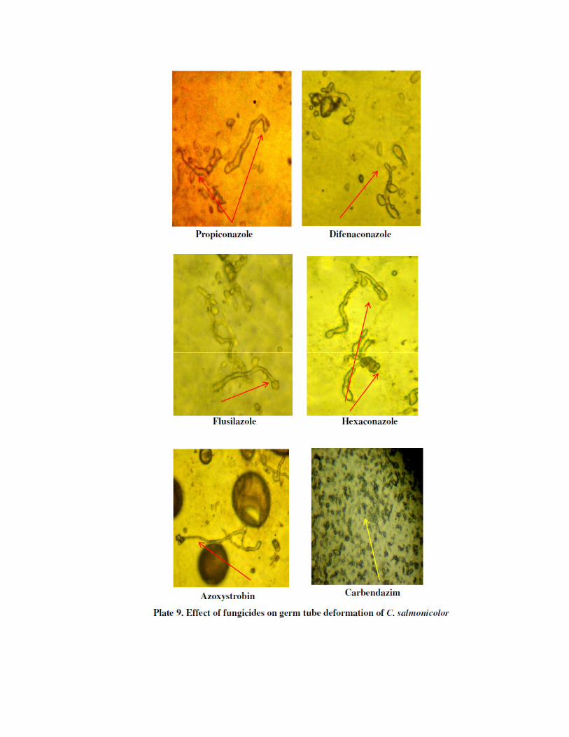
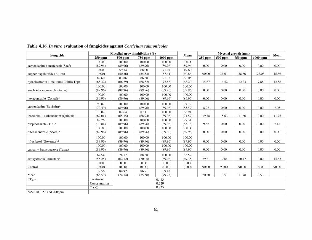
65
Table 4.16. In vitro evaluation of fungicides against Corticium salmonicolor
Mycelial growth inhibition (%) Mycelial growth (mm) Fungicide
250 ppm 500 ppm 750 ppm 1000 ppm Mean
250 ppm 500 ppm 750 ppm 1000 ppm Mean
carbendazim + mancozeb (Saaf)
100.00
(89.96)
100.00
(89.96)
100.00
(89.96)
100.00
(89.96)
100.00
(89.96) 0.00 0.00 0.00 0.00 0.00
copper oxychloride (Blitox)
0.00
(0.00)
59.34
(50.36)
68.00
(55.53)
71.07
(57.44)
49.60
(40.83) 90.00 36.61 28.80 26.03 45.36
pyraclostrobin + metiram (Cabrio Top)
82.60
(65.32)
83.86
(66.29)
86.38
(68.32)
91.35
(72.88)
86.05
(68.20) 15.67 14.52 12.23 7.88 12.58
zineb + hexaconazole (Avtar)
100.00
(89.96)
100.00
(89.96)
100.00
(89.96)
100.00
(89.96)
100.00
(89.96) 0.00 0.00 0.00 0.00 0.00
hexaconazole (Contaf)*
100.00
(89.96)
100.00
(89.96)
100.00
(89.96)
100.00
(89.96)
100.00
(89.96) 0.00 0.00 0.00 0.00 0.00
carbendazim (Bavistin)* 90.87
(72.49)
100.00
(89.96)
100.00
(89.96)
100.00
(89.96)
97.72
(85.59) 8.22 0.00 0.00 0.00 2.05
iprodione + carbendazim (Quintal)
78.02
(62.01)
82.64
(65.35)
87.11
(68.94)
100.00
(89.96)
86.94
(71.57) 19.78 15.63 11.60 0.00 11.75
propiconazole (Tilt)*
89.26
(70.84)
100.00
(89.96)
100.00
(89.96)
100.00
(89.96)
97.31
(85.18) 9.67 0.00 0.00 0.00 2.42
difenaconazole (Score)*
100.00
(89.96)
100.00
(89.96)
100.00
(89.96)
100.00
(89.96)
100.00
(89.96) 0.00 0.00 0.00 0.00 0.00
flusilazol (Governor)*
100.00
(89.96)
100.00
(89.96)
100.00
(89.96)
100.00
(89.96)
100.00
(89.96) 0.00 0.00 0.00 0.00 0.00
captan + hexaconazole (Taqat)
100.00
(89.96)
100.00
(89.96)
100.00
(89.96)
100.00
(89.96)
100.00
(89.96) 0.00 0.00 0.00 0.00 0.00
azoxystrobin (Amistar)*
67.54
(55.25)
78.17
(62.12)
88.38
(70.05)
100.00
(89.96)
83.52
(69.35) 29.21 19.64 10.47 0.00 14.83
Control
0.00
(0.00)
0.00
(0.00)
0.00
(0.00)
0.00
(0.00)
0.00
(0.00) 90.00 90.00 90.00 90.00 90.00
Mean
77.56
(66.59)
84.92
(74.14)
86.91
(75.58)
89.42
(79.23)
20.20 13.57 11.78 9.53
CD0.05 Treatment
Concentration
T x C
0.413
0.229
0.825
*=50,100,150 and 200ppm

66
metiram and captan + hexaconazole were least effective as compared to control
(84.56%). A Strobilurin fungicide like azoxystrobin which is ecofriendly and safe to
use was also found effective in reducing the conidial germination at higher
concentrations. It was found that EBI fungicides do not affect conidial germination
although germ tube length was reported to be inhibited and distorted.
Table 4.17. In vitro effect of fungicides on conidial germination of C.
salmonicolor
Mean conidial germination (%)
Fungicide 250 ppm 500 ppm 750 ppm 1000 ppm
Overall
Mean
carbendazim + mancozeb (Saaf) 0.00 (0.00) 0.00 (0.00) 0.00 (0.00) 0.00 (0.00) 0.00 (0.00)
pyraclostrobin + metiram (Cabrio Top) 34.82 (36.15) 27.69 (31.74) 21.74 (27.78) 11.38 (19.69) 23.91 (28.84)
zineb + hexaconazole (Avtar) 26.40 (30.90) 12.32 (20.53) 8.34 (16.77) 6.38 (14.61) 13.36 (20.70)
hexaconazole (Contaf) * 18.84 (25.71) 9.99 (18.42) 7.42 (15.78) 5.05 (12.96) 10.32 (18.22)
carbendazim (Bavistin) * 0.00 (0.00) 0.00 (0.00) 0.00 (0.00) 0.00 (0.00) 0.00 (0.00)
iprodione + carbendazim (Quintal) 0.00 (0.00) 0.00 (0.00) 0.00 (0.00) 0.00 (0.00) 0.00 (0.00)
propiconazole (Tilt) * 19.64 (26.30) 12.39 (20.60) 8.45 (16.89) 5.14 (13.10) 11.41 (19.22)
difenaconazole (Score) * 5.50 (13.53) 4.04 (11.56) 3.58 (10.89) 2.76 (9.49) 3.97 (11.37)
flusilazol (Governor) * 21.46 (27.58) 17.07 (24.39) 14.44 (22.32) 9.47 (17.91) 15.61 (23.05)
captan + hexaconazole (Taqat) 25.69 (30.44) 22.68 (28.42) 18.77 (25.66) 15.04 (22.81) 20.55 (26.83)
azoxystrobin (Amistar)* 6.19 (14.38) 4.79 (12.63) 3.77 (11.18) 2.32 (8.74) 4.27 (11.73)
Control 84.56 66.84 84.56 (66.84) 84.56 (66.84) 84.56 (66.84) 84.56 (66.84)
Mean 20.26 (22.65) 16.269(19.59) 14.26 (17.84) 11.84 (15.51)
CD0.05 Treatment
Concentration
T x C
0.466
0.269
0.933
*=50,100,150 and 200ppm
4.8.3. Efficacy of fungicide under field conditions
A field experiment was laid out in Randomized Block Design at the farmer’s
field at Kotgarh, district Shimla, H.P. for two consecutive years (2011-12) to evaluate
in vitro effective fungicide (3.8.2) applied as paint against the target disease and the
data pertaining to per cent wound recovery and callus formation were recorded and
presented in Table- 4.18.
The perusal of data (Table 4.18 & Plate 10) indicated that triazoles like,
difenaconazole (250 µl l-1
) and hexaconazole (500 µl l-1
) when applied on scarified
canker lesions as a paint in the month of Nov-Dec. provided 92.04 and 89.00 per cent
wound recovery with more than 10 mm callus formation followed by flusilazole (100
µl l-1
) and carbendazim + mancozeb (2500 µl l-1
) with 87.80 and 86.69 per cent
wound recovery respectively for two consecutive crop seasons (2011-12) than white
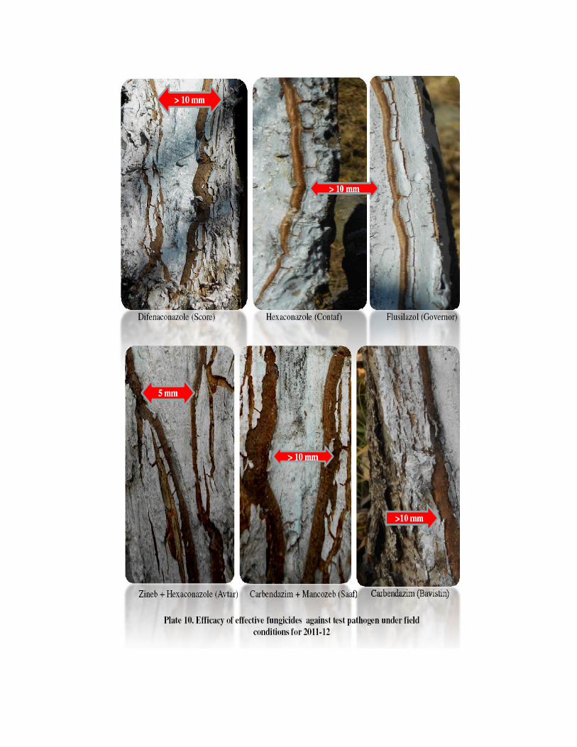

67
paint alone (13.56%) wound recovery with 0.1-5mm callus formation only.
Propiconazole (0.05%) and carbendazim (0.05%) alone was next best in order with
85.62 and 81.73 per cent wound recovery respectively. Whereas iprodione +
carbendazim (0.15%) was found to be least effective with 51.38 per cent wound
recovery with more than 5mm callus formation during 2011-12 crop seasons as
compared to untreated check wherein no wound recovery and callus formation had
taken place.
Table 4.18. Efficacy of fungicide against C. salmonicolor under field condition
for two consecutive years (2011-12)
Callus formation Wound recovery
(%) Fungicide
Conc.
(%)
2011 2012 2011 2012
Pooled
carbendazim + mancozeb (Saaf) 0.25 +++ +++ 86.87
(69.55)
85.58
(67.81)
86.69
(68.65)
pyraclostrobin + metiram
(CabrioTop)
0.10 ++ ++ 65.27
(55.74)
62.55
(52.26)
65.40
(53.97)
zineb + hexaconazole (Avtar) 0.25 +++ +++ 89.91
(70.68)
80.93
(64.10)
84.99
(67.18)
hexaconazole (Contaf) 0.05 +++ +++ 87.04
(69.77)
89.97
(71.71)
89.00
(70.61)
carbendazim (Bavistin) 0.05 +++ +++ 85.85
(67.06)
78.67
(62.48)
81.73
(64.68)
iprodione + carbendazim (Quintal) 0.15 ++ ++ 51.07
(46.67)
49.82
(44.88)
51.38
(45.77)
propiconazole (Tilt) 0.05 +++ +++ 89.02
(70.70)
82.15
(65.16)
85.62
(67.75)
difenaconazole (Score) 0.025 +++ +++ 92.08
(74.00)
91.72
(73.33)
92.04
(73.59)
flusilazol (Governor) 0.01 +++ +++ 90.71
(70.18)
87.29
(69.17)
87.80
(69.66)
captan + hexaconazole (Taqat) 0.20 +++ +++ 73.28
(59.30)
81.41
(64.49)
77.67
(61.81)
azoxystrobin (Amistar) 0.05 ++ ++ 63.39
(53.54)
71.41
(57.66)
68.05
(55.57)
White Paint + + 13.58
(21.60)
13.54
(21.58)
13.56
(21.60)
Control - - 0.00
(0.00)
0.00
(0.00)
0.00
(0.00)
Mean 68.31
(56.06)
67.31
(54.97)
67.99
(55.45)
CD0.05
Fungicides 2.518
Year 0.988
Fungicides x Year 3.561

68
4.8.4. Plant extract
4.8.4.a. In vitro evaluation of botanicals
The efficacy of twenty plant origin biopesticides (Table 4.19 & Plate a & b)
viz. Allium sativum (Cloves), Artemisia roxburghiana (Leaves), Mentha pipereta
(Leaves), Roylea elegans (Leaves), Eucalyptus globulus (Leaves), Vitex negundo
(Leaves), Cymbopogon citratus (Leaves), Melia azedarach (Seed), Adhatoda vasica
(Leaves), Emblica officinalis (Leaves), Prunus persica (Leaves), Juglans regia
(Leaves), Lantana camera (Leaves), Carya illieonsis (Shell), Brassica juncea var.
cunefolia (Leaves), Ocimum sanctum (Leaves) and a cow urine based combinations
(Plate 11b) i.e. Cow urine + Darek + Bana + Walnut leaves, Cow urine + Darek +
Eucalyptus + Bougainvellia leaves, Cow urine + Darek seed + Bana + Artimisia sp.+
Walnut leaves + Kadu, and Cow urine + Darek seed + Bana + Artimisia + Walnut
leaves were evaluated at 10,20,50 and 100 per cent by “Poisoned food technique”(
3.8.3. a.) and the data on mycelial growth inhibition in each treatment was recorded
and presented in Table 4.19.
The data (Table 4.19 & Plate.11a, & b) on per cent growth inhibition of
Corticium salmonicolor by different plant extracts used under in vitro conditions
revealed that aqueous extracts of Adhatoda vasica (Leaves) along with two
combinations of Cow urine + Melia azedarach (seed) + Vitex negundo (L) + Artimisia
roxburghiana (L) + Juglans regia (L) + Roylea elegans (L) and Cow urine + Melia
azedarach (seed) + Vitex negundo (L) + Artimisia roxburghiana (L) + Juglans regia
(L), all in 1:1 ratio exhibited complete (100%) growth inhibition at 10, 20 50 and 100
per cent concentrations respectively. The interaction between treatment and
concentrations were found to be highly significant. Whereas combination of cow
urine + Melia azedarach (Seed) + Vitex negundo (Leaves) + Juglans regia (L), and
cow urine + Melia azedarach (Seed) + Eucalyptus globulus (L) + Bougainvellia
spectabilis (L) resulted 94.45 and 95.20 per cent growth inhibition respectively.


69
Table.4.19. In vitro evaluation of botanicals against C. salmonicolor
Growth inhibition (%) Mycelial growth (mm) Botanical
10 % 20 % 50 % 100 %
Mean
10 % 20 % 50 % 100 %
Mean
Artemisia roxburghiana L. 4.88 (12.71) 6.38 (14.59) 17.51 (24.73) 67.76 (55.39) 24.13 (26.86) 85.6 84.25 74.25 29.05 68.29
Allium sativum L. 33.39 (35.27) 52.06 (46.17) 76.68 (61.10) 100.00 (89.96) 65.53 (58.13) 60.01 43.15 20.98 0.00 31.04
Mentha pipereta L. 84.37 (66.69) 87.70 (69.46) 89.82 (71.37) 92.45 (74.04) 88.58 (70.39) 14.94 11.0 9.15 6.78 10.47
Roylea elegans Wall. 59.69 (50.73) 72.72 (58.49) 76.38 (60.90) 83.94 (66.35) 73.18 (59.12) 36.27 24.55 21.25 14.45 24.13
Eucalyptus globulus Labill. 55.52 (48.15) 59.73 (50.59) 64.04 (53.13) 66.44 (54.58) 61.43 (51.61) 40.00 36.24 32.25 30.20 34.67
Vitex negundo Linn. 25.40 (30.24) 28.49 (32.24) 75.31 (60.18) 84.05 (66.44) 53.31 (47.28) 67.1 64.35 22.27 14.01 41.93
Cymbopogon citratus (DC) Stapf 17.31 (24.57) 51.21 (45.68) 71.46 (57.68) 82.38 (65.16) 55.59 (48.27) 74.58 43.89 25.69 15.86 40.01
Melia azedarach L. 57.01 (49.01) 66.69 (54.73) 72.43 (58.31) 78.36 (62.25) 68.62 (56.07) 38.69 29.98 24.79 19.48 28.24
Adhatoda vasica Nees. 100.00 (89.96) 100.00 (89.96) 100.00 (89.96) 100.00 (89.96) 100.00 (89.96) 0.00 0.00 0.00 0.00 0.00
Emblica officinalis Gaertn. 32.60 (34.80) 36.31 (37.04) 39.14 (38.71) 53.15 (46.78) 40.30 (39.33) 60.00 57.32 54.77 42.15 53.56
Cow urine + Melia azedarach + Vitex nigundo + Juglans regia
(1:1:1:1)
77.79 (61.86) 100.00 (89.96) 100.00 (89.96) 100.00 (89.96) 94.45 (82.94) 20.0 0.00 0.00 0.00 5.00
Prunus persica L. 62.63 (52.30) 68.52 (55.85) 100.00 (89.96) 100.00 (89.96) 82.79 (72.02) 33.64 28.33 0.00 0.00 15.49
Juglans regia L. 0.00 (0.00) 0.00 (0.00) 7.57 (15.95) 13.64 (21.66) 5.30 (9.40) 90.0 90.0 83.18 77.72 85.23
Lantana camera L. 17.94 (25.04) 39.25 (38.77) 46.98 (43.25) 50.17 (45.08) 38.58 (38.04) 73.85 54.68 47.71 44.84 55.27
Carya illieonsis K.Koch. 0.00 (0.00) 0.00 (0.00) 0.00 (0.00) 0.00 (0.00) 0.00 (0.00) 90.00 90.00 90.00 90.00 90.00
Cow urine + Melia azedarach + Eucalyptus globulus +
Bougainvellia spectabilis
80.78 (64.03) 100.00 (89.96) 100.00 (89.96) 100.00 (89.96) 95.20 (83.48) 17.30 0.00 0.00 0.00 4.33
Cow urine + Melia azedarach + Vitex nigundo + Artimisia
roxburghiana + Juglans regia + Roylea elegans (1:1:1:1:1:1)
100.00 (89.96) 100.00 (89.96) 100.00 (89.96) 100.00 (89.96) 100.00 (89.96) 0.000 0.00 0.00 0.00 0.00
Brassica juncea var. cunefolia Roxb. 90.59 (72.11) 100.00 (89.96) 100.00 (89.96) 100.00 (89.96) 97.65 (85.50) 8.42 0.00 0.00 0.00 2.11
Ocimum sanctum L. 83.95 (66.36) 86.68 (68.57) 91.32 (72.84) 100.00 (89.96) 90.49 (74.43) 14.49 11.90 7.80 0.00 8.55
Cow urine + Melia azedarach + Vitex nigundo + Artimisia
roxburghiana + Juglans regia (1:1:1:1:1)
100.00 (89.96) 100.00 (89.96) 100.00 (89.96) 100.00 (89.96) 100.00 (89.96) 0.00 0.00 0.00 0.00 0.00
Control 0.00 (0.00) 0.00 (0.00) 0.00 (0.00) 0.00 (0.00) 0.00 (0.00) 90.00 90.00 90.00 90.00 90.00
Mean 51.61 (45.89) 59.80 (52.95) 68.03 (59.42) 74.87 (65.11) 43.57 36.17 28.77 22.60
CD0.05 Treatment (T) 1.072
Concentration (C) 0.468
T x C 2.144

70
While extracts of Brassica juncea var. cunefolia (mustard leaves), Ocimum
sanctum (tulsi), Mentha pipereta (mentha), Prunus persica (peach leaf) and Roylea
elegans (kadu) were found to provide 97.65, 90.49, 88.58, 82.79 and 73.18 per cent
growth inhibition respectively. While rest of plant extracts obtained from Artimisia
roxburghiana, Allium sativum, Eucalyptus globulus, Vitex negundo, Cymbopogon
citratus, Melia azedarach, Emblica officinalis, Juglans regia, Lantana camera and Carya
illieonsis were found to exhibit moderate to poor growth inhibition against the test
pathogen. It was found that as the concentration of these botanicals increased there is
corresponding increase in per cent growth inhibition.
4.8.4b. Efficacy of botanicals under field conditions against C. salmonicolor
Efficacy of in vitro effective eleven botanicals viz., Mentha pipereta, Roylea
elegans, Melia azedarach, Adhatoda vasica, Ocimum sanctum, Prunus persica, Brassica
juncea var. cunefolia and cow urine based formulations like Cow urine + Melia
azedarach + Vitex negundo + Juglans regia (1:1:1:1) , Cow urine + Melia azedarach +
Vitex negundo + Artimisia roxburghiana + Juglans regia + Roylea elegans (1:1:1:1:1:1),
Cow urine + Melia azedarach + Eucalyptus lobules + Bougainvellia spectabilis
(1:1:1:1), Cow urine + Melia azedarach + Vitex negundo + Artimisia roxburghiana +
Juglans regia (1:1:1:1:1) were evaluated (3.8.3. b) and the data on wound recovery and
callus formation in each treatment was recorded and presented in Table 4.20.
A perusal of the data (Table 4.20 & Plate 12.a) reveals that irrespective of the
concentration mean wound recovery was significantly maximum (58.54 %) in cow urine
based formulation i.e. Cow urine + Melia azedarach + Vitex negundo + Artimisia
roxburghiana + Juglans regia + Roylea elegans (1:1:1:1:1:1) followed by formulation of
Cow urine + Melia azedarach + Vitex negundo + Artimisia roxburghiana + Juglans regia
(1:1:1:1:1) with wound recovery of 55.44 per cent with a callus formulation of 5-10 mm
respectively. Adhatoda vasica alone when added in white paint @ 0.10 per cent provided
52.91 per cent wound recovery with >5 mm callus formation which was statistically at
par with each other. Cow urine based formulation of Cow urine + Melia azedarach +
Vitex negundo + Juglans regia (1:1:1:1) and Cow urine + Melia azedarach + Eucalyptus
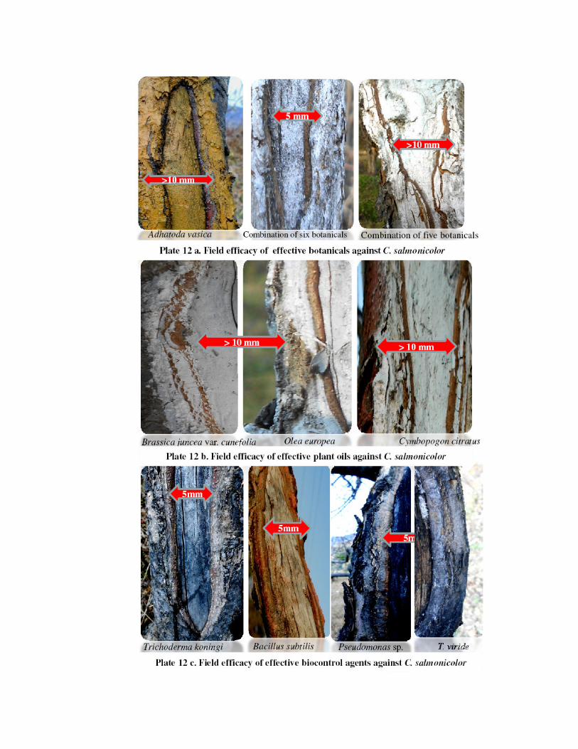
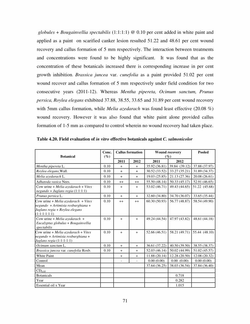
71
globules + Bougainvellia spectabilis (1:1:1:1) @ 0.10 per cent added in white paint and
applied as a paint on scarified canker lesion resulted 51.22 and 48.61 per cent wound
recovery and callus formation of 5 mm respectively. The interaction between treatments
and concentrations were found to be highly significant. It was found that as the
concentration of these botanicals increased there is corresponding increase in per cent
growth inhibition. Brassica juncea var. cunefolia as a paint provided 51.02 per cent
wound recover and callus formation of 5 mm respectively under field condition for two
consecutive years (2011-12). Whereas Mentha pipereta, Ocimum sanctum, Prunus
persica, Roylea elegans exhibited 37.88, 38.55, 33.65 and 31.89 per cent wound recovery
with 5mm callus formation, while Melia azedarach was found least effective (20.08 %)
wound recovery. However it was also found that white paint alone provided callus
formation of 1-5 mm as compared to control wherein no wound recovery had taken place.
Table 4.20. Field evaluation of in vitro effective botanicals against C. salmonicolor
Callus formation Wound recovery
(%) Botanical
Conc.
(%)
2011 2012 2011 2012
Pooled
Mentha pipereta L. 0.10 + + 35.92 (36.81) 39.84 (39.12) 37.88 (37.97)
Roylea elegans Wall. 0.10 + + 30.52 (33.52) 33.27 (35.21) 31.89 (34.37)
Melia azedarach L. 0.10 + + 19.03 (25.85) 21.13 (27.36) 20.08 (26.61)
Adhatoda vasica Nees. 0.10 ++ ++ 55.50 (48.14) 50.33 (45.17) 52.91 (46.65)
Cow urine + Melia azedarach + Vitex
negundo + Juglans regia (1:1:1:1)
0.10 + + 53.02 (46.71) 49.43 (44.65) 51.22 (45.68)
Prunus persica L. 0.10 + + 32.60 (34.80) 34.70 (36.07) 33.65 (35.44)
Cow urine + Melia azedarach + Vitex
negundo + Artimisia roxburghiana +
Juglans regia + Roylea elegans
(1:1:1:1:1:1)
0.10 ++ ++ 60.30 (50.93) 56.77 (48.87) 58.54 (49.90)
Cow urine + Melia azedarach +
Eucalyptus globulus + Bougainvellia
spectabilis
0.10 + + 49.24 (44.54) 47.97 (43.82) 48.61 (44.18)
Cow urine + Melia azedarach + Vitex
negundo + Artimisia roxburghiana +
Juglans regia (1:1:1:1:1)
0.10 + + 52.66 (46.51) 58.21 (49.71) 55.44 (48.10)
Ocimum sanctum L. 0.10 + + 36.61 (37.22) 40.50 (39.50) 38.55 (38.37)
Brassica juncea var. cunefolia Roxb. 0.10 + + 52.03 (46.14) 50.02 (44.99) 51.02 (45.57)
White Paint + + 11.88 (20.14) 12.28 (20.50) 12.08 (20.32)
Control - - 0.00 (0.00) 0.00 (0.00) 0.00 (0.00)
Mean 37.64 (36.25) 38.03 (36.54) 37.84 (36.40)
CD0.05
Botanicals 0.718
Year 0.282
Essential oil x Year 1.015
72
4.8.5 Plant oils
4.8. 5a. In vitro evaluation of plant oils
In vitro efficacy of thirteen plant oils like, Tagetes minuta, Artemisia
roxburghiana, Eugenia caryophyllata, Olea cuspidata, Cymbopogon citratus, Ocimum
sanctum, Azadirachta indica, Brassica juncea var. cunefolia, Eucalyptus globulus,
Prunus armeniaca, Ricinus communis (Seed), Juniperus virginiana (Wood) and Juglans
regia (Kernels) were evaluated against C. salmonicolor by “Poisoned food technique
(3.8.4. a.) and data on per cent mycelial inhibition recorded and presented in Table 4.21
and Plate 13.
It is apparent from the data (Table 4.21 and Plate 13) which revealed that per cent
growth inhibition was significantly maximum (100%) in Olea cuspidata, Cymbopogon
citratus and Brassica juncea var. cunefolia at all concentrations tested, followed by
Eugenia caryophyllata (94.00 %), Ocimum sanctum (83.98 %), and Azadirachta indica
(85.95 %) respectively. Plant oils from Juglans regia, Ricinus communis and Juniperus
virginiana provided 76.06, 78.90, 73.48 per cent growth inhibition respectively, against
the test pathogen. Rest of the essential oils from wild marigold (Tagetes minuta),
eucalyptus (Eucalyptus globulus), and Artemisia roxburghiana were found to be less
effective. It was also recorded that as the concentration of these bio-pesticides increased,
there was a corresponding increase in per cent growth inhibition with the highest at 100
per cent concentration. The interaction between treatment and concentrations were found
to be highly significant. While wild apricot (Prunus armeniaca) was found to be least
effective (15.72%) mycelial inhibition.
4.8. 5b. Evaluation of plant oils under field condition against C. salmonicolor
Nine in vitro effective plant oils viz., Eugenia caryophyllata, Cymbopogon
citratus, Olea europea, Ocimum sanctum, Azadirachta indica, Brassica juncea var.
cunefolia, Ricinus communis, Juniperus virginiana, Juglans regia along with white paint
separately were evaluated at 5 per cent concentration under field condition for two crop
seasons (2011 and 2012) and the data regarding wound recovery (%) and callus
formation are presented in Table 4.22 & Plate 12.b.
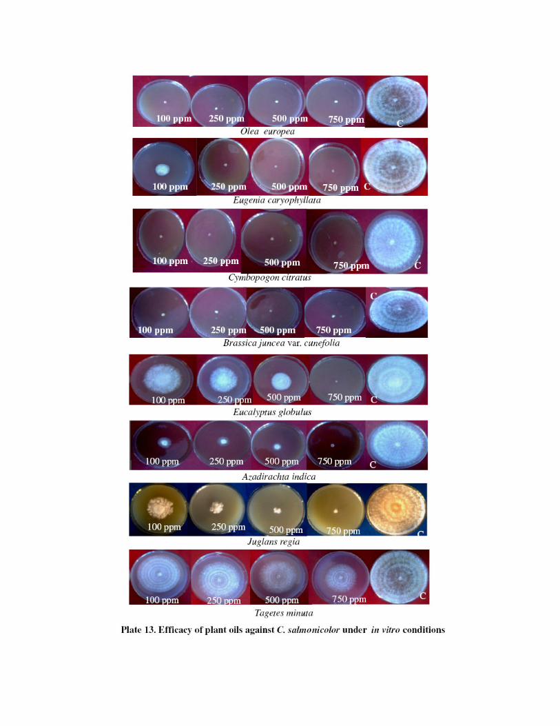
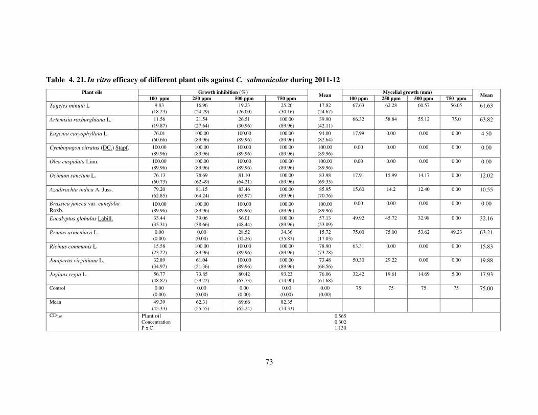
73
Table 4. 21. In vitro efficacy of different plant oils against C. salmonicolor during 2011-12
Growth inhibition (%) Mycelial growth (mm) Plant oils
100 ppm 250 ppm 500 ppm 750 ppm Mean
100 ppm 250 ppm 500 ppm 750 ppm Mean
Tagetes minuta L 9.83
(18.23)
16.96
(24.29)
19.23
(26.00)
25.26
(30.16)
17.82
(24.67)
67.63 62.28 60.57 56.05 61.63
Artemisia roxburghiana L. 11.56
(19.87)
21.54
(27.64)
26.51
(30.96)
100.00
(89.96)
39.90
(42.11)
66.32 58.84 55.12 75.0 63.82
Eugenia caryophyllata L. 76.01
(60.66)
100.00
(89.96)
100.00
(89.96)
100.00
(89.96)
94.00
(82.64)
17.99 0.00 0.00 0.00 4.50
Cymbopogon citratus (DC.) Stapf. 100.00
(89.96)
100.00
(89.96)
100.00
(89.96)
100.00
(89.96)
100.00
(89.96)
0.00 0.00 0.00 0.00 0.00
Olea cuspidata Linn. 100.00
(89.96)
100.00
(89.96)
100.00
(89.96)
100.00
(89.96)
100.00
(89.96)
0.00 0.00 0.00 0.00 0.00
Ocimum sanctum L. 76.13
(60.73)
78.69
(62.49)
81.10
(64.21)
100.00
(89.96)
83.98
(69.35)
17.91 15.99 14.17 0.00 12.02
Azadirachta indica A. Juss. 79.20
(62.85)
81.15
(64.24)
83.46
(65.97)
100.00
(89.96)
85.95
(70.76)
15.60 14.2 12.40 0.00 10.55
Brassica juncea var. cunefolia
Roxb. 100.00
(89.96)
100.00
(89.96)
100.00
(89.96)
100.00
(89.96)
100.00
(89.96)
0.00 0.00 0.00 0.00 0.00
Eucalyptus globulus Labill. 33.44
(35.31)
39.06
(38.66)
56.01
(48.44)
100.00
(89.96)
57.13
(53.09)
49.92 45.72 32.98 0.00 32.16
Prunus armeniaca L. 0.00
(0.00)
0.00
(0.00)
28.52
(32.26)
34.36
(35.87)
15.72
(17.03)
75.00 75.00 53.62 49.23 63.21
Ricinus communis L 15.58
(23.22)
100.00
(89.96)
100.00
(89.96)
100.00
(89.96)
78.90
(73.28)
63.31 0.00 0.00 0.00 15.83
Juniperus virginiana L. 32.89
(34.97)
61.04
(51.36)
100.00
(89.96)
100.00
(89.96)
73.48
(66.56)
50.30 29.22 0.00 0.00 19.88
Juglans regia L. 56.77
(48.87)
73.85
(59.22)
80.42
(63.73)
93.23
(74.90)
76.06
(61.68)
32.42 19.61 14.69 5.00 17.93
Control 0.00
(0.00)
0.00
(0.00)
0.00
(0.00)
0.00
(0.00)
0.00
(0.00)
75 75 75 75 75.00
Mean 49.39
(45.33)
62.31
(55.55)
69.66
(62.24)
82.35
(74.33)
CD0.05
Plant oil Concentration
P x C
0.565
0.302
1.130

74
Table 4.22. Efficacy of in vitro effective plant oils as a paint against C.
salmonicolor under field conditions
Callus formation Wound recovery (%) Plant oils
Conc.
(%) 2011 2012 2011 2012 Pooled
Eugenia caryophyllata L. 0.05 ++ ++ 69.55
(56.48)
64.99
(53.71)
67.77
(55.08)
Cymbopogon citratus
(DC) Stapf.
0.05 +++ +++ 73.58
(59.05)
71.86
(57.94)
72.72
(58.49)
Olea europea L. 0.05 +++ +++ 74.71
(59.79)
76.96
(61.29)
75.83
(60.53)
Ocimum sanctum L. 0.05 + + 47.57
(43.53)
44.60
(41.89)
46.09
(42.74)
Azadirachta indica A.
Juss.
0.05 ++ ++ 54.98
(47.84)
50.44
(45.23)
52.71
(46.53)
Brassica juncea var.
cunefolia Roxb.
0.05 +++ +++ 80.13
(63.51)
82.41
(65.18)
81.27
(64.33)
Ricinus communis L 0.05 ++ + + 43.01
(40.96)
41.25
(39.95)
42.13
(40.64)
Juniperus virginiana L. 0.05 + + 30.63
(33.59)
33.29
(35.21)
31.96
(34.41)
Juglans regia L. 0.05 + + 39.58
(38.97)
41.03
(39.82)
40.31
(39.39)
White Paint - + + 11.72
(20.01)
12.12
(20.36)
11.92
(20.19)
Control - - 0.00
(0.00)
0.00
(0.00)
0.00
(0.00)
Mean
47.77
(42.16)
47.18
(41.87)
47.47
(42.01)
CD0.05
Plant oils
Year
Plant oils x Year
0.560
0.239
0.792
It is clear from the data (Table 4.22 & Plate 12.b.) that significantly maximum
wound recovery (81.27%) was achieved with Brassica juncea var. cunefolia, when
applied as paint against C. salmonicolor under field conditions for two years followed
by Olea europea (75.83 %) with more than 10 mm callus formation respectively.
Cymbopogon citratus, Eugenia caryophyllata, Azadirachta indica, Ocimum sanctum
was found next best in order with 72.72, 67.77, 52.71 and 46.09 per cent wound
recovery respectively. Whereas Ricinus communis and Juglans regia were found less
effective in controlling disease as compared to Juniperus virginiana which was least
effective (31.96%). White paint alone resulted 11.92 per cent wound recovery with 1-
5 mm callus formation than in control wherein no wound recovery and callus
formation occurred.
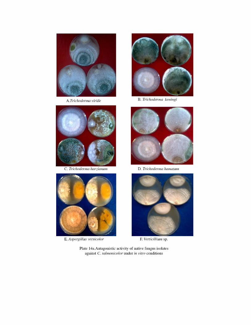
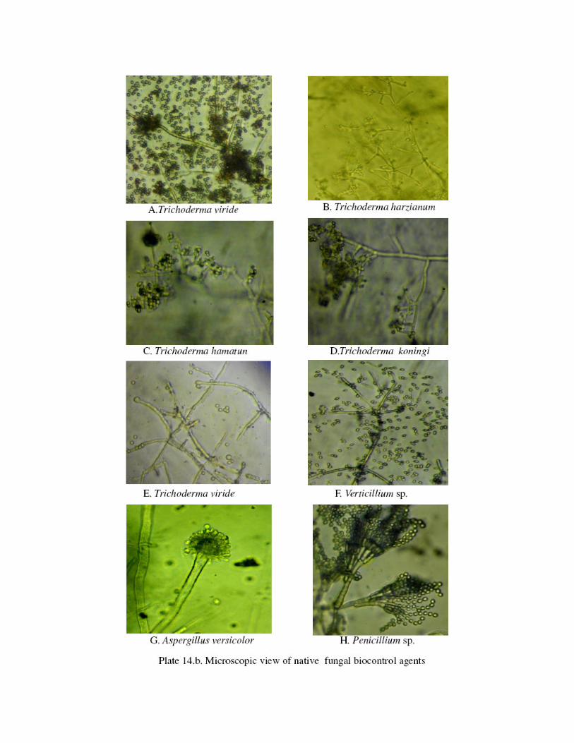

75
4.8.6 Biological control
4.8.6.1. Isolation and Identification of antagonists
The microflora (fungi, bacteria and actinomycete) isolated from in and around
the infected branches and twigs of apple trees grown in different geographical
locations viz., Mandi, Kotgarh, Haripurdhar and Kullu district of Himachal Pradesh
were identified based on their specific characteristic features. Total of seventeen
antagonists (Table 4.22) including 11 fungal biocontrol agents viz., Tv1 (Mandi) and
Tv2 (Kotgarh) isolates of Trichoderma viride; TH1 (Kotgarh) and TH2 (Mandi) of
Trichoderma harzianum, Trichoderma koningi, Th1 (Kullu) and Th2 (Haripurdhar)
isolates of Trichoderma hamatum, Trichoderma viride + Aspergillus sp., Verticillium
sp. P1 of Penicillium sp. (Kotgarh) and Aspergillus versicolor and five bacteria i.e.
BS1 (Kotgarh), BS2 (Kullu) and BS3 (Mandi) of Bacillus subtilis, Pseudomonas sp.
and Pseudomonas aeruginosa and an actinomycete were isolated during the course of
study in 2011and 2012.
Fungal biocontrol agents especially Trichoderma sp. were identified from
National Centre for Fungal Taxonomy, New Delhi by Dr. P.N. Chaudhary and
assigned ID no. as Trichoderma koningi (ID no-5216.12), Trichoderma hamatum (ID
no-5213.12), Trichoderma harzianum (ID no-5212.12), Trichoderma viride (ID no-
5214.12), Trichoderma viride + Aspergillus sp.(ID-5215.12). Whereas, Aspergillus
versicolor was also isolated and identified as yellow tan, pale green or pink colour
colony surface (Plate 14.a &b). Two bacterial antagonists’ viz., Bacillus subtilis and
Pseudomonas sp. were identified on the basis of genomic sequence submitted to
NCBI.
Based on morphological and cultural studies, PGPRs were identified.
Pseudomonas sp. (BS2) was identified as gram – ve rod shaped bacteria, produces
fluorescent pigment (blue), grows on King’s ( Proteose peptone 3 (Difco) – 20gm,
K2HPO4.3H2O – 1.5 gm, MgSO4.7H2O – 1.5 gm, Agar -15 gm, Glycerol – 15 ml),
nutrient agar ((Beef extract- 3 gm , peptone- 5 gm, NaCl-5 gm, Agar –agar-15-20
gm, distilled water - 1000 ml) medium within 24hr whereas, Pseudomonas
aeruginosa (BS3) was identified as gram –ve rod shaped, produces chocolate brown
pigment, growth within 24hr at 30 °C. Bacillus sp. was identified as gram + ve rod

76
shaped, creamy white/light brown colony colour, mucoid, grows on NA/ PDA
medium within 24hr, anaerobic (Plate 14.c.).
Molecular analysis and amplification of 16S DNA and protease apr gene was
achieved by PCR by isolating DNA. Phylogenetic trees were constructed for each
bacterium. There were three clusters viz., cluster I, II and III. Cluster I is further
subdivided in to cluster I. a and I. b. Cluster I. a. consisted of Bacillus subtilis,
Kotgarh isolate (BS1) and JQ927217 Bacillus subtilis strain AZ01, whereas cluster I.
b. consisted of only AB734701 Bacillus subtilis. However cluster II and III consisted
of HM154526 Bacillus subtilis and HE17022 Bacillus subtilis aprN which were found
distant relatives of Kotgarh isolate of Bacillus subtilis (Fig 4.8.6.1.a.)
Fig 4.8.6.1.a. Phylogenetic Tree made using Neighbour Joining method
The phylogenetic analysis and sequence comparison of BS2 isolate revealed its
identity as Pseudomonas sp., The BS2 sequence had been submitted to NCBI and its
GenBank Accession Number is KF564924. Based on phylogenetic analysis, three
clusters were constructed, cluster i., consisted of only DQ095204 Pseudomonas
fluorescens whereas, GQ228659 uncultured Pseudomonas sp. in cluster ii. BS2 isolate
of bacterium was closest to GQ228659 uncultured Pseudomonas sp. in cluster ii with
highest sequence similarities. Cluster iii. consisted of CT573326 Pseudomonas
entomophila and further sub-divided in to cluster iii a and b. CP003588 Pseudomonas
putida ND and CP000712 Pseudomonas putida F1 grouped in cluster iii .a and b.,
which were distant relative to Kullu isolate of Pseudomonas sp (Fig 4.8.6.1.b.).


77
Fig 4.8.6.1.b. Phylogenetic Tree made using Neighbour Joining method
4.8.6.2. In vitro evaluation of biocontrol agents
Of the seventeen antagonists including fungi viz., Tv1 (Mandi) and Tv2
(Kotgarh) isolates of Trichoderma viride; TH1 (Kotgarh) and TH2 (Mandi) of
Trichoderma harzianum, Trichoderma koningi, Th1 (Kullu) and Th2 (Haripurdhar)
isolates of Trichoderma hamatum, Trichoderma viride + Aspergillus sp.,
Verticillium sp. P1 (Kotgarh) of Penicilluim sp. and Aspergillus versicolor and five
bacteria i.e. BS1 (Kotgarh), BS2 (Kullu) and BS3 (Mandi) of Bacillus subtilis,
Pseudomonas sp. and Pseudomonas aeruginosa and an actinomycetes were isolated
and tested under in vitro conditions by dual culture technique and streak plate method
(3.8.5.1.) and the data on percent mycelial inhibition and inhibition zone are presented
in Table 4.23 and Plate 14.a,b & c.
The perusal of data (Table 4.23 & Plate 14.a, b & c.) revealed that all
biocontrol agents either of fungal, bacterial and actinomycetes in origin inhibited the
mycelial growth of C. salmonicolor to various extents over control. However,
maximum inhibition of mycelial growth was observed in Trichoderma koningi (82.62
%) followed by Th1 of Trichoderma hamatum (78.02 %) and Th2 of Trichoderma
hamatum (77.15 %) which were statistically at par with each other. All other isolates
of Trichoderma sp. inhibited the growth of C. salmonicolor to greater extent in
comparison to other fungal flora while minimum per cent inhibition was recorded
with Aspergillus versicolor (40.92%). Amongst the bacterial and actinomycetes sp.
Pseudomonas sp. provided maximum inhibition zone (15.70 mm) followed by

78
Actinomycetes sp. (14.20 mm) which was statistically at par with each other. Among
Bacillus sp.BS1 (Kotgarh) isolate of Bacillus subtilis caused maximum inhibition zone
of 12.3 mm as compared to others. Amongst fungal biocontrol agents only Aspergillus
versicolor and Verticillium sp. provided maximum inhibition zone of 13.4 and 8.40
mm respectively.
Table 4. 23. In vitro efficacy of native biocontrol agents against C. salmonicolor
Antagonist
Growth
inhibition
(%)
Mycelial
growth
(mm)
Zone of
inhibition
(mm)
Trichoderma viride (TV1) 76.00 (60.65) 27.90 -
Trichoderma koningi 82.62 (65.33) 13.00 -
Trichoderma harzianum (TH1) 63.47 (52.79) 27.50 -
Trichoderma harzianum (TH2) 75.31 (60.19) 18.50 -
Trichoderma hamatum (Th1) 78.02 (62.06) 16.50 -
Trichoderma hamatum (Th2) 77.15 (61.50) 17.16 -
Bacillus subtilis (BS1) 51.95 (46.10) 36.05 12.30
Trichoderma viride (TV2) 73.82 (59.21) 19.70 -
Trichoderma viride + Aspergillus sp. 62.82 (52.41) 18.00 -
Pseudomonas sp. 55.17 (47.95) 33.66 15.70
Verticillium sp. 54.72 (47.69) 33.96 8.40
Bacillus subtilis (BS3) 52.75 (46.56) 35.44 7.10
Actinomycetes sp. 75.58 (60.36) 18.33 14.20
Penicilluim sp. (P1) 46.21 (42.81) 40.33 -
Bacillus subtilis (BS2) 45.56 (42.43) 40.83 6.61
Pseudomonas aeruginosa 58.44 (49.86) 31.16 9.83
Aspergillus versicolor 40.92 (39.74) 44.35 13.40
Control 0.00 (0.00) 75.00 75.00
CD0.05 2.400
4.8.6.3. Field evaluation of antagonists
In order to ascertain the efficacy of in vitro effective antagonists against
Corticium salmonicolor, the field trial were carried out with eight biocontrol agents
viz., Trichoderma harzianum, Trichoderma viride, Trichoderma koningi,
Trichoderma hamatum, Aspergillus versicolor, Pseudomonas sp., Bacillus subtilis
(BS1), Actinomycetes sp. under natural epiphytotic conditions by slurry method
(3.8.5.1.a) and the data regarding wound recovery (%) and callus formation was
recorded for two consecutive years are presented in Table 4.24 and Plate 12.c.
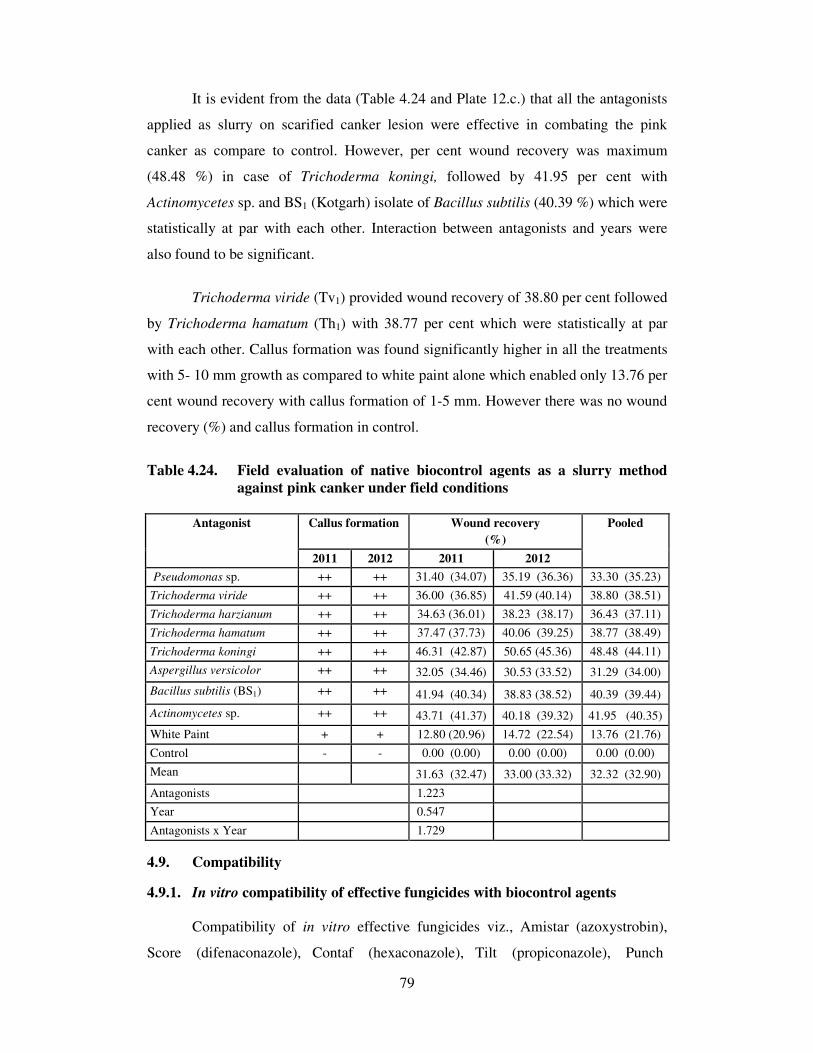
79
It is evident from the data (Table 4.24 and Plate 12.c.) that all the antagonists
applied as slurry on scarified canker lesion were effective in combating the pink
canker as compare to control. However, per cent wound recovery was maximum
(48.48 %) in case of Trichoderma koningi, followed by 41.95 per cent with
Actinomycetes sp. and BS1 (Kotgarh) isolate of Bacillus subtilis (40.39 %) which were
statistically at par with each other. Interaction between antagonists and years were
also found to be significant.
Trichoderma viride (Tv1) provided wound recovery of 38.80 per cent followed
by Trichoderma hamatum (Th1) with 38.77 per cent which were statistically at par
with each other. Callus formation was found significantly higher in all the treatments
with 5- 10 mm growth as compared to white paint alone which enabled only 13.76 per
cent wound recovery with callus formation of 1-5 mm. However there was no wound
recovery (%) and callus formation in control.
Table 4.24. Field evaluation of native biocontrol agents as a slurry method
against pink canker under field conditions
Callus formation Wound recovery
(%)
Antagonist
2011 2012 2011 2012
Pooled
Pseudomonas sp. ++ ++ 31.40 (34.07) 35.19 (36.36) 33.30 (35.23)
Trichoderma viride ++ ++ 36.00 (36.85) 41.59 (40.14) 38.80 (38.51)
Trichoderma harzianum ++ ++ 34.63 (36.01) 38.23 (38.17) 36.43 (37.11)
Trichoderma hamatum ++ ++ 37.47 (37.73) 40.06 (39.25) 38.77 (38.49)
Trichoderma koningi ++ ++ 46.31 (42.87) 50.65 (45.36) 48.48 (44.11)
Aspergillus versicolor ++ ++ 32.05 (34.46) 30.53 (33.52) 31.29 (34.00)
Bacillus subtilis (BS1) ++ ++ 41.94 (40.34) 38.83 (38.52) 40.39 (39.44)
Actinomycetes sp. ++ ++ 43.71 (41.37) 40.18 (39.32) 41.95 (40.35)
White Paint + + 12.80 (20.96) 14.72 (22.54) 13.76 (21.76)
Control - - 0.00 (0.00) 0.00 (0.00) 0.00 (0.00)
Mean 31.63 (32.47) 33.00 (33.32) 32.32 (32.90)
Antagonists 1.223
Year 0.547
Antagonists x Year 1.729
4.9. Compatibility
4.9.1. In vitro compatibility of effective fungicides with biocontrol agents
Compatibility of in vitro effective fungicides viz., Amistar (azoxystrobin),
Score (difenaconazole), Contaf (hexaconazole), Tilt (propiconazole), Punch

80
Table 4.25. Compatibility of effective fungicides with biocontrol agents under in vitro conditions
Mycelial Growth Inhibition (%)
Fungicide Conc.(%) Trichoderma
harzianum
Trichoderma
viride
Trichoderma
koningi
Trichoderma
hamatum
Pseudomonas
sp.
Aspergillus
versicolor
Bacillus subtilis
(BS1)
Actinomycetes
sp.
Mean
Avtar (zineb + hexaconazole) 0.25 0.00
(0.00) 0.00
(0.00) 49.41
(44.60) 91.17
(72.69) 0.00
(0.00) 65.68
(54.13) 0.00
(0.00) 0.00
(0.00)
25.78 (21.43)
Saaf (carbendazim + mancozeb) 0.25 100
(89.96)
100
(89.96)
100
(89.96)
100
(89.96)
0.00
(0.00)
100
(89.96)
0
(0.00)
0
(0.00)
62.50
(56.22)
Taqat (captan + hexaconazole) 0.20 80.00
(63.41)
77.50
(61.66)
70.81
(58.15)
100.0
(89.96)
0.00
(0.00)
100.0
(89.96)
0.00
(0.00)
0.00
(0.00)
53.54
(45.39)
Cabrio Top (pyraclostrobin +
metiram) 0.10
93.46
(75.15)
84.55
(66.83)
100.0
(89.96)
100.0
(89.96)
0.00
(0.00)
100.0
(89.96)
0.00
(0.00)
0.00
(0.00)
59.76
(51.48)
Tilt (propiconazole)* 0.05 100.0
(89.96)
35.11
(36.32)
67.18
(55.03)
96.61
(79.37)
0.00
(0.00)
82.09
(64.96)
0.00
(0.00)
0.00
(0.00)
47.62
(40.71)
Contaf (hexaconazole) 0.05 86.10
(68.09)
17.34
(24.59)
42.57
(40.71)
0.00
(0.00)
0.00
(0.00)
49.49
(44.69)
0.00
(0.00)
0.00
(0.00)
24.44
(22.26)
Governor (flusilazol ) 0.05 81.73
(64.67)
31.40
(34.06)
71.20
(57.52)
29.20
(32.70)
0.00
(0.00)
100.0
(89.96)
0.00
(0.00)
0.00
(0.00)
39.19
(34.86)
Bavistin (carbendazim) 0.05 96.93
(80.00)
100.0
(89.96)
100.0
(89.96)
100.0
(89.96)
0.00
(0.00)
100.0
(89.96)
0.00
(0.00)
0.00
(0.00)
62.12
(54.98)
Score (difenaconazole) 97.36
(80.64)
24.46
(29.62)
88.20
(69.88)
35.19
(36.37)
0.00
(0.00)
100.0
(89.96)
0.00
(0.00)
0.00
(0.00)
43.15
(38.31)
Amistar (azoxystrobin)* 0.025 0.00
(0.00)
39.79
(39.09)
0.00
(0.00)
39.78
(39.08)
0.00
(0.00)
56.81
(48.90)
0.00
(0.00)
0.00
(0.00)
17.05
(15.88)
Quintal (iprodione + carbendazim) 0.15 86.70
(68.59)
97.96
(81.78)
92.48
(74.06)
100.0
(89.96)
0.00
(0.00)
100.0
(89.96)
0.00
(0.00)
0.00
(0.00)
59.64
(50.54)
Control - 75
(59.98 )
75
(59.98)
75
(59.98)
75
(59.98)
75
(59.98)
75
(59.98)
75
(59.98)
75
(59.98)
Mean 74.77
(61.86)
56.93
(50.35)
71.40
(60.89)
72.25
(64.55)
46.42
(40.40)
45.58
(39.80)
6.25
(5.00)
6.25
(5.00)
CD 0.05 1.011
0.862
Fungicides Antagonists
Fungicides x Antagonists 2.859

81
(flusilazole), Bavistin (carbendazim), Saaf (carbendazim + mancozeb), Quintal
(carbendazim+ iprodione), Taqat (contaf + captan), Cabrio Top (pyraclostrobin+
metiram) and Avtar (zineb + hexaconazole) were evaluated at their recommended
dose for their compatibility with effective biocontrol agents under in vitro conditions
following poisoned food technique and the data on mycelial growth inhibition are
presented (Table 4.25 & Plate 15).
It is evident from the data (Table 4.25 and Plate15) that fungicides viz., Avtar
(zineb + hexaconazole) provided compatible reaction with Trichoderma harzianum
(TH1), Trichoderma viride (Tv1), and Pseudomonas sp., Bacillus subtilis (BS1) and
actinomycetes sp., when evaluated at 0.25 per cent, though inhibited mycelial growth
of Trichoderma koningi (49.41%), Trichoderma hamatum (91.17%) and Aspergillus
versicolor (65.68%) respectively. Strobilurin fungicide viz., Amistar (azoxystrobin)
@ 0.025 per cent was found compatible with Trichoderma harzianum (TH1),
Trichoderma koningi, Pseudomonas sp., Bacillus subtilis (BS1) and actinomycetes
sp., although inhibited mycelium of (Tv1; Mandi) isolate of Trichoderma viride (39.79
%), Trichoderma hamatum (39.78%) and Aspergillus versicolor (56.81%)
respectively. Whereas rest of fungicides like, Saaf (carbendazim + mancozeb), Taqat
(captan + hexaconazole), Cabrio Top (pyraclostrobin + metiram), Tilt
(propiconazole), Contaf (hexaconazole), Governor (flusilazol), Bavistin
(carbendazim), Score (difenaconazole) and Quintal (iprodione + carbendazim) at their
recommended dose found compatible with bacterial and actinomycetes sp., but
resulted significant mycelial inhibition of fungal isolates. Bavistin (carbendazim)
either alone or in combination i.e. Saaf (carbendazim + mancozeb) exhibited cent per
cent mycelial inhibition of Trichoderma viride (Tv1), Trichoderma koningi,
Trichoderma hamatum and Aspergillus versicolor except Trichoderma harzianum
(96.93%).
4.9.2. In vitro compatibility of effective plant oils with biocontrol agents
To known the compatibility of in vitro effective plant oils with biocontrol
agents, six plant oils viz., Brassica juncea var. cunefolia, Ocimum sanctum,
Azadirachta indica, Cymbopogon citratus, Eugenia caryophyllata and Olea europea
were evaluated at 5 per cent concentration using poisoned food technique and the data
on per cent mycelial inhibition are presented (Table 4.26 & Plate 16).

82
The perusal of data presented (Table 4.26 & Plate 16) revealed that Brassica
juncea var. cunefolia, Azadirachta indica and Olea europea @ 0.05 per cent found
compatible with all antagonists used. While Ocimum sanctum was found to inhibit
only mycelial growth of Aspergillus versicolor (45.29%) and compatible with rest of
antagonists. Similarly, Eugenia caryophyllata inhibited mycelial growth of
Trichoderma viride (14.10%). Cymbopogon citratus was found to inhibit
Trichoderma hamatum (83.58%), Trichoderma harzianum (89.57%), Aspergillus
versicolor (90.29%), Trichoderma koningi (96.55%) and Trichoderma viride
(27.73%) respectively, but compatible with bacterial antagonists.
4.10 Integrated management of pink canker
4.10.1 Integrated management of pink canker with fungicides and plant oils
Field evaluated most effective and compatible fungicides viz., Score
(difenaconazole), Contaf (hexaconazole), Punch (flusilazole), Saaf (carbendazim +
mancozeb), Taqat (contaf + captan), Avtar (zineb +hexaconazole) and plant oils viz.,
Cymbopogon citratus, Brassica juncea var. cunefolia and Olea europea were tested at
different concentrations against C. salmonicolor under field condition for two
consecutive seasons (2011 and 2012) and data on per cent wound recovery and callus
formation are presented in Table 4.27 and Plate 17.a.
The data presented in Table 4.27 clearly indicated that all combination of
fungicides and plant oils were highly effective in combating pink canker under field
conditions. However, maximum wound recovery (92.94%) and callus formation of
more than 10 mm was observed in combination of Contaf + Brassica juncea var.
cunefolia followed by combinaion of Score + Brassica juncea var. cunefolia (91.86%)
with more than 10 mm callus formation and was statistically at par with each other.
Whereas combination of Saaf + Brassica juncea var. cunefolia, Avtar +Brassica
juncea var. cunefolia, Score+ Cymbopogon citratus and Governor + Brassica
juncea var. cunefolia resulted wound recovery of 88.76, 88.71, 85.02 and 82.28 per
cent wound recovery with more than 10 mm callus formation respectively during both
the year. All other combinations of fungicides and plant oils were highly significant in
controlling pink canker with varying degree of wound recovery and callus formation
as compared to white paint alone which resulted only 16.22 per cent wound recovery
with callus formation of 1-5 mm only during both years. However minimum wound
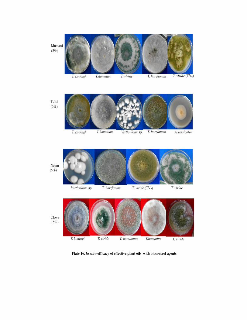

83
Table 4.26. Compatibility of in vitro effective plant oils with biocontrol agents
Mycelial Growth Inhibition (%)
Plant oil Conc.
(%) Trichoderma
harzianum
Trichoderma
viride
Trichoderma
koningi
Trichoderma
hamatum
Pseudomonas
sp.
Aspergillus
versicolor
Bacillus
subtilis (BS1)
Actinomycetes
sp.
Mean
Brassica juncea
var. cunefolia
Roxb.
0.05 0.00
0.00
0.00
0.00
0.00
0.00
0.00
0.00
0.00
0.00
0.00
0.00
0.00
0.00
0.00
0.00
0.00
0.00
Ocimum sanctum
L.
0.05 0.00
0.00
0.00
0.00
0.00
0.00
0.00
0.00
0.00
0.00
45.29
(42.28)
0.00
0.00
0.00
0.00
5.62
(5.28)
Azadirachta indica
A. Juss.
0.05 0.00
0.00
0.00
0.00
0.00
0.00
0.00
0.00
0.00
0.00
0.00
0.00
0.00
0.00
0.00
0.00
0.00
0.00
Cymbopogon
citratus (DC)
Stapf.
0.05 89.57
(71.18)
27.73
(31.76)
96.55
(79.32)
83.58
(66.08)
0.00
0.00
90.29
(71.84)
0.00
0.00
0.00
0.00
48.46
(40.20)
Eugenia
caryophyllata L.
0.05 0.00
0.00
14.10
(22.04)
0.00
0.00
0.00
0.00
0.00
0.00
0.00
0.00
0.00
0.00
0.00
0.00
1.76
(2.75)
Olea europea L. 0.05 0.00
0.00
0.00
0.00
0.00
0.00
0.00
0.00
0.00
0.00
0.00
0.00
0.00
0.00
0.00
0.00
0.00
0.00
Control - 75
(59.98 )
75
(59.98)
75
(59.98)
75
(59.98)
75
(59.98)
75
(59.98)
75
(59.98)
75
(59.98 )
CD 0.05 0.247
0.285
Essential oils
Antagonists
Oils x Antagonists 0.697
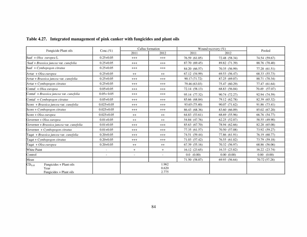
84
Table 4.27. Integrated management of pink canker with fungicides and plant oils
Callus formation Wound recovery (%)
Fungicide Plant oils Conc.(%) 2011 2012 2011 2012
Pooled
Saaf + Olea europea L 0.25+0.05 +++ +++ 76.59 (61.05) 72.48 (58.34) 74.54 (59.67)
Saaf + Brassica juncea var. cunefolia 0.25+0.05 +++ +++ 87.70 (69.45) 89.82 (71.39) 88.76 (70.40)
Saaf + Cymbopogon citratus 0.25+0.05 +++ +++ 84.20 (66.57) 70.35 (56.99) 77.28 (61.51)
Avtar + Olea europea 0.25+0.05 ++ ++ 67.12 (54.99) 69.53 (56.47) 68.33 (55.73)
Avtar + Brassica juncea var. cunefolia 0.25+0.05 +++ +++ 90.17 (71.72) 87.25 (69.07) 88.71 (70.34)
Avtar + Cymbopogon citratus 0.25+0.05 +++ +++ 79.46 (63.03) 75.47 (60.29) 77.47 (61.64)
Contaf + Olea europea 0.05+0.05 +++ +++ 72.14 (58.13) 68.83 (56.04) 70.49 (57.07)
Contaf + Brassica juncea var. cunefolia 0.05+ 0.05 +++ +++ 95.14 (77.32) 90.74 (72.27) 92.94 (74.59)
Contaf + Cymbopogon citratus 0.05+0.05 +++ +++ 85.66 (68.84) 79.12 (62.78) 82.39 (65.32)
Score + Brassica juncea var. cunefolia 0.025+0.05 +++ +++ 93.65 (75.40) 90.07 (71.62) 91.86 (73.41)
Score + Cymbopogon citratus 0.025+0.05 +++ +++ 86.43 (68.36) 83.60 (66.09) 85.02 (67.20)
Score + Olea europea 0.025+0.05 ++ ++ 64.83 (53.61) 68.69 (55.96) 66.76 (54.77)
Governor + Olea europea 0.01+0.05 ++ ++ 54.84 (47.76) 62.25 (52.07) 58.55 (49.90)
Governor + Brassica juncea var. cunefolia 0.01+0.05 +++ +++ 85.63 (67.70) 78.94 (62.66) 82.28 (65.08)
Governor + Cymbopogon citratus 0.01+0.05 +++ +++ 77.35 (61.57) 70.50 (57.08) 73.92 (59.27)
Taqat + Brassica juncea var. cunefolia 0.20+0.05 +++ +++ 74.51 (59.44) 77.86 (61.91) 76.19 (60.77)
Taqat + Cymbopogon citratus 0.20+0.05 +++ +++ 71.03 (57.42) 76.55 (61.02) 73.79 (59.18)
Taqat + Olea europea 0.20+0.05 ++ ++ 67.39 (55.16) 70.32 (56.97) 68.86 (56.06)
White Paint - + + 16.12 (23.65) 16.33 (23.82) 16.22 (23.74)
Control - - - 0.0 (0.00) 0.00 (0.00) 0.00 (0.00)
Mean 71.50 (58.07) 69.93 (56.64) 70.72 (57.28)
CD0.05 Fungicides + Plant oils
Year
Fungicides + Plant oils
1.962
0.620
2.775

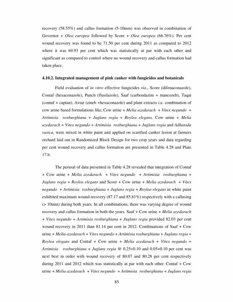
85
recovery (58.55%) and callus formation (5-10mm) was observed in combination of
Governor + Olea europea followed by Score + Olea europea (66.76%). Per cent
wound recovery was found to be 71.50 per cent during 2011 as compared to 2012
where it was 69.93 per cent which was statistically at par with each other and
significant as compared to control where no wound recovery and callus formation had
taken place.
4.10.2. Integrated management of pink canker with fungicides and botanicals
Field evaluation of in vitro effective fungicides viz., Score (difenaconazole),
Contaf (hexaconazole), Punch (flusilazole), Saaf (carbendazim + mancozeb), Taqat
(contaf + captan), Avtar (zineb +hexaconazole) and plant extracts i.e. combination of
cow urine based formulations like, Cow urine + Melia azedarach + Vitex negundo +
Artimisia roxburghiana + Juglans regia + Roylea elegans, Cow urine + Melia
azedarach + Vitex negundo + Artimisia roxburghiana + Juglans regia and Adhatoda
vasica, were mixed in white paint and applied on scarified canker lesion at farmers
orchard laid out in Randomized Block Design for two crop years and data regarding
per cent wound recovery and callus formation are presented in Table 4.28 and Plate
17.b.
The perusal of data presented in Table 4.28 revealed that integration of Contaf
+ Cow urine + Melia azedarach + Vitex negundo + Artimisia roxburghiana +
Juglans regia + Roylea elegans and Score + Cow urine + Melia azedarach + Vitex
negundo + Artimisia roxburghiana + Juglans regia + Roylea elegans in white paint
exhibited maximum wound recovery (87.17 and 85.81%) respectively with a callusing
(> 10mm) during both years. In all combinations, there was varying degree of wound
recovery and callus formation in both the years. Saaf + Cow urine + Melia azedarach
+ Vitex negundo + Artimisia roxburghiana + Juglans regia provided 82.03 per cent
wound recovery in 2011 than 81.14 per cent in 2012. Combinations of Saaf + Cow
urine + Melia azedarach + Vitex negundo + Artimisia roxburghiana + Juglans regia +
Roylea elegans and Contaf + Cow urine + Melia azedarach + Vitex negundo +
Artimisia roxburghiana + Juglans regia @ 0.25+0.10 and 0.05+0.10 per cent was
next best in order with wound recovery of 80.07 and 80.28 per cent respectively
during 2011 and 2012 which was statistically at par with each other. Contaf + Cow
urine + Melia azedarach + Vitex negundo + Artimisia roxburghiana + Juglans regia

86
Table 4. 28. Integration of fungicides with botanicals for the management of pink canker under field condition
Callus formation Wound recovery (%)
Fungicide + botanical Conc. (%) 2011 2012 2011 2012
Pooled
Saaf + Cow urine + Melia azedarach + Vitex negundo + Artimisia roxburghiana + Juglans regia +
Roylea elegans (1:1:1:1:1:1)
0.25+0.10 +++ +++ 78.70 (62.50) 81.44 (64.46) 80.07 (63.46)
Saaf + Cow urine + Melia azedarach + Vitex negundo + Artimisia roxburghiana + Juglans regia
(1:1:1:1:1)
0.25+0.10 ++ ++ 82.03 (64.92) 81.14 (64.25) 81.59 (64.57)
Saaf + Adhatoda vasica 0.25+0.10 ++ ++ 69.46 (56.43) 70.30 (56.96) 69.88 (56.69)
Avtar + Adhatoda vasica 0.25+0.10 +++ +++ 62.31 (52.11) 66.92 (54.87) 64.62 (53.48)
Avtar + Cow urine + Melia azedarach + Vitex negundo + Artimisia roxburghiana + Juglans regia +
Roylea elegans (1:1:1:1:1:1)
0.25+0.10 +++ +++ 70.15 (56.87) 74.70 (59.78) 72.43 (58.30)
Avtar + Cow urine + Melia azedarach + Vitex negundo + Artimisia roxburghiana + Juglans regia
(1:1:1:1:1)
0.25+0.10 +++ +++ 64.56 (53.44) 69.02 (56.16) 66.79 (54.79)
Contaf + Cow urine + Melia azedarach + Vitex negundo + Artimisia roxburghiana + Juglans regia
(1:1:1:1:1)
0.05+0.10 ++ ++ 80.03 (63.43) 80.52 (63.79) 80.28 (63.61)
Contaf + Cow urine + Melia azedarach + Vitex negundo + Artimisia roxburghiana + Juglans regia
+ Roylea elegans (1:1:1:1:1:1)
0.05+0.10 +++ +++ 85.60 (67.68) 88.74 (70.38) 87.17 (68.99)
Contaf + Adhatoda vasica 0.05+0.10 ++ ++ 60.43 (51.00) 62.95 (52.48) 61.69 (51.74)
Score + Cow urine + Melia azedarach + Vitex negundo + Artimisia roxburghiana + Juglans regia +
Roylea elegans (1:1:1:1:1:1)
0.025+0.10 +++ +++ 84.27 (66.67) 87.36 (69.15) 85.81 (67.87)
Score + Adhatoda vasica 0.025+0.10 ++ ++ 57.36 (49.21) 60.26 (50.90) 58.81 (50.05)
Score + Cow urine + Melia azedarach + Vitex negundo + Artimisia roxburghiana + Juglans regia
(1:1:1:1:1)
0.025+0.10 +++ +++ 71.58 (57.76) 75.60 (60.38) 73.59 (59.05)
Governor + Adhatoda vasica 0.05+0.10 +++ +++ 55.62 (48.21) 60.46 (51.02) 58.04 (49.61)
Governor + Cow urine + Melia azedarach + Vitex negundo + Artimisia roxburghiana + Juglans
regia (1:1:1:1:1) 0.05+0.10 +++ +++ 62.41 (52.17) 66.68 (54.72) 64.55 (53.44)
Governor + Cow urine + Melia azedarach + Vitex negundo + Artimisia roxburghiana + Juglans regia
+ Roylea elegans (1:1:1:1:1:1)
0.05+0.10 +++ +++ 70.28 (56.94) 73.86 (59.23) 72.07 (58.08)
Taqat + Cow urine + Melia azedarach + Vitex negundo + Artimisia roxburghiana + Juglans regia +
Roylea elegans (1:1:1:1:1:1)
0.20+0.10 +++ +++ 77.36 (61.56) 71.46 (57.69) 74.41 (59.59)
Taqat + Adhatoda vasica 0.20+0.10 ++ ++ 61.13 (51.41) 59.67 (50.55) 60.40 (50.98)
Taqat + Cow urine + Melia azedarach + Vitex negundo + Artimisia roxburghiana + Juglans regia
(1:1:1:1:1)
0.20+0.10 ++ ++ 69.84 (56.67) 63.68 (52.92) 66.76 (54.77)
White Paint - + + 12.72 (20.88) 13.11 (21.22) 12.92 (21.05)
Control 0.00 (0.00) 0.00 (0.00) 0.00 (0.00)
Mean 63.79 (52.49) 65.39 (53.55) 64.59 (53.01)
CD0.05
Fungicides + botanicals
Year
Fungicides+ botanicals x Year
0.984
0.311
1.392

87
Table 4.29. Integration of compatible fungicides with biocontrol agents for the management of pink canker under field condition
Callus formation Wound recovery (%) Fungicide + biocontrol agent
2011 2012 2011 2012 Pooled
Avtar + Trichoderma harzianum ++ ++ 60.04 (50.77) 56.29 (48.60) 58.17 (49.68)
Avtar + Trichoderma viride ++ ++ 54.81 (47.74) 49.50 (44.69) 52.15 (46.22)
Avtar + Pseudomonas sp. +++ +++ 70.30 (56.95) 72.43 (58.31) 71.36 (57.63)
Avtar + Bacillus subtilis (BS1) +++ +++ 73.11 (58.75) 76.73 (61.14) 74.92 (59.92)
Avtar + Actinomycetes sp. ++ ++ 63.48 (52.80) 60.37 (50.97) 61.93 (51.88)
Saaf + Pseudomonas sp. ++ ++ 57.59 (49.35) 61.27 (51.49) 59.43 (50.42)
Saaf + Bacillus subtilis (BS1) ++ ++ 63.65 (52.90) 69.01 (56.16) 66.33 (54.51)
Saaf + Actinomycetes sp. ++ ++ 59.62 (50.53) 61.48 (51.62) 60.55 (51.07)
Taqat + Pseudomonas sp. + + 51.32 (45.74) 47.59 (43.60) 49.46 (44.67)
Taqat + Bacillus subtilis (BS1) + + 48.64 (44.20) 53.42 (46.94) 51.03 (45.57)
Taqat + Actinomycetes sp. ++ ++ 55.71 (48.26) 58.82 (50.06) 57.27 (49.16)
Score + Pseudomonas sp. ++ ++ 58.06 (49.62) 63.56 (52.85) 60.81 (51.22)
Score + Bacillus subtilis (BS1) +++ +++ 70.07 (56.81) 76.15 (60.74) 73.11 (58.74)
Score + Actinomycetes sp. +++ +++ 77.07 (61.37) 71.28 (57.58) 74.18 (59.43)
Contaf + Trichoderma hamatum +++ +++ 81.37 (64.42) 78.52 (62.37) 79.95 (63.37)
Contaf + Pseudomonas sp. +++ +++ 82.35 (65.14) 78.59 (62.41) 80.47 (63.75)
Contaf + Bacillus subtilis (BS1) ++ ++ 73.42 (58.95) 68.80 (56.02) 71.11 (57.47)
Contaf + Actinomycetes sp. ++ ++ 62.17 (52.03) 57.50 (49.29) 59.84 (50.65)
Governor + Pseudomonas sp. +++ +++ 67.56 (55.26) 71.24 (57.55) 69.40 (56.39)
Governor + Bacillus subtilis (BS1) ++ ++ 69.21 (56.28) 64.23 (53.25) 66.72 (54.75)
Governor + Actinomycetes sp. ++ ++ 64.68 (53.51) 58.57 (49.92) 61.62 (51.70)
White Paint + + 15.17 (22.91) 14.41 (22.30) 14.79 (22.61)
Control - - 0.00 (0.00) 0.00 (0.00) 0.00 (0.00)
Mean 59.97 (50.19) 59.55 (49.91) 59.76 (50.04)
CD0.05 Fungicides + biocontrol agents
Year
Fungicides+ biocontrol agents x Year
0.779
0.230
1.102

88
+ Roylea elegans provided highest wound recovery of 85.60 and 88.74 per cent in
corresponding years, followed by 84.27 and 87.36 per cent with Score + Cow urine +
Melia azedarach + Vitex negundo + Artimisia roxburghiana + Juglans regia +
Roylea elegans, respectively. In the interaction studies between wound recovery and
combination of fungicides + botanicals during 2011 and 2012.Governor + Adhatoda
vasica was found to be least effective (58.04%) whereas interaction between wound
recovery and years were found to be highly significant.
4.10.3. Combined efficacy of fungicides and biocontrol agents against pink
canker
Combined efficacy of most effective fungicides viz., Score (difenaconazole),
Contaf (hexaconazole), Punch (flusilazole), Saaf (carbendazim + mancozeb), Taqat
(contaf + captan), Avtar (zineb + hexaconazole) and antagonists viz., Trichoderma
harzianum, Trichoderma viride, Pseudomonas sp., Bacillus subtilis (BS1) and
Actinomycetes sp. were evaluated at their recommended dose, while biocontrol agents
were mixed with compatible fungicides using slurry method and the data on per cent
wound recovery and callus formation are presented in Table 4.29 and Plate .17.c.
It is clear from the data presented (Table 4.29 & Plate 17.c) that in the
interaction studies between wound recovery and a combination of fungicide +
antagonists resulted significant maximum wound recovery (82.35%) in 2011 than in
2012 with wound recovery of 78.59 per cent in Contaf + Pseudomonas sp.
combination followed by 81.37 and 78.52 per cent wound recovery in combination of
Contaf + Trichoderma hamatum which was statistically at par with each other.
Callusing of more than 10 mm was also found maximum in Contaf + Pseudomonas
sp. and Contaf + Trichoderma hamatum. Combination of Avtar + Bacillus subtilis
(BS1), Score + Bacillus subtilis (BS1), Contaf + Bacillus subtilis (BS1) and Score +
Actinomycetes sp. provided 74.92, 73.11, 71.11 and 74.18 per cent wound recovery
respectively and was found next best in order and was statistically at par with each
other. Wound recovery was found to be 59.97 per cent in 2011 and 59.55 per cent in
2012 and their interaction was found to be significant. However minimum wound
recovery of 51.03, 52.15 and 57.27 per cent was observed in combination of Taqat +
Bacillus subtilis (BS1), Avtar + Trichoderma viride and Taqat + Actinomycetes sp.
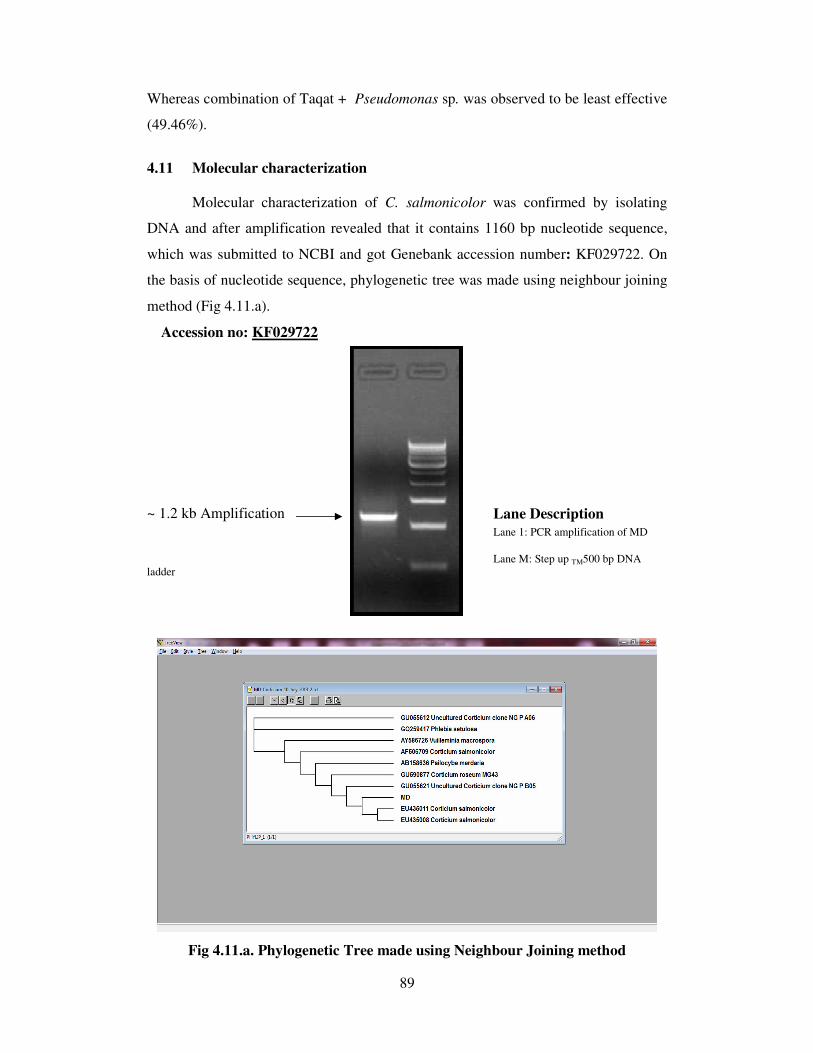
89
Whereas combination of Taqat + Pseudomonas sp. was observed to be least effective
(49.46%).
4.11 Molecular characterization
Molecular characterization of C. salmonicolor was confirmed by isolating
DNA and after amplification revealed that it contains 1160 bp nucleotide sequence,
which was submitted to NCBI and got Genebank accession number: KF029722. On
the basis of nucleotide sequence, phylogenetic tree was made using neighbour joining
method (Fig 4.11.a).
Accession no: KF029722
~ 1.2 kb Amplification Lane Description Lane 1: PCR amplification of MD
Lane M: Step up TM500 bp DNA
ladder
Fig 4.11.a. Phylogenetic Tree made using Neighbour Joining method

90
On the basis of phylogenetic tree, two groups were constructed, C.
salmonicolor isolate of Mandi isolate (MD) was found to be closest to EU435011
Corticium salmonicolor and EU435008. Whereas GU055621 uncultured Corticium
clone NG P B05 and GU590877 Corticium roseum was also found to be nearest
relative. Second group consist of GU055612 uncultured Corticium clone NG P A06
and GQ259417 Phlebia setulosa which were observed to be distant relative to Mandi
isolate of Corticium salmonicolor.

Chapter-5
DISCUSSION
The prevalence of pink canker (Corticium salmonicolor Berk. and Br.) in
different regions in the state is a major constraint in successful apple cultivation in
mid and high hills of Himachal Pradesh. The disease has been responsible for causing
a marked reduction in both quality and quantity of fruits. Poor orchard management
practices, moderate temperature, frequent rains and high relative humidity coupled
with increasing popularity of Delicious varieties, being highly susceptible to canker
diseases including pink canker, have led to the progressive increase of the severity of
this disease for the past few years causing huge losses through girdling of branches,
limbs, blightening and dieback of twigs resulting in death of plants. Although
attempts have been made to manage the disease through spray application of
fungicides (Verma and Munjal, 1980; Kondal, 1986; Shandilya and Agarwala, 1975;
Kondal and Agarwala, 1975) yet satisfactory level of the management is still lacking.
Keeping in view the economic importance of the research gaps and meagre
information available in literature on different aspects, it was thought worthwhile to
investigate the disease with the objectives as outlined in the introducing chapter and
the results obtained are being discussed in the light of available literature as follows:
PREVALENCE OF PINK CANKER
Surveys conducted during 2011 and 2012 crop seasons in four apple growing
areas in low and mid hills of Himachal Pradesh during June-September revealed that
the disease occurred in moderate to severe form in Mandi, Sirmour and Shimla
districts of the state. The disease severity varied at different locations. Maximum
severity (51.84%) was observed at Haripurdhar followed by Nauhradhar (50.31%) of
Sirmour District. Whereas, the maximum disease incidence (68.14%) of pink canker
was at Kotgarh of district Shimla followed by Janjhehli (62.66 %) of Mandi district.
The severity of pink canker varied from 9.88 per cent to 51.84 per cent. The severity
and incidence of pink canker increased with age of the tree and decreased with
altitudes. These findings were in confirmations with Shandilya et al. (1973), Verma

92
(1991) who also reported pink canker disease more prevalent and highly destructive in
Kullu valley and also observed that canker diseases increase with age of the trees and
declined with the increase in altitude.
Gibson and Armitage (1979) reported 55 to 95 per cent losses due to C.
salmonicolor. Infection was severe in 5 to 11 year old plantations of Eucalyptus
tereticornis at low and medium altitudes with warm, humid, receiving high rainfall.
In the present studies also, higher disease incidence and severity (68.14 and
51.84%) recorded in Kotgarh and Haripurdhar could be ascribed to build up of wind
borne inoculum as a result of most intensive cultivation of highly susceptible
“Delicious varieties” and poor orchard management besides the favourable
environmental conditions.
SYMPTOMATOLOGY
Symptom of pink canker disease occurred in mainly four forms namely, a)
Cob-web stage b) Pustuler stage c) Necator pustules and d) Pink incrustation stage.
Cob-web and pustuler stage occur immediately after monsoon rains. Cob-web stage
consists of silky white mycelium that grows over the surface of bark covering large
area and is the first sign of infection. The pustuler stage is readily identifiable when
pink pustules consisting of sterile mycelia produced on the affected area that erupts
out by breaking lenticels appearing white in colour. The necator stage is characterized
by appearance of orange-red necator (conidial) stage being produced late in the season
and usually develops on the upper side of infected branches. Whereas pink
incrustation stage consisting of basidial stage or Corticium stage usually developed on
underside of the branches facing less sunshine hours and characterized by
development of salmon- pink colour on the dying infected branch in the winters.
The characteristic symptoms of the disease as observed during the course of present
investigations on stem and branches are in accordance with those described by other
workers (Singh, 1942; Thakur 1970; Shandilya, 1971; Gupta and Agarwala, 1973 and
Verma, 1978).
ISOLATION AND IDENTIFICATION
Based on morphological, cultural and molecular characters, the identity of the
pathogen was confirmed to be Corticium salmonicolor Berk. and Br., Mandi and
93
Kotgarh isolate appeared to have similar mycelial growth with cottony and abundant
concentric rings whereas, Kullu and Haripurdhar isolate formed sparse and fan shaped
mycelial growth in the culture after 7 days of incubation at 25±1°C. Mycelium of all
four isolates consisted of clamp connection with prominent dolipore septa. All four
isolates produced pink colour under in vitro condition when exposed to shaded natural
light which is in accordance with the characters documented by (Singh, 1942; Thakur
1970 and Verma, 1978; Luz, 1983 and Sharma, 2005).
PATHOGENICITY
Pathogenicity tests of pink canker isolates were conducted on Royal Delicious
variety under laboratory condition by excised twig method and revealed that twigs of
Royal Delicious were readily infected by the C. salmonicolor isolates. Longest
incubation period of 30 days was required for symptoms development on stem
ostensibly due to non-succulent woody tissue. Maximum infection (93.33%) and
average lesion size (65.2 mm) was observed in Mandi isolate followed by Kotgarh
(73.33 % and 52.0 mm ), Kullu (66.67 % and 46.7%) and Haripurdhar isolate (46.67
% and 40.4 mm), respectively after 30 days of incubation. These tests revealed that
Mandi isolate of Corticium salmonicolor Berk. and Br., was more pathogenic in
comparison to Corticium salmonicolor isolates of Kotgarh, Kullu and Haripurdhar
area.
Sharma (2005) also found C. salmonicolor as more pathogenic and destructive
on apple twigs with 80.0 per cent infection and lesion size of 30.0 mm after 30 days
of inoculation.
CULTURAL STUDIES
The results of cultural studies pertaining to the conidial germination (%) and
germ tube length (µm) of C. salmonicolor isolates in distilled water indicated that
significantly maximum conidial germination (88.39 %) and germ tube growth (81.85
µm) was obtained in Mandi isolate followed by Kotgarh (82.88 % and 68.67 µm),
Kullu (74.09 % and 61.61 µm) and Haripurdhar isolate (65.30 % and 51.98 µm)
respectively at 25±1o C after 24 and 48 hrs of incubation. Further imperfect stage of
C. salmonicolor i.e. Necator decretus was induced in the culture after incubation of
94
28-30 days at 25 ±1°C with orange-red Necator spores measuring 45-52 µm, which
serves as a primary source of infection.
Verma (1978) reported 94.81 and 80.16 per cent germination of Necator spore
and basidiospores at 25±1o C after 4 hours of incubation. He further observed that 25
o
C to be the best temperature for germination of Necator spore with 81.48 and 98.26
per cent germination after 4 and 8 hours, respectively however, he induced Necator
stage of pathogen after 20-25 days at 25±1o C which are in conformity with the
present study.
However, leaves and fruits of Royal Delicious cultivar were severely infected
with conidia of C. salmonicolor under in vitro conditions at 25±1°C coupled with 100
% RH. Maximum leaf infection (100%) was obtained after 14 days at 25±1°C coupled
with 100 % RH whereas, fruits were having lesion size of 4.8 cm after 14 days of
inculation with mycelium of the fungus and confirmed that under congenial climatic
conditions it may infect leaves as well as fruits.
Luz (1983) also reported C. salmonicolor infecting stem, leaves and pods of
Cocoa and differed in their mycelial growth pattern, which lends support to the
present findings.
EPIDEMIOLOGICAL STUDIES
The results pertaining to the influence of environmental factors on the disease
development under field conditions revealed that the disease was initiated in second
and third week of June during 2011 and 2012, respectively after the commencement
of rain indicating thereby the influence of rainfall and higher relative humidity on the
disease development resulting in the progress of the disease during the month of
August and September. These results corroborate the findings of Shandilya and
Agarwala (1975) who reported that disease would not develop if the environmental
factors such as relative humidity and temperature are not favourable. Importance of
temperature and relative humidity in disease development has also been reported by
other workers (Hilton, 1958; Luz and Ram, 1980; Verma, 1988 and Mohanan 2008).
During the present investigations, simple correlation coefficient between
disease severity and temperature was negative but highly significant in both crop
95
seasons. Pooled data also showed similar results between meteorological factors and
disease severity. Further the partial correlation coefficient between disease severity
and temperature was highly significant but negative in 2011 and 2012 however,
relative humidity was found positive and highly significant. Simple correlation of
pooled data regarding cumulative rainfall was found to be positive and highly
significant during both the seasons 2011-12. These results corroborate the earlier
findings of Luz and Ram (1980), Ploetz (2007) and Mohanan (2008) on pink disease
(Corticium salmonicolor) of cacao, citrus and teak, respectively indicating their role
in development and spread of disease. However, sunshine hours were found to be
significant but negatively correlated. It also disclosed the fact that pink incrustation or
salmon-coloured crustose phase, consisting of the sexual Corticium stage usually
develops on shaded underside of infected branch or limbs of many plantation crops
viz., cacao, coffee, tea, rubber and teak including apple (Hilton, 1958; Old et al., 2000
and Mohanan, 2008).
Partial correlations and regression equation between disease severity and
meteorological factors further elaborated the role of relative humidity and temperature
in disease development. The present studies revealed that multiple coefficient of
determination between disease severity and group of independent variables was found
to be 0.6753, suggesting that 67.53 per cent disease development is attributed to all
the meteorological factors viz., temperature, relative humidity, cumulative rainfall and
sunshine hours, whereas rest of the variation is due to unexplained factors or unknown
factors not included in the study. However, similar observations have been recorded
in case of eucalyptus pink disease caused by the same pathogen (C. salmonicolor)
where high rainfall and relative humidity with site factor are considered to be major
influencing factors for sudden outbreak of the disease (Mohanan, 2008).
VARIETAL SUSCEPTIBILITY
However, little information is available in the literature to support varietal
screening against the test pathogen on apple. Though, similar observations have been
recorded in case of apple pink canker caused by the same pathogen. Several apple
cultivars have already been screened against pink canker by various workers
(Shandilya and Agarwala, 1975 and Verma, 1978) but only a few have been reported
as resistant. During the present study also, out of nine cultivars screened against the
96
disease, none of the cultivars showed resistant reaction though Tydeman Early
Worcester was found moderately susceptible, while four cultivars viz., William’s
Favourite, Scarlet Spur, Granny Smith and Rich-a-red exhibited susceptible reaction
under field condition. Under in vitro conditions, none of the cultivars exhibited
resistant reaction only, Spur Winter Banana, Zinger Gold, Granny Smith and Scarlet
Spur showed moderately susceptible reaction based on lesion size and pustule
formation.
Whereas, screening against Mandi isolate revealed that three cultivars viz.,
Ace Spur, Zinger Gold and Granny Smith showed moderately resistant, while rest of
cultivars behaved moderately susceptible reaction under flask condition. While these
cultivars screened against Mandi isolate under Petri plate conditions exhibited
susceptible and highly susceptible reaction except Zinger Gold which showed
moderately susceptible reaction. This variation might be due to the persistence of high
humidity under Petri plate condition.
Out of fifteen cultivars of apple screened against Kotgarh, Kullu and
Haripurdhar isolates of C. salmonicolor, none of the cultivars showed resistant
reaction and exhibited moderately susceptible to susceptible reaction under in vitro
conditions.
However, Verma (1978) reported the existence of resistance in certain cultivars viz.,
Boycon, Early Red Bird, Early Shanbury, Early Victory, Irish Peach, Tydeman Early
Worcester, and Walthy Double Red.
DISEASE MANAGEMENT
Disease management through fungicides
Besides eradicative action, chemicals also provide a chemical toxic barrier
against pathogens and are thus unavoidable means of controlling many plant diseases.
Efficacy of carbendazim against pink canker has been well documented in literature
by various workers (Kondal, 1986; Shandilya and Agarwala; 1975 and Verma and
Munjal, 1980). Among the various chemicals evaluated in vitro by poisoned food
technique, difenaconazole, hexaconazole and flusilazol at 50,100,150 and 200 µl l-1
and combi-products like, carbendazim + mancozeb, zineb + hexaconazole and captan
+ hexaconazole at 250, 500, 750 and 1000 µl l-1
exhibited complete mycelium

97
inhibition of Corticium salmonicolor followed by carbendazim (97.72 %),
propiconazole (97.31 %) and pyraclostrobin + metiram (86.05 %), respectively.
Whereas, carbendazim alone or in combination with mancozeb or iprodione was
found to be most effective at all concentrations and caused complete conidial
germination inhibition followed by difenaconazole (3.97%) and azoxystrobin
(4.27%), while pyraclostrobin + metiram and captan + hexaconazole was least
effective as compared to control (84.56%).
These results corroborate the findings of Gupta and Kumar (2008) that
carbendazim was effective in inhibition of conidial germination of Erysiphe pisi.
Whereas, EBI fungicides do not affect conidial germination although germ tube
length is inhibited and distorted (Scheinpflug and Kuck, 1987). The reduction in
conidial formation by difenaconazole, hexaconazole, propiconazole and flusilazole
are in consonance with the findings of Gupta and Shyam (1998) and Gupta and
Sharma (2004).
The per cent wound recovery and callus formation was best contained by
triazoles like, difenaconazole (250 µl l-1
) and hexaconazole (500 µl l-1
) with 92.04 and
89.00 per cent wound recovery with more than 10 mm callus formation followed by
flusilazole (100 µl l-1
) and carbendazim + mancozeb (2500 µl l-1
) with 87.80 and
86.69 per cent wound recovery, respectively for two consecutive cropping seasons
(2011-12) than white paint alone with 13.56 per cent wound recovery. Propiconazole
(0.05%) and carbendazim (0.05%) alone was next best in order with 85.62 and 81.73
per cent wound recovery, respectively. Whereas iprodione + carbendazim (0.15%)
was found to be least effective with 51.38 per cent wound recovery with more than
5mm callus formation during 2011-12 crop seasons as compared to untreated check
wherein no wound recovery and callus formation had taken place.
The effectiveness of combiproduct (systemic and non-systemic) can be
explained as either or both of its constituent are from effective fungicide and thus
exhibiting excellent protective, curative and eradicative and post infection activity to
restrict mycelial growth and inhibit the formation and germination of conidia. Higher
disease control obtained in carbendazim is due to its effect on inhibiting “two track”
type of mitosis which is common in ascomycetes and disruption of mitosis through
inactivation of spindle formation composed of microtubules (Hammerschlag and

98
Sisler 1972 and Davidse 1986). Calixin (tridemorph) paint has already been reported
best against pink canker of apple (Verma and Munjal 1980). EBI fungicides inhibit
the biosynthesis of ergosterol needed for fungal cell wall and subsequent
multiplication thereby inhibiting the fungal growth (Schwinn 1984).
Disease management through botanicals
A huge quantity of chemicals are used to reduce the ravages of various
diseases which are not only detrimental to the environment but are also hazardous to
the human health and needs to be replaced by less detrimental compounds preferably
from natural sources. In the present investigations with plant extracts initially twenty
plant extracts were in vitro screened for their antimicrobial activity against the pink
canker disease. The plant extracts with higher antifungal activity were further tested
in field condition at 10 per cent concentration. Aqueous extracts of Adhatoda vasica
along with two combinations of cow urine + Melia azedarach + Vitex negundo +
Artimisia roxburghiana + Juglans regia + Roylea elegans and cow urine + Melia
azedarach + Vitex negundo + Artimisia roxburghiana + Juglans regia, all in 1:1 ratio
exhibited complete growth inhibition at 10, 20 50 and 100 per cent concentrations
followed by cow urine + Melia azedarach + Vitex negundo + Juglans regia and cow
urine + Melia azedarach + Eucalyptus globulus + Bougainvellia spectabilis resulted
94.45 and 95.20 per cent growth inhibition, respectively under in vitro condition.
The per cent wound recovery was significantly maximum (58.54 %) in cow
urine based formulation i.e. Cow urine + Melia azedarach + Vitex negundo +
Artimisia roxburghiana + Juglans regia + Roylea elegans followed by formulation of
Cow urine + Melia azedarach + Vitex negundo + Artimisia roxburghiana + Juglans
regia with wound recovery of 55.44 per cent with a callus formulation of 5-10 mm
respectively. Adhatoda vasica alone when added in white paint @ 0.10 per cent
provided 52.91 per cent wound recovery with >5 mm callus formation which was
statistically at par with each other. Aqueous extract of 100 per cent Brassica juncea
var. cunefolia as a paint provided 51.02 per cent wound recovey and callus formation
of 5 mm respectively under field condition for two consecutive years (2011-12).
Whereas Mentha pipereta, Ocimum sanctum, Prunus persica and Roylea elegans
exhibited 37.88, 38.55, 33.65 and 31.89 per cent wound recovery with 5mm callus

99
formation, while Melia azedarach was found least effective (20.08 %) in wound
recovery.
The fungicidal/ bactericidal activity of Ocimum sanctum can only be partially
attributed to the volatile compounds it contains (Grover and Rao, 1977).
Antimicrobial activity of Brassica juncea var. cunefolia and Cymbopogon citratus
against many foliar and soil borne pathogens could be due to the presence of alkaloids
i.e. allyl isothiocyanate and citral respectively. The fungicidal/ bactericidal activity of
cow urine may be due to presence of phenol in cow urine that may be instrumental for
its potent antimicrobial nature (Jarald et al., 2008). The residual activity may be
attributed to the ajoenes that are thought to be one of several pharmacologically active
compounds in Allium sativum (Singh et al., 1990 and Singh et al., 1992). Sharma
(2005) reported Emblica officinalis that exhibited maximum antifungal activity
against all the three canker pathogens. Whereas, Dodonia viscosa also proved
effective against C. salmonicolor (pink canker) and Dothidea mali (stem brown) and
Eucalyptus globulus against Sphaeropsis malorum (smoky blight). Antibacterial
activity of extracts of different plants has already been reported (Mangamma and
Sreeramulu, 1991; Abassi et al., 2003). Mycelial growth of various species of
Fusarium was inhibited by the plant extracts of Convolvulus alsinoides and C.
Pluricutis (Furgal, 1984); Allium cepa (El. Sharmi et al.,1986) Adhatoda vasica,
Azadirachta indica, Cinnamomum camphora and Ocimum sanctum (Prasad and
Ojha, 1986); Agave americana, Cassia nodosa (Reddy and Reddy, 1987);
Azadirachta indica (Eswaramoorthy et al; 1989); Allium cepa (Patel, 1989);
Excoecaria agallocha and Acanthus ilicifolius (So, 1990); Agave americana
(Pandey et al 1992); Allium sativum and Sapindus trifoliata (Gohil and Vala,
1996); Neem seed extract (Gour and Sharmaik, 1998); Azadirachta indica,
Atropha belladona, Calotropis procera, Ocimum basilicum, Eucalyptus
amygdalina, and Lantana camera (Bansal and Gupta, 2000).
Disease management through plant oils
The present studies explicitly indicate the efficacy of plant oils against
Corticium salmonicolor both under in vitro and field conditions exhibiting their effect
in wound recovery and subsequent callus formation. The per cent growth inhibition
was significantly maximum (100 %) in Olea cuspidata, Cymbopogon citratus and

100
Brassica juncea var. cunefolia at all concentrations tested, followed by Eugenia
caryophyllata (94.00 %), Ocimum sanctum (83.98 %), and Azadirachta indica (85.95
%) respectively. However, plant oils of Juglans regia, Ricinus communis and
Juniperus virginiana provided 76.06, 78.90, 73.48 per cent growth inhibition
respectively, against the test pathogen. Rest of the essential oils from wild marigold
(Tagetes minuta), eucalyptus (Eucalyptus globulus), wild apricot (Prunus armeniaca)
and Artemisia roxburghiana were found to be less effective. It was also recorded that
as the concentration of these bio-pesticides increased, there was a corresponding
increase in per cent growth inhibition with the highest at 100 per cent concentration.
The interaction between treatment and concentrations were found to be highly
significant. The antifungal and antibacterial activity of essential oils have already
been reported by many workers (Hudson, Kuo and Vimalanathan, 2011; Kota and
Manthri, 2011; Dwivedi and Dwivedi, 2012).
Maximum wound recovery (81.27%) was achieved with Brassica juncea var.
cunefolia followed by Olea europea (75.83 %) with more than 10 mm callus
formation, respectively. Cymbopogon citratus, Eugenia caryophyllata, Azadirachta
indica and Ocimum sanctum was found next best in order with 72.72, 67.77, 52.71
and 46.09 per cent wound recovery, respectively. Whereas Ricinus communis and
Juglans regia were found less effective in controlling disease as compared to
Juniperus virginiana which was least effective (31.96%). White paint alone resulted
11.92 per cent wound recovery with 1-5 mm callus formation than control wherein no
wound recovery and callus formation occurred.
The present studies explicitly indicate the efficacy of Olea cuspidata in
growth inhibition of Corticium salmonicolor might be because of presence of
phenolic compounds like oleuropein, luteolin 7-O-glucoside (Pereira et al., 2007).
Antifungal and antibacterial properties of Brassica juncea var. cunefolia can probably
be due to the presence of allyl isothiocyanate and diallyl trisulfide (Jimmy et al.,
2003). Alkaloids like oxygenated monoterpenes, a-citral or geranial and b-citral or
neral, a major constituent of Cymbopogon citratus, has strong toxic properties against
several bacteria and fungi (Tyagi and Malik, 2010). Wilson et al., (1997) tested 49
essential oils from various plants and found that the oil and clove buds of
Eugenia caryophyllata was effective to control Botrytis cinerea. Singh et al.,
101
(1980) observed inhibitory effects of essential oils of C. martinii, C. oliveri, and
Trachysperumm ammi on Helminthosporium oryzae, as well as inhibitory effects
of the essential oils from rhizomes and leaves of Zingiber chrysanthum on plant
pathogens such as Alternaria sp. and Fusarium sp. A commercial product based on
the formulation of plant extracts and essential oils from pepper, mustard, cassia,
and clove extracts was effective in reducing the population density of F. oxysporum f.
sp. chrysanthemi (Bowers and Locke, 2000). Sharma (2005) revealed maximum
mycelium inhibition of Corticium salmonicolor with the oils of Cymbopogon citratus
(73.47%) followed by Tagetes minuta (71.46%) and Eucalyptus hybrida (67.08%),
while the least inhibition has been observed with Azadirachta indica (48.89%).
Disease management through biocontrol agents
The biological control aims at eradication or management of pathogens
through the activity of other microorganisms. It can be carried out either by
manipulating the existing soil microflora to the disadvantage of the pathogen or by
addition of non-resident antagonists (Cook, 1982). The screening of seventeen
antagonists (five bacteria, eleven fungi and one actinomycetes) for their efficacy
against C. salmonicolor under in vitro condition indicated that maximum inhibition of
mycelial growth was observed in Trichoderma koningi (82.62 %) followed by Th1 of
Trichoderma hamatum (78.02 %) and Th2 of Trichoderma hamatum (77.15 %) which
were statistically at par with each other. All other isolates of Trichoderma sp.
inhibited the growth of C. salmonicolor to greater extent in comparison to other
fungal flora while minimum per cent inhibition was recorded with Aspergillus
versicolor (40.92%).
Amongst the bacterial and actinomycetes sp. Pseudomonas sp. provided
maximum inhibition zone (15.70 mm) followed by Actinomycetes sp. (14.20 mm)
which was statistically at par with each other. Among Bacillus sp. BS1 (Kotgarh)
isolate of Bacillus subtilis caused maximum inhibition zone of 12.3 mm as compared
to others. Amongst fungal biocontrol agents only Aspergillus versicolor and
Verticillium sp. provided maximum inhibition zone of 13.4 and 8.40 mm respectively.
The per cent wound recovery was maximum (48.48 %) in case of Trichoderma
koningi, followed by 41.95 per cent with Actinomycetes sp. and BS1 (Kotgarh) isolate
of Bacillus subtilis (40.39 %) which were statistically at par with each other.

102
Interaction between antagonists and years were also found to be significant.
Trichoderma viride (Tv1) provided wound recovery of 38.80 per cent followed by
Trichoderma hamatum (Th1) with 38.77 per cent wound recovery under field
condition for two years. Bacillus subtilis and Pseudomonas sp. were identified based
on phylogenetic groups and Pseudomonas sp was given GenBank accession no:
KF564924.
The inhibitory effect of Pseudomonas sp. and B. subtilis against C.
salmonicolor was probably due to antibiosis. The results obtained in the present
investigations are in conformity with Sharma (2005) who reported the antagonistic
activity of Trichoderma sp. against C. salmonicolor. Jollands (1983) founded
Trichoderma spp. were antagonistic to C. salmonicolor infecting rubber and oil palm.
Jansen (2005) noticed parasitic fungi (Gliocladium sp., Trichoderma sp., and
Verticillium sp.) showed antagonistic properties against C. salmonicolor affecting
coffee production. Jollands (1983) also reported in vitro antagonistic activity of
various micro-organisms viz., Alternaria sp., Bacillus subtilis, Gliocladium roseum,
Trichoderma harzianum and Trichoderma viride against Corticium salmonicolor
causing pink disease in rubber.
COMPATIBILITY STUDIES
Fungicides have become indispensable in plant disease control therefore; these
must be used in combination with other management practices. Besides the
eradicative action, chemical pesticides also provide protective barrier against the
pathogens and were thus unavoidable in plant disease control. In all, eleven fungicides
were in vitro evaluated against eight antagonistic microbes including fungi, bacteria
and actinomycetes for ascertaining their compatibility.
Fungicides viz., Avtar (zineb + hexaconazole) provided compatible reaction
with Trichoderma harzianum, Trichoderma viride and Pseudomonas sp., Bacillus
subtilis and actinomycetes sp., when evaluated at 0.25 per cent, though inhibited
mycelial growth of Trichoderma koningi (49.41%), Trichoderma hamatum (91.17%)
and Aspergillus versicolor (65.68%) respectively. Strobilurin fungicide viz., Amistar
(azoxystrobin) @ 0.025 per cent was found compatible with Trichoderma harzianum,
Trichoderma koningi, Pseudomonas sp., Bacillus subtilis and actinomycetes sp.,

103
although inhibited mycelium of isolate of Trichoderma viride (39.79 %), Trichoderma
hamatum (39.78%) and Aspergillus versicolor (56.81%) respectively.
Whereas rest of fungicides like, Saaf (carbendazim + mancozeb), Taqat
(captan + hexaconazole), Cabrio Top (pyraclostrobin + metiram), Tilt
(propiconazole), Contaf (hexaconazole), Governor (flusilazol), Bavistin
(carbendazim), Score (difenaconazole) and Quintal (iprodione + carbendazim) at their
recommended dose found compatible with bacterial and actinomycetes sp., but
resulted significant mycelial inhibition of fungal isolates. Bavistin (carbendazim)
either alone or in combination i.e. Saaf (carbendazim + mancozeb) exhibited cent per
cent mycelial inhibition of Trichoderma viride (Tv1), Trichoderma koningi,
Trichoderma hamatum and Aspergillus versicolor except Trichoderma harzianum
(96.93%).
The present findings were in agreement with various workers (Malathi et al.,
2002; Anand et al., 2009; Basha et al., 2010 and Pallavi et al., 2012) who reported
Pseudomonas sp, Bacillus sp and Trichoderma sp. to be compatible with
azoxystrobin, thiophanate-methyl, mancozeb, carbendazim, copper oxychloride
propioconazole and hexaconazole to varying degree under in vitro condition.
Bacterial strains showed tolerance to hexaconazole and carbendazim. Whereas,
Trichoderma sp., were found to be inhibited by PCNB, Emisan, Bavistin, Benlate,
Saaf, Tilt and carboxin under in vitro condition has already been documented by
several workers (Yokomizo et al.,1980; Papavizas et al.,1982; Bhat and Srivastava,
2003; Upadhyay et al., 2004 and Gowdar, et al,. 2006).
Among plant oils viz., Brassica juncea var. cunefolia, Azadirachta indica and
Olea europea @ 0.05 per cent found compatible with all antagonists used. While
Ocimum sanctum was found to inhibit only mycelial growth of Aspergillus versicolor
(45.29%) and compatible with rest of antagonists. Similarly, Eugenia caryophyllata
inhibited mycelial growth of Trichoderma viride (14.10%). Cymbopogon citratus was
found to inhibit growth of Trichoderma hamatum (83.58%), Trichoderma harzianum
(89.57%), Aspergillus versicolor (90.29%), Trichoderma koningi (96.55%) and
Trichoderma viride (27.73%) respectively, but found compatible with bacterial
antagonists. These results are in consonance with the finding of several workers
(Goswami et al., 2006; Bagwan, 2010 and Vanitha, 2010) who reported compatibility
104
of Trichoderma sp. with neem cake oil, mustard oil, lemon grass oils and their
integration with fungicides to manage mangy soil borne and foliar pathogens.
INTEGRATED MANAGEMNET OF PINK CANKER
Disease management practice alone is not sufficient until integrated with other
components, as disease is a complex phenomenon governed by many factors.
The integration of effective fungicides with plant oils, botanicals and
antagonists were important for the successful management of plant diseases (Mondal
et al, 1995). Moreover, there is enhanced enthusiasm in the use of integrated pest
management and eco-friendly technologies for plant disease control.
All combination of fungicides and plant oils were highly effective in
combating pink canker under field conditions. However, maximum wound recovery
(92.94%) and callus formation of more than 10 mm was observed in combination of
Contaf + Brassica juncea var. cunefolia followed by combinaion of Score + Brassica
juncea var. cunefolia (91.86%) with more than 10 mm callus formation and was
statistically at par with each other. Whereas combination of Saaf + Brassica juncea
var. Cunefolia, Avtar +Brassica juncea var. cunefolia, Score+ Cymbopogon citratus
and Governor + Brassica juncea var. cunefolia resulted wound recovery of 88.76,
88.71, 85.02 and 82.28 per cent wound recovery with more than 10 mm callus
formation respectively during both the year. However minimum wound recovery
(58.55%) and callus formation (5-10mm) was observed in combination of Governor +
Olea europea followed by Score + Olea europea (66.76%).
Though no information is available in literature on the combined effect of
different combinations of fungicides and plant oils in management of pink canker, yet
the control of powdery mildew and black spot of roses has already been reported with
combined effect of sodium bicarbonate 1% (v/v) and sunspray ultrafine spray oil
(Horst, 1992). The combination of ketoconazole and essential oil from P.
graveolens was reported to control Trichophyton species. He also concluded that it
reduces dose of ketoconazole and minimizes the side-effects of ketoconazole (Shin
and Lim, 2004). However, efficacy of white paint as a carrier and its toxic nature has
already been noted by Turner, 1980 and Johnson et al., 2009.

105
Incorporation of natural products provides a viable solution to the
environmental problems caused by synthetic pesticides. Identification of these
compounds and their further testing may be an effective approach to minimize the use
of hazardous chemicals (Duke, 1990).
In the present studies regarding integrated management of pink canker in
apple, integration of Contaf + Cow urine + Melia azedarach + Vitex negundo +
Artimisia roxburghiana + Juglans regia + Roylea elegans and Score + Cow urine +
Melia azedarach + Vitex negundo + Artimisia roxburghiana + Juglans regia +
Roylea elegans in white paint exhibited maximum wound recovery (87.17 and
85.81%) respectively with a callusing (> 10mm) during both years. In all
combinations, there was varying degree of wound recovery and callus formation in
both the years. Saaf + Cow urine + Melia azedarach + Vitex negundo + Artimisia
roxburghiana + Juglans regia provided 82.03 per cent wound recovery in 2011 than
81.14 per cent in 2012. Combinations of Saaf + Cow urine + Melia azedarach + Vitex
negundo + Artimisia roxburghiana + Juglans regia + Roylea elegans and Contaf +
Cow urine + Melia azedarach + Vitex negundo + Artimisia roxburghiana + Juglans
regia @ 0.25+0.10 and 0.05+0.10 per cent was next best in order with wound
recovery of 80.07 and 80.28 per cent respectively during 2011 and 2012 which was
statistically at par with each other. Linton and Dick (1990) revealed that presence of
antioxidants and phenols in cow urine exhibit bactericidal to both gram positive and
gram-negative bacteria. Similarly, antifungal activity of cow urine was already
documented by Sathasivam et al. (2010).These results are in agreement with the
findings of Tapwal et al. (2012) who reported compatibility of aqueous extracts of
Parthenium, Adiantum, Urtica, Polystichum and Cannabis sp. with T. viride and
fungicides.
To develop an effective disease management programme, the compatibility of
potential bioagents with fungicides and botanicals is essential. Combination of
chemicals and compatible bioagents in an IDM strategy protects the seeds and
seedlings from soil-borne and seed-borne inoculum (Dubey and Patil, 2001).
In the interaction studies between wound recovery and a combination of
fungicide + antagonists revealed that significant maximum wound recovery (82.35%)
was recorded in 2011 than in 2012 with wound recovery of 78.59 per cent in Contaf +

106
Pseudomonas sp. combination followed by 81.37 and 78.52 per cent wound recovery
in combination of Contaf + Trichoderma hamatum which was statistically at par with
each other. Callusing of more than 10 mm was also found maximum in Contaf +
Pseudomonas sp. and Contaf + Trichoderma hamatum. Combination of Avtar +
Bacillus subtilis (BS1), Score + Bacillus subtilis (BS1), Contaf + Bacillus subtilis
(BS1) and Score + Actinomycetes sp. provided 74.92, 73.11, 71.11 and 74.18 per cent
wound recovery respectively and were found next best in order and were statistically
at par with each other. Wound recovery was found to be 59.97 per cent in 2011 and
59.55 per cent in 2012 and their interaction was found to be significant. However
minimum wound recovery of 51.03, 52.15 and 57.27 per cent was observed in
combination of Taqat + Bacillus subtilis (BS1), Avtar + Trichoderma viride and
Taqat + Actinomycetes sp. Whereas combination of Taqat + Pseudomonas sp. was
observed to be least effective (49.46%).
These results are in consonance with the finding of several workers
(Upadhyay et al., 2004; Vijayaraghavan and Abraham, 2004; Anand et al., 2009;
Tapwal et al., 2012 and Devamma et al., 2012) who reported compatibility of
Trichoderma sp. with azoxystrobin, dithane, carbendazim, blitox and thiophanate-
methyl and their integration to manage many soil borne and foliar pathogens.
MOLECULAR CHARACTERIZATION
Molecular characterization of pink canker associated fungus revealed that the
associated pathogen is C. salmonicolor with its aligned sequence data (1160 bp) and
based on nucleotides homology and phylogenetic analysis the fungus MD (Mandi)
was detected to be Corticium salmonicolor (GenBank Accession Number: KF029722.
Nearest homolog was found to be EU435011 Corticium salmonicolor and EU435008
Corticium salmonicolor. Whereas GU055621 uncultured Corticium clone NG P B05
and GU590877 Corticium roseum was also found to be nearest relative and Phlebia
setulosa was found to be distant relative. These results are in conformity with the
research work done by Fernanda et al. (2007) who confirmed genetic variability of
different isolates of C. salmonicolor and showed vegetative compatibility among
these isolates.

107
From the foregoing discussion it is concluded that pink canker in apple was
found to occur in moderate to severe form in different apple growing areas of Mandi,
Kullu, Shimla and Sirmour districts of Himachal Pradesh. Based on morphological,
cultural and molecular characters, the identity of the pathogen was confirmed to be
Corticium salmonicolor Berk. and Br. Necator decretus was induced in the culture
after incubation of 28-30 days at 25 ±1°C with orange-red Necator spores measuring
45-52 µm, which serves as a primary source of infection. Conidial germination and
germ tube length was found to be higher in Mandi isolate followed by Kotgarh, Kullu
and Haripurdhar isolate. Maximum disease severity (100%) was reported on leaves of
Royal Delicious after 14 days when inoculated with conidia at 25 + 2o C coupled with
100 % RH with lesion size of 4.8cm on fruit of Royal Delicious.
Moderate temperatures (19 + 2o C) coupled with high relative humidity (80%)
were found to favour the disease development. Only Tydeman Early Worcester
showed moderately susceptible under natural epiphytotic conditions. Four cultivars
viz., Ace Spur, Spur Winter Banana and Granny Smith behaved moderately resistant
reaction under flask condition whereas, only Ginger Gold showed moderately
susceptible reaction to Mandi isolate of C. salmonicolor, under Petri plate conditions.
However All varieties screened against Kotgarh, Haripurdhar and Kullu isolates
exhibited moderately susceptible to susceptible reaction under in vitro conditions.
Saaf, Avtar, Contaf, Governor, Score and Taqat provided complete inhibition under in
vitro conditions. Among botanicals only basuti and cow urine based formulation
provided cent per cent inhibition.
Maximum conidial germination inhibition was obtained in carbendazim and
minimum conidial germination was recorded in score. Among plant oils three viz.,
lemon grass, mustard and olive oil provided complete inhibition whereas,
Trichoderma koningi, T. hamatum, Pseudomonas sp., actinomycetes sp. and B.
subtilis exhibited maximum inhibition under in vitro conditions. Avtar was found
compatible with T. harzianum and T. viride. Similarly, Amistar was compatible with
T. koningi and T. harzianum, Contaf was found compatible with T. hamatum and T.
viride at recommended dose.
Application of Score and Contaf in white paint over scarified canker lesion
resulted maximum wound recovery (92.04%) and (89.00%) respectively with >10 mm

108
callus formation. Mustard and olive oil @ 5 % in white paint provided wound
recovery of 81.27 per cent and 75.83 per cent respectively with >10 mm callus
formation. However, cow urine based product @ 10 % exhibited 58.54 per cent and
55.44 per cent wound recovery with 5 mm callusing. T. koningi and actinomycetes sp.
provided 48.48 per cent and 41.95 per cent wound recovery during both years.
Integration of Saaf with mustard oil and Contaf with mustard oil in paint resulted
maximum wound recovery of 91.86 per cent and 92.94 per cent respectively.
Combination of Contaf with cow urine based formulation and Score with cow urine
based formulation provided 87.17% and 85.81% wound recovery respectively.
Among integration of Contaf with T. hamatum and Contaf with Pseudomonas sp.,
when applied as slurry method resulted 80.47 per cent and 79.95 per cent wound
recovery during both crop seasons.
Chapter-6
SUMMARY AND CONCLUSION
The present investigations on pink canker (Corticium salmonicolor Berk. and
Br.) in apple were undertaken with regard to occurrence, identification of the
pathogen, cultural studies, relationship of meteorological factors with disease
development, screening of available cultivars and to evolve effective disease
management strategy using chemicals, plant origin pesticides, bioagents and their
integration. The results obtained are summarized as under:
Pink canker was found to occur in moderate to severe form in different apple
growing areas of Mandi, Kullu, Shimla and Sirmour districts of Himachal Pradesh.
The severity of pink canker varied from 9.88.50 to 51.84 per cent during 2011 and
2012 crop seasons. However, maximum severity (51.84%) was observed at
Haripurdhar followed by Nauhradhar (50.31%) of Sirmour District.
Symptoms of pink canker disease occurred mainly in four forms namely, a)
Cob-web stage b) Pustuler stage c) Necator pustules and d) Pink incrustation stage.
Based on morphological, cultural and molecular characters, the identity of the
pathogen was confirmed to be Corticium salmonicolor Berk. and Br., with GenBank
Accession Number: KF029722. Fungus was observed to attack stems, branches and
twigs more often but also to fruits and leaves of Royal Delicious under favourable
conditions. Longest incubation period of 30 days was required for symptoms
development on stem ostensibly due to non-succulent woody tissue. Maximum
infection (93.33%) and average lesion size (65.2 mm) was observed in Mandi isolate
followed by Kotgarh (73.33 % and 52.0 mm ), Kullu (66.67 % and 46.7%) and
Haripurdhar isolate (46.67 % and 40.4 mm) respectively, after 30 days of incubation.
Maximum conidial germination (88.39 %) and germ tube length (81.85 µm)
was obtained in Mandi isolate followed by Kotgarh (82.88 % and 68.67 µm), Kullu
(74.09 % and 61.61 µm) and Haripurdhar isolate (65.30 % and 51.98 µm) respectively
at 25±1o C after 24 and 48 hrs of incubation. Further imperfect stage , Necator
110
decretus was induced in the culture after incubation of 28-30 days at 25 ±1°C,
measuring 45-52 µm, which serves as a primary source of infection.
The environmental factors viz., mean temperature, relative humidity,
cumulative rainfall and sunshine hours influenced the pink canker development
during 2011 and 2012 crop seasons. Simple and partial correlation coefficient
between disease severity and temperature was negative but highly significant in both
crop seasons, however, relative humidity was found positive and highly significant.
The present studies revealed that multiple coefficient of determination between
disease severity and group of independent variables was found to be 0.6753,
suggesting that 67.53 per cent disease development is attributed to all the
meteorological factors viz., temperature, relative humidity, cumulative rainfall and
sunshine hours, whereas rest of the variation was due to unexplained factors or
unknown factors not included in the study. Thus moderate temperatures (19 + 2o C)
coupled with high humidity (80%) were found to favour the disease.
Out of nine cultivars, Tydeman Early Worcester was found moderately
susceptible, while William’s Favourite, Scarlet Spur, Granny Smith and Rich-a-red
exhibited susceptible reaction under field condition. Under in vitro conditions, none
of cultivars exhibited resistant reaction, while Spur Winter Banana, Zinger Gold,
Granny Smith and Scarlet Spur showed moderately susceptible reaction. Four
cultivars viz., Ace Spur, Spur Winter Banana and Granny Smith behaved moderately
resistant reaction under flask condition whereas, only Ginger Gold showed
moderately susceptible reaction to Mandi isolate of C. salmonicolor, under Petri plate
conditions. However out of fifteen cultivars screened against Kotgarh, Haripurdhar
and Kullu isolates, none was found to exhibit resistant reaction but behaved
moderately susceptible to susceptible reaction under in vitro conditions.
Under in vitro conditions, difenaconazole, hexaconazole and flusilazol at
50,100,150 and 200 µl l-1
and combi-products like, carbendazim + mancozeb, zineb +
hexaconazole and captan + hexaconazole at 250, 500, 750 and 1000 µl l-1
exhibited
complete mycelium inhibition of Corticium salmonicolor followed by carbendazim
(97.72 %), propiconazole (97.31 %) and pyraclostrobin + metiram (86.05 %),
respectively. Whereas, carbendazim alone or in combination with mancozeb or
iprodione was found to be most effective at all concentrations and caused complete
111
conidial germination inhibition followed by difenaconazole (3.97%) and azoxystrobin
(4.27%), while pyraclostrobin + metiram and captan + hexaconazole was least
effective as compared to control (84.56%).
Triazoles like, difenaconazole (250 µl l-1
) and hexaconazole (500 µl l-1
)
provided 92.04 and 89.00 per cent wound recovery with more than 10 mm callus
formation followed by flusilazole (100 µl l-1
) and carbendazim + mancozeb (2500 µl
l-1
) with 87.80 and 86.69 per cent wound recovery respectively for two consecutive
crop seasons (2011-12) than white paint alone (13.56%).
Out of twenty plant extracts, aqueous extracts of Adhatoda vasica along with
two combinations of cow urine + Melia azedarach + Vitex negundo + Artimisia
roxburghiana + Juglans regia + Roylea elegans and cow urine + Melia azedarach +
Vitex negundo + Artimisia roxburghiana + Juglans regia, exhibited complete growth
inhibition at 10, 20 50 and 100 per cent concentrations. The per cent wound recovery
was significantly maximum (58.54 %) in cow urine based formulation i.e. Cow urine
+ Melia azedarach + Vitex negundo + Artimisia roxburghiana + Juglans regia +
Roylea elegans followed by formulation of Cow urine + Melia azedarach + Vitex
negundo + Artimisia roxburghiana + Juglans regia with wound recovery of 55.44 per
cent with a callus formulation of 5-10 mm respectively. Adhatoda vasica alone @
0.10 per cent provided 52.91 per cent wound recovery with >5 mm callus formation.
Out of fourteen plant oils, significantly maximum mycelial inhibition (100 %)
was obtained in Brassica juncea var. cunefolia, Olea cuspidata and Cymbopogon
citratus followed by Eugenia caryophyllata (94.00 %), Ocimum sanctum (83.98 %),
and Azadirachta indica (85.95 %) respectively. Maximum wound recovery (81.27%)
was achieved with Brassica juncea var. cunefolia, followed by Olea europea (75.83
%) with more than 10 mm callus formation respectively.
Seventeen biological control agents screened against test pathogen indicated
that maximum mycelial growth inhibition was observed in Trichoderma koningi
(82.62 %) followed by (Th1) isolate of Trichoderma hamatum (78.02 %) and (Th2)
isolate of T. hamatum (77.15 %) which were statistically at par with each other.
Amongst the bacterial antagonists, Pseudomonas sp., provided maximum inhibition
zone (15.70 mm) followed by Actinomycetes sp. (14.20 mm) which was statistically
112
at par with each other. Bacillus sp.BS1 (Kotgarh) isolate of Bacillus subtilis caused
maximum inhibition zone of 12.3 mm as compared to others. The per cent wound
recovery was higher (48.48 %) in case of Trichoderma koningi, followed by 41.95 per
cent with Actinomycetes sp. and Bacillus subtilis (40.39 %). Molecular
characterization and based on phylogenetic tree bacterial isolates were identified as
Bacillus subtilis and Pseudomonas sp. Pseudomonas sp. was given GenBank
Accession Number: KF564924.
Fungicides viz., Avtar (zineb + hexaconazole) provided compatible reaction
with Trichoderma harzianum, Trichoderma viride and Pseudomonas sp., Bacillus
subtilis and actinomycetes sp. Strobilurin fungicide viz., Amistar (azoxystrobin) was
compatible with Trichoderma harzianum, Trichoderma koningi, Pseudomonas sp.,
Bacillus subtilis and actinomycetes sp. Among plant oils, Brassica juncea var.
cunefolia, Azadirachta indica and Olea europea were found compatible with all
antagonists used.
All combinations of fungicides and plant oils were highly effective in
combating pink canker under field conditions. However, maximum wound recovery
(92.94%) and callus formation of more than 10 mm was observed in combination of
Contaf + Brassica juncea var. cunefolia followed by combinaion of Score + Brassica
juncea var. cunefolia (91.86%).
Integration of Contaf + Cow urine + Melia azedarach + Vitex negundo +
Artimisia roxburghiana + Juglans regia + Roylea elegans and Score + Cow urine +
Melia azedarach + Vitex negundo + Artimisia roxburghiana + Juglans regia +
Roylea elegans in white paint exhibited maximum wound recovery (87.17 and
85.81%) respectively. Among combination of fungicides + antagonists significant
maximum wound recovery was recorded in Contaf + Pseudomonas sp., combination
followed by a combination of Contaf + Trichoderma hamatum.
Molecular characterization of pink canker associated fungus revealed that the
associated pathogen is C. salmonicolor with GenBank Accession Number: KF029722
and its aligned sequence data (1160 bp) and based on nucleotides homology and
phylogenetic analysis.
Chapter-7
REFERENCES
Abbasi P A, Cuppels D A and Lazarovits G. 2003. Effect of foliar applications of
neem oil and fish emulsion on bacterial spot and yield of tomatoes and peppers.
Canadian Journal of Plant Pathology 25 (1): 41 – 48.
Abd El-Moity T II, Papavizas G C and Shatla M N. 1982. Induction of new isolates of
Trichoderma harzianum tolerant to fungicides and their experimental use for
control of white rot of onion. Phytopathology 72: 396-400
Agarwal A and Tripathi H S. 1999. Biological and chemical control of Botrytis gray
moulds of Chickpea. Journal of Mycology and Plant Pathology 29: 52-56.
Agarwala R K and Gupta G K. 1971. Canker diseases ccuplcx of apple trees. In:
Proceedings I hid Symposium, Plant Pathology 27 January - 3 February 1971,
New Delhi, 160p.
Agnihothrudu V.1963. Outbreak and new records. Two species of Pellicularia
parasitic on Albizzia falcatai in Assam. F.A.O.Plant Prot. Bull. 10:143-145.
Almeida L C C de and Luz E D M N. 1986. Ação do vento na disseminação do mal-rosado
do cacaueiro. Revista Theobroma 16: 133-140.
Altona T. 1926.Infection on Tectona grandis by Corticium salmonicolor B.et
Br.Tectona. 19: 31-35.
Altschul S F, Madden T L, Schaffer A A, Zhang J, Zhang Z, Miller W and Lipman D
J. 1997. Gapped BLAST and PSI-BLAST: a new generation of protein database
search programs. Nucleic Acids Res., 25, 3389–3402.
Amaresh Y S and Nargund V B. 2003. Antifungal activity of some plant extracts on
Alternaria helianthi (Hansf). Tubaki and Nishimura causing leaf blight of
sunflower. Indian Journal of Plant Protection 31: 129-130.
Amit K, Bhatii V, Ajay K, Patil S, Bhatia V K A.2011.Screening of various plant
extracts for antifungal activity against Candida species. World Journal of Science
and Technology 1(10): 43-47.
Ana Cristina Pinheiro do Prado, Analu Monalise Aragão, Roseane Fett and Jane Mara
Block.2009. Antioxidant properties of Pecan nut [Carya illinoinensis (Wangenh.)
C. Koch] Shell infusion. Grasasy Aceities 60 (4): 330-335.
Anand T A, Chandrasekaran S, Kuttalam T, Raguchander and R. Samiyappan.2009.
Management of Cucumber (Cucumis sativus L.) Mildews through Azoxystrobin-
Tolerant Pseudomonas fluorescens. J. Agric. Sci. Technol 11: 211-226.
Andrews J H, Kenerly C M and Mordheim E V. 1980. Positional variation in
phylloplane microbial populations within an apple tree canopy. Microbial Ecology
6: 71-84

114
Anggroeni I and Suharti M. 1994. In vitro antagonism of Trichoderma species to
some fungi causing root and stem rot in forest tree. Bulletin Penelitian Hutan 561:
25-40.
Anonymous. 1943. The slide germination method of evaluating protectant fungicides.
Phytopathology 33:627-632.
Anonymous, 2013. Area and production of fruits in Himachal Pradesh. State
Department of Horticulture, Shimla, India web site:
http://www.nhb.org.in/horticulture/default.aspx.
Aranguren M, Garcia A and Grillo H. 1994. In vitro evaluation of three isolates of
Trichoderma antagonists of post-harvest pathogens of citrus fruits. Centro
Agricola 21: 42-46.
Ashraf Muhammad, Muhammad Qasim Hayat, Shazia Jabeen, Nighat Shaheen, Mir
Ajab Khan and Ghazalah Yasmin.2010. Artemisia L. species recognized by the
local community of northern areas of Pakistan as folk therapeutic plants. Journal
of Medicinal Plants Research 4(2):112-119
Bagwan N B. 2010. Evaluation of Trichoderma compatibility with fungicides,
pesticides, organic cakes and botanicals for integerated management of soil borne
diseases of soybean [Glycine max (L.) Merril]. International Journal of Plant
Protection 3(2): 206-209.
Bakshi B K, Ram M A, Reddy Y N Puri and Singh S. 1972. Forest Disease Survey
(Final Technical Report) 117p.
Bally W.1929. Thread blights and pink diseases of coffee. Arch. voor Koffiecult.
Nederl. Indie 3:1-24.
Bansal R K and Gupta R K. 2000. Evaluation of plant extracts against Fusarium
oxysporum, wilt pathogen of fenugreek. Indian Phytopathology 53: 107-108
Basha S T, Suvarna J, Hemalatha T M and Reddy N P E. 2010. Compatibility of
native potential bioagents with different fungicides against Colletotrichum
gloeosporioides penz. causing mango anthracnose. The Bioscan 5(1): 19-20.
Bateson S.1923. Annual report of Mycologists and Agricultural Advisor for 1922.
Supplement to the official gazette, state of North Borneo.pp.80-82.
Berkeley M J and Broome C E. 1873. Enumeration of the fungi of Ceylon. J. Linn.
Soc. 14(2):29-140.
Bhat M N and Srivastava L S. 2003. Evaluation of some fungicides and necm
formulations against six seed brone pathogens and three Trichoderma spp. in
vitro. Plant Disease Research 18: 56-59.
Bilgrami K S, Jamaluddin and Rizwi M A. 1979. Fungi of India Pt.I. List and
references. Today & Tommarrow's Printers & Publishers, New Delhi, 467 pp.
Birmingham W A. 1936. Wood rot and limb blight of fruit trees. Agric. Gaz. N.S.W.,
x1vii, 9, pp.507-511, 5fig.

115
Bitancourt A A.1937.Report on the disease and parasitic fungi observed in the
Phytopathological section during the years 1935 and 1936. Arch. Inst.Biol. Agric.
Anim. S.Paulo 7(4): 315-322.
Bobbarala Varaprasad, Katikala Prasanth Kumar, Naidu K. Chandrasekhar and
Penumajji Somasekhar. 2009. Antifungal activity of selected plant extracts against
phytopathogenic fungi Aspergillus niger F2723. Indian Journal of Science and
Technology 2(4):87-90
Bondar G. 1925.The cacao.II. Diseases and pests of the cacao tree. Bacretaria da
agric. Indus. Comm.Vicao e obras publicas Brazil. pp.126
Booth C. 1971. The genus Fusarium. Commonwealth Mycological Institute, Kew,
Surrey, England. 237p.
Bowers J H and Locke J C. 2000. Effect of botanical extracts on the population
density of Fusarium oxysporum in soil and control of Fusarium wilt in the
greenhouse, Plant Disease 84: 300-305.
Brien R N and Dingley J M.1957.Thrid supplement to a revised list of plant diseases
recorded in new Zealand, 1955-57. N.Z.J.Sci.Tech.Sect.A. 38: 777-781.
Brooks T T and A Sharples.1914.Pink disease. Dept. Agr. Feder. Malay States, Bull.
21,27 pp.
Brooks T T and Sharples A. 1915.Pink disease of plantation rubber. Ann. Appl. Biol.
2: 58-80
Bugnicourt F.1956. Paratitic fungi of cultivated plants of New Caledonia. Nomea
institute francais d, oceanie pp-23.
Burger C F.1924. Report of the Plant Pathologist.Ann. Rep. Florida Agric. Exper.Stat.
for fiscal year ending June,30,1923.pp-52.
Butler E J. 1918. Fungi and disease in plants. Thacker, Spink and Co., Calcutta.
pp.907.
Campbell R. 1989. Biological control of microbial plant pathogens, Cambridge
University press, Cambridge, 218p.
Carpinella M C, Ferrayolic C G, Palacios S M.2005. Antifungal synergistic effect of
scopoletin, a hydroxycoumarin isolated from Melia azedarach L fruits. J Agric
Food Chem. 53: 2922-2927.f
Carter M V and Price T V. 1975. Biological control of Eulypa armeniacae III. A
comparison of chemical, biological and integrated control. Australian Journal of
Agricultural Research 26: 537-543.
Chadha K L and Awasthi R P. 2005.The apple: improvement, production and post
harvest management. Malhotra Publishing House, New Delhi, India.551 p.
Chandrasrikul A.1962. A preliminary host list of plant diseases in Thailand. Dept.
Agr. Thailand, Tech. Bull. 6: 23 pp.
Charleston D S, Kfir R, Vet L E, Dicke M. 2005. Behavior responses of diamond
back moth Plutella xylostella (Lepidoptera: Plutellidae)to extracts derived from
Melia azedarach and Azadirachta indica. Bull Entomol Res 95: 457-465

116
Chauhan R S, Singh B P, Singhal L K.2001. Immunomodulation with kamdhenu Ark
in mice. J Immunol Immunolpathol 71: 89-92.
Chiang L C, Ng L T, Cheng P W, Chiang W & Lin C.2005.Antiviral activities of
extracts and selected pure constituents of Ocimum basilicum. Clinical and
Experimental Pharmacology and Physiology 32(10): 811-816.
Cook R J.1982. Progress toward biological control of plant pathogens with special
reference to take all of wheat. Agric. For. Bull. 5: 22.
D. Yigit, N. Yigit and A. Mavi .2009. Antioxidant and antimicrobial activities of
bitter and sweet apricot (Prunus armeniaca L.) kernels. Brazilian Journal of
Medical and Biological Research 42: 346-352.
Danielson R M and Davey C B. 1973. Non-nutritional factors affecting the growth o f
Trichoderma in culture. Soil Biology and Biochemistry 5: 495-504.
Dastur J F. 1941. Pink disease of orange trees in the central Provinces. Indian J.
Agric. Sci.11: 892-901.
Dastur J F.1946.Notes on Corticium album Dast. and C. salmonicolor B. and Br.
Curr. Sci. 15: 192-93.
Davidse LC.1986. Mode of action of benzimidazoles. Ann. Rev. Phytopathol. 24:43-
65.
Deepthi K C. 2013. Effect of potential biocontrol agents against Sclerotium rolfsii
causing stem rot of groundnut. Int. J. Life Sc. Bt & Pharm. Res. 2(2): 58-65.
Devamma M N, Rajkumari J P and Devi P S. 2012. Fungicide compatible potential
biocontrol agents against Colletotrichum gloeosporioides Penz. causing mango
anthracnose. Current Biotica 5(4): 454-464.
Devi M. 1998. Evaluation of plant extracts against fungal diseases of vegetable
nurseries. MSc. Thesis, Department of Plant Pathology, Himachal Pradesh Krishi
Vishvavidyalya, Palampur (H.P), India, 68p.
Dickinson C H and Bottomley D. 1980. Germination and growth of Alternaria and
Cladosporium in relation to their activity in the phylloplane. Transactions of
Bristish Mycological Society 74: 309-319.
Domsch K H, Gams W and Anderson T H. 1980. Compendium of soil fungi (Vol-I),
Academic Press, Londoa 859p.
Doubrava N S, Dean R A and Kuc J. 1998. Induction of systemic resistance to
anthracnose caused by Colletolrichum lagenarium in cucumber by oxalate and
extracts from spinach and rhubarb leaves. Physiological and Molecular Plant
Pathology 33: 69-79.
Dubey R C and Dwivedi R S. 1991. Fungitoxic properties of some plant extracts
against vegetative growth and sclerotial viability of Macrophomina phaseolina.
Indian Phytopathology 44: 411-413
Dubey S C and Patil B.2001. Determination of tolerance in Thanetophorus
cucumeris, Trichoderma viride, Gliocladium virens and Rhizobium sp. to
fungicides. Indian Phytopathol 54:98-101.

117
Duke J A and Ayensu E S.1985. Medicinal Plants of China. Reference Publications,
Inc. 1985 ISBN 0-917256-20-4
Duke S O. 1990. Natural Pesticides from Plants. In: Advances in New Crops, Janick, J
and J.E. Simon (Eds.). Timber Press, Portland pp: 511-517.
Duke, J.A. and Wain, K.K. 1981. Medicinal plants of the world. Computer index with
more than 85,000 entries. 3 vols.
Dwivedi S K and Dwivedi Neetu.2012.Antifungal activity of some plant extracts
against guava wilt pathogen. International Journal of Environmental Sciences
3(1): 412-420.
El Shami M A, Fadl A F, Taw Fick A K, Sirry A R and Zayak M M.1986. Anti fungal
property of garlic clove juice compared with fungicidal treatments against
Fusarium wilt of water melon, Egyptian Journal of Phytopathology 17: 55-62.
Eswaramoorthy S, Muthusamy S and Mariappan V.1989.Neem, News letter, 6 (1), pp
4-5.
Eusebio M A and Qurmio M J.1981. Canker in Albizia falcataria (L.) Back: its effect
on certain wood properties. Ibid. 6 (4): 181-193.
Eusebio M A, F P Ilagan and Qurmio M J. 1980. Infection trend and control of canker
of moluccan sau (Albizia falcataria (L.) Back) in Bislig, Surigao del Sur.
Sylvatrop 5 (2) : 99-122.
Eusebio M A, Quimio M J and Ilagan F P. 1979. Canker of moluccan sau (Albizia
falcataria (L.) Back) in Bislig. Ibid. 4(4): 191-214.
Falck R. 1907.Wachstungesctze,Wachstumataktoren Und Temperature Werteder
Holzer Storenden. Mycelien 1: 153-154.
Felsenstein J. 1993. PHYLIP (phylogeny inference package), version 3.5c. Seattle:
University of Washington.
Fernanda Luiza de Souza S, Cristina S M, Fernando D A, Welington L A and Aline A
P K.2007. Genetic variability and vegetative compatibility of Erythricium
Salmonicolor isolates. Sci. Agric. (Piracicaba, Braz.), 64(2):162-168.
Figueras R M, Cristani C and Vannacci G. 1996. Sensitivity of Trichoderma isolates
and selected resistant mutants to DMI-fungicidcs. Crop Protection 15: 615-620.
Furgal W. H.1984. Czeca zeszyty Naukowe Akademi, Rolincz, Technizejw.
Olsztytie. Ralnictwo 39: 137-153.
Gaumann J. 1922. Septobasidium bogoriense Pat. Ann. Mycol. 20: 160-173.
Gibson I. A. S . and F. B. Armitage 1979. Mission Report on Disease Problems in
Eucalypt Plantations in Kerala State, India. Food and Agricultural Organization,
Rome. 29p.
Gilman J C. 1957. A manual of soil fungi, Oxford IBH Publication Company, New
Delhi, 450p.

118
Gohil V P and Vala G D. 1996. Effect of extracts of some medicinal plants on the
growth of Fusarium moniliforme. Indian Journal of Mycology and plant
pathology 26(1): 110- 111.
Gomez K A and Gomez A A. 1986. Statistical procedures for agricultural research.
2nd
ed. JohnWilley and Sons, 680p.
Goswami B K, Pandey R K, Rathour, K S, Bhattacharya C and Singh L.2006.
Integrated application of some compatible biocontrol agents along with mustard
oil seed cake and furadan on Meloidogyne incognita infecting tomato plants. J
Zhejiang Univ Sci B. 7 (11): 873–875.
Gour H N and Sharmaik C. 1998. Inhibition of growth, Sporulation and
Phytotoxicity of Fusarium oxysporum f.sp. cumini, a wilt pathogen of cumin
by plant extracts. Indian Journal of Mycology and plant pathology 2: 76- 77.
Gowdar S B, H N Ramesh Babu, V B Nargundi and M Krishnappa. 2006.
compatibility of fungicides with trichoderma harzianum. Agric. Sci. Digest. 26 (4)
: 279 – 281.
Grabowski M and Les B. 1996. Fungi inhabiting cancerations on apple shoots and
their application against Nectria galligena Bres. Phytopathologia Polonica 11:
15-22
Grabowski M. 1994. Study of mycoflora of canccration on apple shoots in orchards
unprotected and chemically protected. Phytopathologia Polonica 19: 107-113
Grover, G and Rao S.1977. Investigations of antimicrobial efficiency from essential
oil from O. sanctum & O. gratissimum. Perfuma, Kosmat 58: 326 - 329.
Gupta G K and Agarwala R K. 1973. Canker diseases of apple trees in Himachal
Pradesh. Indian Phytopathology 3: 189-192.
Gupta G K and Agarwala R K.1970.New canker diseases of apple trees from
Himachal Pradesh. Indian Phytopathology (In press).
Gupta M. 2001. Studies of Phytophthora diseases of strawberry. Ph.D Thesis,
Department of Mycology and Plant Pathology, Dr. Y.S. Parmar, University of
Horticulture and Forestry, Nauni, Solan (H.P), India, 83p.
Gupta SK and Kumar A. 2008. Management of Erysiphae pisi through Strobilurin and
EBI fungicides. Indian Phytopathology 61(2): 184-191.
Gupta SK and Sharma HR.2004. Efficacy of some EBI fungicides against pea
powdery mildew. Pl. Dis. Res.19: 190-191.
Gupta SK and Shyam KR. 1998. Control of powdery mildew and rust of pea by
fungicide. Indian Phytopathol. pp. 184-186.
Gupta V K and Sharma S K.1979.Practical way of combating apple canker in
Himachal Pradesh. Indian Phytopathology 23:163.
Gupta V P, Tewari S K, Govindaiah and Bajpai A K. 1999. Ultraslructurc of
mycoparasitism of Trichoderma, Gliocladium and Laetisaria species on
Botryodiplodia theobromae. Journal of Pathology 147: 19-24.

119
Gupta V. 2004. Compatibility of biocontrol agent Trichoderma harzianum with
pesticides. Journal of Mycology and Plant Pathology 34: 504-505.
Hammer KA, Carson CF, Riley TV.1999.Antimicrobial activity of essential oils and
other plant extracts. J. Applied Microbiol. 86: 985-90.
Hammerschlag RS and Sisler HD.1972. Differential action of benomyl and methyl -2-
bendimidazole carbamate (MBC) in Saccharomyces cerevisiae. Pestic. Biochem.
Physiol 3: 42-54.
Hilton, R.N. 1958. Pink disease of Hevea caused by Corticium salmonicolor Berk.
and Br. J. Rubb. Res. Inst. Malaya 15: 275-292.
Holland T H.1925. Progress report of the Experiment station Peradeniya. Trop. Agric.
Colombo 15: 39-40.
Hopkins J C F.1937. A programme for the control of diseases of apple trees in
Southern Rhodesia. Rhod. Agric. J. 34: 619-630.
Horst R K, Kawamoto S O and Porter L L.1992. Effect of sodium bicarbonate and
oils on the control of powdery mildew and black spot of roses. Plant disease 76
(3): 247-251.
Huang H C and Hoes J A. 1976. Penetration and infection of Sclerotinia sclerotiorum
by Coniothyrium minitans. Canadian Journal of Botany 54: 406-410.
Hudson James, Kuo Michael and Vimalanathan Selvarani. 2011.The Antimicrobial
Properties of Cedar Leaf (Thuja plicata) Oil; A Safe and Efficient
Decontamination Agent for Buildings. Int. J. Environ. Res. Public Health 8: 4477-
4487.
Isman BM, Koul O, Luezynski A, Kaminski J .1990. Insecticides and antifeadant
bioactivities of neem oil and relationship to azadirachtin content. J. Agric. Food
Chem. 28: 1406-1411.
Jansen Anna-Elisabeth. 2005. Plant Protection in Coffee Recommendations for the
Common Code for the Coffee Community-Initiative.
Jarald Edwin, Edwin Sheeja, Tiwari Vaibhav, Rajesh and Emmanuel
Toppo.2008.Antioxidant and antimicrobial activities of cow urine. Global Journal
of Pharmacology 2 (2): 20-22.
Jimmy C. Yu, Zi-Tao Jiang, Rong L I and Sze Man Chan.2003. Chemical
Composition of the Essential Oils of Brassica juncea (L.) Coss. Grown in
Different Regions, Hebei,Shaanxi and Shandong, of China. Journal of Food and
Drug Analysis 2(1): 22-26
John C. 1973. Effect of environmental factors on plant disease. Ann. Rev. of
Phytopathol 11: 343-364.
Johnson S, Saikia N and Sahu R. 2009. Lead in paints. Centre for Science and
Environment Pollution Monitoring Laboratory India Habitat Center, Tughlakabad
Institutional Area, New Delhi.p.32.

120
Jollands P. 1983. Laboratory investigations on fungicides and biological agents to
control three diseases of rubber, oil palm and their potential application. Tropical
Pest Management 29: 33-38.
Kalenich F S and Padalko L I. 1996. The epiphytic microflora of the apple
phylloplane and its role in biological scab control. In: International Conference on
Integrated Fruit Production at Cadzyna, Poland, 1995, F Polesny, W Mullcr, R
W Olszak (eds). Bulletin OII.V/SROP 19: 193-196.
Kalshoven L C B. 1928. Injuries, pests and diseases of Mahugony (Swietenia
mahagoni and S. macrophylla) cultivated in Java. Mede. Inst. Voor Plantenziekten
69: 126.
Karpagavalli S. 1997. Effect of different fungicides on the growth of Trichoderma
lutrzianum. Indian Journal of Plant Protection 25: 82-83.
Karunanithi K and Usman K M. 1999. Effect of soil drenching of fungicides on the
survival of Trichoderma viride. Tropical Agricultural Research and Extension 2:
72-73.
Kaura S K, Gupta S K, Chowdhury J B .1998. Morphological and oil content
variation in seeds of Azadirachta indica (neem) from northern and Agricultural
Univ. Hisar, India.
Kim S C, Won C N, Lee E K, Son J S and Han C D. 1970. Studies on the apple tree
canker caused by Valsa mali. Journal of Plant Protection 9: 81-84.
Kobayashi T. 1978. Forest tree diseases observed in the Philippines. Nettai-ringyo
(Trop.For.) 48: 23-29.
Kochuthresiamma J, Kothandarman R, Mathew J and Jayarathnam K. 1996. A soil
actinomycete antagonistic to Corticium salmonicolor causing pink disease of
rubber. Indian Journal of Natural Rubber Research 41: 26-30.
Kochuthresiamma Joseph, Kothandaraman R, Mathew J Jayarathnam K.1991. A soil
actinomycete antagonistic to Corticium salmonicolor causing pink disease of
rubber. Indian Journal of Natural Rubber Research 4(2): 126-130
Kondal M R and Agarwala R K.1975. Control trials on pink disease of apple plants.
Indian Journal of Mycology and Plant Pathology 5(1): 102-103
Kondal M R. 1986. Pink canker of apple. Indian Horticulture 31:20 p.
Kota Chaitanya Sravanthi and Manthri Sarvani.2011.Antibacterial activity of Ricinus
communis leaf extract. International Journal of Pharmaceutical Sciences and
Research 2(5): 1259-1261
Kumar R. 2004. Studies on leaf spot diseases of apple and their management. MSc
Thcsis, Department of Mycology and Plant Pathology, Dr. Y.S. Parmar,
University of Horticulture and Forestry, Nauni, Solan (H.P), India, 71p.
Lawrence M B.1987. Progress in Essential Oils. Perfum. Flav. 13: 39 - 40.
Leach E Q.1946. Trinidad and Tobago Administration Report of the Director of
Agriculture for the year 1945.pp.20 (RAM.26:188)
Lee H A and Yates H S.1919. Pink disease of citrus. Philipp. J. Sci.14: 6.
121
Lee H A.1922.Observations on previously unreported or noteworthy plant diseases in
the Philippines. Philipp. Agric. Rev. 14: 422-434.
Leng H, Liu D, Cai R, Liu X and Jiang C.1982. On the pink disease of apple. Acta
Phytopathologica Sinica 12(1): 39-44.
Linton, A.H. and H.M. Dick, 1990. Topley and Wilson's principles of bacteriology,
virology and immunity. Edn. Edward Arnold, London 1: 8p.
Liu P S W. 1977. A supplement to a host list of plant diseases in Sabah, Malaysia.
CMI Phytopathol. Pap. 21, 50 pp.
Liyanage A de S.1983.Review of the Plant Pathology Department. Annual Review of
Rubber Research Institute of Sri Lanka 3: 53-68
Luz E D M N and Ram A. 1980. Methods of inoculating Corticium salmonicolor into
cacao. Revista Theobroma 10(3): 123-134.
Luz E D M N.1983.Morphological and pathological comparison of Corticium
salmonicolor Berk & Br. isolates from cocoa in Bahia, Brazil. Journal Revista
Theobroma 13 (3): 165-173
Majumdar M K. 1972. Control of microflora and related production of mycotoxins in
stored sorghum, rice and groundnut. Infestation Control and Pesticide Discipline,
CITRI, Mysore, India.
Malathi P, Viswanathan R, Padmanaban P, Mohanraj D and Ramesh Sunder A. 2002.
Compatibility of biocontrol agents with fungicides against red rot disease of
sugarcane. Sugar Tech. 4(3&4):131-136.
Mangamma T F and Sreeramulu A. 1991. Garlic extract inhibitory to growth of
Xanthomonas campestris pv.vesicatoria.Indian Phytopathology 44: 373 – 374.
Massee G. 1898. Fungi exotici. Kew Bulletin, No. 138: 119p.
Mc Donald J. 1924. Annual report of the Mycologist for the year 1923.Ann. Rep.
Kenya Dept. of agric. For the year ending 31st December,1923.pp.81-85.
Mc Donald J. 1928. Annual report of the Mycologist for the year 1927.Ann. Rep.
Kenya Dept. of agric. For the year ending 31st December,1927.pp.225-230.
Mc Donald J. 1929. Notes on diseases of coffee in Kenya. Kenya Dept. of Agric.
Bull. 7,3p.
Mc Kinney H H. 1923.Influence of soil temperature and moisture on infection of
wheat seedlings by Helminthosporium sativum. Journal of Agricultural Research
26: 195-197.
Mclzer R R and Berton O. 1988. Survey of wood attacking fungi in apple orchards of
Santa Catarina State, Brazil. Acta Horticulturae 232: 219-222.
Menakshi Bhat, Sandeep K K, Kothiwale Amrutha R, Tirmale S Y B and Joshi B N.
2011. Antidiabetic properties of Azadirachta indica and Bougainvellia spectabilis:
in vivo studies in Murine Diabetes Model. Evidence- based Complementary and
Alternative medicine vol. 2011(2011).
122
Mitchell J.1923. Ceylon Rubber Research Scheme. The work done in 1921-23.
Malayan Tin and Rubber J. 12: 929-930.
Mitra S K.1928.Mycology. Ann. Rep. Dept. of Agric. Assam for the year 1927-
28.pp36-37.
Mohanan C. 2008. Outbreak of Pink Disease in Young Teak Plantations. Forest
Protection Division, Kerala Forest Research Institute, Kerala, India. 1(1):8p.
Mondal G, Srivastava K D and Aggarwal R. 1995. Antagonistic effect of
Trichoderma spp on Ustilago segetum var tritici and their compatibility with
fungicides and biocides. Indian Phytopathology 48: 466-470.
Mordue J E M and Gibson I A S. 1976. Corticium salmonicolor. Ibid. Set 52, No.
511, 2 pp.
Mukerji K G. 1983. Biocontrol of plant diseases, pp 1-19. In: Recent Advances in
Plant Pathology, A Hussain, K Singh, B P Singh and V P Agnihotri (eds). Print
House, Lucknow, India, 521 p.
Mukhopadhyay A N and Mukherjee P K.1966.Fungi as fungicides. International
Journal of Tropical Plant Diseases 14:1-7.
Munnecke D E. 1972. Factors affecting the efficacy of fungicides in soil. Annual
Review of Phytopathology 10: 375-398.
Muthusamy M. 1999. Biofungicidcs effective tools for the management of plant
diseases. Pestologyli: 185-187.
Narasimhan M J.1933. Black rot of coffee in Mysore. Phytopathol 23: 875-886.
Neveling C H. 1956.Report of the Secretary for Agriculture for the year ended 31st
August, 1955.Farm. S. Afr. 31:176p.
Nitika Chauhan. 2000. Department of Pharmacognosy, ISF College of Pharmacy,
Moga, Punjab P. 203. http://www.pharmasm.com/pdf_files/ISP%202012.pdf.
O'Donnell J and Dickinson C H. 1980. Pathogenicity of Alternaria and Cladosporium
isolates on Phaseolus. Transactions of British Mycological Society 14: 335-342.
Old K M, See L S, Sharma J K and Yuan Z Q. 2000. A manual of diseases of tropical
acacias in Australia, South-East Asia and India. Center for International Forestry
Research: Jakarata, Indonesia.104 p.
Oshiba S, Imai, H, and Tamada, T.1991. Oral antitumour drug for lung cancer.
Europe. Pat. 393-973.
Pallavi R V, Nepolean P, Balamurugan A, Jayanthi R, Beulah T and Premkumar
R.2012. In vitro studies of biocontrol agents and fungicides tolerance against grey
blight disease in tea. Asian Pacific Journal of Tropical Biomedicine S435-S438.
Pandey J C, Kumar R and Gupta R C.1992.Possibility of biological control of
rhizome rot of ginger by different antagonists, Progressive Horticulture 24(3-4):
227-232.
Pandey M K, Singh A K and Singh R B. 2002. Mycotoxic potential of some higher
plants. Plant Disease Research 17: 51-56.
123
Papavizas G C and Lumsden R D. 1980. Biological control of soil borne fungal
propagules. Annual Review of Phytopathology 18: 389-413.
Papavizas G C, Lewis J A and Abd-El, Moity T H. 1982. Evaluation of new biotypes
of Trichoderma harzianum for tolerance to benomyl and enhanced biocontrol
capabilities. Phytopathology . 126-132.
Papavizas G C. 1981. Survival of Trichoderma harzianum in soil and in pea and bean
rhizosphere. Phytopathology 41: 121-125.
Papavizas G C. 1985. Trichoderma and Gliocladium biology, ecology and potential
for biocontrol. Annual Review of Phytopathology 23: 23-24.
Parekh, J and Chanda S. 2007. In vitro screening of antibacterial activity of aqueous
and alcoholic extracts of various Indian plant species against selected pathogens
from Enterobacteriaceae. Afr. J. Microbiol. Res. 1(6): 92-99.
Parimelazhagan T and Francis K. 1999. Antifungal activity of Clerodendron viscosum
against Curwlaria lunata in rice seeds. Journal of Mycology and Plant Pathology
29: 139-141.
Park M.1932.Report on the work of the Mycological Division. Ceylon Administration
Reports. Report of the Director of Agriculture for1931.pp.103-111.
Park M.1935.Report on the work of the Mycological Division. Ceylon Administration
Reports. Report of the Director of Agriculture for1933.pp.126-133.
Parmar C, Kaushal M K. Aegle marmelos. 1982. In: Wild Fruits.Kalyani Publishers,
New Delhi, India, 1–5.
Patel J A. 1989. Studies on wilt of Sugarcane under South Gujarath conditions.
M.Sc. (Ag.) Thesis. Gujarat Agriculture University.
Pathak N, Vidaya Sagar D, Vasudevan P and Mittal PK.1999.Larvicidal action of
essential oils from plant against the vector mosquito Anopheles stephensi (Liston),
Culex quinquefasciatus (say) Aedes aegypti (L). Pest Control 42: 53-55.
Payghami E. 1984. Study of mycoflora of apricot foliage and their antagonism with
Stigmina carpophila (I.cv.) Ellis, causal agent of shot hole disease. Iranian
Journal of Plant Pathology 20: 13-14.
Peregrine, W T H and Ahmad K B.1982. Brunei: a first annotated list of plant
diseases and associated organisms. CMI Phytopathol 27: 878 pp.
Pereira Ana P, Isabel C F R Ferreira , Filipa Marcelino , Patricia Valentão, Paula B,
Andrade, Rosa Seabra, Leticia Estevinho, Albino Bento and José Alberto Pereira.
2007. Phenolic Compounds and Antimicrobial Activity of Olive (Olea europaea
L. Cv. Cobrançosa) Leaves. Molecules 12: 1153-1162
Petch T. 1911. Physiology and diseases of Hevea brasiliensis, McMillan and Co.,
London, 209 p.
Petch T.1923. The diseases of tea bush. Macmillan and Co., London. xii +220pp.
Ploetz R C. 2007. Diseases of tropical perennial crops: Challenging problems in
diverse environments. Plant Disease 91, 644-663.
124
Prasad A K and Ojha N L. 1986. Antifungal evaluatiion of leaf extracts for the control
at some cucurbitaceous fruit rot diseases. Abstract. Indian Phytopathology 39:
153.
Rajlakshmi V K and Pillai P N R.1975. Necator stage of Corticium salmonicolor
(Pellicularia salmonicolor) in culture. Indian Phytopathol 28: 112-118.
Ram A, Bastos S T G and Figueiredo J M de.1982. Chemical control of Corticium
salmonicolor (Berk.) & Br., causal agent of pink disease of cocoa. Revista
Theobroma 12(4): 241-247.
Ramakrishnan T S and Pillay R P N. 1962. Rubber Board Bulletin 5: 120-126.
Ramarethinam S, Murugusan, N U and Marimutbu S. 2001. Compatibility studies of
fungicides with Trichoderma viride used in commercial formulation - Biocurf-F.
Pestology 25: 2-6
Rangari P.K , Patole V.C, Chaudhari N.A, Borhade P.S., Devkar T.2012.
Antimicrobial activity of Adhatoda vasica (VasakA). International Journal of
Pharmascholars 1(1): 70-74.
Rant A R. 1910. De diamoer oepaszeikte. Peysmanoia 20p.
Rant A R.1911.Bull.Ord.Bot.Buitensory ser. 2, 10.4
Rant A. 1912. Ubcr die djamoer- oepas krankhcit und ubcr das Cortlicium javanicum
Zimm. Bulletin Jard Bot de Buitenzorg, Sec 2, No.4, ppl-50.
Rao B S. 1972. Changing aspect of rubber diseases in Malaya. F.A.O. Plant Prot.
Bull. 20:1-8.
Razdan V K and Puttoo B L. 2002. Antagonism of Wilsonomyces carpophilus causing
shot hole of stone and nut fruits by phylloplanc microflora. Plant Disease
Research 17: 256-260.
Reddy V K and Reddy S M. 1987. Screening of indigenous plants for their antifungal
principle, Pesticides 21: 17-18.
ResplandyR and Resplandy A. 1959.Synthesis in vitro of alkaloid substances by
pathogenic fungi. C. B. Acad. Sci. Paris 248: 1400-1402.
Rifai M A. 1969. A revision of genus Trichoderma. Mycology Papers 116: 59p.
Sakuma T. 1990. Canker disease of deciduous fruit trees in Japan. Pestic. Inf. 57:12-
14.
Sarbhoy A K, Lai G and Varshney J L.1975. Fungi of India. Navyug Traders, New
Delhi, 148p.
Sartori H.E.2003.Ozone-Eternal purifier of earth and cleanser of all living beings
Sea-Health Imports.MD.,Ph.D, D. Sc. Ltd.
Sathasivam A, Muthuselvam M and Rajendran R. 2010. Antimicrobial Activities of
Cow Urine Distillate against Some Clinical Pathogens. Muthaiyah Research
Foundation, Thanjavur, Tamilnadu, India. Global Journal of Pharmacology 4 (1):
41-44.

125
Schaad N W.1980.Laboratory guide for identification of plant pathogenic
bacteria.Department of Plant Pathology University of Georgia. Experimental Ga.
for Bacteriological Committee of American Pathological Society, St. Paul,
Minnesota.
Scheinpflug H and Kuck KH. 1987. Sterol biosynthesis inhibiting piperazine,
pyridines, pyrimidines and azole fungicides. In: Modern Selective fungicides-
Properties, Applications and Mechanisms of action. (Ed.H.Lyr.), VEB Gustav
Fisher. Verlag, Jena. pp. 173-204.
Schwarf M.B. 1925.Pink disease of teak. Medea. Inst.voor Plantensiekten 68: 17
Schwinn Franz J.1984. Ergosterol biosynthesis inhibitors. An overview of their
history and contribution to medicine and agriculture. Pest Management Science
15(1): 40-47.
Seth S K, Bakshi B K, Reddy, M A R and Singh S. 1978. Pink disease of eucalyptus
in India. European Journal Forest Pathology 8: 200-216
Shandilya T R.1971.Study of perennial canker of apple (Malus pumilla Mill.) in
Kullu Valley and their control. M. Sc Thesis, College of Agriculture, Agriculture
Complex, Solan, 97 p.
Shandilya T R and Agarwala R K.1975.Pink disease of apple in Himachal Pradesh
can be controlled. Pesticides 9: 50-51
Shandilya T R and Kaul J L. 1973. Cankers that damage apple trees in Himachal
Pradesh. Pesticides 16-18.
Shandilya T R, Thakur M S and Agarwala R K. 1973. Effect of age and altitude on
the incidence of canker diseases of apple in Himachal Pradesh. Indian Journal of
Mycology and Plant Pathology 3: 102-103.
Sharma A. 2002. Non-chemical management of soil borne fungal diseases of apple
nurseries. PhD. Thesis, Department of Mycology and Plant Pathology, Dr. Y.S.
Parmar, University of Horticulture and Forestry, Nauni, Solan (H.P), India, 109p.
Sharma D D, Gupta V P and Chandrashckar D S. 1999. Compatibility of certain
biocontrol agents with chemical pesticides and fertilizers. Indian Journal of
Sericulture 38: 79-82.
Sharma I M and Bhardwaj S S. 1999. Canker and foliar diseases of apple, pp 15-33.
In: Diseases of Horticultural Crops: Fruits, L R Verma and R C Sharma (eds).
Indus Publishing Co. New Delhi, India, 724p.
Sharma I M and Ram Ved. 2010. Occurrence and Management of Important Cankers
in Apple. Journal of Mycology and Plant Pathology 40(2): 213-218
Sharma J K, Mohanan C and Florence E J M. 1984. Outbreak of pink disease caused
by Corticium salmonicolor in Eucalyptus grandis in Kerala, India. Tropical Pest
Management 30: 253–255.
Sharma P, Singh S D and Rawal P. 2003.Antifungal activity of some plant extracts
and oils against seed borne pathogens of pea. Plant Disease Research 18:16-20

126
Sharma S K. 1988. Prevalence of apple cankers in Himachal Pradesh and their
control. Indian Journal of Plant Pathology 6: 125-127.
Sharma S. 2005. Biological control of major cankers of apple. Ph.D Thesis, The
Department of Mycology and Plant Pathology, Dr. Y.S. Parmar University of
Horticulture and Forestry, Nauni Solan. H.P. India.p.125.
Sharples A.1927. Annual report of the Mycologist for 1926. Malayan Agric. J. 15:
152-159.
Sharples A.1936. Disease and pests of rubber trees, McMillan and Co.Ltd.,
London,480p.
Shin S and Lim S.2004. Antifungal effects of herbal essential oils alone and in
combination with ketoconazole against Trichophyton spp. Journal of Applied
Microbiology 97 (6): 1289-1296.
Shivpuri A, Sharma O P and Jhamaria S L. 1997. Fungitoxic properties of plant
extracts against pathogenic fungi. Journal of Mycology and Plant Pathology 27:
29-31.
Simmonds J H.1931. Cobweb or pink disease of citrus. Quensl Agric. J. 36:16-16
Sindhan G S, Hood J and Prashar R D. 1999. Effect of some plant extracts on the
vegetative growth of root rot causing fungi. Journal of Mycology and Plant
Pathology 29: 110-111.
Singh A K, Kickshit A, Sharma M L and Dixit S N. 1980. Fungitoxic activity of some
essential oils. Economic Botany 34: 186-190.
Singh D. 1985. Studies on apple canker caused by Sphaeropsis malorum Berk, and its
control. Ph.D. Thesis, College of Agriculture, H.P. Krishi Vishva Vidhyala S.N.S.
Nagar, Solan, 202p.
Singh K G. 1980. A check list of host and disease in Malaysia. Bull. Minist. Agr.
Malaysia 154: 280 pp.
Singh U B. 1942. Stem brown disease of apple in Kumaon. Indian Journal of
Agricultural Sciences 12: 368-380.
Singh U B. 1943. Pink disease of apple in Kumaon. Indian Journal of Agricultural
Science 12:368-380
Singh U P, Chauhan V B, Wagner K G and Kumar A.1992.Effect of ajoene, a
compound derived from garlic (Allium sativum) on Phytophthora drechsleri f. sp.
cajani. Mycologia 84(1):105-108.
Singh U P, Pandey V N, Wagner K G and Singh K P. 1990.Antifungal activity of
ajoene, a constituent of garlic (Allium sativum).Canadian Journal of Botany 68:
1354-1356.
So, M L.1990. Antifungal activities of mangrove plants, Proceedings of the 3rd
International Conference on Plant Protection in the tropics, Malaysian Plant
Protection Society Malaysia 2: 95-98.
127
Sood R and Dohroo N P. 2003. Efficacy of botanicals in vitro against Phyllosticta
zingiberi causing leaf spots of zinger. Plant Disease Research 18: 174-175.
Souri Effat, Amin Gholamreza B, Hassan Farsam, Hassan Jalalizadeha and Saba
Barezi..2008.Screening of Thirteen Medicinal Plant Extracts for Antioxidant
Activity. Iranian Journal of Pharmaceutical Research 7 (2): 149-154.
South F W.1921. Work of the inspection staff. July-Sept., 1921. Agric. Bull. Fed.
Malay States 9:200-203.
Staner P. 1931. Some disease of Hevea. Bull. Agric. Congo Beles 21: 649-658.
Subha Rao M K. 1936. Report of the mycologist. Adm. Rep. Tea Sci. Dept. Unit.,
Plant Assoc. India, 1935-36: 46-54
Subramaniam S V and Ramaswamy V. 1987. Histopathological observations on pink
disease of eucalyptus. Current Science 56: 1042-1044
Sutton J C and Peng G. 1993. Manipulation and vectoring of biocontrol organisms to
manage foliage and fruit diseases in cropping systems. Annual Review of
Phytopaihologyil: 473-493.
Tai F R. 1979. Sylloge fungorum Sinicorum. Sci. Press, Peking, 1527pp.
Taniwaki M II, Bleinroth E W, Martin Z J De. 1989. Patulin producing moulds in
apple and apple juice. Coletanea do Insiitulo de Tecnologia de Alimentos 19: 42-
49.
Tapwal A, Kumar R, Gautam N and Pandey S. 2012. Compatibility of Trichoderma
viride for Selected Fungicides and Botanicals. International Journal of Plant
Pathology 3: 89-94.
Tempany H A .1922.Annual report of the dept. of Agriculture, Mauritius for the year
1921.pp.21.
Teodoro N G. 1937. An enumeration of Philippine fungi. Tech. Bull. Dept. Agr. &
Commerce 4: 585 pp.
Thakur M S.1970.Studies on the perennial cankers of apple trees in Rajgarh area and
their control. M. Sc Thesis, Himachal Agricultural College and Research Institute,
Solan. 73p.
Thompson A.1924. Annual report of the Mycologist for 1923. Malayan Agric. J. 12:
246-251.
Tims E C. 1963. Corticium salmonicolor in the United States. Plant Disease Reporter
47: 1055-1059.
Triharso J Kaselan and Christanti.1975. List of diseases of important economic crop
plants already reported in Indonesia. Bull. Fak. Pertanian, Univ. Gadjah Mada.
14: 60 pp.
Tronsmo A. 1989. Effect of fungicides and insecticides on growth of Botrytis cinerea.
Trichoderma viride and Trichoderma harzianum. Norwegian Journal of
Agricultural Sciences 3:151-156.

128
Tronsmo A. 1991. Biological and integrated control of Botrytis cinerea on apple with
Trichoderma harzianum Biological Control 1: 59-62.
Tunstall R C.1925.Stem disease caused by Corticium sp. J. Indian Tea Assoc.1924 4:
256-58.
Turner G J.1971. Fungi and plant diseases in Sarawak. CMI Phytop 13: 55 pp.
Turner G P A.1980. Introduction to paint chemistry and principles of paint
technology.ICI Paints Division Slough, Berkshire 2: 361p.
Tyagi A K and Malik Anushree. 2010.Liquid and vapour-phase antifungal activities
of selected essential oils against Candida albicans: microscopic observations and
chemical characterization of Cymbopogon citrates. BMC Complementary and
Alternative Medicine 10: 65p.
Upadhyay J P, Lai H C and Roy S. 2004. Effect of fungicides, cakes and plant by -
products on the development of Trichoderma viride. Journal of Mycology and
Plant Pathology 34: 527-529.
Upadhyay R S and Rai B. 1983. Mycoparasitism with reference to biological control
of plant diseases, pp 48-72 In: Recent Advances in Plant Pathology, A Hussain, K
Singh, B P Singh and V P Agnihotri (cds). Print House, Lucknow, India, 521 p.
Upadhyaya M L and Gupta R C. 1990. Effects of extracts of some medicinal plants
OQ the growth of Cui-.ulariu lunula. Indian Journal of Mycology and Plant
Pathology 20: 144-145.
Utkhede R S and Rahe J E.1983.Interactions of antagonists and pathogens in
biological control of onion white rot. Phytopathology 73: 890.
Van Der Goot P.1934.diseases and pests of cultivated plants in the Dutch East Indies
in 1932. Meded. Inst. Voor. Plantenziekten 83.80pp.
Van Hall C J J. 1924. Diseases and pests of economic crops in the Dutch East Indies
in 1923. Maded. Inst. Voor. Plantensiekten 64: 47pp
Van Hall C J J. 1926. Diseases and pests of economic crops in the Dutch East Indies
in 1925. Maded. Inst. Voor. Plantensiekten 70:51pp.
Van Hall C J J.1921. Diseases and pests in cultivated plants in the Dutch East Indies
in 1932. Medea. Inst.voorPlantensiekten 46-50p.
Vanitha S. 2010. Developing new botanical formulation using plant oils and testing
their physical stability and antifungal activity against Alternaria
chlamydospora causing leaf blight in Solanum nigrum. Res. J. Agric. Sci. 1: 385-
390
Venkatarayan S V.1950. Notes on some species of Corticium and Pelliculaiai. Indian
Phytopathol 3: 81-86.
Verma K S and Munjal R L. 1980.Studies on toxicity of various fungicidal sprays and
paints on the control of pink disease (Corticium salmonicolor) of apple in Rajgarh
area of Himachal Pradesh. Pesticides 14: 25-27
Verma K S and Munjal R L.1983.Histopathology of Corticium salmonicolor on apple
tree. Indian Phytopathology 36: 751-752.

129
Verma K S. 1978.Studies on apple cankers caused by Corticium salmonicolor Berk.
& Br. In Rajgarh area of H.P. and its control. Ph.D.Thesis,The Department of
Botany and Plant Pathology,H.P.Krishi Vishava Vidhyalaya,Solan,113p.
Verma K S. 1991.Factor affecting the development of pink canker of apple. Plant
Disease Research 6: 40-45
Verma K S.1988. Role of necator stage of Corticium salmonicolor in initiation of
pink canker disease of apple in Himachal Pradesh. Plant Disease Research 3: 226-
228
Vidya S S Dharmagadda, Mamta Tandonb and Padma Vasudevan.2005.Biocidal
activity of essential oils of Lantana camara, Ocimum sanctum and Tagetus petula.
Journal of Scientific and Industrial Research 64: 53-56.
Vidyasekaran P and Muthamilan. 1995.Development of formulations of Pseudomonas
fluorescens for control of chickpea wilt”, Plant Diseases79, pp.782-786.
Vijayaraghavan R and Abraham K .2004.“Compatibility of biocontrol agents with
pesticides and fertilizers used in black pepper gardens”, Journal of Mycology and
Plant Pathology 34: 506-510.
Vincens F. 1921. Summary of the scientific Inst. of Indo-China from January 1, 1919
to July 1, 1921.pp 21.
Vincent J H.1947.Distortion of fungal hyphae in the presence of certain inhibitors.
Nature 15: 580.
Vozenilkova B. 1996. The effectiveness of fungicides on Sclerotinia sclerotium (Lib).
deBary and Trichoderma under in vitro. Fylotechnicha Rada 13: 81-89.
Wallace G B. 1944. Plant Pathology: ex-Specialist and Research work of Dept. of
Agriculture, Tanganyika Territory,1943. pp. 24-27.
Widmer FA, FlieBbach E, Laczkó J, Schulze-Aurich and Zeyer J.2001. Assessing soil
biological characteristics: a comparison of bulk soil community DNA-, PLFA-,
and Biolog™-analyses. Soil Biology and Biochemistry 33( 7,8,) : 1029-1036
Williams T H and Liu P S W.1976. A host list of plant diseases in Sabah, Malaysiya.
CMI Phytopathol. Pap. 19. 67 pp.
Wilson C L, J M Solar, Ghaouth A E I and Wisniewski M E.1997.Rapid
evaluation of plant extracts and essential oils for antifungal activity against
Botrytis cinerea, Plant Disease, 81: 204-210.
Wiltshire S P. 1956. Plant disease in British Columbia dependencies: a half yearly
report. F.A.O Plant Protection Bulletin 5: 66.
Yokomizo N K S, Souza N L De, Valdebenito R M and Tokeshi II. 1980. Sensitivity
in vitro of isolates of Sclerotium rolfsii Sacc. and Trichoderma spp. to fungicides.
Summa Phytopathologica 6: 73-78.
Zimmermann A.1901. Uber einige auf tropicchea kulturpfangen beobachtete pilze, J.
Barkt.Centralb.Bot.7.51-52.

130
Dr. Y.S. Parmar University of Horticulture and Forestry
Nauni, Solan-173 230 (H.P.)
Department of Plant Pathology
Title of Thesis : “Studies on epidemiology and management of
pink canker (Corticium salmonicolor Berk. &
Br.) in apple”
Name of the student : Durga Prashad Admission Number : H-2009-14-D Degree Awarded : Ph.D (Plant Pathology)
Year of Award of Degree : 2013
Major Advisor : Dr. Ved Ram, Sr. Plant Pathologist
Major subject/ Discipline : Department of Plant Pathology
Minor Field (s) : i) Fruit Science
: ii) Entomology and Apiculture
No. of pages in Thesis : 130+II
No. of words in abstract : 292
ABSTRACT
Pink canker (Corticium salmonicolor Berk. and Br.) in apple is one of the major limiting
factor in its cultivation affecting both yield and fruit quality in Himachal Pradesh. Present
investigations were undertaken with an objective to study the prevalence of the disease, role of abiotic
environmental factors in disease development and to devise suitable disease management strategies.
Pink canker was found to occur in moderate to severe form in different apple growing areas of Mandi,
Shimla, Kullu and Sirmour districts of Himachal Pradesh. Based on morphological, cultural and
molecular characters, the pathogen was identified as Corticium salmonicolor Berk. and Br. Moderate
temperatures (19 + 2o C) coupled with high RH (80%) favoured disease development. Conidial
germination and germ tube length was high in Mandi isolate. Out of nine cultivars only, Tydeman
Early Worcester was found moderately susceptible under field conditions. Ace Spur, Spur Winter
Banana and Granny Smith exhibited moderately resistant reaction under flask condition. Under
congenial condition, the pathogen infected leaves and fruits of Royal Delicious. Among integration of
fungicide and plant oils, integration of Contaf + Brassica juncea var. cunefolia and combinaion of
Score + Brassica juncea var. cunefolia provided maximum wound recovery (92.94% and 91.86%
respectively) and callus formation (>10 mm). The combination of Contaf + Cow urine + Melia
azedarach + Vitex negundo + Artimisia roxburghiana + Juglans regia + Roylea elegans and Score +
Cow urine + Melia azedarach + Vitex negundo + Artimisia roxburghiana + Juglans regia + Roylea
elegans in white paint exhibited maximum wound recovery (87.17% and 85.81% respectively) and
callus formation (>10 mm) during 2011-12. Among combination of fungicides + antagonists,
significantly maximum wound recovery (80.47%) was recorded in Contaf + Pseudomonas sp.
followed by a combination of Contaf + Trichoderma hamatum (79.95%).
Major Advisor Signature of the student
Countersigned
Professor and Head
Department of Plant Pathology
Dr. Y.S. Parmar University of Horticulture & Forestry
Nauni, Solan (H.P.) 173 230

i
APPENDIX-I
COMPOSITION OF VARIOUS SOLID AND LIQUD MEDIA USED IN PRESENT
INVESTIGATIONS
1. Nutrient sodium chloride agar (NSA)
Beef extract 3.0 g
Peptone 5.0 g
Sodium chloride 5.0 g
Agar 20.0 g
Distilled water 1000 ml
2. Potato dextrose agar (PDA)
Potato (peeled) 250.0 g
Dextrose 20.0 g
Agar-Agar 20.0 g
Distilled water 1000 ml
3. White paint
Pigment viz., 7-8%
White lead
Zinc oxide and titanium dioxide
Binder 30%
Solvent viz., 60%
Toluene, Xylene
Methyl ethyl ketone (MEK)
Additives 2-3%
4. Nutrient sodium chloride broth
Beef extract 3.0 g
Peptone 5.0 g
Sodium chloride (NaCl) 5.0 g
Distilled water 1000 ml
6. Malt extract agar
Malt- extract 20.00g
Agar-agar 20.00g
Distilled water 1000ml
5. King’s media
Proteose peptone ҂3 (Difco) 20.00gm
K2HPO4.3H2O 1.5 gm,
MgSO4.7H2O 1.5 gm
Agar-agar 15 gm
Glycerol 15 ml
Distilled water 1000 ml

ii
APPENDIX-II
Analysis of Variance Table for combinations of botanicals and fungicides
Source of Variation DF Sum of Squares Mean Squares F-Calculated Significance
Replication 2 0.714
Factor A 1 33.319 33.319 45.642 0.00000
Factor B 19 29,083.137 1,530.691 2,096.800 0.00000
Interaction A X B 19 121.272 6.383 8.743 0.00000
Error 78 56.941 0.730
Total 119 29,295.383
TABLE OF SEM, SED AND C.D.
Factors C.D. SE(d) SE(m)
Factor (A) 0.311 0.156 0.110
Factor (B) 0.984 0.493 0.349
Factor (A X B) 1.392 0.698 0.493
Analysis of Variance Table for combinations of fungicides and plant oils
Source of Variation DF Sum of Squares Mean Squares F-Calculated Significance
Replication 2 5.552
Factor A 1 61.243 61.243 21.102 0.00002
Factor B 19 34,110.045 1,795.266 618.588 0.00000
Interaction A X B 19 383.700 20.195 6.958 0.00000
Error 78 226.371 2.902
Total 119 34,786.912
TABLE OF SEM, SED AND C.D.
Factors C.D. SE(d) SE(m)
Factor(A) 0.620 0.311 0.220
Factor(B) 1.962 0.984 0.695
Factor(A X B) 2.775 1.391 0.984
Analysis of Variance Table for conidial germination with fungicides
Source of Variation DF Sum of Squares Mean Squares F-Calculated Significance
Factor A 3 977.101 325.700 986.447 0.00000
Factor B 11 43,928.469 3,993.497 12,095.086 0.00000
Interaction A X B 33 809.812 24.540 74.323 -0.00000
Error 96 31.697 0.330
Total 143 45,747.079
TABLE OF SEM, SED AND C.D.
Factors C.D. SE(d) SE(m)
Factor(A) 0.269 0.135 0.096
Factor(B) 0.466 0.235 0.166
Factor(A X B) 0.933 0.469 0.332
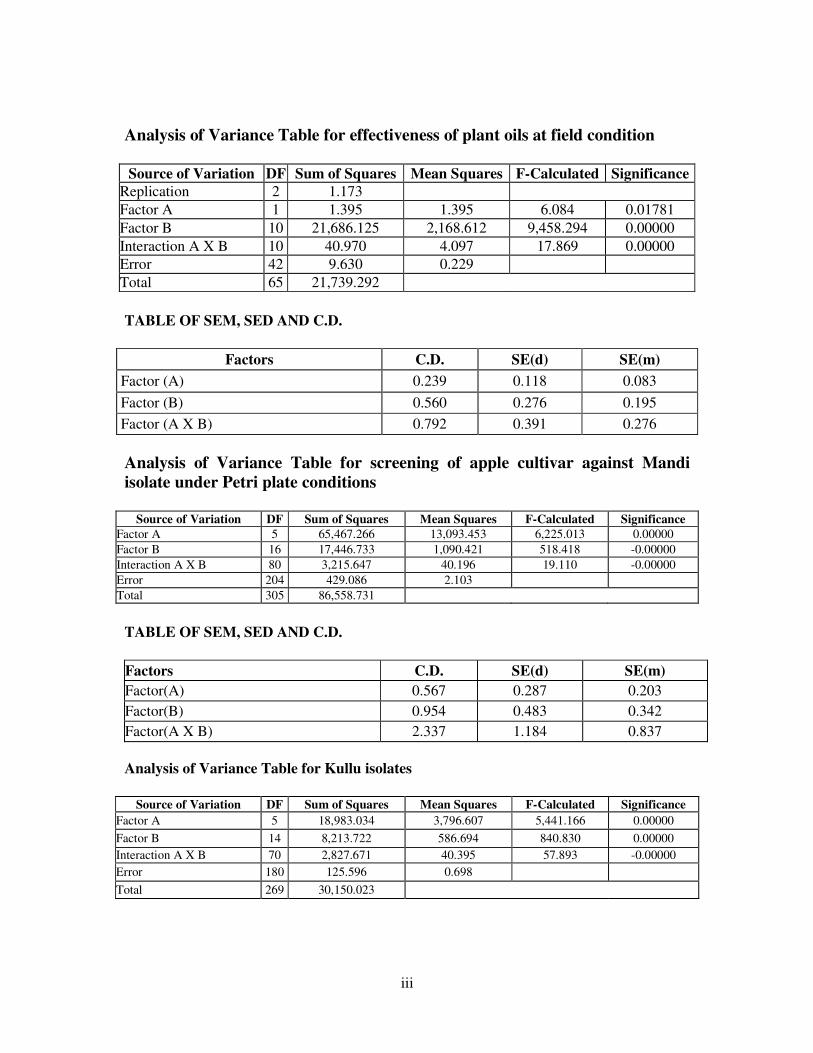
iii
Analysis of Variance Table for effectiveness of plant oils at field condition
Source of Variation DF Sum of Squares Mean Squares F-Calculated Significance
Replication 2 1.173
Factor A 1 1.395 1.395 6.084 0.01781
Factor B 10 21,686.125 2,168.612 9,458.294 0.00000
Interaction A X B 10 40.970 4.097 17.869 0.00000
Error 42 9.630 0.229
Total 65 21,739.292
TABLE OF SEM, SED AND C.D.
Factors C.D. SE(d) SE(m)
Factor (A) 0.239 0.118 0.083
Factor (B) 0.560 0.276 0.195
Factor (A X B) 0.792 0.391 0.276
Analysis of Variance Table for screening of apple cultivar against Mandi
isolate under Petri plate conditions
Source of Variation DF Sum of Squares Mean Squares F-Calculated Significance
Factor A 5 65,467.266 13,093.453 6,225.013 0.00000
Factor B 16 17,446.733 1,090.421 518.418 -0.00000
Interaction A X B 80 3,215.647 40.196 19.110 -0.00000
Error 204 429.086 2.103
Total 305 86,558.731
TABLE OF SEM, SED AND C.D.
Factors C.D. SE(d) SE(m)
Factor(A) 0.567 0.287 0.203
Factor(B) 0.954 0.483 0.342
Factor(A X B) 2.337 1.184 0.837
Analysis of Variance Table for Kullu isolates
Source of Variation DF Sum of Squares Mean Squares F-Calculated Significance
Factor A 5 18,983.034 3,796.607 5,441.166 0.00000
Factor B 14 8,213.722 586.694 840.830 0.00000
Interaction A X B 70 2,827.671 40.395 57.893 -0.00000
Error 180 125.596 0.698
Total 269 30,150.023

iv
TABLE OF SEM,SED AND C.D.
Factors C.D. SE(d) SE(m)
Factor(A) 0.348 0.176 0.125
Factor(B) 0.550 0.278 0.197
Factor(A X B) 1.347 0.682 0.482
Analysis of Variance Table Compatibility of plant oils with biocontrol agents
Source of Variation DF Sum of Squares Mean Squares F-Calculated Significance
Factor A 5 30,053.090 6,010.618 32,582.216 0.00000
Factor B 7 6,581.162 940.166 5,096.429 0.00000
Interaction A X B 35 26,624.209 760.692 4,123.540 0.00000
Error 96 17.710 0.184
Total 143 63,276.169
TABLE OF SEM,SED AND C.D.
Factors C.D. SE(d) SE(m)
Factor(A) 0.247 0.124 0.088
Factor(B) 0.285 0.143 0.101
Factor(A X B) 0.697 0.351 0.248
Analysis of Variance Table for screening against Haripurdhar isolate
Source of Variation DF Sum of
Squares Mean Squares F-Calculated Significance
Factor A 5 8,095.390 1,619.078 4,030.374 0.00000
Factor B 14 1,029.109 73.508 182.983 -0.00000
Interaction A X B 70 1,029.394 14.706 36.607 -0.00000
Error 180 72.309 0.402
Total 269 10,226.203
TABLE OF SEM, SED AND C.D.
Factors C.D. SE(d) SE(m)
Factor(A) 0.264 0.134 0.094
Factor(B) 0.417 0.211 0.149
Factor(A X B) 1.022 0.518 0.366
CURRICULUM VITAE
Name : Durga Prashad
Father’s Name : Sh. G.P. Bhandari
Date of Birth : 11.03.1983
Sex : Male
Marital Status : Unmarried
Nationality : Indian
Educational Qualification:
Certificate/degree Class/grade Board/University Year
Matric
10+2
B.Sc Horticulture
M.Sc.Vegetable
Pathology
Second
Third
First
First
HPSEB,Dharamshala
HPSEB,Dharamshala
Dr. Y.S. Parmar
UHF,Nauni,Solan(H.P.)
Dr. Y.S. Parmar
UHF,Nauni,Solan(H.P.)
1999
2001
2007
2010
Wheather sponsored by some state/ : NA
Central Govt./Univ./SAARC
Scholarship/Stipend/Fellowship, any : University Stipend
other financial assistance received
during the study period
( Durga Prashad )