doc1-dependent recruitment of nurd reveals antagonism … · cell reports article doc1-dependent...
TRANSCRIPT

DOC1-Dependent Recruitment of NURD Reveals Antagonism withSWI/SNF during Epithelial-Mesenchymal Transition in Oral CancerCellsMohd-Sarip, A., Teeuwssen, M., Bot, A. G., De Herdt, M. J., Willems, S. M., Baatenburg de Jong, R. J., ...Verrijzer, C. P. (2017). DOC1-Dependent Recruitment of NURD Reveals Antagonism with SWI/SNF duringEpithelial-Mesenchymal Transition in Oral Cancer Cells. Cell Reports, 20(1), 61-75.https://doi.org/10.1016/j.celrep.2017.06.020
Published in:Cell Reports
Document Version:Publisher's PDF, also known as Version of record
Queen's University Belfast - Research Portal:Link to publication record in Queen's University Belfast Research Portal
Publisher rightsCopyright 2017 Elsevier.This manuscript is distributed under a Creative Commons Attribution-NonCommercial-NoDerivs License(https://creativecommons.org/licenses/by-nc-nd/4.0/), which permits distribution and reproduction for non-commercial purposes, provided theauthor and source are cited.
General rightsCopyright for the publications made accessible via the Queen's University Belfast Research Portal is retained by the author(s) and / or othercopyright owners and it is a condition of accessing these publications that users recognise and abide by the legal requirements associatedwith these rights.
Take down policyThe Research Portal is Queen's institutional repository that provides access to Queen's research output. Every effort has been made toensure that content in the Research Portal does not infringe any person's rights, or applicable UK laws. If you discover content in theResearch Portal that you believe breaches copyright or violates any law, please contact [email protected].
Download date:13. Jun. 2019

Article
DOC1-Dependent Recruitm
ent of NURD RevealsAntagonism with SWI/SNF during Epithelial-Mesenchymal Transition in Oral Cancer CellsGraphical Abstract
Highlights
d DOC1 re-expression in oral cancer cells causes a reversal
of EMT
d DOC1 promotes NURD binding to a subset of target loci
d NURD and SWI/SNF compete for chromatin access,
generating opposite epigenetic states
d Remodeler antagonism controls chromatin reprogramming
of EMT
Mohd-Sarip et al., 2017, Cell Reports 20, 61–75July 5, 2017 ª 2017 The Authors.http://dx.doi.org/10.1016/j.celrep.2017.06.020
Authors
Adone Mohd-Sarip, Miriam Teeuwssen,
Alice G. Bot, ..., Jeroen A. Demmers,
Riccardo Fodde, C. Peter Verrijzer
[email protected] (A.M.-S.),[email protected] (C.P.V.)
In Brief
Mohd-Sarip et al. find that DOC1-
dependent recruitment of NURD leads to
reversal of the epithelial-mesenchymal
transition (EMT) in oral cancer cells.
Promoter binding of NURD drives SWI/
SNF eviction, formation of repressive
chromatin, and transcriptional repression
of master regulators of EMT. The authors
propose that remodeler antagonism
controls reprogramming of EMT at the
chromatin level.
Accession Numbers
GSE97839

Cell Reports
Article
DOC1-Dependent Recruitment of NURD RevealsAntagonism with SWI/SNF during Epithelial-Mesenchymal Transition in Oral Cancer CellsAdone Mohd-Sarip,1,2,* Miriam Teeuwssen,3 Alice G. Bot,2 Maria J. De Herdt,4 Stefan M. Willems,5
Robert J. Baatenburg de Jong,4 Leendert H.J. Looijenga,3 Diana Zatreanu,2,10 Karel Bezstarosti,6 Job van Riet,7,8
Edwin Oole,9 Wilfred F.J. van Ijcken,9 Harmen J.G. van de Werken,7,8 Jeroen A. Demmers,6 Riccardo Fodde,3
and C. Peter Verrijzer2,11,*1Centre for Cancer Research and Cell Biology, Queen’s University Belfast, Belfast BT9 7BL, UK2Department of Biochemistry3Department of Pathology4Department of Otorhinolaryngology and Head and Neck Surgery, Erasmus MC Cancer Institute
Erasmus University Medical Center, P.O. Box 1738, 3000 DR, Rotterdam, the Netherlands5Department of Pathology, University Medical Center Utrecht, Heidelberglaan 100, 3584 CX, Utrecht, the Netherlands6Proteomics Centre7Cancer Computational Biology Center, Erasmus MC Cancer Institute8Department of Urology9Center for Biomics
Erasmus University Medical Center, P.O. Box 1738, 3000 DR, Rotterdam, the Netherlands10Present address: Mechanisms of Transcription Laboratory, The Francis Crick Institute, 1 Midland Road, L3-4384, London NW1 1AT, UK11Lead Contact*Correspondence: [email protected] (A.M.-S.), [email protected] (C.P.V.)
http://dx.doi.org/10.1016/j.celrep.2017.06.020
SUMMARY
The Nucleosome Remodeling and Deacetylase(NURD) complex is a key regulator of cell differentia-tion that has also been implicated in tumorigenesis.Loss of the NURD subunit Deleted in Oral Cancer 1(DOC1) is associated with human oral squamouscell carcinomas (OSCCs). Here,we show that restora-tion of DOC1 expression in OSCC cells leads to areversal of epithelial-mesenchymal transition (EMT).This is caused by the DOC1-dependent targetingof NURD to repress key transcriptional regulatorsof EMT. NURD recruitment drives extensive epige-netic reprogramming, including eviction of the SWI/SNF remodeler, formation of inaccessible chro-matin, H3K27 deacetylation, and binding of PRC2and KDM1A, followed by H3K27 methylation andH3K4 demethylation. Strikingly, depletion of SWI/SNF mimics the effects of DOC1 re-expression. Ourresults suggest that SWI/SNF and NURD functionantagonistically to control chromatin state andtranscription. We propose that disturbance of thisdynamic equilibrium may lead to defects in geneexpression that promote oncogenesis.
INTRODUCTION
ATP-dependent chromatin remodeling complexes (remodelers)
control expression of the eukaryotic genome through the mobi-
This is an open access article under the CC BY-N
lization of nucleosomes. The nucleosome is the basic repeat
unit of eukaryotic chromatin, comprising 147 bp of DNA, wrap-
ped tightly around a protein core formed by an octamer of his-
tones (Luger et al., 1997). Nucleosome positioning and stability
determine the accessibility of regulatory DNA elements, thereby
providing a pervasive mode of gene expression control. Conse-
quently, remodelers play a central role in transcriptional regu-
lation by mediating the assembly, sliding, restructuring, or ejec-
tion of nucleosomes (Becker and Workman, 2013; Narlikar
et al., 2013). There are four major families of remodelers, each
named after its ATPase subunit: SWI/SNF (Switch/Sucrose
Non-fermentable), INO80, ISWI, and CHD. In addition to the
central ATPase, remodeler complexes have unique sets of
tightly associated proteins that determine targeting and regulate
activity.
A second mechanism to control chromatin state involves a
plethora of post-translational histone modifications, which can
direct the recruitment of regulatory proteins (including remodel-
ers) and modulate the folding of the chromatin fiber (Patel and
Wang, 2013; Zentner and Henikoff, 2013). Prominent modifi-
cations include acetylation, methylation, and phosphorylation
of specific residues on the histone N-terminal tails, which
protrude from the nucleosome. There is a clear correlation be-
tween specific histone modifications and transcriptional state.
For example, acetylation of histone H3K27 (H3K27ac) marks
active genes, whereas tri-methylation of the same residue
(H3K27me3) is associated with gene silencing by the Polycomb
system. Although they mediate completely different biochemical
reactions, remodelers and histone-modifying enzymes function
in a closely integrated manner to determine chromatin state
(Swygert and Peterson, 2014). In agreement with their central
Cell Reports 20, 61–75, July 5, 2017 ª 2017 The Authors. 61C-ND license (http://creativecommons.org/licenses/by-nc-nd/4.0/).

DOC1
Tubulin
+DOC1
Mascot Score
01-5050-100100-200200-500500-10001000-2000
>30002000-3000
0
20
40
60
80
100
PositiveMixedNegative
E F
Merge
DOC1
+DOC1G
H
I J
HeLa
CHD4 IP
DOC1 IP
CHD4 IP DOC1 IP
CHD3
MTA1
CHD4
MTA2MTA3GATAD2A
RBBP4RBBP7
HDAC1HDAC2
GATAD2B
MBD3MBD2
CHD5
CDK2KDM1A
SCC9
DOC1- - DOC1
DOC1
GATAD2A/B
MBD2RBBP4/7 MTA1/2
HDAC1/2CHD3/4
+ DOC1
GATAD2A/B
MBD2RBBP4/7 MTA1/2
HDAC1/2CHD3/4DOC1
LPBL
SSEL
SCA C DB
normal tongue oral squamous cell carcinomasD
OC
1 st
aini
ng (%
)
Mock
CHD4Inp
utMoc
kDOC1
Input
+DOC1 +DOC1
MTA1
DOC1CHD4
KDM1AHDAC1
CDK2
Mock
DOC1
Input
Mock
CHD4Inp
ut
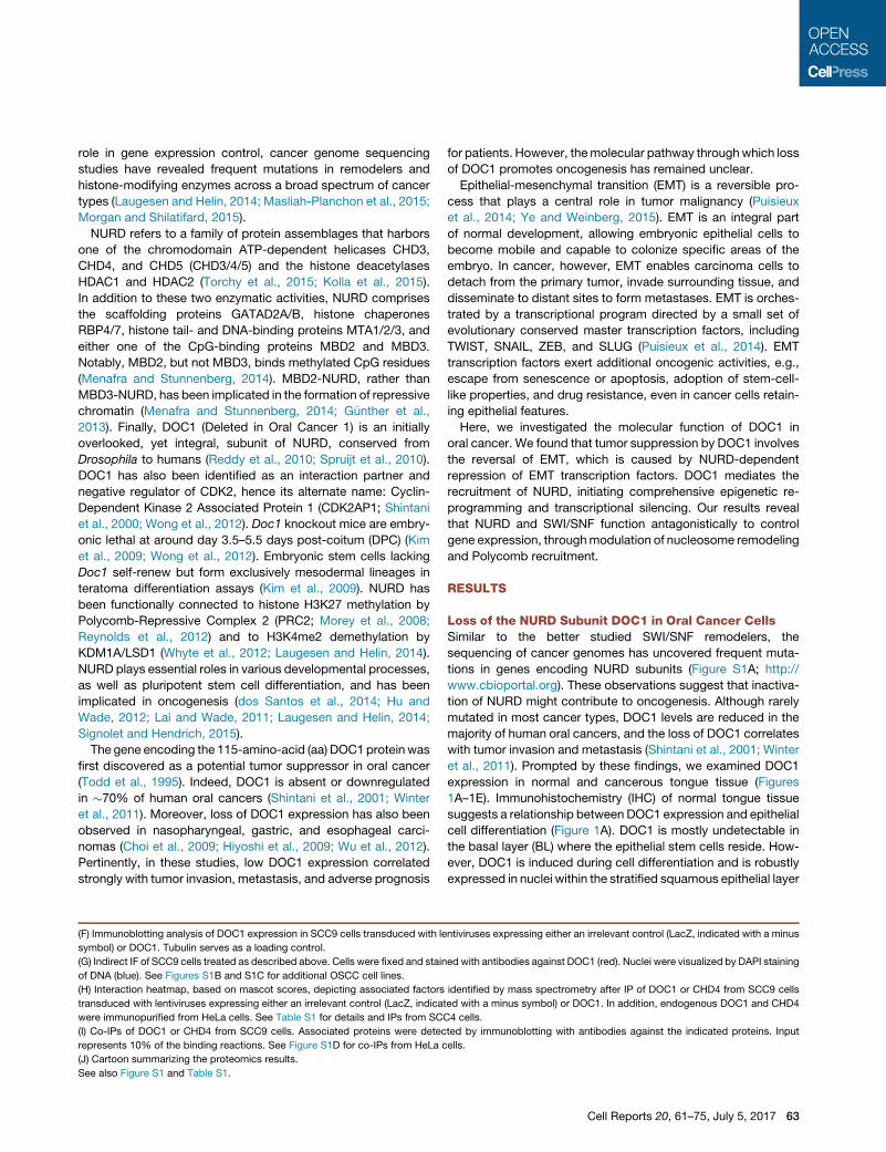
Figure 1. Re-expressed DOC1 in OSCC Cells Integrates into NURD
(A) Photomicrograph depicting DOC1 (brown), detected by immunohistochemistry, in a hematoxylin counterstained section of normal tongue epithelium. The
underlying connective tissue of the lamina propria (LP), the BL, SSEL, and keratinized SC are indicated. Our anti-DOC1 antibodies strongly stain the nuclei of
the SSEL.
(B–D) DOC1 expression in tongue carcinoma. Examples are of tumors that: (B) were negative for DOC1, (C) comprise a mixture of DOC1-negative and -positive
cells, and (D) were strongly positive for DOC1. Scale bars, 200 mm (top row) or 50 mm (bottom row).
(E) Quantification of the DOC1 expression in 36 tongue carcinomas.
(legend continued on next page)
62 Cell Reports 20, 61–75, July 5, 2017
role in gene expression control, cancer genome sequencing
studies have revealed frequent mutations in remodelers and
histone-modifying enzymes across a broad spectrum of cancer
types (Laugesen and Helin, 2014; Masliah-Planchon et al., 2015;
Morgan and Shilatifard, 2015).
NURD refers to a family of protein assemblages that harbors
one of the chromodomain ATP-dependent helicases CHD3,
CHD4, and CHD5 (CHD3/4/5) and the histone deacetylases
HDAC1 and HDAC2 (Torchy et al., 2015; Kolla et al., 2015).
In addition to these two enzymatic activities, NURD comprises
the scaffolding proteins GATAD2A/B, histone chaperones
RBP4/7, histone tail- and DNA-binding proteins MTA1/2/3, and
either one of the CpG-binding proteins MBD2 and MBD3.
Notably, MBD2, but not MBD3, binds methylated CpG residues
(Menafra and Stunnenberg, 2014). MBD2-NURD, rather than
MBD3-NURD, has been implicated in the formation of repressive
chromatin (Menafra and Stunnenberg, 2014; G€unther et al.,
2013). Finally, DOC1 (Deleted in Oral Cancer 1) is an initially
overlooked, yet integral, subunit of NURD, conserved from
Drosophila to humans (Reddy et al., 2010; Spruijt et al., 2010).
DOC1 has also been identified as an interaction partner and
negative regulator of CDK2, hence its alternate name: Cyclin-
Dependent Kinase 2 Associated Protein 1 (CDK2AP1; Shintani
et al., 2000; Wong et al., 2012). Doc1 knockout mice are embry-
onic lethal at around day 3.5–5.5 days post-coitum (DPC) (Kim
et al., 2009; Wong et al., 2012). Embryonic stem cells lacking
Doc1 self-renew but form exclusively mesodermal lineages in
teratoma differentiation assays (Kim et al., 2009). NURD has
been functionally connected to histone H3K27 methylation by
Polycomb-Repressive Complex 2 (PRC2; Morey et al., 2008;
Reynolds et al., 2012) and to H3K4me2 demethylation by
KDM1A/LSD1 (Whyte et al., 2012; Laugesen and Helin, 2014).
NURD plays essential roles in various developmental processes,
as well as pluripotent stem cell differentiation, and has been
implicated in oncogenesis (dos Santos et al., 2014; Hu and
Wade, 2012; Lai and Wade, 2011; Laugesen and Helin, 2014;
Signolet and Hendrich, 2015).
The gene encoding the 115-amino-acid (aa) DOC1 protein was
first discovered as a potential tumor suppressor in oral cancer
(Todd et al., 1995). Indeed, DOC1 is absent or downregulated
in �70% of human oral cancers (Shintani et al., 2001; Winter
et al., 2011). Moreover, loss of DOC1 expression has also been
observed in nasopharyngeal, gastric, and esophageal carci-
nomas (Choi et al., 2009; Hiyoshi et al., 2009; Wu et al., 2012).
Pertinently, in these studies, low DOC1 expression correlated
strongly with tumor invasion, metastasis, and adverse prognosis
(F) Immunoblotting analysis of DOC1 expression in SCC9 cells transduced with le
symbol) or DOC1. Tubulin serves as a loading control.
(G) Indirect IF of SCC9 cells treated as described above. Cells were fixed and stain
of DNA (blue). See Figures S1B and S1C for additional OSCC cell lines.
(H) Interaction heatmap, based on mascot scores, depicting associated factors
transduced with lentiviruses expressing either an irrelevant control (LacZ, indica
were immunopurified from HeLa cells. See Table S1 for details and IPs from SC
(I) Co-IPs of DOC1 or CHD4 from SCC9 cells. Associated proteins were detec
represents 10% of the binding reactions. See Figure S1D for co-IPs from HeLa c
(J) Cartoon summarizing the proteomics results.
See also Figure S1 and Table S1.
for patients. However, themolecular pathway throughwhich loss
of DOC1 promotes oncogenesis has remained unclear.
Epithelial-mesenchymal transition (EMT) is a reversible pro-
cess that plays a central role in tumor malignancy (Puisieux
et al., 2014; Ye and Weinberg, 2015). EMT is an integral part
of normal development, allowing embryonic epithelial cells to
become mobile and capable to colonize specific areas of the
embryo. In cancer, however, EMT enables carcinoma cells to
detach from the primary tumor, invade surrounding tissue, and
disseminate to distant sites to form metastases. EMT is orches-
trated by a transcriptional program directed by a small set of
evolutionary conserved master transcription factors, including
TWIST, SNAIL, ZEB, and SLUG (Puisieux et al., 2014). EMT
transcription factors exert additional oncogenic activities, e.g.,
escape from senescence or apoptosis, adoption of stem-cell-
like properties, and drug resistance, even in cancer cells retain-
ing epithelial features.
Here, we investigated the molecular function of DOC1 in
oral cancer. We found that tumor suppression by DOC1 involves
the reversal of EMT, which is caused by NURD-dependent
repression of EMT transcription factors. DOC1 mediates the
recruitment of NURD, initiating comprehensive epigenetic re-
programming and transcriptional silencing. Our results reveal
that NURD and SWI/SNF function antagonistically to control
gene expression, throughmodulation of nucleosome remodeling
and Polycomb recruitment.
RESULTS
Loss of the NURD Subunit DOC1 in Oral Cancer CellsSimilar to the better studied SWI/SNF remodelers, the
sequencing of cancer genomes has uncovered frequent muta-
tions in genes encoding NURD subunits (Figure S1A; http://
www.cbioportal.org). These observations suggest that inactiva-
tion of NURD might contribute to oncogenesis. Although rarely
mutated in most cancer types, DOC1 levels are reduced in the
majority of human oral cancers, and the loss of DOC1 correlates
with tumor invasion and metastasis (Shintani et al., 2001; Winter
et al., 2011). Prompted by these findings, we examined DOC1
expression in normal and cancerous tongue tissue (Figures
1A–1E). Immunohistochemistry (IHC) of normal tongue tissue
suggests a relationship between DOC1 expression and epithelial
cell differentiation (Figure 1A). DOC1 is mostly undetectable in
the basal layer (BL) where the epithelial stem cells reside. How-
ever, DOC1 is induced during cell differentiation and is robustly
expressed in nuclei within the stratified squamous epithelial layer
ntiviruses expressing either an irrelevant control (LacZ, indicated with a minus
ed with antibodies against DOC1 (red). Nuclei were visualized by DAPI staining
identified by mass spectrometry after IP of DOC1 or CHD4 from SCC9 cells
ted with a minus symbol) or DOC1. In addition, endogenous DOC1 and CHD4
C4 cells.
ted by immunoblotting with antibodies against the indicated proteins. Input
ells.
Cell Reports 20, 61–75, July 5, 2017 63
A
C
B
DAPIPhalloidin
+DOC1
D
DAPIVIM
E-CAD
N-CAD
+DOC1
Time (day)
OD
490
mm
1 2 3 4 5 6 70
0.20.40.60.81.01.2
+DOC1
+DOC1
Day 0
Day 3
E
+DOC1
VIMN-CADE-CADTUB
DOC1
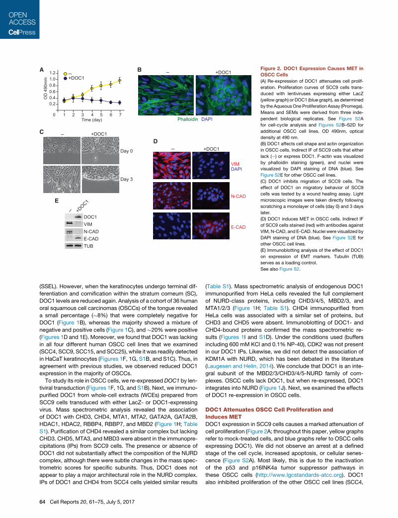
Figure 2. DOC1 Expression Causes MET in
OSCC Cells
(A) Re-expression of DOC1 attenuates cell prolif-
eration. Proliferation curves of SCC9 cells trans-
duced with lentiviruses expressing either LacZ
(yellow graph) or DOC1 (blue graph), as determined
by the AqueousOne Proliferation Assay (Promega).
Means and SEMs were derived from three inde-
pendent biological replicates. See Figure S2A
for cell-cycle analysis and Figures S2B–S2D for
additional OSCC cell lines. OD 490nm, optical
density at 490 nm.
(B) DOC1 affects cell shape and actin organization
in OSCC cells. Indirect IF of SCC9 cells that either
lack (�) or express DOC1. F-actin was visualized
by phalloidin staining (green), and nuclei were
visualized by DAPI staining of DNA (blue). See
Figure S2E for other OSCC cell lines.
(C) DOC1 inhibits migration of SCC9 cells. The
effect of DOC1 on migratory behavior of SCC9
cells was tested by a wound healing assay. Light
microscopic images were taken directly following
scratching a monolayer of cells (day 0) and 3 days
later.
(D) DOC1 induces MET in OSCC cells. Indirect IF
of SCC9 cells stained (red) with antibodies against
VIM, N-CAD, and E-CAD. Nuclei were visualized by
DAPI staining of DNA (blue). See Figure S2E for
other OSCC cell lines.
(E) Immunoblotting analysis of the effect of DOC1
on expression of EMT markers. Tubulin (TUB)
serves as a loading control.
See also Figure S2.
(SSEL). However, when the keratinocytes undergo terminal dif-
ferentiation and cornification within the stratum corneum (SC),
DOC1 levels are reduced again. Analysis of a cohort of 36 human
oral squamous cell carcinomas (OSCCs) of the tongue revealed
a small percentage (�8%) that were completely negative for
DOC1 (Figure 1B), whereas the majority showed a mixture of
negative and positive cells (Figure 1C), and �20% were positive
(Figures 1D and 1E). Moreover, we found that DOC1 was lacking
in all four different human OSCC cell lines that we examined
(SCC4, SCC9, SCC15, and SCC25), while it was readily detected
in HaCaT keratinocytes (Figures 1F, 1G, S1B, and S1C). Thus, in
agreement with previous studies, we observed reduced DOC1
expression in the majority of OSCCs.
To study its role in OSCC cells, we re-expressedDOC1 by len-
tiviral transduction (Figures 1F, 1G, and S1B). Next, we immuno-
purified DOC1 from whole-cell extracts (WCEs) prepared from
SCC9 cells transduced with either LacZ- or DOC1-expressing
virus. Mass spectrometric analysis revealed the association
of DOC1 with CHD3, CHD4, MTA1, MTA2, GATA2A, GATA2B,
HDAC1, HDAC2, RBBP4, RBBP7, and MBD2 (Figure 1H; Table
S1). Purification of CHD4 revealed a similar complex but lacking
CHD3. CHD5, MTA3, and MBD3 were absent in the immunopre-
cipitations (IPs) from SCC9 cells. The presence or absence of
DOC1 did not substantially affect the composition of the NURD
complex, although there were subtle changes in the mass spec-
trometric scores for specific subunits. Thus, DOC1 does not
appear to play a major architectural role in the NURD complex.
IPs of DOC1 and CHD4 from SCC4 cells yielded similar results
64 Cell Reports 20, 61–75, July 5, 2017
(Table S1). Mass spectrometric analysis of endogenous DOC1
immunopurified from HeLa cells revealed the full complement
of NURD-class proteins, including CHD3/4/5, MBD2/3, and
MTA1/2/3 (Figure 1H; Table S1). CHD4 immunopurified from
HeLa cells was associated with a similar set of proteins, but
CHD3 and CHD5 were absent. Immunoblotting of DOC1- and
CHD4-bound proteins confirmed the mass spectrometric re-
sults (Figures 1I and S1D). Under the conditions used (buffers
including 600 mM KCl and 0.1% NP-40), CDK2 was not present
in our DOC1 IPs. Likewise, we did not detect the association of
KDM1A with NURD, which has been debated in the literature
(Laugesen and Helin, 2014). We conclude that DOC1 is an inte-
gral subunit of the MBD2/3/CHD3/4/5-NURD family of com-
plexes. OSCC cells lack DOC1, but when re-expressed, DOC1
integrates into NURD (Figure 1J). Next, we examined the effects
of DOC1 re-expression in OSCC cells.
DOC1 Attenuates OSCC Cell Proliferation andInduces METDOC1 expression in SCC9 cells causes a marked attenuation of
cell proliferation (Figure 2A; throughout this paper, yellow graphs
refer to mock-treated cells, and blue graphs refer to OSCC cells
expressing DOC1). We did not observe an arrest at a defined
stage of the cell cycle, increased apoptosis, or cellular senes-
cence (Figure S2A). Most likely, this is due to the inactivation
of the p53 and p16INK4a tumor suppressor pathways in
these OSCC cells (http://www.lgcstandards-atcc.org). DOC1
also inhibited proliferation of the other OSCC cell lines (SCC4,
SCC15, and SSC25; Figures S2B–S2D). Surprisingly, DOC1 re-
expression induced marked changes in SCC9 cell morphology
and actin organization, as visualized by phalloidin staining (Fig-
ure 2B). Compared to cells transduced with a control vector,
which have a more fibroblast-like appearance, DOC1-express-
ing cells acquire a more cobblestone-like morphology with
epithelial features. Moreover, upon DOC1 expression, prominent
stress fibers are replaced by amore cortical actin organization. A
scratch test revealed that DOC1-expressing SCC9 cells are less
migratory and form layers of tightly attached cells (Figure 2C).
These results suggest that expression of DOC1 induces a
mesenchymal-to-epithelial transition (MET). To test this possibil-
ity, we examined the expression of a number of canonical EMT
markers. Immunofluorescence (IF) microscopy revealed a strong
reduction of the mesenchymal markers vimentin (VIM) and
N-cadherin (N-CAD) after DOC1 expression, whereas the epithe-
lial marker E-cadherin (E-CAD) was upregulated (Figures 2D and
S2E). The observed changes in expression of these EMT
markers were confirmed by immunoblotting (Figure 2E).
In conclusion, we examined the effects of DOC1 re-expression
in OSCC cells that lack this integral subunit of NURD. DOC1 effi-
ciently incorporates into NURD and triggers the differentiation of
cells from a quasi-mesenchymal (SCC9 and SCC15) or quasi-
epithelial (SCC4 and SCC25) appearance toward an epithelial
phenotype. Therefore, DOC1-induced cell differentiation is,
strictly speaking, a partial MET. For the sake of brevity, however,
we will, hereinafter, refer to this process as MET. This transition
involves changes in actin organization, cell shape, expression of
key EMT markers, reduced cell migration, and attenuated cell
proliferation. These observations suggest that loss of DOC1 con-
tributes to the development of OSCC by inhibiting epithelial dif-
ferentiation and by conferring tumor cells with a mesenchymal-
like and, possibly, more invasive phenotype.
DOC1 Functions as Part of NURDTo test whether the effects of DOC1 re-expression in OSCC cells
depend on the chromatin remodeling activity of the NURD com-
plex, we depleted its ATPase CHD4 (Figure 3A). Following short
hairpin RNA (shRNA)-mediated knockdown of CHD4 in SCC9
cells, DOC1 expression failed to trigger MET. There was no
induction of E-CAD, whereas VIM and N-CAD expression was
not reduced. Loss of CHD4, in the absence of DOC1 expression,
did not affect the expression of EMT markers. Regardless of the
presence or absence of DOC1, knockdown of CHD4 led to
reduced cell proliferation (Figure 3B). Likewise, depletion of
MBD2 or MTA2 caused a loss of cell viability and blocked the
ability of DOC1 to promote MET (Figures S3A and S3B). Thus,
once NURD lacks DOC1, loss of additional NURD subunits com-
promises cell viability but has little effect on the expression of
EMTmarkers. Thus, the capacity of DOC1 to driveMET depends
on NURD, and cells lacking DOC1 still depend on the remaining
NURD for viability.
Next, we used shRNAs to deplete either DOC1, CHD4, MBD2,
or MTA2 in HaCaT cells, a spontaneously immortalized, human
keratinocyte line (Figures 3C, 3D, and S3C–S3E). Under our
culture conditions, HaCaT cells have an epithelial phenotype.
Upon knockdown of DOC1 or other NURD subunits, there was
reduced expression of the epithelial marker E-CAD, whereas
the mesenchymal markers N-CAD and VIM were induced (Fig-
ures 3C and S3C). In agreement with our earlier results (Fig-
ure 1J), loss of DOC1 did not affect the stability of other NURD
subunits (Figure S3D). However, loss of MBD2 or MTA2 affected
CHD4 levels, suggesting that these subunits are important for
the structural integrity of NURD. Importantly, depletion of either
DOC1, CHD4, MBD2, or MTA2 caused substantially reduced
cell numbers (Figures 3D and S3E). Thus, the intact NURD com-
plex is required for optimal viability of HaCaT cells. Collectively,
these observations support the notion that DOC1 functions as an
integral part of NURD.
DOC1-Dependent Recruitment of NURD Drives METThe EMT program is orchestrated by a set of master regulators
that form an integrated transcriptional network with extensive
cross-regulation. Expression of DOC1 in OSCC cells leads to
downregulation of all major EMT transcription factors, concom-
itant with cell differentiation toward an epithelial phenotype (Fig-
ure 4A). To determine which of these might be directly regulated
by NURD, we used chromatin IP (ChIP)-qPCR. We monitored
CHD4 binding to selected promoter regions in either the absence
or presence of DOC1. CHD4 ChIPs revealed strong DOC1-
dependent binding to the promoters of Twist1, Twist2, and
Zeb2 and weaker binding to the Snail, Slug, and Zeb1 promoters
(Figure 4B). CHD4 binding to two previously identified targets of
NURD, Crabp1 and Rassf10 (G€unther et al., 2013), was indepen-
dent of DOC1. The binding pattern of DOC1was similar to that of
CHD4 (Figure 4C). Collectively, these results suggest that DOC1
is a gene-selective subunit of NURD, required for the binding and
repression of key EMT transcription factor genes.
NURD-mediated repression of crucial master regulators of
EMT provides an attractive molecular mechanism to explain
DOC1-induced MET in OSCC cells. To test this hypothesis, we
transduced lentiviruses that expressed shRNAs directed against
either Twist1 or Twist2 or a control virus (mock). Depletion of
either TWIST1 or TWIST2, in the absence of DOC1 expression,
suffices to induce MET, as indicated by actin reorganization,
downregulation of VIM and N-CAD, and induction of E-CAD (Fig-
ures 4D and 4E). TWIST1 and TWIST2 appear both to be required
for EMT. As observed for DOC1 re-expression in OSCC cells,
loss of TWIST1/2 inhibited cell proliferation (Figure 4F). These re-
sults establish that downregulation of TWIST1 or TWIST2 can
mimic the main effects of DOC1 re-expression in OSCC cells.
These results suggest that DOC1 initiates MET in oral cancer
cells by directing NURD to repress master regulators of EMT.
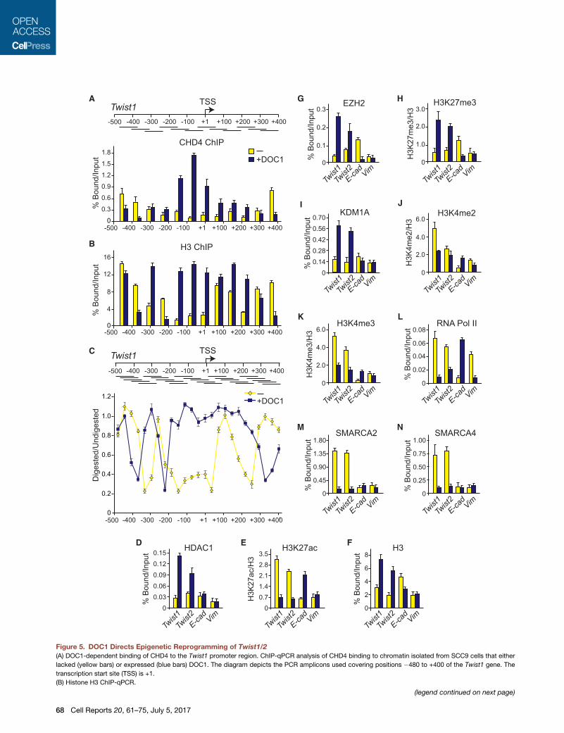
NURD Recruitment Causes Extensive ChromatinReorganizationTo explore the impact of NURD recruitment on the local chro-
matin structure, we first determined its precise localization within
�900 bp of the Twist1 promoter region (�500 to +400 bp, relative
to the transcription start site; TSS). ChIP-qPCR revealed DOC1-
dependent CHD4 binding, directly upstream of the Twist1 tran-
scription start site (Figure 5A). Histone H3 ChIPs revealed a
prominent nucleosome-depleted region (NDR) in the absence
of DOC1 when Twist1 is expressed (Figure 5B). Following
DOC1 expression and NURD binding, there is a dramatic
nucleosome repositioning, leading to occupancy of the NDR.
Cell Reports 20, 61–75, July 5, 2017 65

A
# ce
lls (%
)
0
20
40
60
80
100 Mock KD
CHD4 KDDOC1 KD
DB
# ce
lls (%
)
0
20
40
60
80
100
CHD4 KD
CHD4
VIM
PhalloidinDAPI
CHD4VIM
E-CAD
N-CAD
Mock KD+DOC1 +DOC1
C
PhalloidinDAPI
VIM
E-CAD
N-CAD
CHD4 KDMock KD DOC1 KD
DOC1
CHD4
HaCaT
Mock KD
CHD4 KD
+DOC1
+DOC1
SCC9
HaCaTSCC9
Figure 3. DOC1-Induced MET Depends on CHD4
(A) Indirect IF of SCC9 cells that either lack or express DOC1, in combination with shRNA-mediated knockdown (KD) of CHD4. Cells were stained using the
indicated antibodies.
(B) Effects of DOC1 expression in combination with CHD4 KD on cell proliferation were determined 3 days after KD, as described in the legend for Figure 2A.
(C) Indirect IF of HaCaT cells after KD of DOC1 or CHD4. Cells were stained using the indicated antibodies.
(D) HaCaT cell numbers were determined 3 days following KD of DOC1 or CHD4.
Means and SEMs were derived from three independent biological replicates.
See also Figure S3.
High-resolution micrococcal nuclease (MNase) sensitivity map-
ping showed that, in the absence of DOC1, the Twist1 promoter
DNA was highly accessible to nuclease digestion (Figure 5C). In
addition, MNase mapping established that the �250-bp NDR is
flanked by well-positioned nucleosomes. DOC1 expression in-
66 Cell Reports 20, 61–75, July 5, 2017
duces extensive chromatin reorganization, leading to complete
occlusion of the NDR and a shift in the position of the flanking nu-
cleosomes. Thus, DOC1-mediated recruitment of NURD to the
Twist1 promoter induces a switch from an open to closed nucle-
osomal organization.

C
B
D
E
% B
ound
/Inpu
t
0
DOC1 ChIP
0.3
0.6
0.9
1.2
SnailSlugTwist1
Zeb1Zeb2
Twist2
E-cadVim
Crabp1
Rassf10
+DOC1
% B
ound
/Inpu
t
0
0.2
0.4
0.6
0.8
1.0
CHD4 ChIP
SnailSlugTwist1
Zeb1Zeb2
Twist2
VimE-cad
Crabp1
Rassf10
+DOC1
TWIST2
TWIST1
PhalloidinDAPI
VIM
E-CAD
N-CAD
TWIST2 KDMock KD TWIST1 KD
# ce
lls (%
)
0
20
40
60
80
100
Mock KD
TWIST2 KDTWIST1 KD
TWIST1/2 KD
F
Mock K
D
Twist
1 KD
Twist
2 KD
E-CAD
TWIST1
TWIST2
VIM
N-CAD
TUB
DOC1
CHD4
HeLa
A+DOC1
Rel
ativ
e m
RN
A ex
pres
sion
0
1.0
2.0
3.0
7.5
E-cadVimN-cadMmp2
SnailSlugTwist1
Zeb1
Twist2
Zeb2
Figure 4. DOC1-Mediated Repression of TWIST1/2 Drives MET
(A) Effect of DOC1 on the expression of EMT transcription factors. mRNA was isolated from SCC9 cells that either lacked (yellow bars) or expressed (blue bars)
DOC1. Relative levels of mRNA were determined by qRT-PCR. Gapdh was used for normalization. Means and SDs were derived from three independent bio-
logical replicates.
(B) DOC1 is required for CHD4 binding to the Twist1, Twist2, and Zeb2 promoters. ChIP-qPCR analysis of DOC1 binding to the promoters of EMT transcription
factors, E-cadherin, Vimentin, Crabp1, and Rassf10. Chromatin was isolated from SCC9 cells that either lacked (yellow bars) or expressed DOC1 (blue bars).
Means and SDs were derived from three independent biological replicates.
(C) ChIP-qPCR analysis of DOC1 binding. Means and SEM were derived from three independent biological replicates.
(D) Depletion of TWIST1 or TWIST2 suffices forMET. Indirect IF of SCC9 cells after knockdown (KD) of either TWIST1 or TWIST2. Cells were stainedwith phalloidin
or the indicated antibodies.
(E) Immunoblotting analysis of the effect of DOC1 on the expression of EMT markers, using antibodies against the indicated proteins.
(F) Effect of KD of TWIST1 or TWIST2 on cell proliferation were determined 3 days after KD, as described in the legend to Figure 2A. Means and SEMs were
derived from three independent biological replicates.
Cell Reports 20, 61–75, July 5, 2017 67
B
C
A G
I
HEZH2
% B
ound
/Inpu
t
0
0.1
0.2
0.3
VimTwist1Twist2E-cad
H3K27me3
0
1.0
2.0
3.0
H3K
27m
e3/H
3
Twist1Twist2E-cadVim
K
0
H3K4me3
2.0
4.0
6.0
H3K
4me3
/H3
Twist1Twist2E-cadVim
0
RNA Pol II
0.02
0.04
0.06
0.08
Twist2
Twist1
E-cadVim
% B
ound
/Inpu
t
L
J
M N
0
SMARCA4
0.25
0.50
0.75
1.00
Twist1Twist2E-cadVim
% B
ound
/Inpu
t
0
SMARCA2
0.45
0.90
1.35
1.80
Twist2
Twist1
E-cadVim
% B
ound
/Inpu
t
0
KDM1A
0.140.280.42
0.70
% B
ound
/Inpu
t
0.56
Twist1Twist2E-cadVim
0
H3K4me2
2.0
4.0
6.0
H3K
4me2
/H3
Twist1Twist2E-cadVim
H3 ChIP
0+400+200+100+1 +300-400 -300 -200 -100-500
% B
ound
/Inpu
t
4
8
12
16
Twist1+400+200+100+1 +300-400 -300 -200 -100-500
TSS
CHD4 ChIP
0+400+200+100+1 +300-400 -300 -200 -100-500
% B
ound
/Inpu
t
0.3
0.6
0.9
1.2
1.8
1.5 +DOC1
E F H3
Twist1Twist2
% B
ound
/Inpu
t
0
2
4
6
8
E-cadVim
% B
ound
/Inpu
t
Twist1Twist2
0
HDAC1
0.03
0.090.120.15
0.06
E-cadVim
H3K27ac
H3K
27ac
/H3
Twist2
Twist1
00.71.42.12.83.5
E-cadVim
D
Twist1+400+200+100+1 +300-400 -300 -200 -100-500
TSS
0
0.4
0.6
0.8
1.0
1.2
+400+200+100+1 +300-400 -300 -200 -100-500
0.2
Dig
este
d/U
ndig
este
d
+DOC1
Figure 5. DOC1 Directs Epigenetic Reprogramming of Twist1/2
(A) DOC1-dependent binding of CHD4 to the Twist1 promoter region. ChIP-qPCR analysis of CHD4 binding to chromatin isolated from SCC9 cells that either
lacked (yellow bars) or expressed (blue bars) DOC1. The diagram depicts the PCR amplicons used covering positions �480 to +400 of the Twist1 gene. The
transcription start site (TSS) is +1.
(B) Histone H3 ChIP-qPCR.
(legend continued on next page)
68 Cell Reports 20, 61–75, July 5, 2017

In addition to nucleosome remodeling, NURD mediates his-
tone deacetylation. As expected, HDAC1 was readily recruited
to the Twist1 and Twist2 promoters following DOC1 expression
(Figure 5D). Concomitantly, there was a drop in the level of
H3K27 acetylation, corrected for histone H3 occupancy (Fig-
ure 5E). Similar to the Twist1 promoter, histone H3 ChIP revealed
DOC1-induced nucleosome occupancy at the Twist2 promoter
(Figure 5F). H3K27 deacetylation by NURD has been linked to
the recruitment of PRC2 (Reynolds et al., 2012). Indeed, in the
presence of DOC1, we observed binding of the PRC2 enzymatic
subunit EZH2, accompanied by increased levels of H3K27me3
(Figures 5G and 5H). Moreover, DOC1 expression was followed
by binding of the KDM1A, with concomitant loss of H3K4me2
and H3K4me3 (Figures 5I and 5K). The transfer from an active
to a repressed chromatin state was accompanied by loss of
RNA polymerase II (RNA Pol II; Figure 5L). Similar to what we
observed for the Twist1/2 promoters, DOC1-dependent binding
of NURD to the promoter region of Zeb2 induced formation of a
repressive chromatin structure (Figures S4A–S4K). Thus, NURD
recruitment initiates the comprehensive epigenetic reprogram-
ming of the Twist1/2 and Zeb2 genes.
Previously, we reported that the SWI/SNF remodeler counter-
acts chromatin binding of Polycomb repressors (Kia et al., 2008).
Therefore, we wondered whether SWI/SNF might be associated
with the active Twist1/2 and Zeb2 promoters to prevent Poly-
comb repression. We performed ChIP assays using antibodies
directed against either SMARCA4/BRG1 or SMARCA2/hBRM,
the mutually exclusive ATPase subunits of SWI/SNF assem-
blages. Both SMARCA2 and SMARCA4 bound the active
Twist1/2 and Zeb2 promoters but were displaced following
DOC1-driven binding of NURD (Figures 5M, 5N, and S4L–
S4N). These observations raised the possibility that SWI/SNF
and NURD act antagonistically in the control of the Twist1/2
and Zeb2 genes.
Loss of SWI/SNF Phenocopies the Effects of DOC1Re-expressionTo test the idea that SWI/SNF and NURD might have opposing
effects on the EMT program, we determined the consequences
of SWI/SNF depletion in the absence of DOC1 induction. Deple-
tion of either SMARCA2 or SMARCA4 had only weak effects
on SCC9 cell phenotype (Figure S5A). However, knockdown of
both SWI/SNF ATPases induced a strong MET. Loss of both
SMARCA2 and SMARCA4 (SMARCA2/4) led to actin fiber re-or-
ganization and a change from a fibroblast-like morphology to an
epithelial cell shape (Figure 6A). We observed the downregula-
tion of VIM and N-CAD, whereas E-CAD was induced (Figures
6B and S5B). Moreover, there was a loss of Twist1/2 and Zeb2
(C) DOC1-induced changes in nucleosome organization. High-resolutionMNase a
graph) or expressed (blue graph) DOC1. The MNase accessibility profile was d
undigested product using the delta C(t) method. Ratios were plotted against the
(D–N) ChIP-qPCR analysis of chromatin at the Twist1, Twist2, E-cadherin, and Vim
H3, (G) EZH2, (H) H3K27me3, (I) KDM1A, (J) H3K4me2, (K) H3K4me3, (L) RNA Po
that either lacked (yellow bars) or expressed DOC1 (blue bars). Protein ChIP signa
were normalized to H3 signals.
Means and SEMs for all experiments in this figure were derived from three ind
Figure S4.
See also Figure S4.
expression after SMARCA2/4 depletion (Figure 6C; yellow indi-
cates mock, and red indicates SMARCA2/4 knockdown). Loss
of either SMARCA2 or SMARCA4 alone gave an intermediate
effect, suggesting that both remodelers stimulate Twist1/2
and Zeb2 transcription (Figure S5C). Finally, depletion of
SMARCA2/4 led to diminished cell numbers (Figure S5D).
Thus, the functional consequences of SWI/SNF depletion are
similar to those of DOC1 re-expression: reduced cell prolifera-
tion, attenuated expression of EMT transcription factors, and
MET. Our results suggest that SWI/SNF and NURD compete
for chromatin binding at Twist1/2 and Zeb2 promoters and
generate opposite transcriptional states. To test this idea,
we examined the impact of SWI/SNF depletion on chromatin
organization.
Remodeler Antagonism Controls EpigeneticReprogramming of EMTBoth SMARCA2 and SMARCA4 bind to the Twist1/2 and Zeb2
promoters (Figures 6D, 6E, S6B, and S6C). Knockdown of
SMARCA2/4 caused a loss of ChIP signals, confirming the spec-
ificity of our antibodies. Following SWI/SNF depletion, the NDR
disappears and the Twist1 promoter DNA is now occluded by
nucleosomes (Figures 6F and S5E). The pattern of MNase
accessibility after the knockdown of SWI/SNF is remarkably
similar to that following DOC1 expression (compare Figures 5C
and 6F). CHD4 and HDAC1 ChIPs showed that depletion of
SWI/SNF suffices to allow NURD binding to the Twist1/2 and
Zeb2 promoters, in spite of the absence of DOC1 (Figures 6G,
S5F, S6D, and S6E). These results show that NURD devoid of
DOC1 still has an intrinsic, albeit weakened, ability to bind the
Twist1/2 and Zeb2 promoters. The chromatin changes caused
by SWI/SNF depletion are remarkably similar to those observed
after DOC1 re-expression (Figures 6H–6J, S5G–S5K, and S6F–
S6M). Concomitant with NURD recruitment after SWI/SNF
knockdown, the level of H3K27ac dropped, PRC2 bound, and
H3K27ac was replaced by H3K27me3. In addition, KDM1A is
recruited, accompanied by H3K4 demethylation. In agreement
with the repression of Twist1/2 and Zeb2 transcription, RNA
Pol II is lost following the knockdown of SWI/SNF. Thus, SWI/
SNF depletion in OSCCcells has similar effects on the epigenetic
setting of EMT master regulators as DOC1 re-expression.
In summary, SWI/SNF prevents the binding of NURD lacking
DOC1 to the Twist1/2 and Zeb2 promoters. Conversely, upon in-
clusion of DOC1 in the complex, NURD displaces SWI/SNF. The
replacement of SWI/SNF by NURD results in the transition from
an open to a closed chromatin structure. Moreover, chromatin
binding by PRC2 is blocked by SWI/SNF but promoted by
NURD. Thus, SWI/SNF and NURD compete for binding and
ccessibility mapping on chromatin isolated from cells that either lacked (yellow
etermined by normalizing the amount of digested PCR product to that of the
midpoint of the corresponding PCR amplicons shown in the diagram on top.
entin promoters using antibodies directed against (D) HDAC1, (E) H3K27ac, (F)
lI, (M) SMARCA2, and (N) SMARCA4. Chromatin was isolated from SCC9 cells
ls are presented as percentage of input chromatin. Histone modification ChIPs
ependent biological replicates. Results for the Zeb2 promoter are shown in
Cell Reports 20, 61–75, July 5, 2017 69

A
B
Mock K
D
SMARCA2/4 K
D
E-CAD
VIM
N-CAD
TUB
SMARCA4
SMARCA2
CHD4
PhalloidinDAPI
Mock KD SMARCA2/4 KD
PhalloidinDAPI
SMARCA2
SMARCA4
D
SMARCA2
0
0.2
0.4
0.6
0.8
Twist2
Twist1
E-cadVim
% B
ound
/Inpu
t
E
SMARCA4
0
1.0
2.0
3.0
% B
ound
/Inpu
t
Twist2
Twist1
E-cadVim
GCHD4
0
0.2
0.4
0.6
% B
ound
/Inpu
t
Twist2
Twist1
E-cadVim
HH3K27ac
0
0.5
1.0
1.5
2.0
H3K
27ac
/H3
Twist2
Twist1
E-cadVim
I
EZH2
% B
ound
/Inpu
t
0
0.6
1.2
1.8
Twist2
Twist1
E-cadVim
J
H3K27me3
0
2.0
4.0
6.0
H3K
27m
e3/H
3
Twist2
Twist1
E-cadVim
FTwist1
+400+200+100+1 +300-400 -300 -200 -100-500
TSS
+400+200+100+1 +3000
0.4
0.6
0.8
1.0
1.2
-400 -300 -200 -100-500
0.2
Dig
este
d/U
ndig
este
dMock KD SMARCA2/4 KD
Mock KD SMARCA2/4 KD
Rel
ativ
e m
RN
A ex
pres
sion
0
0.5
1.0
1.5
2.0
2.5
Twist1Twist2
VimN-cadE-cad
Zeb2
CMock KD SMARCA2/4 KD
(legend on next page)
70 Cell Reports 20, 61–75, July 5, 2017
generate opposite chromatin states. We propose that a distur-
bance in the balance between these antagonistic remodelers
can set off a cascade of chromatin reprograming that promotes
oncogenesis.
DOC1 Assists NURD Recruitment to CpG IslandsTo investigate the impact of DOC1 on the genome-wide binding
of NURD, we performed CHD4 ChIP sequencing (ChIP-seq)
on chromatin from SCC9 cells. We identified 4,902 CHD4
consensus peaks in DOC1-expressing cells, compared to
3,949 in cells lacking DOC1. This observation indicates that
DOC1 is important for binding to a subset of NURD loci. We
note that the ChIP-seq uncovered DOC1-dependent binding to
additional genes involved in EMT, as illustrated with a few exam-
ples in Figure 7A. Analysis of the genomic distribution of CHD4
revealed that about 60% (no DOC1) to 67% (+DOC1) of the bind-
ing sites correspond to genic regions; in particular, promoters
and introns (Figure 7B). DOC1 appears to enhance promoter
binding by NURD, which increased from �23% to �35% of all
mapped binding sites (Figures 7B and 7C). Strikingly, DOC1
expression led to a substantially higher proportion of CHD4 bind-
ing at CpG islands (Figure 7D). Taken together, genome-wide
binding analysis confirmed that DOC1 promotes NURD binding
to a subset of target loci. In particular, our results support a
role for DOC1 in NURD recruitment to CpG islands.
DISCUSSION
ATP-dependent chromatin remodelers are frequently mutated in
human cancers. However, themolecular basis of the association
between mutations in specific remodeler subunits and particular
types of cancer is poorly understood. Here, we showed that the
loss of DOC1 in oral cancer cells leads to a failure of NURD to
bind and repress master transcriptional regulators of EMT. Re-
expression of DOC1 in OSCC cells restores NURD recruitment
to key target genes, a switch fromopen toclosedchromatin, tran-
scriptional repression, and reversal of EMT (MET). Consistent
with the transcriptional repression we observed after DOC1-
dependent NURD recruitment, theOSCCcells we studied harbor
MBD2-NURD (Figure 1), the NURD variant implicated in the for-
mation of repressive chromatin (G€unther et al., 2013). In agree-
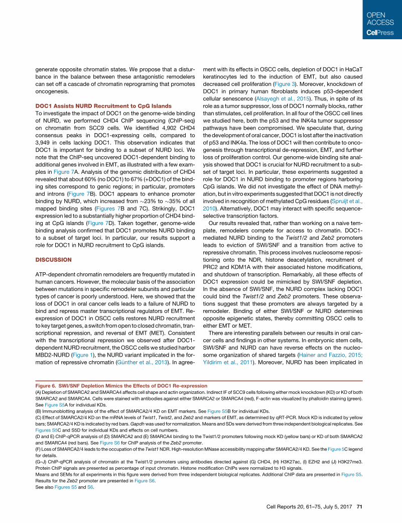
Figure 6. SWI/SNF Depletion Mimics the Effects of DOC1 Re-expressi
(A) Depletion of SMARCA2 and SMARCA4 affects cell shape and actin organizatio
SMARCA2 and SMARCA4. Cells were stained with antibodies against either SMA
See Figure S5A for individual KDs.
(B) Immunoblotting analysis of the effect of SMARCA2/4 KD on EMT markers. S
(C) Effect of SMARCA2/4 KD on the mRNA levels of Twist1, Twist2, and Zeb2 and
bars; SMARCA2/4 KD is indicated by red bars.Gapdhwas used for normalization.
Figures S5C and S5D for individual KDs and effects on cell numbers.
(D and E) ChIP-qPCR analysis of (D) SMARCA2 and (E) SMARCA4 binding to the
and SMARCA4 (red bars). See Figure S6 for ChIP analysis of the Zeb2 promoter
(F) Loss of SMARCA2/4 leads to the occupation of the Twist1NDR. High-resolution
for details.
(G–J) ChIP-qPCR analysis of chromatin at the Twist1/2 promoters using antibo
Protein ChIP signals are presented as percentage of input chromatin. Histone m
Means and SEMs for all experiments in this figure were derived from three indep
Results for the Zeb2 promoter are presented in Figure S6.
See also Figures S5 and S6.
ment with its effects in OSCC cells, depletion of DOC1 in HaCaT
keratinocytes led to the induction of EMT, but also caused
decreased cell proliferation (Figure 3). Moreover, knockdown of
DOC1 in primary human fibroblasts induces p53-dependent
cellular senescence (Alsayegh et al., 2015). Thus, in spite of its
role as a tumor suppressor, loss of DOC1 normally blocks, rather
than stimulates, cell proliferation. In all four of the OSCC cell lines
we studied here, both the p53 and the INK4a tumor suppressor
pathways have been compromised. We speculate that, during
thedevelopment of oral cancer,DOC1 is lost after the inactivation
of p53 and INK4a. The loss of DOC1 will then contribute to onco-
genesis through transcriptional de-repression, EMT, and further
loss of proliferation control. Our genome-wide binding site anal-
ysis showed that DOC1 is crucial for NURD recruitment to a sub-
set of target loci. In particular, these experiments suggested a
role for DOC1 in NURD binding to promoter regions harboring
CpG islands. We did not investigate the effect of DNA methyl-
ation, but in vitro experiments suggested thatDOC1 is notdirectly
involved in recognition of methylated CpG residues (Spruijt et al.,
2010). Alternatively, DOC1 may interact with specific sequence-
selective transcription factors.
Our results revealed that, rather than working on a naive tem-
plate, remodelers compete for access to chromatin. DOC1-
mediated NURD binding to the Twist1/2 and Zeb2 promoters
leads to eviction of SWI/SNF and a transition from active to
repressive chromatin. This process involves nucleosome reposi-
tioning onto the NDR, histone deacetylation, recruitment of
PRC2 and KDM1A with their associated histone modifications,
and shutdown of transcription. Remarkably, all these effects of
DOC1 expression could be mimicked by SWI/SNF depletion.
In the absence of SWI/SNF, the NURD complex lacking DOC1
could bind the Twist1/2 and Zeb2 promoters. These observa-
tions suggest that these promoters are always targeted by a
remodeler. Binding of either SWI/SNF or NURD determines
opposite epigenetic states, thereby committing OSCC cells to
either EMT or MET.
There are interesting parallels between our results in oral can-
cer cells and findings in other systems. In embryonic stem cells,
SWI/SNF and NURD can have reverse effects on the nucleo-
some organization of shared targets (Hainer and Fazzio, 2015;
Yildirim et al., 2011). Moreover, NURD has been implicated in
on
n. Indirect IF of SCC9 cells following either mock knockdown (KD) or KD of both
RCA2 or SMARCA4 (red), F-actin was visualized by phalloidin staining (green).
ee Figure S5B for individual KDs.
markers of EMT, as determined by qRT-PCR. Mock KD is indicated by yellow
Means and SDswere derived from three independent biological replicates. See
Twist1/2 promoters following mock KD (yellow bars) or KD of both SMARCA2
.
MNase accessibility mapping after SMARCA2/4 KD. See the Figure 5C legend
dies directed against (G) CHD4, (H) H3K27ac, (I) EZH2 and (J) H3K27me3.
odification ChIPs were normalized to H3 signals.
endent biological replicates. Additional ChIP data are presented in Figure S5.
Cell Reports 20, 61–75, July 5, 2017 71

B
D
10
20
30
40
0
% c
onse
nsus
pea
ksov
erla
ppin
g w
ith C
pG is
land
s
+DOC1
CpG Islands
C
2e-04
-3000-1500 TSS 1500 3000
4e-04
6e-04
8e-04+DOC1
Distance from TSS (bp)
Consensus peaks
Rea
d co
unt f
requ
ency
A
+DOC10
15
POLR2LEFCAB4APNPLA2 CD151
TSPAN4
0
15
CGI
+DOC1
0
15
SNAI1CGI
15
+DOC1
0
15
SNAI20
15
+DOC10
15
VIM0
15
CGI
Other gene featuresIntrons
Distal intergenicPromoter (<=1kb)
23%40%
25%12%
+DOC1
35%33%
11% 21%
Figure 7. DOC1 Promotes NURD Binding to a Subset of Loci
(A) Genome browser track examples illustrating DOC1-dependent binding of CHD4 to CpG islands (CGI, green) and the Snail (SNAI1), Slug (SNAI2), and Vimentin
genes. Read coverage of CHD4 ChIPs in the absence (yellow) or presence (blue) of DOC1. MACS2-called peaks are highlighted as gray bars.
(B) Distribution of CHD4 consensus peaks to their nearest genomic feature. CHD4 ChIP-seq on chromatin from SCC9 cells that either lack or express DOC1.
Genomic features that corresponded to <4%of total peaks were aggregated into ‘‘other gene features,’’ comprising: exons, 1–3 kb from promoter; 50 UTR,%3 kb
downstream; and 30 UTR. Consensus peaks were derived from three (�DOC1) or two (+DOC1) biological replicates.
(C) Averaged CHD4 peak density (read count frequency) around the aligned transcription start sites (TSSs) of all known human genes (UCSC, hg19). �DOC1 is
indicated in yellow, and +DOC1 is indicated in blue.
(D) Tukey-style boxplots representing the relative frequency of ChIP-seq peaks on human CpG islands.
Polycomb repression in flies and mice (Kehle et al., 1998; Morey
et al., 2008; Reynolds et al., 2012; Sparmann et al., 2013). The
link between NURD and Polycomb might involve a direct molec-
72 Cell Reports 20, 61–75, July 5, 2017
ular mechanism, e.g., H3K27 deacetylation by NURDmight pro-
mote PRC2 binding (Reynolds et al., 2012). Alternatively, tran-
scriptional repression by NURD might allow the default binding

of PRC2 to CpG islands of silenced genes (Riising et al., 2014).
Our results in OSCC cells emphasize the importance of the dy-
namic balance between NURD, Polycomb, and SWI/SNF func-
tion in human cancer.
It is instructive to compare the function of DOC1 in OSCCswith
that of the SWI/SNF subunit SMARCB1/hSNF5 inmalignant rhab-
doid tumors (MRTs). MRTs are an extremely aggressive pediatric
cancer caused by the loss of SMARCB1 (Masliah-Planchon et al.,
2015;Wilson andRoberts, 2011). The absence of SMARCB1 pre-
cludes SWI/SNF binding to key tumor suppressor genes, leading
to a failure to block Polycomb repression (Kia et al., 2008; Wil-
son et al., 2010). We showed previously that re-expression
ofSMARCB1 inMRTcells restoresSWI/SNF recruitment, causing
Polycomb eviction and activation of the p16INK4a and p15INK4b
tumor suppressors (Kia et al., 2008). Thus, in contrast to NURD,
SWI/SNF antagonizes Polycomb repression. Although the loss
ofDOC1 inOSCCsor that of SMARCB1 inMRTsgenerates oppo-
site epigenetic states of their target genes, in both cases, this is
caused by failed remodeler recruitment. The loss of a single sub-
unit, such as DOC1 or SMARCB1, does not abrogate all other re-
modeler functions. For example, OSCC cells are still dependent
on CHD4, MBD2, and MTA2 (Figures 3 and S3), and MRT cells
require SMARCA4 for survival (Wang et al., 2009).
We suggest that subunit-dependent gene selection is a major
cause of the association between the loss of specific remodeler
subunits and particular types of cancer. Our results emphasize
that gene control involves a dynamic equilibrium between
opposing chromatin modulating enzymes rather than a static
chromatin state. Disturbances in this balance can initiate a
cascade of chromatin reprogramming events that drives onco-
genesis. Such an intertwined system of epigenetic regulation
suggests therapeutic strategies aimed at restoring the balance
between antagonistic activities.
EXPERIMENTAL PROCEDURES
Cell-Based Assays
Tumor analysis and IF were performed using standard procedures. FLAG-
tagged DOC1 was expressed using lentiviral transduction, followed by selec-
tion for expression of the lentiviral vector with blasticidin. DOC1-expressing
cells were analyzed 2–10 days after transduction, but typically at day 4.
shRNAs for knockdown experiments were delivered by lentiviral transduction,
and cells were selected for blasticidin resistance and analyzed 4 days after
transduction. For the wound-healing assay, cells were plated to confluence,
and then a scratch was introduced with a pipette tip. Images were captured
at 0 and 72 hr following scratching. Cell numbers were determined by using
the Aqueous One Solution Cell Proliferation Assay (Promega). Means and
SEMs were derived from three independent biological replicates. See the
Supplemental Experimental Procedures for details, cloning, sequences, and
antibodies used.
Biochemical Procedures
Most procedures were performed essentially as described previously (Chalk-
ley and Verrijzer, 2004). WCEs were prepared by sonication in RIPA buffer
(50 mM Tris [pH 7.5], 150 mM NaCl, 0.1% SDS v/v, 0.5% deoxycholate v/v,
1% NP-40 v/v, and protease inhibitors). Excess debris was removed by
centrifugation. For IPs, WCEs prepared from �107 cells were incubated with
antibodies crosslinked to Protein A-Sepharose beads (Sigma), followed by
sequential washes with HEMG/300 buffer (25 mM HEPES-KOH [pH 7.6],
0.1 mM EDTA, 12.5 mM MgCl2, 10% glycerol, 0.1% NP-4, 300 mM KCl, and
protease inhibitors), followed by washes with HEMG/600 mMNaCl, and finally
HEMG/100 mm NaCl. Bound proteins were eluted by pH shock with glycine
buffer (100 mM glycine, 150 mM NaCl [pH 2.5]). For mass spectrometric ana-
lyses, proteins were TCA (trichloroacetic acid) precipitated, resolved by SDS-
PAGE, processed, and analyzed by nanoflow liquid chromatography-tandem
mass spectrometry, as described previously (Moshkin et al., 2009). For co-IP-
western blot experiments, cell extracts were incubated with antibodies cross-
linked to Protein A-Sepharose beads. Beads were washed with HEMG/
400 mM NaCl, HEMG/200 mM NaCl, and then bound proteins were dissolved
in SDS loading buffer. Proteins were resolved by SDS-PAGE followed by
immunoblotting. See the Supplemental Experimental Procedures for details
and a list of antibodies used.
Chromatin Analysis and RNA Procedures
ChIP assays were performed using standard procedures. ChIP using species-
and isotype-matched immunoglobulins were used to determine background
levels. qPCR analyses were performed on immunoprecipitated DNA. The
enrichment of specific DNA sequences was calculated by using the DCT
method. All ChIP data presented are the result of at least three biological repli-
cate experiments and triplicate qPCR reactions. Results were averaged, and
SEs were determined. ChIPs against histone marks were normalized against
histone H3. High-resolution MNase mapping was performed essentially as
described previously (Sekinger et al., 2005; Rafati et al., 2011). For ChIP-
seq, samples from three biological replicates were prepared according to
the NEXTflex ChIP-Seq Kit (Bioo Scientific). ChIP libraries were sequenced ac-
cording to the Illumina TruSeq Rapid v2 protocol on the HiSeq2500. Trimmed
ChIP-seq reads were aligned to the human genome (hg19). Narrow peak
calling was performed by MACS2, with a q-value cutoff of 0.01 using mock
controls (�_IgG/+DOC1_IgG) per sample to reduce background noise and
artifacts. One experiment (+DOC1, replicate #2) was removed from further
analysis due to quality concerns. Consensus peak sets per condition
(�DOC1, +DOC1) were generated using DiffBind (v2.2.8). Peaks were anno-
tated using ChIPseeker (v1.10.3) and UCSC (University of California, Santa
Cruz) hg19 annotations. For gene expression analysis, total RNA was isolated
using the TriPure Isolation Reagent (Roche Diagnostics). RTwas carried out on
�1 mg total RNA using SuperScript II RNase H Reverse Transcriptase (Invitro-
gen) and oligo(dT) or random hexamer primers. Real-time qPCR (MylQ;
Bio-Rad) was performed with the GoTaq qPCR Master Mix (Promega).Gapdh
was used for normalization. See the Supplemental Experimental Procedures
for details and a list of antibodies used.
ACCESSION NUMBERS
The accession number for the ChIP-seq data reported in this paper is GEO:
GSE97839.
SUPPLEMENTAL INFORMATION
Supplemental Information includes Supplemental Experimental Procedures,
six figures, and one table and can be found with this article online at http://
dx.doi.org/10.1016/j.celrep.2017.06.020.
AUTHOR CONTRIBUTIONS
A.M.-S. and C.P.V. designed the research, analyzed the results, and wrote the
manuscript, with input from all other authors. A.M.-S. performed all molecular
and cellular assays. A.G.B. and D.Z. assisted with the IPs, IF, and cell culture.
K.B. and J.A.D. performed the proteomic analysis. M.T., R.F., M.J.D.H.,
S.M.W., R.J.B.d.J., and L.H.J.L. were responsible for the oral cancer analysis.
W.F.J.v.I. and E.O. performed next-generation sequencing. J.v.R. and
H.J.G.v.d.W. performed analysis of ChIP-seq data.
ACKNOWLEDGMENTS
This work was supported in part by grants from The Netherlands Institute for
Regenerative Medicine Consortium (FES0908) and the Netherlands Prote-
omics Centre to C.P.V.
Cell Reports 20, 61–75, July 5, 2017 73

Received: July 1, 2016
Revised: April 24, 2017
Accepted: June 4, 2017
Published: July 5, 2017
REFERENCES
Alsayegh, K.N., Gadepalli, V.S., Iyer, S., and Rao, R.R. (2015). Knockdown of
CDK2AP1 in primary human fibroblasts induces p53 dependent senescence.
PLoS ONE 10, e0120782.
Becker, P.B., and Workman, J.L. (2013). Nucleosome remodeling and epige-
netics. Cold Spring Harb. Perspect. Biol. 5, a017905.
Chalkley, G.E., and Verrijzer, C.P. (2004). Immuno-depletion and purification
strategies to study chromatin-remodeling factors in vitro. Methods Enzymol.
377, 421–442.
Choi, M.G., Sohn, T.S., Park, S.B., Paik, Y.H., Noh, J.H., Kim, K.M., Park, C.K.,
and Kim, S. (2009). Decreased expression of p12 is associated with more
advanced tumor invasion in human gastric cancer tissues. Eur. Surg. Res.
42, 223–229.
dos Santos, R.L., Tosti, L., Radzisheuskaya, A., Caballero, I.M., Kaji, K., Hen-
drich, B., and Silva, J.C. (2014). MBD3/NuRD facilitates induction of pluripo-
tency in a context-dependent manner. Cell Stem Cell 15, 102–110.
G€unther, K., Rust, M., Leers, J., Boettger, T., Scharfe, M., Jarek, M., Bartkuhn,
M., and Renkawitz, R. (2013). Differential roles for MBD2 and MBD3 at meth-
ylated CpG islands, active promoters and binding to exon sequences. Nucleic
Acids Res. 41, 3010–3021.
Hainer, S.J., and Fazzio, T.G. (2015). Regulation of nucleosome architecture
and factor binding revealed by nuclease footprinting of the ESC genome.
Cell Rep. 13, 61–69.
Hiyoshi, Y., Watanabe, M., Hirashima, K., Karashima, R., Sato, N., Imamura,
Y., Nagai, Y., Yoshida, N., Toyama, E., Hayashi, N., and Baba, H. (2009).
p12CDK2-AP1 is associated with tumor progression and a poor prognosis in
esophageal squamous cell carcinoma. Oncol. Rep. 22, 35–39.
Hu, G., and Wade, P.A. (2012). NuRD and pluripotency: a complex balancing
act. Cell Stem Cell 10, 497–503.
Kehle, J., Beuchle, D., Treuheit, S., Christen, B., Kennison, J.A., Bienz, M., and
M€uller, J. (1998). dMi-2, a Hunchback-interacting protein that functions in
polycomb repression. Science 282, 1897–1900.
Kia, S.K., Gorski, M.M., Giannakopoulos, S., and Verrijzer, C.P. (2008). SWI/
SNF mediates polycomb eviction and epigenetic reprogramming of the
INK4b-ARF-INK4a locus. Mol. Cell. Biol. 28, 3457–3464.
Kim, Y., McBride, J., Kimlin, L., Pae, E.K., Deshpande, A., and Wong, D.T.
(2009). Targeted inactivation of p12, CDK2 associating protein 1, leads to early
embryonic lethality. PLoS ONE 4, e4518.
Kolla, V., Naraparaju, K., Zhuang, T., Higashi, M., Kolla, S., Blobel, G.A., and
Brodeur, G.M. (2015). The tumour suppressor CHD5 forms a NuRD-type chro-
matin remodelling complex. Biochem. J. 468, 345–352.
Lai, A.Y., and Wade, P.A. (2011). Cancer biology and NuRD: a multifaceted
chromatin remodelling complex. Nat. Rev. Cancer 11, 588–596.
Laugesen, A., and Helin, K. (2014). Chromatin repressive complexes in stem
cells, development, and cancer. Cell Stem Cell 14, 735–751.
Luger, K., Mader, A.W., Richmond, R.K., Sargent, D.F., and Richmond, T.J.
(1997). Crystal structure of the nucleosome core particle at 2.8 A resolution.
Nature 389, 251–260.
Masliah-Planchon, J., Bieche, I., Guinebretiere, J.M., Bourdeaut, F., and
Delattre, O. (2015). SWI/SNF chromatin remodeling and human malignancies.
Annu. Rev. Pathol. 10, 145–171.
Menafra, R., and Stunnenberg, H.G. (2014). MBD2 and MBD3: elusive func-
tions and mechanisms. Front. Genet. 5, 428.
Morey, L., Brenner, C., Fazi, F., Villa, R., Gutierrez, A., Buschbeck, M., Nervi,
C., Minucci, S., Fuks, F., and Di Croce, L. (2008). MBD3, a component of the
NuRD complex, facilitates chromatin alteration and deposition of epigenetic
marks. Mol. Cell. Biol. 28, 5912–5923.
74 Cell Reports 20, 61–75, July 5, 2017
Morgan, M.A., and Shilatifard, A. (2015). Chromatin signatures of cancer.
Genes Dev. 29, 238–249.
Moshkin, Y.M., Kan, T.W., Goodfellow, H., Bezstarosti, K., Maeda, R.K.,
Pilyugin, M., Karch, F., Bray, S.J., Demmers, J.A., and Verrijzer, C.P. (2009).
Histone chaperones ASF1 and NAP1 differentially modulate removal of active
histone marks by LID-RPD3 complexes during NOTCH silencing. Mol. Cell 35,
782–793.
Narlikar, G.J., Sundaramoorthy, R., andOwen-Hughes, T. (2013). Mechanisms
and functions of ATP-dependent chromatin-remodeling enzymes. Cell 154,
490–503.
Patel, D.J., and Wang, Z. (2013). Readout of epigenetic modifications. Annu.
Rev. Biochem. 82, 81–118.
Puisieux, A., Brabletz, T., and Caramel, J. (2014). Oncogenic roles of EMT-
inducing transcription factors. Nat. Cell Biol. 16, 488–494.
Rafati, H., Parra, M., Hakre, S., Moshkin, Y., Verdin, E., and Mahmoudi, T.
(2011). Repressive LTR nucleosome positioning by the BAF complex is
required for HIV latency. PLoS Biol. 9, e1001206.
Reddy, B.A., Bajpe, P.K., Bassett, A., Moshkin, Y.M., Kozhevnikova, E.,
Bezstarosti, K., Demmers, J.A., Travers, A.A., and Verrijzer, C.P. (2010).
Drosophila transcription factor Tramtrack69 binds MEP1 to recruit the chro-
matin remodeler NuRD. Mol. Cell. Biol. 30, 5234–5244.
Reynolds, N., Salmon-Divon, M., Dvinge, H., Hynes-Allen, A., Balasooriya, G.,
Leaford, D., Behrens, A., Bertone, P., and Hendrich, B. (2012). NuRD-medi-
ated deacetylation of H3K27 facilitates recruitment of Polycomb Repressive
Complex 2 to direct gene repression. EMBO J. 31, 593–605.
Riising, E.M., Comet, I., Leblanc, B., Wu, X., Johansen, J.V., and Helin, K.
(2014). Gene silencing triggers polycomb repressive complex 2 recruitment
to CpG islands genome wide. Mol. Cell 55, 347–360.
Sekinger, E.A., Moqtaderi, Z., and Struhl, K. (2005). Intrinsic histone-DNA in-
teractions and low nucleosome density are important for preferential accessi-
bility of promoter regions in yeast. Mol. Cell 18, 735–748.
Shintani, S., Ohyama, H., Zhang, X., McBride, J., Matsuo, K., Tsuji, T., Hu,
M.G., Hu, G., Kohno, Y., Lerman, M., et al. (2000). p12(DOC-1) is a novel cy-
clin-dependent kinase 2-associated protein. Mol. Cell. Biol. 20, 6300–6307.
Shintani, S., Mihara, M., Terakado, N., Nakahara, Y., Matsumura, T., Kohno,
Y., Ohyama, H., McBride, J., Kent, R., Todd, R., et al. (2001). Reduction of
p12DOC-1 expression is a negative prognostic indicator in patients with surgi-
cally resected oral squamous cell carcinoma. Clin. Cancer Res. 7, 2776–2782.
Signolet, J., and Hendrich, B. (2015). The function of chromatin modifiers in
lineage commitment and cell fate specification. FEBS J. 282, 1692–1702.
Sparmann, A., Xie, Y., Verhoeven, E., Vermeulen, M., Lancini, C., Gargiulo, G.,
Hulsman, D., Mann, M., Knoblich, J.A., and van Lohuizen, M. (2013). The chro-
modomain helicase Chd4 is required for Polycomb-mediated inhibition of as-
troglial differentiation. EMBO J. 32, 1598–1612.
Spruijt, C.G., Bartels, S.J., Brinkman, A.B., Tjeertes, J.V., Poser, I., Stunnen-
berg, H.G., and Vermeulen, M. (2010). CDK2AP1/DOC-1 is a bona fide subunit
of the Mi-2/NuRD complex. Mol. Biosyst. 6, 1700–1706.
Swygert, S.G., and Peterson, C.L. (2014). Chromatin dynamics: interplay
between remodeling enzymes and histone modifications. Biochim. Biophys.
Acta 1839, 728–736.
Todd, R., McBride, J., Tsuji, T., Donoff, R.B., Nagai, M., Chou, M.Y., Chiang,
T., and Wong, D.T. (1995). Deleted in oral cancer-1 (doc-1), a novel oral tumor
suppressor gene. FASEB J. 9, 1362–1370.
Torchy, M.P., Hamiche, A., and Klaholz, B.P. (2015). Structure and function
insights into the NuRD chromatin remodeling complex. Cell. Mol. Life Sci.
72, 2491–2507.
Wang, X., Sansam, C.G., Thom, C.S., Metzger, D., Evans, J.A., Nguyen, P.T.,
and Roberts, C.W. (2009). Oncogenesis caused by loss of the SNF5 tumor
suppressor is dependent on activity of BRG1, the ATPase of the SWI/SNF
chromatin remodeling complex. Cancer Res. 69, 8094–8101.
Whyte, W.A., Bilodeau, S., Orlando, D.A., Hoke, H.A., Frampton, G.M., Foster,
C.T., Cowley, S.M., and Young, R.A. (2012). Enhancer decommissioning by
LSD1 during embryonic stem cell differentiation. Nature 482, 221–225.
Wilson, B.G., and Roberts, C.W. (2011). SWI/SNF nucleosome remodellers
and cancer. Nat. Rev. Cancer 11, 481–492.
Wilson, B.G., Wang, X., Shen, X., McKenna, E.S., Lemieux, M.E., Cho, Y.J.,
Koellhoffer, E.C., Pomeroy, S.L., Orkin, S.H., and Roberts, C.W. (2010). Epige-
netic antagonism between Polycomb and SWI/SNF complexes during onco-
genic transformation. Cancer Cell 18, 316–328.
Winter, J., Pantelis, A., Reich, R., Jepsen, S., Allam, J.P., Novak, N., and Wen-
ghoefer, M. (2011). Risk estimation for a malignant transformation of oral le-
sions by S100A7 and Doc-1 gene expression. Cancer Invest. 29, 478–484.
Wong, D.T., Kim, J.J., Khalid, O., Sun, H.H., and Kim, Y. (2012). Double edge:
CDK2AP1 in cell-cycle regulation and epigenetic regulation. J. Dent. Res. 91,
235–241.
Wu, L.C., Chen, Y.L., Wu, W.R., Li, C.F., Huang, H.Y., Lee, S.W., Chang, S.L.,
Lin, C.Y., Chen, Y.H., Hsu, H.P., et al. (2012). Expression of cyclin-dependent
kinase 2-associated protein 1 confers an independent prognosticator in naso-
pharyngeal carcinoma: a cohort study. J. Clin. Pathol. 65, 795–801.
Ye, X., and Weinberg, R.A. (2015). Epithelial-mesenchymal plasticity: a central
regulator of cancer progression. Trends Cell Biol. 25, 675–686.
Yildirim, O., Li, R., Hung, J.H., Chen, P.B., Dong, X., Ee, L.S., Weng, Z., Rando,
O.J., and Fazzio, T.G. (2011). Mbd3/NURD complex regulates expression
of 5-hydroxymethylcytosine marked genes in embryonic stem cells. Cell
147, 1498–1510.
Zentner, G.E., and Henikoff, S. (2013). Regulation of nucleosome dynamics by
histone modifications. Nat. Struct. Mol. Biol. 20, 259–266.
Cell Reports 20, 61–75, July 5, 2017 75