do you know this syndrome? você conhece esta síndrome? · syndrome in question case report ......
TRANSCRIPT
639
An Bras Dermatol. 2005;80(6):643-5.
Do you know this syndrome?*
Você conhece esta síndrome? *
Roberto Rheingantz da Cunha Filho 1 Roberto Lopes Gervini 2 André Cartell3
Syndrome in question
CASE REPORTSeventy-year-old male patient, who, from the
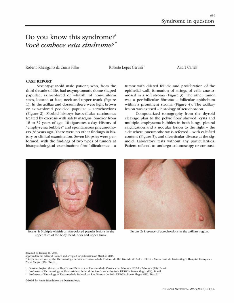
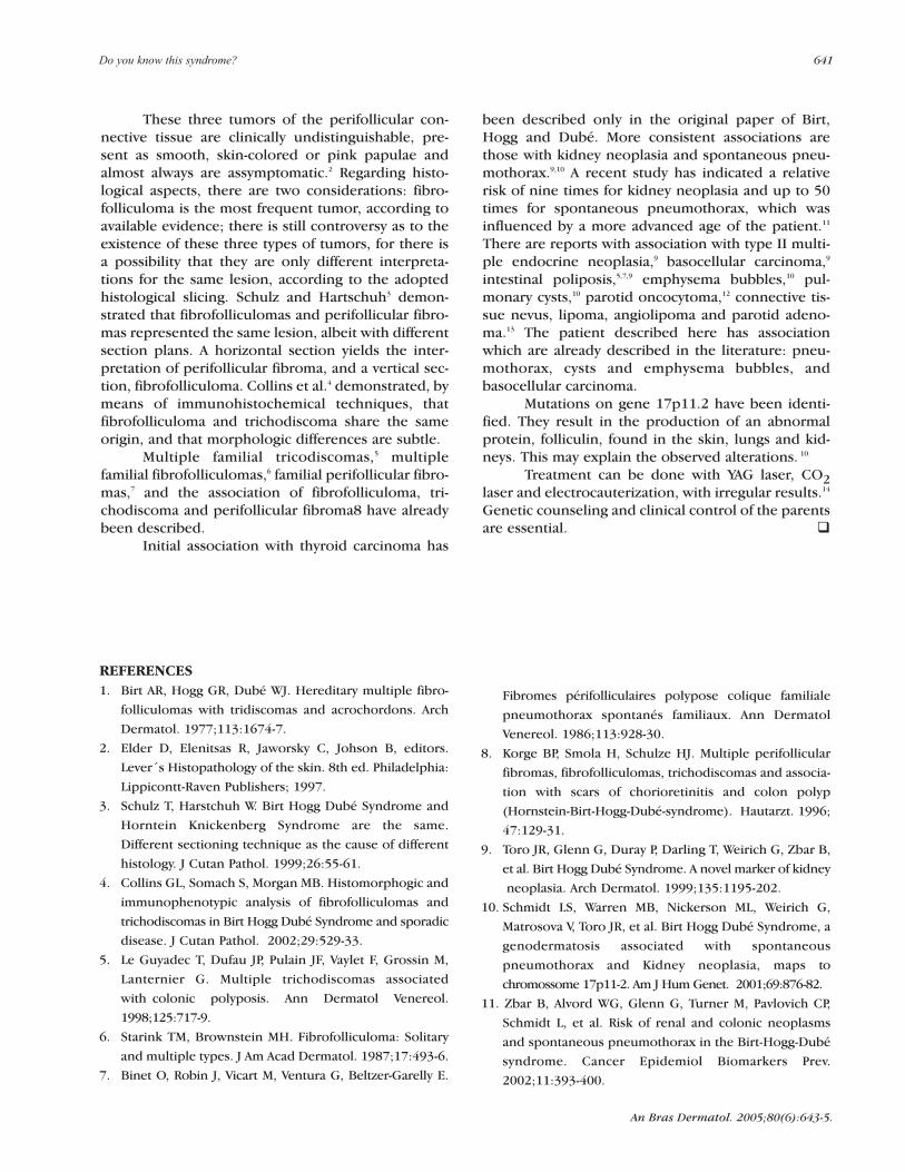
third decade of life, had assymptomatic dome-shapedpapullae, skin-colored or whitish, of non-uniformsizes, located at face, neck and upper trunk (Figure1). In the axillae and dorsum there were light brownor skin-colored pedicled papullae – acrochordons(Figure 2). Morbid history: basocellular carcinomastreated by exeresis with safety margins. Smoker from18 to 32 years of age, 10 cigarettes a day. History of“emphysema bubbles” and spontaneous pneumotho-rax 38 years ago. There were no other findings in his-tory or clinical examination. Seven biopsies were per-formed, with the findings of two types of tumors athistopathological examination: fibrofolliculomas – a
Received on January 16, 2004.Approved by the Editorial Council and accepted for publication on March 2, 2005.* Work carried out at the Dermatology Service at Universidade Federal do Rio Grande do Sul - UFRGS – Santa Casa de Porto Alegre Hospital Complex -Porto Alegre (RS), Brazil.
1 Dermatologist. Master in Health and Behavior at Universidade Católica de Pelotas - UCPel - Pelotas - (RS), Brazil.2 Professor of Dermatology at Universidade Federal do Rio Grande do Sul - UFRGS - Porto Alegre (RS), Brazil. 3 Professor of Pathology at Universidade Federal do Rio Grande do Sul - UFRGS - Porto Alegre (RS), Brazil.
©2005 by Anais Brasileiros de Dermatologia
tumor with dilated follicle and proliferation of theepithelial wall, formation of strings of cells anasto-mosed in a soft stroma (Figure 3). The other tumorwas a perifollicular fibroma – follicular epitheliumwithin a prominent stroma (Figure 4). The axillarylesion was excised – histology of acrochordon.
Computarized tomography from the thyroidcleavage plan to the pelvic floor showed: cysts andmultiple emphysema bubbles in both lungs, pleuralcalcification and a nodular lesion to the right – theside where pneumothorax is referred – with calcifiedcontent (Figure 5), and diverticular disease at the sig-moid. Laboratory tests without any particularities.Patient refused to undergo colonoscopy or contrast-
FIGURE 1: Multiple whitish or skin-colored papular lesions in theupper third of the body: head, neck and upper trunk.
FIGURE 2: Presence of acrochordons in the axilllary region.

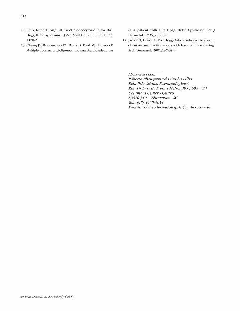
FIGURE 6:Patient 2:multipleskin-coloredor whitishpapulae onthe face.
640 Cunha Filho RR, Gervini RL, Cartell A.
An Bras Dermatol. 2005;80(6):643-5.
ed study of the intestine. The patient reported that his father, three
brothers and a sister also had similar lesions in theface, being the father and himself the most affected.He had 11 brothers and sisters, one had passed awaybecause of disseminated non-pigmented skin tumors,which he could not specify. The affected sister was 80and had similar lesions that had begun at 30 years ofage (Figure 6). Anatomopathologic exams demon-strated perifollicular fibromas. There were no otherclinical, laboratorial or imaging alterations.Management has been guidance and medical follow-
up with emphasis on possible associated diseases.
COMMENTSIn 1977, dermatologist Birt, pathologist Hogg
and intern physician Dubé described the syndromethat has their names, characterized by an eruption offace, neck and upper trunk tumors, with dominantautossomic inheritance.1 Lesions are fibrofolliculo-mas, trichodiscomas and acrochodons. Throughoutthe last 28 years, new and more frequent associa-tions, histopathological features and gene mutationshave been discovered.
FIGURE 3: Hyperplastic, dilated follicle, with keratin plugging andepithelial wall proliferation, forming cell strings within a relatively
well-delineated proliferation of soft connective tissue. The strings anastomose, and are also located by the
sebaceous gland (HE, 100x)
FIGURE 4: Perifollicular fibroma: dilated central follicle and concentric proliferation of collagen fibers (HE, 160x).
FIGURE 5: Tomography: cysts, emphysema bubbles and pleuralthickening with rough residual calcification on the right lung.
An Bras Dermatol. 2005;80(6):643-5.
Do you know this syndrome? 641
REFERENCES1. Birt AR, Hogg GR, Dubé WJ. Hereditary multiple fibro-
folliculomas with tridiscomas and acrochordons. Arch
Dermatol. 1977;113:1674-7.
2. Elder D, Elenitsas R, Jaworsky C, Johson B, editors.
Lever´s Histopathology of the skin. 8th ed. Philadelphia:
Lippicontt-Raven Publishers; 1997.
3. Schulz T, Harstchuh W. Birt Hogg Dubé Syndrome and
Horntein Knickenberg Syndrome are the same.
Different sectioning technique as the cause of different
histology. J Cutan Pathol. 1999;26:55-61.
4. Collins GL, Somach S, Morgan MB. Histomorphogic and
immunophenotypic analysis of fibrofolliculomas and
trichodiscomas in Birt Hogg Dubé Syndrome and sporadic
disease. J Cutan Pathol. 2002;29:529-33.
5. Le Guyadec T, Dufau JP, Pulain JF, Vaylet F, Grossin M,
Lanternier G. Multiple trichodiscomas associated
with colonic polyposis. Ann Dermatol Venereol.
1998;125:717-9.
6. Starink TM, Brownstein MH. Fibrofolliculoma: Solitary
and multiple types. J Am Acad Dermatol. 1987;17:493-6.
7. Binet O, Robin J, Vicart M, Ventura G, Beltzer-Garelly E.
These three tumors of the perifollicular con-nective tissue are clinically undistinguishable, pre-sent as smooth, skin-colored or pink papulae andalmost always are assymptomatic.2 Regarding histo-logical aspects, there are two considerations: fibro-folliculoma is the most frequent tumor, according toavailable evidence; there is still controversy as to theexistence of these three types of tumors, for there isa possibility that they are only different interpreta-tions for the same lesion, according to the adoptedhistological slicing. Schulz and Hartschuh3 demon-strated that fibrofolliculomas and perifollicular fibro-mas represented the same lesion, albeit with differentsection plans. A horizontal section yields the inter-pretation of perifollicular fibroma, and a vertical sec-tion, fibrofolliculoma. Collins et al.4 demonstrated, bymeans of immunohistochemical techniques, thatfibrofolliculoma and trichodiscoma share the sameorigin, and that morphologic differences are subtle.
Multiple familial tricodiscomas,5 multiplefamilial fibrofolliculomas,6 familial perifollicular fibro-mas,7 and the association of fibrofolliculoma, tri-chodiscoma and perifollicular fibroma8 have alreadybeen described.
Initial association with thyroid carcinoma has
been described only in the original paper of Birt,Hogg and Dubé. More consistent associations arethose with kidney neoplasia and spontaneous pneu-mothorax.9,10 A recent study has indicated a relativerisk of nine times for kidney neoplasia and up to 50times for spontaneous pneumothorax, which wasinfluenced by a more advanced age of the patient.11
There are reports with association with type II multi-ple endocrine neoplasia,9 basocellular carcinoma,9
intestinal poliposis,5,7,9 emphysema bubbles,10 pul-monary cysts,10 parotid oncocytoma,12 connective tis-sue nevus, lipoma, angiolipoma and parotid adeno-ma.13 The patient described here has associationwhich are already described in the literature: pneu-mothorax, cysts and emphysema bubbles, andbasocellular carcinoma.
Mutations on gene 17p11.2 have been identi-fied. They result in the production of an abnormalprotein, folliculin, found in the skin, lungs and kid-neys. This may explain the observed alterations. 10
Treatment can be done with YAG laser, CO2laser and electrocauterization, with irregular results.14
Genetic counseling and clinical control of the parentsare essential. �
Fibromes périfolliculaires polypose colique familiale
pneumothorax spontanés familiaux. Ann Dermatol
Venereol. 1986;113:928-30.
8. Korge BP, Smola H, Schulze HJ. Multiple perifollicular
fibromas, fibrofolliculomas, trichodiscomas and associa-
tion with scars of chorioretinitis and colon polyp
(Hornstein-Birt-Hogg-Dubé-syndrome). Hautarzt. 1996;
47:129-31.
9. Toro JR, Glenn G, Duray P, Darling T, Weirich G, Zbar B,
et al. Birt Hogg Dubé Syndrome. A novel marker of kidney
neoplasia. Arch Dermatol. 1999;135:1195-202.
10. Schmidt LS, Warren MB, Nickerson ML, Weirich G,
Matrosova V, Toro JR, et al. Birt Hogg Dubé Syndrome, a
genodermatosis associated with spontaneous
pneumothorax and Kidney neoplasia, maps to
chromossome 17p11-2. Am J Hum Genet. 2001;69:876-82.
11. Zbar B, Alvord WG, Glenn G, Turner M, Pavlovich CP,
Schmidt L, et al. Risk of renal and colonic neoplasms
and spontaneous pneumothorax in the Birt-Hogg-Dubé
syndrome. Cancer Epidemiol Biomarkers Prev.
2002;11:393-400.
An Bras Dermatol. 2005;80(6):646-53.
642
MAILING ADDRESS:Roberto Rheingantz da Cunha Filho Bela Pele Clínica Dermatológica®Rua Dr Luiz de Freitas Melro, 395 / 604 – EdColumbia Center - Centro89010-310 Blumenau SC Tel.: (47) 3035-4053 E-mail: [email protected]
12. Liu V, Kwan T, Page EH. Parotid oncocytoma in the Birt-
Hogg-Dubé syndrome. J Am Acad Dermatol. 2000; 43:
1120-2.
13. Chung JY, Ramos-Caso FA, Beers B, Ford MJ, Flowers F.
Multiple lipomas, angiolipomas and parathyroid adenomas
in a patient with Birt Hogg Dubé Syndrome. Int J
Dermatol. 1996;35:365-8.
14. Jacob CI, Dover JS. Birt-Hogg-Dubé syndrome: treatment
of cutaneous manifestations with laser skin resurfacing.
Arch Dermatol. 2001;137:98-9.