do antidepressants regulate how cortisol affects the brain?
TRANSCRIPT

Psychoneuroendocrinology 29 (2004) 423–447www.elsevier.com/locate/psyneuen
2003 Curt P. Richter Award Winner
Do antidepressants regulate how cortisolaffects the brain?
Carmine M. Pariantea,∗, Sarah A. Thomasb,Simon Lovestonea, Andrew Makoffa, Robert W. Kerwina
a Institute of Psychiatry, King’s College London, 1 Windsor Walk, Denmark Hill, London SE5 8AF,UK
b Centre for Neuroscience Research, King’s College London, London, UK
Abstract
Although the effects of antidepressants on glucocorticoid hormones and their receptors arerelevant for the therapeutic action of these drugs, the molecular mechanisms underlying theseeffects are unclear. Studies in depressed patients, animals and cellular models have demon-strated that antidepressants increase glucocorticoid receptor (GR) and mineralocorticoid recep-tor (MR) expression and function; this, in turn, is associated with enhanced negative feedbackby endogenous glucocorticoids, and thus with reduced resting and stimulated hypothalamic–pituitary–adrenal (HPA) axis activity. In a series of studies conducted over the last few years,we have shown that antidepressants modulate GR function in vitro by inhibiting membranesteroid transporters that regulate the intracellular concentration of glucocorticoids. In thispaper, we will review the effects of membrane steroid transporters and antidepressants oncorticosteroid receptors. We will then present our unpublished data on GR live microscopyin vitro, showing that ligand-induced translocation of the GR starts within 30 seconds and iscompleted within minutes. Furthermore, we will present our new data using an in situ brainperfusion model in anaesthetised guinea-pigs, showing that entry of cortisol to the brain ofthese animals is limited at the blood–brain barrier (BBB). Finally, we will present a compre-hensive discussion of our published findings on the effects of chemically unrelated antidepress-ants on membrane steroid transporters, in mouse fibroblasts and rat cortical neurones. Wepropose that antidepressants in humans could inhibit steroid transporters localised on the BBBand in neurones, like the multidrug resistancep-glycoprotein, and thus increase the access ofcortisol to the brain and the glucocorticoid-mediated negative feedback on the HPA axis.Enhanced cortisol action in the brain might prove to be a successful approach to maximisetherapeutic antidepressant effects.
∗ Corresponding author. Tel.:+44-20-7848-0807; fax:+44-20-7848-0051.E-mail address: [email protected] (C.M. Pariante).
0306-4530/$ - see front matter 2003 Elsevier Ltd. All rights reserved.doi:10.1016/j.psyneuen.2003.10.009

424 C.M. Pariante et al. / Psychoneuroendocrinology 29 (2004) 423–447
2003 Elsevier Ltd. All rights reserved.
Keywords: Antidepressant; Glucocorticoid receptor; Hypothalamic–pituitary–adrenal axis; Mineralocort-icoid receptor; Multidrug resistance; p-Glycoprotein
1. Introduction
Patients with major depression show hyperactivity of the hypothalamic–pituitary–adrenal (HPA) axis, which is thought to participate in the development of thedepressive symptoms (Nemeroff, 1996; Holsboer, 2000; Pariante & Miller, 2001;Pariante, 2003). One explanation for the HPA axis hyperactivity is an impaired feed-back inhibition by the endogenous glucocorticoid, cortisol. This feedback is mediatedby the glucocorticoid receptor (GR) and the mineralocorticoid receptor (MR) in thebrain (de Kloet et al., 1998; McEwen, 2000). Patients with major depression exhibitimpaired HPA axis negative feedback in the context of elevated circulating levelsof cortisol (Nemeroff, 1996), and the GR is important in the regulation of the HPAwhen endogenous levels of cortisol are high (de Kloet et al., 1998). Consistent withthis, the function of GR is reduced in depressed patients (GR resistance) and antide-pressants reverse these putative GR changes (Holsboer, 2000; McQuade & Young,2000; Pariante & Miller, 2001).
We have shown that antidepressants modulate GR function in vitro by inhibitingmembrane steroid transporters that regulate the intracellular concentration of gluco-corticoids (Pariante et al., 1997, 2001a,b, 2003a,b). Moreover, we have proposedthat antidepressants in humans could inhibit steroid transporters localised on theblood–brain barrier (BBB) and in neurones, like the multidrug resistance (MDR) p-glycoprotein (PGP), and thus increase the access of cortisol to the brain and theglucocorticoid-mediated negative feedback on the HPA axis (Pariante et al., 2001a,2003a,b). In this paper, we will review the role of membrane steroid transportersin regulating GR and MR function and the access of glucocorticoids to the brain.Furthermore, we will present our unpublished data on GR live microscopy in vitroand on cortisol access to the brain in guinea-pigs. We will then summarise the clinicaland experimental evidence showing that antidepressants regulate corticosteroidreceptors and the HPA axis, and that membrane steroid transporters could mediatethese effects. Finally, we will present a comprehensive discussion of our publishedfindings on the effects of antidepressants on membrane steroid transporters.
2. Corticosteroid receptors and the access of glucocorticoids to the brain
2.1. The role of corticosteroid receptors in HPA axis functioning
HPA axis activity is governed by the secretion of corticotrophin hormone-releasingfactor (CRF) and vasopressin (AVP) from the hypothalamus, which in turn activates

425C.M. Pariante et al. / Psychoneuroendocrinology 29 (2004) 423–447
the secretion of corticotrophin (adrenocorticotrophic hormone, ACTH) from the pitu-itary, which finally stimulates the secretion of glucocorticoids (cortisol in humansand corticosterone in rodents) from the adrenal cortex (Nemeroff, 1996; de Kloet etal., 1998; Ebrecht et al., 2000). Glucocorticoids then interact with their receptors inmultiple target tissues, including the HPA axis where they are responsible for feed-back inhibition of the secretion of ACTH from the pituitary and CRF from the hypo-thalamus (Nemeroff, 1996; de Kloet et al., 1998; Young et al., 1998; McEwen, 2000).The MR has a high affinity for endogenous glucocorticoids, and is believed to playa role in the regulation of circadian fluctuations of these hormones. In contrast tothe MR, the GR has a lower affinity for endogenous glucocorticoids, and is believedto be more important in the regulation of the response to stress, when endogenouslevels of glucocorticoids are high (de Kloet et al., 1998).
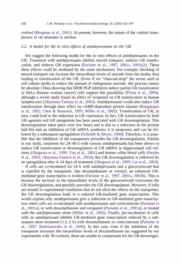
These receptors are ligand-induced transcription factors belonging to thesteroid/thyroid receptor superfamily (de Kloet et al., 1998; Reul et al., 2000; Pari-ante & Miller, 2001; Kalman & Spencer, 2002). According to the “nucleocytoplasmictraffic” model, these receptors reside predominantly in the cytoplasm in an inactiveform until they bind their glucocorticoid ligands. Ligand binding results in receptoractivation and translocation to the nucleus (Guiochon-Mantel et al., 1996; Nishi etal., 2001). Within the nucleus, corticosteroid receptors regulate gene expression viabinding to specific DNA sequences (glucocorticoid response elements, GREs). Sev-eral recent studies have confirmed the hormone-dependent translocation of the cyto-plasmic MR and GR into the nucleus using time-lapse video microscopy of live cells(Htun et al., 1996; Yang & DeFranco, 1996; Nishi et al., 1999, 2001). Fig. 1 presentsthe video microscopy of GR translocation in live cells using a new chimeric proteinof GR and green fluorescent protein (GFP–GR) that we have recently developed (seefigure legend for details of the method). In steroid-free conditions, CHO cells showa diffuse cytoplasmic and nuclear fluorescence; after treatment with dexamethasone,translocation of GFP–GR starts within 30 s and is completed within minutes.
2.2. Membrane steroid transporters and HPA axis functioning
Membrane steroid transporters regulate the function of the GR and the MR bymodulating the intracellular access of steroid hormones (de Kloet et al., 1998). Someglucocorticoids, like cortisol and dexamethasone, are actively excreted from cells bymembrane transporters belonging to the ATP-binding cassette family of transporters(Ueda et al., 1992; Kralli & Yamamoto, 1996; Medh et al., 1998). One of thesetransporters, the MDR PGP, has been extensively described to regulate intracellularconcentrations of steroids, to secrete naturally occurring metabolites and toxic sub-stances directly into the urinary or gastrointestinal tracts, and to confer treatmentresistance to tumour cells by expelling anticancer agents (Ueda et al., 1992; Shabbitset al., 2001). The MDR PGP is a polypeptide chain consisting of two similar halves,each containing six putative transmembrane segments and an intracellular ATP-bind-ing site. Hydrolysis of ATP provides the energy for drug export, which can occuragainst a large concentration gradient (Schinkel, 1999; Ferte, 2000). Contrary to mostknown transporters, PGP and related steroid transporters do not move substrates

426 C.M. Pariante et al. / Psychoneuroendocrinology 29 (2004) 423–447
Fig. 1. A new green fluorescent protein (GFP)–GR chimera. The GFP–GR is a chimeric protein ofmurine GR with green fluorescent protein (GFP), derived from the mouse wild-type GR cDNA vectorpHWrec (a kind gift from M. Danielsen, Georgetown University School of Medicine, Washington, DC)(Danielson, Northrop & Ringold, 1986). After eliminating the stop codon in the 5�-untranslated regionof the GR by polymerase chain reaction (PCR), the BglII–Xbal fragment (containing the GR cDNA) wascloned into the corresponding site of the pEGFP-C3 plasmid. The resulting GFP–GR protein contains theC-terminus of GFP fused in-frame to the N-terminus of the GR. We successfully transfected the GFP–GR in CHO, COS-7 and L929 cells, and in rat cortical and hippocampal neurones. Using live microscopyof CHO cells, in steroid-free conditions cells show a diffuse cytoplasmic and nuclear fluorescence (A).After treatment with dexamethasone, translocation of the GFP–GR starts within 30 s and is completedwithin minutes (B–D). For these experiments, CHO cells were cultured in coverslips and transientlytransfected with GFP–GR. Cells were maintained at 37 °C on a heated stage during the experiment.Images were captured at baseline (A) and at time periods after dexamethasone (10 M) was added to theculture medium (B, 60 s; C, 90 s; D, 120 s).
across the cell membrane, but capture substrates from the cell membrane (while thesesubstrates enter the cells by passive diffusion) and pump them out, thus preventingthem from entering the cells (Goodsell, 1999).
The MDR PGP has been described in normal animal and human tissues. In theadrenal gland, MDR PGP regulates the secretion of glucocorticoids into the bloodstream (Ueda et al., 1992); in lymphocytes, it regulates GR sensitivity to steroids(Bourgeois et al., 1993; Szabo et al., 1999); in the endothelial cells of the BBB, itlimits the access of dexamethasone and cortisol to both mouse and human brain (deKloet et al., 1998; Meijer et al., 1998; Karssen et al., 2001). Furthermore, recentstudies have suggested that MDR PGP can regulate corticosterone access to the brainand HPA axis activity in rats and mice. In fact, rodents have two isoforms of PGP:the mdr1a and the mdr1b. The mdr1a PGP is predominantly expressed at the BBB(Regina et al., 1998) and does not expel corticosterone, the endogenous glucocort-icoid, from the brain of rodents (de Kloet et al., 1998; Karssen et al., 2001). However,the mdr1b PGP transports corticosterone (Wolf & Horwitz, 1992; Uhr et al., 2002).This isoform is predominantly expressed in the adrenal and the ovaries (Lee et al.,2001), but is also expressed in the brain (Regina et al., 1998), particularly in thehippocampus (Kwan et al., 2002). Although mdr1b PGP has not been detected inbrain capillaries (Regina et al., 1998), it is expressed in rat brain endothelial cellsin vitro (Felix & Barrand, 2002). Consistent with the hypothesis that mdr1b PGPregulates the effects of corticosterone on the brain in rodents, mice that are knockoutfor mdr1a and mdr1b PGP genes show increased access of corticosterone to the brain

427C.M. Pariante et al. / Psychoneuroendocrinology 29 (2004) 423–447
and increased negative feedback on the HPA axis by corticosterone (Muller et al.,2003). Therefore, there is some evidence suggesting that membrane transport ofcorticosterone across the BBB regulates HPA axis function in rodents.
2.3. The access of cortisol to the brain of guinea-pigs
Cortisol, the endogenous glucocorticoid in humans, is transported by MDR PGPat the BBB in mice, and by human MDR PGP in vitro (Ueda et al., 1992; Karssenet al., 2001; Uhr et al., 2002). Moreover, two postmortem studies have suggestedthat this mechanism can limit the access of cortisol to human brain (Brooksbank etal., 1973; Karssen et al., 2001). Recently, we have used an in situ brain perfusionmodel to study transport of radioactive cortisol across the BBB and the blood–cer-ebrospinal fluid (CSF) barrier in anaesthetised guinea-pigs (Thomas & Segal, 1997,1998; Thomas et al., 2001; Gibbs & Thomas, 2002; Gibbs et al., 2003). Guinea-pigsare very similar to humans not only in the organization of the BBB, blood–CSFbarrier and CSF flux, but also in the fact that cortisol is their main glucocorticoid(Keightley & Fuller, 1996). Therefore, it is conceivable that humans will be moresimilar to guinea-pigs than to mice in the regulation of the access of cortisol to thebrain. In these experiments, guinea-pigs were anaesthetised, and the carotid arterieswere perfused in situ with a warmed, oxygenated artificial plasma (see Table 1 andits legend for results and details of the method). After a 20-min pre-perfusion withartificial plasma (to eliminate endogenous cortisol from the brain), radiolabelled cor-tisol (2.5 nM) and sucrose (vascular marker) were infused into the inflowing plasmafor 10 or 20 min. A CSF sample was taken and the animal was then decapitated.The results show that entry of cortisol to the brain of guinea-pigs is limited at theBBB (Table 1). In summary: (1) entry of radioactive cortisol into the brain rangesfrom 17% to 21.5% of plasma cortisol, and entry into the CSF ranges from 4% to7.5% of plasma cortisol; (2) entry of radioactive cortisol into the pituitary, whichdoes not have a BBB, is approximately 100% of plasma cortisol; and (3) in thepresence of an excess of unlabelled cortisol (30 µM), the radioactive signal decreasesin the hippocampus, but not in the rest of the brain. The discrepancy in cortisolconcentration between plasma and brain, but not between plasma and pituitary, sup-ports the presence of a functional efflux system for cortisol at the BBB. These pre-liminary findings are remarkably consistent with the results obtained by other labora-tories when radioactive cortisol is administered subcutaneously in mice (Karssen etal., 2001; Uhr et al., 2002). Moreover, Karssen et al. (2001, 2002) also have demon-strated in mice that an excess of unlabelled glucocorticoid does not reduce the radio-active signal in the brain, except in the hippocampus (see below for the discussionof these data).
3. The effects of antidepressants on the HPA axis and on corticosteroidreceptors
3.1. The HPA axis and corticosteroid receptors in major depression
Hyperactivity of the HPA axis in major depression is driven by the hypersecretionof CRF (and possibly AVP) in the hypothalamus (Owens & Nemeroff, 1993; Nemer-

428 C.M. Pariante et al. / Psychoneuroendocrinology 29 (2004) 423–447
Table 1Brain and CSF uptake of radiolabelled cortisol in guinea-pigs
3H-cortisol perfusion 3H-cortisol perfusion Excess of unlabelled cortisol+3H-(10 min) (20 min) cortisol perfusion (10 min)
3 Animals 3 Animals 2 Animals
CSF 4.0 ± 2 7.5 ± 2 6.9 ± 2Pituitary 100.5 ± 44 150.4 ± 27 47.0 ± 3Brain 17.0 ± 5 21.5 ± 5 16.0 ± 3Hippocampus 14.1 ± 6 15.6 ± 9 8.7 ± 1Capillary 3.3 ± 0.5 3.5 ± 0.5 1.7 ± 1
All experimental procedures were within the guidelines of the Animals (Scientific Procedures) Act, 1986.Adult Dunkin-Hartley guinea-pigs were anaesthetised and heparinised. The carotid arteries were perfusedin situ with a warmed (37 °C), oxygenated (95% O2; 5% CO2) artificial plasma. With the start of perfusionthe jugular veins were sectioned. After perfusion with plasma for 20 min, radiolabelled cortisol (2.5 nM)and sucrose (vascular marker) were infused into the inflowing plasma. After a further 10 or 20 min, acisterna magna CSF sample was taken and the animal decapitated. Brain, CSF and plasma samples weretaken for radioactive analysis. The uptake is expressed as mean ± SEM percentage ratio of tissue toplasma radioactivities, and corrected for vascular space. Using the capillary depletion analysis, whichseparates the level of radioactivity that has reached the brain from that in the cerebral endothelial cells(“Capillary” ), there is no evidence that the measured signal represents cortisol in the endothelial cellsrather than in the brain parenchyma. We also established that radioactive cortisol does not bind to thedextran/albumin present in the perfusion fluid (using ultrafiltration centrifugal dialysis and proteinprecipitation). The bilateral brain perfusion technique has several advantages, including: (1) the concen-trations of radioactive glucocorticoids in the artificial plasma can be kept constant; (2) the movement ofradioactive glucocorticoids into regions of the brain and into the CSF can be explored simultaneously;and (3) the levels of radioactivity in the endothelial cells of the BBB are separated from the levels inthe brain tissue. The discrepancy between plasma and brain cortisol concentrations further supports thepresence of a functional efflux system at the BBB, like MDR PGP.
off, 1996; Holsboer, 2000). These increased levels of CRF in the hypothalamus arerelated, at least in part, to altered feedback inhibition by endogenous glucocorticoids(de Kloet et al., 1998; Holsboer, 2000; McQuade & Young, 2000; Pariante & Miller,2001; Pariante et al., 2002; Pariante, 2003). Consistent with the fact that patientswith major depression exhibit impaired HPA negative feedback in the context ofelevated circulating levels of cortisol, when the negative feedback is largely mediatedby the GR, a multitude of studies have demonstrated that GR-mediated feedbackinhibition is impaired in major depression. These patients have nonsuppression ofcortisol secretion following dexamethasone (dexamethasone suppression test), non-suppression of ACTH secretion following hydrocortisone (fast-feedback test) andlack of inhibition of ACTH responses to CRF following dexamethasone pre-treat-ment (DEX/CRF test) (Young et al., 1991; Ribeiro et al., 1993; Heuser et al., 1994,1996; Nemeroff, 1996; Holsboer, 2000). The only study that specifically looked atMR-mediated negative feedback in depression found that this pathway is intact (orpossibly oversensitive) in these patients (Young et al., 2003).
In further support to the notion that patients with major depression exhibit

429C.M. Pariante et al. / Psychoneuroendocrinology 29 (2004) 423–447
impaired GR-mediated HPA negative feedback, a number of studies have demon-strated that GR function is also reduced in other tissues of depressed patients, asshown by a decreased sensitivity to the effects of glucocorticoids on immune andmetabolic functions in the absence of a reduction in GR expression (Pariante &Miller, 2001). In fact, there is no consistent evidence of reduced GR expression inblood mononuclear cells or fibroblasts from depressed patients (Pariante & Miller,2001). However, studies that have examined postmortem brains have found reducedcorticosteroid receptors expression: Lopez et al. (1998) found decreased MR (butnot GR) mRNA levels in the hippocampus of six suicide victims with a history ofdepression; Webster et al. (2002) found decreased GR mRNA in the frontal cortexand hippocampus of patients with nonpsychotic depression, bipolar disorder andschizophrenia. These latter results suggest that the stress of having a psychiatricdisorder may be more relevant to changes in brain GR or MR expression thandepression per se (Cotter & Pariante, 2002).
3.2. The effects of antidepressants on the HPA axis and on corticosteroidreceptors
The most striking support to the hypothesis that abnormalities in the corticosteroidreceptors contribute to the pathophysiology of major depression derives from animalsand in vitro studies demonstrating a direct effect of antidepressants on the GR andthe MR, leading to increased receptor expression and function, and thus to increasednegative feedback on the HPA axis. These studies support the clinical evidence thatsuccessful antidepressant treatment is associated with resolution of the impairmentin the HPA axis negative feedback by glucocorticoids (Linkowski et al., 1987; Rib-eiro et al., 1993; Heuser et al., 1996) and of glucocorticoid resistance in immunecells (Wodarz et al., 1992).
A number of studies have shown that long-term antidepressant treatment upregul-ates GR and MR in the brain, including in the hippocampus and in the hypothalamus,and decreases basal and stress-induced glucocorticoid secretion. The vast majorityof studies using tricyclic antidepressants, like desipramine, amitriptyline and imipra-mine, or using electroconvulsive shock, has shown antidepressant-induced upregul-ation of brain GR, MR, or both (Kitayama et al., 1988; Young et al., 1990; Peifferet al., 1991; Brady et al., 1991; Seckl and Fink, 1992; Pepin et al., 1992b; Reul etal., 1993; Przegalinski et al., 1993; Przegalinski and Budziszewska, 1993; Bud-ziszewska et al., 1994; Peeters et al., 1994; Rossby et al., 1995; Yau et al., 1995;Eiring and Sulser, 1997; Johansson et al., 1998). Studies examining selective sero-tonin reuptake inhibitor (SSRI) antidepressants, like fluoxetine, citalopram and zimel-idine, have found that chronic treatment with these antidepressants upregulates MRexpression, while it has no effect on GR expression (Seckl and Fink, 1992; Bradyet al., 1992; Budziszewska et al., 1994; Rossby et al., 1995; Lopez et al., 1998;Bjartmar et al., 2000; Yau et al., 2002), although one study found that a 2-daytreatment with fluoxetine increases both MR and GR mRNAs in the hippocampus(Semont et al., 2000).
Interestingly, the few studies that have looked at shorter durations of treatment

430 C.M. Pariante et al. / Psychoneuroendocrinology 29 (2004) 423–447
have found that acutely (within 3–9 days) antidepressants induce a decrease of GRand MR expression. For example, Reul et al. (1993) showed a decreased GR andMR expression after 3–7 days of amitriptyline, and Yau et al. (2001) showed thatfluoxetine and venlafaxine (a serotonin and noradrenaline reuptake inhibitor, SNRI)induce downregulation of GR and MR expression at 9 days. On the opposite end,studies looking at longer time points have indicated that GR expression returns tocontrol levels after 6–9 weeks of treatment with antidepressants, while MR upregul-ation persists (Reul et al., 1993, 1994; Yau et al., 1995).
Several of these studies have also shown that treatment with antidepressants inrodents is associated with a reduction in basal and stress-induced HPA axis activity,and that—surprisingly—GR or MR upregulation is not a prerequisite for thisreduction (Pariante & Miller, 2001). For example, Delbende et al. (1991) showedthat a single injection of the antidepressant tianeptine, given 1–3 h before a tuberestraint stress, significantly reduces the stress-induced ACTH and corticosteronerelease in rats: an acute effect unlikely to be related to GR or MR upregulation.Brady et al. (1992) found that a 2-week treatment with fluoxetine, idazoxan orphenelzine reduces basal corticosterone levels in the absence of GR or MR upregul-ation. Reul et al. (1993) showed a decrease in adrenal weight, likely representing adecrease in HPA axis function, in rats treated with amitriptyline for 5 days, togetherwith a decrease in hippocampal GR binding. Montkowski et al. (1995) demonstratedthat long-term antidepressant treatment with moclobemide (a monoamine-oxidaseinhibitor) induces normalization of the HPA axis in the absence of any changes inGR binding. Finally, Yau et al. (2001) have found that a 9-day treatment with fluoxe-tine or venlafaxine induces a reduction in HPA axis activity together with the down-regulation of GR and MR. As we will discuss below, our research also demonstratesthat antidepressants increase GR function in vitro in the absence of GR upregulation.
A potent tool for clarifying the mechanisms underlying antidepressant-inducedincrease in corticosteroid receptor function and expression has been the study of theeffects of antidepressants in in vitro cell culture systems. Antidepressants regulateGR translocation, GR function and GR expression in neuronal cell cultures (Pepinet al., 1989; Okugawa et al., 1999; Hery et al., 2000; Yau et al., 2001; Lai et al.,2003; Herr et al., 2003), fibroblasts (Pepin et al., 1992a; Pariante et al., 1997, 2001a,2003a,b; Budziszewska et al., 2000; Miller et al., 2002) and human peripheral bloodmononuclear cells (Vedder et al., 1999; Heiske et al., 2003; Okuyama-Tamura etal., 2003). These experimental systems do not contain noradrenaline or serotoninreuptake sites within synaptic connections (Okugawa et al., 1999; Hery et al., 2000;Lai et al., 2003). Therefore, these systems allow the study of molecular effects thatare unrelated to the inhibition of noradrenaline or serotonin reuptake, the mechanismconsidered to be crucial in the therapeutic action of antidepressants (Nestler, 1998;Schafer, 1999). Indeed, antidepressant-induced GR upregulation in vitro is notblocked by antagonists of alpha or beta adrenergic receptors or of 5HT1a or 5HT2serotonergic receptors (Okugawa et al., 1999; Lai et al., 2003). These intriguing invitro findings confirm data in animals showing that inhibition of noradrenaline reup-take is not relevant for the antidepressant-induced changes in the GR or MRexpression. In fact, desipramine has been shown to induce GR upregulation in rats

431C.M. Pariante et al. / Psychoneuroendocrinology 29 (2004) 423–447
even following neurotoxic lesioning of noradrenergic neurones with DSP4 (Rossbyet al., 1995). Moreover, the noradrenaline reuptake inhibitor, oxaprotiline, consist-ently has shown no effects on GR or MR expression in animals (Budziszewska etal., 1994; Eiring and Sulser, 1997).
Some evidence suggests a possible role of the membrane steroid transporters inthe regulation of corticosteroid receptor function in major depression or during anti-depressant treatment. In vitro expression of MDR PGP induces GR resistance inlymphocytes by limiting the intracellular access of glucocorticoids (Bourgeois et al.,1993), thus reproducing a condition similar to that described in lymphocytes ofpatients with major depression. Moreover, previous studies have suggested that anti-depressants interact with the MDR PGP in animals and in vitro. For example, thetricyclic antidepressant clomipramine, at 10 mg/kg/day for 2 days, completelyinhibits MDR activity in subcutaneous tumours in mice (Merry et al., 1991). Of noteis that the dose used in this study is within the range (10–20 mg/kg/day) used inmost animal studies showing GR or MR upregulation by tricyclic antidepressants(Pariante & Miller, 2001). In vitro, tricyclic antidepressants (at concentrations similarto those used in the studies on the GR) blocks the MDR PGP-mediated efflux ofrhodamine 123 from human colon cancer cells (Varga et al., 1996), cells from aleukemia cell line (Szabo et al., 1999) and human peripheral blood mononuclearcells (Szabo et al., 1999). Finally, two recent papers have described that amitriptylineand citalopram are transported by the MDR PGP (Uhr et al., 2000; Uhr & Grauer,2003). Our hypothesis—that antidepressants modulate corticosteroid receptors byinhibiting membrane steroid transporters—is a potential explanation of how chemi-cally and pharmacologically unrelated antidepressants may have similar effects onthe GR and the MR, without affecting the reuptake of serotonin or noradrenaline.In fact, inhibition of PGP and other membrane steroid transporters is not receptor-mediated and is related to the drugs physiochemical properties, that is, lipophilicity,electric charge and ability to accept hydrogen bonds (Ford, 1996; Castaing et al.,2000; Ekins et al., 2002).
4. Antidepressants enhance GR function in vitro by modulating membranesteroid transporters
4.1. Antidepressants potentiate GR function in vitro in the absence of GRupregulation
In their pivotal paper, Pepin et al. (1992a) used a fibroblast cell line to show that24 h treatment with desipramine enhances GR function (GR-mediated genetranscription) as measured by increased activity of a transiently transfected reportergene whose regulation is dependent on GREs. Desipramine was also found to induceupregulation of GR protein after 72 h of treatment. Based on these data, the authorshypothesised that antidepressants directly induce GR upregulation in vitro. Our workhas originated from this seminal paper, and has led us to propose that the mechanismby which antidepressants regulate corticosteroid receptor in vitro, and possibly in

432 C.M. Pariante et al. / Psychoneuroendocrinology 29 (2004) 423–447
animals, is the inhibition of membrane steroid transporters, leading to increased intra-cellular levels of glucocorticoids. The results of our studies are summarised inTable 2.
We have used L929 mouse fibroblast cells, stably transfected with the MMTV–chloramphenicol acetyltransferase (MMTV–CAT) reporter gene (LMCAT cells).Expression of the CAT reporter gene (Sanchez et al., 1994) by these cells is underglucocorticoid control by virtue of several GREs residing within the MMTV pro-moter, which lies upstream of the CAT reporter gene. Work conducted in this andother laboratories has shown that these cells have a membrane steroid transporterthat is virtually identical to MDR PGP in its substrates and modulators (Kralli &Yamamoto, 1996; Medh et al., 1998; Marsaud et al., 1998; Pariante et al., 2001a,b).More recently, Webster and Carlstedt-Duke (2002) have demonstrated MDR PGPmRNA in these cells. Therefore, L929/LMCAT cells have the unique advantage ofexpressing endogenous (not transfected) membrane steroid transporters, while alsoallowing the direct examination of GR function (Sanchez et al., 1994; Pariante etal., 1997, 1999, 2001a,b, 2003a,b; Budziszewska et al., 2000). These cells have beenused in a variety of studies looking at molecular determinants of GR function.
Our first finding on the effects of antidepressants on the GR was that 24 h treatmentwith desipramine (at 1 and 10 µM concentrations) induces translocation of the GRfrom the cytoplasm to the nucleus in the absence of any glucocorticoids, andpotentiates GR translocation induced by a low (10 nM) concentration of dexame-thasone (Pariante et al., 1997). These results were confirmed using immunocytoch-emistry as well as western blotting of the GR in the cytosolic and nuclear fractions.In a subsequent series of experiments, we found that co-incubation of cells for 24 hwith dexamethasone and several, chemically unrelated antidepressants—the tricyclicsclomipramine, desipramine and amitriptyline, and the SSRIs fluoxetine, paroxetineand citalopram—induces an increase in GR function (GR-mediated genetranscription) compared to cells treated with dexamethasone alone (Table 2, 1st col-umn; see table legend for details of the method). At a concentration of antidepressantsof 10 µM, this potentiation is particularly intense in cells treated with dexamethasoneand clomipramine (more than 10-fold) while is approximately +70% to +170% forthe other antidepressants (Pariante et al., 1997, 2001a, 2003b). A smaller(approximately 15–25%) potentiation is evident at a lower (1 µM) concentration ofantidepressants (Pariante et al., 1997, 2001a, 2003b). We did not elicit any effectsof these antidepressants alone on GR-mediated gene transcription (Pariante et al.,1997, 2001a, 2003b). Finally, this potentiation is not related to an increase in GRexpression, since we found that desipramine and clomipramine reduce, rather thanincrease, GR expression after 24 h of treatment (see below) (Pariante et al., 1997,2003a).
4.2. Antidepressants potentiate GR-mediated gene transcription by inhibitingmembrane steroid transporters
We hypothesised that the effects of antidepressants on GR-mediated gene tran-scription are mediated by the L929/LMCAT cells membrane steroid transporter. In

433C.M. Pariante et al. / Psychoneuroendocrinology 29 (2004) 423–447T
able
2Su
mm
ary
ofou
rin
vitr
ost
udie
son
antid
epre
ssan
tsan
dG
R
GR
-med
iate
dge
netr
ansc
ript
ion
inL
MC
AT
Intr
acel
lula
rac
cum
ulat
ion
inL
MC
AT
Intr
acel
lula
rW
este
rnbl
ottin
gof
the
cells
cells
accu
mul
atio
nin
rat
GR
neur
ones
Dex
Cor
tisol
Cor
ticos
.D
ex+
Cor
tisol
Cor
ticos
.C
ortis
ol+
Cor
tisol
Cor
ticos
.L
MC
AT
Rat
vera
pam
ilve
rapa
mil
cells
neur
ones
Clo
mip
ram
ine
↑↑
↓↓
↑=
=↑
↓↓
↓Fl
uoxe
tine
↑↑
==
↑↓
Des
ipra
min
e↑
↑↓
Paro
xetin
e↑
↑↓
Cita
lopr
am↑
Am
itrip
tylin
e↑
Cel
lsw
ere
trea
ted
for
24h
with
clom
ipra
min
e(1
0µM
),flu
oxet
ine
(10
µM),
desi
pram
ine
(10
µM),
paro
xetin
e(1
0µM
),ci
talo
pram
(10
µM)
oram
itrip
tylin
e(1
0µM
).↑I
ndic
ates
anan
tidep
ress
ant-
indu
ced
incr
ease
;↓I
ndic
ates
anan
tidep
ress
ant-
indu
ced
decr
ease
;=
Indi
cate
sno
effe
ctof
the
antid
epre
ssan
t.G
R-
med
iate
dge
netr
ansc
ript
ion
inL
MC
AT
cells
:L
MC
AT
cells
wer
etr
eate
dw
ithve
hicl
eor
the
antid
epre
ssan
tsin
co-i
ncub
atio
nw
ithde
xam
etha
sone
(Dex
)(1
0nM
),co
rtis
ol(5
0nM
),co
rtic
oste
rone
(cor
ticos
.)(5
0nM
),or
dexa
met
haso
ne(2
.5nM
)+
vera
pam
il(1
00µM
).M
easu
rem
ent
ofC
AT
was
perf
orm
edus
ing
aco
lori
met
ric
enzy
me
imm
unoa
ssay
(Roc
heD
iagn
ostic
,U
K).
Res
ults
wer
eno
rmal
ised
with
resp
ect
toce
llnu
mbe
rby
mea
sure
men
tof
met
abol
icac
tivity
bycl
eava
geof
the
tetr
azol
ium
salt
WST
-1(R
oche
Dia
gnos
tic).
Intr
acel
lula
rac
cum
ulat
ion
ofra
dioa
ctiv
egl
ucoc
ortic
oids
inL
MC
AT
cells
orra
tne
uron
es:
LM
CA
Tce
llsor
one-
wee
kol
dra
tpr
imar
yne
uron
esw
ere
trea
ted
with
vehi
cle
orth
ean
tidep
ress
ants
for
24h,
and
then
incu
bate
dfo
r1.
5h
with
3H
-cor
tisol
(50
nM),
or3H
-cor
ticos
tero
ne(5
0nM
),or
3H
-co
rtis
ol(5
0nM
)+
vera
pam
il(1
00M
),at
37°C
,in
aC
O2
incu
bato
r.C
ells
wer
eth
ensc
rape
din
toly
sis
buff
eran
dtr
ansf
erre
dto
vial
sfo
rliq
uid
scin
tilla
tion
coun
ting.
The
radi
oact
ive
sign
al,a
sm
easu
red
bysc
intil
latio
nco
untin
g,is
prop
ortio
nal
toth
ein
trac
ellu
lar
conc
entr
atio
nof
the
radi
oact
ive
gluc
ocor
ticoi
d.R
esul
tsw
ere
norm
alis
edw
ithre
spec
tto
cell
num
ber,
asab
ove.
Wes
tern
blot
ting
ofth
eG
Rin
LM
CA
Tce
llsor
rat
neur
ones
:L
MC
AT
cells
and
one-
wee
kol
dra
tpr
imar
yne
uron
esw
ere
trea
ted
with
vehi
cle
orcl
omip
ram
ine
for
24h.
Cel
lsw
ere
lyse
dby
scra
ping
into
hot
(100
°C)
2XSD
SPA
GE
sam
ple
buff
er.
GR
pres
ent
inth
ely
sate
sw
asan
alys
edby
wes
tern
blot
usin
gth
ean
ti-G
Rpo
lycl
onal
antib
ody
GR
57,
and
quan
tified
byde
nsito
met
ric
anal
ysis
.D
ata
from
Pari
ante
etal
.(1
997,
2001
,20
03a,
b)

434 C.M. Pariante et al. / Psychoneuroendocrinology 29 (2004) 423–447
particular, we hypothesised that antidepressants enhance GR-mediated gene tran-scription in the presence of dexamethasone by inhibiting the membrane steroid trans-porters, and therefore increasing dexamethasone intracellular concentrations. To testthis hypothesis, we co-incubated the antidepressants with two glucocorticoids thatare differentially affected by the transporter: cortisol and corticosterone. In fact, workby us and others in LMCAT cells demonstrated that steroid transporters inhibitors,like verapamil, H-89, and cyclosporin, increase GR-mediated gene transcription onlyin the presence of glucocorticoids that are expelled by the transporters, like dexame-thasone and cortisol, but not in the presence of corticosterone. If the inhibition ofthe transporter were the mechanism by which antidepressants increase GR-mediatedgene transcription, then we would see these effects also in the presence of cortisol,but not in the presence of corticosterone (Medh et al., 1998; Marsaud et al., 1998;Pariante et al., 2001a). We focused these experiments on four antidepressants: clomi-pramine, a tricyclic serotonin reuptake inhibitor; desipramine, a tricyclic noradrena-line reuptake inhibitor; paroxetine, an inhibitor of both noradrenaline and serotoninreuptake; and fluoxetine, an SSRI (Pariante et al., 2001a, 2003b). We used cortisoland corticosterone at a low (50 nM) concentration that, similar to dexamethasone(10 nM), induces only partial GR activation. As hypothesised, we found that allantidepressants strongly potentiate GR-mediated gene transcription in the presenceof cortisol, but not in the presence of corticosterone (Table 2, 2nd and 3rd columns).In fact, we found that all antidepressants, except fluoxetine, induce less GR-mediatedgene transcription in the presence of corticosterone compared to cells treated withcorticosterone alone. Interestingly, clomipramine, which gives the strongest potenti-ation in the presence of dexamethasone, also gives the strongest potentiation in thepresence of cortisol (approximately fourfold) and the largest inhibition in the pres-ence of corticosterone (approximately 35% inhibition).
To corroborate these findings, we examined the effects of clomipramine or fluoxe-tine in the presence of dexamethasone and the steroid transporter inhibitor, verapamil.If inhibition of the steroid transporter were the mechanism by which antidepressantsincrease GR-mediated gene transcription in the presence of dexamethasone, thiseffect would disappear in the presence of verapamil. As hypothesised, we found thatboth clomipramine and fluoxetine do not potentiate GR-mediated gene transcriptionin the presence of dexamethasone after the membrane steroid transporter is blockedby verapamil (Table 2, 4th column). Moreover, consistent with the experiments inthe presence of corticosterone, clomipramine reduces dexamethasone-induced GR-mediated gene transcription in the presence of verapamil, while fluoxetine has noeffect.
4.3. Antidepressants increase intracellular accumulation of radioactiveglucocorticoids in LMCAT cells and neurones
Because the increased GR-mediated gene transcription only provides indirect evi-dence of increased intracellular levels of glucocorticoids, we examined whether anti-depressants directly increase the intracellular levels of radioactive glucocorticoids in

435C.M. Pariante et al. / Psychoneuroendocrinology 29 (2004) 423–447
LMCAT cells. Moreover, we examined whether a similar, antidepressant-sensitive,membrane transport of glucocorticoids is present in cultured rat primary neurones.
We developed the assay to measure intracellular accumulation of radioactive gluc-ocorticoids from Bourgeois et al. (1993) (Pariante et al., 2003a,b) (see legend ofTable 2 for details of the method). We found that there is a linear relationshipbetween 3H-cortisol concentrations in the media (from 1 nM to 1 µM) and the levelsof intracellular accumulation in LMCAT cells. Moreover, at all concentrations of3H-cortisol, verapamil increases the intracellular levels of the glucocorticoid. Wealso found that an excess of unlabelled cortisol induces an increase (+80%) of theintracellular accumulation of radioactive cortisol, while unlabelled corticosteroneresults in a reduction (�40%) of the intracellular accumulation of radioactive cortico-sterone. We interpreted these finding as showing that the unlabelled cortisol competesfor the radioactive cortisol at the efflux system, thus increasing the intracellularaccumulation of the radioactive cortisol. However, because corticosterone is nottransported, the unlabelled corticosterone can compete with the radioactive corticos-terone at GR binding sites or at uptake sites for corticosterone (Pariante et al., 2003a)(see below).
By using this experimental design in primary cultures of rat cortical neurones, wealso showed that a membrane transport of cortisol is present in neurones. First, wefound that unlabelled cortisol induces a small increase of the intracellular accumu-lation of radioactive cortisol in these cells (+7%), while unlabelled corticosteroneinduces a decrease (�15%) in the intracellular accumulation of radioactive corticos-terone; second, we found that verapamil increases the intracellular accumulation of3H-cortisol (Pariante et al., 2003a).
We found that clomipramine and fluoxetine induce an increase in the intracellularaccumulation of 3H-cortisol in LMCAT cells: approximately +80% for clomipramineand +5% for fluoxetine (a small but statistically significant effect) (Table 2, 5thcolumn) (Pariante et al., 2003a,b). In the presence of 3H-corticosterone, clomipram-ine has no effect, while fluoxetine induces a small (�15%) reduction of the intra-cellular accumulation (Table 2, 6th column) (Pariante et al., 2003a,b). Because theeffects of 24 h fluoxetine on the intracellular accumulation of 3H-cortisol was rela-tively small, we also conducted another series of experiments in which cells weretreated with fluoxetine for 72 h. We found that fluoxetine induces a larger increaseof 3H-cortisol intracellular accumulation after this longer incubation (+15%), whilein the presence of 3H-corticosterone has no effect (Pariante et al., 2003b). Consistentwith our hypothesis and the data on GR-mediated gene transcription, treatment ofLMCAT cells with clomipramine in the presence of verapamil induces no increaseof intracellular accumulation of 3H-cortisol (Table 2, 7th column).
Finally, we wanted to test whether the cortisol transporter in rat primary neuronescan be inhibited by clomipramine. Consistent with the data in LMCAT cells, wefound that clomipramine induces an increase (+20%) in the intracellular accumu-lation of 3H-cortisol compared to cells treated with vehicle, while in the presence of3H-corticosterone clomipramine induces a small (�11%) reduction of the intracellu-lar accumulation (Table 2, 8th and 9th columns) (Pariante et al., 2003a).

436 C.M. Pariante et al. / Psychoneuroendocrinology 29 (2004) 423–447
4.4. Antidepressants acutely reduce GR expression in vitro
As mentioned above, we found that some antidepressants decrease, rather thanincrease, GR function in the presence of corticosterone or in the presence of dexame-thasone and verapamil (Pariante et al., 2001a). We also found that pre-incubation ofcells with desipramine followed by treatment with dexamethasone reduces GR-mediated gene transcription (Pariante et al., 1997). This latter finding has been repli-cated by Budziszewska et al. (2000), who also found that pre-incubation of LMCATcells with various antidepressants (including desipramine) reduces GR-mediated genetranscription induced by a subsequent treatment with corticosterone or dexame-thasone. Finally, Miller et al. (2002) have recently found that incubation of LMCATcells with desipramine alone induces a small decrease in the unstimulated GR-mediated gene transcription. Based on these results, we hypothesised that a reductionin GR-mediated gene transcription by antidepressants is present under experimentalconditions that do not elicit the effects on the transporter, like in the presence ofglucocorticoid that are not expelled by the transporters, in the presence of transportersinhibitors, or in the absence of any glucocorticoids. Moreover, we hypothesised thatthese inhibitory effects are due to a reduction in GR expression. Indeed, in our firstpaper, we found that 24–96 h of treatment with desipramine induces a 10–25%reduction in cytosolic GR binding in L929 cells; this reduction is still present afterthe cells have recovered for 24 h in media with no desipramine (Pariante et al.,1997). Since 24 h desipramine also induces activation and translocation of the GRfrom the cytoplasm to the nucleus, and this is associated with a decrease of GR inthe cytosolic fraction, we had originally interpreted the desipramine-induced decreasein GR binding as representing a greater proportion of GR in the nucleus (Parianteet al., 1997). However, these results can also be interpreted as an overall decreasein the total number of GRs. Therefore, we examined the effects of clomipramine onGR expression, assessed by whole-cell western blot, in LMCAT cells and primaryrat cortical neurones (see legend of Table 2 for details of the method) (Pariante etal., 2003a). We used clomipramine because it gives the largest inhibition of GR-mediated gene transcription in the presence of corticosterone, compared to the otherantidepressants tested. In both cell types, treatment with clomipramine for 24 hresults in a reduction of GR levels: 50% reduction in LMCAT cells and 80%reduction in neurones (Pariante et al., 2003a) (Table 2, 10th and 11th columns).
5. A model for brain sensitivity to glucocorticoids and the effects ofantidepressants on the HPA axis
5.1. Physiological relevance of our findings
Our work, in vitro and in guinea-pigs, corroborates consistent lines of evidencefrom different laboratories showing that membrane steroid transporters localised onthe BBB are crucial in the regulation of brain sensitivity to glucocorticoids (de Kloetet al., 1998; Meijer et al., 1998; Karssen et al., 2001, 2002; Uhr et al., 2002). First,

437C.M. Pariante et al. / Psychoneuroendocrinology 29 (2004) 423–447
we have presented preliminary data suggesting that a functional efflux system forcortisol exists at the BBB of guinea-pigs, which have cortisol as their main endogen-ous glucocorticoid. Second, we have shown in vitro that the effect of steroid transportis present for a large range of concentrations of cortisol (1 nM–1 µM) that comprisesthe physiological levels of this hormone in humans (Orth & Kovacs, 1998). Finally,we have shown that neurones (rat cortical neurones) show a functional transportof cortisol.
Our data in guinea-pigs show that an excess of unlabelled cortisol does not reducethe levels of radioactive cortisol in the brain, except in the hippocampus. Theseresults corroborate data by Karssen et al. (2001, 2002) in mdr1a PGP knockout mice,also showing that brain labelling by a radioactive glucocorticoid is not reduced byan excess of unlabelled glucocorticoid, except in the hippocampus. While these dataindicate that most of the glucocorticoid in the brain is unbound to GR, the resultsin the hippocampus suggest that the unlabelled hormone can compete with the radio-active cortisol either at hippocampal MR binding sites or at an uptake system forglucocorticoids in this region (Thomas & Segal, 1997; Karssen et al., 2001, 2002;Gibbs et al., 2003). Similar to our work in guinea-pigs, our studies in vitro alsodemonstrate that most of the hormone in the cells is GR-unbound. In LMCAT cells,the 50% reduction in GR expression by clomipramine leads to no change in intra-cellular accumulation of radioactive corticosterone; in neurones, the 80% reductionof GR expression by clomipramine leads only to a 11% reduction in the intracellularaccumulation of radioactive corticosterone (Pariante et al., 2003a). Our in vitro dataalso suggest that an uptake system for corticosterone is present in LMCAT cells(Pariante et al., 2003a,b). In fact, since most of the intracellular glucocorticoid is GR-unbound, the 40% reduction of 3H-corticosterone intracellular levels by the excess ofunlabelled corticosterone cannot be explained by competition at the GR binding sites(Pariante et al., 2003a,b). Membrane uptake systems for glucocorticoids have beendescribed before (Bossuyt et al., 1996; Lackner et al., 1998), and co-existence of bothefflux and uptake transporters for glucocorticoids has been described in hepatocytes(Bossuyt et al., 1996). We are now investigating these putative uptake systems inthe hippocampus of guinea-pigs and in LMCAT cells.
To our knowledge, we have been the first to describe a functional membranetransport of glucocorticoids in neurones. Human neuroblastoma cells express MDRPGP (Kurowski & Berthold, 1998), and cells from the mouse hippocampal cell lineHT22 express a membrane steroid transporter that is blocked by verapamil (Herr etal., 2000). However, no immunolabelling of PGP has been found in adult rat brainneurones (Matsuoka et al., 1999), although PGP and PGP-like transporters can beexpressed at very low levels that are undetected by immunocytochemistry (Kralli &Yamamoto, 1996; Marsaud et al., 1998). Interestingly, immunoreactive and mRNAsignals for another family of membrane transporters, the multidrug resistance-asso-ciated proteins (MRPs), have been found in rat neuronal cultures (Hirrlinger et al.,2002) and in dysplastic neurones from brain samples of epileptic patients (Sisodiyaet al., 2002). The MRPs are organic anion transporters that participate to MDR(Leslie et al., 2001). The function and expression of MRPs are modulated by cortisol(Mulder et al., 1996) and MRP-overexpressing cells are resistant to the effects of
438 C.M. Pariante et al. / Psychoneuroendocrinology 29 (2004) 423–447
cortisol (Bergman et al., 2001). At present, however, the nature of the cortisol trans-porters in rat neurones is unclear.
5.2. A model for the in vitro effects of antidepressants on the GR
We suggest the following model for the in vitro effects of antidepressants on theGR. Treatment with antidepressants inhibits steroid transport, induces GR translo-cation, and reduces GR expression (Pariante et al., 1997, 2001a, 2003a,b). Thesethree effects could be mediated by the same mechanism. For example, blocking ofsteroid transport can increase the intracellular levels of steroids from the media, thusleading to translocation of the GR. (Even if we “charcoal-strip” the serum used incell culture media to reduce the amount of endogenous steroids, this process cannotbe absolute.) Data showing that MDR PGP inhibitors induce partial GR translocationin HeLa (human ovarian cancer) cells support this possibility (Prima et al., 2000),although a recent study found no effect of verapamil on GR translocation in humanlymphocytes (Okuyama-Tamura et al., 2003). Antidepressants could also induce GRtranslocation through their effect on cAMP-dependent protein kinases (Rangarajanet al., 1992; Chen & Rasenick, 1995; Miller et al., 2002). Translocation of GR, inturn, could lead to the reduction in GR expression. In fact, GR translocation by bothGR agonists and GR antagonists has been associated with GR downregulation. Thisdownregulation takes place over few hours and is due to a reduction in the proteinhalf-life and an inhibition of GR mRNA synthesis; it is temporary and can be fol-lowed by a subsequent upregulation (Schmidt & Meyer, 1994). Therefore, it is poss-ible that the inhibition of the transporters precedes the GR downregulation. Similarto our study, treatment for 24–48 h with various antidepressants has been shown toinduce GR translocation or downregulation of GR mRNA in hippocampal cell cul-tures (Okugawa et al., 1999; Yau et al., 2001) and human white blood cells (Heiskeet al., 2003; Okuyama-Tamura et al., 2003); this GR downregulation is followed byan upregulation after 4–14 days of treatment (Okugawa et al., 1999; Lai et al., 2003).
If cells are co-incubated for 24 h with antidepressants and a glucocorticoid thatis expelled by the transporter, like dexamethasone or cortisol, an enhanced GR-mediated gene transcription is evident (Pariante et al., 1997, 2001a, 2003b). This isbecause the increase in the intracellular levels of the glucocorticoid overcomes theGR downregulation, and possibly precedes the GR downregulation. However, if cellsare treated in experimental conditions that do not elicit the effects on the transporter,the GR downregulation leads to a reduced GR-mediated gene transcription. Thiswould explain why antidepressants give a reduction in GR-mediated gene transcrip-tion when cells are co-incubated with antidepressants and corticosterone (Pariante etal., 2001a), or with dexamethasone and verapamil (Pariante et al., 2001a), or treatedwith the antidepressants alone (Miller et al., 2002). Finally, pre-incubation of cellswith an antidepressant inhibits GR-mediated gene transcription induced by a sub-sequent short treatment (1.5–2 h) with dexamethasone or corticosterone (Pariante etal., 1997; Budziszewska et al., 2000). In this case, even if the inhibition of thetransporter increases the intracellular levels of dexamethasone (as suggested by ourexperiments with 3H-cortisol), these are unable to compensate for the GR downregul-

439C.M. Pariante et al. / Psychoneuroendocrinology 29 (2004) 423–447
ation, possibly because of the short incubation, or possibly because the GR downreg-ulation is present before the glucocorticoid is added (differently than in the co-incu-bation experiments).
It is of note that fluoxetine seems different from the other antidepressants testedin the fact that, after 24 h, it does not reduce GR-mediated gene transcription in thepresence of corticosterone or in the presence of dexamethasone and verapamil(Pariante et al., 2003b). However, data from other laboratories show that fluoxetinereduces GR mRNA in vitro after longer incubation (Lai et al., 2003). Fluoxetine isdifferent from the other antidepressants also because it is not transported by PGP,while tricyclic antidepressants as well as citalopram are transported (Uhr et al., 2000;Uhr & Grauer, 2003). Not all PGP inhibitors are transported by PGP; for example,progesterone is a potent inhibitor but it is not transported (Ford, 1996). It is possiblethat fluoxetine has a different mechanism for its effects on the transporter.
A recent important paper offers further support to our model. Herr et al. (2003)have confirmed, in the mouse hippocampal cell line HT22, that chemically unrelatedantidepressants (including desipramine, clomipramine and fluoxetine) increase GR-mediated gene transcription in the presence of dexamethasone. Moreover, they haveshown that treatment of cells with desipramine in the presence of verapamil (20 µM)reduces (although does not abolish) these enhancing effects of desipramine on GR-mediated gene transcription. It is of note that the concentration of verapamil usedin this paper (20 µM) is lower than the concentration used in our work (100 µM),and this could explain the residual effects of desipramine. In fact, previous studieshave shown, at least in LMCAT cells, that verapamil induces only partial inhibitionof the cells steroid tranporter at concentrations of 10 µM and 50 µM (Marsaud etal., 1998; Medh et al., 1998); moreover, our original dose-finding experiments inthese cells have shown that verapamil (100 µM) induces a larger potentiation of GRtranslocation in the presence of dexamethasone than verapamil (50 µM) (Pariante etal., 2001b). Therefore, we have consistently used verapamil (100 µM) in our work(Pariante et al., 2001a, 2001b, 2003a, 2003b). However, we cannot exclude that theresidual effects of desipramine on GR function in these hippocampal cells are dueto activation of other intracellular pathways leading to enhanced GR function.
5.3. A model for the effects of antidepressants in animals
As we discussed above, mice that are knockout for mdr1a and mdr1b PGP showincreased access of corticosterone to the brain and increased negative feedback onthe HPA axis by corticosterone (Uhr et al., 2002; Muller et al., 2003). By inhibitingmembrane steroid transporters, antidepressants could directly increase the access ofcorticosterone, or of other steroids that are transported by PGP like aldosterone (Uedaet al., 1992), to the brain of mice, thus enhancing GR and MR activation. Althoughin the brain the highest expression of PGP has been found at the BBB (Lee et al.,2001), our data show that this effect could also occur directly at neuronal level(Pariante et al., 2003a). In turn, the increased access of glucocorticoids to the braincould lead to the decrease in HPA axis activity and the GR and MR downregulationdescribed in rats after 3–9 days of treatment with antidepressants (Reul et al., 1993;

440 C.M. Pariante et al. / Psychoneuroendocrinology 29 (2004) 423–447
Yau et al., 2001). This receptor downregulation is consistent with our data in vitroand, theoretically, could explain part of the lag time seen with antidepressants forthe onset of their therapeutic action. After 14 days of more of treatment, antidepress-ants induce GR and MR upregulation in the brain, as a compensatory mechanismfollowing the initial downregulation or as a consequence of the reduced HPA axisactivity. Consistent with our model, pre-treatment with nifedipine, an MDR PGPinhibitor, prevents the hippocampal GR upregulation induced by antidepressants(Przegalinski et al., 1993). This antidepressant-induced reduction in circulatingcorticosterone levels could also participate to the neuroprotective effects of thesedrugs (Duman et al., 2001).
5.4. In conclusion, a model for the effects of antidepressants in humans
Fig. 2. A model of the effects of antidepressants on MDR PGP, cortisol access to the brain and regulationof HPA axis function in humans The CRF in the hypothalamus stimulates the secretion of ACTH fromthe pituitary, which stimulates the secretion of cortisol from the adrenal gland. In turn, the circulatingcortisol binds to the GR in the brain to exert its negative feedback on the HPA axis. Plasma cortisolcannot freely enter the brain by passive diffusion (continuous arrow), because its access is limited byMDR PGP and other membrane steroid transporters localised at the BBB and possibly in neurones. Thesetransporters capture cortisol from the apical membrane of the endothelial cells of the BBB, while it isentering the cells by passive diffusion, and expel the hormone back into the plasma (dotted arrow).Therefore, membrane steroid transporters seem to participate in the regulation of GR-mediated negativefeedback and HPA axis activity. Antidepressants may inhibit membrane steroid transporters at the BBBand in neurones, so that more cortisol is able to enter the brain. This leads to increased activation ofbrain GR (and MR), increased negative feedback on the HPA axis and, finally, normalisation of HPAaxis hyperactivity in depressed patients.

441C.M. Pariante et al. / Psychoneuroendocrinology 29 (2004) 423–447
We have demonstrated that antidepressants regulate the intracellular levels of cor-tisol by inhibiting membrane steroid transporters, in LMCAT cells and neurones, thatare similar to MDR PGP in their substrates and modulators. Cortisol, the endogenousglucocorticoid in humans, is transported by the PGP at the BBB (Karssen et al.,2001). We propose that antidepressants in humans inhibit the steroid transporterslocalised on the BBB and in neurones, and thus increase the access of cortisol tothe brain. In turn, facilitation of GR and MR activation by antidepressants may leadto an increased negative feedback by circulating glucocorticoids on the HPA axis,and then to resolution of glucocorticoid hypersecretion (see model in Fig. 2). Thismodel is supported by studies showing that HPA hyperactivity in depressed patientsbegins to normalise after 7–9 days of antidepressant treatment, and that this precedesthe therapeutic effects on depressive symptoms (Heuser et al., 1996; Holsboer, 2000).Moreover, this model is consistent with clinical studies showing that treatment withGR and MR agonists, including cortisol, has antidepressant effects in humans (Dinanet al., 1997; Bouwer et al., 2000; DeBattista et al., 2000). Hypothetically, theincreased access of cortisol to the brain could balance the decreased function andexpression of corticosteroid receptors in the brain of depressed patients. Or, perhaps,this effect could compensate for a “ reduction of cortisol levels” in the brain ofdepressed patients, as described by Brooksbank et al. (1973). We believe thatenhanced cortisol action in the brain might prove to be a successful approach tomaximise therapeutic antidepressant effects (Pariante, 2003).
Acknowledgements
This research started at the Department of Psychiatry and Behavioural Sciencesof Emory University in Atlanta, Georgia, where Dr. Pariante worked as a ResearchFellow from 1995 to 1997, and is now a Clinical Assistant Professor. He is deeplygrateful to Professor Andrew H. Miller and Professor Charles B. Nemeroff for theirteaching and support. Dr. Pariante’s research in the UK has been funded by a Grantfrom the Psychiatry Research Trust, by a Medical Research Council Clinical TrainingFellowship, a Travel Award from the Wellcome Trust and a NARSAD Young Inves-tigator Award. His research in the UK has been supervised by all the co-authors onthis paper, and also by Professor Stuart Checkley, Professor Stafford Ligthman andProfessor Robin Murray.
References
Bergman, A.M., Pinedo, H.M., Peters, G.J., 2001. Steroids affect collateral sensitivity to gemcitabine ofmultidrug-resistant human lung cancer cells. Eur. J. Pharmacol. 416, 19–24.
Bjartmar, L., Johansson, I.M., Marcusson, J., Ross, S.B., Seckl, J.R., Olsson, T., 2000. Selective effects onNGFI-A, MR, GR and NGFI-B hippocampal mRNA expression after chronic treatment with differentsubclasses of antidepressants in the rat. Psychopharmacology (Berl) 151, 7–12.
Bossuyt, X., Muller, M., Hagenbuch, B., Meier, P.J., 1996. Polyspecific drug and steroid clearance byan organic anion transporter of mammalian liver. J. Pharmacol. Exp. Ther. 276, 891–896.

442 C.M. Pariante et al. / Psychoneuroendocrinology 29 (2004) 423–447
Bourgeois, S., Gruol, D.J., Newby, R.F., Rajah, F.M., 1993. Expression of an mdr gene is associatedwith a new form of resistance to dexamethasone-induced apoptosis. Mol. Endocrinol. 7, 840–851.
Bouwer, C., Claassen, J., Dinan, T.G., Nemeroff, C.B., 2000. Prednisone augmentation in treatment-resistant depression with fatigue and hypocortisolaemia: a case series. Depress. Anxiety 12, 44–50.
Brady, L.S., Whitfield, H.J. Jr., Fox, R.J., Gold, P.W., Herkenham, M., 1991. Long-term antidepressantadministration alters corticotropin-releasing hormone, tyrosine hydroxylase, and mineralocorticoidreceptor gene expression in rat brain. Therapeutic implications. J. Clin. Invest. 87, 831–837.
Brady, L.S., Gold, P.W., Herkenham, M., Lynn, A.B., Whitfield, H.J. Jr., 1992. The antidepressantsfluoxetine, idazoxan and phenelzine alter corticotropin-releasing hormone and tyrosine hydroxylasemRNA levels in rat brain: therapeutic implications. Brain Res. 572, 117–125.
Brooksbank, B.W., Brammall, M.A., Shaw, D.M., 1973. Estimation of cortisol, cortisone and corticos-terone in cerebral cortex, hypothalamus and other regions of the human brain after natural death andafter death by suicide. Steroids Lipids Res. 4, 162–183.
Budziszewska, B., Siwanowicz, J., Przegalinski, E., 1994. The effect of chronic treatment with antidepress-ant drugs on the corticosteroid receptor levels in the rat hippocampus. Pol. J. Pharmacol. 46, 147–152.
Budziszewska, B., Jaworska-Feil, L., Kajta, M., Lason, W., 2000. Antidepressant drugs inhibit glucocort-icoid receptor-mediated gene transcription—a possible mechanism. Br. J. Pharmacol. 130, 1385–1393.
Castaing, M., Brouant, P., Loiseau, A., Santelli-Rouvier, C., Santelli, M., Alibert-Franco, S., Mahamoud,A., Barbe, J., 2000. Membrane permeation by multidrug-resistance-modulators and non-modulators:effects of hydrophobicity and electric charge. J. Pharm. Pharmacol. 52, 289–296.
Chen, J., Rasenick, M.M., 1995. Chronic treatment of C6 glioma cells with antidepressant drugs increasesfunctional coupling between a G protein (Gs) and adenylyl cyclase. J. Neurochem. 64, 724–732.
Cotter, D., Pariante, C.M., 2002. Stress and the progression of the developmental hypothesis of schizo-phrenia. Br. J. Psychiatry 181, 363–365.
Danielsen, M., Northrop, J.P., Ringold, G.M., 1986. The mouse glucocorticoid receptor: mapping offunctional domains by cloning, sequencing and expression of wild-type and mutant receptor proteins.EMBO J. 5, 2513–2522.
de Kloet, E.R., Vreugdenhil, E., Oitzl, M.S., Joels, M., 1998. Brain corticosteroid receptor balance inhealth and disease. Endocr. Rev. 19, 269–301.
DeBattista, C., Posener, J.A., Kalehzan, B.M., Schatzberg, A.F., 2000. Acute antidepressant effects ofintravenous hydrocortisone and CRH in depressed patients: a double-blind, placebo-controlled study.Am. J. Psychiatry 157, 1334–1337.
Delbende, C., Contesse, V., Mocaer, E., Kamoun, A., Vaudry, H., 1991. The novel antidepressant, tianep-tine, reduces stress-evoked stimulation of the hypothalamo–pituitary–adrenal axis. Eur. J. Pharmacol.202, 391–396.
Dinan, T.G., Lavelle, E., Cooney, J., Burnett, F., Scott, L., Dash, A., Thakore, J., Berti, C., 1997. Dexame-thasone augmentation in treatment-resistant depression. Acta Psychiatr. Scand. 95, 58–61.
Duman, R.S., Nakagawa, S., Malberg, J., 2001. Regulation of adult neurogenesis by antidepressant treat-ment. Neuropsychopharmacology 25, 836–844.
Ebrecht, M., Buske-Kirschbaum, A., Hellhammer, D., Kern, S., Rohleder, N., Walker, B., Kirschbaum,C., 2000. Tissue specificity of glucocorticoid sensitivity in healthy adults. J. Clin. Endocrinol. Metab.85, 3733–3739.
Eiring, A., Sulser, F., 1997. Increased synaptic availability of norepinephrine following desipramine isnot essential for increases in GR mRNA. Short communication. J. Neural Transm. 104, 1255–1258.
Ekins, S., Kim, R.B., Leake, B.F., Dantzig, A.H., Schuetz, E.G., Lan, L.B., Yasuda, K., Shepard, R.L.,Winter, M.A., Schuetz, J.D., Wikel, J.H., Wrighton, S.A., 2002. Application of three-dimensionalquantitative structure-activity relationships of P-glycoprotein inhibitors and substrates. Mol. Pharma-col. 61, 974–981.
Felix, R.A., Barrand, M.A., 2002. P-glycoprotein expression in rat brain endothelial cells: evidence forregulation by transient oxidative stress. J. Neurochem. 80, 64–72.
Ferte, J., 2000. Analysis of the tangled relationships between P-glycoprotein-mediated multidrug resist-ance and the lipid phase of the cell membrane. Eur. J. Biochem. 267, 277–294.
Ford, J.M., 1996. Experimental reversal of P-glycoprotein-mediated multidrug resistance by pharmaco-logical chemosensitisers. Eur. J. Cancer 32A, 991–1001.

443C.M. Pariante et al. / Psychoneuroendocrinology 29 (2004) 423–447
Gibbs, J.E., Thomas, S.A., 2002. The distribution of the anti-HIV drug, 2�3�-dideoxycytidine (ddC), acrossthe blood–brain and blood–cerebrospinal fluid barriers and the influence of organic anion transportinhibitors. J. Neurochem. 80, 392–404.
Gibbs, J., Jayabalan, P., Thomas, S.A., 2003. Mechanisms by which 2�3�-dideoxyinosine (ddI) crossesthe guinea-pig CNS barriers; relevance to HIV therapy. J. Neurochem. 84, 725–734.
Goodsell, D.S., 1999. The molecular perspective: the multidrug transporter. Stem Cells 17, 377–378.Guiochon-Mantel, A., Delabre, K., Lescop, P., Milgrom, E., 1996. The Ernst Schering Poster Award.
Intracellular traffic of steroid hormone receptors. J. Steroid Biochem. Mol. Biol. 56, 3–9.Heiske, A., Jesberg, J., Krieg, J.C., Vedder, H., 2003. Differential effects of antidepressants on glucocort-
icoid receptors in human primary blood cells and human monocytic u-937 cells. Neuropsychopharmac-ology 28, 807–817.
Herr, A.S., Tsolakidou, A.F., Yassouridis, A., Holsboer, F., Rein, T., 2003. Antidepressants differentiallyinfluence the transcriptional activity of the glucocorticoid receptor in vitro. Neuroendocrinology 78,12–22.
Herr, A.S., Wochnik, G.M., Rosenhagen, M.C., Holsboer, F., Rein, T., 2000. Rifampicin is not an activatorof glucocorticoid receptor. Mol. Pharmacol. 57, 732–737.
Hery, M., Semont, A., Fache, M.P., Faudon, M., Hery, F., 2000. The effects of serotonin on glucocorticoidreceptor binding in rat raphe nuclei and hippocampal cells in culture. J. Neurochem. 74, 406–413.
Heuser, I., Yassouridis, A., Holsboer, F., 1994. The combined dexamethasone/CRH test: a refined labora-tory test for psychiatric disorders. J. Psychiatr. Res. 28, 341–356.
Heuser, I.J., Schweiger, U., Gotthardt, U., Schmider, J., Lammers, C.H., Dettling, M., Yassouridis, A.,Holsboer, F., 1996. Pituitary–adrenal-system regulation and psychopathology during amitriptylinetreatment in elderly depressed patients and normal comparison subjects. Am. J. Psychiatry 153, 93–99.
Hirrlinger, J., Konig, J., Dringen, R., 2002. Expression of mRNAs of multidrug resistance proteins (Mrps)in cultured rat astrocytes, oligodendrocytes, microglial cells and neurones. J. Neurochem. 82, 716–719.
Holsboer, F., 2000. The corticosteroid receptor hypothesis of depression. Neuropsychopharmacology 23,477–501.
Htun, H., Barsony, J., Renyi, I., Gould, D.L., Hager, G.L., 1996. Visualization of glucocorticoid receptortranslocation and intranuclear organization in living cells with a green fluorescent protein chimera.Proc. Natl. Acad. Sci. USA 93, 4845–4850.
Johansson, I.M., Bjartmar, L., Marcusson, J., Ross, S.B., Seckl, J.R., Olsson, T., 1998. Chronic amitripty-line treatment induces hippocampal NGFI-A, glucocorticoid receptor and mineralocorticoid receptormRNA expression in rats. Mol. Brain Res. 62, 92–95.
Kalman, B.A., Spencer, R.L., 2002. Rapid corticosteroid-dependent regulation of mineralocorticoid recep-tor protein expression in rat brain. Endocrinology 143, 4184–4195.
Karssen, A.M., Meijer, O.C., van der Sandt, I., Lucassen, P.J., de Lange, E.C., de Boer, A.G., de Kloet,E.R., 2001. Multidrug resistance P-glycoprotein hampers the access of cortisol but not of corticosteroneto mouse and human brain. Endocrinology 142, 2686–2694.
Karssen, A.M., Meijer, O.C., van der Sandt, I., de Boer, A.G., de Lange, E.C., de Kloet, E.R., 2002. Therole of the efflux transporter P-glycoprotein in brain penetration of prednisolone. J. Endocrinol. 175,251–260.
Keightley, M.C., Fuller, P.J., 1996. Anomalies in the endocrine axes of the guinea pig: relevance tohuman physiology and disease. Endocr. Rev. 17, 30–44.
Kitayama, I., Janson, A.M., Cintra, A., Fuxe, K., Agnati, L.F., Ogren, S.O., Harfstrand, A., Eneroth, P.,Gustafsson, J.A., 1988. Effects of chronic imipramine treatment on glucocorticoid receptor immunore-activity in various regions of the rat brain. Evidence for selective increases of glucocorticoid receptorimmunoreactivity in the locus coeruleus and in 5-hydroxytryptamine nerve cell groups of the rostralventromedial medulla. J. Neural Transm. 73, 191–203.
Kralli, A., Yamamoto, K.R., 1996. An FK506-sensitive transporter selectively decreases intracellular lev-els and potency of steroid hormones. J. Biol. Chem. 271, 17152–17156.
Kurowski, C., Berthold, F., 1998. Presence of classical multidrug resistance and P-glycoprotein expressionin human neuroblastoma cells. Ann. Oncol. 9, 1009–1014.
Kwan, P., Sills, G.J., Butler, E., Gant, T.W., Meldrum, B.S., Brodie, M.J., 2002. Regional expression of

444 C.M. Pariante et al. / Psychoneuroendocrinology 29 (2004) 423–447
multidrug resistance genes in genetically epilepsy-prone rat brain after a single audiogenic seizure.Epilepsia 43, 1318–1323.
Lackner, C., Daufeldt, S., Wildt, L., Allera, A., 1998. Glucocorticoid-recognizing and -effector sites inrat liver plasma membrane. Kinetics of corticosterone uptake by isolated membrane vesicles. III. Speci-ficity and stereospecificity. J. Steroid Biochem. Mol. Biol. 64, 69–82.
Lai, M., McCormick, J.A., Chapman, K.E., Kelly, P.A.T., Seckl, J.R., Yau, J.L.W., 2003. Differentialregulation of corticosteroid receptors by monoamine neurotransmitters and antidepressant drugs inprimary hippocampal culture. Neuroscience 118, 975–984.
Lee, G., Dallas, S., Hong, M., Bendayan, R., 2001. Drug transporters in the central nervous system: brainbarriers and brain parenchyma considerations. Pharmacol. Rev. 53, 569–596.
Leslie, E.M., Deeley, R.G., Cole, S.P., 2001. Toxicological relevance of the multidrug resistance protein1, MRP1 (ABCC1) and related transporters. Toxicology 167, 3–23.
Linkowski, P., Mendlewicz, J., Kerkhofs, M., Leclercq, R., Golstein, J., Brasseur, M., Copinschi, G., VanCauter, E., 1987. 24-Hour profiles of adrenocorticotropin, cortisol, and growth hormone in majordepressive illness: effect of antidepressant treatment. J. Clin. Endocrinol. Metab. 65, 141–152.
Lopez, J.F., Chalmers, D.T., Little, K.Y., Watson, S.J., 1998. A.E. Bennett Research Award. Regulationof serotonin1A, glucocorticoid, and mineralocorticoid receptor in rat and human hippocampus: impli-cations for the neurobiology of depression. Biol. Psychiatry 43, 547–573.
Marsaud, V., Mercier-Bodard, C., Fortin, D., Le Bihan, S., Renoir, J.M., 1998. Dexamethasone and triam-cinolone acetonide accumulation in mouse fibroblasts is differently modulated by the immunosuppres-sants cyclosporin A, FK506, rapamycin and their analogues, as well as by other P-glycoprotein ligands.J. Steroid Biochem. Mol. Biol. 66, 11–25.
Matsuoka, Y., Okazaki, M., Kitamura, Y., Taniguchi, T., 1999. Developmental expression of P-glyco-protein (multidrug resistance gene product) in the rat brain. J. Neurobiol. 39, 383–392.
McEwen, B.S., 2000. The neurobiology of stress: from serendipity to clinical relevance. Brain Res. 886,172–189.
McQuade, R., Young, A.H., 2000. Future therapeutic targets in mood disorders: the glucocorticoid recep-tor. Br. J. Psychiatry 177, 390–395.
Medh, R.D., Lay, R.H., Schmidt, T.J., 1998. Agonist-specific modulation of glucocorticoid receptor-mediated transcription by immunosuppressants. Mol. Cell Endocrinol. 138, 11–23.
Meijer, O.C., de Lange, E.C., Breimer, D.D., de Boer, A.G., Workel, J.O., de Kloet, E.R., 1998. Pen-etration of dexamethasone into brain glucocorticoid targets is enhanced in mdr1A P-glycoproteinknockout mice. Endocrinology 139, 1789–1793.
Merry, S., Hamilton, T.G., Flanigan, P., Freshney, R.I., Kaye, S.B., 1991. Circumvention of pleiotropicdrug resistance in subcutaneous tumours in vivo with verapamil and clomipramine. Eur. J. Cancer 27,31–34.
Miller, A.H., Vogt, G.J., Pearce, B.D., 2002. The phosphodiesterase type 4 inhibitor, rolipram, enhancesglucocorticoid receptor function. Neuropsychopharmacology 27, 939–948.
Montkowski, A., Barden, N., Wotjak, C., Stec, I., Ganster, J., Meaney, M., Engelmann, M., Reul, J.M.,Landgraf, R., Holsboer, F., 1995. Long-term antidepressant treatment reduces behavioural deficits intransgenic mice with impaired glucocorticoid receptor function. J. Neuroendocrinol. 7, 841–845.
Mulder, H.S., Pinedo, H.M., Timmer, A.T., Rao, B.R., Lankelma, J., 1996. Multidrug resistance-modifyingcomponents in human plasma with potential clinical significance. J. Exp. Ther. Oncol. 1, 13–22.
Muller, M.B., Keck, M.E., Binder, E.B., Kresse, A.E., Hagemeyer, T.P., Landgraf, R., Holsboer, F., Uhr,M., 2003. ABCB1 (MDR1)-type P-glycoproteins at the blood-brain barrier modulate the activity ofthe hypothalamic-pituitary-adrenocortical system: implications for affective discorder. Neuropsycho-pharmacology 28, 1991–1999.
Nemeroff, C.B., 1996. The corticotropin-releasing factor (CRF) hypothesis of depression: new findingsand new directions. Mol. Psychiatry 1, 336–342.
Nestler, E.J., 1998. Antidepressant treatments in the 21st century. Biol. Psychiatry 44, 526–533.Nishi, M., Takenaka, N., Morita, N., Ito, T., Ozawa, H., Kawata, M., 1999. Real-time imaging of glucocor-
ticoid receptor dynamics in living neurons and glial cells in comparison with non-neural cells. Eur.J. Neurosci. 11, 1927–1936.
Nishi, M., Ogawa, H., Ito, T., Matsuda, K.I., Kawata, M., 2001. Dynamic changes in subcellular localiz-

445C.M. Pariante et al. / Psychoneuroendocrinology 29 (2004) 423–447
ation of mineralocorticoid receptor in living cells: in comparison with glucocorticoid receptor usingdual-color labeling with green fluorescent protein spectral variants. Mol. Endocrinol. 15, 1077–1092.
Okugawa, G., Omori, K., Suzukawa, J., Fujiseki, Y., Kinoshita, T., Inagaki, C., 1999. Long-term treatmentwith antidepressants increases glucocorticoid receptor binding and gene expression in cultured rathippocampal neurones. J. Neuroendocrinol. 11, 887–895.
Okuyama-Tamura, M., Mikuni, M., Kojima, I., 2003. Modulation of the human glucocorticoid receptorfunction by antidepressive compounds. Neurosci. Lett. 342, 206–210.
Orth, D., Kovacs, W., 1998. The adrenal cortex. In: Wilson, J., Foster, D., Kronenberg, H., Larsen, P.(Eds.), Williams Textbook of Endocrinology, ninth ed. W.B. Saunders Company, Philadelphia, pp.517–664.
Owens, M.J., Nemeroff, C.B., 1993. The role of corticotropin-releasing factor in the pathophysiology ofaffective and anxiety disorders: laboratory and clinical studies. Ciba Found. Symp. 172, 296–308.
Pariante, C.M., 2003. Depression, stress and the adrenal axis. J. Neuroendocrinol. 15, 811–812.Pariante, C.M., Miller, A.H., 2001. Glucocorticoid receptors in major depression: relevance to pathophysi-
ology and treatment. Biol. Psychiatry 49, 391–404.Pariante, C.M., Pearce, B.D., Pisell, T.L., Owens, M.J., Miller, A.H., 1997. Steroid-independent translo-
cation of the glucocorticoid receptor by the antidepressant desipramine. Mol. Pharmacol. 52, 571–581.Pariante, C.M., Pearce, B.D., Pisell, T.L., Sanchez, C.I., Po, C., Su, C., Miller, A.H., 1999. The proin-
flammatory cytokine, interleukin-1alpha, reduces glucocorticoid receptor translocation and function.Endocrinology 140, 4359–4366.
Pariante, C.M., Makoff, A., Lovestone, S., Feroli, S., Heyden, A., Miller, A.H., Kerwin, R.W., 2001a.Antidepressants enhance glucocorticoid receptor function in vitro by modulating the membrane steroidtransporters. Br. J. Pharmacol. 134, 1335–1343.
Pariante, C.M., Pearce, B.D., Pisell, T.L., Su, C., Miller, A.H., 2001b. The steroid receptor antagonistsRU40555 and RU486 activate glucocorticoid receptor translocation and are not excreted by the steroidhormones transporter in L929 cells. J. Endocrinol. 169, 309–320.
Pariante, C.M., Papadopoulos, A.S., Poon, L., Checkley, S.A., English, J., Kerwin, R.W., Lightman, S.,2002. A novel prednisolone suppression test for the hypothalamic–pituitary–adrenal axis. Biol. Psy-chiatry 51, 922–930.
Pariante, C.M., Hye, A., Williamson, R., Makoff, A., Lovestone, S., Kerwin, R.W., 2003a. The antide-pressant clomipramine regulates cortisol intracellular concentrations and glucocorticoid receptorexpression in fibroblasts and rat primary neurones. Neuropsychopharmacology 28, 1553–1561.
Pariante, C.M., Kim, R.B., Makoff, A., Kerwin, R.W., 2003b. The antidepressant fluoxetine enhancesglucocorticoid receptor function in vitro by modulating membrane steroid transporters. Br. J. Pharma-col. 139, 1111–1118.
Peeters, B.W., van der, H.R., Gubbels, D.G., Vanderheyden, P.M., 1994. Effects of chronic antidepressanttreatment on the hypothalamic–pituitary–adrenal axis of Wistar rats. Ann. N.Y. Acad. Sci. 746,449–452.
Peiffer, A., Veilleux, S., Barden, N., 1991. Antidepressant and other centrally acting drugs regulate gluco-corticoid receptor messenger RNA levels in rat brain. Psychoneuroendocrinology 16, 505–515.
Pepin, M.C., Beaulieu, S., Barden, N., 1989. Antidepressants regulate glucocorticoid receptor messengerRNA concentrations in primary neuronal cultures. Mol. Brain Res. 6, 77–83.
Pepin, M.C., Govindan, M.V., Barden, N., 1992a. Increased glucocorticoid receptor gene promoter activityafter antidepressant treatment. Mol. Pharmacol. 41, 1016–1022.
Pepin, M.C., Pothier, F., Barden, N., 1992b. Antidepressant drug action in a transgenic mouse model ofthe endocrine changes seen in depression. Mol. Pharmacol. 42, 991–995.
Prima, V., Depoix, C., Masselot, B., Formstecher, P., Lefebvre, P., 2000. Alteration of the glucocorticoidreceptor subcellular localization by non steroidal compounds. J. Steroid Biochem. Mol. Biol. 72, 1–12.
Przegalinski, E., Budziszewska, B., 1993. The effect of long-term treatment with antidepressant drugs onthe hippocampal mineralocorticoid and glucocorticoid receptors in rats. Neurosci. Lett. 161, 215–218.
Przegalinski, E., Budziszewska, B., Siwanowicz, J., Jaworska, L., 1993. The effect of repeated combinedtreatment with nifedipine and antidepressant drugs or electroconvulsive shock on the hippocampalcorticosteroid receptors in rats. Neuropharmacology 32, 1397–1400.

446 C.M. Pariante et al. / Psychoneuroendocrinology 29 (2004) 423–447
Rangarajan, P.N., Umesono, K., Evans, R.M., 1992. Modulation of glucocorticoid receptor function byprotein kinase A. Mol. Endocrinol. 6, 1451–1457.
Regina, A., Koman, A., Piciotti, M., El Hafny, B., Center, M.S., Bergmann, R., Couraud, P.O., Roux,F., 1998. Mrp1 multidrug resistance-associated protein and P-glycoprotein expression in rat brainmicrovessel endothelial cells. J. Neurochem. 71, 705–715.
Reul, J.M., Stec, I., Soder, M., Holsboer, F., 1993. Chronic treatment of rats with the antidepressantamitriptyline attenuates the activity of the hypothalamic–pituitary–adrenocortical system. Endocrin-ology 133, 312–320.
Reul, J.M., Labeur, M.S., Grigoriadis, D.E., De Souza, E.B., Holsboer, F., 1994. Hypothalamic–pituitary–adrenocortical axis changes in the rat after long-term treatment with the reversible monoamine oxidase-A inhibitor moclobemide. Neuroendocrinology 60, 509–519.
Reul, J.M., Gesing, A., Droste, S., Stec, I.S., Weber, A., Bachmann, C., Bilang-Bleuel, A., Holsboer, F.,Linthorst, A.C., 2000. The brain mineralocorticoid receptor: greedy for ligand, mysterious in function.Eur. J. Pharmacol. 405, 235–249.
Ribeiro, S.C., Tandon, R., Grunhaus, L., Greden, J.F., 1993. The DST as a predictor of outcome indepression: a meta-analysis. Am. J. Psychiatry 150, 1618–1629.
Rossby, S.P., Nalepa, I., Huang, M., Perrin, C., Burt, A.M., Schmidt, D.E., Gillespie, D.D., Sulser, F.,1995. Norepinephrine-independent regulation of GRII mRNA in vivo by a tricyclic antidepressant.Brain Res. 687, 79–82.
Sanchez, E.R., Hu, J.L., Zhong, S., Shen, P., Greene, M.J., Housley, P.R., 1994. Potentiation of glucocort-icoid receptor-mediated gene expression by heat and chemical shock. Mol. Endocrinol. 8, 408–421.
Schafer, W.R., 1999. How do antidepressants work? Prospects for genetic analysis of drug mechanisms.Cell 98, 551–554.
Schinkel, A.H., 1999. P-Glycoprotein, a gatekeeper in the blood–brain barrier. Adv. Drug Deliv. Rev.36, 179–194.
Schmidt, T.J., Meyer, A.S., 1994. Autoregulation of corticosteroid receptors. How, when, where, andwhy? Receptor 4, 229–257.
Seckl, J.R., Fink, G., 1992. Antidepressants increase glucocorticoid and mineralocorticoid receptor mRNAexpression in rat hippocampus in vivo. Neuroendocrinology 55, 621–626.
Semont, A., Fache, M., Hery, F., Faudon, M., Youssouf, F., Hery, M., 2000. Regulation of central corticos-teroid receptors following short-term activation of serotonin transmission by 5-hydroxy-l-tryptophanor fluoxetine. J. Neuroendocrinol. 12, 736–744.
Shabbits, J.A., Krishna, R., Mayer, L.D., 2001. Molecular and pharmacological strategies to overcomemultidrug resistance. Expert. Rev. Anticancer Ther. 1, 585–594.
Sisodiya, S.M., Lin, W.R., Harding, B.N., Squier, M.V., Thom, M., 2002. Drug resistance in epilepsy:expression of drug resistance proteins in common causes of refractory epilepsy. Brain 125, 22–31.
Szabo, D., Szabo, G. Jr., Ocsovszki, I., Aszalos, A., Molnar, J., 1999. Anti-psychotic drugs reverse multid-rug resistance of tumor cell lines and human AML cells ex-vivo. Cancer Lett. 139, 115–119.
Thomas, S.A., Segal, M.B., 1997. Saturation kinetics, specificity and NBMPR sensitivity of thymidineentry into the central nervous system. Brain Res. 760, 59–67.
Thomas, S.A., Segal, M.B., 1998. The transport of the anti-HIV drug, 2�,3�-didehydro-3�-deoxythymidine(D4T), across the blood–brain and blood–cerebrospinal fluid barriers. Br. J. Pharmacol. 125, 49–54.
Thomas, S.A., Bye, A., Segal, M.B., 2001. Transport characteristics of the anti-human immunodeficiencyvirus nucleoside analog, abacavir, into brain and cerebrospinal fluid. J. Pharmacol. Exp. Ther. 298,947–953.
Ueda, K., Okamura, N., Hirai, M., Tanigawara, Y., Saeki, T., Kioka, N., Komano, T., Hori, R., 1992.Human P-glycoprotein transports cortisol, aldosterone, and dexamethasone, but not progesterone. J.Biol. Chem. 267, 24248–24252.
Uhr, M., Grauer, M.T., 2003. abcb1ab P-glycoprotein is involved in the uptake of citalopram and trimipra-mine into the brain of mice. J. Psychiatr. Res. 37, 179–185.
Uhr, M., Steckler, T., Yassouridis, A., Holsboer, F., 2000. Penetration of amitriptyline, but not of fluoxet-ine, into brain is enhanced in mice with blood–brain barrier deficiency due to mdr1a P-glycoproteingene disruption. Neuropsychopharmacology 22, 380–387.
Uhr, M., Holsboer, F., Muller, M.B., 2002. Penetration of endogenous steroid hormones corticosterone,
447C.M. Pariante et al. / Psychoneuroendocrinology 29 (2004) 423–447
cortisol, aldosterone and progesterone into the brain is enhanced in mice deficient for both mdr1a andmdr1b P-glycoproteins. J. Neuroendocrinol. 14, 753–759.
Varga, A., Nugel, H., Baehr, R., Marx, U., Hever, A., Nacsa, J., Ocsovszky, I., Molnar, J., 1996. Reversalof multidrug resistance by amitriptyline in vitro. Anticancer Res. 16, 209–211.
Vedder, H., Bening-Abu-Shach, U., Lanquillon, S., Krieg, J.C., 1999. Regulation of glucocorticoid recep-tor-mRNA in human blood cells by amitriptyline and dexamethasone. J. Psychiatr. Res. 33, 303–308.
Webster, J.I., Carlstedt-Duke, J., 2002. Involvement of multidrug resistance proteins (MDR) in the modu-lation of glucocorticoid response. J. Steroid Biochem. Mol. Biol. 82, 277–288.
Webster, M.J., Knable, M.B., O’Grady, J., Orthmann, J., Weickert, C.S., 2002. Regional specificity ofbrain glucocorticoid receptor mRNA alterations in subjects with schizophrenia and mood disorders.Mol. Psychiatry 7 (985-94), 924.
Wodarz, N., Rupprecht, R., Kornhuber, J., Schmitz, B., Wild, K., Riederer, P., 1992. Cell-mediatedimmunity and its glucocorticoid-sensitivity after clinical recovery from severe major depressive dis-order. J. Affect. Disord. 25, 31–38.
Wolf, D.C., Horwitz, S.B., 1992. P-glycoprotein transports corticosterone and is photoaffinity-labeled bythe steroid. Int. J. Cancer 52, 141–146.
Yang, J., DeFranco, D.B., 1996. Assessment of glucocorticoid receptor-heat shock protein 90 interactionsin vivo during nucleocytoplasmic trafficking. Mol. Endocrinol. 10, 3–13.
Yau, J.L., Olsson, T., Morris, R.G., Meaney, M.J., Seckl, J.R., 1995. Glucocorticoids, hippocampalcorticosteroid receptor gene expression and antidepressant treatment: relationship with spatial learningin young and aged rats. Neuroscience 66, 571–581.
Yau, J.L., Noble, J., Hibberd, C., Seckl, J.R., 2001. Short-term administration of fluoxetine and venlafax-ine decreases corticosteroid receptor mRNA expression in the rat hippocampus. Neurosci. Lett. 306,161–164.
Yau, J.L., Hibberd, C., Noble, J., Seckl, J.R., 2002. The effect of chronic fluoxetine treatment on braincorticosteroid receptor mRNA expression and spatial memory in young and aged rats. Mol. BrainRes. 106, 117–123.
Young, E.A., Spencer, R.L., McEwen, B.S., 1990. Changes at multiple levels of the hypothalamo–pituitaryadrenal axis following repeated electrically induced seizures. Psychoneuroendocrinology 15, 165–172.
Young, E.A., Haskett, R.F., Murphy-Weinberg, V., Watson, S.J., Akil, H., 1991. Loss of glucocorticoidfast feedback in depression. Arch. Gen. Psychiatry 48, 693–699.
Young, E.A., Lopez, J.F., Murphy-Weinberg, V., Watson, S.J., Akil, H., 1998. The role of mineralocort-icoid receptors in hypothalamic–pituitary–adrenal axis regulation in humans. J. Clin. Endocrinol.Metab. 83, 3339–3345.
Young, E.A., Lopez, J.F., Murphy-Weinberg, V., Watson, S.J., Akil, H., 2003. Mineralocorticoid receptorfunction in major depression. Arch. Gen. Psychiatry 60, 24–28.