dna-protein interaction at the replication termini of plasmid r6k
TRANSCRIPT

DNA-protein interaction at the replication termini of plasmid R6K Frakash Rao S i s t a / Clyde A. Hutchison III,^ and Deepak Bastia^'^
^Department of Microbiology and Immunology, Duke University Medical Center, Durham, North Carolina 27710 USA; ^Department of Microbiology and Immunology, University of North Carolina, Chapel Hill, North Carolina 27599 USA
Understanding the molecular mechanism of specific and polarized termination of DNA replication at a sequence-specific replication terminus requires detailed analyses of the interaction of terminator protein {ter) with specific DNA sequences (T], constituting the replication terminus. Such analyses should provide the structural basis of the functional polarity of replication inhibition observed in vivo and in vitro at T sites. With this objective in mind, we have purified the replication terminator protein of Escherichia coli to homogeneity and have analyzed the interaction of the protein with the replication termini of R6K, using chemical probes and by site-directed mutagenesis. The results show that one monomer of ter protein binds to a single T site with an equilibrium dissociation constant of 5 x 10"' moles/liter. Furthermore, a combination of alkylation interference and protection, hydroxyradical footprinting, and site-directed mutagenesis has revealed the phosphate groups and base residues of the T core sequence that make contacts with ter protein and those residues that are important for both DNA-protein interaction and for termination of replication in vivo. The overall picture that emerges from these analyses reveals that ter forms an asymmetric complex with a T sequence. Thus, the asymmetric ter-r complex provides a structural basis for the functional polarity of the arrest of a moving replication fork at a T site.
[Key Words: Termination of replication; terminator protein; DNA-prote in interaction]
Received August 31, 1990; revised version accepted November 8, 1990.
The termination of DNA replication of the chromosome of Escherichia coli and those of several of its plasmids occur at unique DNA sequences called replication termini (T) (Crosa et al. 1976; Kuempel et al. 1977: Louarn et al. 1977; Kolter and Helinski 1978; Bastia et al. 1981a,b; Sista et al. 1989). The termination process is interesting because of its potential as a biochemical coordination point or a linkage between a round of DNA replication and subsequent cell division.
Recently, several significant steps of the molecular process of replication termination have come to light. Replication termination in E. coli and R6K is effected by the interaction of a terminator protein [ter], encoded by the host, with the T sites (Sista et al. 1989). The existence of ter protein was first suggested by in vitro replication experiments that used hybrid replicons with ori of colEl and T of R6K, replicated in cell extracts of E. coli (without a resident plasmid). The results showed specific termination of replication in vitro at T (Germino and Bastia 1981). The ter protein has been purified 6000-fold from wild-type bacterial cells (Sista et al. 1989) and from cells containing an overproducer plasmid (Khatri et al. 1989) to near homogeneity. Genetic analyses of the bacterial DNA by Kuempel and co-workers revealed the structural gene tus, which encodes the ter protein (Hill et al. 1989). Chemical footprinting with Cu-phen-
'Corresponding author.
anthroline revealed the T sequences of R6K recognized by the ter protein (Sista et al. 1989). Curiously, the T sites block the movement of the replication fork in a polarized fashion (Kuempel et al. 1977; Koriuchi and Hi-daka 1988; Sista et al. 1989).
The mechanism of action of the ter protein was revealed by the discovery that the protein is a polarized contrahelicase, that is, the protein, when bound to a T sequence, blocks ATP-dependent unwinding of double-stranded DNA catalyzed by dnaB helicase. Furthermore, polarity of the contrahelicase activity is manifested in the inhibition of DNA unwinding in only one orientation of the double-stranded T sequence present on the helicase substrate (Khatri et al. 1989). The polarity of the block to fork movement seen in vivo (Horiuchi and Hi-daka 1988; Sista et al. 1989) and in vitro (Khatri et al. 1989; Lee et al. 1989) correlates with the polarity of the contrahelicase activity in vitro (Khatri et al. 1989). That the arrangement of the ter protein bound to the T sites would provide the structural basis of the functional polarity is a reasonable expectation.
To understand further the structural basis of the polarized replication block, we have endeavored to analyze, by using chemical probes, the arrangement of the ter protein bound to sequences and, by site-directed mutagenesis, the specific residues of the core T sequence that are essential for DNA binding. In this paper we show that single-point mutations at four key residues of
74 GENES & DEVELOPMENT 5:74-82 © 1991 by Cold Spring Harbor Laboratory Press ISSN 0890-9369/91 $1.00
Cold Spring Harbor Laboratory Press on February 9, 2018 - Published by genesdev.cshlp.orgDownloaded from

T-Ter interaction
the core sequence can abolish DNA binding and that these mutants completely abolish termination of replication in vivo at the mutant T sites.
We have estimated the stoichiometry of DNA-protein interaction from mobility shift and protein cross-linking experiments and have also determined the equilibrium dissociation constant of T-ter complex. The results reveal a monomer of ter protein bound at each site v/ith asymmetric contact points that correlate well with the functional polarity or asymmetry in the block to replication fork movement. The two-dimensional picture of T-ter complex should be useful in future attempts to analyze three-dimensional structure of the complex by X-ray crystallography. The structural information is likely to explain why ter protein blocks ATP-dependent unwinding of DNA in only one orientation of the T sequence (Khatri et al. 1989).
Results
Stoichiometry of r - t e r complex and its equilibrium dissociation constant
The relative locations of the two terminator sites constituting the replication terminus of R6K are shown in Figure 1. The leftward terminator T^ retards replication forks traveling right to left, whereas the right terminator TR blocks forks moving from left to right. That is, the TL and TR sequences, which are inverted repeats of each other, impose polarized blocks to fork movement.
To investigate a possible structural basis of this polarity, we purified the replication terminator protein ter to near homogeneity, as described previously (Khatri et al. 1989), with the exception that the protein was loaded onto a phosphocellulose column without prior treatment with 6 M urea. The protein behaved as a monomer upon gel filtration through a Superose-12 column (data not shown).
We incubated a fragment of DNA containing TR with excess of ter protein and determined the mobility of the TR-ter complex and that of uncomplexed TR DNA by electrophoresis in a 5% polyacrylamide gel. From the relative mobility of the DNA-pro te in complex, we estimated that a single monomer of ter binds to each T site (see Bading 1989).
We confirmed that ter exists as a monomer in solution and binds to each T site as a monomer by performing protein cross-linking experiments with a water-soluble bifunctional cross-linker sulfo-maleimido-benzoyl-N-
a b c d e f g h i
Tl X
ii 0 50 100 150 200
Figure 1. Physical map of the replication terminus region of plasmid R6K (after Bastia et al. 1981a,b; Horiuchi and Hidaka 1988). TL and TR are the two termini that block forks moving right to left and left to right, respectively, as indicated by heavy arrow^s. The two terminator sites are separated by —85 bp.
200-
97-88-43-
29-I l i i «•««••* 1 , ^ . . W
Figure 2. SDS-polyacrylamide gel showing the effect of the cross-linker SMBS on the mobilities of dnaB protein, ter protein, and ter-T complex, [m] Molecular weight markers; [a] dnaB (5 |xg) protein; (b) dnaB (5 |xg) cross-linked for 30 min with 0.1 mM SMBS; (c) dnaB (5 \xg] cross-linked with 0.5 mM SMBS for 10 min at 37°C; {d) ter protein (2 jjig); (e) ter protein (2 (jLg) incubated with 0.1 mM SMBS for 10 min at 37°C; (/) ter protein (2 |xg) incubated with 0.5 mM SMBS for 10 min; (g) ter protein (2 fi,g) bound to T£ DNA (excess) and incubated with buffer without cross-linker; [h] ter protein (2 ixg) bound to TL DNA (excess) and incubated with 0.1 mM SMBS for 10 min at 37°C; (i) ter protein (2 |jLg) complexed to excess of TL DNA and incubated with 0.5 mM SMBS for 10 min at 37°C. A part of the reaction mixture was loaded onto 3-17% SDS-polyacrylamide gradient gels. The gels were stained with silver.
hydroxysuccinimide ester (SMBS), as described (Lerner et al. 1985). The cross-linker reacts with a primary amino group of a monomer forming a maleimide which, in turn, reacts with a free sulfhydryl group of an available partner of a dimeric (or oligomeric) protein forming cross-linked dimers [or oligomers). We employed purified dnaB protein, which forms hexamers in solution and binds to single-stranded DNA as a hexamer, as a positive control. We attempted to cross-link ter protein to itself in solution and also in a complex bound to T sites. The cross-linked products were electrophoresed in 3 .5-17% gradient polyacrylamide gels and stained with silver or Coomassie blue. The results (Fig. 2) show that dnaB protein, upon cross-linking with SMBS, generated di-, tri-tetra-, penta-, and hexamers (Fig. 2, lanes b and c). Under similar ratios of protein to cross-linker, ter protein by itself (Fig. 2, lanes e and f) or as a complex with T (lanes h and i) remained as a monomer. Our interpretation of the results is that ter protein not only remains as a monomer in solution but also binds to T sites as a monomer. The trivial alternative possibility that dnaB is susceptible to cross-linking and that ter is not has to be discounted because both proteins have primary amino and sulfhydryl groups capable of reacting with the SMBS cross-linker.
The relative equilibrium dissociation constant of 7-ter complex was calculated according to a procedure described by Fried and Crothers (1981) and by Ptashne and co-workers (Koudelka et al. 1987). We titrated a fixed and known amount of a ^^P-labeled 100-bp piece of DNA containing a single T site, with serial dilutions of homogeneous ter protein (1.275 mg/ml) and measured the percentage of free and complexed DNA by gel elec-
GENES & DEVELOPMENT 75
Cold Spring Harbor Laboratory Press on February 9, 2018 - Published by genesdev.cshlp.orgDownloaded from

Sista et al.
trophoresis and autoradiography. The K^ was estimated from the concemration of ter that yielded half-maximal binding of available T sites (Fig. 3). The K^ was calculated to be 5 X 10"^ moles/liter. Although it is difficult to compare K^ of ter with that of other DNA-binding proteins reported in the literature, due to small variations in experimental conditions, the binding affinity of ter for T is approximately less than an order of magnitude of that of lac repressor for lac operator. The possible relevance of this observation will be discussed later.
Domain of the T sequence recognized by ter as determined by hydroxyradical footprinting
In a previous paper, using less than homogeneous ter and Cu-phenonthral ine as a chemical probe, we showed that ter bound to 22 bp of T sequence. We have refined and extended this observation by using the higher resolution provided by O H " radical footprinting (Tullius and Dombroski 1987).
A representative autoradiogram showing the hydroxyradical footprints of the top and bottom strands of TR of R6K is shown in Figure 4 (top). A summary of the footprint is shown in Figure 4 (bottom). The results show that the footprint of ter extends over the core sequence GA/TGTGTTGT on both strands of the DNA. Similar results were obtained with TL (not shown), which contains the same core sequence but is merely an inverted repeat of TR. The hydroxyradical footprint helped us to identify the main targets of sequence for further analysis by site-directed mutagenesis.
Methylation protection analyses r^ and TR
Methylation protection experiments using dimethylsul-fate (DMS) were performed to determine the arrangement of ter about the T sites. DNA fragments containing TL and TR were 3 ' - or 5'-end-labeled with • P, complexed
M - + M M - + M
« m
880
883
I I -r
!l - • -874 a* -876
M -879
m -882
ii i ATTGAGTGTfGTAACtActA ^ TAACTCACAACATTGATGAT R
Hydroxyradical footprint
Figure 4. Hydroxyradical footprints of TR sites. [ToTp] The autoradiogram at the left set represents the top and the right set the bottom strands of TR. (M) G-* A Maxam-Gilbert cleavage markers (of 5' or 3' end-labeled TR); ( - ) cleavage pattern of free DNA; (-I-) cleavage pattern of DNA bound to ter protein. The dots show^ the contact points of protein with DNA. [Bottom] The summary of hydroxyradical footprint of tei 2i\. TR site. The dots show the contact points of protein on DNA.
lUU
90
80
1 ° Q 60 • o
§ 50 ^ 40
5 30
20 10 0
-
,
•
1
K,= 5XI0' a
• /
•
•
1 • 1
9 moles/lit .
y-
(^m
. -
-
1
2 0 0 400 600 800
dilution factor
1000
Figure 3. Graph showing titration of a known amount of labeled T DNA with various dilutions of a stock solution of purified ter protein. The free and bound DNAs were separated by electrophoresis in nondenaturing acrylamide gels, the gels were dried and autoradiographed, and the labeled DNA bands were quantitated by densitometry. The dilution factor refers to the number of fold dilution of a stock solution of 1.275 mg/ml of ter protein. The K^ was calculated to be 5 x 10"' moles/liter.
with ter, and treated with DMS. The reductions or enhancements of methylation at A and G residues were determined by the usual procedures of p elimination, following depurination of methylated purines (Ogata and Gilbert 1978). The methylation protection/enhancement of purine residues of TL and TR are shown in Figure 5 (left and right, respectively). The results readily show enhancement of bases at 776 and 784 of the top strand of TL and attenuation at 775 of the bottom strand in the presence of ter protein.
Similar results were obtained with TR site. Thus, residues 874 and 882 were enhanced, and 876 and 879 were blocked in the top strand. The bottom strand showed blockage of methylation at residues 883 and 886 (Fig. 5, right).
The results show that ter protein contacts both A and G residues and is therefore arranged over both the major and minor grooves of T - D N A (Siebenlist and Gilbert 1980).
Some residues outside the core TL sequence (see Fig. 5, left, lanes c and d), for example, beyond coordinate 775,
76 GENES & DEVELOPMENT
Cold Spring Harbor Laboratory Press on February 9, 2018 - Published by genesdev.cshlp.orgDownloaded from

T-Ter interaction
a b
mm
ii«i
wn
c d a to
imm
S i
770-•775
-.776
•« m^rm
787-8f6- <* * •
883-
886-
t* Figure 5. Methylation protection at TL and TR sites. (Le/t; lanes (3 and c) Cleavage of the bottom and top strand, respectively, of TL D N A ; (lanes b and d] cleavage of the bottom and top strands, respectively, of T^-ter complex. {Right; lanes a and c) Cleavage patterns of top and bottom strands, respectively, of naked TR DNA; (lanes b and d] cleavage patterns of the top and bottom strands, respectively, of T^-ter complex. (Solid arrowhead) Enhanced cleavage; (solid circle) attenuated cleavage. For each lane of the gels, 1400 units of ter and 10,000 cpm end-labeled DNA vkrere used.
s h o w e d s o m e p ro t ec t i on in s o m e gels. However , t he se p a t t e r n s w e r e e i the r n o t reproduc ib le or w e r e ou t s ide t h e m i n i m a l s equence a n d w e r e n o t e x a m i n e d fur ther .
Methylation interference experiments
W e a t t e m p t e d to ident ify t h e p u r i n e res idues in b o t h TL and TR, w h i c h are essen t ia l for T-ter i n t e r ac t i on by identifying t h o s e res idues w h i c h , u p o n m e t h y l a t i o n w i t h D M S , failed to c o m p l e x w i t h ter p ro t e in (Siebenlis t and Gi lber t 1980).
A u t o r a d i o g r a m s of m e t h y l a t i o n in te r ference gels at TL and TR are s h o w n in Figure 6 (left and r ight , respect ively) . T h e r e su l t s c lear ly reveal t h a t t h e G a t 775 at t h e top s t rand of TL a n d t h o s e a t 779 and 782 at t h e b o t t o m prev e n t b i n d i n g to ter after m e t h y l a t i o n . S imi lar r e su l t s we re ob ta ined w i t h TR s i te (Fig. 6, r ight) . T h u s , t h e G at 883 of t h e b o t t o m s t r and a n d t hose a t 876 a n d 879 of t h e top s t r and s h o w clear a n d reproduc ib le m e t h y l a t i o n inter ference (Fig. 6, r ight) .
Ethylation interference analyses
W e a t t e m p t e d to ident ify t h e p h o s p h a t e res idues t h a t are i m p o r t a n t for b ind ing of ter t o TL and TR by e t h y l a t i n g t h e
end- labeled D N A s w i t h e thy ln i t ro sourea . Represen ta t ive r e su l t s of t h e e t h y l a t i o n in te r fe rence at t h e b o t t o m s t rand of TL and top s t r and of TR are s h o w n (Fig. 7, left a n d right). T h e a r r o w s ind ica t e t h e p h o s p h a t e r e s idues
a b c d e f
W T ^ ™ a b C
ik£^^ *" * * *"* 't I * • « • « •
Ml Ml
SI: i*^
pm v^! . _ ^
782 >
r79« I It:
mm^t^ -^879
1883
jr Figure 6. Autoradiograms showing methylation interference analyses of ter protein bound to T sites. 100,000 cpm of TL or TR DNA end-labeled at one 5' end were partially methylated at purine residues with DMS and then incubated with saturating amounts of tei protein. The pro te in-DNA complex and any noncomplexed DNA (due to methylation at critical purine residues that prevented binding to ter] were separated by electrophoresis in nondenaturing 8% polyacrylamide gels. The D N A -protein complex and free DNA were separately eluted and cleaved by depurination and subsequent 3 elimination. Some methylated DNA that was not incubated with protein was also similarly cleaved. The cleavage products were resolved in 8% sequencing gels. [Left; lanes a-c) Bottom strands of TL; (lanes d-f] top strands of T^. (Lanes a and d] Cleavage pattern of methylated TL bottom and top strands of TL that were not incubated with ter; (lanes b and e] cleavage pattern of bottom and top strands of TL that failed to bind to ter protein; (lanes c and /) cleavage patterns of bottom and top strands of TL that bound to ter and showed lower mobility in the nondenaturing gel. Arrowheads show methylation at critical purine residues that interfered with ter binding to TL. [Right] Cleavage patterns of the bottom strand of TR (lanes a-c] and the top strands of TR (lanes d-f]. (Lanes a and e) Cleavage patterns of bottom and top strands of TR that were methylated but not complexed with ter; (lanes b and /) cleavage patterns of bottom and top strands of TR that failed to bind to ter as a result of methylation; (lanes c and d] cleavage patterns of bottom and top strands of TR that bound to the labeled TR and showed gel shift. Arrowheads show critical purine residues that, upon methylation, interfered with ter binding.
GENES & DEVELOPMENT 77
Cold Spring Harbor Laboratory Press on February 9, 2018 - Published by genesdev.cshlp.orgDownloaded from

Sista et al.
F G H
?: T-G T-G-T-G-T
^ H '•782
879>
376»"
Figure 7. Autoradiograms showing ethylation interference of tei binding to replication termini. [Left] Bottom strand of TL- (£) G ^^ A reaction markers; {F and G] ethylation and cleavage of naked DNA; [H] ethylated DNA that failed to form complex with tei. Note that the phosphates from 779 to 782 showed consistent interference and closely correspond to similar residues at the top strand of TR. Note that TL and TR are inverted repeats of a consensus sequence. {Right) Top strand of TL. {A] G —* A reaction; [B and C) ethylation and cleavage of noncom-plexed DNA: (D) ethylated DNA that failed to complex with tei (1400 units]. A longer run of lane D is shown (arrow). Only the phosphate groups from 876 to 879 showed consistent interference, reproducibly, in five separate experiments. Although bands preceding 876 are darker, these did not show consistent interference patterns (in five experiments) and are therefore not marked. The phosphates between 876 and 879 showed consistent interference.
that showed reproducible interference. Although other darker bands suggestive of interference v ere sometimes observed preceding residue 876 (e.g., Fig. 7, right, lane D), preceding the GAGTGTTG core sequence, these were not reproducible in at least five separate experiments. Thus, only those phosphate residues that showed reproducible interference patterns are summarized in Figures 7 and 8. The summary of the alkylation protection, enhancement, and interference experiments summarized in Figure 8 shows that most of the purine and phosphate contacts are located in the GT/AGTGTTGT core sequence and its complementary strand, present in both TL and TR.
Determination of the bases critical for protein binding and in vivo replication termination as determined by site-directed mutagenesis
We introduced point mutations into 10 of 14 bases constituting the core sequence TGTGTGTTGTAACT of T by automated oligonucleotide synthesis using mixed precursors. The identities of the mutants were established by DNA sequence analyses (Hutchison et al. 1986). The wild-type core sequence residues have been numbered 1-14 (Fig. 9, middle). The mutants are labeled as 1 A, 2T, etc., which means that 1A has a T ^ A trans-
version and 2T has a G ^ T transversion, and so forth. The wild-type T site and each of the 10 mutants were analyzed for their ability to bind to saturating amounts of ter by gel mobility-shift assay. The results shown in Figure 9 (top) indicate that mutants lA, 2T, 4C, and 7C retain the ability to bind to ter. Mutants 12C and 14A seem to bind to ter with less affinity, although the last conclusion should be regarded as tentative until the equilibrium dissociation constants of 12C-ter and 14A-ter complex relative to wild-type i-ter complex have been determined. In contrast, mutants 6C, 8A, 9A, and lOA appear to have lost the ability to bind to ter, as shown by almost complete absence of a mobility shift in the presence of an amount of ter that completely complexes an equivalent amount of wild type.
Mutants 6C, 8A, 9A, and lOA (6c, 8a, 9a, and 10a in Fig. 9, bottom) were examined further with regard to their ability to terminate replication in vivo. Replication intermediates were prepared from normal T in pUC18 and pUC19, as well as the mutants cloned into pUC18 and pUC19 vectors. In pUCI8, the T sequences should be in a functional orientation (r), whereas in pUCl9, T sequences should be in a nonfunctional orientation (w) with respect to the direction of fork movement (Sista et al. 1989). The replication intermediates with the unidirectional fork stalled at T in pUC18 (but not in pUC19) were cut with £coRI, thus generating a double Y (in effect, a single Y because the Y at the T end would be very small; see Fig. 10, bottom). The termination intermediates were resolved from linear DNA by agarose gel electrophoresis, transferred to nylon membranes, and probed with ^^P-labeled pUC18 probe. The autoradio-gram of the filter shows a band of low mobility [arrow in Fig. 9, bottom (wt, r)]. The band was missing in the wild-type T sequence, as expected, in pUC19 [Fig. 9, bottom (wt, w)]. The band marked by the arrow, when eluted from a preparative gel and examined by electron micros-
871 ^ ^ i i i^i
5'TGAGTGTTGTAACTACTA" ACTCACAACATTGATGAT5'
,770 ^ i 4 787
5ATTTAGTTACAACACACA TAAATCAATGTTGTGTGT5'
Figure 8. Summary of alkylation protection and interference experiments at TL and TR. Arrows indicate phosphate groups necessary for binding, as revealed by ethylation interference. (•) Residues showing reduced methylation with DMS; (A) residues showing enhanced methylation; (^ / ) residues that show methylation interference. Note that the phosphate and base contacts are mostly located around the GTGTGTTGT core sequence.
78 GENES & DEVELOPMENT
Cold Spring Harbor Laboratory Press on February 9, 2018 - Published by genesdev.cshlp.orgDownloaded from

T-Ter interaction
1A 2T 4C 6C 7C 8A 9A 10A 12C 14A
T f"tl--#l***^ 5 10 15
Wildtype 5'- TGTGTGTTGTAACTA-3'
Wt Wt 6c 8a 9a lOa r w r r r r
Figure 9. Autoradiogram showing the phenotype of single-point mutants of core T sequence. [Top] Mobihty shift experiments showing the abihty of wild-type (wt) and mutant T (1A-14A) to bind to saturating amounts of ter protein. lA, 2T, etc., mean that the mutants have undergone T ^ A transver-sion at the first residue (see Fig. 9, middle) and a G T trans-version at the second residue, and so forth. Note that the mutants 6C, 8A, 9A, and lOA fail to bind to ter-, mutants 12C and 14A appear to show somewhat reduced binding. [Middle] Numbering of wild-type T sequence. [Bottom] In vivo replication intermediates (linearized with £coRI) of wild-type and mutant T in the right (functional) orientation (r) or wrong (nonfunctional) orientations (w). We have also examined the wrong orientations of mutants, and, as expected, they do not have a functional T site (data not shown). The autoradiogram of a 1% agarose gel probed with labeled pUC18 probe shows a band of low mobility [-^] that represents the terminated DNA (as determined by electron microscopy; Fig. 11). The terminated intermediates were not visible in the mutants 6c,r; 8a,r; 9a,r; and 10a,r, even after exposure to film for up to 1 week.
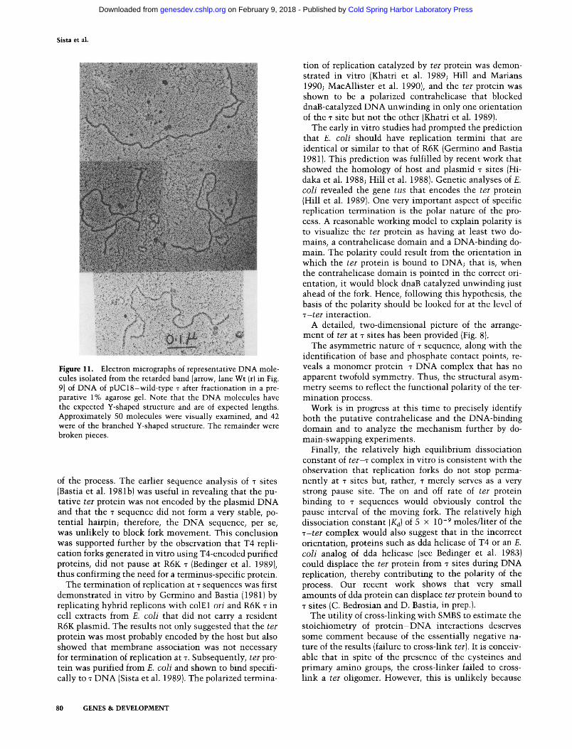
copy, revealed expected Y-shaped DNA molecules of correct dimensions (Fig. 11).
The mutants, similarly cloned in pUC18 and pUC19, were examined to determine whether they terminated DNA replication, as indicated by the band of arrested termination intermediates. The results show that 6C, 8A, 9A, and lOA failed to produce the termination intermediates in pUClS (see Fig. 9, bottom, lanes 6c,r; 8a,r; 9a,r; and 10a,r). No termination intermediates were expected or seen in corresponding pUC19 clones. It should be noted that the wild-type r sample seems to have trace amounts of open circular DNA that migrates just above the line (Fig. 9, bottom). To ensure that we did not miss observing small amounts of termination intermediates in the mutants, we overexposed the gel for up to 1 week after probing with labeled DNA. Under these conditions, whereas the wild-type T revealed the Y-shaped DNA band after 10-12 hr of exposure, no such bands were detected in 6C,r; 8A,r; 9A,r; and 10A,r mutants even after prolonged overexposure (not shown). A summary of the DNA-binding and in vivo termination experiments performed with the mutants is shown in Figure 10 (top).
The results show that those mutants failing to bind to ter also failed to terminate DNA replication in vivo. Conversely, mutants that showed apparently normal or somewhat reduced binding to ter terminated replication in vivo. The efficiency of replication termination was not quantitated.
Discussion
DNA replication can be divided into three steps, namely, initiation, ongoing replication, and termination (Kornberg 1980). The termination step of DNA replication was, until recently, mechanistically the least known of the three steps. Part of the reason for a delay in efforts to understand the termination step, until recently, was the lack of specific termination site in some DNA (Lai and Nathans 1975) and the apparent dispensability of the terminator site in those chromosomes that contained specific termini (e.g., R6K; see Crosa et al. 1976; Bastia et al. 1981a).
Recently, the consensus sequence has not only been found in E. coli (Hidaka et al. 1988; Hill et al. 1988) but also in several of its plasmids; analogous sequences that specifically block fork movement have been found in yeast (Brewer and Fangman 1988) and in the Epstein-Barr virus (EBV) chromosome (Gahn and Schildkraut 1989). In yeast rDNA, the transcription of the DNA is believed to block replication forks from entering the transcribed region (Brewer and Fangman 1988).
Despite the early gaps in our knowledge of termination of replication, more recently, rapid progress has been made in understanding the molecular mechanism
T-Mutants
Core " sequence
I 2 3 4 5 6 7 8 9 10 II 12 13 14 -T- G - T - G - T - 6 - T - T - G - T - A - A - C - T -
4-4 , -l. J - 4 . t > U > l - -i -i
Mutants A T C C C A A A C A
DNA + + binding
Replication" "^ termination
T EcoR!
pUCIST
Figure 10. Summary of the phenotypes of single-base mutants of sequence. [Top] Note that 6C, 8A, 9A, and lOA failed to bind to ter [-] and failed to terminate replication in pUC18 background (also as expected in pUC19). Although 12A and 14T showed reduced binding, they still terminated replication of pUC18. [Bottom] Diagram showing the expected termination intermediate of pUC18-T (wild-type) DNA. Note that EcoRL cleavage would generate a double Y, although the Y at the T end would be very small and, therefore, not detectable by electron microscopy.
GENES & DEVELOPMENT 79
Cold Spring Harbor Laboratory Press on February 9, 2018 - Published by genesdev.cshlp.orgDownloaded from
Sista et al.
"^^x i ^ ^
' . ' . ^ ' ' . • , • . / A ' • • • » . ^ ! ' > - ' ' - •
«
, ?*'. ".'!/-
Figure 11. Electron micrographs of representative DNA molecules isolated from the retarded band [arrow, lane Wt (r) in Fig. 9] of DNA of pUC 18-wild-type T after fractionation in a preparative 1% agarose gel. Note that the DNA molecules have the expected Y-shaped structure and are of expected lengths. Approximately 50 molecules were visually examined, and 42 were of the branched Y-shaped structure. The remainder were broken pieces.
of the process. The earlier sequence analysis of T sites (Bastia et al. 1981b) was useful in revealing that the putative ter protein was not encoded by the plasmid DNA and that the T sequence did not form a very stable, potential hairpin; therefore, the DNA sequence, per se, was unlikely to block fork movement. This conclusion was supported further by the observation that T4 replication forks generated in vitro using T4-encoded purified proteins, did not pause at R6K T (Bedinger et al. 1989), thus confirming the need for a terminus-specific protein.
The termination of replication at T sequences was first demonstrated in vitro by Germino and Bastia (1981) by replicating hybrid replicons with colEl oh and R6K T in cell extracts from E. coli that did not carry a resident R6K plasmid. The results not only suggested that the ter protein was most probably encoded by the host but also showed that membrane association was not necessary for termination of replication at T. Subsequently, ter protein was purified from E. coli and shown to bind specifically to T DNA (Sista et al. 1989). The polarized termina
tion of replication catalyzed by ter protein was demonstrated in vitro (Khatri et al. 1989; Hill and Marians 1990; MacAllister et al. 1990), and the ter protein was shown to be a polarized contrahelicase that blocked dnaB-catalyzed DNA unwinding in only one orientation of the T site but not the other (Khatri et al. 1989).
The early in vitro studies had prompted the prediction that E. coli should have replication termini that are identical or similar to that of R6K (Germino and Bastia 1981). This prediction was fulfilled by recent work that showed the homology of host and plasmid T sites (Hi-daka et al. 1988; Hill et al. 1988). Genetic analyses of E. coli revealed the gene tus that encodes the ter protein (Hill et al. 1989). One very important aspect of specific replication termination is the polar nature of the process. A reasonable working model to explain polarity is to visualize the ter protein as having at least two domains, a contrahelicase domain and a DNA-binding domain. The polarity could result from the orientation in which the ter protein is bound to DNA; that is, when the contrahelicase domain is pointed in the correct orientation, it would block dnaB catalyzed unwinding just ahead of the fork. Hence, following this hypothesis, the basis of the polarity should be looked for at the level of T-ter interaction.
A detailed, two-dimensional picture of the arrangement of ter at T sites has been provided (Fig. 8).
The asymmetric nature of T sequence, along with the identification of base and phosphate contact points, reveals a monomer protein- t DNA complex that has no apparent twofold symmetry. Thus, the structural asymmetry seems to reflect the functional polarity of the termination process.
Work is in progress at this time to precisely identify both the putative contrahelicase and the DNA-binding domain and to analyze the mechanism further by domain-swapping experiments.
Finally, the relatively high equilibrium dissociation constant of te r - r complex in vitro is consistent with the observation that replication forks do not stop permanently at T sites but, rather, T merely serves as a very strong pause site. The on and off rate of ter protein binding to T sequences would obviously control the pause interval of the moving fork. The relatively high dissociation constant {Ki) of 5 x 10"^ moles/liter of the -7-ter complex would also suggest that in the incorrect orientation, proteins such as dda helicase of T4 or an E. coli analog of dda helicase (see Bedinger et al. 1983) could displace the ter protein from T sites during DNA replication, thereby contributing to the polarity of the process. Our recent work shows that very small amounts of dda protein can displace ter protein bound to T sites (C. Bedrosian and D. Bastia, in prep.).
The utility of cross-linking with SMBS to estimate the stoichiometry of pro te in-DNA interactions deserves some comment because of the essentially negative nature of the results (failure to cross-link ter]. It is conceivable that in spite of the presence of the cysteines and primary amino groups, the cross-linker failed to crosslink a ter oligomer. However, this is unlikely because
80 GENES & DEVELOPMENT
Cold Spring Harbor Laboratory Press on February 9, 2018 - Published by genesdev.cshlp.orgDownloaded from

T-Ter interaction
t h e a t t a c h m e n t of cross- l inker to ter is s t rongly indi cated by t h e i n d u c t i o n of he t e rogene i ty of ter bands after SMBS t r e a t m e n t (Fig. 2, cf. l anes d a n d g w i t h l anes e, f, h, and i). F u r t h e r m o r e , t h e gel shift, gel f i l t rat ion, and c ross - l ink ing r e su l t s are all cons i s t en t w i t h each o ther .
Materials and methods
Bacterial and plasmid strains
The strains have been described previously (Sista et al. 1989; Khatri et al. 1989).
carbon replica of the mica surface was prepared and shadowed with plat inum-pal ladium as described (Mukherjee et al.l988).
Isolation of in vivo termination intermediates and their resolution by agarose gel electrophoresis
Bacterial cultures containing the appropriate plasmids were grown in standard L broth with 50 ixg/ml of ampicillin to a cell density of 1 x 10^ to 2 x 10^ cells/ml and harvested. The cell lysis, DNA isolation, and analyses of the replication intermediates have been described (Sista et al. 1989). All DNA samples were cut with £coRI before analysis by gel electrophoresis.
Measurement of Kj
A stock solution of 1.275 mg/ml ter protein was serially diluted, and an aliquot of each dilution was used to titrate a known and fixed amount of a labeled T site DNA (50,000 cpm of 2 X 10* cpm/fJLg of DNA). The protein was present in vast molar excess. The bound and unbound DNA at each protein concentration was quantitated after polyacrylamide gel electrophoresis and autoradiography. The relative K^ is the protein concentration that promotes half-maximal occupation of available T sites (Fried and Crothers 1981: Koudelka et al. 1987). The binding buffer contained 40 mM Tris-HCl (pH 7.5), 1 mM EDTA, 4 mM MgClj, and 50 mM potassium glutamate.
Hydroxyradical footprinting
Footprinting was carried out using 3 ' - or 5'-end-labeled TL or TR DNA as described (Tullius and Dombroski 1988).
Saturation site-directed mutagenesis
Mutations were introduced at each residue of the core T sequence by automated synthesis of mixed oligonucleotide preparations (Hutchison et al. 1986; Murray et al. 1988). Two complementary oligonucleotides, 5'-CAATCTCTTGTGTGT-TGTAACTAAATCATCGA-3' (a 32-mer) and 5'-AGCTTC-GATGATTTAGTTACAACACACAAGAGATTGAGCT-3' (a 40-mer), were synthesized. A small amount (6.5%) of an equi-molar mixture of all four phosphoramidites was added to each pure precursor to introduce an average of 1.5 single-base substitutions per oligonucleotide molecule. The two synthetic products were gel purified and annealed to give duplexes with protruding Hindlll (5') and Sstl (3') sticky ends. These duplexes were cloned into M13mpl l , and individual isolates were sequenced to identify single-base substitution mutations (for further details, see Hutchison et al. 1986).
Methylation protection and methylation and ethylation interference
Protection and interference were performed as described previously (Ogata and Gilbert 1978; Siebenhst and Gilbert 1980; Germino and Bastia 1983).
Gel mobility shift
This assay was performed as described (Fried and Crothers 1981), except that the buffer contained 40 mM Tris-Cl (pH 7.5), 1 mM EDTA, and 50 mM potassium glutamate.
Electron microscopy
The DNA samples were sprayed on freshly cleaved mica, and a
Cross-linking with SMBS
Purified ter protein and dnaB and te r - r complex were cross-linked with SMBS essentially as described by Lerner et al. (1985), with the exception that water-soluble SMBS was added directly to the protein solutions (whereas MBS used by Lerner et al. was dissolved first in dimethylsulfoxide) to concentrations of 0.1 and 0.3 mM.
A c k n o w l e d g m e n t s
This work was supported by grants from the National Institutes of Health (NIH) and the National Cancer Institute to D.B. and by a grant from NIH to C.H. We thank Mr. Tim Oliver for electron microscopy, Ms. Hilda Smith for the preparation of this manuscript, Drs. S. Mukherjee and S. Natarajan for discussions and comments, and Dr. R. Joshi for help with protein cross-linking gels.
The publication costs of this article were defrayed in part by payment of page charges. This article must therefore be hereby marked "advertisement" in accordance with 18 USC section 1734 solely to indicate this fact.
References
Bading, H. 1988. Determination of the molecular weight of DNA-bound protein(s) responsible for electrophoretic mobility shift of linear DNA fragments exemplified with viral myb proteins. Nucleic Acids Res. 16: 5241-5248.
Bastia, D., J. Germino, l.H. Crosa, and P. Hale. 1981a. Molecular cloning of the replication terminus of plasmid R6K. Gene 41: 81 -89 .
Bastia, D., J. Germino, J.H. Crosa, and T. Ram. 1981b. The nucleotide sequence surrounding the replication terminus of R6K. Proc. Natl. Acad. Sci. 72: 2095-2099.
Bedinger, P., M. Hochstrasser, C.V. longeneel, and B.M. Alberts. 1983. Properties of T4 bacteriophage DNA replication apparatus: The dda DNA helicase is required to pass a bound RNA polymerase molecule. Cell 34: 115-123.
Bedinger, P., M. Munn, and B.M. Alberts. 1989. Sequence-specific pausing during in vitro DNA replication on double-stranded DNA templates. /. Biol. Chem. 264: 16880-16886.
Brewer, B. and W.L. Fangman. 1988. A replication fork barrier at the 3'-end of yeast ribosomal RNA genes. Cell 55: 637-643.
Crosa, l.H., L. Luttrop, and S. Falkow. 1976. Mode of replication of the conjugative R plasmid RSF 1040 in Escherichia coli. f. Bacterial. 126: 454-466.
Fried, M. and D. Crothers. 1981. Equilibria and kinetics of lac repressor-operator interaction by polyacrylamide gel electrophoresis. Nucleic Acids Res. 9: 6505-6525.
Gahn, T. and C. Schildkraut. 1989. The Epstein-Barr virus origin of plasmid replication, OriP contains both the initiation
GENES & DEVELOPMENT 81
Cold Spring Harbor Laboratory Press on February 9, 2018 - Published by genesdev.cshlp.orgDownloaded from
Sista et al.
and termination sites of DNA replication. Cell 58: 527-535. Germino, J. and D. Bastia. 1981. Termination of DNA replica
tion in vitro at a sequence-specific replication terminus. Cell 23: 681-687.
-. 1983. Interaction of the plasmid R6K-encoded replication initiator protein with its binding sites on DNA. Cell 34: 125-134.
Hidaka, M., M. Akiyama, and T. Horiuchi. 1988. A consensus sequence of three DNA replication terminus sites on the E. coli chromosome is highly homologous to the Ter R sites of the R6K plasmid. Cell 55: 467-475.
Hill, T.M. and K. Marians. 1990. E. coli tus protein acts to arrest the progression of DNA replication forks in vitro. Proc. Natl. Acad. Sci. 87: 2481-2485.
Hill, T.M., A.l. Pelletier, M.L. Tecklenberg, and P.L. Kuempel. 1988. Identification of DNA sequence from the E. coli terminus region that halts replication forks. Cell 55: 459-466.
Hill, T.M., M. Tecklenberg, A. Pelletier, and P.L. Kuempel. 1989. Tus the transacting gene required for termination of DNA replication in Escherichia coli encodes a DNA-binding protein. Proc. Natl. Acad. Sci. 86: 1593-1597.
Horiuchi, T. and M. Hidaka. 1988. Core sequence of two separable terminus sites of R6K plasmid that exhibit polar inhibition of replication is a 20 bp inverted repeat. Cell 54: 5 1 5 -523.
Hutchison III, C.A., S, Nordeen, K. Vogt, and M. Edgell. 1986. A complete library of point substitution mutations in the glucocorticoid response elements of mouse mammary tumor virus. Proc. Natl. Acad. Sci. 83: 710-714.
Khatri, G.S., T. MacAUister, P.R. Sista, and D. Bastia. 1989. The replication terminator protein of E. coli is a DNA sequence-specific contrahelicase. Cell 59: 667-674.
Kolter, R. and D. Helinski. 1978. Activity of the replication terminus of R6K in hybrid replicons in £. coli. f. Mol. Biol. 124:425-441.
Kornberg, A. 1980. In DNA Replication. W.H. Freeman, San Francisco.
Koudelka, G.B., S.C. Harrisson, and M. Ptashne. 1987. Effect of noncontacted bases on the affinity of 434 operator for 434 repressor and Cro. Nature 306: 886-888.
Kuempel, P., S.A. Duerr, and N.R. Seeley. 1977. Terminus region of E. coli inhibits replication forks. Proc. Natl. Acad. Sci. 74 :3927-3931.
Lai, C.S. and D. Nathans. 1975. Non-specific termination of SV40 DNA replication. /. Mol. Biol. 97: 113-118.
Lee, E.-U., A. Kornberg, M. Hidaka, T. Kobayashi, and T. Horiuchi. 1989. Escherichia coli replication termination protein impedes the action of helicases. Proc. Natl. Acad. Sci. 86: 9104-9108.
Lerner, R., N. Green, H. Alexander, F.-T. Liu, J.G. Sutcliffe, and T.M. Shinnick. 1985. Chemically synthesized peptides predicted from the nucleotide sequence of hepatitis B virus genome elicit antibody reactive with native envelope protein and Dane particles. Proc. Natl. Acad. Sci. 78: 3 4 0 3 -3407.
Louarn, J., D. Patte, and J.M. Louarn. 1977. Evidence for a fixed termination site of chromosome replication in £. coli, K12. J. Mol. Biol. 115:295-314.
MacAUister, T., G.S. Khatri, and D. Bastia. 1990. Sequence-specific and polarized termination in vitro: Complementation of extracts of t u s " £. coli by purified ter protein and analysis of termination intermediates. Proc. Natl. Acad. Sci. 87: 2828-2832.
Mukherjee, S., H. Erickson, and D. Bastia. 1988. Enhancer-origin interaction in plasmid R6K involves a DNA loop mediated by initiator protein. Cell 52: 375-383.
Murray, R., C.A. Hutchison III, and J.A. Frelinger. 1988. Saturation mutagenesis of a major histocompatibility complex protein domain: Identification of a single conserved amino acid important for allorecognition. Proc. Natl. Acad. Sci. 85: 3535-3539.
Ogata, R. and N. Gilbert. 1978. An amino-terminal fragment of lac repressor binds to the lac operator. Proc. Natl. Acad. Sci. 75:5851-5854.
Siebenlist, U. and W. Gilbert. 1980. Contact points between Escherichia coli RNA polymerase and an early promoter of phage T7. Proc. Natl. Acad. Sci. 77: 122-126.
Sista, P.A., S. Mukherjee, P. Patel, G.S. Khatri, and D. Bastia. 1989. A host-encoded DNA binding protein promotes termination of plasmid replication at a sequence-specific replication terminus. Proc. Natl. Acad. Sci. 86: 3026-3030.
Tullius, T.D. and B.A. Dombroski. 1986. Hydroxy-radical "footprinting": High resolution information about DNA-protein contacts and application to repressor and Cro protein. Proc. Natl. Acad. Sci. 83: 5469-5473.
82 GENES & DEVELOPMENT
Cold Spring Harbor Laboratory Press on February 9, 2018 - Published by genesdev.cshlp.orgDownloaded from

10.1101/gad.5.1.74Access the most recent version at doi: 5:1991, Genes Dev.
P R Sista, C A Hutchinson and D Bastia DNA-protein interaction at the replication termini of plasmid R6K.
References
http://genesdev.cshlp.org/content/5/1/74.full.html#ref-list-1
This article cites 32 articles, 13 of which can be accessed free at:
License
ServiceEmail Alerting
click here.right corner of the article or
Receive free email alerts when new articles cite this article - sign up in the box at the top
Copyright © Cold Spring Harbor Laboratory Press
Cold Spring Harbor Laboratory Press on February 9, 2018 - Published by genesdev.cshlp.orgDownloaded from