dna based vaccines against tuberculosis; recent progress
TRANSCRIPT

Mobed A. Iran J Immunol. 2020; 17(4):255-274. https://doi.org/10.22034/iji.2020.87480.1806.
Iran.J.Immunol. VOL.17 NO.4 December 2020 255
REVIEW ARTICLE
DNA Based Vaccines against Tuberculosis; Recent Progress and Discovery Research
in Vaccine Development and Delivery System
Ahmad Mobed1,2* 1Physical Medicine and Rehabilitation Research Center, Aging Research Institute, 2Neuroscience Research Center, Tabriz University of Medical Sciences, Tabriz, Iran ABSTRACT Tuberculosis is believed to be one of the leading sources of death round the world; nevertheless, Bacillus Calmette Guérin (BCG) is the solitary vaccine utilized to prevent TB. Despite the protective effect of this vaccine on children, its efficiency remains under question in adults. We conducted the present study to provide an overview of the DNA based vaccine against Tuberculosis (TB) and highlight the vaccine delivery advances and limitations in TB treatment. This study also aimed to bring a review of mycobacterial antigens, including heat shock protein 65 (Hsp65), antigen 85A (Ag85A), early secretory antigenic target (EAST-6), antigen 85B (Ag85B), and heat shock protein X (HspX) as the most extensively considered antigens used to strategy vaccines against M. tuberculosis. Received: 2020-07-22, Revised: 2020-08-24, Accepted: 2020-09-16. Citation: Mobed A. DNA Based Vaccines against Tuberculosis; Recent Progress and Discovery Research in Vaccine Development and Delivery System. Iran J Immunol. 2020; 17(4):255-274.
Keywords: DNA Vaccine, Mycobacterium Tuberculosis, Mycobacterial Antigen, Vaccine Delivery --------------------------------------------------------------------------------------------------------------------------------------------------------------- *Corresponding author: Dr. Ahmad Mobed, Physical Medicine and Rehabilitation Research Center, Aging Research Institute, Tabriz University of Medical Sciences, Tabriz, Iran, e-mail: [email protected]

Mobed A.
Iran.J.Immunol. VOL.17 NO.4 December 2020 256
INTRODUCTION To date, vaccines have always been one of the most important medical interventions in the world (1). In spite of great scientific advances , there are still several challenges in this regard (2); for instance, very limited time for clinical trials, particularly the trials associated with emerging and fatal infections. Rapid vaccine interventions could be specifically beneficial in case of emerging infections such as Ebola virus (EBOV), ZIKA virus (ZIKV), and recently coronavirus (COVID-19) (3). There are certain factors to be taken into consideration concerning vaccines, such as rapid development, simple and low cost to organize, stable in different temperature (3,4). According to WHO, in 2018, an estimated 10 million people were affected by tuberculosis (TB) worldwide, including 1.1 million children, 3.2 million women, and 5.7 million men. However, these cases were all around the world and age groups, TB is preventable and curable (5,6). Currently, TB vaccination is one of the best approaches to preventing and controlling Mtb infection. Accordingly, the BCG vaccine as a live attenuated strain of M. bovis, is the only accepted vaccine against TB (7). Due to the many advantages over traditional vaccines, the DNA vaccine development is increasing more rapidly (8). DNA vaccines permit the expression of imported genes in a host and their demonstration of specific encoded proteins to the immune system, which subsequently results in the initiation of wide-ranging, antigen-specific humoral, and cellular immunity (9). Certain advantages of employing DNA vaccines over traditional vaccines include simple production, low production cost, stability, promising safety profile of plasmid incorporation in humans, facility to be administered frequently since plasmid vector competences are not influenced by pre-existing neutralizing antibodies, and ultimately, being able to target multiple antigens. Vaccine delivery is one of the major challenges in DNA vaccines (10,11). Advances in DNA Vaccine Delivery. The paramount features of DNA vaccine technology could be the fact that they are produced on an industrial scale and do not require the cultivation of the target pathogen (12,13). On the other hand, there are certain delivery-associated obstacles concerning DNA vaccines compared to traditional ones; DNA based vaccines require accurate physical injection and effective gene expression following injection (14). To overcome these problems, electroporation (EP) system and mechanically powered jet injection system was established appropriately (15-17). EP requires needle syringe injection of DNA into the target region while jet-injection is most commonly employed for subcutaneous or intramuscular administration (18,19). Recently, a new type of needleless device has been established in order to control the injection depth and speed of using a computer armed in DNA vaccination (20). Undoubtedly, in the last decades, the micro-needle method has attracted a great deal of scientific attention regarding drug delivery. Simple drugs delivery in the outer membrane of skin layer, tough barrier, and inactivating stratum corneum (SC) are the paramount properties of micro-needle based drug delivery. In other words, the micro-needle method significantly facilitates the admission of large hydrophilic molecules from physiological membranes (21,22). This method also provides a unique perspective on the drug delivery of high molecular weight and hydrophilic compounds through the skin (23). Moreover, not only it is completely painless and safe, but also significantly accelerates the healing and recovery of the skin. It also eliminates the risk of bacterial infection (24,25). Owing to these unique features, their use in cosmetic and restorative injections is significantly increasing. Additionally, considering the efficiency of the micro-needle method, it is suggested to be applied in oral mucosal, gastrointestinal, and

DNA based vaccines against tuberculosis
Iran.J.Immunol. VOL.17 NO.4 December 2020 257
ocular drug delivery. In sum, micro-needle technique is extensively applied for drug delivery and it is predictable that several micro-needle-associated applications would develop for other organs and tissues. Dissolving micro-needles, coated micro-needles, solid removable micro-needle, hydrogel forming micro-needles, and hollow micro-needles are the most imperative types of micro-needle procedure (Figure 1, A,B). Figure 1.C represents the scheme of nano-vaccine possible cellular and humoral mechanisms.
Figure 1. A) Representation of routine/old and micro-needle based injection methods on human skin. As can be seen in this figure, applying micro-needles technique, stratum corneum penetrated to the reaching the viable epidermis. Inserting the hypodermic needles into the muscle tissues or subcutaneous ruptured the skin (26). B) High-voltage electroporation technique where applied for a short time to create local aqueous penetrable areas in between lipid membranes by destabilization (27). C) The scheme of nano-vaccine possible cellular and humoral mechanisms (28).

Mobed A.
Iran.J.Immunol. VOL.17 NO.4 December 2020 258
Suitable vaccine administration/delivery is the key factor for a successful vaccination. Most vaccines are normally administered via the intramuscular (IM) or subcutaneous (SC) routes. Hypodermic injections are related to pain and distress that might lead to poor patient agreement. Thus, they require highly skilled personnel for administration (36). In these injections, there is a risk of disease transmission due to the probability of needle-stick damages or reuse of contaminated needles. Deficient vaccine supply or limitation of vaccine production might be also challenging in instances when quantity vaccination is required. In global vaccination, its limitations and production problems will be challenging (36). Table.1. Lists of the immune-biological administration through micro-needles and its associated effects.
Vaccine Microneedle type Significances Ref.
Live attenuated
BCG
Dissolving microneedle
Vaccination using microneedle by (BCG) powder did not triggered severe bruise and inflammation as compared
to intradermal injection
[29]
Hepatitis B
surface antigen
Dissolving microneedle
The antigenicity of the Hepatitis B adjuvant continued for long-time with
only a 10% loss [30]
Rabies
vaccination Dissolving
microneedle Used vaccine dose ten-fold lower than
full-dose intramuscular vaccination [31]
IgG and IgG1 Hollow microneedle Ability to monitoring of immune
response [32]
Ag85B DNA
vaccine Dissolving
microneedle Microneedle patch
(MNP)
Effective protection and offer a novel approach against TB
[33]
BCG-polysaccharide DNA vaccine
MNPs eliminate the side effects of syringes, Finding revealed that BCG-
can be clinically administrated in powder form
[34]
Nanoparticles
based TB Vaccine
Hollow microneedle Effective protection and offer a novel
approach in immune response [35]
To be medically appreciated, DNA vaccines need to get into the nucleus before they can express antigen molecules. Naturally, the introduction of DNA vaccines requires proper perforation of existing cell membranes (37, 38). Several studies have demonstrated that direct injections of DNA vaccines could stimulate an immune response in smaller animals, and the delivery of the DNA to target cells is not optimum, particularly in higher animals (38, 39). Table 2 depicts certain DNA vaccine delivery methods which are of importance.
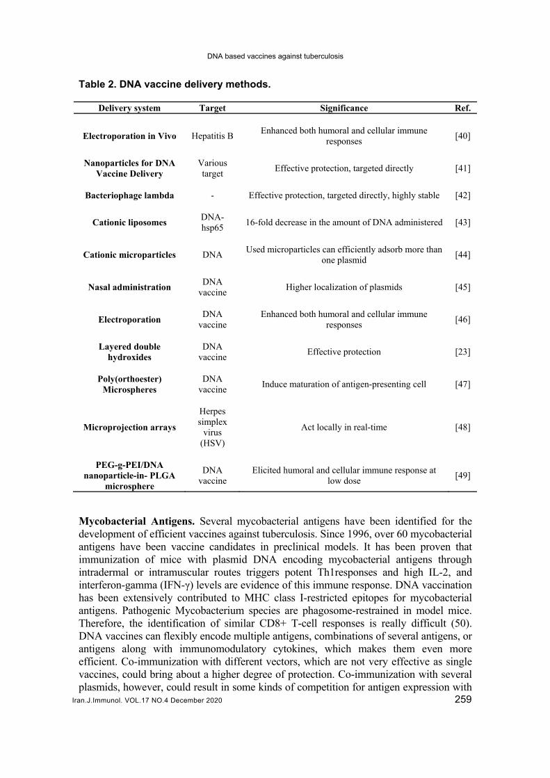
DNA based vaccines against tuberculosis
Iran.J.Immunol. VOL.17 NO.4 December 2020 259
Table 2. DNA vaccine delivery methods.
Delivery system Target Significance Ref.
Electroporation in Vivo Hepatitis B Enhanced both humoral and cellular immune
responses [40]
Nanoparticles for DNA Vaccine Delivery
Various target
Effective protection, targeted directly [41]
Bacteriophage lambda - Effective protection, targeted directly, highly stable [42]
Cationic liposomes DNA-hsp65
16-fold decrease in the amount of DNA administered [43]
Cationic microparticles DNA Used microparticles can efficiently adsorb more than
one plasmid [44]
Nasal administration DNA
vaccine Higher localization of plasmids [45]
Electroporation DNA
vaccine Enhanced both humoral and cellular immune
responses [46]
Layered double hydroxides
DNA vaccine
Effective protection [23]
Poly(orthoester) Microspheres
DNA vaccine
Induce maturation of antigen-presenting cell [47]
Microprojection arrays
Herpes simplex
virus (HSV)
Act locally in real-time [48]
PEG-g-PEI/DNA nanoparticle-in- PLGA
microsphere
DNA vaccine
Elicited humoral and cellular immune response at low dose
[49]
Mycobacterial Antigens. Several mycobacterial antigens have been identified for the development of efficient vaccines against tuberculosis. Since 1996, over 60 mycobacterial antigens have been vaccine candidates in preclinical models. It has been proven that immunization of mice with plasmid DNA encoding mycobacterial antigens through intradermal or intramuscular routes triggers potent Th1responses and high IL-2, and interferon-gamma (IFN-γ) levels are evidence of this immune response. DNA vaccination has been extensively contributed to MHC class I-restricted epitopes for mycobacterial antigens. Pathogenic Mycobacterium species are phagosome-restrained in model mice. Therefore, the identification of similar CD8+ T-cell responses is really difficult (50). DNA vaccines can flexibly encode multiple antigens, combinations of several antigens, or antigens along with immunomodulatory cytokines, which makes them even more efficient. Co-immunization with different vectors, which are not very effective as single vaccines, could bring about a higher degree of protection. Co-immunization with several plasmids, however, could result in some kinds of competition for antigen expression with

Mobed A.
Iran.J.Immunol. VOL.17 NO.4 December 2020 260
several plasmids, however, could result in a competition for antigen expression, which limits antigen expression and processing efficiency. On the other hand, in comparison to the combination of plasmids, the application of hybrid genes or multi-promoter plasmid vectors is simpler and more cost-effective for industrial purposes (51). Heat shock protein 65 (Hsp65), early secretory antigenic target (EAST-6), antigen 85A (Ag85A), antigen 85B (Ag85B), and heat shock protein X (HspX) are the most extensively considered antigens applied to develop vaccines against M. tuberculosis (52). Secretory and surface proteins (EAST-6) of M. tuberculosis are the paramount antigens expressed through immunization possible against tuberculosis. Encoded by three different genes on the mycobacterial genome, antigen 85 (Ag85) complex involves Mycobacterium tuberculosis (Mtb) and secretory proteins of Bacillus Calmette-Guerin (BCG). This compound contains three related antigens: Ag85C (32.5 kDa), Ag85A (32 kDa), and Ag85B (30 kDa). These proteins are mycolyl transferase enzymes that are important for the biosynthesis of the mycobacterial cell wall throughout tuberculosis pathogenesis (52). Ag85A and Ag85B. Ag85A and Ag85B are two major Mtb secretory proteins that have pivotal roles in eliciting strong humoral and cell-mediated immune responses and protecting against tuberculosis in animal models. Ag85 complex plays its main role in tuberculosis pathogenesis likely via its physiological function in the biosynthesis of M. tuberculosis cell wall lipids. In addition, owing to its ability to bind with fibronectin, it is an essential constituent in tuberculosis pathogenesis. This protein complex stimulates the adhesion of mycobacteria to the mucosal surfaces and, therefore, facilitates its entry to the host cell (53). Some trainings have focused on Ag85A as suitable antigen for developing DNA vaccines against M. tuberculosis. During 2000, Audrey Tanghe and coworkers explored the immunogenicity of a DNA vaccine encoding Ag85A in mice. They determined that Ag85A DNA vaccine is a high-quality technique to convince protective Th1 immune responses (54). Meanwhile, another work by the same authors revealed that DNA priming vaccination monitored by exogenous protein-boosting characterizes a well-organized way of growing the immunogenicity and protective effect of DNA vaccine encoding Ag85 (55). In 2002, S. D’Souza vaccinated mice with a plasmid DNA carrying Ag85A. He reported that the level of T-cell-derived Th1-type cytokines were higher in response to Ag85A (56). Sugawa et al., administered a DNA vaccine encoded Ag85A of tuberculosis for pigs via epidermal gene gun bombardment and determined its protecting ability. According to their results, peptide boosting and dosage played the key roles in prompting higher protective responses by DNA vaccination (57). Ruyi Liu et al. studied the immunogenicity of a multi-epitope DNA vaccine encoding some mycobacterial antigens including Ag85A in mouse models in 2008 (58). They indicated that this DNA vaccine induced stronger immune reactions with high levels of specific IgG antibody. Relevant studies during 2009 also put Ag85A forward as a model antigen for DNA vaccine studies (58). Fayaz Ahmad Mir fabricated a novel strategy for DNA vaccine encoding Ag85A in concoction with other mycobacterial antigens (59). Based on his results, developing vaccines may be a potential approach to fighting against tuberculosis (59). Another study planned four functional Tcell epitopes as well as Ag85A, and considered cellular and humoral responses elicited by this DNA vaccine in mice (60). In 2012, Ahn et al. performed a head-to-head comparison of seven tuberculosis antigens delivered as DNA vaccines and evaluated their associated immune responses and immune protection in mouse models (61). The obtained results pointed out that the Ag85A vaccines in combination with chemotherapy reduced bacterial load. The current study verified that DNA vaccination directed to a stronger Th1 cytokine response against
DNA based vaccines against tuberculosis
Iran.J.Immunol. VOL.17 NO.4 December 2020 261
tuberculosis (61). In 2014, Kun Tan et al. industrialized a recombinant BCG strain expressing Ag85A. They complemented he protective effects of this recombinant vaccine and immune responses with a former BCG prime booster regimen and reported that their novel vaccine elevated both TNF-α and iNOS responses in the lung and produced higher IFN-γ responses. Therefore, this DNA vaccine provided better control of bacterial growth in the lung and spleen of vaccinated mice (62). Zahra Meshkat directed a survey in 2016, in which the immunogenicity of Ag85A DNA vaccine was analyzed with enzyme-linked immunosorbent assay (ELISA). She established that the levels of IFN-γ and IL-12 increased significantly in mice vaccinated with Ag85A DNA vaccine in comparison with control BCG groups (63). In another study in the same year, Yan Liang constructed a chimeric DNA vaccine which was encoded Ag85A of M. tuberculosis (64). The administration of this DNA vaccine implied results opposed to the previous reports. Even though in contrast to the previous findings, these results indicated that DNA vaccines were either beneficial or at least not harmful, they resulted in increased mortality in mice. A study conducted in 2017 by Baghani et al. recommended that a DNA vaccine encoding Ag85A of M. tuberculosis might be applied for exploring immune responses in animal models (65). In another research, Li Sun united AG85A DNA vaccine with the IL-15 as a molecular adjuvant in order to examine the potential immune response. They perceived a greater Th1 immune response in mouse models vaccinated by a DNA vaccine expressing Ag85A-IL-15 transgenes (66). As another member of Ag85 complex, Ag85B has been also studied as an antigen to design DNA vaccines against tuberculosis. In 2002, D’Souza et al. studied Ag85B in combination with Ag85A and PstS3. Their obtained results indicated that formulation in Vaxfectin had an increasing effect on the protective efficiency of the Ag85B DNA vaccine (56). Xia Tian evaluated the immunogenic activity and protective efficacy of a divalent DNA vaccine encoding Ag85B and MPT 64 of M. tuberculosis (67). He elucidated that both humoral and Th1 cellular responses caused by this bivalent vaccine were significantly higher than those of BCG (67). In 2009, Wanhong Yao et al. investigated the immune responses and efficacy of Ag85B DNA vaccine in mice. This study revealed that Ag85B- spleen lymphocyte and specific antibodies proliferative responses motivated by DNA co-expressing bovine herpesvirus 1 VP22 (BVP22) and Ag85B were significantly greater than those perceived in mice vaccinated with Ag85B only (68). Following several studies, Haifeng Gao and coworker expressed that Ag85B in a chimeric DNA vaccine carried further mycobacterial antigens. Based on their findings, this DNA vaccine could effectively induce greater specific cell-mediated immune responses (69). In 2013, Cervantes fabricated a DNA vaccine via the fusion of Ag85B genes and β defensin 2. Mice were vaccinated with constructed DNA vaccine and showed that this vaccination resulted in similar level of protection as BCG vaccines (70, 71). Yet after encountering certain challenges with highly virulent M. tuberculosis strain, the animals which were prime-boosted with BCG followed by DNA vaccine showed remarkably higher survival (71). In the same year, Jomkhwan Meerak utilized a DNA vaccine to discover whether autophagy increases immune responses alongside DNA vaccination. For this reason, DNA vaccine with autophagy convincing created greater Ag85B-specific antibody levels in comparison to Ag85/b plasmid with the wild type mTOR construct, Ag85B alone, and vaccinated control group (72). The findings confirmed that the primeboosted DNA vaccination with a lentivirus encoding Ag85B considerably improved bothCD8+ cytotoxic T lymphocytes and T helper type I responses in comparison to the DNA and protein-based vaccines (73).

Mobed A.
Iran.J.Immunol. VOL.17 NO.4 December 2020 262
Table 3. DNA vaccine based on Ag85A and Ag85B antigens.
Antigen Model cloned Vector Cell line Strain Immune Response
Ref.
ESAT6 Ag85B
mouse skin - murine
defensin-2 (mBD2)
TB H37Rv Th1 adaptive
response [70]
Ag85A
Escherichia coli DH5
Recombinant plasmid DNA
C57BL/6 mice
H37Rv,VR1020
IFNγ, IL-2
[55]
Ag85A mice VR1020 mice Kurono strain (ATCC25618)
IFN-γ [57]
Ag85B
female BALB/c inbred mice
pmTOR-KD
HEK293T
cells
TB
IFN-γ,IL-2
[72]
ESAT-6 and
Ag85A
Escherichia coli strains DH5α
and BL21 (DE3)
pDE22
C57BL/6
mice
M. tuberculosis H37Rv, M. bovisBCG
China
IFN-γ, IL-10,
TNF-α
[62]
Ag85A C57BL/6 mice pcDNA3.1 (HEK293)c
ells -
IL 2
[66]
Ag85B E. coli BL21
(DE3) pcDNA3.1(+)
female C57BL/ 6
Mtb H37Rv IFN-γ [68]
Ag85B and Rv3425
mice pVax 293T cell
line bovis BCG
H37Rv - [73]
Ag85B and MPT64
C57BL/6 mice
pJW4303 E.
coli DH5α H37Rv and M.
bovisBCG IFN-γ,IL-4
[67]
Ag85B ESAT-6, Ag85A, CFP-10
female BALB/c
mice
pcDNA3.1
-
PPD,BCG
IFN-γ, TNF-α, IL-4
[69]
cfp10 , Ag85A E. coli strain
JM109 pCDNA3.1 HeLa cells TB H37Rv
TNFα, IL-2, IFNγ
[65]
Ag85B and MPT64
C57BL/6 mice pJW4303 E. coli DH5α
H37Rv and M. bovis BCG
IFN-γ,IL-4 [67]
Hsp60 Family. The mycobacterial heat shock protein (Hsp65, 65 kDa), as a member of the Hsp60 family, is the major antigen of M. tuberculosis. From a general point of view, Hsp65 is analogous to the eukaryotic Hsp70 chaperon. Matrix metallopeptidase 9 (MMP9) could break down the Hsp65 and subsequently form immunogenic peptides that disturb host adaptive immunity. A highly important point in the design of DNA vaccine based on antigen Hsp65 is its similarity to more than
DNA based vaccines against tuberculosis
Iran.J.Immunol. VOL.17 NO.4 December 2020 263
50 amino acids with mammalian cells, which makes autoimmune reactions, such as systemic lupus erythematosus, acute anterior uveitis, and arthritis possibility (74). Moreover, it has been demonstrated that M. leprae hsp65 induces noticeable humoral and cell-mediated immunity in guinea pigs infected with Mycobacterium tuberculosis (74). For note, the pathophysiological significance of the H antigen is very wide and requires further research (74). In other words, on top of demonstrating important antigens, hsp65 could be a peptidase with the potential of generating or destroying other biologically active molecules probably involved in vaccine production processes (74). Furthermore, hsp65 has been studied as a candidate antigen for DNA vaccines during the last decades. Shi Changhong fabricated human IL-2 genes and plasmid DNA vaccine expressing hsp65, 10 years ago. He demonstrated amplified Th1 cellular response and greater levels of IL-2 and IFN-γ in mice (75). These findings recommended that the applied vaccine improved protective effects of DNA vaccine against tuberculosis and their immunogenicity (75). Another study was developed based on a DNA vaccine encoding numerous mycobacterial antigens as well as hsp65. Its results revealed that DNA vaccination is an effective way of tempting improved specific cell-mediated immunity against tuberculosis in mice (69). In an investigation conducted by Yan Dong et al. in 2013, a bicistronic DNA vaccine was made, which expressed hsp65 and EAST-6 in mice. They perceived a greater titer of the IFN-γ and IL-2 secretion, antibody, and lymphocyte proliferation, which was suggestive of a real immune response prompted by DNA vaccine (76). In another research by Qingmin Wanget et al., the immune responses induced by a DNA vaccine with the hsp65 gene were studied. They showed that UbGr-hsp65 DNA vaccine prompted a Th1-polarized immune response with meaningfully enhanced IFN-γ and proliferative T cell response from spleen when compared with the responses in the hsp65 DNA vaccine group. Consequently, this study verified that UbGr fusion could increase specific cell-mediated immune responses (77). During induction of infection with macrophage and oxygen decline, HspX could not stimulate IFN- immune responsesin BCG vaccinated animal model or in newborn BCG immunization. The HspX (16 kDa a-crystallin homologue) protein in TB which, encoded by the acr gene. This gen is one of the most important helper of the small heat shock protein family of chaperones. During the move from log-phase growth toward stationary phase, HspX expression rises and turn into one of the highest plentiful proteins in the stationary phase (78). Nevertheless, the cellular contented of 16-kDa protein, though low in log-phase bacteria, increased to a maximum at 10 days and persisted at this high level until 50th days. This revealed that this protein is a steady molecule with a low turnover rate. Our results showed that the regulation of (78). Due to the slowly growing heat shock protein, X antigen is an unusual protein of M. tuberculosis complex whereas it is classified as a heat shock protein (74). Members of the sHSP family revealed in M. tuberculosis, and Acr2 is heat stress-induced Ribosome-associated protein (17.8 kDa). The full sequence similarity between Acr2 and Acr1 is over 40% and the crystal-like domain is over 5%. Some Mycobacteria such as M. smegmatis and M. marinarium have Acr3 protein that are homologous with both Acr1 and Acr2. This protein is closely similar to the single sHSP in M. leprae (79). In Mycobacteria strain, from which acr gene was removed despite normal growth in culture, the development of macrophages decreased. DosR transcription factor, regulated by histidine sensor

Mobed A.
Iran.J.Immunol. VOL.17 NO.4 December 2020 264
kinases, controlled the gene expression, and regulated by histidine sensor kinases. During stresses such as S-nitrosoglutathione, ethanol, and hypoxia the dos R regulon induced Acr1 transcription, yet not under heat shock (79), heat shock powerfully regulated Acr2 genes. Additionally, negative regulation was monitored by the hspR heat shock regulator while positive regulation was monitored by other sigma factors. H and E factors, combined with heat and oxidative stress (79). Both tuberculosis acceptor and non-acceptor macrophages, which were not stimulated by interferon, were also involved in the regulation of the ECR gene. In mouse models, although mutations in the ECR1 gene reduce pathogenesis, mutations in gene 2 have no effect on pathogenicity. Recent studies have revealed that Acr2 is the main target of humoral and T cell immunity throughout the early phases of infection in humans. This finding could be significant for emerging improved vaccines (80). Chaperone-like activity is one of the paramount aspects and potentials of HspX antigen, a cytosolic protein, which has been mostly considered. In some cases, as well as pulmonary TB patients, the HspX antigen was able for induction of IgG antibodies among from 34- 62%. So, HSPX have been used with other antigen to expand commercial sero-diagnostic trials. In up-to-date microarray method B-cell epitopes of HSPX accepted through peptide (81). The results of studies show that the HSPX antigen is compressed in the cell wall in TB anaerobic and microaerophilic cultures.Under increased expression of HspX antigen hsp60 promoter makes recombinant M. smegmatis or else M. tuberculosis that this less vulnerable mycobacterium to autolysis and have a slow initial growth ratio. Consequently, HspX is of other key roles, for instance adjusting to intracellular environments in early stages and continuing long-term viability of M. tuberculosis in vivo (82). Approximately 77% of chronic TB patients have detectable anti-HspX since HspX is an extra immunogenic protein. Although the higher level of IFN- responses to HspX protein could be used as an interpreter of latent TB infection, individual with active TB had little IgG antibody against HspX. Additionally in mice, HspX antigen-induced high levels of IFN-γ (82). According several studies HspX prompted a high level of INF in the latent stage of TB patients (83). The obtained findings herein indicated that the latent stage of infection and stationary phase are the appropriate phases to apply HspX antigen in order to fabricate a suitable TB vaccine. An arrangement of Ag85A and HspX antigen is the potential reactive TB antigen throughout the stationary stage and the main TB antigen, correspondingly. In another study, this arrangement was utilized for improving a prophylactic vaccine and maintain protective from the acute phase to the chronic phase of infection. In this regard, a TB aerosol from mouse models was applied to estimate the primed combination of HspX antigen only in stationary and early phases (83). Our findings demonstrated that once the mice are vaccinated with combined antigen, a greater level of IFN-γ secretion was prompted compared to that prompted via a single antigen, Ag85A or HspX antigen. Even though both HspX antigens and Ag85A could be applied as a TB vaccine candidate, a mixture of these antigens might synergistically induce IFN-γ production. |Comparing the naïve group with BCG vaccinated group, the bacterial reduction (around 1.2 log (p<0.001) was shown in both lungs and spleen. Furthermore, in comparison with other investigational vaccines, the attained protective efficacy of the vaccine was considerably higher. In sum, the protective efficacy of the stationary phase of infection with HspX antigen vaccine permitted its combination with Ag85A antigen to obtain greater and further

DNA based vaccines against tuberculosis
Iran.J.Immunol. VOL.17 NO.4 December 2020 265
stable protective efficacy in the stationary and early stages of infection (84). It is necessary to evaluation of the protection level of the combined antigen vaccine is of great necessity, particularly against hypervirulent M. tuberculosis strains, such as HN878, ny669, or W-beijing in the chronic, intense, and stationary degrees of the disease (85). According to the findings of Taylor et al., HspX is capable of stimulating both short and long-term protective effects. Current studies have proven that IFN-γ production in cells from the mice vaccinated with HspX is substantially more than that of those vaccinated with BCG. Additionally, the ability of HspX to excite CD4+ T cells has been assessed. For this reason, spleen cells from mice were analyzed a half year following the vaccination. The amount of CD4+ T cells and plenitude of cytokines TNF-α, IL-2, and IFN-γ in the cells of the mice immunized with HspX were more significant than those of the mice vaccinated with BCG. Accordingly, HspX might be an efficient TB immunization candidate (86). The Anti-tumor Potential of HspX Antigen. It has been demonstrated that heat shock protein X (HspX) in Mycobacterium tuberculosis has significant potentials as an immune adjuvant in DC-based tumor immunotherapy. This treatment helps to induce the tumorreactive T cell responses, particularly in tumor-specific CTLs. Pro-inflammatory cytokine production (TNF-a, IL-1b, IL-6, and IFN-b) and DC maturation are induced by HspX protein through TLR4, the binding mediated by both TRIF and MyD88 signaling pathways. In a study by Jung ID et al., two models of metastasis and tumor progression were employed to evaluate HspX-stimulated DCs in vivo. They reported that the activation of naive T cells increased following the administration of HspX-stimulated DCs, effectively polarizing the CD4+ and CD8+ T cells to secrete IFN-γ. In addition, it has been shown that in therapeutic experimental animals, the cytotoxicity of splenocytes against HPV-16 E7 (E7)–expressing TC-1 murine tumor cells enhanced. In conclusion, HspX-stimulated DCs result in high therapeutic response rates with tumor-targeted Th1-type T cell immunity. These results suggest that HspX could be applied for the treatment of tumors in which the exquisite immunological power and specificity of DCs were harnessed. It has also been reported that HspX has the potential to be used as a promising candidate for TB vaccines owing to its ability to induce Th1-type T cell immunity. Jung ID et al. suggested that the interaction of HspX with DCs as a TLR ligand might be the mechanism in which host immunity against M. tuberculosis is boosted. Therefore, they suggested that, specifically in the context of DC-based immunotherapy, HspX could be used as a key adjuvant in cancer therapeutic vaccination (87). They showed that HspX could enhance both Th1 polarization and DC activation as a potent TLR4 agonist through TRIF and MyD88 signaling pathways (87). Interestingly, it has been shown that strong induction of Ag-specific CD8+ T cell–mediated immune responses are arbitrated by HspX, which triggered the reversion of tumor growth and metastasis in vivo. The major effect of HspX as an immune adjuvant is believed to be promising, which will open a new outlook for the improvement of new immunotherapeutic approaches for better clinical results (88).
Mobed A.
Iran.J.Immunol. VOL.17 NO.4 December 2020 266
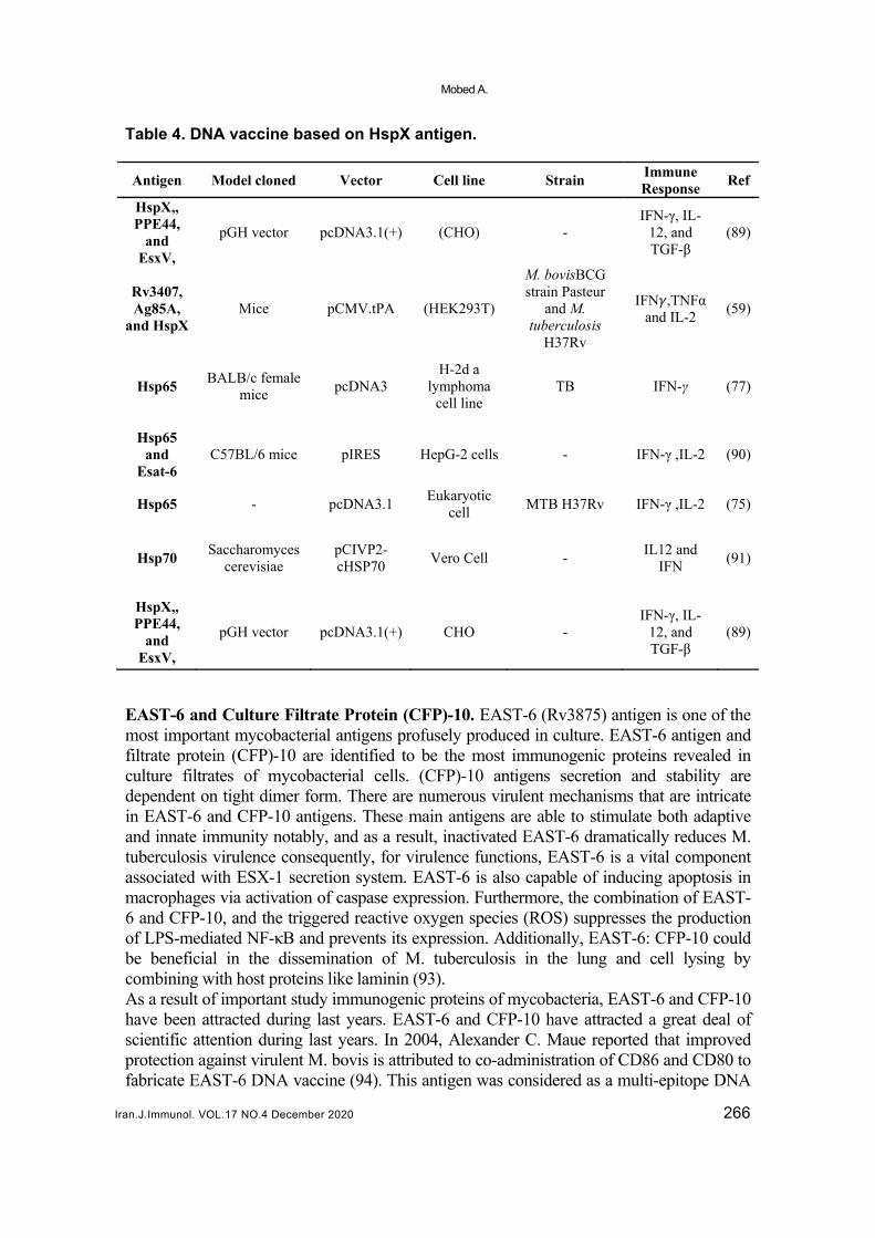
Table 4. DNA vaccine based on HspX antigen.
Antigen Model cloned Vector Cell line Strain Immune Response
Ref
HspX,, PPE44,
and EsxV,
pGH vector pcDNA3.1(+) (CHO) - IFN-γ, IL-
12, and TGF-β
(89)
Rv3407, Ag85A,
and HspX Mice pCMV.tPA (HEK293T)
M. bovisBCG strain Pasteur
and M. tuberculosis
H37Rv
IFN ,TNFα and IL-2
(59)
Hsp65 BALB/c female
mice pcDNA3
H-2d a lymphoma
cell line TB IFN-γ (77)
Hsp65 and
Esat-6 C57BL/6 mice pIRES HepG-2 cells - IFN-γ ,IL-2 (90)
Hsp65 - pcDNA3.1 Eukaryotic
cell MTB H37Rv IFN-γ ,IL-2 (75)
Hsp70 Saccharomyces
cerevisiae pCIVP2-cHSP70
Vero Cell - IL12 and
IFN (91)
HspX,, PPE44,
and EsxV,
pGH vector pcDNA3.1(+) CHO - IFN-γ, IL-
12, and TGF-β
(89)
EAST-6 and Culture Filtrate Protein (CFP)-10. EAST-6 (Rv3875) antigen is one of the most important mycobacterial antigens profusely produced in culture. EAST-6 antigen and filtrate protein (CFP)-10 are identified to be the most immunogenic proteins revealed in culture filtrates of mycobacterial cells. (CFP)-10 antigens secretion and stability are dependent on tight dimer form. There are numerous virulent mechanisms that are intricate in EAST-6 and CFP-10 antigens. These main antigens are able to stimulate both adaptive and innate immunity notably, and as a result, inactivated EAST-6 dramatically reduces M. tuberculosis virulence consequently, for virulence functions, EAST-6 is a vital component associated with ESX-1 secretion system. EAST-6 is also capable of inducing apoptosis in macrophages via activation of caspase expression. Furthermore, the combination of EAST-6 and CFP-10, and the triggered reactive oxygen species (ROS) suppresses the production of LPS-mediated NF-κB and prevents its expression. Additionally, EAST-6: CFP-10 could be beneficial in the dissemination of M. tuberculosis in the lung and cell lysing by combining with host proteins like laminin (93). As a result of important study immunogenic proteins of mycobacteria, EAST-6 and CFP-10 have been attracted during last years. EAST-6 and CFP-10 have attracted a great deal of scientific attention during last years. In 2004, Alexander C. Maue reported that improved protection against virulent M. bovis is attributed to co-administration of CD86 and CD80 to fabricate EAST-6 DNA vaccine (94). This antigen was considered as a multi-epitope DNA

DNA based vaccines against tuberculosis
Iran.J.Immunol. VOL.17 NO.4 December 2020 267
vaccine encoding Hsp70, Ag85A, and EAST-6 by Ruyi et al. in 2008. Their findings illustrated that multi-epitope DNA vaccine induced robust immune responses in mice leading to high levels of IgG, and stimulating IFN-γ secretion. In a similar study, Haifeng Gao fabricated a recombinant plasmid containing EAST-6, CFP-10, Ag85A, and Ag85B genes as a DNA vaccine for mice. Cellular responses occurred once. Cellular responses occurred when immunization with this constructed vaccine (69). In 2013, two DNA vaccines complete fusion of beta-defensin-2, and antigens EAST-6 and Ag85B created. These vaccines were managed to mouse models with BCG and equally in control case. This study improved BCG vaccination in the group vaccinated with a combination of DNA vaccines and BCG (70). Nanoparticle-based DNA vaccine containing EST-6 epitopes in the similar year was established by Ganzhu Feng. This vaccine was administered to mice and led into increased T-cell responses, which demonstrates immunogenic and protective effects of it (95). In a similar study, a DNA vaccine expressing EAST-6 and Hsp65 with a cytokine such as a molecular adjuvant was investigated. Utilizing this DNA vaccine stimulated considerably higher antibody titers, as well as lymphocyte proliferation, IFN-γ and IL-2 levels compared to the groups vaccinated with other recombinant plasmids. On the other hand, in another study in 2014, a recombinant BCG strain expressing a combination protein of EAST-6 and Ag85 was fabricated (76). The immune responses and protective effects of this DNA vaccine were assayed. Due to the improvement of BCG with this DNA vaccine in the lung, higher IFN-γ levels and notably improved secretion of TNF-α and iNOS levels were stimulated; this immune response is associated with better control of bacterial growth (76). A chimeric DNA vaccine bringing EAST-6 and Ag85A genes was made in 2016. Yan Liang et al. cured mice with this DNA vaccine and reported faster mortality due to vaccination. Therefore, contrary to the literature, they stated that individual antigens were promising or at least harmless. Thus, it was determined that EAST-6 is not suitable as a therapeutic vaccine (64). Table 5. DNA vaccine based on EAST-6.
Antigen Model cloned Vector Cell line Strain Immune Response
Ref
Esat-6/3e female C57BL/6
mice pIRES - M.tb H37Rv
IFN-γ -IL-12-IL-4
(76)
ESAT-6 breed cattle VR101
2 COS-7cells
M. bovisstrain 1315
IFNγ (94)
ESAT6 -Ag85B mouse skin - murine
defensin-2 (mBD2)
TB H37Rv Th1 adaptive
response (70)
ESAT-6 and Ag85A
Escherichia coli strains DH5α
and BL21 (DE3) pDE22
C57BL/6 mice
M. tuberculosis H37Rv, M. bovisBCG
China
IFN-γ, IL-10, TNF-α
(62)
Hsp65 and Esat-6
C57BL/6 mice pIRES HepG-2
cells - IFN-γ ,IL-2 (76)
Ag85B ESAT-6, Ag85A, CFP-10
female BALB/c mice
pcDNA3.1
- PPD,BCG IFN-γ, TNF-α, IL-4
(69)
Mobed A.
Iran.J.Immunol. VOL.17 NO.4 December 2020 268
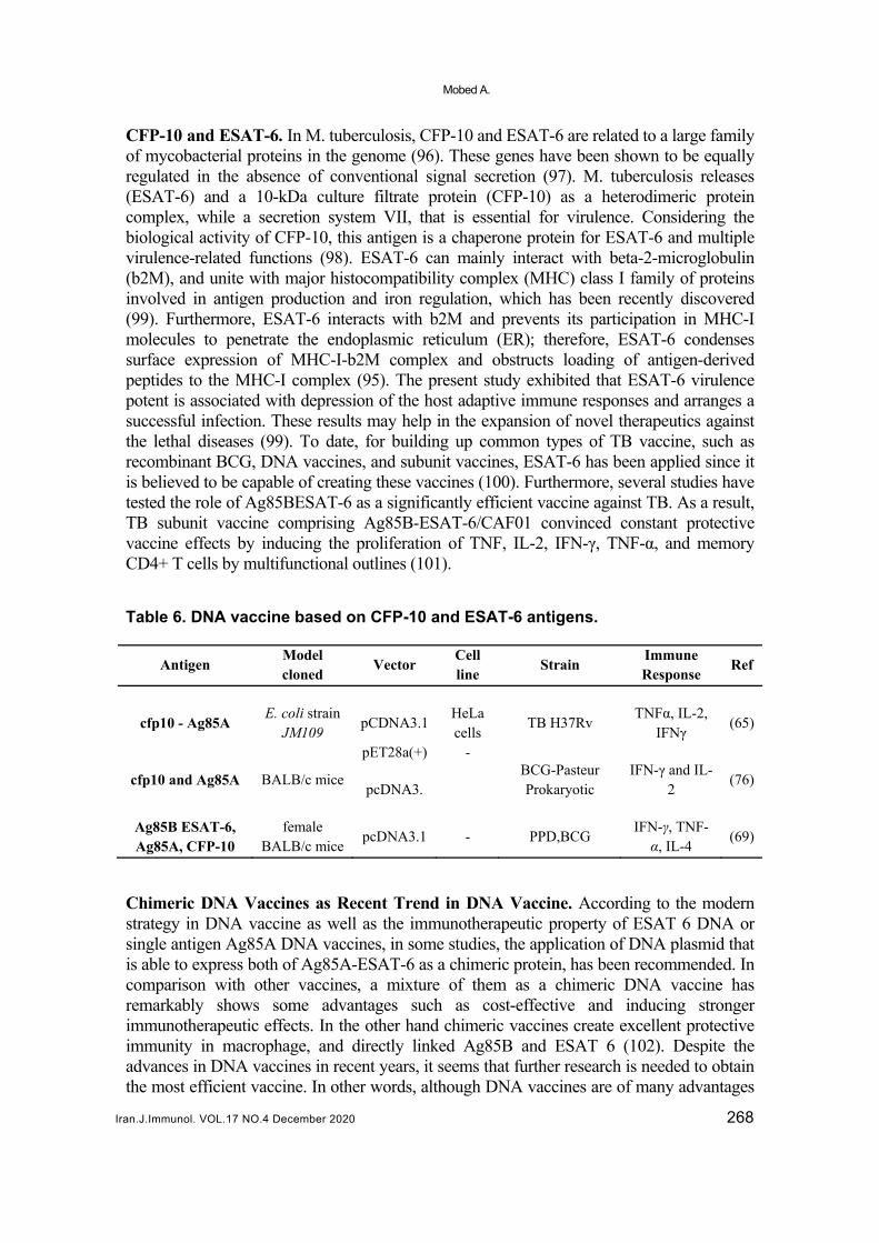
CFP-10 and ESAT-6. In M. tuberculosis, CFP-10 and ESAT-6 are related to a large family of mycobacterial proteins in the genome (96). These genes have been shown to be equally regulated in the absence of conventional signal secretion (97). M. tuberculosis releases (ESAT-6) and a 10-kDa culture filtrate protein (CFP-10) as a heterodimeric protein complex, while a secretion system VII, that is essential for virulence. Considering the biological activity of CFP-10, this antigen is a chaperone protein for ESAT-6 and multiple virulence-related functions (98). ESAT-6 can mainly interact with beta-2-microglobulin (b2M), and unite with major histocompatibility complex (MHC) class I family of proteins involved in antigen production and iron regulation, which has been recently discovered (99). Furthermore, ESAT-6 interacts with b2M and prevents its participation in MHC-I molecules to penetrate the endoplasmic reticulum (ER); therefore, ESAT-6 condenses surface expression of MHC-I-b2M complex and obstructs loading of antigen-derived peptides to the MHC-I complex (95). The present study exhibited that ESAT-6 virulence potent is associated with depression of the host adaptive immune responses and arranges a successful infection. These results may help in the expansion of novel therapeutics against the lethal diseases (99). To date, for building up common types of TB vaccine, such as recombinant BCG, DNA vaccines, and subunit vaccines, ESAT-6 has been applied since it is believed to be capable of creating these vaccines (100). Furthermore, several studies have tested the role of Ag85BESAT-6 as a significantly efficient vaccine against TB. As a result, TB subunit vaccine comprising Ag85B-ESAT-6/CAF01 convinced constant protective vaccine effects by inducing the proliferation of TNF, IL-2, IFN-γ, TNF-α, and memory CD4+ T cells by multifunctional outlines (101). Table 6. DNA vaccine based on CFP-10 and ESAT-6 antigens.
Antigen Model cloned
Vector Cell line
Strain Immune Response
Ref
cfp10 - Ag85A E. coli strain
JM109 pCDNA3.1
HeLa cells
TB H37Rv TNFα, IL-2,
IFNγ (65)
cfp10 and Ag85A BALB/c mice
pET28a(+)
pcDNA3.
-
BCG-Pasteur Prokaryotic
IFN-γ and IL-2
(76)
Ag85B ESAT-6, Ag85A, CFP-10
female BALB/c mice
pcDNA3.1 - PPD,BCG IFN-γ, TNF-α, IL-4
(69)
Chimeric DNA Vaccines as Recent Trend in DNA Vaccine. According to the modern strategy in DNA vaccine as well as the immunotherapeutic property of ESAT 6 DNA or single antigen Ag85A DNA vaccines, in some studies, the application of DNA plasmid that is able to express both of Ag85A-ESAT-6 as a chimeric protein, has been recommended. In comparison with other vaccines, a mixture of them as a chimeric DNA vaccine has remarkably shows some advantages such as cost-effective and inducing stronger immunotherapeutic effects. In the other hand chimeric vaccines create excellent protective immunity in macrophage, and directly linked Ag85B and ESAT 6 (102). Despite the advances in DNA vaccines in recent years, it seems that further research is needed to obtain the most efficient vaccine. In other words, although DNA vaccines are of many advantages

DNA based vaccines against tuberculosis
Iran.J.Immunol. VOL.17 NO.4 December 2020 269
over older vaccines, they have disadvantages and limitations; hence, further comprehensive researches are required to address these limitations. Table 7 represents some of the most important advantages and disadvantages of DNA vaccines. Table 7. Advantages and disadvantages of DNA vaccine.
Advantages Disadvantages Ref
No risk for infection Restricted to protein immune-gens (not valuable for
non-protein based antigens such as bacterial polysaccharides).
(103-107)
Antigen presentation by both MHC class I and class II
molecules Risk of affecting genes controlling cell growth.
Simplicity of expansion and manufacture
Possibility of inducing antibody production against DNA.
Stability for storage and transport
Possibility of tolerance to the antigen (protein) produced.
Long-term persistence of immune-gen
Potential for atypical processing of bacterial and parasite proteins.
In vivo expression guarantees protein more closely resembles normal eukaryotic structure,
with associated post-translational modifications
DISCUSSION In conclusion, the obtained results in this work confirmed that a DNA vaccine immunization considerably increased DNA-mediated protection and had therapeutic effects against TB. DNA vaccines offer a number of advantages over certain other types of vaccines, such as the stimulation of strong immune response to their general manufacturing platform and their relatively low engineering costs. In view of their robust potential for inducing memory responses, DNA vaccines are mainly suitable for preparing immune responses. Moreover, DNA vaccine technology may help antigen detection by facilitating the screening of candidate vaccines. Furthermore, our findings revealed that vaccine delivery is one of the most important challenges in the development of DNA vaccines, and it seems that with the progress in this field, the effectiveness of the vaccine will increase dramatically. In conclusion, we recommend further research regarding the efficiency of DNA vaccines against TB focusing on the exact antigens and improving immune responses and immunogenicity.

Mobed A.
Iran.J.Immunol. VOL.17 NO.4 December 2020 270
ACKNOWLEDGEMENT This review study was supported by Aging Research Institute of Tabriz University of Medical Sciences, Iran. No funding was available. REFERENCES
1. Peeples L. News Feature: Avoiding pitfalls in the pursuit of a COVID-19 vaccine. Proc Natl Acad Sci USA. 2020; 117:8218-21.
2. Jiang F, Deng L, Zhang L, Cai Y, Cheung CW, Xia Z. Review of the clinical characteristics of coronavirus disease 2019 (COVID-19). J Gen Intern Med. 2020; 35:1545-49.
3. Saif LJ. Vaccines for COVID-19: perspectives, prospects, and challenges based on candidate SARS, MERS, and animal coronavirus vaccines. Euro Med J. 2020.
4. Ahmed SF, Quadeer AA, McKay MR. Preliminary identification of potential vaccine targets for the COVID-19 coronavirus (SARS-CoV-2) based on SARS-CoV immunological studies. Viruses. 2020; 12:254.
5. Babawo L, Sellu E, George A, Kaikai D. A Five Year Incidence Trend Analysis of Tuberculosis (TB) and HIV/AIDs Co-Infection at Bo Government Hospital, Southern Sierra Leone. J Public Health Dis Prev. 2020; 3:102.
6. Yu W, Pu-Xuan L, Tan W. Overview of Tuberculosis, Tuberculosis Control in Migrating Population, Singapore, Springer; 2020; 1-10.
7. Sable SB, Posey JE, Scriba TJ. Tuberculosis Vaccine Development: Progress in Clinical Evaluation. Clin. Microbiol. Rev. 2019; 33:e00100-19.
8. Ma K. Weiner DB. DNA vaccines: Ready for prime time. Nat Rev Genet. 2008; 9:776-88. 9. Baliban SM, Michael A, Shammassian B, Mudakha S, Khan AS, Cocklin S, et al. An optimized,
synthetic DNA vaccine encoding the toxin A and toxin B receptor binding domains of Clostridium difficile induces protective antibody responses in vivo. Infect. Immun. 2014; 82:4080-91.
10. Peeridogaheh H, Teimourpour R, Moradi B, Yousefipour M, Gholoobi A, Baghani A, et al. Evaluation of immune responses to a DNA vaccine encoding Ag85a-Cfp10 antigen of Mycobacterium tuberculosis in an animal model. Jundishapur J Microbiol. 2019; 12:e65689.
11. Lim M, Badruddoza AZM, Firdous J, Azad M, Mannan A, Al-Hilal TA, et al. Engineered Nanodelivery Systems to Improve DNA Vaccine Technologies. Pharmaceutics. 2020; 12:30.
12. Lamolinara A, Stramucci L, Hysi A, Iezzi M, Marchini C, Mariotti M, et al. Intradermal DNA electroporation induces cellular and humoral immune response and confers protection against HER2/neu tumor. J Immunol Res. 2015; 2015:159145.
13. Ghaffarifar F, Jorjani O, Sharifi Z, Dalimi A, Hassan ZM, Tabatabaie F, et al. Enhancement of immune response induced by DNA vaccine cocktail expressing complete LACK and TSA genes against Leishmania major. Apmis. 2013; 121:290-8.
14. Lambert PH, Laurent PE. Intradermal vaccine delivery: will new delivery systems transform vaccine administration? Vaccine. 2008; 26:3197-208.
15. Hunter IW, Taberner AJ, Hogan NC. Delivery of a solid body and/or a fluid using a linear Lorentz-force actuated needle-free jet injection system. Google Patents; 2014.
16. Stadelmann B, Kemmerrer S, Campillo Agusti A, Ho E, Lovell N, Masterson S. Electroporation device with detachable needle array with lock-out system. Google Patents; 2019.
17. Tang J, Cai Y, Liang J, Tan Z, Tang X, Zhang C, et al. In vivo electroporation of a codon-optimized BERopt DNA vaccine protects mice from pathogenic Mycobacterium tuberculosis aerosol challenge. Tuberculosis. 2018; 113:65-75.
18. Kwon T-R, Seok J, Jang J-H, Kwon MK, Oh CT, Choi EJ, et al. Needle-free jet injection of hyaluronic acid improves skin remodeling in a mouse model. Eur J Pharm Biopharm. 2016; 105:69-74.
19. Papania MJ, Zehrung D, Jarrahian C. Technologies to Improve Immunization. Plotkin's Vaccines: Elsevier; 2018. p. 1320-53. e17. https://www.ncbi.nlm.nih.gov/pmc/articles/PMC7152424/pdf/main.pdf
20. Kojic N, Goyal P, Lou CH, Corwin MJ. An innovative needle-free injection system: comparison to 1 ml standard subcutaneous injection. AAPS Pharm Sci Tech. 2017; 18:2965-70.

DNA based vaccines against tuberculosis
Iran.J.Immunol. VOL.17 NO.4 December 2020 271
21. van der Maaden K, Jiskoot W, Bouwstra J. Microneedle technologies for (trans) dermal drug and vaccine delivery. J Control Release. 2012; 161:645-55.
22. Kim YC, Park JH, Prausnitz MR. Microneedles for drug and vaccine delivery. Adv Drug Deliv Rev. 2012; 64:1547-68.
23. Donnelly RF, Majithiya R, Singh TRR, Morrow DI, Garland MJ, Demir YK, et al. Design, optimization and characterisation of polymeric microneedle arrays prepared by a novel laser-based micromoulding technique. Pharm Res. 2011; 28:41-57.
24. Sullivan SP, Koutsonanos DG, del Pilar Martin M, Lee JW, Zarnitsyn V, Choi SO, et al. Dissolving polymer microneedle patches for influenza vaccination. Nat Med. 2010; 16:915-20.
25. Hiraishi Y, Nandakumar S, Choi S-O, Lee JW, Kim Y-C, Posey JE, et al. Bacillus Calmette-Guerin vaccination using a microneedle patch. Vaccine. 2011; 29:2626-36.
26. Leone M, Monkare J, Bouwstra J, Kersten G. Dissolving microneedle patches for dermal vaccination. Pharm Res. 2017; 34:2223-40.
27. Sharma M. Transdermal and intravenous nano drug delivery systems: present and future. Applications of Targeted Nano Drugs and Delivery Systems. Elsevier; 2019. p. 499-550.
28. Liang X, Duan J, Li X, Zhu X, Chen Y, Wang X, et al. Improved vaccine-induced immune responses via a ROS-triggered nanoparticle-based antigen delivery system. Nanoscale. 2018; 10:9489-503.
29. Chen F, Yan Q, Yu Y, Wu MX. BCG vaccine powder-laden and dissolvable microneedle arrays for lesion-free vaccination. J Control Release. 2017; 255:36-44.
30. Poirier D, Renaud F, Dewar V, Strodiot L, Wauters F, Janimak J, et al. Hepatitis B surface antigen incorporated in dissolvable microneedle array patch is antigenic and thermostable. Biomaterials. 2017; 145:256-65.
31. Arya JM, Dewitt K, Scott-Garrard M, Chiang Y-W, Prausnitz MR. Rabies vaccination in dogs using a dissolving microneedle patch. J Control Release. 2016; 239:19-26.
32. Du G, Hathout RM, Nasr M, Nejadnik MR, Tu J, Koning RI, et al. Intradermal vaccination with hollow microneedles: a comparative study of various protein antigen and adjuvant encapsulated nanoparticles. J Control Release. 2017; 266:109-18.
33. Rzhevskiy AS, Singh TRR, Donnelly RF, Anissimov YG. Microneedles as the technique of drug delivery enhancement in diverse organs and tissues. J Control Release. 2018; 270:184-202.
34. Yan Q, Liu H, Cheng Z, Xue Y, Cheng Z, Dai X, et al. Immunotherapeutic effect of BCG-polysaccharide nucleic acid powder on Mycobacterium tuberculosis-infected mice using microneedle patches. Drug Deliv. 2017; 24:1648-53.
35. Niu L, Chu LY, Burton SA, Hansen KJ, Panyam J. Intradermal delivery of vaccine nanoparticles using hollow microneedle array generates enhanced and balanced immune response. J Control Release. 2019; 294:268-78.
36. Hegde NR, Kaveri SV, Bayry J. Recent advances in the administration of vaccines for infectious diseases: microneedles as painless delivery devices for mass vaccination. Drug Discov Today. 2011; 16:1061-8.
37. Nishikawa M, Huang L. Nonviral vectors in the new millennium: delivery barriers in gene transfer. Hum Gene Ther. 2001; 12:861-70.
38. Roos AK, Eriksson F, Walters DC, Pisa P, King AD. Optimization of skin electroporation in mice to increase tolerability of DNA vaccine delivery to patients. Mol Ther. 2009; 17:1637-42.
39. Mumper RJ, Ledebur HC. Dendritic cell delivery of plasmid DNA. Applications for controlled genetic immunization. Mol Biotechnol. 2001; 19:79-95.
40. Widera G, Austin M, Rabussay D, Goldbeck C, Barnett SW, Chen M, et al. Increased DNA Vaccine Delivery and Immunogenicity by Electroporation In Vivo. J Immunol. 2000; 164:4635-40.
41. Shah MAA, He N, Li Z, Ali Z, Zhang L. Nanoparticles for DNA vaccine delivery. J Biomed Nanotechnol. 2014; 10:2332-49.
42. Jepson CD, March JB. Bacteriophage lambda is a highly stable DNA vaccine delivery vehicle. Vaccine. 2004; 22:2413-9.
43. Rosada RS, de la Torre LG, Frantz FG, Trombone AP, Zárate-Bladés CR, Fonseca DM, et al. Protection against tuberculosis by a single intranasal administration of DNA-hsp65 vaccine complexed with cationic liposomes. BMC Immunol. 2008; 9:38.
44. Briones M, Singh M, Ugozzoli M, Kazzaz J. The preparation, characterization, and evaluation of cationic microparticles for DNA vaccine delivery. Pharm Res. 2001; 18:709-12.

Mobed A.
Iran.J.Immunol. VOL.17 NO.4 December 2020 272
45. Oh YK, Kim JP, Hwang TS, Ko JJ, Kim JM, Yang JS, et al. Nasal absorption and biodistribution of plasmid DNA: an alternative route of DNA vaccine delivery. Vaccine. 2001; 19:4519-25.
46. Medi BM, Singh J. Skin targeted DNA vaccine delivery using electroporation in rabbits: II. Safety. Int J Pharm. 2006; 308:61-8.
47. Nguyen DN, Raghavan SS, Tashima LM, Lin EC, Fredette SJ, Langer RS, et al. Enhancement of poly(orthoester) microspheres for DNA vaccine delivery by blending with poly(ethylenimine). Biomaterials. 2008; 29:2783-93.
48. Kask AS, Chen X, Marshak JO, Dong L, Saracino M, Chen D, et al. DNA vaccine delivery by densely-packed and short microprojection arrays to skin protects against vaginal HSV-2 challenge. Vaccine. 2010; 28:7483-91.
49. Lu Y, Wu F, Duan W, Mu X, Fang S, Lu N, et al. Engineering a “PEG-g-PEI/DNA nanoparticle-in-PLGA microsphere” hybrid controlled release system to enhance immunogenicity of DNA vaccine. Mater Sci Eng C Mater Biol Appl. 2020; 106:110294.
50. Kaufmann SH, Hussey G, Lambert PH. New vaccines for tuberculosis. Lancet. 2010; 375:2110-9. 51. Bruffaerts N, Huygen K, Romano M. DNA vaccines against tuberculosis. Expert Opin Biol
Ther. 2014; 14:1801-13. 52. Yuk JM, Jo EK. Host immune responses to mycobacterial antigens and their implications for the
development of a vaccine to control tuberculosis. Clin Exp Vaccine Res. 2014; 3:155-67. 53. Sun R, Skeiky YA, Izzo A, Dheenadhayalan V, Imam Z, Penn E, et al. Novel recombinant BCG
expressing perfringolysin O and the over-expression of key immunodominant antigens; pre-clinical characterization, safety and protection against challenge with Mycobacterium tuberculosis. Vaccine. 2009; 27:4412-23.
54. Tanghe A, Denis O, Lambrecht B, Motte V, van den Berg T, Huygen K. Tuberculosis DNA vaccine encoding Ag85A is immunogenic and protective when administered by intramuscular needle injection but not by epidermal gene gun bombardment. Infect Immun. 2000; 68:3854-60.
55. Tanghe A, D'Souza S, Rosseels V, Denis O, Ottenhoff TH, Dalemans W, et al. Improved immunogenicity and protective efficacy of a tuberculosis DNA vaccine encoding Ag85 by protein boosting. Infect Immun. 2001; 69:3041-7.
56. D'souza S, Rosseels V, Denis O, Tanghe A, De Smet N, Jurion F, et al. Improved tuberculosis DNA vaccines by formulation in cationic lipids. Infect Immun. 2002; 70:3681-8.
57. Sugawara I, Yamada H, Udagawa T, Huygen K. Vaccination of guinea pigs with DNA encoding Ag85A by gene gun bombardment. Tuberculosis. 2003; 83:331-7.
58. Draghia-Akli R, Smith LC. Electrokinetic enhancement of plasmid delivery in vivo: Marcel Dekker, Inc., New York; 2003.
59. Mir FA, Kaufmann SH, Eddine AN. A multicistronic DNA vaccine induces significant protection against tuberculosis in mice and offers flexibility in the expressed antigen repertoire. Clin Vaccine Immunol. 2009; 16:1467-75.
60. Gao H, Li K, Yu S, Xiong S. A novel DNA vaccine containing multiple TB‐specific epitopes cast in a natural structure elicits enhanced Th1 immunity compared with BCG. Microbiol Immunol. 2009; 53:541-9.
61. Ahn S, Jeon B, Kim K, Kwack J, Lee E, Park K, et al. Mtb32 is a promising tuberculosis antigen for DNA vaccination in pre-and post-exposure mouse models. Gene Ther. 2012; 19:570-5.
62. Tan K, Tan K, Liang J, Liang J, Teng X, Teng X, et al. Comparison of BCG prime-DNA booster and rBCG regimens for protection against tuberculosis. Hum Vaccin Immunother. 2014; 10:391-8.
63. Meshkat Z, Teimourpour A, Rashidian S, Arzanlou M, Teimourpour R. Immunogenicity of a DNA Vaccine Encoding Ag85a-Tb10. 4 Antigens from Mycobacterium Tuberculosis. Iran J Immunol. 2016; 13:289-95.
64. Liang Y, Bai X, Zhang J, Song J, Yang Y, Yu Q, et al. Ag85A/ESAT-6 chimeric DNA vaccine induces an adverse response in tuberculosis-infected mice. Mol Med Rep. 2016; 14:1146-52.
65. Baghani A, Yousefi M, Safdari H, Teimourpour R, Gholoobi A, Meshkat Z. Designing and construction a DNA vaccine encoding the fusion fragment of cfp10 and Ag85A immunodominant genes of Mycobacterium tuberculosis. Archives of Medical Laboratory Sciences. 2017; 2:135-140
66. Sun L, Yuan Q, Xu T, Yao L, Feng J, Ma J, et al. Novel adjuvant for immunization against tuberculosis: DNA vaccine expressing Mycobacterium tuberculosis antigen 85A and interleukin-15 fusion product elicits strong immune responses in mice. Biotechnol Lett.. 2017; 39:1159-66.

DNA based vaccines against tuberculosis
Iran.J.Immunol. VOL.17 NO.4 December 2020 273
67. Tian X, Cai H, Zhu YX. Protection of mice with a divalent tuberculosis DNA vaccine encoding antigens Ag85B and MPT64. Acta Biochim Biophys Sin (Shanghai). 2004; 36:269-76.
68. Yao W, Liu S, Qu X, Xiao S, Liu Y, Liu J. Enhanced immune response and protection efficacy of a DNA vaccine constructed by linkage of the Mycobacterium tuberculosis Ag85B-encoding gene with the BVP22-encoding gene. J Med Microbiol. 2009; 58:462-8.
69. Gao H, Yue Y, Hu L, Xu W, Xiong S. A novel DNA vaccine containing multiple TB-specific epitopes casted in a natural structure (ECANS) confers protective immunity against pulmonary mycobacterial challenge. Vaccine. 2009; 27:5313-9.
70. Cervantes-Villagrana AR, Hernandez-Pando R, Biragyn A, Castaneda-Delgado J, Bodogai M, Martínez-Fierro M, et al. Prime-boost BCG vaccination with DNA vaccines based in β-defensin-2 and mycobacterial antigens ESAT6 or Ag85B improve protection in a tuberculosis experimental model. Vaccine. 2013; 31:676-84.
71. Rivas-Santiago B, Cervantes-Villagrana AR. Novel approaches to tuberculosis prevention: DNA vaccines. Scand J Infect Dis. 2014; 46:161-8.
72. Meerak J, Wanichwecharungruang SP, Palaga T. Enhancement of immune response to a DNA vaccine against Mycobacterium tuberculosis Ag85B by incorporation of an autophagy inducing system. Vaccine. 2013; 31:784-90.
73. Xu Y, Yang E, Wang J, Li R, Li G, Liu G, et al. Prime–boost bacillus Calmette–Guérin vaccination with lentivirus‐vectored and DNA‐based vaccines expressing antigens Ag85B and Rv3425 improves protective efficacy against Mycobacterium tuberculosis in mice. Immunology. 2014; 143:277-86.
74. Portaro FC, Hayashi MA, De Arauz LJ, Palma MS, Assakura MT, Silva CL, et al. The Mycobacterium leprae hsp65 displays proteolytic activity. Mutagenesis studies indicate that the M. leprae hsp65 proteolytic activity is catalytically related to the HslVU protease. Biochemistry. 2002; 41:7400-6.
75. Changhong S, Hai Z, Limei W, Jiaze A, Li X, Tingfen Z, et al. Therapeutic efficacy of a tuberculosis DNA vaccine encoding heat shock protein 65 of Mycobacterium tuberculosis and the human interleukin 2 fusion gene. Tuberculosis. 2009; 89:54-61.
76. Dong Y, Gong JY, Liu X, Li JW. Enhanced immune response of a bicistronic DNA vaccine expressing fusion antigen Hsp65-Esat-6 of Mycobacterium Tuberculosis with GM-CSF as a molecular adjuvant. Braz Arch. Biol Technol. 2013; 56:757-65.
77. Wang Q, Lei C, Liu Q. Ub combination enhanced cellular immune response elicited by HSP65 DNA vaccine against Mycobacterium tuberculosis. World Journal of Vaccines. 2013; 3:89-97.
78. Hu Y, Coates AR. Transcription of the Stationary-Phase-AssociatedhspX Gene of Mycobacterium tuberculosis Is Inversely Related to Synthesis of the 16-Kilodalton Protein. J Bacteriol. 1999; 181:1380-7.
79. Kennaway CK, Benesch JL, Gohlke U, Wang L, Robinson CV, Orlova EV, et al. Dodecameric structure of the small heat shock protein Acr1 from Mycobacterium tuberculosis. J Biol Chem. 2005; 280:33419-25.
80. Pang X, Howard ST. Regulation of the α-crystallin gene acr2 by the MprAB two-component system of Mycobacterium tuberculosis. J Bacteriol. 2007; 189:6213-21.
81. Khalid R, Afzal M, Khurshid S, Paracha RZ, Khan IH, Akhtar MW. Fusion molecules of heat shock protein HSPX with other antigens of Mycobacterium tuberculosis show high potential in serodiagnosis of tuberculosis. PloS One. 2016; 11:e0163349.
82. Shi C, Chen L, Chen Z, Zhang Y, Zhou Z, Lu J, et al. Enhanced protection against tuberculosis by vaccination with recombinant BCG over-expressing HspX protein. Vaccine. 2010; 28:5237-44.
83. Fallow A. Beijing lineage strains of Mycobacterium tuberculosis are natural mutants of the DosT sensor kinase. McGill University; 2010.
84. Aagaard CS, Hoang TTKT, Vingsbo Lundberg C, Dietrich J, Andersen P. Quality and vaccine efficacy of CD4+ T cell responses directed to dominant and subdominant epitopes in ESAT-6 from Mycobacterium tuberculosis. J Immunol. 2009; 183:2659-68.
85. Kim JS, Kim WS, Choi HG, Jang B, Lee K, Park JH, et al. Mycobacterium tuberculosis RpfB drives Th1-type T cell immunity via a TLR4-dependent activation of dendritic cells. J Leukoc Biol. 2013; 94:733-49.
86. Even-Or O, Samira S, Ellis R, Kedar E, Barenholz Y. Adjuvanted influenza vaccines. Expert Rev Vaccines. 2013; 12:1095-108.

Mobed A.
Iran.J.Immunol. VOL.17 NO.4 December 2020 274
87. Jung ID, Shin SJ, Lee MG, Kang TH, Han HD, Lee SJ, et al. Enhancement of Tumor-Specific T Cell–Mediated Immunity in Dendritic Cell–Based Vaccines by Mycobacterium tuberculosis Heat Shock Protein X. J Immunol. 2014; 193:1233-45.
88. Zheng YQ, Naguib YW, Dong Y, Shi YC, Bou S, Cui Z. Applications of bacillus Calmette–Guerin and recombinant bacillus Calmette–Guerin in vaccine development and tumor immunotherapy. Expert Rev Vaccines. 2015;14:1255-75.
89. Moradi B, Sankian M, Amini Y, Meshkat Z. Construction of a Novel DNA Vaccine Candidate Encoding an HspX-PPE44-EsxV Fusion Antigen of Mycobacterium tuberculosis. Rep Biochem Mol Biol. 2016; 4:89-97.
90. Chong P, Hsieh SY, Liu CC, Chou AH, Chang JY, Wu SC, et al. Production of EV71 vaccine candidates. Hum Vaccin Immunother. 2012; 8:1775-83.
91. Maity HK, Dey S, Mohan CM, Khulape SA, Pathak DC, Vakharia VN. Protective efficacy of a DNA vaccine construct encoding the VP2 gene of infectious bursal disease and a truncated HSP70 of Mycobacterium tuberculosis in chickens. Vaccine. 2015; 33:1033-9.
92. Feng Y, Yang X, Liu Z, Liu Y, Su B, Ding Y, et al. Continuous treatment with recombinant Mycobacterium tuberculosis CFP-10-ESAT-6 protein activated human monocyte while deactivated LPS-stimulated macrophage. Biochem Biophys Res Commun. 2008; 365:534-40.
93. Yang H, Chen H, Liu Z, Ma H, Qin L, Jin R, et al. A novel B-cell epitope identified within Mycobacterium tuberculosis CFP10/ESAT-6 protein. PloS One. 2013; 8:e52848.
94. Maue AC, Waters WR, Palmer MV, Whipple DL, Minion FC, Brown WC, et al. CD80 and CD86, but not CD154, augment DNA vaccine-induced protection in experimental bovine tuberculosis. Vaccine. 2004; 23:769-79.
95. Feng G, Jiang Q, Xia M, Lu Y, Qiu W, Zhao D, et al. Enhanced immune response and protective effects of nano-chitosan-based DNA vaccine encoding T cell epitopes of Esat-6 and FL against Mycobacterium tuberculosis infection. PLoS One. 2013; 8:e61135.
96. Rashidian S, Teimourpour R, Meshkat Z. Designing and construction of a DNA vaccine encoding tb10. 4 gene of Mycobacterium tuberculosis. Iran J Pathol. 2016; 11:112-9.
97. Daleke MH, Ummels R, Bawono P, Heringa J, Vandenbroucke-Grauls CM, Luirink J, et al. General secretion signal for the mycobacterial type VII secretion pathway. Proc Natl Acad Sci USA. 2012; 109:11342-7.
98. Guinn KM, Hickey MJ, Mathur SK, Zakel KL, Grotzke JE, Lewinsohn DM, et al. Individual RD1‐region genes are required for export of ESAT‐6/CFP‐10 and for virulence of Mycobacterium tuberculosis. Mol Microbiol. 2004; 51:359-70.
99. Sreejit G, Ahmed A, Parveen N, Jha V, Valluri VL, Ghosh S, et al. The ESAT-6 protein of Mycobacterium tuberculosis interacts with beta-2-microglobulin (β2M) affecting antigen presentation function of macrophage. PLoS Pathog. 2014; 10:e1004446.
100. van Zyl Smit R, Pai M, Yew W, Leung C, Zumla A, Bateman E, et al. Global lung health: the colliding epidemics of tuberculosis, tobacco smoking, HIV and COPD. Eur Respir J. 2010; 35:27-33.
101. Orme IM. Preclinical testing of new vaccines for tuberculosis: a comprehensive review. Vaccine. 2006; 24:2-19.
102. Huygen K. On the use of DNA vaccines for the prophylaxis of mycobacterial diseases. Infect Immun. 2003; 71:1613-21.
103. Robinson HL, Pertmer TM. DNA vaccines for viral infections: basic studies and applications. Adv Virus Res. 2000; 55:1-74.
104. Soltani S, Farahani A, Dastranj M, Momenifar N, Mohajeri P, Emamie A. DNA vaccine: Methods and mechanisms. Adv Hum Biol. 2018; 8:132-9.
105. Kofta W, Wedrychowicz H. c-DNA vaccination against parasitic infections: advantages and disadvantages. Vet Parasitol. 2001; 100:3-12.
106. Hasson SSAA, Al-Busaidi JKZ, Sallam TA. The past, current and future trends in DNA vaccine immunisations. Asian Pac J Trop Biomed. 2015; 5:344-53.
107. Stachyra A, Góra-Sochacka A, Sirko A. DNA vaccines against influenza. Acta Biochim Pol. 2014; 61: 515-22.