dmd 40:856–864, 2012 the impact of single...
TRANSCRIPT

The Impact of Single Nucleotide Polymorphisms on HumanAldehyde Oxidase□S
Tobias Hartmann, Mineko Terao, Enrico Garattini, Christian Teutloff, Joshua F. Alfaro,Jeffrey P. Jones, and Silke Leimkuhler
Department of Molecular Enzymology, Institute of Biochemistry and Biology, University of Potsdam, Potsdam, Germany (T.H.,S.L.); Laboratory of Molecular Biology, Department of Biochemistry and Molecular Pharmacology, Istituto di Ricerche
Farmacologiche Mario Negri, Milano, Italy (M.T., E.G.); Institute for Experimental Physics, Free University of Berlin, Berlin,Germany (C.T.); and Department of Chemistry, Washington State University, Pullman, Washington (J.F.A., J.P.J.)
Received November 10, 2011; accepted January 25, 2012
ABSTRACT:
Aldehyde oxidase (AO) is a complex molybdo-flavoprotein thatbelongs to the xanthine oxidase family. AO is active as a ho-modimer, and each 150-kDa monomer binds two distinct [2Fe2S]clusters, FAD, and the molybdenum cofactor. AO has an importantrole in the metabolism of drugs based on its broad substratespecificity oxidizing aromatic aza-heterocycles, for example, N1-methylnicotinamide and N-methylphthalazinium, or aldehydes,such as benzaldehyde, retinal, and vanillin. Sequencing the 35coding exons of the human AOX1 gene in a sample of 180 Italianindividuals led to the identification of relatively frequent, synony-mous, missense and nonsense single-nucleotide polymorphisms(SNPs). Human aldehyde oxidase (hAOX1) was purified after het-erologous expression in Escherichia coli. The recombinant protein
was obtained with a purity of 95% and a yield of 50 �g/l E. coliculture. Site-directed mutagenesis of the hAOX1 cDNA allowed thepurification of protein variants bearing the amino acid changesR802C, R921H, N1135S, and H1297R, which correspond to some ofthe identified SNPs. The hAOX1 variants were purified and com-pared with the wild-type protein relative to activity, oligomerizationstate, and metal content. Our data show that the mutation of eachamino acid residue has a variable impact on the ability of hAOX1 tometabolize selected substrates. Thus, the human population ischaracterized by the presence of functionally inactive hAOX1 al-lelic variants as well as variants encoding enzymes with differentcatalytic activities. Our results indicate that the presence of theseallelic variants should be considered for the design of future drugs.
Introduction
Aldehyde oxidase (AO) (EC1.2.3.1) is a molybdo-flavoenzymepresent in the cytosolic compartment of many tissues in variousanimal species, including humans (Garattini et al., 2008, 2009). AO isa member of the xanthine oxidase (XO) family, which consists ofcomplex metalloflavoproteins containing two [2Fe2S] clusters, FAD,and the molybdenum cofactor (Moco) as the catalytically active units(Hille, 1996). The AO holoenzyme is a homodimer, and each 150-kDamonomer is characterized by three separate domains: the 20-kDaN-terminal domain binds the two distinct [2Fe2S] clusters, FeSI andFeSII, the 40-kDa central domain binds FAD, and the 80-kDa C-ter-minal domain binds Moco (Garattini et al., 2003). Members of the XO
family of molybdoenzymes are characterized by an equatorial sulfurligand at the Moco that is essential for enzyme activity (Edmondsonet al., 1972; Wahl and Rajagopalan, 1982).
AO is found in most animal species, including fish and insects(Garattini et al., 2008). In humans, AO is encoded by a singlefunctional gene, hAOX1. Although hAOX1 orthologs are found inalmost all mammalian organisms, the number of functional AOXgenes varies according to the species considered. Rodents contain thelargest number of AOX functional genes: Aox1, Aox3, Aox4, andAox3l1 (Garattini et al., 2009). These genes arose from a series ofgene duplication events from a common ancestor and are clustered ona short region of mouse chromosome 1 and rat chromosome 9. All ofthe products of the mammalian AOX genes have high amino acidsequence similarity and are expressed in a tissue-specific manner indifferent organisms (Garattini et al., 2003; Terao et al., 2006). It isbelieved that the various AOX isoforms recognize distinct substratesand carry out different physiological tasks. The tissue distribution ofmouse AOX3 (mAOX3) is superimposable with that of mAOX1, andthe two enzymes are synthesized predominantly in liver, lung, andtestis (Vila et al., 2004). The expression of mAOX4 is limited to theHarderian gland, esophagus and skin, whereas mAOX3L1 expressionis restricted to the nasal mucosa (Terao et al., 2000). Except for
This work was supported by the Cluster of Excellence “Unifying Concepts inCatalysis” (to C.T. and S.L.) coordinated by the Technische Universitat Berlin andfunded by the Deutsche Forschungsgemeinschaft and by grants from the Asso-ciazione Italiana per la Ricerca contro il Cancro and the Fondazione Italo Monzino(to E.G.).
Article, publication date, and citation information can be found athttp://dmd.aspetjournals.org.
http://dx.doi.org/10.1124/dmd.111.043828.□S The online version of this article (available at http://dmd.aspetjournals.org)
contains supplemental material.
ABBREVIATIONS: AO, aldehyde oxidase; AOX1, aldehyde oxidase 1; FM, fast metabolizer; MCSF, Moco sulfurase; Moco, molybdenum cofactor;PAGE, polyacrylamide gel electrophoresis; PCR, polymerase chain reaction; PM, poor metabolizer; SNP, single nucleotide polymorphism; XDH,xanthine dehydrogenase; XO, xanthine oxidase; Vis, visible; MPT, molybdopterin.
1521-009X/12/4005-856–864$25.00DRUG METABOLISM AND DISPOSITION Vol. 40, No. 5Copyright © 2012 by The American Society for Pharmacology and Experimental Therapeutics 43828/3760655DMD 40:856–864, 2012
856
http://dmd.aspetjournals.org/content/suppl/2016/01/27/dmd.111.043828.DC2http://dmd.aspetjournals.org/content/suppl/2012/01/25/dmd.111.043828.DC1
Supplemental material to this article can be found at: at A
SPET
Journals on August 25, 2018
dmd.aspetjournals.org
Dow
nloaded from
mAOX4, which metabolizes retinaldehyde into retinoic acid and playsa role in skin homeostasis (Terao et al., 2009), very little is knownabout specific substrates and the physiological role of mAOX1 or anyof the other AOX homologs.
In spite of this lack of information on the physiological significanceof the AO enzymes, hAOX1 long has been recognized as a prominentdrug-metabolizing enzyme (Obach et al., 2004; Pryde et al., 2010;Garattini and Terao, 2011). AOX1 is characterized by broad substratespecificity, catalyzing the oxidation of a wide range of endogenousand exogenous aldehydes as well as N-heterocyclic aromatic com-pounds (Kitamura et al., 2006). In addition, AOX1 catalyzes thereduction of a variety of functional groups, including sulfoxides,N-oxides, azo dyes, and N-hydroxycarbamoyl substituents in thepresence of an appropriate donor. N-Heterocyclic drugs, such asmethotrexate, 6-mercaptopurine, cinchona alkaloids, and famciclovir,also are oxidized by this enzyme (Obach et al., 2004; Kitamura et al.,2006). Finally, AOX1 is involved in the oxidation of intermediarydrug metabolites, such as the conversion of cyclic iminium ionsarising from cytochrome P450-catalyzed oxidation of pyrrolidines andpiperidines into lactams or the oxidation of aldehydes derived fromalcohol-containing drugs (Beedham, 1997).
Marked species differences have been well documented for thealdehyde-catalyzed metabolism of drugs, including methotrexate andfamciclovir (Rashidi et al., 1997; Jordan et al., 1999; Kitamura et al.,1999a,b). Interindividual variability in hAOX1 in vitro activity hasbeen reported in humans (Kitamura et al., 1999b; Al-Salmy, 2001),although the underlying determinants of these variations have neverbeen investigated (Beedham et al., 2003). Gender may be one suchdeterminant. In fact, male mice exhibit a 2- to 4-fold higher AOactivity than female mice (Beedham, 1985; Kurosaki et al., 1999;Al-Salmy, 2001). It also has been suggested that factors such as age,cigarette smoking, drug usage, and disease states, such as cancer, alsomay account for the interindividual variability of hAOX1 activity(Pryde et al., 2010). One final and prominent source of interindividualvariation is represented by missense single-nucleotide polymorphisms(SNPs) affecting the catalytic activity of the hAOX1 enzyme. Numer-ous SNPs of the hAOX1 gene are available in the National Center forBiotechnology Information dbSNP database, although for the majorityof the data on frequency in the human population are not available. Inaddition, no single human SNP has been characterized for its effect onthe catalytic activity of the purified enzyme. The only availablefunctional studies on AOX1 SNPs were reported in Donryu rats(Adachi et al., 2007; Itoh et al., 2007a,b,c). Functional characteriza-tion of the identified SNPs permitted the classification of these ratsinto ultrarapid metabolizers, extensive metabolizers, and poor me-tabolizers according to the AOX1 mutation considered.
Interindividual differences in hAOX1 activity are of primaryimportance for the clinical use of drugs known to be metabolizedby the enzyme. They also are very important factors to be consid-ered in the development of new drugs. For these reasons, wedecided to focus on this aspect of hAOX1 biology. In this article,we report on the identification of relatively frequent hAOX1 SNPsand define their frequency in the Italian population. The mostfrequent missense SNPs were selected to produce the correspond-ing recombinant hAOX1 variant proteins, using an efficient Esch-erichia coli expression system. The enzymatic and kinetic charac-teristics of the canonical and variant hAOX1 proteins werecompared. Our data provide evidence for the existence of frequenthAOX1 allelic variants defining fast and poor metabolizers in thehuman population.
Materials and Methods
Identification of SNPs. Blood samples (3 ml) were collected from 180 (67males and 113 females) volunteers after signing written consent forms. Thirtyof these individuals were healthy volunteers, whereas the remaining 150 wereindividuals recruited for an unrelated epidemiological study. All of the sampleswere treated anonymously according to the guidelines of the internal EthicalCommittee of the Istituto Mario Negri. The genomic DNA was extracted fromblood samples by a semiautomated vacuum-based nucleic acids extractor(AB6100; Applied Biosystems, Foster City, CA). Oligonucleotides were syn-thesized by Invitrogen (Carlsbad, CA). The genomic DNA fragments contain-ing each of the exon sequences were amplified using the oligonucleotideslisted in Supplemental Table 1, using the Taq DNA polymerase kit (AppliedBiosystems). The amplified polymerase chain reaction (PCR) products (30–60ng) were sequenced in 96-well plates by Primm, S.r.l. (Milan, Italy).
Cloning, Expression, and Purification of hAOX1 and Its Variants.Cloning of hAOX1 cDNA into pQE-30 Xa vector and PCR mutagenesis of thevariants R921H and N1135S were done after Alfaro et al. (2009). For site-directed mutagenesis of hAOX1 R802C and H1297R variants, the expressionvector pQE-30 Xa including hAOX1 cDNA was used as a template, resultingin plasmids pTHAO1 and pTHAO2. All of the constructs used express hAOX1as an N-terminal fusion protein with a His6 tag.
For expression in E. coli, the constructs were transformed into TP1000(�mobAB) cells (Palmer et al., 1996). E. coli were grown at 30°C in Luria-Bertanimedium supplemented with 150 �g/ml ampicillin, 1 mM molybdate, and 20 �Misopropyl �-D-thiogalactoside. Cells were harvested by centrifugation after 24 h ofgrowth and resuspended in 50 mM sodium phosphate buffer (pH 8.0) containing300 mM NaCl and frozen at �20°C until purification.
Cells were lysed twice in a cell disruptor system at 1.35 kbar (ConstantSystems, Northhampton, UK). Cell fragments were removed by centrifugation,and the supernatant was mixed with 4 ml of nickel-nitrilotriacetic acid resin(QIAGEN GmbH, Hilden, Germany) per 14 liters of cell growth and incubatedfor 20 min at low stirring speed. The mixture was transferred to a column andwashed with 10 column volumes of both 50 mM sodium phosphate and 300mM NaCl (pH 8.0) containing 10 mM imidazole and the same buffer contain-ing 20 mM imidazole. Proteins were eluted from the resin with 50 mM sodiumphosphate and 300 mM NaCl (pH 8.0) containing 250 mM imidazole. Thebuffer of the eluted proteins was changed to 50 mM Tris (pH 7.5) by usingPD-10 columns (GE Healthcare, Chalfont St. Giles, Buckinghamshire, UK).For the stabilization of the protein, 2.5 mM dithiothreitol was added to thebuffer. For further purification, hAOX1 was loaded on a Mono Q 5/50 GLcolumn (GE Healthcare) equilibrated with 50 mM Tris (pH 7.5) containing 1mM EDTA and eluted with a linear gradient of the same buffer containing 1M NaCl. Fractions were analyzed by SDS-polyacrylamide gel electrophoresis(PAGE), and the ones containing hAOX1 were combined. hAOX1 was puri-fied further on a Superdex 200 column (GE Healthcare), equilibrated in 50 mMTris-HCl (pH 7.5) containing 200 mM NaCl and 1 mM EDTA. Only thefractions containing the dimeric form of hAOX1 were combined and used forthe kinetic studies.
SDS-PAGE. SDS-PAGE was performed using 12% polyacrylamide gelsdescribed by Laemmli (1970). Staining of the proteins was done using Coo-massie Blue R.
Enzyme Assays. Steady-state enzyme kinetics were performed with puri-fied hAOX1in 50 mM Tris buffer (pH 7.5) containing 200 mM NaCl and 1mM EDTA at 25°C in a final volume of 500 �l. The substrates benzaldehyde,phthalazine, and chloroquinazolinone were used in a range from 0.1 to 100 �Musing dichlorophenolindophenol (100 �M) as an electron acceptor. Forphenanthridine, molecular oxygen was used as an electron acceptor, and theproduct phenanthridone was detected at 450 nm. Total enzyme concentrationvaried between 200 and 600 nM. Reactions were monitored over a range of60 s. Activities were calculated using the extinction coefficients of 16,100 M�1
cm�1 at 600 nm for dichlorophenolindophenol and 4775 M�1 cm�1 at 450 nmfor phenanthridone. Data obtained from three individual measurements werefitted nonlinearly using the Michaelis-Menten equation to obtain the kineticconstants KM and turnover numbers.
Metal and Molybdopterin Analysis. The molybdenum and iron content ofpurified hAOX1 was determined by inductively coupled plasma optical emis-sion spectroscopy on an Optima 2100 DV (PerkinElmer Life and Analytical
857CHARACTERIZATION OF SINGLE NUCLEOTIDE POLYMORPHISMS OF hAOX1
at ASPE
T Journals on A
ugust 25, 2018dm
d.aspetjournals.orgD
ownloaded from

Sciences, Waltham, MA). Five-hundred-microliter protein samples with a finalconcentration of 5 to 10 �M were wet-ashed in the same volume of 65% nitricacid by incubation at 100°C for 24 h. The samples were diluted with 4 ml ofwater. The multielement standard solution XVI (Merck, Darmstadt, Germany)was used as a reference. The resulting mass concentrations were calculated asa percentage of protein saturation of both molybdenum and two [2Fe2S] inrelation to a theoretical 100% saturation. Molybdopterin (MPT) was convertedto its fluorescent derivative Form A by adjusting the pH of the supernatant to2.5 with HCl and heating at 95°C for 30 min in the presence of iodine (Johnsonet al., 1984). Excess iodine was removed by the addition of 55 �l of 1% w/vascorbic acid, and the sample was adjusted with 1 M Tris to pH 8.3. Form Awas obtained from phospho-Form A by the addition of 40 mM MgCl2 and 1unit of calf intestine alkaline phosphatase. Form A was isolated with 10 mMacetic acid on a QAE ion exchange column (Sigma-Aldrich, St. Louis, MO),which was equilibrated in H2O. Form A was identified and quantified byhigh-performance liquid chromatography analysis with a C18 reversed-phasehigh-performance liquid chromatography column [4.6 � 250 mm; HypersilODS (Thermo Fisher Scientific, Waltham, MA); particle size 5 �m] with 5mM ammonium acetate and 15% v/v methanol at an isocratic flow rate of 1ml/min. In-line fluorescence was monitored by an 1100 series detector (AgilentTechnologies, Santa Clara, CA) with excitation at 383 nm and emission at 450 nm.
EPR Spectroscopy. Continuous-wave EPR spectra at 9.4 GHz X-bandwere recorded on a home-built spectrometer (microwave bridge,ER041MR, Bruker, Newark, DE; lock-in amplifier, SR810, Stanford Re-search, Stanford, CA; microwave counter, 53181A, Agilent Technologies)equipped with a Bruker SHQ resonator. An ESR 910 helium flow cryostatwith an ITC503 temperature controller (Oxford, Oxfordshire, UK) wereused for temperature control. The magnetic field was calibrated using aLi/LiF standard with a known g value of 2.002293 � 0.000002. hAOX1samples with concentrations of 50 �M were reduced with a 20-fold excessof dithionite anaerobically on an argon flow prepared in quartz tubes witha 4-mm outer diameter. Reduction was achieved by adding a small volumeof an anaerobic dithionite solution to generate reduced FeII/FeIII in the FeSclusters. Sample tubes were frozen rapidly in deep-cold ethanol after acolor change confirmed successful reduction.
Results
SNPs of the hAOX1 Gene. The hAOX1 gene is characterized bya very complex structure consisting of 35 coding exons (Garattini etal., 2008). To identify relatively frequent SNPs in the coding region ofthe gene, we amplified by PCR and sequenced each hAOX1 exonfrom genomic DNA belonging to a cohort of 180 volunteers repre-sentative of the Italian population. The couple of exon-specific oli-gonucleotides used for the amplification and corresponding to thesequences upstream and downstream of each exon in intronic regionsare shown in Supplemental Table 1. The presence of bona fide SNPswas verified by sequencing in both directions. In this population, weidentified one nonsense mutation, five nonsynonymous SNPs, and onesynonymous SNP (Table 1). The nonsense mutation was located inexon 5 and is predicted to result in a very short, nonfunctional proteinof 126 amino acids. This mutation was relatively frequent and causedhaploinsufficiency in eight individuals. The most frequent missensemutation was an A/G transition resulting in the substitution H/R at
position 1297. The SNP was located in exon 34, and it was observedin 13 individuals, six of which were homozygous for the trait. Exon 34appeared to be a mutational hotspot, because another relatively fre-quent SNP (C/T) resulting in the substitution S/L at position 1271 anda synonymous SNP were located in this exon. All of the individualscarrying the S1271L SNP were heterozygous for the trait. A relativelyfrequent missense SNP corresponding to an A/G transition, causingthe substitution N/S at position 1135, was identified in exon 30. Twofurther and rare nonsynonymous SNPs were identified in exons 22and 25. They were the result of C/T (R802C) and G/A (R921H)transitions, respectively.
Expression and Purification of hAOX1. A prerequisite for thefunctional characterization of the identified SNPs was the optimiza-tion of the system used for the purification of a catalytically activehAOX1 protein (Alfaro et al., 2009). For this purpose, recombinanthAOX1 was expressed as an N-terminal His6 tag fusion protein in E.coli TP1000 using the protocol described for mAOX1 (Schumann etal., 2009) with slight modifications. The protein was purified usingnickel-nitrilotriacetic acid, anion exchange, and size-exclusion chro-matography. We isolated 95% pure protein (Fig. 1) with a yield of 50�g/l cell growth. After size-exclusion chromatography, two majorpeaks were detected corresponding to calculated molecular masses of300 and 150 kDa, which represented the dimeric and monomericforms of hAOX1 (Fig. 1). The ratio between dimeric and monomericforms was calculated to be 1.5:1. The formation of protein aggregatesalso was observed; however, aggregation was prevented by the addi-tion of 2.5 mM dithiothreitol during the purification. Analysis of thepurified hAOX1 protein by 12% SDS-PAGE revealed a dominantprotein band corresponding to the calculated molecular mass of 150kDa for hAOX1 (Fig. 1). Three other less intense bands of lowermolecular masses were visible by SDS-PAGE. The three bands wereanalyzed by matrix-assisted laser desorption ionization peptide map-ping and identified to be the degradation products of hAOX1. Similarbands also were observed in purified mAOX3 (Mahro et al., 2011)and AOX1 protein preparations of mouse and rat origin (Kundu et al.,2007; Schumann et al., 2009). These products were never detectableupon native PAGE (Kundu et al., 2007; Mahro et al., 2011) and fastprotein liquid chromatography. All of this suggested that the observeddegradation products are generated by the reductive conditions intrin-sic to SDS-PAGE.
The UV-visible (UV/Vis) spectra of hAOX1 in its oxidized form dis-played the typical features of molybdo-flavoenzymes (Fig. 1). The 280:450nm absorbance ratio of 5.0 for the recombinant protein indicated the highpurity of the enzyme. The 450:550 nm absorbance ratio in the UV/Visspectrum was calculated to be 3.0 for the recombinant enzyme, demonstrat-ing full saturation with FAD. In addition, the FAD content of hAOX1 wasdetermined to be 100% by quantification of the AMP content of the protein.
Protein expression over an extended time at low temperature allowsE. coli cells to produce sufficient amounts of Mo-MPT for almost
TABLE 1
Nonsense, missense, and synonymous polymorphisms of the hAOX1 gene in the Italian population
The SNP data were deposited in the National Center for Biotechnology Information database under the accession numbers indicated in the last column.
Exon Nucleotide Amino Acid Allelic Frequency Heterozygotes Homozygotes RefSNP(rs)
5 C/G Y126stop 0.026 8/153 0/153 ss46933348122 C/T R802C 0.006 2/180 0/180 rs4130976825 G/A R921H 0.003 1/180 0/180 rs5619963530 A/G N1135S 0.029 1/154 4/154 rs5575465534 C/T S1271L 0.039 14/178 0/178 rs14178603034 A/G H1297R 0.053 7/178 6/178 rs373172234 C/T L1268silent 0.022 8/178 0/178 ss469333482
858 HARTMANN ET AL.
at ASPE
T Journals on A
ugust 25, 2018dm
d.aspetjournals.orgD
ownloaded from

complete saturation of the enzyme with this cofactor (Leimkuhler etal., 2004). The main problem of the recombinant system is poorinsertion of the terminal sulfido ligand that is essential for enzymeactivity. We determined 60% saturation of recombinant hAOX1 withmolybdenum and 70% saturation of the 2�[2Fe2S] clusters with iron(Fig. 2). The amount of active enzyme containing the Mo�S ligandwas calculated from the reduction spectra. For this purpose, wecompared the ratio of oxidized to benzaldehyde-reduced hAOX1 withthe amount of the enzyme reduced in relation to the fully reducedenzyme with dithionite under anaerobic conditions. This methodshowed a 30% saturation ratio of sulfurated Mo-MPT in hAOX1.Thus, only 50% of Mo-MPT contained the terminal sulfur ligand. Toincrease the catalytically active fraction of hAOX1, we coexpressedhAOX1 and human Moco sulfurase (hMCSF) cDNAs (Ichida et al.,2001). However, an analysis of purified hAOX1 showed no increasein the levels of sulfurated Mo-MPT. A band corresponding to hMCSFwas not detected in cell lysates after separation by SDS-PAGE,suggesting that the majority of hMCSF was expressed in inclusionbodies in an inactive form. Thus, in all of the subsequent purificationexperiments, we decided to express only hAOX1 cDNA in TP1000cells.
EPR Spectroscopy of the hAOX1 [2Fe2S] Clusters. The two[2Fe2S] clusters were characterized by EPR spectroscopy. Figure 3shows the EPR spectra of recombinant and dithionite-reduced wild-type hAOX1 along with the corresponding simulations. The spectradisplay signals from several superimposed paramagnetic species,which are observed usually in enzymes belonging to the XO family.However, the most prominent signals are the characteristic EPRsignals assigned to the two iron-sulfur centers FeSI and FeSII, whichare similar for all of the members of the XO family that have beendescribed to date (Hille, 1996; Parschat et al., 2001). FeSI has EPRproperties showing a slightly rhombic g tensor, similar to those ofmany other [2Fe2S] proteins, being fully developed at relatively hightemperatures (up to T � 80 K), whereas FeSII has unusual EPRproperties for [2Fe2S] species with a more rhombic g tensor, showinglines with varying line widths that can only be observed clearly at
much lower temperatures (T � 30 K). The g values and line widthswere evaluated by simulating the spectra using the EasySpin toolboxfor MATLAB (Stoll and Schweiger, 2006). In both proteins, FeSI hassimilar EPR properties, including the typical, rhombic g tensor asobserved for other [2Fe2S] clusters of the ferredoxin type (Table 2).Under the experimental conditions used to reduce the proteins, noclear signals of the reduced flavin semiquinone (FAD) or Moco(MoV) were observed. The line widths and positions of the FeS signalsat lower temperatures were affected by magnetic interactions. The gtensors obtained and line widths for FeSI and FeSII of hAOX1 arealmost identical to the values published previously for mAOX1(Schumann et al., 2009) or mAOX3, indicating high structural simi-larity between the proteins as expected from amino acid sequenceidentities of 83% and 61%, respectively.
Purification and Characterization of hAOX1 PolymorphicVariants. We studied the four most frequent hAOX1 SNPs,
FIG. 2. Saturation of hAOX1 with molybdenum and iron. Determination of thecofactor saturation of hAOX1 by inductively coupled plasma optical emissionspectroscopy. The iron content corresponds to the saturation of both FeSI and FeSIIclusters. The wild-type protein and all of its variants show a similar saturation of2�[2Fe2S] but vary in their molybdenum content saturations between 60 and 80%(D, dimer; M, monomer). The percentage values are related to the theoretical fullcomplement of Moco and the 2�[2Fe2S] clusters.
FIG. 1. Characterization of hAOX1. Shown are the size-exclusion chromatogram of Superdex 200-purified hAOX1 and its UV/Vis absorption spectrum. A, elution profileof a size-exclusion chromatography (Superdex 200) shows two peaks of wild-type hAOX1 protein corresponding to the dimeric (1) and monomeric (2) forms in solution.The purity of the protein was determined by SDS-PAGE (D, dimer; M, monomer). Stars indicate the degradation products of hAOX1 as determined by mass spectroscopy.B, UV/Vis spectrum of air-oxidized hAOX1 in 50 mM Tris (pH 7.5) at 25°C showing characteristic absorptions of the protein-bound FAD at 450 nm and a shoulder forthe iron-sulfur clusters at 550 nm. The spectrum of the protein dimer is shown in black, and the spectrum of the monomeric portion is shown in gray.
859CHARACTERIZATION OF SINGLE NUCLEOTIDE POLYMORPHISMS OF hAOX1
at ASPE
T Journals on A
ugust 25, 2018dm
d.aspetjournals.orgD
ownloaded from

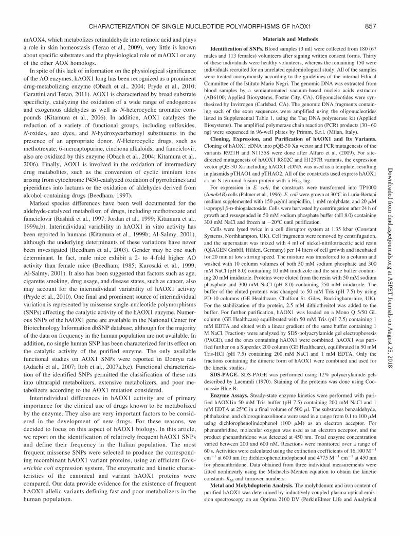
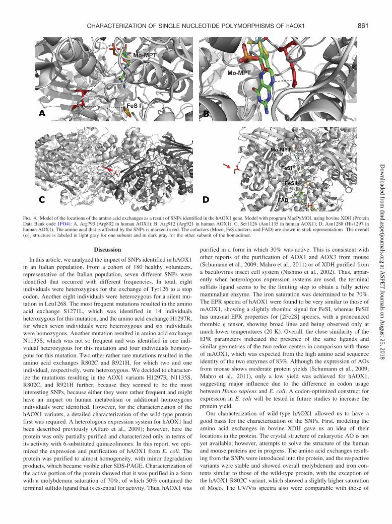
R802C, R921H, N1135S, and H1297R, as to their influences onenzyme activity, protein dimerization, and cofactor insertion. AOsbelong to the XO family of molybdoenzymes and are characterizedby high amino acid identity to the XO and xanthine dehydrogenase(XDH) forms of xanthine oxidoreductase (Garattini et al., 2008).Given the 50% identity between the two proteins, we modeled thestructure of hAOX1 against the crystal structure of XDH from Bostaurus (Protein Data Bank code 1FO4) to define the spatial local-ization of the amino acid residues modified by the SNPs (Fig. 4).The hAOX1 model indicates that Arg802 and Arg921 are inproximity to the Moco and FeSI sites, Asn1135 is located close tothe dimerization domain, and His1297 maps to the surface of theprotein.
All of the hAOX1 variants were expressed and purified undersimilar conditions as the wild-type protein. Differences in the oli-gomerization states of the variants and wild-type hAOX1 were iden-
tified by size-exclusion chromatography. All of the variants showedan altered dimer/monomer ratio. In the case of the R802C variant,only 40% of the protein was in its dimeric form (Fig. 5). In contrast,all of the other hAOX1 variants showed a higher dimer/monomerratio, indicating a higher proportion of the active protein. Mutation ofeach of the four amino acid residues did not affect the insertion of the[2Fe2S] clusters, because the iron content was similar to that ofwild-type hAOX1. In fact, similar to wild-type hAOX1, all of thevariants showed 70% saturation with iron (Fig. 2). In the R802Cvariant, molybdenum saturation was increased to 80%, which isslightly higher than the 70% saturation observed in the wild-typeprotein and all of the other variants (Fig. 2). The variant hAOX1proteins were purified and showed purities comparable with that ofthe wild-type protein. The UV/Vis absorption spectra of the purifiedvariants showed the same characteristic features of all of the molybdo-flavoenzymes (Fig. 5).
Steady-State Kinetics of Wild-Type hAOX1 and Its Variants.To determine the impact of SNPs on enzymatic activity, steady-statekinetics were performed on the variants hAOX1-R802C, hAOX1-R921H, hAOX1-N1135S, and hAOX1-H1297R. All of the variantsgenerated were tested for their abilities to catalyze the conversion ofbenzaldehyde, phthalazine, phenanthridine, and chloroquinazolinoneas substrates (Table 3). The wild-type hAOX1 showed activities withall of the substrates tested with kcat values in the range from 5 to 12min�1 and KM values between 1 and 7 �M. The best substrate wasphenanthridine with a kcat value of 12.2 min�1.
For a comparison, the dimeric portion of the hAOX1 variants alsowas subjected to steady-state kinetic analysis. The R802C variantshowed kinetic data most comparable with those of the wild-typeprotein. For hAOX1-N1135S and hAOX1-H1297R, the turnovernumbers were increased 2.5-fold with phenanthridine as the substrate.With the other substrates, the kcat and KM values for the two variantsremained in the same range as those of the wild-type protein. hAOX1-R921H showed a 3.7- to 1.5-fold decrease in kcat values with most ofthe substrates in comparison with those of the wild-type protein,whereas the KM values remained comparable. Only with phenanthri-dine as a substrate was the protein completely inactive. The resultsshow that amino acid substitution has different influences on thekinetic constants depending on the substrate. The inactivity or reducedactivity of the R921H variant might be explained by the fact thatarginine is highly conserved in all of the members of the XO family,and with its close proximity to the pterin molecule of the Mo-MPTcofactor, it might influence the geometry of the bound Mo-MPTmolecule, thus affecting catalytic turnover but not affecting the bind-ing of the substrate. However, because the different mutations af-fected the dimer/monomer ratio of the protein variants, we alsoconsidered the total amount of hAOX1 expressed and calculated theturnover number, taking into account the inactive portion of themonomeric protein. The normalized values are shown in Table 3. Ingeneral, the results show the same trends as those with the activeportion of the protein.
TABLE 2
EPR line widths and g values of FeSI and FeSII from hAOX1
g Values Line Widths (mT)a
Protein Cluster gx gy gz �Bx �By �Bz
hAOX1 FeSI 2.0115 1.924 1.900 4.1 2.4 3.7FeSII 2.085 1.975 1.906 9.2 3.3 2.8
mAOX1† FeSI 2.019 1.927 1.912 2.6 2.6 3.2FeSII 2.085 1.971 1.90 7.4 4.0 4.0
a The variable line width was included as g strain in the simulations.† Values for mAOX1 are taken from Schumann et al. (2009).
FIG. 3. EPR spectra of wild-type hAOX1. Experimental continuous-wave EPR spectraof dithionite-reduced wild-type hAOX1 samples at pH 7.0 (trace a) together with thecorresponding simulation (trace b). For simulation parameters, see Table 2. The flavinsemiquinone and MoV were not detected under these experimental conditions andtherefore neglected in all of the simulations: a, wild-type hAOX1; b, simulation ofcomplete spectrum; c, simulation of FeSI; d, simulation of FeSII. Experimental condi-tions: T � 20 K; microwave power, 1 mW; microwave frequency, 9.385 GHz;modulation amplitude, 0.5 mT; modulation frequency, 100 kHz.
860 HARTMANN ET AL.
at ASPE
T Journals on A
ugust 25, 2018dm
d.aspetjournals.orgD
ownloaded from
Discussion
In this article, we analyzed the impact of SNPs identified in hAOX1in an Italian population. From a cohort of 180 healthy volunteers,representative of the Italian population, seven different SNPs wereidentified that occurred with different frequencies. In total, eightindividuals were heterozygous for the exchange of Tyr126 to a stopcodon. Another eight individuals were heterozygous for a silent mu-tation in Leu1268. The most frequent mutations resulted in the aminoacid exchange S1271L, which was identified in 14 individualsheterozygous for this mutation, and the amino acid exchange H1297R,for which seven individuals were heterozygous and six individualswere homozygous. Another mutation resulted in amino acid exchangeN1135S, which was not so frequent and was identified in one indi-vidual heterozygous for this mutation and four individuals homozy-gous for this mutation. Two other rather rare mutations resulted in theamino acid exchanges R802C and R921H, for which two and oneindividual, respectively, were heterozygous. We decided to character-ize the mutations resulting in the AOX1 variants H1297R, N1135S,R802C, and R921H further, because they seemed to be the mostinteresting SNPs, because either they were rather frequent and mighthave an impact on human metabolism or additional homozygousindividuals were identified. However, for the characterization of thehAOX1 variants, a detailed characterization of the wild-type proteinfirst was required. A heterologous expression system for hAOX1 hadbeen described previously (Alfaro et al., 2009); however, here theprotein was only partially purified and characterized only in terms ofits activity with 6-substituted quinazolinones. In this report, we opti-mized the expression and purification of hAOX1 from E. coli. Theprotein was purified to almost homogeneity, with minor degradationproducts, which became visible after SDS-PAGE. Characterization ofthe active portion of the protein showed that it was purified in a formwith a molybdenum saturation of 70%, of which 50% contained theterminal sulfido ligand that is essential for activity. Thus, hAOX1 was
purified in a form in which 30% was active. This is consistent withother reports of the purification of AOX1 and AOX3 from mouse(Schumann et al., 2009; Mahro et al., 2011) or of XDH purified froma baculovirus insect cell system (Nishino et al., 2002). Thus, appar-ently when heterologous expression systems are used, the terminalsulfido ligand seems to be the limiting step to obtain a fully activemammalian enzyme. The iron saturation was determined to be 70%.The EPR spectra of hAOX1 were found to be very similar to those ofmAOX1, showing a slightly rhombic signal for FeSI, whereas FeSIIhas unusual EPR properties for [2Fe2S] species, with a pronouncedrhombic g tensor, showing broad lines and being observed only atmuch lower temperatures (20 K). Overall, the close similarity of theEPR parameters indicated the presence of the same ligands andsimilar geometries of the two redox centers in comparison with thoseof mAOX1, which was expected from the high amino acid sequenceidentity of the two enzymes of 83%. Although the expression of AOsfrom mouse shows moderate protein yields (Schumann et al., 2009;Mahro et al., 2011), only a low yield was achieved for hAOX1,suggesting major influence due to the difference in codon usagebetween Homo sapiens and E. coli. A codon-optimized construct forexpression in E. coli will be tested in future studies to increase theprotein yield.
Our characterization of wild-type hAOX1 allowed us to have agood basis for the characterization of the SNPs. First, modeling theamino acid exchanges in bovine XDH gave us an idea of theirlocations in the protein. The crystal structure of eukaryotic AO is notyet available; however, attempts to solve the structure of the humanand mouse proteins are in progress. The amino acid exchanges result-ing from the SNPs were introduced into the protein, and the respectivevariants were stable and showed overall molybdenum and iron con-tents similar to those of the wild-type protein, with the exception ofthe hAOX1-R802C variant, which showed a slightly higher saturationof Moco. The UV/Vis spectra also were comparable with those of
FIG. 4. Model of the locations of the amino acid exchanges as a result of SNPs identified in the hAOX1 gene. Model with program MacPyMOL using bovine XDH (ProteinData Bank code 1FO4): A, Arg793 (Arg802 in human AOX1); B, Arg912 (Arg921 in human AOX1); C, Ser1126 (Asn1135 in human AOX1); D, Asn1288 (His1297 inhuman AOX1). The amino acid that is affected by the SNPs is marked in red. The cofactors (Moco, FeS clusters, and FAD) are shown in stick representations. The overall(�)2 structure is labeled in light gray for one subunit and in dark gray for the other subunit of the homodimer.
861CHARACTERIZATION OF SINGLE NUCLEOTIDE POLYMORPHISMS OF hAOX1
at ASPE
T Journals on A
ugust 25, 2018dm
d.aspetjournals.orgD
ownloaded from

wild-type hAOX1, suggesting complete saturation of the FAD cofac-tor. However, in contrast to the overall cofactor composition, crucialchanges were observed in protein quaternary structures. Although thewild-type enzyme is stable in its monomeric and dimeric forms in aratio of 1:1.5, hAOX1-R921H, hAOX1-N1125S, and hAOX1-H1297R were purified mainly as stable dimers. In contrast, hAOX1-R802C showed much higher levels of monomer in solution in a ratioof 1.5:1, resulting in a higher proportion of inactive protein. A changein the monomer/dimer ratio has been reported previously in AO andXDH enzymes with similar variants in proximity to the FeS clusters.In SNPs identified in Donryu rats, the amino acid exchange G101S inproximity to FeSII also resulted in the production of the monomericform of AOX1 (Itoh et al., 2007c). In addition, in a human patientsuffering from xanthinuria I, a mutation resulting in the amino acidexchange R149C was identified in the XDH gene (Sakamoto et al.,2001). The arginine is located close to FeSI, and when this mutationwas introduced in the Rhodobacter capsulatus xdhB gene and thecorresponding RcXDH-R135C was characterized, the purified proteinalso existed in two forms, a monomeric inactive form and a dimericactive form (Leimkuhler et al., 2003). Further analyses of the mono-mer/dimer behavior of R. capsulatus XDH resulted in a model inwhich it was proposed that dimerization requires that the two FeSclusters are assembled correctly before Moco can be inserted and theprotein can dimerize via the Moco domain (Schumann et al., 2008).Thus, amino acid exchanges in proximity to the FeS clusters influence
the structure of the protein in a manner that dimerization is no longereffective.
We studied the activities of wild-type hAOX1 in comparison withthose of selected variants based on the SNPs with four selectedsubstrates, benzaldehyde, phthalazine, phenanthridine, and chloro-quinazolinone. Our results show that the SNPs can be classified intothree groups in general: fast metabolizers (FMs), poor metabolizers(PMs), and no affect on catalytic efficiency. Taking into account onlythe active portion of the protein, hAOX1-R802C and, even morepronounced, hAOX1-R921H are PMs, because both variants had a2.4- to 1.5-fold reduced activity with most of the substrates tested. Inaddition, the R921H variant was identified to be inactive with phenan-thridine; thus, the influence of the amino acid substitution on enzymeactivity varies depending on the substrate. hAOX1-N1135S andhAOX1-H1297R can be classified as FMs, because an increasedcatalytic efficiency of 2- to 4-fold was observed depending onthe substrate tested (taking into account only the active portion of theprotein). In general, for the catalytic efficiencies, we calculated theoverall activity of the purified enzyme, taking into account the activeand inactive portions of the protein. In general, the same trends forthe kinetic values were obtained for the variants in comparison withthose of the wild-type protein. hAOX1-R921H showed that this res-idue is important for maintaining the catalytic activity of hAOX1, inparticular with phenanthridine as the substrate. The residue Arg921lies close to Moco and affected mainly the monomer/dimer ratio but
FIG. 5. Size-exclusion chromatography profiles and UV/Vis spectra of hAOX1 variants identified in SNPs. Elution profiles of hAOX1 variants using size-exclusionchromatography on a Superdex 200 column and UV/Vis spectra of purified proteins are shown (A, R802C; B, R921H; C, N1135S; D, H1297R). In comparison with thewild-type protein, hAOX1 variants show different dimer/monomer ratios in solution. R802C is mainly purified in its monomeric form (A), whereas all of the other studiedvariants almost completely exist in their dimeric forms (B–D). For all of the variants, similar degradation products were visualized by SDS-PAGE as in the wild-type (D,Dimer; M, Monomer; stars, degradation products of hAOX1; 1, peak corresponding to the AOX1 dimer; 2, peak corresponding to the AOX1 monomer).
862 HARTMANN ET AL.
at ASPE
T Journals on A
ugust 25, 2018dm
d.aspetjournals.orgD
ownloaded from

also the overall activity of the protein. This might suggest that notonly substrate turnover and binding were impaired but also intramo-lecular electron transfer. Thus, the positive charge of the argininemight affect substrate binding and intramolecular electron transfer,which can only poorly be substituted by His. In contrast, in hAOX1-R802C, the catalytic efficiency of the protein was not affected, and weobtained similar values in comparison with those of wild-typehAOX1. Both hAOX1-N1135S and hAOX1-H1297R are consideredto be FM variants of hAOX1. Especially with phthalazine and phenan-thridine, a 2- to 4-fold higher catalytic efficiency was obtained. Bothamino acids are located at the surface of the protein and seem toinfluence the stability of hAOX1. Thus, the more polar and positivecharged residues seem to affect the surface charge of the protein,which results in higher stability of hAOX1. This also might influenceits interaction with other proteins and/or posttranslational modifica-tions of the protein, as suggested by Itoh et al. (2007c) in a report onthe characterization of SNPs in Donryu rats.
Our studies reveal the importance of considering individual differ-ences based on SNPs for drug design. We analyzed 180 healthyindividuals representative of the Italian population and identified thetotal occurrence of 51 SNPs in hAOX1, and a total of 10 werehomozygous for the SNP. Two of the SNPs resulted in FM and one inPM individuals. hAOX1 is an important enzyme responsible for themetabolism of a number of drugs containing aldehydes and the moreprevalent nitrogen heterocycles (Kitamura et al., 2006; Torres et al.,2007). The fraction of drugs metabolized by hAOX1 is likely toincrease over the next decade. Thus, it should be carefully consideredwhich dose of the drug is administered to an individual, because ourresults indicate that there might be a difference in hAOX1 activity indifferent individuals containing SNPs that additionally varies depend-ing on the substrate used; thus, in FMs the drug might be cleared toofast and have no effect, whereas in PMs the drug might reach a toxicdose that would cause more severe side effects. In addition, someindividuals were identified with SNPs that resulted in nonsense mu-tations, which might affect even the overall hAOX1 content and thusthe overall activity more drastically when the active protein amount is
expressed only from one allele. In the future, hAOX1 activities shouldbe measured in individuals with known SNPs, or a pharmacokineticstudy with a hAOX1-cleared drug in genotyped individuals should beconducted to confirm our results on the purified enzyme variants.
Acknowledgments
We thank Manfred Nimtz (Helmholtz Center for Infection Research, Braun-schweig, Germany) for matrix-assisted laser desorption ionization peptidemapping and T. Nishino and T. Matsumura (Nippon Medical School, Tokyo,Japan) for providing hMCSF cDNA.
Authorship Contributions
Participated in research design: Hartmann, Terao, Garattini, Teutloff,Jones, and Leimkuhler.
Conducted experiments: Hartmann, Terao, Teutloff, and Alfaro.Performed data analysis: Hartmann, Terao, Garattini, Teutloff, and Leimkuhler.Wrote or contributed to the writing of the manuscript: Hartmann, Garattini,
Jones, and Leimkuhler.
References
Adachi M, Itoh K, Masubuchi A, Watanabe N, and Tanaka Y (2007) Construction and expressionof mutant cDNAs responsible for genetic polymorphism in aldehyde oxidase in Donryu strainrats. J Biochem Mol Biol 40:1021–1027.
Al-Salmy HS (2001) Individual variation in hepatic aldehyde oxidase activity. IUBMB Life51:249–253.
Alfaro JF, Joswig-Jones CA, Ouyang W, Nichols J, Crouch GJ, and Jones JP (2009) Purificationand mechanism of human aldehyde oxidase expressed in Escherichia coli. Drug Metab Dispos37:2393–2398.
Beedham C (1985) Molybdenum hydroxylases as drug-metabolizing enzymes. Drug Metab Rev16:119–156.
Beedham C (1997) The role of non-P450 enzymes in drug oxidation. Pharm World Sci19:255–263.
Beedham C, Miceli JJ, and Obach RS (2003) Ziprasidone metabolism, aldehyde oxidase, andclinical implications. J Clin Psychopharmacol 23:229–232.
Edmondson D, Massey V, Palmer G, Beacham LM 3rd, and Elion GB (1972) The resolution ofactive and inactive xanthine oxidase by affinity chromatography. J Biol Chem 247:1597–1604.
Garattini E and Terao M (2011) Increasing recognition of the importance of aldehyde oxidase indrug development and discovery. Drug Metab Rev 43:374–386.
Garattini E, Fratelli M, and Terao M (2008) Mammalian aldehyde oxidases: genetics, evolutionand biochemistry. Cell Mol Life Sci 65:1019–1048.
Garattini E, Fratelli M, and Terao M (2009) The mammalian aldehyde oxidase gene family. HumGenomics 4:119–130.
Garattini E, Mendel R, Romao MJ, Wright R, and Terao M (2003) Mammalian molybdo-
TABLE 3
Steady-state kinetics of hAOX1 and its variants corresponding to SNPs
Benzaldehyde, phthalazine, phenanthridine, and chloroquinazolinone were used as substrates to cover a range from aldehydes to N-heterocyclic compounds. Assays were performedphotometrically using 2,6-dichlorphenolindophenol as a final electron acceptor. With phenanthridine, molecular oxygen was used as the electron acceptor. Data are mean values from threeindependent measurements (�S.D.).
Substrate Protein Portion Kinetic Parameters WT R802C R921H N1135S H1297R
Benzaldehyde KM, �M 7.1 � 0.6 7.6 � 1.9 6.3 � 1.2 6.7 � 2.8 5.2 � 1.8Active portion kcat, min�1 6.4 � 0.1 5.3 � 0.3 1.7 � 0.1 6.2 � 0.3 6.4 � 0.3
kcat/KM, 1/min � �M 0.91 � 0.17 0.70 � 0.16 0.27 � 0.08 0.92 � 0.11 1.23 � 0.17Total protein kcat, min�1 2.7 � 0.1 1.8 � 0.3 1.1 � 0.1 3.8 � 0.3 2.6 � 0.3
kcat/KM, 1/min � �M 0.38 � 0.17 0.24 � 0.16 0.17 � 0.08 0.57 � 0.11 0.50 � 0.17Phthalazine KM, �M 1.3 � 0.3 0.9 � 0.3 1.6 � 0.3 1.2 � 0.1 1.3 � 0.2
Active portion kcat, min�1 5.6 � 0.2 5.2 � 0.3 2.4 � 0.1 7.2 � 0.1 5.4 � 0.1kcat/KM, 1/min � �M 4.31 � 0.67 5.78 � 1.00 1.50 � 0.33 6.00 � 1.00 4.15 � 0.50
Total protein kcat, min�1 2.3 � 0.2 1.8 � 0.3 1.5 � 0.1 4.4 � 0.1 2.9 � 0.1kcat/KM, 1/min � �M 1.77 � 0.67 2.00 � 1.00 0.94 � 0.33 3.67 � 1.00 2.23 � 0.50
Phenanthridine KM, �M 3.9 � 0.8 4.4 � 0.4 N.D. 6.1 � 1.0 4.1 � 0.7Active portion kcat, min�1 12.2 � 0.5 10.2 � 0.2 N.D. 32.6 � 1.1 31.5 � 1.0
kcat/KM, 1/min � �M 3.13 � 0.63 2.32 � 0.50 5.34 � 1.10 7.68 � 1.43Total protein kcat, min�1 5.3 � 0.5 3.4 � 0.2 N.D. 19.9 � 1.1 16.9 � 1.0
kcat/KM, 1/min � �M 1.36 � 0.63 0.77 � 0.50 3.26 � 1.10 4.12 � 1.43Chloroquinazolinone KM, �M 5.2 � 0.7 4.7 � 0.7 4.5 � 0.8 4.1 � 0.5 5.8 � 0.5
Active portion kcat, min�1 5.6 � 0.1 5.4 � 0.2 3.6 � 0.1 6.5 � 0.1 6.7 � 0.2kcat/KM, 1/min � �M 1.08 � 0.14 1.15 � 0.29 0.80 � 0.13 1.59 � 0.20 1.16 � 0.4
Total protein kcat, min�1 2.3 � 0.1 1.8 � 0.2 2.2 � 0.1 4.0 � 0.1 3.6 � 0.2kcat/KM, 1/min � �M 0.44 � 0.14 0.38 � 0.29 0.49 � 0.13 0.98 � 0.20 0.62 � 0.4
Oligomerization in solution Dimer Percentage 58 39 83 90 78Monomer Percentage 38 58 13 7 17Multimer Percentage 4 3 4 3 5
N.D., no activity was detectable; WT, wild type.
863CHARACTERIZATION OF SINGLE NUCLEOTIDE POLYMORPHISMS OF hAOX1
at ASPE
T Journals on A
ugust 25, 2018dm
d.aspetjournals.orgD
ownloaded from

flavoenzymes, an expanding family of proteins: structure, genetics, regulation, function andpathophysiology. Biochem J 372:15–32.
Hille R (1996) The Mononuclear Molybdenum Enzymes. Chem Rev 96:2757–2816.Ichida K, Matsumura T, Sakuma R, Hosoya T, and Nishino T (2001) Mutation of human
molybdenum cofactor sulfurase gene is responsible for classical xanthinuria type II. BiochemBiophys Res Commun 282:1194–1200.
Itoh K, Maruyama H, Adachi M, Hoshino K, Watanabe N, and Tanaka Y (2007a) Lack of dimerformation ability in rat strains with low aldehyde oxidase activity. Xenobiotica 37:709–716.
Itoh K, Maruyama H, Adachi M, Hoshino K, Watanabe N, and Tanaka Y (2007b) Lack offormation of aldehyde oxidase dimer possibly due to 377G�A nucleotide substitution. DrugMetab Dispos 35:1860–1864.
Itoh K, Masubuchi A, Sasaki T, Adachi M, Watanabe N, Nagata K, Yamazoe Y, Hiratsuka M,Mizugaki M, and Tanaka Y (2007c) Genetic polymorphism of aldehyde oxidase in Donryurats. Drug Metab Dispos 35:734–739.
Johnson JL, Hainline BE, Rajagopalan KV, and Arison BH (1984) The pterin component of themolybdenum cofactor. Structural characterization of two fluorescent derivatives. J Biol Chem259:5414–5422.
Jordan CG, Rashidi MR, Laljee H, Clarke SE, Brown JE, and Beedham C (1999) Aldehydeoxidase-catalysed oxidation of methotrexate in the liver of guinea-pig, rabbit and man.J Pharm Pharmacol 51:411–418.
Kitamura S, Nakatani K, Sugihara K, and Ohta S (1999a) Strain differences of the ability tohydroxylate methotrexate in rats. Comp Biochem Physiol C Pharmacol Toxicol Endocrinol122:331–336.
Kitamura S, Sugihara K, and Ohta S (2006) Drug-metabolizing ability of molybdenum hydroxy-lases. Drug Metab Pharmacokinet 21:83–98.
Kitamura S, Sugihara K, Nakatani K, Ohta S, Ohhara T, Ninomiya S, Green CE, and Tyson CA(1999b) Variation of hepatic methotrexate 7-hydroxylase activity in animals and humans.IUBMB Life 48:607–611.
Kundu TK, Hille R, Velayutham M, and Zweier JL (2007) Characterization of superoxideproduction from aldehyde oxidase: an important source of oxidants in biological tissues. ArchBiochem Biophys 460:113–121.
Kurosaki M, Demontis S, Barzago MM, Garattini E, and Terao M (1999) Molecular cloning ofthe cDNA coding for mouse aldehyde oxidase: tissue distribution and regulation in vivo bytestosterone. Biochem J 341:71–80.
Laemmli UK (1970) Cleavage of structural proteins during the assembly of the head ofbacteriophage T4. Nature 227:680–685.
Leimkuhler S, Hodson R, George GN, and Rajagopalan KV (2003) Recombinant Rhodobactercapsulatus xanthine dehydrogenase, a useful model system for the characterization of proteinvariants leading to xanthinuria I in humans. J Biol Chem 278:20802–20811.
Leimkuhler S, Stockert AL, Igarashi K, Nishino T, and Hille R (2004) The role of active siteglutamate residues in catalysis of Rhodobacter capsulatus xanthine dehydrogenase. J BiolChem 279:40437–40444.
Mahro M, Coelho C, Trincao J, Rodrigues D, Terao M, Garattini E, Saggu M, Lendzian F,Hildebrandt P, Romao MJ, et al. (2011) Characterization and crystallization of mouse aldehydeoxidase 3: from mouse liver to Escherichia coli heterologous protein expression. Drug MetabDispos 39:1939–1945.
Nishino T, Amaya Y, Kawamoto S, Kashima Y, Okamoto K, and Nishino T (2002) Purificationand characterization of multiple forms of rat liver xanthine oxidoreductase expressed inbaculovirus-insect cell system. J Biochem 132:597–606.
Obach RS, Huynh P, Allen MC, and Beedham C (2004) Human liver aldehyde oxidase:inhibition by 239 drugs. J Clin Pharmacol 44:7–19.
Palmer T, Santini CL, Iobbi-Nivol C, Eaves DJ, Boxer DH, and Giordano G (1996) Involvementof the narJ and mob gene products in distinct steps in the biosynthesis of the molybdoenzymenitrate reductase in Escherichia coli. Mol Microbiol 20:875–884.
Parschat K, Canne C, Huttermann J, Kappl R, and Fetzner S (2001) Xanthine dehydrogenasefrom Pseudomonas putida 86: specificity, oxidation-reduction potentials of its redox-activecenters, and first EPR characterization. Biochim Biophys Acta 1544:151–165.
Pryde DC, Dalvie D, Hu Q, Jones P, Obach RS, and Tran TD (2010) Aldehyde oxidase: anenzyme of emerging importance in drug discovery. J Med Chem 53:8441–8460.
Rashidi MR, Smith JA, Clarke SE, and Beedham C (1997) In vitro oxidation of famciclovir and6-deoxypenciclovir by aldehyde oxidase from human, guinea pig, rabbit, and rat liver. DrugMetab Dispos 25:805–813.
Sakamoto N, Yamamoto T, Moriwaki Y, Teranishi T, Toyoda M, Onishi Y, Kuroda S, SakaguchiK, Fujisawa T, Maeda M, et al. (2001) Identification of a new point mutation in the humanxanthine dehydrogenase gene responsible for a case of classical type I xanthinuria. Hum Genet108:279–283.
Schumann S, Saggu M, Moller N, Anker SD, Lendzian F, Hildebrandt P, and Leimkuhler S(2008) The mechanism of assembly and cofactor insertion into Rhodobacter capsulatusxanthine dehydrogenase. J Biol Chem 283:16602–16611.
Schumann S, Terao M, Garattini E, Saggu M, Lendzian F, Hildebrandt P, and Leimkuhler S(2009) Site directed mutagenesis of amino acid residues at the active site of mouse aldehydeoxidase AOX1. PLoS One 4:e5348.
Stoll S and Schweiger A (2006) EasySpin, a comprehensive software package for spectralsimulation and analysis in EPR. J Magn Reson 178:42–55.
Terao M, Kurosaki M, Barzago MM, Fratelli M, Bagnati R, Bastone A, Giudice C, Scanziani E,Mancuso A, Tiveron C, et al. (2009) Role of the molybdoflavoenzyme aldehyde oxidasehomolog 2 in the biosynthesis of retinoic acid: generation and characterization of a knockoutmouse. Mol Cell Biol 29:357–377.
Terao M, Kurosaki M, Barzago MM, Varasano E, Boldetti A, Bastone A, Fratelli M, andGarattini E (2006) Avian and canine aldehyde oxidases. Novel insights into the biology andevolution of molybdo-flavoenzymes. J Biol Chem 281:19748–19761.
Terao M, Kurosaki M, Saltini G, Demontis S, Marini M, Salmona M, and Garattini E (2000)Cloning of the cDNAs coding for two novel molybdo-flavoproteins showing high similaritywith aldehyde oxidase and xanthine oxidoreductase. J Biol Chem 275:30690–30700.
Torres RA, Korzekwa KR, McMasters DR, Fandozzi CM, and Jones JP (2007) Use of densityfunctional calculations to predict the regioselectivity of drugs and molecules metabolized byaldehyde oxidase. J Med Chem 50:4642–4647.
Vila R, Kurosaki M, Barzago MM, Kolek M, Bastone A, Colombo L, Salmona M, Terao M, andGarattini E (2004) Regulation and biochemistry of mouse molybdo-flavoenzymes. The DBA/2mouse is selectively deficient in the expression of aldehyde oxidase homologues 1 and 2 andrepresents a unique source for the purification and characterization of aldehyde oxidase. J BiolChem 279:8668–8683.
Wahl RC and Rajagopalan KV (1982) Evidence for the inorganic nature of the cyanolyzablesulfur of molybdenum hydroxylases. J Biol Chem 257:1354–1359.
Address correspondence to: Dr. Silke Leimkuhler, Department of Molecular Enzy-mology, Institute of Biochemistry and Biology, University of Potsdam, Karl-Liebknecht-Strasse 24-25, 14476 Potsdam, Germany. E-mail: [email protected]
864 HARTMANN ET AL.
at ASPE
T Journals on A
ugust 25, 2018dm
d.aspetjournals.orgD
ownloaded from