division of specialty medicine - south tees hospitals nhs ... · with itch or raised wcc / plt. o...
TRANSCRIPT

October 2014 V3
Haematology Handbook
Division of Specialty Medicine
Author: Dr Jamie Maddox
Issue Date: 01/11/2014
Review Date: 31/10/2016

1 October 2014 V3
HHaaeemmaattoollooggyy
HHaannddbbooookk
Although this document is designed for use in General Practice, it can also be used in hospital medicine as a tool for investigating common haematological abnormalities and for guiding appropriate referral. Within hospital practice, the causes for haematological abnormalities will be skewed towards acute causes (e.g. acute illness, medications) rather than chronic conditions. Com-morbidities should also be taken into account. Further guidance on anticoagulation and blood transfusion can be found on the ‘Policies’ > ‘Clinical Guidelines & Protocols’ page of the South Tees Intranet. The handbook can also be accessed via the internet: http://stm-pathfinder/pathology/maindocuments.asp (South Tees) http://nww.stpathfinder.nhs.uk/pathology/maindocuments.asp (N3 link)

2 October 2014 V3
Contents Section Page Anaemia............................................................................................... 3-4
High haemoglobin................................................................................ 5
Iron deficiency...................................................................................... 6-7
High ferritin........................................................................................... 8
Macrocytosis........................................................................................ 9
Low white cell count ............................................................................. 10
High white cell count............................................................................ 11
Lymphocytosis..................................................................................... 12
Newly diagnosed early CLL................................................................. 13
Low platelet count................................................................................ 14
High platelet count............................................................................... 15
Paraprotein.......................................................................................... 16-17
Polyclonal immunoglobulins................................................................ 18
Raised ESR / plasma viscosity............................................................ 19
Lymphadenopathy............................................................................... 20
Splenomegaly...................................................................................... 21
Antibiotic prophylaxis and vaccination following splenectomy…….. 22-23
Sweats……………………………………………………………………… 24
Easy bruising....................................................................................... 25
Abnormal coagulation.......................................................................... 26
Thrombophilia ..................................................................................... 27-28

3 October 2014 V3
Clinical Problem: Anaemia The most common cause of anaemia will be iron deficiency presenting with a low MCV/MCH and low serum ferritin (see separate protocol). There are however many other possible causes of anaemia so co-morbidities, medication, diet, ethnicity and family history are all also relevant. Initially consider the MCV as this can help narrow down the cause of anaemia:
Low MCV- iron deficiency, some anaemia of chronic disease (otherwise MCV normal) or haemoglobinopathy.
Normal MCV- some anaemia of chronic disease, acute bleeding, mixed iron and B12/folate deficiency and most non-haematinic causes of anaemia.
High MCV – B12/folate deficiency, liver disease, hypothyroidism, some bone marrow disorders (e.g. myelodysplastic syndrome) or increased red cell destruction. A mildly increased MCV can also be seen in pregnancy.
Differential Diagnosis:
Iron deficiency- low MCV/MCH. Low serum ferritin, although this can be masked by inflammation. Usually due to chronic blood loss.
Acute bleeding, even if not yet iron deficient. May have increased reticulocytes.
Anaemia of chronic disease- normal or low MCV/MCH. Normal or increased ferritin with low transferrin saturation. Raised inflammatory markers.
Megaloblastic anaemia (B12, folate or drugs) - high MCV and megaloblastic features on blood film. Low serum B12 or folate.
Increased red cell destruction- inherited or acquired haemolytic anaemia with raised reticulocytes, bilirubin and LDH. Positive Direct Coombs test if autoimmune. Blood film may show spherocytes.
Bone marrow disorder– blood film appearances may be suggestive. May also have low WCC and PLT.
Drugs- e.g. GI bleeding with NSAIDs; bone marrow suppression with chemotherapy, azathioprine or methotrexate; red cell haemolysis with dapsone or sulphasalazine.
Renal impairment- often with normal MCV. Incidence increases as renal function declines, although the cause of renal impairment is also relevant (e.g. diabetic nephropathy tends to be more anaemic than expected from eGFR alone).
Liver disease- often with high MCV, although could be normal or low if GI bleeding due to varicies.
Endocrine- thyroid, parathyroid, pituitary or adrenal dysfunction.
Physiological, e.g. mild drop in haemoglobin is normal in pregnancy. Examination:
Depends on suspected cause
Include abdominal examination and rectal exam if considering iron deficiency anaemia or occult bleeding.
Include examination for lymphadenopathy, splenomegaly and hepatomegaly if considering haematological disorder.

4 October 2014 V3
Baseline investigations:
Depending on suspected cause, additional investigation may include: o Low MCV: blood film, SMAC, ferritin / iron studies, CRP. Screening for
a haemoglobinopathy is normally carried out automatically by the laboratory if the red cell indices and / or ethnic origin are suggestive.
o Normal MCV: blood film, reticulocytes, SMAC, ferritin, B12/folate, CRP o High MCV: blood film, reticulocytes, SMAC, TSH, B12/folate
Serum immunoglobulins and urinary Bence-Jones protein if myeloma is being considered (normal or high MCV).
Consider HIV serology in patients with unexplained anaemia. Referral: Consider Haematology referral if:
Suspected haematological disorder (bone marrow disorder, haemolytic anaemia).
Unexplained symptomatic anaemia with haemoglobin <10g/dL (female) / <11g/dL (male), especially if progressive.
We do not routinely see haemoglobinopathy carriers, however an information sheet is sent to the patient’s General Practice.
Other specialty referrals based on suspected cause. If anaemia of chronic disease but no obvious explanation for the inflammatory process then consider referral to General Medicine.

5 October 2014 V3
Clinical Problem: High haemoglobin / haematocrit Investigation is suggested if the haematocrit is persistently (>2 months) >0.52 (male) or >0.48 (female). This may be due to increased red cell mass (true polycythaemia) or reduced plasma volume (relative polycythaemia). If the haematocrit is >0.6 (male) or >0.56 (female) then it can be assumed that there is a true increase in red cell mass. Differential Diagnosis:
Relative polycythaemia – may be due to a variety of causes such as stress, dehydration, diuretics, oedema, hypertension, obesity, sleep apnoea, cigarettes or alcohol.
True polycythaemia o Reactive to hypoxia- e.g. pulmonary disease, heavy smoking, sleep
apnoea or congenital cyanotic heart disease. o Abnormal high erythropoietin- e.g. secondary to various tumours, renal
transplant or renal artery stenosis. o Polycythaemia vera- bone marrow disorder with uncontrolled red cell
proliferation. Usually positive for JAK2 V617F mutation. Associated with thrombotic and haemorrhagic complications. May be associated with itch or raised WCC / PLT.
o Endocrine- Cushing’s syndrome, Conn’s syndrome, phaeochromocytoma or androgens (including testosterone supplements and anabolic steroids).
o Inherited erythropoietin or haemoglobin variants (rare). Examination:
Features of cardiac or pulmonary disease.
Features of hyperviscosity (fatigue, headache, retinal haemorrhage, blurred vision, paraesthesia, slowed thought, chest / abdominal pain, myalgia).
Splenomegaly suggests polycythaemia vera although this is not a consistent finding.
Baseline investigations:
FBC, blood film, U+E, LFT, calcium, ferritin (high haematocrit despite low ferritin suggests polycythaemia vera. DO NOT give iron replacement).
Further tests as indicated by history, e.g. oxygen saturations, CXR
Approximately 98% of patients with polycythaemia vera will have a JAK2 V617F mutation or a JAK2 exon 12 mutation (testing to be performed by Haematology Department).
Referral:
Haematocrit persistently >0.52 (male) or >0.48 (female) without obvious cause or if suspicion of polycythaemia vera.
URGENT referral if features of hyperviscosity.
Other referrals as directed by suspected cause, e.g. may benefit from long-term oxygen therapy if chronic hypoxia.
References:
McMullin MF, et al. Guidelines for the diagnosis, investigation and management of polycythaemia / erythrocytosis. Br J Haematol 2005; 130: 174-195.
6 October 2014 V3
Clinical Problem: Iron deficiency Iron deficiency often shows as a hypochromic microcytic anaemia (low MCH and MCV) with a low ferritin value. Diagnostic problems can occur in the presence of an acute or chronic inflammatory condition. This can cause a falsely elevated ferritin despite true iron deficiency. It can also cause anaemia of chronic disease with a mildly low MCV and MCH despite normal iron stores. This picture may become easier to interpret if the underlying disease can be controlled. Anaemia with a markedly low MCV / MCH can also be seen with a haemoglobinopathy. Consider if the MCV/MCH is lower than expected, normal ferritin and non-Caucasian ethnic origin. The laboratory will normally automatically add on haemoglobinopathy screening in this situation. Differential Diagnosis:
Blood loss- most commonly gastrointestinal (NSAIDs or pathological lesion) or gynaecological.
Malabsorption- such as coeliac disease.
Poor intake- unusual as a sole cause, except with extreme diets. Examination:
Abdominal examination including rectal exam.
Gynaecological examination as appropriate. Baseline investigations:
FBC, blood film (elliptocytes or pencil cells suggest iron deficiency), ferritin. Check inflammatory markers if ferritin unexpectedly normal.
Coeliac serology
Urinalysis
Consider the possibility of atrophic gastritis (anti-parietal cell & anti-intrinsic factor antibodies) or H. pylori infection (blood or faecal antigen tests) as a cause of unexplained or refractory iron deficiency anaemia.
Faecal occult blood testing is of NO benefit and neither confirms or excludes gastrointestinal pathology.
Treatment:
Standard treatment is ferrous sulphate 200mg tds until anaemia resolves then a further 3 months to build iron stores.
If intolerant of ferrous sulphate then try alternate iron preparations. These may be better tolerated although the iron content may be lower.
Referral:
The majority of patients will require referral to Gastroenterology to look for an underlying cause (see chart).
Consider Gynaecology referral based on symptoms.
Haematology referral if considering parenteral iron therapy- i.e. unresponsive or intolerant of oral iron. Patients will still need referral to Gastroenterology / Gynaecology to identify and treat the underlying cause.
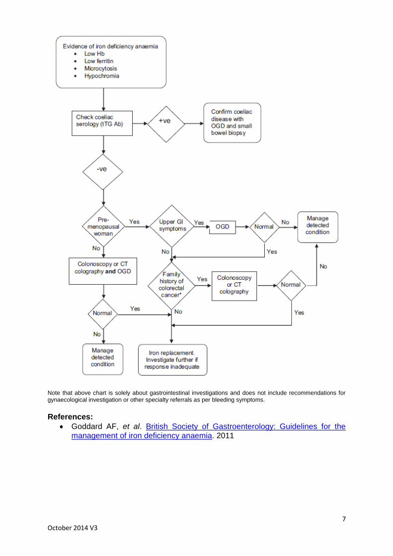
7 October 2014 V3
Note that above chart is solely about gastrointestinal investigations and does not include recommendations for gynaecological investigation or other specialty referrals as per bleeding symptoms.
References:
Goddard AF, et al. British Society of Gastroenterology: Guidelines for the management of iron deficiency anaemia. 2011

8 October 2014 V3
Clinical Problem: High ferritin In healthy subjects the serum ferritin concentration correlates with iron stores, however high levels do not necessarily reflect iron overload. Ferritin behaves as an acute phase reactant in many inflammatory diseases and tissue damage, particularly to the liver, can release large amounts of ferritin into the plasma. Differential diagnosis:
Acute phase reaction- infection, inflammation or malignancy.
Release of tissue ferritin by damage, especially to iron rich organs- e.g. chronic liver disease (including alcoholic liver disease and cirrhosis) or splenic infarction.
True iron overload: genetic haemochromatosis, iron overload secondary to repeated blood transfusions, massive ineffective erythropoeisis (thalassaemia, sideroblastic anaemia), porphyria cutanea tarda or rare genetic causes of iron overload (e.g. aceruloplasminaemia).
Baseline investigations:
FBC, LFT, CRP, iron studies (in particular transferrin saturation), TSH and blood glucose. Repeat serum ferritin levels >3 months apart. If the fasting transferrin saturation is normal then this strongly suggests a falsely elevated ferritin rather than true iron overload.
Genetic screening for haemochromatosis is indicated if positive family history OR if persistently raised ferritin with high transferrin saturation. This test can be sent from General Practice.
If persistently raised ferritin with raised CRP and/or normal transferrin saturation then consider alternate causes.
Referral:
If genetic haemochromatosis suspected / confirmed then patient management is overseen by Gastroenterology. A referral to Gastroenterology would also be appropriate in patients found to have liver disease on investigation of a high ferritin value.
If family screening is required for genetic haemochromatosis (parents, siblings, children) this can be undertaken in General Practice. Screen includes ferritin, transferrin saturation and genetic studies. If genetic counselling is required then we suggest refer to the clinical genetics service.
For raised serum ferritin is associated with an acute phase response then the underlying condition may require further investigation and management.
Refer to Haematology if true iron overload (raised transferrin saturation) with uncertain cause (e.g. negative tests for genetic haemochromatosis).
References
British Committee for Standards in Haematology. Guidelines on diagnosis and therapy. Genetic Haemochromatosis. 2000

9 October 2014 V3
Clinical Problem: Macrocytosis (high MCV) The most common cause of a high MCV is alcohol excess, however other causes should be considered.
Differential diagnosis:
Artefact – e.g. delayed sample processing or cold haemagglutinins.
Physiological – mild macrocytosis normal in pregnancy.
Reticulocytosis – suggests marrow response to red cell destruction or blood loss.
Excess alcohol intake - target cells or stomatocytes on a blood film report suggests alcohol excess or liver dysfunction.
Abnormal liver function
Hypothyroidism
Vitamin B12 or folate deficiency - hypersegmented neutrophils and / or oval macrocytes on a blood film report suggest a megaloblastic anaemia.
Drugs e.g. hydroxycarbamide, azathioprine and many chemotherapy agents. Can give the appearances of a megaloblastic anaemia.
Bone marrow disorder, e.g. myelodysplastic syndrome – blood film may be suggestive and may also have leucopenia or thrombocytopenia.
Baseline investigations:
Repeat FBC (ensure sample gets to laboratory on same day), blood film, reticulocytes, LFT, TFT, B12 / folate.
Note that a mild reduction in vitamin B12 levels can be seen as an artefact in pregnancy or women taking OCP / HRT.
Referral:
Further management / referral based on suspected cause.
Consider haematology referral if suspected red cell haemolysis or bone marrow disorder.
We do not see patients with an unexplained macrocytosis but an otherwise normal blood count. These patients can be monitored in General Practice with 6-12 monthly blood counts.

10 October 2014 V3
Clinical Problem: Low White Cell Count (<4 x10⁹/L) A low white cell count (WCC) is most commonly seen as a transient feature following viral infection. Where there was been no recent history of infection or where the low WCC is persistent then other causes should be considered. Differential Diagnosis Neutropenia:
Constitutional – Ethnic variation in Black Africans and some Mediterranean patients includes a neutrophil count down to 0.8 x10⁹/L.
Congenital – consider in young patients if no previous normal blood counts. Variable inheritance and severity.
Post-viral- may persist for several weeks and be followed by a prolonged autoimmune neutropenia lasting for several months.
Drug-induced- especially chemotherapy, phenothiazines and other anti-psychotics, anti-epileptics, anti-arrhythmics, anti-thyroid drugs, antibiotics, ACE-inhibitors, sulphasalazine and NSAIDs.
Autoimmune- may be seen in association with other autoimmune disease.
Bone marrow failure- may give isolated neutropenia but more commonly associated with anaemia and / or thrombocytopenia.
Unusual causes such as Felty’s syndrome. Lymphopenia:
Infection- acute or chronic infection, especially HIV or TB
Autoimmune disease / connective tissue disease
Steroid therapy
Cardiac failure
Malignancy Examination:
Should include baseline observations. Examine for hepato-splenomegaly and for lymphadenopathy. Local signs of infection
Baseline investigations:
Blood film should be examined. Consider autoimmune screen or viral serology (including HIV) as appropriate.
Management:
Consider switching non-essential medications if suspicion of drug-induced neutropenia. Neutrophil count should begin to recover within a few days.
If neutrophil count <1.0x10⁹/L then patients should report any fever or symptoms of infection immediately and may need hospitalisation for intravenous broad spectrum antibiotics.
Referral: Consider Haematology referral if:
Unexplained neutropenia <1.0x10⁹/L.
Unexplained neutropenia <1.5 x10⁹/L if persistent or in association with other cytopenias.
We do not routinely investigate isolated lymphopenia.

11 October 2014 V3

12 October 2014 V3
Clinical Problem: Raised White Cell Count (see separate sheet for
lymphocytosis) Note the particular cell line increased, i.e. neutrophilia, monocytosis, eosinophilia or lymphocytosis.
Differential Diagnosis: Neutrophilia
Reactive- most commonly seen as a reactive feature secondary to infection, inflammation, trauma or malignancy. May have increased monocytes and platelets. May develop anaemia of chronic disease.
Corticosteroids.
Chronic myeloid leukaemia (CML) – rare condition, often with coexisting splenomegaly, eosinophilia, basophilia and primitive cells in the peripheral blood. These characteristic features will often be picked-up by the haematology laboratory.
Monocytosis
Reactive - most commonly seen as a reactive feature, often with a neutrophilia.
Chronic myelomonocytic leukaemia (CMML) – often with cytopenias and dysplastic features on a blood film.
Eosinophilia
Most common causes are allergy / atopy or drug reaction.
Less common causes include parasitic infection, tuberculosis, HIV, malignancy, connective tissue disease / vasculitis, sarcoidosis, skin disease or pulmonary disease (e.g. allergic bronchopulmonary aspergillosis, Loffler’s syndrome). Can be seen in haematological malignancies.
Examination:
Difficult to give specific recommendations given wide-range of possible causes.
Examine for hepatomegaly and splenomegaly if suspect CML or CMML.
If unexplained eosinophilia then consider eosinophil related organ damage which can cause cardiac, pulmonary, gastrointestinal, renal, musculoskeletal, neurological and skin damage.
Baseline investigations:
FBC, blood film, inflammatory markers.
Other investigations as directed by symptoms and suspected underlying cause.
Referral: Consider Haematology referral if:
Suspicion of haematological malignancy - chronic myeloid leukaemia or chronic myelomonocytic leukaemia.
Sustained eosinophilia (>1.5 x10⁹/L) in absence of a secondary cause or with suspicion of associated organ damage.
If raised WCC as part of an unexplained inflammatory process then consider referral to General Medicine.

13 October 2014 V3
Clinical Problem: Lymphocytosis (>4 x 10⁹/L) A mild lymphocytosis has two main causes, either reactive (especially in children) or related to a lymphoproliferative disorder (especially in the elderly). Differential Diagnosis:
Normal - mild lymphocytosis normal post-splenectomy or in children <5 years.
Viral infection
Bacterial infection- especially pertussis or chronic tuberculosis.
Low-grade lymphoproliferative disorder (film report comment such as `smear cells` or ‘small, mature lymphocytes’ may point towards this diagnosis).
Examination:
Look for evidence of recent infection.
Check for lymphadenopathy and splenomegaly. Baseline investigations:
Consider likelihood of viral aetiology and relevance of viral serology.
If isolated lymphocytosis with no other disturbance in blood counts then repeat in 6-8 weeks to see if resolved.
If persisting, or blood film comments suggest a lymphoproliferative disorder, then consider sending second EDTA (purple) tube to haematology marked `immunophenotyping – chronic panel` to assess for a clonal population.
Note that the diagnosis of an asymptomatic early-stage lymphoproliferative disorder (e.g. CLL) generally requires no immediate treatment and often does not influence the life expectancy of an elderly patient. Therefore consider the possibility of monitoring the blood count 6-12 monthly in General Practice rather than causing undue concern to the patient by confirmatory testing. See following letter for recommendations on monitoring and referral. Referral: Consider Haematology referral if:
Associated anaemia or thrombocytopenia.
Immunophenotyping confirms lymphoproliferative disorder.
Lymphadenopathy or splenomegaly.
B symptoms – weight loss, drenching night sweats, extreme lethargy.
It would be unusual to have significant symptoms with a lymphocyte count <20-30 x109/L and no palpable lymphadenopathy.

14 October 2014 V3
Letter regarding patient with newly diagnosed early CLL Dear Dr …, Your patient has been found on peripheral blood immunophenotyping (cell marker studies) to have evidence of B cell chronic lymphocytic leukaemia (CLL). This is a common incidental finding, and having early CLL does not always indicate the need for referral to the Haematology Department. The decision to refer needs to take in to account a number of factors. As a guide, the following may suggest that it is acceptable to monitor your patient in Primary Care:
Being asymptomatic
No or minimal lymphadenopathy or hepato-splenomegaly
Normal haemoglobin, neutrophil and platelet counts
Known or long-standing, relatively stable lymphocytosis
Relatively mild to moderate lymphocytosis (e.g. < 30x109/l)
Significant symptomatic co-morbidities or advanced age If you are happy to monitor the patient in Primary Care, we would suggest:
Initially 3 monthly FBC, and if lymphocytosis is stable, reduce to 4-6 monthly
Monitor haemoglobin and platelet count as well as the lymphocyte count
Monitor and document any lymphadenopathy or hepato-splenomegaly (6 monthly exam)
The following may suggest the possible need to refer:
Progressively falling haemoglobin or platelet count
Progressive lymphadenopathy or hepato-splenomegaly
Large symptomatic lymphadenopathy
Lymphocyte count doubles in less than one year
Systemic symptoms such as unexplained weight loss or drenching night sweats
Patient anxiety and request for referral If you wish to offer literature on CLL to your patient, you may consider the info sheet provided by MacMillan:
http://www.macmillan.org.uk/Cancerinformation/Cancertypes/Leukaemiachroniclymphocytic/CLL.aspx
We hope the above guidance is helpful. If you need verbal advice, the on-call Haematology doctor is available via the hospital switchboard during working hours. Yours sincerely,

15 October 2014 V3
Clinical Problem: Low platelets (platelets <150 x 109/L) A low platelet count is defined a count <150 x109/L. This is usually asymptomatic until the platelet count falls <50 x109/L, with spontaneous bleeding being more common once <20-30 x109/L. Symptoms tend to include bruising, petechiae and mucosal bleeding, however more serious bleeds, e.g. intra-cerebral, can also occur. Due to the risk of bleeding, drugs such as NSAIDs or anticoagulants should generally be avoided when the platelet count is <70 x109/L. Differential diagnosis:
Spurious: e.g. platelet aggregation in blood sample.
Infection related, especially viral, including HIV.
Alcohol excess
Liver disease / splenomegaly.
Autoimmune, especially if history of autoimmune disease, e.g. SLE.
Chronic DIC, e.g. malignancy.
Drug effect
Bone marrow failure- for example haematological malignancy or metastatic solid tumour. Often with low haemoglobin / white cell count and suspicious blood film features.
Examination should include assessment of haemorrhagic features, e.g. bruising, petechiae or mucosal bleeding. Also check for lymphadenopathy, hepatomegaly or splenomegaly. Baseline testing: FBC, blood film, coagulation screen (PT, APTT, fibrinogen), U+E, LFT, TSH. Patients should be considered for HIV testing, especially if no other cause of thrombocytopenia is apparent. Referral: Consider Haematology referral if:
Co-existing anaemia or leucopenia.
Unexplained platelet count <50 x109/L (repeat immediately to ensure not a sample error).
Unexplained platelet count <100 x109/L for >3 months. Haematological referral is inappropriate where there is a known non-haematological cause, for example metastatic solid tumour or liver disease.

16 October 2014 V3
Clinical Problem: High platelets (platelets >450 x 109/L) A high platelet count is often reactive, in the same way that a plasma viscosity or ESR may be elevated. If there is no obvious underlying cause then other possibilities should be considered.
Differential diagnosis:
Normal – post-splenectomy
Reactive to infection, inflammation or malignancy. May be associated with neutrophil leucocytosis and anaemia of chronic disease.
In response to blood loss or trauma. If chronic blood loss may also see iron deficiency anaemia.
Related to a myeloproliferative disorder, e.g. essential thrombocythaemia (isolated thrombocytosis); polycythaemia vera (also raised haematocrit); or proliferative phase myelofibrosis (splenomegaly often a feature).
Examination:
Examine for hepatomegaly or splenomegaly. Baseline investigations:
FBC, blood film, U+E, LFT, calcium, inflammatory markers
Ferritin if anaemia (may be elevated if raised inflammatory markers)
Chest x-ray
Other investigations depending on history / examination and clinical suspicion Referral: Consider haematology referral if:
Persistently raised platelets with no obvious underlying cause / normal inflammatory markers.
Splenomegaly or blood film suggestion of a primary bone marrow disorder. If a raised platelet count is part of an unexplained inflammatory condition (e.g. raised WCC, anaemia of chronic disease, raised inflammatory markers) then consider referral to General Medicine.
References:
Harrison CN, et al. Guideline for investigation and management of adults and children presenting with a thrombocytosis. Br J Haematol 2010; 149: 352-375.

17 October 2014 V3
Clinical Problem: Paraprotein A paraprotein is a monoclonal band of immunoglobulin found in the serum or urine. In the urine it is uncommon to find intact immunoglobulin as this is a large unfiltered protein, however immunoglobulin light chains can be identified if present above the renal threshold for reabsorption (Bence-Jones protein). Monoclonal immunoglobulin is produced by a clonal B-lymphocyte population which can be benign (e.g. monoclonal gammopathy of undetermined significance [MGUS]) or malignant (e.g. multiple myeloma or B-cell lymphoma). An IgM paraprotein is extremely rare in myeloma but can be seen as a MGUS or in B-cell lymphomas. Testing for a paraprotein is normally performed when there is a concern over underlying multiple myeloma (e.g. unexplained bone pain, anaemia, renal impairment, hypercalcaemia, increased total protein or decreased immunoglobulins) or to investigate an unexplained raised plasma viscosity or ESR. Testing is performed by protein electrophoresis of the serum and urine (present in ~98-99% cases myeloma). Non-directed screening is not recommended. Differential Diagnosis:
MGUS- benign condition found in 3% of the population over the age of 50 years or 5% of those over 70 years.
Multiple myeloma Solitary plasmacytoma – solitary area of plasma cell infiltration which can be
present in the bone or as an extramedullary mass. Amyloidosis- rare disorder but poor outlook and often diagnosed late. Amyloid
protein deposition in various organs including heart, liver, kidneys, gastrointestinal tract, peripheral nerves and spleen. Requires specialist investigation if suspected.
B-cell lymphoproliferative disorder- usually presents with features such as lymphadenopathy or systemic symptoms such as weight loss or drenching night sweats.
Examination:
Examine for lymphadenopathy and splenomegaly. Baseline investigations:
FBC, plasma viscosity, U+E, calcium, immunoglobulins, urine BJP. Any symptoms of bone pain should be investigated by plain radiography.
Distinguishing MGUS from symptomatic multiple myeloma can largely be done with simple tests available in the community. This involves looking for signs of myeloma-related features such as anaemia, hypercalcaemia, renal impairment, bone lesions, symptomatic hyperviscosity or recurrent bacterial infections. Referral:
Symptoms or signs suggestive of a paraprotein-related disorder (e.g. myeloma, amyloidosis, plasmacytoma, lymphoma).
Paraprotein with unexplained anaemia, renal impairment or hypercalcaemia.
Lytic lesions or unexpected osteoporosis on plain radiography.
IgG paraprotein >15 g/l, IgA or IgM paraprotein >10 g/l or any value of IgD or IgE paraprotein. Bence-Jones proteinuria >500 mg/ml.

18 October 2014 V3
URGENT REFERRAL if renal failure, symptomatic hypercalcaemia, hyperviscosity or suspicion of spinal cord compression. Follow-up of MGUS: With time, MGUS may progress or the underlying condition may reveal itself, so long-term follow up is required. Progression is not just to multiple myeloma, but also to other paraprotein disorders (e.g. B-cell lymphoma, amyloidosis). The approximate yearly risk of disease progression is about a tenth of the paraprotein value in grams per litre (e.g. 10 g/l = about 1% per year) and the vast majority of patients will therefore die from unrelated conditions. If patients are being monitored in the community, recommendations are for blood testing (FBC, U+E, LFT, calcium, immunoglobulins) initially 3-4 monthly, and up to 6-12 monthly if the paraprotein is stable. Re-referral to haematology is advised if the paraprotein increases by >25% (absolute increase of >5 g/l), if there is unexplained anaemia, renal impairment or hypercalcaemia, or if there are any other concerning symptoms or signs of a paraprotein-related disorder. References:
Bird J, et al. UK Myeloma Forum (UKMF) and Nordic Myeloma Study Group (NMSG): Guidelines for the investigation of newly detected M-proteins and the management of monoclonal gammopathy of undetermined significance (MGUS). Br J Haematol 2009; 147: 22-24

19 October 2014 V3
Clinical Problem: Polyclonal immunoglobulins / hypergammaglobulinaemia Raised levels of immunoglobulins are a common finding as part of an acute phase response. If a distinct monoclonal protein is detected then refer to separate protocol for paraprotein investigation. Differential Diagnosis: The list of potential causes is vast and includes many infectious, inflammatory, malignant or autoimmune conditions. Particularly high levels (IgG >30g/L) are most commonly associated with:
Liver disease (~50%), inc. autoimmune hepatitis, viral hepatitis, primary biliary cirrhosis or alcohol induced liver disease
Connective tissue disease (~25%), inc. Sjögren syndrome, rheumatoid arthritis or systemic lupus erythematosus
Solid tumour (~5%)
Chronic infection (~5%) including HIV
Haematological disorders (<5%) such as lymphoma or leukaemia
Examination: Given the wide range of possible causes a thorough general examination should be performed. The finding of lymphadenopathy should raise concern over an underling haematological malignancy, however, as shown above, these are not a common cause of a polyclonal gammopathy. Baseline investigations:
It is difficult to give specific recommendations given the wide range of possible causes so investigations should be led by clinical features.
If no obvious cause then as a baseline consider: FBC, U+E, LFT, calcium, CRP and autoimmune screen. Consider viral serology (hepatitis, HIV). Consider radiological imaging such as CXR and abdominal ultrasound.
Consider Haematology referral if:
Features suggesting haematological malignancy (laboratory results, signs or symptoms).
If no obvious cause but systemically unwell then consider referral to General Medicine.
References:
Dispenzieri A, et al. Retrospective cohort study of 148 patients with polyclonal gammopathy. Mayo Clinic Proc 2001; 76: 476-487.

20 October 2014 V3
Clinical Problem: Raised erythrocyte sedimentation rate (ESR) or plasma viscosity (PV) A raised plasma viscosity (PV) or erythrocyte sedimentation rate (ESR) is a non-specific marker of underlying infection, inflammation, trauma or malignancy. Particularly high values are associated with multiple myeloma due to a serum paraprotein. Note that the ESR, and less so the PV, are both affected by patient age and in pregnancy. The ESR is also affected by age of the sample (should be received within 4 hours) and by the haemoglobin value (can be falsely high if anaemia). Differential diagnosis:
Acute or chronic infection, including tuberculosis or HIV.
Other inflammatory disorders, e.g. inflammatory bowel disease, sarcoidosis.
Malignancy- solid organ or haematological (esp. multiple myeloma).
Collagen disorders, e.g. rheumatoid arthritis, SLE, temporal arteritis, vasculitis.
Baseline investigations:
Full history and examination to look for potential cause.
If no obvious explanation then screen for multiple myeloma: FBC, U+E, serum calcium, serum immunoglobulins and paraprotein. Universal container of morning urine for Bence-Jones protein. Plain x-ray if bone pain.
Note that polyclonal raised immunoglobulins can be seen with an underlying inflammatory condition, but do not suggest multiple myeloma (see separate protocol for polyclonal hypergammaglobulinaemia).
Consider occult infection, inflammation or malignancy. ‘Routine’ investigations such as CXR, autoimmune screen and urinalysis are often performed however investigations should be led by the clinical history.
Referral Haematology referral if:
Features suggesting haematological malignancy / multiple myeloma (laboratory results, signs or symptoms).
Monoclonal band / Bence-Jones protein detected or immuneparesis (reduction in immunoglobulins). See paraprotein investigation.
Hyperviscosity symptoms – e.g. headaches, visual disturbance / retinal haemorrhages, breathlessness (often only seen if PV >4 cp).
Unexplained persistent raised PV without the above features may warrant referral to General Medicine.

21 October 2014 V3
Clinical Problem: Lymphadenopathy Palpable lymph nodes are most commonly noted in the neck, axillae or groin. The most common cause will be inflamed lymph nodes in response to a local infection, however if no obvious inflammatory cause is present or if nodes are persistent then other causes should also be considered. Differential diagnosis:
Infection: acute bacterial or viral (often tender) or chronic e.g. tuberculosis, HIV.
Local skin disease
Haematological malignancy, esp. lymphoma or leukaemia
Metastatic solid tumour
Others: collagen disorder (SLE, RA), sarcoidosis, Kikushi disease and other rarer causes.
Investigation:
As per symptoms, e.g. viral serology (e.g. EBV, CMV, toxoplasma, HIV) or CXR.
Investigation of persistent (≥3 weeks) unexplained lymphadenopathy should include FBC, blood film and inflammatory markers.
NICE guidelines also recommend as urgent chest x-ray with persistent cervical or supraclavicular lymphadenopathy.
Examination:
Look for local cause, e.g. infection, skin disorder.
Examine for cervical, supraclavicular, axillary and inguinal lymph nodes.
Palpate abdomen for hepatomegaly or splenomegaly. Referral:
NICE suggest referral if: persistence for ≥6 weeks; increasing size; size >2cm; widespread nodes or systemic symptoms.
Features suggesting haematological malignancy include drenching night sweats, fever, weight loss (>10% within 6 months), cytopenias, generalised itch, alcohol-induced nodal pain (rare but associated with Hodgkin lymphoma) or hepato-splenomegaly.
If suspicion of haematological malignancy then may be referred to Haematology however a node biopsy is almost always required and a more direct referral route is initially to ENT (localised cervical node) or the general surgeons (widespread nodes or localised nodes non-cervical nodes).
An exception is if there is a peripheral blood lymphocytosis, in which case flow cytometry of peripheral blood lymphocytes may give a diagnosis without need for a biopsy (see separate protocol for investigation of lymphocytosis).
References:
NICE Referral Guidelines for Suspected Cancer CG27. July 2005.

22 October 2014 V3
Clinical Problem: Splenomegaly Splenomegaly may be detected due to local symptoms, such as left upper quadrant discomfort or early satiety, but more commonly it is an incidental finding on radiological imaging. An enlarged spleen is >12-13cm length, although with mild splenomegaly (up to approximately 14cm) and no associated systemic symptoms we often find no underlying cause. Differential Diagnosis:
Liver disease with portal hypertension
Haematological (red cell destruction) - acquired haemolytic anaemia, red cell membrane disorder or haemoglobinopathy.
Haematological (malignancy) – related to myeloproliferative disorder, lymphoma or leukaemia (may be related to systemic symptoms such as weight loss or drenching night sweats).
Autoimmune disease – such as rheumatoid arthritis or systemic lupus.
Infection – bacterial (e.g. endocarditis, tuberculosis), viral (e.g. HIV, EBV, CMV, hepatitis) or protozoal (e.g. tuberculosis, malaria, leishmaniasis, schistosomiasis).
Rare causes, e.g. sarcoidosis, amyloidosis or storage disorders such as Gaucher disease.
Massive splenomegaly (>20cm) is only seen with the causes shown in bold. Examination: Depends on suspected cause but include basic observations, stigmata of endocarditis, hepatomegaly / signs of liver disease and lymphadenopathy. Baseline investigations:
Full blood count, blood film, reticulocytes, U+E, LFT, LDH, immunoglobulins, Direct Coombs test. Check HIV serology.
Other testing depending on suspected cause, e.g. haemoglobinopathy screen, autoimmune screen, infection serology / cultures, serum ACE, etc.
Referral: Haematology referral if:
Suspected haematological disorder – red cell disorder or malignancy.
Unexplained splenomegaly but systemic symptoms or clinical concern.
Unexplained splenomegaly >14cm in size. If systemically well, no obvious cause and only borderline splenomegaly then suggest repeat US scan in 4-6 months to look for evidence of further enlargement.

23 October 2014 V3
Antibiotic prophylaxis and vaccination following splenectomy Patients without a functioning spleen are at increased risk of severe life-threatening
infection but this risk can be reduced by education, vaccination and antibacterial
prophylaxis.
Patients may have had their spleen removed surgically or have poor splenic function
from a variety of conditions (e.g. sickle cell disease, amyloidosis, tumour infiltration,
chronic GVHD, splenic irradiation, coeliac disease, inflammatory bowel disease).
The lack of splenic function may be obvious (e.g. post splenectomy or congenital
asplenia) or may be assumed from Howell-Jolly bodies on a blood film (specific but
not completely sensitive). The radiological finding of a small spleen does not always
indicate poor function so a blood film should also be examined.
The risk of severe infection comes from encapsulated bacteria (pneumococcus,
haemophilus, meningococcus) as well as more unusual infections such as malaria,
babesia (tick borne infection mainly seen in north-eastern USA and southern
Europe) and capnocytophaga (dog or cat bites).
Patient Education
Patients should be aware that they have an increased risk of severe infection.
They should know about the need for vaccination and antibacterial prophylaxis.
They should know what actions to take if they suspect infection. They may wish to wear a Medic Alert bracelet and should carry a card with written information about their condition, relevant clinical details and contact telephone details.
They should be aware of the risks from animal bites, mosquito bites and from tick bites. Travel advice should include appropriate malaria chemoprophylaxis and strategies to reduce mosquito bites.
Vaccination (age 5 and above) [for younger patients see details in BCSH guideline, referenced]
Vaccines should preferably be given at least 2 weeks before splenectomy (or at
least two weeks post-splenectomy if not done previously). Patients on a short-
course of chemotherapy or immunosuppressants may get a better response if
vaccines are given at least 3 months after completion.
Pre-splenectomy:
Pneumococcal polysaccharide vaccine: Pneumovax II 0.5ml i.m. or s.c.
Combined haemophilus B and meningococcus C vaccine: Menitorix 0.5ml s.c.
Four to six weeks later:
Conjugate meningococcus A, C, W125 and Y vaccine: Menveo 0.5ml i.m.
Check pneumococcal antibody titres; if inadequate response to Pneumovax then two doses of Prevenar-13 0.5ml i.m. given one month apart.

24 October 2014 V3
Long-term:
Annual influenza vaccine
Booster pneumococcal vaccine – either 5 yearly or guided by antibody titres
Booster meningococcal vaccine if travelling to high-risk area (e.g. sub-Saharan Africa or Saudi Arabia)
*JCUH pharmacy currently do not routinely stock Menveo however it should be
available through the patients general practice. Otherwise a more narrow spectrum
meningococcal vaccine can be given.
Antibacterial prophylaxis
Antibacterial prophylaxis should be offered to all patients. It should be actively
encouraged in patients with high risk features, however patients without these
features may decide not to use regular antibiotics after appropriate counselling.
Regardless, all patients should have a supply of antibiotics at home to take in an
emergency.
High risk features:
Age less than 16 years or age greater than 50 years
Inadequate response to pneumococcal vaccination
Previous invasive pneumococcal infection
Underlying haematological malignancy or immunosuppression
Patients are also at a higher risk in the immediate post-operative period and for the
first two years after splenectomy.
Antibacterial prophylaxis should be with penicillin V 500mg b.d. (or a macrolide if
penicillin allergic).
References:
Davies JM, Lewis MPN et al. Review of guidelines for the prevention and
treatment of infection in patients with an absent or dysfunctional spleen:
Prepared on behalf of the British Committee for Standards in Haematology by
a Working Party of the Haemato-Oncology Task Force. Br J Haematol 2011;
155: 308-317

25 October 2014 V3
Clinical Problem: Sweats
We are often referred patients with ‘sweats’ for investigation of possible
haematological malignancy. Although it is true that haematological malignancy can
cause drenching night sweats, it is unusual for this to be the sole presenting feature.
Other systemic features include unexplained weight loss (>10% body weight within 6
months), lymphadenopathy / splenomegaly or an abnormal full blood count.
Differential Diagnosis:
Infection – acute or chronic infections including tuberculosis, endocarditis and HIV infection.
Endocrine – menopause, hyperthyroid, diabetes mellitus, phaeochromocytoma, carcinoid syndrome or acromegaly.
Neurological – Parkinsonism or autonomic neuropathy.
Malignancy – Haematological malignancy including myeloproliferative disorders or lymphoma.
Medication – in particular antidepressants (SSRIs, especially venlafaxine, and tricyclics), hormonal agents (e.g. tamoxifen, GnRH agonists) or NSAIDs.
Withdrawal from alcohol or other illicit substances. Examination:
Examine for lymphadenopathy, splenomegaly or hepatomegaly. Baseline investigations:
Full blood count, blood film, basic biochemistry, LDH, immunoglobulins, TSH, glucose.
Other tests depending on suspected cause, e.g. chest x-ray, hormone levels, withdrawal of suspected medications.
Referral:
Features to suggest haematological malignancy.
Other speciality referrals depending on suspected cause.
References:
Paisley AN, Buckler HM. Investigating secondary hyperhidrosis. Br Med J 2010; 341: 4475.

26 October 2014 V3
Clinical Problem - Easy bruising (also relevant for other unexplained
haemorrhagic symptoms)
Easy bruising is a relatively common symptom, especially in females or the elderly, although it is uncommon to find a significant underlying disorder.
Easy bruising as a solitary symptom is unlikely to be significant however suspicion is higher when accompanied by other haemorrhagic problems (e.g. epistaxis, gum bleeding, menorrhagia or bleeding at previous childbirth, surgery or dental extraction) or if there is a family history of a bleeding disorder.
A life-long history of a bleeding tendency may point towards an inherited disorder while a more recent history suggests an acquired cause. Differential diagnosis: Acquired:
Trauma, including non-accidental injury
Low platelets, especially if <50 x109/l, multiple causes – see above.
Abnormal platelet function – e.g. uraemia, NSAIDs, clopidogrel, anti-depressants, anticonvulsants.
Abnormal coagulation – e.g. liver disease, anticoagulants.
Vascular defect – e.g. corticosteroids, senile purpura.
Autoimmune disease – e.g. thyroid disease. Inherited:
Vascular disorders, all rare and generally associated with other features, e.g. Marfan syndrome.
Inherited platelet disorders, rare and often mild.
Inherited coagulation factor deficiency, e.g. all rare except Von Willebrand disease.
Easy bruising or other haemorrhagic symptoms are very subjective. An anxious patient may worry about minor symptoms while a patient with a true bleeding disorder may disregard significant symptoms as being normal for their experience. Patients with bleeding disorders often have multiple haemorrhagic symptoms or symptoms which have required medical intervention. Examination should look for current bruises (size, distribution), petechiae (very suggestive of vascular or platelet disorder) or mucosal bleeding. Also check for lymphadenopathy, hepatomegaly or splenomegaly. Baseline investigations therefore include: FBC, blood film, coagulation screen (inc. PT, APTT and fibrinogen), U+E, liver function and thyroid function. Referral: Patients with significant or multiple symptoms or abnormal baseline investigations should also be referred to Haematology, as further investigation is best performed in this setting.

27 October 2014 V3
Clinical Problem: Abnormal Coagulation Blood clotting tests should be performed when there is a suspicion of an underlying bleeding disorder. Screening in the absence of symptoms rarely detects significant abnormalities but may detect insignificant problems prompting unnecessary referral and further testing. It should be remembered that abnormal clotting tests do not always indicate a bleeding disorder and that not all bleeding disorders produce abnormal clotting results.
Isolated prolonged prothrombin time (PT) or PT > APTT
Isolated prolonged activated partial thromboplastin time (APTT) or APTT > PT
Prolonged PT and APTT
Warfarin, rivaroxaban, apixaban Vitamin K deficiency Liver disease (early) Inherited – FVII deficiency
Lupus anticoagulant Heparin Dabigatran Inherited – FXII, FXI, FIX or FVIII deficiency Often seen as a mild unexplained abnormality
Liver disease (late) Disseminated intravascular coagulation (DIC) Hypo- / dys-fibrinogenaemia (inherited or acquired) Inherited – FX, FV or prothrombin deficiency.
Examination:
Evidence of bleeding disorder (e.g. unexplained bruises).
Features of liver disease Baseline investigations:
FBC, PT, APTT, mixing studies, thrombin time, fibrinogen, U+E, LFT Any further relevant coagulation tests may be performed automatically by the laboratory if enough sample is available and if the clinical details suggest bleeding symptoms. If unexplained consistently prolonged PT then repeat sample after trial dose of 10mg vitamin K. If unexplained consistently prolonged APTT then initially screen for lupus anticoagulant, especially if mixing does not show complete correction. If unexplained consistently prolonged APTT with complete correction on mixing then request coagulation factor assays, especially if personal or family history of abnormal bleeding. Referral:
Haematology referral if suspicion of inherited or acquired bleeding disorder regardless of clotting test results (see protocol on easy bruising for more specific details).
Not for haematology referral if a non-haematological cause is apparent, e.g. liver disease.

28 October 2014 V3
Clinical Problem: Thrombophilia Individuals who have an increased risk of developing a pathological thrombosis are described as having a thrombophilia. There are both inherited and acquired causes of thrombophilia. Low-risk thrombophilias such as the factor V Leiden mutation or the prothrombin G20210A mutation are common in the population (each ~3-5% population) and are weak risk factors for thrombosis. Rarer thrombophilias such as deficiencies of antithrombin, protein C or protein S (each ~0.1-0.2% population) are stronger risk factors for thrombosis. Requests for thrombophilia testing are most commonly encountered following an episode of venous thrombosis or if there is a family history of thrombosis or thrombophilia. Thrombophilia testing should only be performed if it will influence clinical management and should not be performed indiscriminately in these situations. Inherited thrombophilia testing is also not recommended following arterial thrombosis (except in the rare case of arterial embolism via a patent foramen ovale or arterial thrombosis in children). It should be noted that results are unlikely to influence the intensity or duration of anticoagulation following VTE and are typically unrelated to the risk of recurrence. Indications for screening: Venous thrombosis
Patients who develop unprovoked venous thromboembolism at <40 years old.
Patients who have recurrent unprovoked VTE, although likely to need long term anticoagulation regardless of result.
Patients with an unprovoked VTE who have a strong family history of unprovoked VTE (at least two other family members).
Patients with unprovoked VTE at an unusual site, e.g. upper limb (unless related to central line), intra-abdominal thrombosis or cerebral vein.
Family screening
Family members of patients with a known thrombophilia, where knowledge of a thrombophilic defect may be useful (e.g. daughters of child-bearing age).
Family members of a patient with early or recurrent VTE, where knowledge of a thrombophilic defect may be useful (e.g. daughters of child-bearing age).
Family members in a family with recurrent VTE (two or more symptomatic relatives), where knowledge of a thrombophilic defect may be useful (e.g. daughters of child-bearing age).
Obstetrics
Testing before commencing COC/HRT is generally not recommended, as even if a first degree family member is known to have a thrombosis, a negative result does not exclude an increased risk of venous thrombosis. This is especially true where it is not known if a thrombophilia is present in the index case.
It is important to recognise that thrombophilia testing is not comprehensive and only includes the most common abnormalities.

29 October 2014 V3
Testing cannot be performed at the time of an acute thrombotic event or while a patient is on anticoagulation. The majority of patients will require samples to be taken 4–6 weeks after the completion of anticoagulation. Referral:
Consider haematology referral if patient meets criteria for testing as mentioned above. We would prefer to perform the counselling and testing ourselves (samples need to arrive at the laboratory as soon as possible) rather than see patients following testing performed in the community.
References:
Baglin T, et al. Clinical guidelines for testing of hereditable thrombophilia. Br J Haematol 2010; 149: 209-220