diversity in the bacterial genus dickeya grouping plant
TRANSCRIPT

© 2019 by the author. This is an open access article distributed under the conditions of the Creative Commons by Attribution License, which permits unrestricted use, distribution, and reproduction in any medium or format, provided the original work is correctly cited.
Open Access
OBM Genetics
Original Research
Diversity in the Bacterial Genus Dickeya Grouping Plant Pathogens and
Waterways Isolates
Jacques Pédron *, Frédérique Van Gijsegem *
Sorbonne Université, INRA, Institute of Ecology and Environmental Sciences-Paris (iEES-Paris),
75252 Paris cedex, France; E-Mails: [email protected]; [email protected]
* Correspondences: Jacques Pédron, Frédérique Van Gijsegem; E-Mails: [email protected];
Academic Editors: Ben Krause-Kyora, Sergey Pisarenko, Dmitry Kovalev and Alexander Kulichenko
Special Issue: Bacterial Genomes
OBM Genetics
2019, volume 3, issue 4
doi:10.21926/obm.genet.1904098
Received: July 25, 2019
Accepted: November 07, 2019
Published: November 26, 2019
Abstract
Background: Genus Dickeya comprises aggressive soft rot plant pathogens with wide
geographic distribution and host ranges. Ten Dickeya species were characterized. Seven of
them (Dickeya chrysanthemi, D. dadantii, D. dianthicola, D. fangzhongdai, D. solani, D.
paradisiaca, and D. zeae) group causative agents of maceration-associated diseases that
impact a wide variety of crops or ornamentals as well as isolates from fresh water. The other
three species (D. aquatica, D. lacustris, D. undicola) were recently isolated only from water
sources, so far. Here, we analyzed the Dickeya genetic diversity in relation to species
affiliation and habitats.
Methods: We compared the genomes of 59 Dickeya strains isolated from various hosts and
from different environments, determined their relatedness both at the genetic level (ANI)
and their pan-genome content and carried out SiLix analysis to explore the occurrence of
orthologous or species/strain-specific gene families.
Results: Our study revealed significant conservation of virulence-associated genes in most
Dickeya species (including “water-specific” ones). We also identified the genome-specific
OBM Genetics 2019; 3(4), doi:10.21926/obm.genet.1904098
Page 2/22
traits of the various species and highlighted the intra-species diversities. At the species level,
we observed a contrasting diversity with some species grouping highly related strains while
others were much more diverse or at the limit of subdivision in separate species. The
diversity was not related to diversity in habitat or geographical origin neither in the extent of
the species accessory genome nor in the number of strain-specific genes.
Conclusions: The genus Dickeya pangenome analysis did not highlight strain clustering
following host/environment of isolation. The different Dickeya species present few specific
characteristics and fewer specific gene losses, which are frequently found in specialized
human pathogens, reflecting the broad host range of this genus. No “water-specific” genes
were identified that would indicate possible spread of Dickeya via waterways and implicate
irrigation water as a potential threat for economically important crops.
Keywords
Comparative genomics; pectinolytic; enterobacteria; plant pathology; soft rot; blackleg;
potato
1. Introduction
Soft rot Pectobacteriaceae and Dickeya – are Enterobacterales responsible for considerable
economic losses in several important crops and ornamental plants and were listed as one of the
top ten phytopathogenic bacteria *1+. Recent severe outbreaks in potato, one of the primary food
crops worldwide, resulted from the action of a cohort of bacteria belonging to different
Pectobacterium and Dickeya species in a complex population dynamics history *2+. These bacteria
often exhibit a very broad host range and are able to infect both monocot and dicot plants *2, 3+.
Their hallmark is the ability to macerate plant tissues by disintegrating plant cell walls leading to
cell lysis and liberation of the cell content. They do so by producing and secreting a battery of plant
cell wall degrading enzymes (PCWDEs) that cause maceration of the plant tissue. Their virulence
relies however on several other factors that allow them to adapt to environmental changes
encountered in planta and to face the stresses produced by plant defense responses *4+.
The genus Dickeya was formed in 2005 by the reclassification of former Erwinia chrysanthemi
into six species as Dickeya chrysanthemi, D. dadantii, D. diffenbachiae, D. dianthicola, D. zeae and
D. paradisiaca *5+. Subsequently, D. dieffenbachiae was reclassified as a subspecies of D. dadantii
based on DNA-DNA hybridization analysis and MLSA *6+. Each of these species comprises strains
isolated from various plant hosts including both dicots and monocots and does not possess real
host specificity *5+. Three new Dickeya species have also been described: D. aquatica isolated from
freshwaters in Europe *7+, D. solani associated with severe outbreaks in the 2000s in potato in
Europe and Israel *8, 9+ and D. fangzhongdai isolated from diseased pear trees but also from
several monocots, including orchids, in Asia and Europe and from freshwaters *3, 10, 11+.
Comparative analyses of these Dickeya proteomes allowed Duprey et al. *12+ to infer the complex
evolutionary history of the genus Dickeya. A recent systematic search for Dickeya in waterways led
to the characterization of two new Dickeya species, D. lacustris *13+ and D. undicola *14+ that were
isolated exclusively from freshwaters or from the plant rhizosphere at pond edges so far.

OBM Genetics 2019; 3(4), doi:10.21926/obm.genet.1904098
Page 3/22
Thus, the genus Dickeya thus currently comprises ten characterized species that group bacterial
isolates from various environments including monocot versus dicot plants, diseased plants versus
freshwaters. The purpose of this study was to analyze the inter- and intra-specific diversity in
genus Dickeya with a special emphasis on the two new species D. undicola and D. lacustris, isolated
from waterways.
2. Materials and Methods
2.1 Genome Collection and Annotation
We downloaded the complete and draft genomes of all Dickeyas available as of March 2019
from the NCBI (https://www.ncbi.nlm.nih.gov/), except D. aquatica 174/2, whose complete
genome was downloaded from the ASAP database (http://asap.ahabs.wisc.edu/asap/home.php).
Coding sequences were predicted using the Rapid Annotation Subsystem Technology (RAST) server
*15+ using default parameters.
2.2 Species Delimitation
Average nucleotide identity (ANI) was computed using the pyani python module *16+ with the
BLASTp algorithm. In silico DNA-DNA hybridization (DDH) was carried out and analyzed according
to Meier-Kolthoff et al. *17+, using a dedicated pipeline (http://ggdc.dsmz.de/). Species threshold
was set to 96% and 70% for ANI and DDH, respectively. Genome synteny was visualized by a dot
plot using the D-Genies web site (*18+, http://dgenies.toulouse.inra.fr/).
2.3 Phylogenetic Analysis
In silico multilocus sequence analysis (MLSA) was performed on 1191 concatenated amino acid
orthologous sequences (82093 variable sites) of the 59 genomes. Pectobacterium atrosepticum
21A was used as an outgroup. The clustering of orthologous sequences into homologous families
was conducted using the SiLix software package *19+, using an identity threshold of 70% and a
minimal overlap of 80%. Orthologous sequences were aligned using the Muscle software *20+ and
then concatenated. SeaView (version 4.6.5), a multi-platform program for molecular phylogeny,
was used to implement specified tools and options *21+. Alignments were curated using Gblocks
*22+ and phylogenic analysis was performed using the BioNJ algorithm *23+ with 100 bootstraps
value. Mega7 tool *24+ was used to visualize the phylogenetic tree.
For the pangenome clustering, hierarchical clustering was performed for the pan-genome as
described by Meric et al. *25+. Briefly, a presence/absence matrix (0/1) for the pangenome was
constructed. Manhattan distances were calculated and used for hierarchical clustering to generate
the tree. The plotted distance between two genomes revealed the proportion of protein families
where their present/absent status differs. Thus, in the pan-genome hierarchical clustering
analyses, protein families that were not conserved vary in their presence or absence between
genomes. Protein families occurring only in a single genome (singletons) were not included in the
analysis. Mega7 tool *24+ was used to visualize the phylogenetic tree with the BioNJ algorithm.

OBM Genetics 2019; 3(4), doi:10.21926/obm.genet.1904098
Page 4/22
2.4 Assessment of Core Genome, Pan Genome, and Species-Specific Genes
Genome-to-genome comparisons were performed by bi-directional protein-protein BLAST
sequence comparison of translated open reading frames (ORFs) with a 10−5 e-value threshold.
Genes were defined using the RAST server *15+ applying default parameters and without a
minimum length cut-off. Orthologous sequences were clustered into homologous families using
the SiLix software package *19+, with a constraint of 70% amino acid identity and 80% overlap.
3. Results and Discussion
3.1 Contrasting Diversities among Dickeya Species
To investigate the diversity of the genus Dickeya, we analyzed 59 Dickeya genomes from
databases, including the 49 genomes analyzed by Duprey et al. *12+, two additional genomes of
recently described D. undicola species *14+ and the genome of the new D. lacustris species *13+.
Our panel comprised 20 fully sequenced genomes and 39 draft genomes consisting of one to
nearly 200 scaffolds (Table 1). A significant number of the analyzed strains were isolated from
potato (Solanum tuberosum) diseased tissues (19 isolates) reflecting the important economic
implications of the recent outbreaks that occurred in this crop in Europe and in the USA *2+. The
other strains were collected from monocots or other dicots plants, from the rhizosphere of healthy
potato plants (2 isolates) or from natural freshwaters (14 isolates). Geographically, the analyzed
bacteria originated from all around the world but the distribution was not uniform with half of
them isolated from Europe and only one isolated from Comores islands, Africa (Table 1).
As already described *26, 12+, a whole-genome MLSA analysis of the 59 studied genomes
confirmed that all strains fit in one of the ten characterized Dickeya species except Dickeya sp.
NCPPB569 that was proposed to be a new species *12+ (Figure 1).
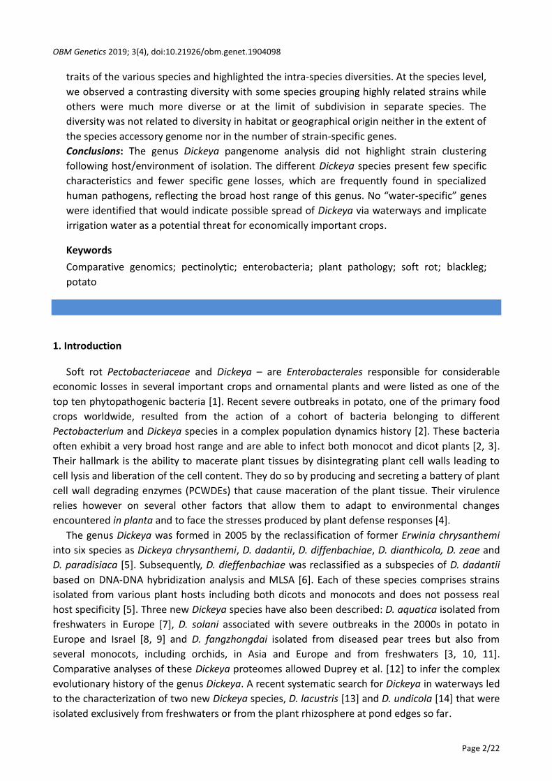
Based on MLSA analysis, Dickeya species could be further organized in different branches, clade
I grouping D. chrysanthemi, D. zeae and Dickeya sp. NCPPB569, clade II grouping the new D.
undicola species with D. solani, D. dadantii, D. fangzhongdai and D. dianthicola, a third clade
grouping D. lacustris/D. aquatica clade, and the D. paradisiaca branch (Figure 1).
We analyzed the relatedness between strains of similar species by calculating the two-by-two
average nucleotide identities (ANI) for each Dickeya genome (Figure 1) and highlighted contrasting
situations. Bacteria belonging to D. aquatica, D. dianthicola and D. undicola group shared around
99% or more ANI among them (Figure 1), revealing very high sequence conservation. However,
only three genomes of D. aquatica and D. undicola are available until now and they may not reflect
the complete diversity of these two species. The high conservation observed in these three species
does not correlate to similar geographical origins or similar hosts. Indeed, if all D. dianthicola
strains (except one) were isolated from diseased potato, they originate from locations all over the
world (Table 1). Similarly, while the three D. undicola strains were isolated from freshwaters, they
were from either France or Malaysia.

OBM Genetics 2019; 3(4), doi:10.21926/obm.genet.1904098
Page 5/22
Table 1 General features of the different Dickeya strains/genomes analyzed in this study.
Strains
Genome
length # scaffolds
# of
CDS
Isolated
from
Geographic
origin
Year of
isolation
D. aquatica 174/2(T) 4.50 complete 4535 river water UK
D. aquatica DW_0440 4.34 47 4353 river water Finland
D. aquatica CSL RW240 4.39 26 4398 river water UK
D. chrysanthemi 1591 4.81 complete 4604 Zea mays USA 1957
D. chrysanthemi L11 4.77 144 4691 lake water Malaysia 2014
D. chrysanthemi NCPPB3533 4.73 23 4583 Solanum tuberosum USA
D. chrysanthemi NCPPB402 (T) 4.70 21 4606 Chrysanthemum morifolium USA 1956
D. chrysanthemi NCPPB516 4.62 8 4542 Parthenium argentatum Denmark 1957
D. dadantii 3937 4.92 complete 4271 Saintpaulia ionantha France 1977
D. dadantii DSM18020 (T) 5.00 complete 4732 Pelargonium capitatum Comoros 1960
D. dadantii subsp. dieffenbachiae
NCPPB2976 (T) 4.82 14 4652 Dieffenbachia sp. USA 1977
D. dadantii NCPPB3537 4.81 1 4508 Solanum tuberosum Peru 1987
D. dianthicola DE440 4.87 55 4792 Solanum tuberosum USA 2016
D. dianthicola GBBC2039 4.80 32 4607 Solanum tuberosum Belgium
D. dianthicola IPO980 4.84 27 4493 Solanum tuberosum Netherlands 1991
D. dianthicola ME23 4.91 complete 4790 Solanum tuberosum USA
D. dianthicola NCPPB3534 4.87 1 4663 Solanum tuberosum Netherlands 1987
D. dianthicola NCPPB453 (T) 4.68 1 4477 Dianthus caryophyllus UK 1956

OBM Genetics 2019; 3(4), doi:10.21926/obm.genet.1904098
Page 6/22
D. dianthicola RNS04.9 4.72 complete 4567 Solanum tuberosum France 2004
D. dianthicola SS70 4.80 62 4665 Solanum tuberosum Pakistan 2017
D. dianthicola WV516 4.91 103 4795 Solanum tuberosum USA 2016
D. fangzhongdai B16 4.89 53 4580 Phalaenopsis orchid Slovenia 2010
D. fangzhongdai DMS101947 (T) 5.03 complete 4638 Pyrus pyrifolia China 2009
D. fangzhongdai M005 5.11 138 4788 waterfall Malaysia 2013
D. fangzhongdai M074 4.95 145 4631 waterfall Malaysia 2013
D. fangzhongdai MK7 4.93 21 4589 river water Scotland
D. fangzhongdai NCPPB3274 4.99 1 4656 Aglaonema St Lucia 1983
D. fangzhongdai ND14b 5.05 complete 4616 waterfall Malaysia 2013
D. fangzhongdai PA1 4.98 complete 4548 Phalaenopsis orchid China 2011
D. fangzhongdai S1 4.94 51 4652 Phalaenopsis orchid Slovenia 2012
D. lacustris S29 (T) 4,31 118 4377 lake water France 2017
D. paradisiaca 703 4.68 complete 4526 Solanum tuberosum Australia
D. paradisiaca NCPPB2511 (T) 4.63 1 4548 Musa paradisiaca Colombia 1970
D. solani Ds0432 4.92 complete 4498 Solanum tuberosum Finland 2004
D. solani F012 4.88 25 4509 Solanum tuberosum Russia 2010
D. solani IFB0099 4.93 complete 4516 Solanum tuberosum Poland 2005
D. solani IFB0223 4.94 complete 4516 potato rhizosphere Germany 2005
D. solani IFB0221 4.88 38 4518 potato rhizosphere Germany 2005
D. solani IPO2222 (T) 4.92 complete 4530 Solanum tuberosum Netherlands 2007

OBM Genetics 2019; 3(4), doi:10.21926/obm.genet.1904098
Page 7/22
D. solani PPO9019 4.96 complete 4641 muscari Netherlands 2006
D. solani PPO9134 4.87 22 4546 hyacinth Netherlands 2008
D. solani RNS05.1.2A 4.99 37 4718 Solanum tuberosum France 2005
D. solani RNS08.23.3.1.A 4.92 complete 4536 Solanum tuberosum France 2008
D. undicola 2B12 4.35 77 4178 lake water Malaysia 2014
D. undicola FVG01 4.61 178 4525 water France 2017
D. undicola FVG10 4.54 202 4484 water France 2016
Dickeya sp. NCPPB569 4.22 6 4235 Saccharum Australia
Dickeya sp. Secpp_1600 * 5.11 complete 4713 radish China 2016
D. zeae 586 4.82 complete 4515 Philodendron Schott USA
D. zeae CSL_RW192 4.70 4 4587 river water UK
D. zeae DZ2Q 4.65 26 4456 Oryza sativa Italy
D. zeae EC1 4.53 complete 4260 Oryza sativa China 1997
D. zeae MK19 4.67 4 4494 river water UK
D. zeae MS1 4.75 58 4589 Musa China 2009
D. zeae MS2 4.74 complete 4529 Musa paradisiaca China 2014
D. zeae NCPPB2538 (T) 4.56 7 4380 Zea mays USA 1970
D. zeae NCPPB3531 4.63 2 4354 Solanum tuberosum Australia
D. zeae NCPPB3532 4.56 1 4390 Solanum tuberosum Australia
D. zeae ZJU1202 4.59 188 4417 Oryza sativa China 2002
* Our analyses showed that the Secpp_1600 strain belongs to D. fangzhongdai.
OBM Genetics 2019; 3(4), doi:10.21926/obm.genet.1904098
Page 8/22
Figure 1 Phylogeny of the Dickeya genus. A. Phylogenic tree built up from the
concatenated sequences of 1191 homologous protein sequences (82093 variable sites).
One hundred bootstrap replicates were performed to assess the statistical support of
each node. Only one node (labelled ◇) was below 100% (bootstrap value 49%).
Pectobacterium atrosepticum 21A was used as an outgroup. The numbers of protein
families common to all genomes of the different clades/nodes are written in the left
part. ANI values ranges for each species are given. B. Pangenome tree: distance was
calculated from a presence/absence matrix of the pangenome (see Materials and
Methods). Origins: M: monocot; D: dicot; R: potato rhizosphere; : potato; : water.
The stars indicate differences between the phylogenic and pangenome trees.

OBM Genetics 2019; 3(4), doi:10.21926/obm.genet.1904098
Page 9/22
Similarly, no link was observed between plant host origin and genome variability. For instance,
as already reported *27, 28, 29+, most D. solani strains exhibiting very high ANI values above 99.9%,
were isolated either from potato or from ornamentals, pointing to a clonal origin and possible
contamination of potato from nearby fields planted with ornamentals *30+. However, one D. solani
genome, RNS05.1.2 was found to be more divergent with an ANI value of about 98% with the
other D. solani genomes *28, 29+. This divergence is not related to a different host or geographical
origins because, like other D. solani strains of our panel, this strain was isolated from potato in
France *28+.
Strains grouped in the D. fangzhongdai, D. zeae, D. dadantii and D. chrysanthemi species were
more diverse and, in some cases, fell below the limits of species definition, now generally
recognized as 95%–96% for average identity value (ANI) and 70% for digitally derived DNA-DNA
hybridization (dDDH) *31, 32+. While all D. dadantii and D. chrysanthemi strains harbored more
than 96/70% ANI/dDDH values between genomes of the same species, these values were at the
limits of species definition for some D. fangzhongdai or even fell below these limits for D. zeae
genomes. In both cases, a further subcladal organization was observed in these species and the
distribution of the analyzed genomes could be assigned to three branches, two subclades grouping
genomes that share ANI values ranging from 97% to 99%, and one divergent strain (NCPPB3274 in
D. fangzhongdai and 586 in D. zeae) (Figure 1) *33+. The ANI/dDDH values of genomes belonging to
the two D. zeae subclades were as low as 94/58.2% (Table S1), indicating the separation of these
two lineages into two distinct subspecies or even separate species. Further phenotypic
characterizations and analysis of more genomes are needed to solve this ambiguity.
3.2 Dickeya Species Share a Large Panel of Genes
In order to further address the relationships between the different Dickeya species, we carried
out a pan-genome analysis of this genus and compared the protein-coding sequences of the 59
genomes using the SiLix gene family clustering tool. Proteins were classified as homologous to
others in a given family if the amino acid identities were above 70% with 80% minimal overlap.
Bacterial pan-genomes could be divided into three gene classes as i) the core genome that groups
protein families shared by all members of the analyzed genus/species, ii) the specific genome
gathering protein families unique to one genome or a genome group, iii) the accessory genome
including protein families present in more than one but not all genomes of a group. The genus
Dickeya core genome comprises 1800 genes representing nearly 40% (37.5% to 43%) of a single
genome content (Figure 1). These common genes encompass most of the genes shown to be
involved in virulence (for a review, *4+). The hallmark of virulence of the soft rot causing Dickeya is
the production and secretion of a battery of plant cell wall degrading enzymes, such as pectinases,
cellulase, and proteases. The out type 2 secretion system (T2SS) drives the secretion of nine
pectate (PelABCDEILNZ), one pectin (Pnl), one or two rhamnogalacturonate (RhiE, RhiEbis) lyases
and various enzymes that strip off the pectin decorations (pectin methylesterases PemA and
PemB, acetyl esterase PaeY) as well as the cellulase CelZ. As recently described *12+, the
corresponding encoding genes are largely conserved in the Dickeya genus (Table 2). Like all
members of clade II, D. undicola possesses all PCWDE-encoding genes except the gene encoding
the second rhamnogalacturonate lyase. A notable exception is PelA whose gene is truncated in all
D. dianthicola genomes. Likewise, D. lacustris harbors almost the same PCWDE battery as its

OBM Genetics 2019; 3(4), doi:10.21926/obm.genet.1904098
Page 10/22
closest relative D. aquatica, except that it has only one copy of the pelD/E genes and possesses the
Pel10 encoding gene otherwise present only in D. fangzhongdai and some D. chrysanthemi strains.
D. paradisiaca and Dickeya sp. 569 were found to be less equipped, and they both lacked PelA,
PelI, Rhi, and PemB. In addition, PelZ, and PnlG were absent in Dickeya sp. 569 while D. paradisiaca
harbored only one copy of the PelD/E isoenzymes and lacked PaeY acetyl esterase.
Table 2 Conservation of genes encoding pectinolytic enzymes.
pelA peD/E pelB/C pelI pelL/N pelZ rhiE rhiE
bis pel10 pnlG pemA pemB paeY
D. dadantii 1 2 2 1 2 1 1 no no 1 1 1 1
D. solani 1 2 2 1 2 1 1 no no 1 1 1 1
D. dianthicola trunc 2 2 1 2 1 1 var no var 1 1 1
D. fangzhongdai 1 2 2 1 2 1 1 no 1 1 1 1 1
D. undicola 1 2 2 1 2 1 1 no no 1 1 1 1
D. chrysanthemi 1 2 2 1 2 1 var 1 var no 1 no 1
D. zeae 1 2 2 1 2 1 1 1 no var 1 1 1
Dickeya sp. 569 1 1 2 1 2 1 1 1 1 no 1 no 1
D. lacustris 1 2 2 1 2 1 1 1 no 1 1 no 1
D. aquatica no 1 div 2 no 2 1 no no no no 1 no no
D. paradisiaca no 1 2 1 1 no no no no 1 1 no 1
Trunc: truncated, div: divergent, var: variable.
All Dickeya species except D. paradisiaca were found to possess a Prt T1SS that allows the
secretion of up to four metalloproteases (PrtABCG). The genes encoding the three PrtABC
proteases are also largely shared while the prtG gene is absent in D. dianthicola, D. undicola, D.
chrysanthemi and Dickeya sp. NCPPB569 (Table 3).
Four other protein secretion systems (T3SS to T6SS) might be present in gram-negative bacteria
where they are involved in interactions with eukaryotic cells or in bacterial intra- and inter-
competitions *34+. All four TSS were present in the D. dadantii 3937 model strain but T5SS
comprised very long proteins truncated in the draft genomes and difficult to tackle. So, we
excluded T5SS from our study. The different Dickeya species possess variable sets of T3SS, T4SS,
and T6SS; D. paradisiaca and Dickeya. sp. NCPPB569 do not have any; D. lacustris and D. aquatica
possess only T3SS, and for the other species, the presence and number of these TSS are variable
(Table 3).
Upon plant colonization, bacteria encounter various stressful environmental conditions like the
low pH in apoplast, as well as osmotic and oxidative stresses. The D. dadantii 3937 model strain is
well equipped to cope with these stresses *35+. Like many other bacteria, it responds to oxidative
stress by producing antioxidant enzymes like catalases (KatE, KatG), superoxide dismutases (SodA,
SodC) and alkylhydroperoxide reductase (AhpCF) as well as repair systems like the peptide

OBM Genetics 2019; 3(4), doi:10.21926/obm.genet.1904098
Page 11/22
methionine sulfoxide reductase (MsrA), an enzyme repairing oxidative damage caused to proteins
*36+, or the Suf and Isc systems, involved in the repair of damaged Fe/S clusters containing proteins
*37, 38+. It also produces the ROS scavenging pigment indigoïdine, It can also resist antibacterial
peptides produced by plants through the Sap system and detoxify nitric oxide produced by plants
during infection with the nitric oxide dioxygenase HmpX *39, 40+. Most of the encoding genes are
well conserved in different Dickeya species (Table 3). However, the suf gene cluster is present only
in clade II as well as in D. chrysanthemi, HmpX is truncated in D. undicola and absent in Dickeya sp.
569 and D. paradisiaca. The indigoïdine encoding genes are absent in D. paradisiaca and D.
lacustris. (Table 3), although strikingly, D. lacustris was more resistant to oxidative stress than D.
dadantii despite the lack of indigoïdine production *13+.
Table 3 Conservation of virulence-related factors.
AvrL/M T1SS
prt prtG prtABC T3SS
T4SS
VirB* T4SSb* T6SS kat sod ind suf hmpX
D. dadantii 2 1 1 3 1 var var 1 2 2 yes yes 1
D. solani 2 1 1 3 1 1 var 1 2 2 yes yes 1
D. dianthicola 1 1 no 2 or 3 1 1 no 1 2 2 yes yes 1
D. fangzhongdai 2 1 1 3 1 var var 1 2 2 yes yes 1
D. undicola 1 1 no 3 1 no var 1 2 2 yes yes trunc
D. chrysanthemi 2 1 no 3 1 1 var 1 2 2 yes yes 1
D. zeae 0 or 1 1 1 3 1 var no 1 2 2 yes no 1
Dickeya sp. 569 no 1 no 1 no no no no 1 1 yes no no
D. lacustris 2 1 1 3 1 no no no 2 2 no no 1
D. aquatica 2 1 1 1 to 3 1 no no no 2 2 yes no 1
D. paradisiaca no no no no no no no no 2 2 no no no
*Two types of T4SS might be present in bacteria, either associated to protein secretion
encoded by the virB operon or associated with plasmid conjugation encoded by a
VirD2/VirD4/Trb locus present in an integrative conjugative transposon element (ICE) (T4SSSb).
Trunc: truncated, var: variable.
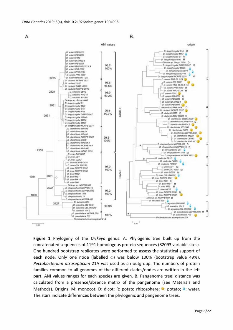
At the species level, core genomes are very large comprising nearly 3000 to 4000 protein
families, i.e., around two-thirds of the genetic content in a strain (Figure 2).
This high genetic conservation persists in clades that group different species. Indeed, the D.
solani/D. dadantii clade, D. undicola/D. fangzhongdai clade and D. lacustris/D. aquatica clade also
share around 3000 protein families (Figure 1). The D. zeae/D. chrysanthemi/Dickeya sp. NCPPB569
clade I is less related because it shares only around 2500 protein families, similar to the number of
protein families shared by all five clade II Dickeya species (Figure 1).
Therefore, even at the genus level, Dickeya members share a very large pool of genes as
compared to other Enterobacterales species. For comparison, using less stringent conditions (50%

OBM Genetics 2019; 3(4), doi:10.21926/obm.genet.1904098
Page 12/22
identity on 50% of the length of the proteins) the E. coli core genome includes only around 1500
orthologous genes *41+. Furthermore, most virulence determinants are shared by most Dickeya
species with the notable exceptions of D. paradisiaca and the Dickeya sp. 569 strain.
Figure 2 Conserved and strain-specific gene content in the different Dickeya species.
The protein families present in the core, accessory (in brackets) and strain-specific (in
petals) are depicted.
3.3 Accessory Genome and Strain-Specific Genes
The sizes of the accessory genome of species, which groups protein families present in more
than one but not all genomes of a given species, ranged from about 400 to more than 2100 protein
families (Figure 2). To analyze them further, we performed a pan-genome clustering analysis that
builds a hierarchical clustering based on the proportion of the presence/absence status of each
gene family in each genome (Figure 1B). The allocation in species determined by the whole
84 183
120
B16
3524 (1831)
177
111
64 516
NC
PP
B
32
74
141
M074
351 M005
221
158
265 112
17
4/2
3930 (318)
D. aquatica D. fangzhongdai D. dadantii
427
266 382
39
37
NCPPB 3537
DSM 18020
3467 (1041)
660
NC
PP
B
35
37
D. undicola D.dianthicola D. chrysanthemi
D. solani D.zeae
380
488 570
2B
12
3560 (437)
132
493 499
159
1
NCPPB
516 3286
(1445)
131 844
39 46
74
DE
440
3634 (1489)
133
159
69
141
147 IPO 980
356
459 191
337
58
6
3115 (2531)
243
231
MS
2
283 203
282
406
NCPPB 3531
194
114 EC1
3 5
12
Ds 0
43
2
3977 (691)
475 RNS
05.1.2
30 PPO 9134
137 3
IPO
2
22
2
19 IFB
0221
9 IFB
0099
11

OBM Genetics 2019; 3(4), doi:10.21926/obm.genet.1904098
Page 13/22
genome MLSA analysis is conserved reflecting the high number of protein families present in the
core genome of each species. Strikingly, however, the relatedness between Dickeya species at the
pan-genome level is quite different.
For instance, D. chrysanthemi did not cluster with D. zeae anymore but was now closer to clade
II species, D. undicola fell outside clade II and D. paradisiaca was closer to P. atrosepticum 21A than
to any Dickeya species (Figure 1B). At the species level, the distribution of D. zeae strains into two
branches as per the MLSA and ANI analyses was conserved in the pan-genome distribution, further
substantiating the separation into two subspecies/species. No correlation was observed between
this pan-genome clustering and habitat or geographical origin (Figure 1B and Table 1) except in D.
undicola (Figure 1B). Indeed, in D. undicola, the two strains isolated from France were closer to
each other than to the 2B12 strain in the pan-genome clustering, while in the MLSA core genome
analysis, D. undicola FVG10 was closer to 2B12 than to the other strain from France. Although the
current analysis was based on only three genomes, it points to a possible closeness of the
accessory genomes in relation to their geographical origin.
We also determined the number of protein families specific to each analyzed genome, and
absent from all other Dickeya genomes considered in this study (Figure 2). We found that D. solani
strains harbored very few strain-specific protein families (except 05.1.1.2), reflecting the already
reported clonal origin of these strains *27, 28, 29, 30+. Three D. dianthicola strains (DE440, WV516,
and ME23) also possess only a few dozen strain-specific genes coupled with very high ANI values (>
99.9%) among them. Interestingly, these three strains were isolated from potato in the USA,
pointing to the same clonal origin as reported for the recent blackleg outbreak devastating potato
crops in this country *2+. For the other strains, the number of strain-specific genes ranged from a
few dozen to several hundreds. Further, we did not observe any link between the proximity of the
core genomes revealed by ANI values in a given species and the extent of the strain-specific
genome. For example, the members of the D. dianthicola and D. undicola species exhibited ANI
values above 99% within a given species but harbored up to several hundred strain-specific genes,
in the same range as D. dadantii members that shared only 96.7% ANI among them.
3.4 Features Specific to Each Dickeya Species
To tackle the functions that might be specific to each Dickeya species, we identified the species-
specific genes present in all members of one species and missing in all other Dickeya genomes
analyzed in this study (corresponding proteins harboring less than 70% identity). The number of
species-specific genes ranged from 20 to 560 gene families (Table 4), although most of these
species-specific genes encode hypothetical proteins or proteins with undefined function.
This may be due to the fact that the RAST CDS definition program considers very short proteins
(less than 30 aa) for which no annotations are available. Expression profiling analyses of D. solani
and D. dianthicola strains, however, revealed that most of the small CDS are expressed and may be
differentially expressed in macerated tubers versus their growth in rich medium *42+. Annotation
of the species-specific genes is listed in Table S2. Out of the five D. undicola genes with a known or
predicted function, two genes encode regulatory proteins, and interestingly, a few (1 to 7) species-
specific regulatory genes are present in all Dickeya species except D. dadantii. Several species-
specific genes clustered in genomic regions, grouping at least five genes. D. aquatica harbors seven
such genomic regions that group 5 to 33 genes. These regions mainly contain genes encoding

OBM Genetics 2019; 3(4), doi:10.21926/obm.genet.1904098
Page 14/22
hypothetical proteins. Further, GR1 encodes an ABC-transport system related to nickel transporters
and GR2 harbors genes related to secondary metabolism (Table S2). Most D. chrysanthemi species-
specific genes with a known function are grouped in a genomic region of 26 genes that contain the
Flp/Tad operon and its associated two-component regulatory system (Table S2). The genes in this
operon encode the synthesis of a type IVb pilus shown to be involved in biofilm formation and
virulence in several bacteria including Pectobacteria *43+.
Table 4 Dickeya species and D. aquatica/D. lacustris clade specificities.
Species-specific
gene families
Of which
hypothetical
Species
specifically
absent genes
Of which
hypothetical
D. solani 122 101 0
D. dadantii 22 22 0
D. dianthicola 164 114 10 0
D.fangzhongdai 20 12 0
D. undicola 129 120 3 0
D. chrysanthemi 77 54 2 0
D. zeae 48 30 0
D. aquatica 560 481 9 3
D. lacustris ND 8 0
D. paradisiaca ND 143 32
Clade-specific gene
families
Of which
hypothetical
Clade specifically
absent genes
Of which
hypothetical
D. aquatica/
D. lacustris clade 239 124 54 5
ND: not determined because too few genomes were available.
In animal pathogens, diversity in virulence at the species level is often related to the absence
instead of the presence of genes, suggesting that high virulence is related to specialization as it
was described for Yersinia pestis *44+. We thus examined if species specificity may reside in the
absence of specific protein families that are present in all other Dickeya species. Very few such
families are present in Dickeya species, less than ten in all, except in D. paradisiaca that lacks 124
such genes (Table S2). As already described above, those families absent from D. paradisiaca
include several proteins associated with virulence like those encoding the Prt type 1 secretion
system of proteases, the pectin acetylesterase PaeY, a catalase, part of the achromobactine
siderophore biosynthesis pathway as well as several genes related to metabolism and regulation
(Table S2). Strikingly, D. paradisiaca strains possess all genes encoding proteins involved in
flagellum biosynthesis, although, these genes are more related to orthologous genes in Lonsdalea
and Brenneria than to their Dickeya counterparts.
OBM Genetics 2019; 3(4), doi:10.21926/obm.genet.1904098
Page 15/22
3.5 The D. aquatica/ D. lacustris Clade
The D. aquatica/D. lacustris clade is the only clade grouping strains isolated only from water,
hence, we analyzed the genomic specificities of this clade. Members of this clade share 239 genes
that are absent in all other Dickeya species (or exhibit less than 70% identity) (Table 4). Besides
genes encoding hypothetical proteins or proteins with undefined function (124 genes), these
clade-specific genes could be divided mainly into categories related to metabolism (45 genes),
transport (14 genes), regulation (6 genes), and resistance to stress (9 genes) (Table S3). Several of
these genes grouped into nine genomic regions (GR) defined as regions clustering at least five
genes (Table 5). Five of these genomic regions include genes involved in interesting
pathways/features.
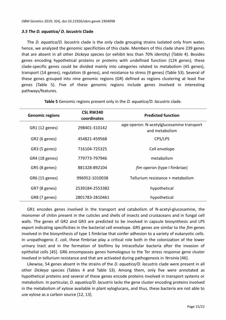
Table 5 Genomic regions present only in the D. aquatica/D. lacustris clade.
Genomic regions CSL RW240
coordinates Predicted function
GR1 (12 genes) 298401-310142 aga operon: N-acetylglucosamine transport
and metabolism
GR2 (6 genes) 454821-459568 CPS/LPS
GR3 (5 genes) 716104-725325 Cell envelope
GR4 (18 genes) 779773-797946 metabolism
GR5 (8 genes) 881328-892104 fim operon (type I fimbriae)
GR6 (15 genes) 996952-1010038 Tellurium resistance + metabolism
GR7 (8 genes) 2539184-2553382 hypothetical
GR8 (7 genes) 2801783-2810461 hypothetical
GR1 encodes genes involved in the transport and catabolism of N-acetyl-glucosamine, the
monomer of chitin present in the cuticles and shells of insects and crustaceans and in fungal cell
walls. The genes of GR2 and GR3 are predicted to be involved in capsule biosynthesis and LPS
export indicating specificities in the bacterial cell envelope. GR5 genes are similar to the fim genes
involved in the biosynthesis of type 1 fimbriae that confer adhesion to a variety of eukaryotic cells.
In uropathogenic E. coli, these fimbriae play a critical role both in the colonization of the lower
urinary tract and in the formation of biofilms by intracellular bacteria after the invasion of
epithelial cells *45+. GR6 encompasses genes homologous to the Ter stress response gene cluster
involved in tellurium resistance and that are activated during pathogenesis in Yersinia *46+.
Likewise, 54 genes absent in the strains of the D. aquatica/D. lacustris clade were present in all
other Dickeya species (Tables 4 and Table S3). Among them, only five were annotated as
hypothetical proteins and several of these genes encode proteins involved in transport systems or
metabolism. In particular, D. aquatica/D. lacustris lacks the gene cluster encoding proteins involved
in the metabolism of xylose available in plant xyloglucans, and thus, these bacteria are not able to
use xylose as a carbon source *12, 13+.
OBM Genetics 2019; 3(4), doi:10.21926/obm.genet.1904098
Page 16/22
For comparison, we performed a similar analysis with the members of the D. zeae/D.
chrysanthemi/Dickeya sp. 569 clade I. This clade shares only nine genes that are absent in all other
Dickeya species and conversely, no genes are absent in its members while present in all other
Dickeya.
Therefore, although D. aquatica and D. lacustris were isolated from water until now, they are
well equipped for invading plants as they possess most of the virulence-related genes (see 3.2)
and, contrary to D. paradisiaca, none of the genes absent in these species are known to be
involved in interactions with plants, except the genes involved in xyloglucan catabolism.
Accordingly, D. aquatica was shown to be capable of macerating acidic fruits like tomato or
cucumber *12+.
3.6 Does D. paradisiaca belong to the Dickeya Genus?
Contrary to the definition of bacterial species, the delimitation of genus adjuncts is still a matter
of debate. One of the proposed limits is an average nucleotide identity above 80% *32+. In the case
of D. paradisiaca, the ANI values ranged from 78 to 80% with the other Dickeya species (at or
below this limit), D. aquatica/D. lacustris being the more distant clade. Furthermore, D.
paradisiaca lacks several enzymes involved in pectinolysis as well as virulence determinants (see
3.2). It was however considered as the earlier branching lineage in the Dickeya genus in evolution
studies that analyzed the emergence of the different virulence-related genes *47, 12+. Finally, in
our pan-genome analysis (Figure 1B), D. paradisiaca clusters with P. atrosepticum, outside the
Dickeya genus.
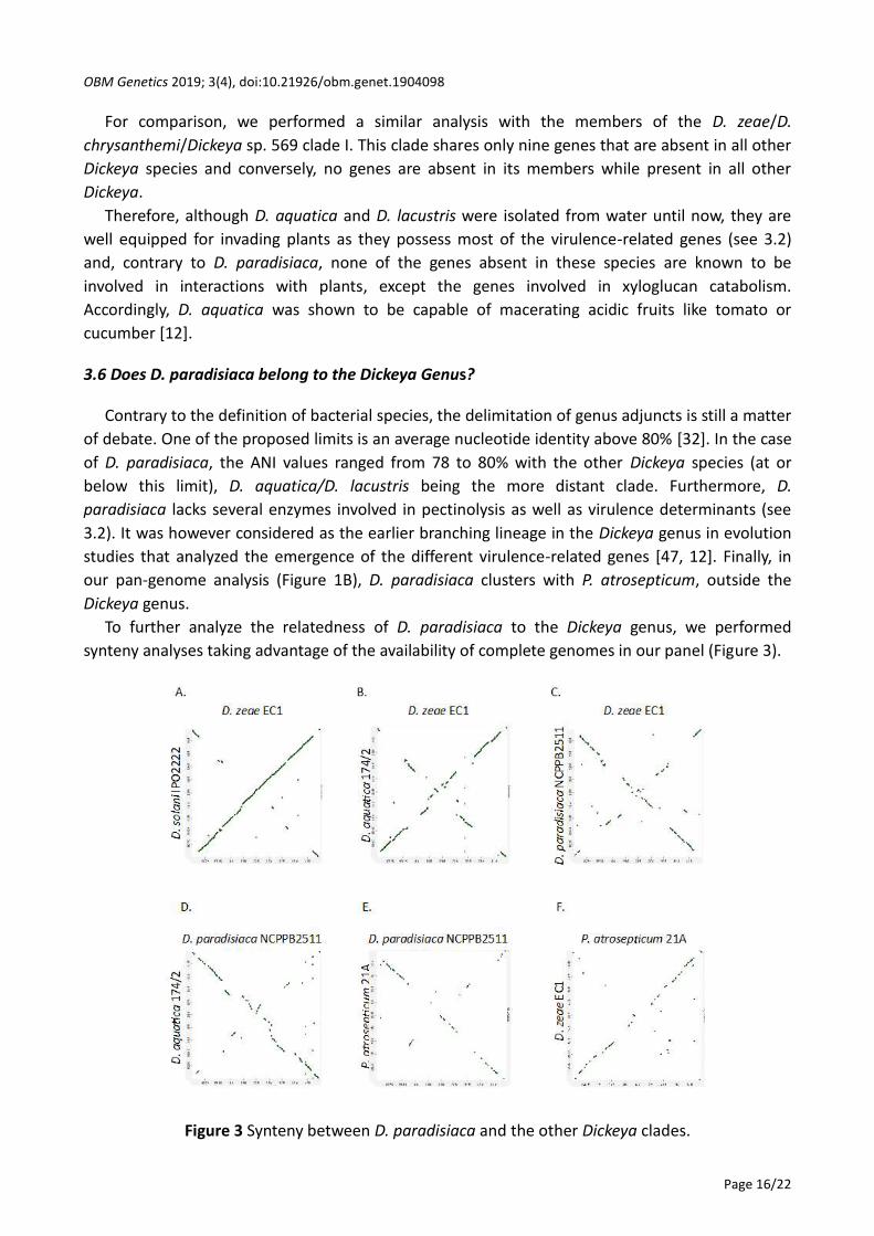
To further analyze the relatedness of D. paradisiaca to the Dickeya genus, we performed
synteny analyses taking advantage of the availability of complete genomes in our panel (Figure 3).
Figure 3 Synteny between D. paradisiaca and the other Dickeya clades.

OBM Genetics 2019; 3(4), doi:10.21926/obm.genet.1904098
Page 17/22
The synteny of clade II members of Dickeya species is very high as already described using the
MAUVE program *27, 42+. This high synteny was conserved between the clade I and clade II
clusters that show collinearity with only a few inversions of genomic regions, as exemplified by the
D. zeae EC1/D. solani IPO2222 synteny (Figure 3A). In D. aquatica 174/2, although the inversed
regions were larger compared to these clusters the syntenic regions were still dense (Figure 3B). In
contrast, the syntenic regions between D. paradisiaca NCPPB2511 and members of clades I and II
were more scattered (Figure 3C). Comparison between D. paradisiaca and D. aquatica gave similar
results (Figure 3D). For comparison, we also analyzed the synteny between Dickeya and the
Pectobacterium related genus exemplified by a P. atrosepticum strain (Figure 3E and Figure 3F).
The homologous regions were even more scattered despite a quite similar overall organization of
D. zeae and P. atrosepticum. This analysis revealed a gradient of synteny between the members of
the genus Dickeya with a very high closeness between the genomes of species belonging to clade I
and clade II while less homologous regions were observed in D. paradisiaca, and an intermediate
synteny was revealed in D. aquatica. Although D. aquatica was proposed as the second deepest
branching lineage in the genus Dickeya, it was not more syntenic to D. paradisiaca than to the
other Dickeya species. Nevertheless, our analyses only comprise two D. paradisiaca genomes (the
only ones available) and further phenotypic, as well as genomic analyses of a larger number of
strains, should be carried out before proposing a possible categorization of this bacterial group
into different genera.
4. Conclusions
One hallmark of the genus Dickeya is the high genetic closeness among its members. Indeed,
despite using high stringency parameters (70% identity with minimal overlap of 80%), all genomes
analyzed shared around as much as 1800 protein families and this was also true for D. paradisiaca
that is at the limit of affiliation to the genus Dickeya. At the species level, very few gene
specificities exist and even fewer specific gene losses frequently found in specialized human
pathogens. This, accompanied by high conservation of the characterized virulence-related genes in
most species, may account for the broad host range of the different Dickeya species that can infect
both monocot and dicot plants. Nevertheless, such a high relatedness in genetic content does not
necessarily imply similarity in strategies to adapt to the plant hosts. In fact, transcriptomic studies
during maceration of potato tubers revealed differences in gene expression even in very close
Dickeya species like D. solani and D. dianthicola *42+. Likewise, pangenome analysis does not
highlight strain clustering according to host or environment of their origin, further substantiating
the lack of specialization to a given environment. Among the 239 protein families present only in
the quite distant D. aquatica/D. lacustris clade that groups the strains isolated only from water, we
did not find any genetic trait that might contribute to habitat specificity. Instead, the members of
this clade shared two gene clusters that play a role in the interactions of animal pathogens with
their hosts and the other one facilitates interaction with insects and fungi. No protein family was
shared by the three Dickeya species found only in water so far, corroborating the absence of
“water-specific” genes already reported in D. fangzhongdai water isolates *33+. None of the
protein families shown to play a role in interactions with plants were found missing in the D.
aquatica/D. lacustris clade. Hence, their spread via waterways, specifically irrigation water might
be a potential threat for economically important crops.
OBM Genetics 2019; 3(4), doi:10.21926/obm.genet.1904098
Page 18/22
Earlier studies, by compiling hundreds of strains previously identified as Erwinia chrysanthemi,
defined seven Dickeya species *3, 26, 48+. In addition, the strain NCPPB569 (isolated from sugar
cane plantations in Australia) is significantly distinct to belong to an additional new species *3, 26,
48+. Recent studies in aquatic environments allowed the identification of three new Dickeya
species that were found only in water so far. It would be interesting to extend the search for
Dickeya isolates to new environments, for instance, insects that are recognized as hosts for some
Dickeya *49+ to explore if this will further broaden the diversity of genus Dickeya.
Acknowledgments
We thank Nicole Hugouvieux-Cotte-Pattat for sharing the D. lacustris genomic sequence before
publication and for fruitful discussions.
Additional Materials
The following additional materials are uploaded at the page of this paper.
1. Table S1: Relatedness of the Dickeya species. in silico DNA-DNA hybridization (DDH, upper
triangle) and Average Nucleotide Identity (ANI, lower triangle) values are presented. The specific
threshold value is 70% for DDH and 96% for ANI. Strains fulfilling these thresholds are highlighted
in tangerine.
2. Table S2: Dickeya species-specific and species specifically absent gene families.
3. Table S2: Gene families specific to and specifically absent from the D. aquatica/D. lacustris
clade.
Author Contributions
JP performed the bioinformatics analyses. Both authors analysed the data and wrote the
manuscript.
Funding
This research was supported by the COMBICONTROL ((ANR-17-CE32-0004-04) and SPREE (ANR-
17-CE32-0004-04) projects financed by the French National Agency for Research.
Competing Interests
The authors have declared that no competing interests exist.
References
1. Mansfield J, Genin S, Magori S, Citovsky V, Sriariyanum M, Ronald P, et al. Top 10 plant
pathogenic bacteria in molecular plant pathology. Mol Plant Pathol. 2012; 13: 614-629.
2. Charkowski AO. The changing face of bacterial soft-rot diseases. Annu Rev Phytopathol. 2018;
56: 269-288.
3. Suharjo R, Sawada H, Takikawa Y. Phylogenetic study of Japanese Dickeya spp. and
development of new rapid identification methods using PCR-RFLP. J Gen Plant Pathol. 2014;
80: 237-254.

OBM Genetics 2019; 3(4), doi:10.21926/obm.genet.1904098
Page 19/22
4. Charkowski A, Blanco C, Condemine G, Expert D, Franza T, Hayes C, et al. The role of secretion
systems and small molecules in soft-rot Enterobacteriaceae pathogenicity. Annu Rev
Phytopathol. 2012; 50: 425-449.
5. Samson R, Legendre JB, Christen R, Fischer-Le Saux M, Achouak W, et al. Transfer of
Pectobacterium chrysanthemi (Burkholder et al 1953) Brenner et al 1973 and Brenneria
paradisiaca to the genus Dickeya gen. nov. as Dickeya chrysanthemi comb. nov. and Dickeya
paradisiaca comb. nov. and delineation of four novel species. Int J Syst Evol Microbiol. 2005;
55: 1415-1427.
6. Brady CL, Cleenwerck I, Denman S, Venter SN, Rodrıguez-Palenzuela P, Coutinho TA, et al.
Proposal to reclassify Brenneria quercina (Hildebrand & Schroth 1967) Hauben et al. 1999 into
a novel genus, Lonsdalea gen. nov., as Lonsdalea quercina comb. nov., descriptions of
Lonsdalea quercina subsp. quercina comb. nov., Lonsdalea quercina subsp. Iberica subsp. nov.,
and Lonsdalea quercina subsp. britannica subsp. nov., emendation of the description of the
genus Brenneria, reclassification of Dickeya dieffenbachiae as Dickeya A new clade of Dickeya
dadantii subsp. dieffenbachiae comb. nov., and emendation of the description of Dickeya
dadantii. Int J Syst Evol Microbiol. 2012; 62: 1592-1602.
7. Parkinson N, DeVos P, Pirhonen M, Elphinstone J. Dickeya aquatica sp. nov., isolated from
waterways. Int J Syst Evol Microbiol. 2014; 64: 2264-2266.
8. Toth IK, van der Wolf JM, Saddler G, Lojkowska E, Helias V, Pirhonen M, et al. Dickeya species:
An emerging problem for potato production in Europe. Plant Pathol. 2011; 60: 385-399.
9. van der Wolf JM, Nijhuis EH, Kowalewska MJ, Saddler GS, Parkinson N, Elphinstone JG, et al.
Dickeya solani sp. nov., a pectinolytic plant-pathogenic bacterium isolated from potato
(Solanum tuberosum). Int J Syst Evol Microbiol. 2014; 64: 768-774
10. Tian Y, Zhao Y, Yuan X, Yi J, Fan J, Xu Z, et al. Dickeya fangzhongdai sp. nov., a plant
pathogenic bacterium isolated from pear trees (Pyrus pyrifolia). Int J Syst Evol Microbiol. 2016;
66: 2831-2835.
11. Alič Š, Van Gijsegem F, Pédron J, Ravnikar M, Dreo T. Diversity within the novel Dickeya
fangzhongdai sp., isolated from infected orchids, water and pears. Plant Pathol. 2018; 67:
1612-1620.
12. Duprey A, Taib N, Leonard S, Garin T, Flandrois, JP Nasser W, et al. The phytopathogenic
nature of Dickeya aquatica 174/2 and the dynamic early evolution of Dickeya pathogenicity.
Env Microbiol. 2019; 21: 2809-2835.
13. Hugouvieux-Cotte-Pattat N, Jacot-des-Combes C, Briolay J. Dickeya lacustris sp. nov., a
pectinolytic bacterium isolated from lakes in the French region of La Dombes. Int J Syst Evol
Microbiol. 2019; 69: 721-726.
14. Oulghazi S, Pédron J, Cigna J, Lau YY, Moumni M, Van Gijsegem F, et al. Dickeya undicola sp.
nov., a novel species for pectinolytic isolates from surface waters in Europe and Asia. Int J Syst
Evol Microbiol. 2019; 69: doi: 10.1099/ijsem.0.003497
15. Aziz RK, Bartels D, Best AA, Dejongh M, Disz T, Edwards RA, et al. The RAST server: Rapid
annotations using subsystems technology. BMC Genomics .2008; 9: 75.
16. Pritchard L, Glover RH, Humphris S, Elphinstone JG, Tothc IK. Genomics and taxonomy in
diagnostics for food security: Soft-rotting enterobacterial plant pathogens. Anal Methods.
2016; 8: 12-24
OBM Genetics 2019; 3(4), doi:10.21926/obm.genet.1904098
Page 20/22
17. Meier-Kolthoff JP, Auch AF, Klenk H-P, G ker M. Genome sequence-based species
delimitation with confidence intervals and improved distance functions. BMC Bioinformatics.
2013; 14: 60.
18. Cabanettes F, Klopp C. D-GENIES: Dot plot large genomes in an interactive, efficient and
simple way. PeerJ. 2018; 6: e4958.
19. Miele V, Penel S, Duret L. Ultra-fast sequence clustering from similarity networks with SiLiX.
BMC Bioinformatics. 2011; 12: 116.
20. Edgar RC. MUSCLE: Multiple sequence alignment with high accuracy and high throughput.
Nucleic Acids Res. 2004; 32: 1792-1797.
21. Gouy M, Guindon S, Gascuel O. SeaView version 4: A multiplatform graphical user interface
for sequence alignment and phylo- genetic tree building. Mol Biol Evol, 2010; 27: 221-224.
22. Castresana J. Selection of conserved blocks from multiple alignments for their use in
phylogenetic analysis. Mol Biol Evol. 2000; 17: 540-552.
23. Gascuel O. BIONJ: An improved version of the NJ algorithm based on a simple model of
sequence data. Mol Biol Evol. 1997; 14: 685-695.
24. Kumar S, Stecher G, Tamura K. MEGA7: Molecular evolutionary genetics analysis version 7.0
for bigger datasets. Mol Biol Evol. 2016; 33: 1870-1874
25. Méric G, Yahara K, Mageiros L, Pascoe B, Maiden MCJ, Jolley KA, et al. A reference pan-
genome approach to comparative bacterial genomics: Identification of novel epidemiological
markers in pathogenic Campylobacter. PLoS One. 2014; 9: e92798.
26. Zhang Y, Fan Q, Loria R. A re-evaluation of the taxonomy of phytopathogenic genera Dickeya
and Pectobacterium using whole-genome sequencing data. Syst Appl Microbiol. 2016; 39:
252-259.
27. Pédron J, Mondy S, Raoul des Essarts Y, Van Gijsegem F, Faure D. Genomic and metabolic
comparison with Dickeya dadantii 3937 reveals the emerging Dickeya solani potato pathogen
to display distinctive metabolic activities and T5SS/T6SS-related toxin repertoire. BMC
Genomics. 2014; 15: 1-13
28. Khayi S, Blin P, Pédron J, Chong TM, Chan KG, Moumni M, et al. Population genomics reveals
additive and replacing horizontal gene transfers in the emerging pathogen Dickeya solani.
BMC Genomics. 2015; 16: 1-13.
29. Golanowska M, Potrykus M, Motyka-Pomagruk A, Kabza M, Bacci G, Galardini M, et al.
Comparison of highly and weakly virulent Dickeya solani strains, with a view on the
pangenome and panregulon of this species. Front Microbiol. 2018; 9: 1940.
30. Sławiak M, Beckhoven JRCM, Speksnijder AGCL, Czajkowski R, Grabe G, Wolf JM. Biochemical
and genetical analysis reveal a new clade of biovar 3 Dickeya spp. strains isolated from potato
in Europe. Eur J Plant Pathol. 2009; 125.
31. Richter M, Rosselló-Móra R. Shifting the genomic gold standard for the prokaryotic species
definition. Proc Natl Am Sci USA. 2009; 106: 19126-19131
32. Konstantinidis KT, Tiedje JM. Genomic insights that advance the species definition for
prokaryotes. Proc Natl Am Sci USA. 2005; 102: 2567-2572.
33. Alic Š, Pedron J, Dreo T, Van Gijsegem F. Genomic characterisation of the new Dickeya
fangzhongdai species regrouping plant pathogens and environmental isolates. BMC Genomics.
2019; 20: 34.
34. Tseng TT, Tyler BM, Setubal JC. Protein secretion systems in bacterial-host associations, and their

OBM Genetics 2019; 3(4), doi:10.21926/obm.genet.1904098
Page 21/22
description in the gene ontology. BMC Microbiol. 2009; 9: S2.
35. Reverchon S, Nasser W. Dickeya ecology, environment sensing and regulation of virulence
programme. Environ Microbiol Rep. 2013; 5: 622-636.
36. El Hassouni M, Chambost JP, Expert D, Van Gijsegem F, Barras F. Identification of peptide
methionine sulfoxide reductase as a virulence determinant of the plant pathogen Erwinia
chrysanthemi. Proc Natl Acad Sci. 1999; 96: 887-892
37. Nachin L, El Hassouni M, Loiseau L, Expert D, Barras F. SoxR-dependent response to oxidative
stress and virulence of Erwinia chrysanthemi: The key role of SufC, an orphan ABC ATPase.
Mol Microbiol. 2001; 39: 960-972.
38. Rincon-Enriquez G, Crete P, Barras F, Py B. Biogenesis of Fe/S proteins and pathogenicity: IscR
plays a key role in allowing Erwinia chrysanthemi to adapt to hostile conditions. Mol Microbiol.
2008; 67: 1257-1273.
39. López-Solanilla E, Llama-Palacios A, Collmer A, García-Olmedo F, Rodríguez-Palenzuela P.
Relative effects on virulence of mutations in the sap, pel, and hrp loci of Erwinia chrysanthemi.
Mol Plant Microbe Interact. 2001; 14: 386-393.
40. Boccara M, Mills CE, Zeier J, Anzi C, Lamb C, Poole RK, et al. Flavohaemoglobin HmpX from
Erwinia chrysanthemi confers nitrosative stress tolerance and affects the plant hypersensitive
reaction by intercepting nitric oxide produced by the host. Plant J. 2005; 43: 226-237.
41. Lukjancenko O, Wassenaar TM, Ussery DW. Comparison of 61 sequenced Escherichia coli
genomes. Microb Ecol. 2010; 60: 708-720.
42. des Essarts YR, Pédron J, Blin P, Van Dijk E, Faure D, Van Gijsegem F. Common and distinctive
adaptive traits expressed in Dickeya dianthicola and Dickeya solani pathogens when exploiting
potato plant host. Envi Microbiol. 2019; 21: 1004-1018.
43. Nykyri J, Mattinen L, Niemi O, Adhikari S, Koiv V, Somervuo P, et al. Role and regulation of
the Flp/Tad pilus in the virulence of Pectobacterium atrosepticum SCRI1043 and
Pectobacterium wasabiae SCC3193. PLoS One. 2013; 8: e73718.
44. Merhej V, Georgiades K, Raoult D. Postgenomic analysis of bacterial pathogens repertoire
reveals genome reduction rather than virulence factors. Brief Funct Genomics. 2013; 12: 291-
304.
45. Wright KJ, Seed PC, Hultgren SJ. Development of intracellular bacterial communities of
uropathogenic Escherichia coli depends on type 1 pili. Cell Microbiol. 2007; 9: 2230-2241.
46. Ponnusamy D, Hartson SD, Clinkenbeard KD. Intracellular Yersinia pestis expresses general
stress response and tellurite resistance proteins in mouse macrophages. Vet Microbiol. 2011;
150: 146-151.
47. Duprey A, Nasser W, Leonard S, Brochier-Armanet C, Reverchon S. Transcriptional start site
turnover in the evolution of bacterial paralogous genes: The pelE-pelD virulence genes in
Dickeya. FEBS J. 2016; 283: 4192-4207.
48. Parkinson N, Stead D, Bew J, Heeney J, Tsror L, Elphinstone J. Dickeya species relatedness and
clade structure determined by comparison of recA sequences. Int J Syst Evol Microbiol. 2009;
59: 2388-2393.
49. Rossmann S, Dees MW, Perminow J, Meadow R, Brurberg MB. Soft rot Enterobacteriaceae are
carried by a large range of insect species in potato fields. Appl Environ Microbiol. 2018; 84:
e00281-18.
OBM Genetics 2019; 3(4), doi:10.21926/obm.genet.1904098
Page 22/22
Enjoy OBM Genetics by:
1. Submitting a manuscript
2. Joining in volunteer reviewer bank
3. Joining Editorial Board
4. Guest editing a special issue
For more details, please visit:
http://www.lidsen.com/journals/genetics
OBM Genetics