disulfiram copper nanoparticles prepared with a stabilized
TRANSCRIPT

Disulfiram Copper Nanoparticles Prepared with a Stabilized MetalIon Ligand Complex Method for Treating Drug-Resistant ProstateCancersWu Chen,† Wen Yang,‡ Pengyu Chen,‡ Yongzhuo Huang,*,§ and Feng Li*,†
†Department of Drug Discovery and Development, Harrison School of Pharmacy, and ‡Materials Research and Education Center,Materials Engineering, Department of Mechanical Engineering, Auburn University, Auburn, Alabama 36849, United States§State Key Laboratory of Drug Research, Shanghai Institute of Materia Medica, Chinese Academy of Sciences, Shanghai 201203,China
*S Supporting Information
ABSTRACT: Disulfiram (DSF), an alcohol-aversion drug, has been explored for cancertreatment. Copper diethyldithiocarbamate (Cu(DDC)2) complex formed by DSF andcopper ions is a major active ingredient for its anticancer activity. Direct administration ofCu(DDC)2 is a promising strategy to enhance the anticancer efficacy of DSF. However,efficient drug delivery remains a significant challenge for Cu(DDC)2 and hinders its clinicaluse. In this study, we developed a facile stabilized metal ion ligand complex (SMILE)method to prepare Cu(DDC)2 nanoparticles (NPs). The SMILE method could prepareCu(DDC)2 NPs with different types of stabilizers including 1,2-distearoyl-sn-glycerol-3-phosphoethanolamine−poly(ethylene glycol) (PEG) 2000, D-α-tocopherol PEG 1000succinate, methoxy PEG 5000-b-poly(L-lactide) 5000, and other generally recognized assafe excipients approved by the US Food and Drug Administration. The optimizedformulations demonstrated excellent drug-loading efficiency (close to 100%), high drugconcentrations (increased drug concentration by over 200-fold compared to the traditionalmicelle formulation), and an optimal particle size in the sub-100 nm range. Cu(DDC)2NPs exhibited outstanding stability in serum for 72 h and can also be stored at room temperature for at least 1 month. Theanticancer effects of Cu(DDC)2 NP formulations were determined by multiple assays including 3-(4,5-dimethyl-thiazol-2-yl)-2,5-diphenyl tetrazolium bromide assay, colony-forming assay, calcein-AM/propidium iodide staining, and others. Cu(DDC)2NPs showed excellent activity against drug-resistant prostate cancer cells and other cancer cells with a half-maximal inhibitoryconcentration (IC50) of around 100 nM. Our study also demonstrated that Cu(DDC)2 NPs induced cell death in drug-resistantprostate cancer cells (DU145-TXR) through paraptosis, which is a nonapoptotic cell death. To our best knowledge, the SMILEmethod provides, for the first time, a simple yet efficient process for generating Cu(DDC)2 NPs with high drug concentration,excellent loading efficiency, and desirable physicochemical properties. This method could potentially address drug deliverychallenges of DSF/copper-based chemotherapy and facilitate its clinical translation.
KEYWORDS: disulfiram, copper diethyldithiocarbamate, drug resistance, prostate cancer, nanoparticle, drug delivery, paraptosis
1. INTRODUCTION
Prostate cancer is the most common male cancer in America;about one man in nine will have prostate cancer. Prostatecancer is also the second leading cause of cancer-associateddeath among males in the USA (https://www.cancer.org/cancer/prostate-cancer/about/key-statistics.html). Althoughmultiple therapeutic strategies, such as hormone therapy andradiation, have been used successfully for early-stage prostatecancer, it inevitably progresses to an aggressive form ofcastration-resistant prostate cancer (CRPC). Although early-stage localized prostate cancer can be effectively treated withless difficulty, the treatment of late-stage aggressive andmetastatic forms of prostate cancer presents a significantchallenge. Chemotherapy with taxanes is often used as a first-line drug when prostate cancer has migrated outside theprostate gland and becomes resistant to hormone therapy.1,2
However, taxane therapy failed to show good responses in halfof the patients. Even for patients with good initial responses,they may eventually develop drug-resistant prostate cancer.2−6
Because of the lack of effective therapy, patients with drug-resistant prostate cancer have a very poor prognosis. Both theprivate sector and academia have put forth substantial efforts todevelop new therapies for drug-resistant prostate cancer. Newdrugs, such as ixabepilone and cabazitaxel, have beendeveloped for treating refractory cancer. However, thedevelopment of a new drug molecule usually takes a longtime and requires significant investment, which eventuallyresults in a high cost of the treatment. Therefore, the
Received: August 31, 2018Accepted: October 30, 2018Published: November 16, 2018
Research Article
www.acsami.orgCite This: ACS Appl. Mater. Interfaces 2018, 10, 41118−41128
© 2018 American Chemical Society 41118 DOI: 10.1021/acsami.8b14940ACS Appl. Mater. Interfaces 2018, 10, 41118−41128
Dow
nloa
ded
via
AU
BU
RN
UN
IV o
n A
ugus
t 6, 2
019
at 1
4:50
:45
(UTC
).Se
e ht
tps:
//pub
s.acs
.org
/sha
ringg
uide
lines
for o
ptio
ns o
n ho
w to
legi
timat
ely
shar
e pu
blis
hed
artic
les.

development of effective but more affordable therapeuticagents for drug-resistant prostate cancer will greatly benefitpatients with this disease. Particularly, it will help thosepatients with financial constraints, which limit their access tooncology drugs affixed with skyrocketing prices.Drug repurposing or repositioning has received increasing
attention as an alternative strategy for drug discovery anddevelopment.7 Disulfiram (DSF) is an alcohol-aversion drugused for treating alcohol dependence for over 60 years.Recently, DSF has been repurposed as an anticancer agent andshows excellent anticancer activity when it is combined withcopper ions (Cu2+).8,9 DSF/Cu has been shown to inhibit theproteasome/poly-Ub protein degradation pathway by targetingthe nuclear protein localization 4 (NPL4) protein.9,10 DSF/Cucould also inhibit cancer stem cells11,12 and sensitize resistantcancer to chemotherapy drugs by inhibiting P-gp.13 Severalongoing clinical trials are testing the DSF/copper combinationin patients with glioblastoma and metastatic CRPC.14 Thedrug repurposing strategy will expedite the drug developmentprocess and facilitate the clinical translation.15 The anticanceractivity of DSF/copper is greatly dependent on the formationof the active metabolite, copper diethyldithiocarbamate(Cu(DDC)2).
10 Because of the poor in vivo stability andrapid degradation of DSF,9,11 the coadministration of DSF andcopper to patients yielded extremely low in vivo concentrationsof Cu(DDC)2, which would significantly compromise theanticancer efficacy and result in poor clinical outcomes. Thus,the administration of the preformed Cu(DDC)2 complex isexpected to be a more effective approach to obtain higheractive drug concentrations and to achieve better anticancerefficacy.16
Because of the low water solubility of Cu(DDC)2, there is agreat need to develop a formulation designed to increaseCu(DDC)2 solubility, thereby making it acceptable for clinicaluse.16,17 Nanotechnology-based approaches have been used forthe delivery of Cu(DDC)2.
16,18,19 In previous studies, thedirect loading of Cu(DDC)2 into nanoparticles (NPs) onlyachieved low drug-loading efficiency and low drug concen-tration.19 Recently, Metaplex technology or PRCosomes havebeen developed to prepare an injectable formulation ofCu(DDC)2.
20 This breakthrough technology utilizes liposomesas a nanoscale reaction vessel to synthesize Cu(DDC)2 NPsfrom copper ions (Cu2+) and diethyldithiocarbamate (DDC−)within the aqueous core of liposomes.16 The in situ formed
Cu(DDC)2 NPs were incorporated inside liposomes and thusstabilized by liposome membranes. However, this methodinvolves complicated preparation and purification processes,which will present difficulties and high expenses in large-scalemanufacturing.In this study, we developed a novel stabilized metal ion
ligand complex (SMILE) technology to prepare Cu(DDC)2NPs. The SMILE technology involves a simple formulationand a straightforward, cost-effective preparation process, whichthus can be easily scaled up during manufacturing and producedesirable Cu(DDC)2 NPs at a reasonable price. This methodcould prepare Cu(DDC)2 NPs with high drug concentrations,high loading efficiencies, and well-controlled physicochemicalproperties. We also investigated the anticancer activity andanticancer mechanism of the developed Cu(DDC)2 NPs.
2. MATERIALS AND METHODS2.1. Materials. 1,2-Distearoyl-sn-glycerol-3-phosphoethanol-
amine−poly(ethylene glycol) 2000 (DSPE−PEG) was purchasedfrom NOF Corporation. Methoxy PEG 5000-b-poly(L-lactide) 5000(PEG−PLA) was purchased from Polymer Source Inc. D-α-Tocopherol PEG 1000 succinate (TPGS) was purchased fromSigma-Aldrich. Other chemicals, solvents, and supplies werepurchased from VWR International.
2.2. Preparation of Cu(DDC)2 NPs. 2.2.1. Preparation ofCu(DDC)2-Loaded Micelles. Cu(DDC)2-loaded micelles were pre-pared with a film-dispersion method as described in previousreports.21−24 Briefly, 40 mg of micelle-forming materials and a givenamount of Cu(DDC)2 were dissolved in 0.5 mL of dichloromethane(CH2Cl2), and the solvent was removed under reduced pressure toform a film. The resulting film was hydrated in 1 mL deionized waterand sonicated for 5 min. Unloaded Cu(DDC)2 was removed bycentrifugation at 12 000 rpm for 5 min. The supernatant was collectedand filtered with 0.45 μM membrane filters.
2.2.2. Preparation of Cu(DDC)2 NPs with the SMILE Technology.Cu(DDC)2 NPs were prepared by mixing sodium diethyldithiocarba-matetrihydrate (DDC-Na) and copper chloride aqueous (CuCl2)solution containing a stabilizer. The molar ratio between DDC-Naand CuCl2 was 2:1. Briefly, DDC-Na and CuCl2 were dissolved in 1%(w/v) stabilizer to get a DDC-Na solution and a CuCl2 solution,respectively. Then, DDC-Na solution and CuCl2 solution were mixedand vortexed for 1 min to form Cu(DDC)2 NPs. The resulting NPformulation was centrifuged at 10 000 rpm for 10 min and filteredwith the 0.45 μM membrane to remove large aggregations (Scheme1).
2.3. Determination of Particle Size and Morphology.2.3.1. Particle Size. The particle size and size distribution of NPs
Scheme 1. Preparation of Cu(DDC)2 NPs with the SMILE Method
ACS Applied Materials & Interfaces Research Article
DOI: 10.1021/acsami.8b14940ACS Appl. Mater. Interfaces 2018, 10, 41118−41128
41119
were determined by dynamic light scattering (DLS) using theMalvern Nano ZS.25 Briefly, a Cu(DDC)2 NP sample (200 μL) wasadded to a microcuvette. The particle size and size distribution weredetermined based on DLS at a 173° scattering angle.2.3.2. Transmission Electron Microscope. The morphology of NPs
was characterized using a high-resolution transmission electronmicroscope (JEM-2100F, JEOL). Samples were loaded onto a gridand stained with 1% uranyl acetate. The grid was visualized under theelectron microscope.2.4. Nuclear Magnetic Resonance. 1H NMR spectra were
recorded on a Varian 400 MHz spectrometer using deuteratedchloroform (CDCl3) or deuterated water (D2O) as a solvent.2.5. Drug Concentration and Loading Efficiency. The
concentration of Cu(DDC)2 NPs was determined with a UV−visspectrometer. Briefly, Cu(DDC)2 NP samples were diluted withdimethylformamide, and the absorbance at 435 nm was determined.The Cu(DDC)2 concentration was calculated based on the standardcurve generated with different concentrations of Cu(DDC)2. Thedrug-loading efficiency was calculated using the following equation:
Drug loading efficiency (%)
(actual drug concentration/theoretical drug concentration)
100%
=
×
2.6. Determination of the Stability of Cu(DDC)2 NPs.2.6.1. Serum Stability. Cu(DDC)2 NPs were mixed and incubatedwith 10% fetal bovine serum (FBS) at room temperature. The particlesize was determined with DLS at the different time points.2.6.2. Long-Term Storage Stability. Cu(DDC)2 NPs were kept at
room temperature, and drug concentrations were determined ondifferent days during storage.
2.7. MTT Cytotoxicity Assay. MCF-7 breast cancer cells(ATCC) were cultured in a medium composed of Roswell ParkMemorial Institute (RPMI) 1640 with 10% FBS and 1% antibiotic−antimycotic. MDA-MB-231 cells (ATCC) were cultured in a 1:1mixture of Dulbecco’s modified Eagle’s medium and Ham’s F-12medium, with 10% FBS and 1% antibiotic−antimycotic. DU145-TXRdrug-resistant prostate cancer cells were obtained from Dr. EvanKeller at the University of Michigan and cultured in a RPMI 1640medium supplemented with 10% FBS, 1% antibiotic−antimycotic,and 40 nM paclitaxel. Cells were cultured at 37 °C in a humidifiedatmosphere containing 5% CO2.Cells were seeded into a 96-well plate at a density of 5000 cells/
well and incubated overnight. Then, a series of differentconcentrations of Cu(DDC)2 NPs diluted in a cell culture mediumwere prepared and added to each well (100 μL/well). At differenttime points, cytotoxicity was determined with the 3-(4,5-dimethyl-thiazol-2-yl)-2,5-diphenyl tetrazolium bromide (MTT) assay. Theabsorbance was determined with a microplate spectrophotometer at awavelength of 570 nm and a reference wavelength of 670 nm.Cytotoxicity was calculated using the following equation:
A ACell viability (%) ( / ) 100%test control= ×
The half-maximal inhibitory concentration (IC50) was calculatedwith SigmaPlot software based on a dose−response curve.26,27
2.8. Colony-Forming Assay. Cells were seeded in a 24-well plateat a density of 500 cells per well and incubated overnight. Then, cellswere treated with different formulations for 2 h and further cultured ina fresh cell culture medium for 1 week. Colonies were fixed with 100%methanol and stained with crystal violet.28
2.9. Determination of Intracellular Reactive OxygenSpecies. The generation of reactive oxygen species (ROS) wasdetermined with a 2′,7′-dichlorodihydrofluorescein diacetate
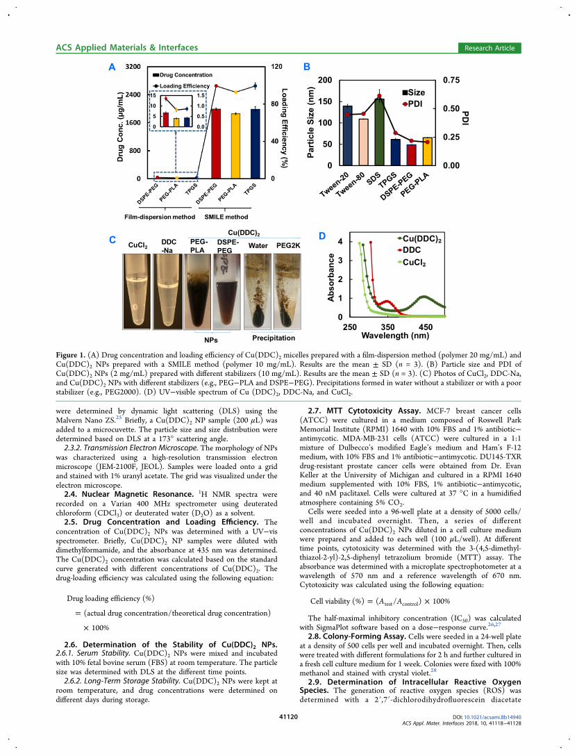
Figure 1. (A) Drug concentration and loading efficiency of Cu(DDC)2 micelles prepared with a film-dispersion method (polymer 20 mg/mL) andCu(DDC)2 NPs prepared with a SMILE method (polymer 10 mg/mL). Results are the mean ± SD (n = 3). (B) Particle size and PDI ofCu(DDC)2 NPs (2 mg/mL) prepared with different stabilizers (10 mg/mL). Results are the mean ± SD (n = 3). (C) Photos of CuCl2, DDC-Na,and Cu(DDC)2 NPs with different stabilizers (e.g., PEG−PLA and DSPE−PEG). Precipitations formed in water without a stabilizer or with a poorstabilizer (e.g., PEG2000). (D) UV−visible spectrum of Cu (DDC)2, DDC-Na, and CuCl2.
ACS Applied Materials & Interfaces Research Article
DOI: 10.1021/acsami.8b14940ACS Appl. Mater. Interfaces 2018, 10, 41118−41128
41120

(H2DCFDA) dye method.29 Briefly, cells were seeded in a dark-walled, clear-bottomed 96-well plate with 50 000 cells per well andincubated at 37 °C overnight before study. Cells were incubated with20 μM H2DCFDA in Hank’s buffered salt solution (HBSS, pH = 7.4)for 30 min at 37 °C in the dark and treated with differentformulations. Then, fluorescence was determined with a Cytation 5Imaging Reader at EX485nm/EM535nm.2.10. Live/Dead Staining with Calcein-AM/Propidium
Iodide. Cells were seeded at a density of 5000 cells per well in a96-well plate. After overnight incubation, cells were treated withdifferent formulations for 24 h and then stained with a solutioncomposed of calcein-AM and propidium iodide (PI) in pH 7.4phosphate-buffered saline (PBS). Cell samples were analyzed with aCytation 5 Cell Imaging Multi-Mode Reader. Viable and dead cellscan be identified by the green fluorescence (viable) and the redfluorescence (dead), respectively. The fluorescence intensities weredetermined quantitatively at EX480nm/EM530nm (viable cells) andEX530nm/EM620nm (dead cells).2.11. Caspase 3/7 Activities. Cells were seeded in a 96-well plate
at a density of 20 000 cells per well and incubated overnight. Aftertreating cells with different formulations, the culture medium wasremoved and 70 μL Caspase Glo 3 reagent (Promega, Madison, WI)was added to each well. After gently mixing the content in each well,the plate was incubated at room temperature for 30 min under darkconditions. Finally, 50 μL of the reaction solution was measured usinga luminometer (Cytation 5 Imaging Reader).27,30
2.12. Cell Morphology. Cells seeded in a 96-well plate weretreated with Cu(DDC)2 NPs. The NP-induced change of cellmorphology was observed with a Cytation 5 Cell Imaging Multi-Mode Reader. We also cotreated cells with cycloheximide (CHX, aprotein synthesis and paraptosis inhibitor) or chloroquine (CQ, anautophagy inhibitor) and observed their effects on Cu(DDC)2 NP-induced cell morphology change.2.13. Endoplasmic Reticulum Staining. Cells were stained with
endoplasmic reticulum (ER)-specific dye and Hoechst 33342 prior tomicroscopy. Briefly, cells were seeded in a clear-bottomed, black-walled 96-well plate at a density of 20 000 cells per well. Aftertreatment with different formulations for 8 h, cells were incubatedwith a staining solution containing ER track dye (ER-ID green, EnzoLife Sciences Inc.) and Hoechst 33342 for 30 min at 37 °C avoidinglight. Then, cells were washed with PBS and observed under afluorescence microscope.
3. RESULTS
3.1. Preparation of Cu(DDC)2 NPs with the SMILEMethod. Because of the poor water solubility of Cu(DDC)2,there is an emerging need to develop an injectable formulationfor its clinical use.16,17 In this study, we first attempted to use aclassical film-dispersion method to prepare Cu(DDC)2-loadedmicelles. However, the drug-loading efficiency was extremelylow. The drug concentrations varied among different polymerstested, but none of them achieved satisfactory drugconcentration and loading efficiency. The resulted drugconcentration was below 10 μg/mL, and the drug-loadingefficiency was below 2% (left columns and the inserted panel inFigure 1A). Therefore, we developed the SMILE technology asan innovative method to prepare Cu(DDC)2 NPs. With theSMILE method, we successfully prepared Cu(DDC)2 NPswith a high drug concentration (2 mg/mL) and a high loadingefficiency (close to 100%) using different stabilizers includingDSPE−PEG, PEG−PLA, and TPGS (Figure 1A). We alsoextensively investigated the effects of theoretical drugconcentrations and different stabilizers on actual drugconcentrations and loading efficiencies (Figures S1 and S2).At the theoretical drug concentrations of 2 or 4 mg/mL, bothTPGS and DSPE−PEG at concentrations ranging from 0.5 to4% could all generate NPs with high drug-loading efficiency
(close to 100%). The drug-loading efficiency was slightly lowerin 4 mg/mL theoretical drug concentration groups comparedto that in the 2 mg/mL groups. When the polymer stabilizerconcentrations were at high levels (e.g., 2 or 4% of TPGS andDSPE−PEG, respectively), those groups with lower theoreticaldrug concentrations (e.g., 0.5 and 1 mg/mL of Cu(DDC)2)showed significantly decreased Cu(DDC)2 drug-loadingefficiencies. However, when the polymer stabilizer concen-trations were at low levels (e.g., 0.5 or 1%), those groups withsimilar low theoretical drug concentrations (e.g., 0.5 and 1 mg/mL of Cu(DDC)2) showed much higher drug-loadingefficiencies.We also studied the effects of theoretical drug concentration
and stabilizer concentration on the particle size andpolydispersity index (PDI). When the theoretical drugconcentration was 2 or 4 mg/mL, NPs exhibited a size of60−70 nm if prepared with the TPGS stabilizer at theconcentrations ranging from 0.5 to 4%. These NPs alsoshowed a good size distribution as indicated by the small PDIvalue (Figure S3). However, when the theoretical drugconcentration was set to be 1 or 0.5 mg/mL, the particlesize varied significantly depending on the concentrations ofTPGS used. These NPs also showed large PDI values,indicating a broad particle size distribution. A similar trendwas also observed in NPs prepared with DSPE−PEG as thestabilizer. We could prepare NPs of well-controlled particle sizewhen theoretical drug concentrations were 2 or 4 mg/mL withDSPE−PEG at concentrations ranging from 0.5 to 4%. Theparticle sizes were in the range of 40−80 nm depending on theDSPE−PEG and drug concentrations (Figure S4). All theseNPs showed a narrow particle size distribution (small PDIvalue). At lower theoretical drug concentrations (0.5 or 1 mg/mL), NPs showed a large variation of particle sizes and largePDI values. These results suggested that the interactionbetween DDC− and Cu2+ during the complex formation wascritical for the NP preparation. This interaction is greatlyinfluenced by the concentrations of DDC− and Cu2+. Althoughstabilizers are essential for the preparation and stabilization ofCu(DDC)2 NPs, high concentrations of stabilizers (e.g., 2 or4%) may interfere with the interaction between DDC− andCu2+ and thus have a negative impact on the NP formation.These negative effects were more significant at lowerconcentrations of DDC− and Cu2+. Therefore, the optimizedtheoretical drug concentration (2 mg/mL) and stabilizerconcentration (1%) were used in the rest of the study unlessotherwise specified.We further explored the preparation of Cu(DDC)2 NPs with
additional stabilizers including Tween-20, Tween-80, andsodium dodecyl sulfate. All of these stabilizers could besuccessfully used to prepare Cu(DDC)2 NPs with particle sizesranging from 50 to 150 nm. The selection of stabilizers had apronounced impact on the particle size and PDI (Figure 1B).Compared with the micelle solutions of DSPE and TPGS, thecorresponding Cu(DDC)2 NPs showed significant largerparticle sizes. In contrast, the PEG−PLA micelle and PEG−PLA/Cu(DDC)2 NPs had similar particle sizes (Figure S5).We characterized the blank PEG−PLA and PEG−PLA/Cu(DDC)2 NPs using transmission electron microscopy(TEM). Both of them showed spherical morphology andsimilar particle sizes (Figure S6).The formation of Cu(DDC)2 NPs was also confirmed by
colorimetric visualization and by UV−vis spectroscopy. TheSMILE method produced stable Cu(DDC)2 NPs with a dark
ACS Applied Materials & Interfaces Research Article
DOI: 10.1021/acsami.8b14940ACS Appl. Mater. Interfaces 2018, 10, 41118−41128
41121

color in the presence of appropriate stabilizers (e.g., PEG−PLA and DSPE−PEG), while precipitation formed with a poorstabilizer or without a stabilizer (Figure 1C). The UV−visspectrum showed that Cu(DDC)2 has a characteristic peak ofaround 450 nm, which is absent in pure DDC-Na or CuCl2solutions (Figure 1D). We also used 1H NMR spectroscopy toconfirm the formation of NPs and study the structure of PEG−PLA Cu(DDC)2 NPs (Figure 2). When all components were
dissolved in CDCl3, the peaks from the hydrophilic PEG block(3.6 ppm), the hydrophobic PLA block (5.1 and 1.7 ppm), andCu(DDC)2 (0.7 ppm) were all observed. When PEG−PLACu(DDC)2 NPs were prepared in D2O, the peaks correspond-ing to the hydrophobic PLA block and Cu(DDC)2 weresignificantly diminished in contrast to the strong peaks for thehydrophilic PEG block. These results demonstrated that thePEG−PLA/Cu(DDC)2 NPs had a core−shell structure withCu(DDC)2 embedded inside the hydrophobic core of PEG−PLA micelles. The PEG−PLA surrounding Cu(DDC)2 NPscould stabilize the NPs and prevent aggregation formation.The selection of stabilizers also had a great influence on NP
stability. We determined the stability of NPs in the presence of10% serum. PEG−PLA/Cu(DDC)2 NPs showed excellentstability and did not have a significant change in particle sizeand PDI after incubation for at least 72 h. Although there wasno obvious precipitation in TPGS and DSPE−PEG Cu-(DDC)2 NP groups, particle sizes significantly increased overtime (Figure 3A,B). We also determined the long-term stabilityof Cu(DDC)2 NPs prepared with different stabilizers,including TPGS, DSPE−PEG, and PEG−PLA. NPs were
kept at room temperature, and changes in drug concentrationwere determined. As shown in Figure 3C, PEG−PLA/Cu(DDC)2 NPs demonstrated excellent stability with aminor decrease of drug concentration after 30 days of storageat room temperature. In contrast, the drug concentrations ofDSPE−PEG and TPGS NPs were significantly decreased,indicating their poor storage stability.
3.2. Anticancer Activity of Cu(DDC)2 NPs. Theanticancer activity of Cu(DDC)2 NPs in drug-resistantDU145-TXR cells was first determined using the MTT assay.As shown in Figure 4A, the DU145-TXR cell is resistant topaclitaxel with an IC50 of 2575 nM. Cu(DDC)2 NPs preparedwith PEG−PLA, TPGS, and DSPE−PEG all showed potentanticancer activities at 48 h treatment. The IC50 values were85, 172, and 193 nM, respectively. The treatment of equivalentconcentrations of CuCl2, DDC-Na, blank PEG−PLA, blankDSPE−PEG, or TPGS did not show significant cell toxicity.The anticancer effects also depended on the treatment timewith increased anticancer effects occurring after prolongedtreatment. Cells treated with DSPE−PEG/Cu(DDC)2 NPsshowed an IC50 of 216 nM at 24 h and 138 nM at 72 h (Figure4B). Similarly, the cells treated with TPGS/Cu(DDC)2 NPsshowed an IC50 of 280 nM at 24 h and 149 nM at 72 h (Figure4C). Cu(DDC)2 NPs also demonstrated excellent anticanceractivity in other cancer cells including MDA-MB-231 cells andMCF-7 cells. The IC50 values of TPGS/Cu(DDC)2 NP andPEG−PLA Cu(DDC)2 NPs on MDA-MB-231 cells were 123and 104 nM, respectively (Figure 4D). We further tested cellviability based on calcein AM and PI staining of cancer cellstreated with Cu(DDC)2 NPs. The treatment caused a dose-dependent increase in membrane permeability. The redstaining of dead cells increased with increasing drugconcentrations. Concurrently, the green fluorescence signalof living cells decreased with increasing drug concentrations.These were evaluated qualitatively with fluorescence imaging(Figures 5A and S7) and quantitatively by determining greenand red fluorescence intensities, respectively (Figure 5B). Inaddition, we probed the anticancer effects of Cu(DDC)2 NPswith the colony formation assay. The treatment of blankPEG−PLA did not show any noticeable effects on colonyformation by DU145-TXR cells. The treatment of PEG−PLA/Cu(DDC)2 NPs showed significant inhibition of colonyformation. The inhibition effects were significantly enhancedwith the increased NP concentration. The colony formationwas almost completely inhibited at the Cu(DDC)2 NPconcentration of 0.2 μM (Figure 5C).
3.3. Cu(DDC)2 NP-Induced Cell Death through Para-ptosis. The anticancer mechanisms of DSF and the DSF/copper combination have been investigated in multiplestudies.31−33 The proteasome/poly-Ub protein degradationpathway has been recognized as one of the major targets.9,10
The morphology of DU145-TXR cells was observed under abright-field microscope. The Cu(DDC)2 NPs includedextensive cytoplasmic vacuolation in DU145-TXR cells (FigureS8). The vacuoles could be observed as early as 8 h after thetreatment and continuously increased in size. Cytoplasmicvacuolation was also observed in Cu(DDC)2 NP-treated MCF-7 cells (Figure S9). To examine the origin of the vacuoles, cellswere stained with ER-tracker dyes. As shown in Figure 6A, ERin the control cells or those treated with paclitaxel (0.5 μM)had a typical reticulate structure. The treatment of Cu(DDC)2NPs induced the formation of cytoplasmic vacuoles, whichwere positive for ER-specific markers, indicating that they
Figure 2. Core−shell structure of PEG−PLA Cu(DDC)2 NPs. (A)1H NMR of Cu(DDC)2 in CDCl3; (B)
1H NMR of PEG−PLA andCu(DDC)2 dissolved in CDCl3; and (C) 1H NMR of PEG−PLA/Cu(DDC)2 NPs formed in D2O. PEG−PLA and Cu(DDC)2concentrations in (B,C) were 10 and 2 mg/mL, respectively.
ACS Applied Materials & Interfaces Research Article
DOI: 10.1021/acsami.8b14940ACS Appl. Mater. Interfaces 2018, 10, 41118−41128
41122

originated from the ER. The treatment of an autophagyinhibitor, CQ, did not show any significant effects onCu(DDC)2 NP-induced cytoplasmic vacuolation, provingthat cytoplasmic vacuolization was not caused by autophago-some accumulation (Figure 6B). In contrast, cytoplasmicvacuolation was inhibited by the cotreatment with CHX, aninhibitor of paraptosis (Figure 6B). The inhibition ofcytoplasmic vacuolization was also observed after 24 h oftreatment (Figure S10).The paraptosis is caspase-independent cell death. Therefore,
we also determined caspase 3/7 activities of DU145-RXR cellstreated with Cu(DDC)2 NPs. The treatment with Cu(DDC)2NPs (0.5 μM) did not cause significant increase of caspase 3/7activities compared to a negative control group of cells treatedwith an equivalent amount of PEG−PLA. However, thedoxorubicin (20 μM) and lapatinib (10 μM) combination-treated cells showed a significant higher caspase 3/7 activity(Figure 7A). In addition, we examined the induction ofintracellular ROS in DU145-TXR cells. As shown in Figure 7B,the treatment of Cu(DDC)2 NPs (5.5 or 0.55 μM) did notincrease the ROS levels compared with the negative controlHBSS-treated group, though the ROS level was increased inthe H2O2 (100 μM) positive control-treated group.
4. DISCUSSION
In this study, we developed an innovative SMILE technologyto prepare Cu(DDC)2 NP formulations with high drugconcentration and high drug-loading efficiency. The drugconcentration achieved with this method is at least 2 mg/mL,which will meet the needs for in vivo applications. Cu(DDC)2NPs were formed in situ by mixing DDC-Na and CuCl2 in thepresence of a stabilizer. Stabilizers successfully used in thismethod for NP preparation include excipients from the FDA-approved generally recognized as safe excipient list or those
with well-accepted safety profiles. The use of these excipientscan avoid potential safety and other regulatory concernsassociated with excipients. In addition, the SMILE technologyis also a “green” method for NP preparation without usingtoxic organic solvents. The fabrication of Cu(DDC)2 NPs withthe SMILE technology does not involve complicated NPpreparation nor postpreparation purification process. Thecomplicated production process is a notorious obstacle forthe commercialization of nanotechnology drug products. TheSMILE technology provided an innovative solution to solvethis problem and pave the way for its clinical translation andcommercialization. We could further improve the fabricationprocess using microfluidics-based mixers, which should have aneven better control of the mixing process and improve theproperties of NPs. The SMILE technology can also be easilyscaled up for mass production. All of the above advantages ofSMILE technology would make it a promising method forpreparing Cu(DDC)2 NPs with great potential for clinical use.The selection of stabilizers has a great influence on the
physical−chemical properties of Cu(DDC)2 NPs includingparticle size, drug-loading efficiency, and stability. Theamphiphilic nature of stabilizers (i.e., surfactant-like materials)is critical for the formation and stabilization of Cu(DDC)2NPs. The use of hydrophilic polymers (such as PEG) resultedin the lack of Cu(DDC)2 NP formation. The presence ofamphiphilic stabilizers could prevent the uncontrolled growthof NPs and avoid the formation of large aggregations. Thehydrophobic moieties of the stabilizers interact with Cu-(DDC)2 NPs, and the hydrophilic block forms a stericprotection layer on the surface of NPs. As a result, a core−shell-structured nanoassembly was formed. This core−shellstructure of NPs was confirmed by the 1H NMR in our study(Figure 2). The interaction between a hydrophobic compo-nent of stabilizers and Cu(DDC)2 NPs is an important factor
Figure 3. Cu(DDC)2 NP stability. NPs prepared with different stabilizers were incubated in 10% FBS in PBS solution. Particle size (A) and PDI(B) were determined at different time points. (C) Storage stability of Cu(DDC)2 NPs. NPs were kept at room temperature, and Cu(DDC)2concentration was determined at different time points. Results are the mean ± SD (n = 3).
ACS Applied Materials & Interfaces Research Article
DOI: 10.1021/acsami.8b14940ACS Appl. Mater. Interfaces 2018, 10, 41118−41128
41123

for the NP preparation. In our study, the use of PEG 2000(PEG2K) failed to form Cu(DDC)2 NPs, although PEG hasbeen used in multiple studies to improve the NP stabilities andprevent their aggregation. This is probably due to the lack ofhydrophobic components in the PEG, which was unable tohave strong hydrophobic interactions with Cu(DDC)2 NPs.The difference in the interactions between stabilizers andCu(DDC)2 could also explain the varied stabilities of NPsprepared with different stabilizers such as PEG−PLA, TPGS,and DSPE−PEG. Stabilizers with a large hydrophobiccomponent (e.g., PEG−PLA) could result in strongerinteractions with Cu(DDC)2 than TPGS or DSPE−PEG,which have relatively smaller hydrophobic components.Therefore, we could engineer the hydrophobic componentsof stabilizers to improve their performances in preparingCu(DDC)2 NPs.The PEG−PLA Cu(DDC)2 NPs developed in this study
demonstrated excellent storage stability and serum stability.We could further improve the stability by designing andscreening additional stabilizers, which might show strongerinteractions with Cu(DDC)2. A freeze-dried formulation couldalso be an option for its clinical application. Another alternativeis that Cu(DDC)2 NPs could be prepared immediately before
use. Because of the simple and straightforward preparationmethod, a kit could be developed to allow the preparation ofCu(DDC)2 NPs by health-care practitioners without extensivespecial training.In this study, Cu(DDC)2 NPs demonstrated excellent
anticancer activity against drug-resistant prostate DU145-TXR cells. The drug resistance is a significant issue forprostate cancer treatment. The development of Cu(DDC)2NPs will potentially lead to a novel effective and affordabledrug for patients with drug-resistant prostate cancers. Cu-(DDC)2 NPs also showed activity against MCF-7 breast canceras well as triple negative MDA-MB-231 breast cancer cells,indicating its potential as a broad spectrum anticancer agent.The anticancer mechanism of DSF or DSF/Cu combinationhas been investigated in several previous studies. DSF/Cu caninhibit proteasome/poly-Ub protein degradation pathway bytargeting the NPL4 protein, which is upstream to theproteasome.9,10 Briefly, Cu(DDC)2 binds to the NPL4 protein,induces NPL4-P97 aggregation, and disables the P97-NPL4-UFD1 pathway. The inhibition of poly-Ub protein degradationleads to poly-Ub protein accumulation in the ER, therebycausing ER stress and unfolded protein response,9 increasesintracellular Ca2+ concentration,34 impairs mitochondria
Figure 4.MTT assay. (A) DU145-TXR cells were treated with different formulations for 48 h. DU145-TXR treated with DSPE−PEG/Cu(DDC)2NPs (B) and TPGS/Cu(DDC)2 (C) for 24, 48, and 72 h. (D) MDA-MB-231 cells treated with Cu(DDC)2 NPs for 48 h. Results are the mean ±SD (n = 4).
ACS Applied Materials & Interfaces Research Article
DOI: 10.1021/acsami.8b14940ACS Appl. Mater. Interfaces 2018, 10, 41118−41128
41124

function,35 and finally causes cell death via apoptosis ornonapoptotic cell death (e.g., paraptosis and autophagy).36
Apoptosis is a type of cell death demonstrating hallmarks suchas caspase activation, chromatin condensation, nuclear
Figure 5. Calcein-AM/PI staining. DU145-TXR cells in a 96-well plate were treated with various concentrations (μM) of TPGS/Cu(DDC)2 NPsas well as a negative control (TPGS only). Twenty-four hours after treatment, cells were stained with calcein-AM and PI and then analyzed by usingfluorescence imaging (A) and by determining fluorescence intensity (B). Results are the mean ± SD (n = 4). (C) Cell colony assay. DU145-TXRcells were seeded at a density of 500 cells per well. The following day, cells were treated with 0.2 or 0.1 μM PEG−PLA/Cu(DDC)2 and blankPEG−PLA for 2 h. Cells were cultured for 1 additional week. Cell colonies were fixed with methanol and visualized with crystal violet staining.
Figure 6. Cu(DDC)2 NPs induced cell paraptosis. (A) DU145-TXR cells were treated with blank PEG−PLA, paclitaxel (0.5 μM), or PEG−PLA/Cu(DDC)2 NPs (0.5 μM) for 8 h and stained with ER-ID green dye and Hoechst 33342. PEG−PLA/Cu(DDC)2 NPs treatment inducedcytoplasmic vacuolation resulted from dilated ER. Right panel: Merged fluorescence and bright filed images. (B) Morphology of Du145-TXR cellstreated with Cu(DDC)2 NPs (0.5 μM) alone and in combination with CHX (20 μM) or CQ (20 μM) for 8 h.
ACS Applied Materials & Interfaces Research Article
DOI: 10.1021/acsami.8b14940ACS Appl. Mater. Interfaces 2018, 10, 41118−41128
41125
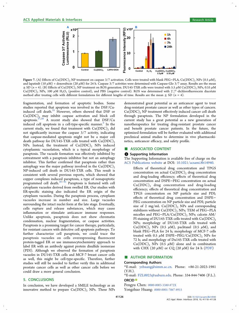
fragmentation, and formation of apoptotic bodies. Somestudies reported that apoptosis was involved in the DSF/Cu-induced cell death.35 However, others showed that DSF orCu(DDC)2 may inhibit caspase activation and block cellapoptosis.37,38 A recent study also showed that DSF/Cuinduced cell apoptosis in a cell-type-specific manner.9 In thecurrent study, we found that treatment with Cu(DDC)2 didnot significantly increase the caspase 3/7 activity, indicatingthat caspase-mediated apoptosis might not be a major celldeath pathway for DU145-TXR cells treated with Cu(DDC)2NPs. Instead, the treatment of Cu(DDC)2 NPs inducedcytoplasmic vacuolation, which is a typical morphology ofparaptosis. The vacuole formation was effectively inhibited bycotreatment with a paraptosis inhibitor but not an autophagyinhibitor. This further confirmed that paraptosis rather thanautophagy was the major pathway involved in the Cu(DDC)2NP-induced cell death in DU145-TXR cells. This result isconsistent with several previous reports, which showed thatcopper complexes induced paraptosis, a type of nonapoptoticprogrammed cell death.38−41 Paraptosis is featured with cellcytoplasm vacuoles derived from swelled ER. Our studies withER-specific staining also indicated the ER origin of thecytoplasm vacuoles. During the paraptosis process, cytoplasmvacuoles increase in number and size. Large vacuolessurrounding the intact nuclei form at the late stage. Eventually,cells rupture and release substances, which may causeinflammation or stimulate anticancer immune responses.Unlike apoptosis, paraptosis does not show chromatincondensation, nuclear fragmentation, or caspase activities.Paraptosis is a promising target for cancer therapy, particularly,for resistant cancers with defective cell apoptosis pathways. Tofurther characterize cell paraptosis, we could trace theparaptosis vacuoles on cells overexpressing fluorescentprotein-tagged ER or use immunocytochemistry approach tolabel ER with an antibody against protein disulfide isomerase(PDI). Although we observed the formation of paraptosisvacuoles in DU145-TXR cells and MCF-7 breast cancer cellsas well, this might be cell-type-specific. Therefore, furtherstudies will still be needed to further verify this in additionalprostate cancer cells as well as other cancer cells before wecould draw a more general conclusion.
5. CONCLUSIONSIn conclusion, we have developed a SMILE technology as aninnovative method to prepare Cu(DDC)2 NPs. These NPs
demonstrated great potential as an anticancer agent to treatdrug-resistant prostate cancer as well as other types of cancers.Cu(DDC)2 NP treatment effectively induced cancer cell deaththrough paraptosis. The NP formulation developed in thecurrent study has a great potential as a new generation ofnanotherapeutics for treating drug-resistant prostate cancerand benefit prostate cancer patients. In the future, theoptimized formulation will be further evaluated with additionalpreclinical animal studies to determine in vivo pharmacoki-netics, anticancer efficacy, and safety profile.
■ ASSOCIATED CONTENT*S Supporting InformationThe Supporting Information is available free of charge on theACS Publications website at DOI: 10.1021/acsami.8b14940.
Effects of theoretical drug concentration and TPGSconcentration on actual Cu(DDC)2 drug concentrationand drug-loading efficiency; effects of theoretical drugconcentration and DSPE−PEG concentration on actualCu(DDC)2 drug concentration and drug-loadingefficiency; effects of theoretical drug concentration andTPGS concentration on NP particle size and PDI;effects of theoretical drug concentration and DSPE−PEG concentration on NP particle size and PDI; particlesize of 2 mg/mL Cu(DDC)2 NPs and correspondingstabilizers without Cu(DDC)2 NPs; TEM of PEG−PLAmicelles and PEG−PLA/Cu(DDC)2 NPs; calcein AM/PI staining of DU145-TXR cells treated with Cu(DDC)2NPs; morphology of DU145-TXR cells treated withCu(DDC)2 NPs (0.5 μM), paclitaxel (0.5 μM), andblank PEG−PLA for 24 h; morphology of MCF-7 cellstreated with 0.5 μM DSPE−PEG/Cu(DDC)2 NPs for72 h; and morphology of Du145-TXR cells treated withCu(DDC)2 NPs (0.5 μM) alone and in combinationwith CHX (20 μM) or CQ (20 μM) for 24 h (PDF)
■ AUTHOR INFORMATIONCorresponding Authors*E-mail: [email protected]. Phone: +86-21-2023-1981(Y.H.).*E-mail: [email protected]. Phone: 334-844-7406 (F.L.).ORCIDPengyu Chen: 0000-0003-3380-872XYongzhuo Huang: 0000-0001-7067-8915
Figure 7. (A) Effects of Cu(DDC)2 NP treatment on caspase 3/7 activation. Cells were treated with blank PEG−PLA, Cu(DDC)2 NPs (0.5 μM),and lapatinib (10 μM) + doxorubicin (20 μM) for 24 h. Caspase 3/7 activities were determined with Caspase-Glo 3/7 assay. Results are the mean± SD (n = 4). (B) Effects of Cu(DDC)2 NP treatment on ROS generation. DU145-TXR cells were treated with 5.5 μM Cu(DDC)2 NPs, 0.55 μMCu(DDC)2 NPs, 100 μM H2O2 (positive control), and PBS (negative control). ROS was determined with 2′,7′-dichlorofluorescein diacetatemethod after treating cells with different formulations for different lengths of time. Results are the mean ± SD (n = 4).
ACS Applied Materials & Interfaces Research Article
DOI: 10.1021/acsami.8b14940ACS Appl. Mater. Interfaces 2018, 10, 41118−41128
41126

Feng Li: 0000-0002-8559-3831NotesThe authors declare no competing financial interest.
■ ACKNOWLEDGMENTSThis work was financially supported by Auburn Universitystartup fund (F.L.) and National Science Foundation grant no.CBET-1701363 (P.C.). A sincere thank you to Dr. DeadreJohnson for proofreading of this paper.
■ REFERENCES(1) Basch, E.; Loblaw, D. A.; Rumble, R. B. Systemic Therapy inMen With Metastatic Castration-Resistant Prostate Cancer: AmericanSociety of Clinical Oncology and Cancer Care Ontario ClinicalPractice Guideline Summary. J. Oncol. Pract. 2014, 10, e418−e420.(2) Hwang, C. Overcoming Docetaxel Resistance in ProstateCancer: a Perspective Review. Ther. Adv. Med. Oncol. 2012, 4,329−340.(3) Theyer, G.; Schirmbock, M.; Thalhammer, T.; Sherwood, E. R.;Baumgartner, G.; Hamilton, G. Role of the MDR-1-encoded MultipleDrug Resistance Phenotype in Prostate Cancer Cell Lines. J. Urol.1993, 150, 1544−1547.(4) van Brussel, J. P.; Mickisch, G. H. J. Multidrug Resistance inProstate Cancer. Oncol. Res. Treat. 2003, 26, 175−181.(5) Li, F.; Mahato, R. I. MicroRNAs and Drug Resistance in ProstateCancers. Mol. Pharm. 2014, 11, 2539−2552.(6) Li, F.; Mahato, R. I. miRNAs as Targets for Cancer Treatment:Therapeutics Design and Delivery. Preface. Adv. Drug Deliv. Rev.2015, 81, 5−6.(7) Sleire, L.; Førde, H. E.; Netland, I. A.; Leiss, L.; Skeie, B. S.;Enger, P. Ø. Drug Repurposing in Cancer. Pharmacol. Res. 2017, 124,74−91.(8) Lun, X.; Wells, J. C.; Grinshtein, N.; King, J. C.; Hao, X.; Dang,N.-H.; Wang, X.; Aman, A.; Uehling, D.; Datti, A.; Wrana, J. L.;Easaw, J. C.; Luchman, A.; Weiss, S.; Cairncross, J. G.; Kaplan, D. R.;Robbins, S. M.; Senger, D. L. Disulfiram when Combined withCopper Enhances the Therapeutic Effects of Temozolomide for theTreatment of Glioblastoma. Clin. Cancer Res. 2016, 22, 3860−3875.(9) Skrott, Z.; Mistrik, M.; Andersen, K. K.; Friis, S.; Majera, D.;Gursky, J.; Ozdian, T.; Bartkova, J.; Turi, Z.; Moudry, P.; Kraus, M.;Michalova, M.; Vaclavkova, J.; Dzubak, P.; Vrobel, I.; Pouckova, P.;Sedlacek, J.; Miklovicova, A.; Kutt, A.; Li, J.; Mattova, J.; Driessen, C.;Dou, Q. P.; Olsen, J.; Hajduch, M.; Cvek, B.; Deshaies, R. J.; Bartek, J.Alcohol-abuse Drug Disulfiram Targets Cancer via p97 SegregaseAdaptor NPL4. Nature 2017, 552, 194−199.(10) Bruning, A.; Kast, R. E. Oxidizing to death. Cell Cycle 2014, 13,1513−1514.(11) Wang, Z.; Tan, J.; McConville, C.; Kannappan, V.; Tawari, P.E.; Brown, J.; Ding, J.; Armesilla, A. L.; Irache, J. M.; Mei, Q.-B.; Tan,Y.; Liu, Y.; Jiang, W.; Bian, X.-W.; Wang, W. Poly Lactic-co-glycolicAcid Controlled Delivery of Disulfiram to Target Liver Cancer Stem-like Cells. Nanomedicine 2017, 13, 641−657.(12) Liu, P.; Kumar, I. S.; Brown, S.; Kannappan, V.; Tawari, P. E.;Tang, J. Z.; Jiang, W.; Armesilla, A. L.; Darling, J. L.; Wang, W.Disulfiram Targets Cancer Stem-like Cells and Reverses Resistanceand Cross-resistance in Acquired Paclitaxel-resistant Triple-negativeBreast Cancer Cells. Br. J. Cancer 2013, 109, 1876−1885.(13) Loo, T. W.; Clarke, D. M. Blockage of Drug Resistance in vitroby Disulfiram, a Drug Used to Treat Alcoholism. J. Natl. Cancer Inst.2000, 92, 898−902.(14) https://clinicaltrials.gov (NCT02963051, NCT02678975,NCT03363659).(15) Pushpakom, S.; Iorio, F.; Eyers, P. A.; Escott, K. J.; Hopper, S.;Wells, A.; Doig, A.; Guilliams, T.; Latimer, J.; McNamee, C.; Norris,A.; Sanseau, P.; Cavalla, D.; Pirmohamed, M. Drug Repurposing:Progress, Challenges and Recommendations. Nat. Rev. Drug Discov.2018, DOI: 10.1038/nrd.2018.168.
(16) Wehbe, M.; Anantha, M.; Shi, M.; Leung, A. W.-y.; Dragowska,W.; Sanche, L.; Bally, M. Development and Optimization of anInjectable Formulation of Copper Diethyldithiocarbamate, an ActiveAnticancer Agent. Int. J. Nanomed. 2017, 12, 4129−4146.(17) Zhao, P.; Yin, W.; Wu, A.; Tang, Y.; Wang, J.; Pan, Z.; Lin, T.;Zhang, M.; Chen, B.; Duan, Y.; Huang, Y. Dual-Targeting to CancerCells and M2 Macrophages via Biomimetic Delivery of MannosylatedAlbumin Nanoparticles for Drug-Resistant Cancer Therapy. Adv.Funct. Mater. 2017, 27, 1700403.(18) Marengo, A.; Forciniti, S.; Dando, I.; Dalla Pozza, E.; Stella, B.;Tsapis, N.; Yagoubi, N.; Fanelli, G.; Fattal, E.; Heeschen, C.; Palmieri,M.; Arpicco, S. Pancreatic Cancer Stem Cell Proliferation is StronglyInhibited by Diethyldithiocarbamate-copper Complex Loaded intoHyaluronic Acid Decorated Liposomes. Biochim. Biophys. Acta, Gen.Subj. 2018, 1863, 61−72.(19) Zhao, P.; Wang, Y.; Kang, X.; Wu, A.; Yin, W.; Tang, Y.; Wang,J.; Zhang, M.; Duan, Y.; Huang, Y. Dual-targeting BiomimeticDelivery for Anti-glioma Activity via Remodeling the TumorMicroenvironment and Directing Macrophage-mediated Immuno-therapy. Chem. Sci. 2018, 9, 2674−2689.(20) Wehbe, M.; Chernov, L.; Chen, K.; Bally, M. B. PRCosomes:Pretty Reactive Complexes Formed in Liposomes. J. Drug Target.2016, 24, 787−796.(21) Wang, H.; Li, F.; Du, C.; Wang, H.; Mahato, R. I.; Huang, Y.Doxorubicin and Lapatinib Combination Nanomedicine for TreatingResistant Breast Cancer. Mol. Pharm. 2014, 11, 2600−2611.(22) Li, F.; Snow-Davis, C.; Du, C.; Bondarev, M. L.; Saulsbury, M.D.; Heyliger, S. O. Preparation and Characterization of LipophilicDoxorubicin Pro-drug Micelles. J. Visualized Exp. 2016, 114, e54338.(23) Li, F.; Danquah, M.; Singh, S.; Wu, H.; Mahato, R. I. Paclitaxel-and Lapatinib-loaded Lipopolymer Micelles Overcome MultidrugResistance in Prostate Cancer. Drug Delivery Transl. Res. 2011, 1,420−428.(24) Lu, W.; Li, F.; Mahato, R. I. Poly(ethylene glycol)-block-poly(2-methyl-2-benzoxycarbonyl-propylene carbonate) Micelles forRapamycin Delivery: in vitro Characterization and Biodistribution. J.Pharm. Sci. 2011, 100, 2418−2429.(25) Li, F.; Danquah, M.; Mahato, R. I. Synthesis and Character-ization of Amphiphilic Lipopolymers for Micellar Drug Delivery.Biomacromolecules 2010, 11, 2610−2620.(26) Wang, C.-Y.; Li, F.; Yang, Y.; Guo, H.-Y.; Wu, C.-X.; Wang, S.Recombinant Baculovirus Containing theDiphtheria Toxin AGene forMalignant Glioma Therapy. Cancer Res. 2006, 66, 5798−5806.(27) Li, F.; Lu, Y.; Li, W.; Miller, D. D.; Mahato, R. I. Synthesis,Formulation and in vitro Evaluation of a Novel MicrotubuleDestabilizer, SMART-100. J. Controlled Release 2010, 143, 151−158.(28) Crowley, L. C.; Christensen, M. E.; Waterhouse, N. J.Measuring Survival of Adherent Cells with the Colony-FormingAssay. Cold Spring Harb. Protoc. 2016, 2016, pdb.prot087171.(29) Wadhwa, S.; Mumper, R. J. Intracellular Delivery of theReactive Oxygen Species Generating Agentd-Penicillamine uponConjugation to Poly-l-glutamic Acid. Mol. Pharm. 2010, 7, 854−862.(30) Danquah, M.; Li, F.; Duke, C. B., 3rd; Miller, D. D.; Mahato, R.I. Micellar Delivery of Bicalutamide and Embelin for TreatingProstate Cancer. Pharm. Res. 2009, 26, 2081−2092.(31) Skrott, Z.; Cvek, B. Diethyldithiocarbamate Complex withCopper: the Mechanism of Action in Cancer Cells. Mini Rev. Med.Chem. 2012, 12, 1184−1192.(32) Wang, N.-n.; Wang, L.-H.; Li, Y.; Fu, S.-Y.; Xue, X.; Jia, L.-N.;Yuan, X.-Z.; Wang, Y.-T.; Tang, X.; Yang, J.-Y.; Wu, C.-F. TargetingALDH2 with Disulfiram/Copper Reverses the Resistance of CancerCells to Microtubule Inhibitors. Exp. Cell Res. 2018, 362, 72−82.(33) Denoyer, D.; Masaldan, S.; La Fontaine, S.; Cater, M. A.Targeting Copper in Cancer Therapy: “Copper That Cancer”.Metallomics 2015, 7, 1459−1476.(34) Hoyer-Hansen, M.; Jaattela, M. Connecting EndoplasmicReticulum Stress to Autophagy by Unfolded Protein Response andCalcium. Cell Death Differ. 2007, 14, 1576−1582.
ACS Applied Materials & Interfaces Research Article
DOI: 10.1021/acsami.8b14940ACS Appl. Mater. Interfaces 2018, 10, 41118−41128
41127

(35) Yang, Y.; Zhang, K.; Wang, Y.; Li, M.; Sun, X.; Liang, Z.; Wang,L.; Chen, L.; Yang, H.; Zhu, L. Disulfiram Chelated with CopperPromotes Apoptosis in Human Breast Cancer Cells by Impairing theMitochondria Functions. Scanning 2016, 38, 825−836.(36) Breckenridge, D. G.; Germain, M.; Mathai, J. P.; Nguyen, M.;Shore, G. C. Regulation of Apoptosis by Endoplasmic ReticulumPathways. Oncogene 2003, 22, 8608−8618.(37) Nobel, C. S. I.; Kimland, M.; Nicholson, D. W.; Orrenius, S.;Slater, A. F. G. Disulfiram is a Potent Inhibitor of Proteases of theCaspase Family. Chem. Res. Toxicol. 1997, 10, 1319−1324.(38) Tardito, S.; Bassanetti, I.; Bignardi, C.; Elviri, L.; Tegoni, M.;Mucchino, C.; Bussolati, O.; Franchi-Gazzola, R.; Marchio, L. CopperBinding Agents Acting as Copper Ionophores Lead to CaspaseInhibition and Paraptotic Cell Death in Human Cancer Cells. J. Am.Chem. Soc. 2011, 133, 6235−6242.(39) Zhou, Y.; Huang, F.; Yang, Y.; Wang, P.; Zhang, Z.; Tang, Y.;Shen, Y.; Wang, K. Paraptosis-Inducing Nanomedicine OvercomesCancer Drug Resistance for a Potent Cancer Therapy. Small 2018, 14,1702446.(40) Chen, X.; Zhang, X.; Chen, J.; Yang, Q.; Yang, L.; Xu, D.;Zhang, P.; Wang, X.; Liu, J. Hinokitiol Copper Complex InhibitsProteasomal Deubiquitination and Induces Paraptosis-like Cell Deathin Human Cancer Cells. Eur. J. Pharmacol. 2017, 815, 147−155.(41) Barilli, A.; Atzeri, C.; Bassanetti, I.; Ingoglia, F.; Dall’Asta, V.;Bussolati, O.; Maffini, M.; Mucchino, C.; Marchio, L. Oxidative StressInduced by Copper and Iron Complexes with 8-hydroxyquinolineDerivatives Causes Paraptotic Death of HeLa Cancer Cells. Mol.Pharm. 2014, 11, 1151−1163.
ACS Applied Materials & Interfaces Research Article
DOI: 10.1021/acsami.8b14940ACS Appl. Mater. Interfaces 2018, 10, 41118−41128
41128