disturbances of circulation, lab 1: edema and...
TRANSCRIPT
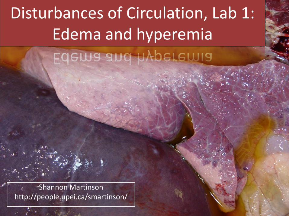
Disturbances of Circulation, Lab 1: Edema and hyperemia
Shannon Martinsonhttp://people.upei.ca/smartinson/
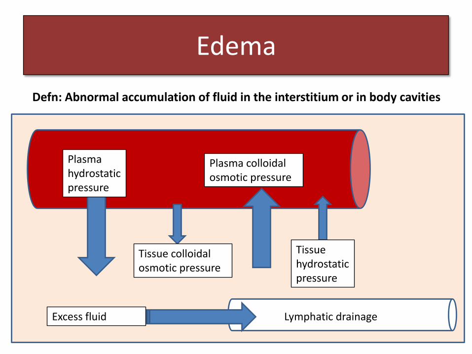
Edema
Defn: Abnormal accumulation of fluid in the interstitium or in body cavities
Plasma hydrostatic pressure
Tissue colloidal osmotic pressure
Plasma colloidal osmotic pressure
Tissue hydrostatic pressure
Excess fluid Lymphatic drainage
hronic local passive hyperemia
1) ↓Plasma colloidal osmotic pressure
• Hypoproteinemia– protein losing enteropathy or nephropathy, liver disease
• Generalized edema
2) ↑Blood hydrostatic pressure
• Generalized edema: heart failure
• Localized edema: Occlusion of the local venous return
3) Lymphatic obstruction
• Localized edema
4) Sodium retention
• Generalized – seen with renal disease
5) ↑Vascular permeability
• Edema of inflammation
• Localized
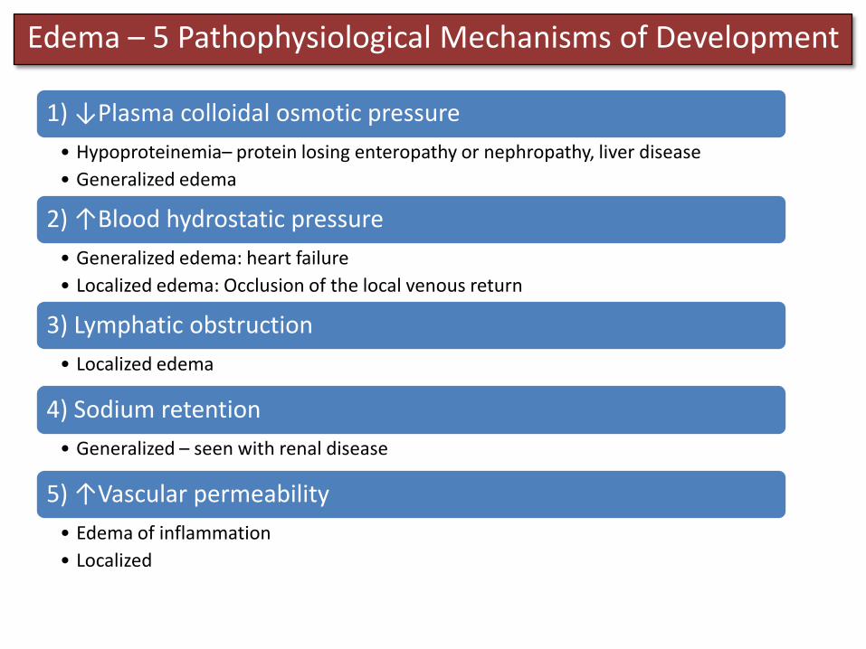
Edema – 5 Pathophysiological Mechanisms of Development

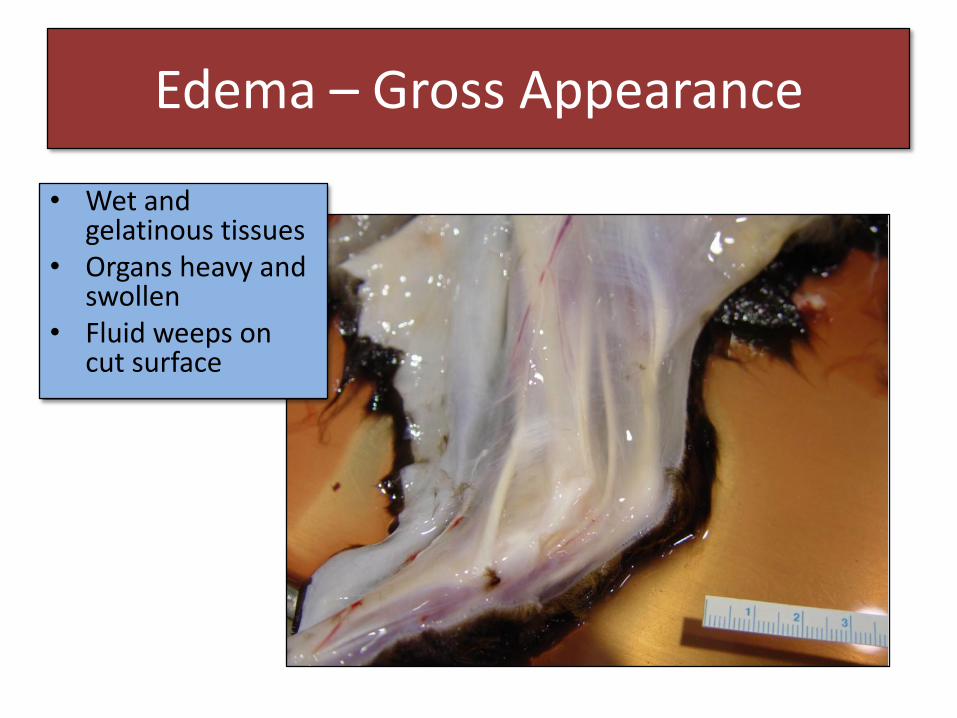
Edema – Gross Appearance
• Wet and gelatinous tissues
• Organs heavy and swollen
• Fluid weeps on cut surface
Edema – Gross Appearance
• Wet and gelatinous tissues
• Organs heavy and swollen
• Fluid weeps on cut surface
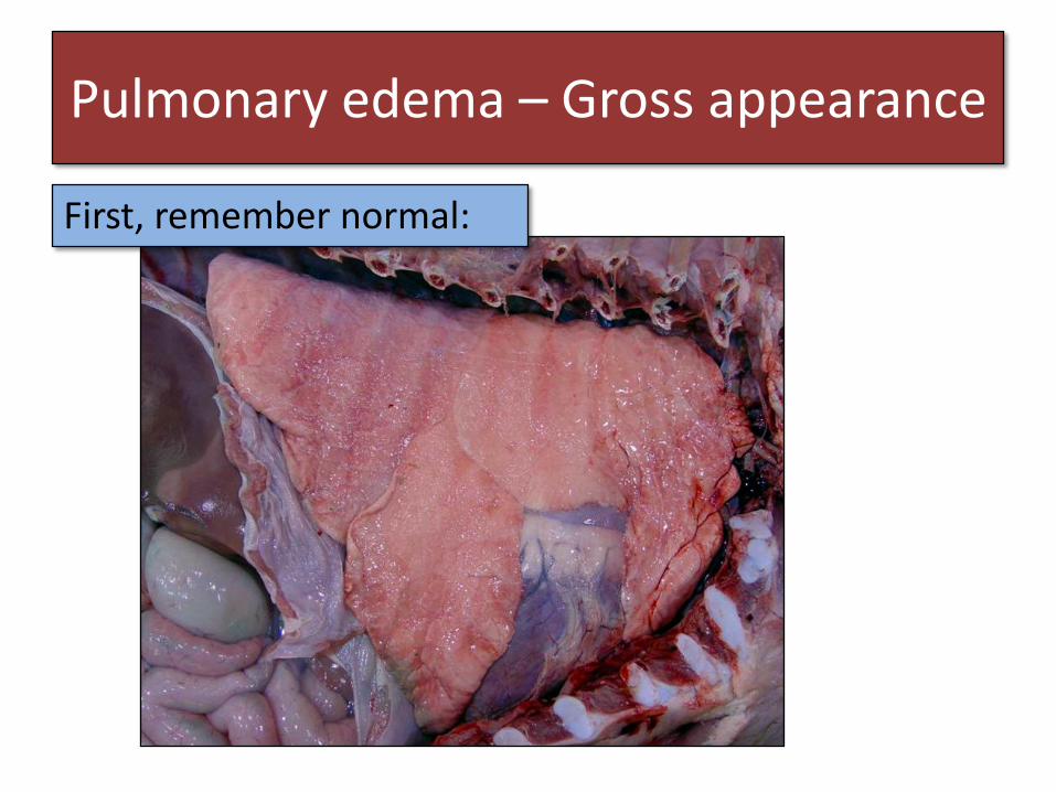
Pulmonary edema – Gross appearance
First, remember normal:
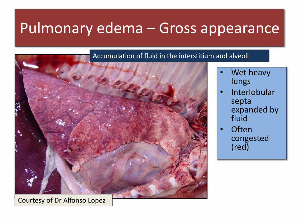
Pulmonary edema – Gross appearance
Courtesy of Dr Alfonso Lopez
• Wet heavy lungs
• Interlobular septa expanded by fluid
• Often congested (red)
Accumulation of fluid in the interstitium and alveoli
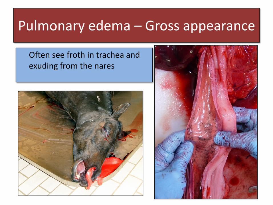
Often see froth in trachea and exuding from the nares
Pulmonary edema – Gross appearance

Edema – Example 1
What do you see?
Swelling of the foot below the leg band
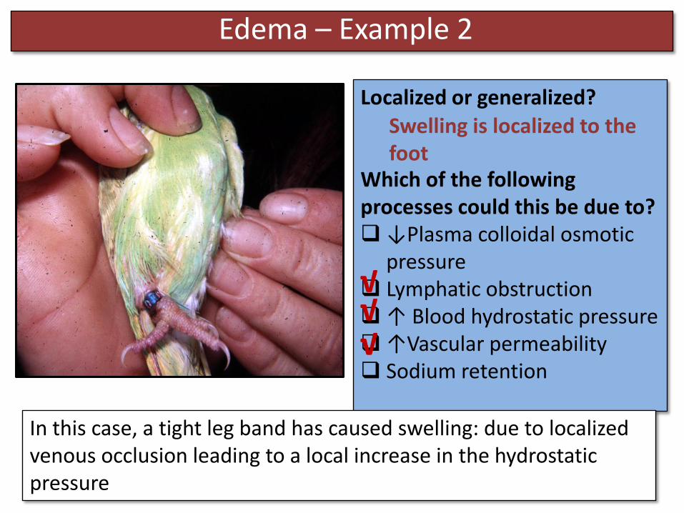
Edema – Example 2
Localized or generalized?
Which of the following processes could this be due to?↓Plasma colloidal osmotic
pressure Lymphatic obstruction↑ Blood hydrostatic pressure↑Vascular permeability Sodium retention
Swelling is localized to the foot
√√
√
In this case, a tight leg band has caused swelling: due to localized venous occlusion leading to a local increase in the hydrostatic pressure
Edema – Example 2
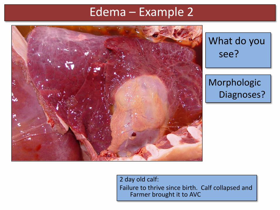
2 day old calf: Failure to thrive since birth. Calf collapsed and
Farmer brought it to AVC
What do you see?
Morphologic Diagnoses?
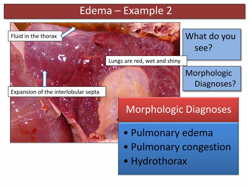
Edema – Example 2
What do you see?
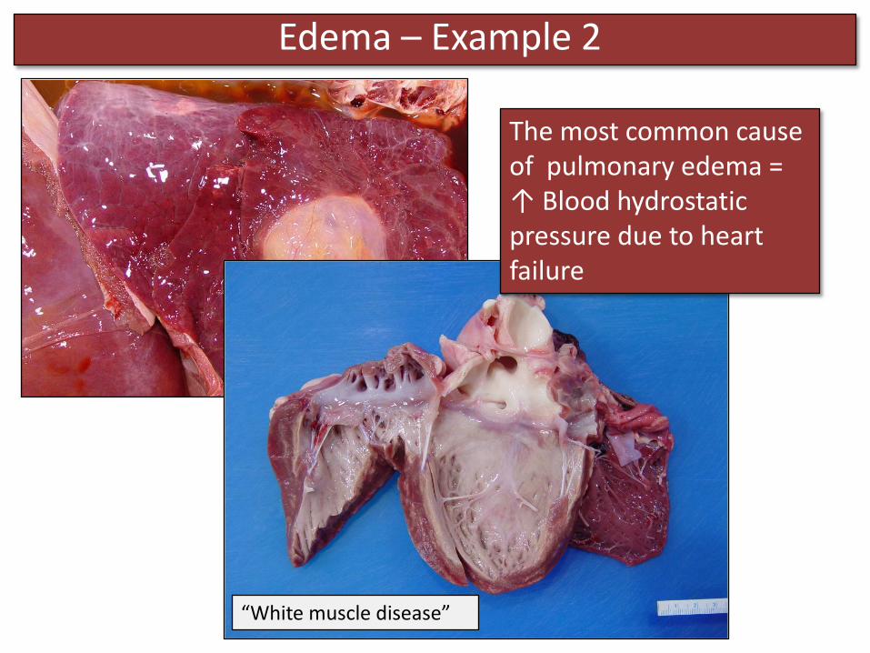
Lungs are red, wet and shiny
Fluid in the thorax
Expansion of the interlobular septa
Morphologic Diagnoses
• Pulmonary edema
• Pulmonary congestion
• Hydrothorax
Morphologic Diagnoses?
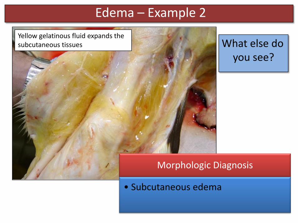
Edema – Example 2
What else do you see?
Yellow gelatinous fluid expands the subcutaneous tissues
Morphologic Diagnosis
• Subcutaneous edema
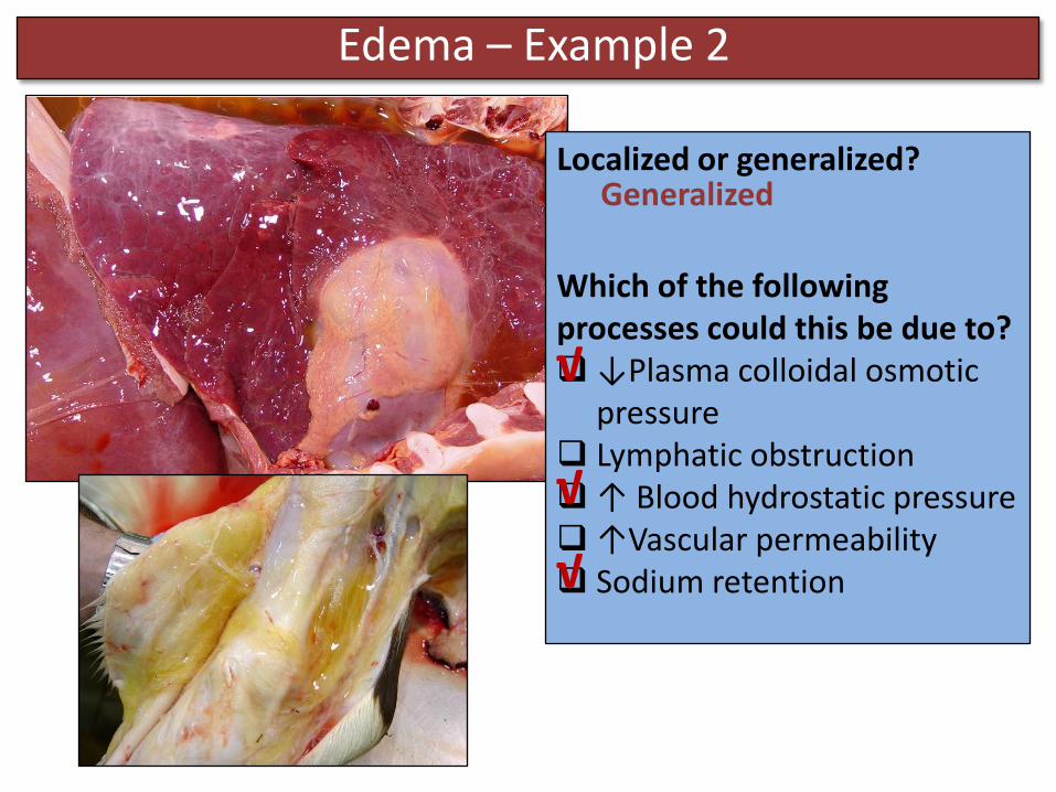
Localized or generalized?
Which of the following processes could this be due to?↓Plasma colloidal osmotic
pressure Lymphatic obstruction↑ Blood hydrostatic pressure↑Vascular permeability Sodium retention
Edema – Example 2
Generalized
√
√
√
Edema – Example 2
The most common cause of pulmonary edema = ↑ Blood hydrostatic pressure due to heart failure
“White muscle disease”
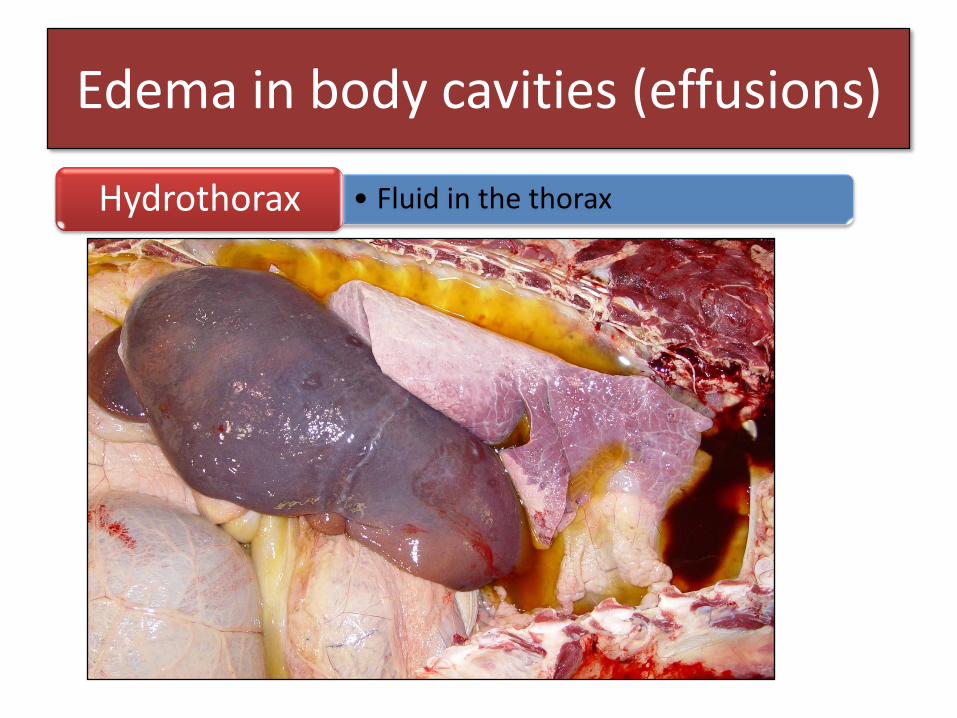
Edema in body cavities (effusions)
• Fluid in the thoraxHydrothorax
Edema in body cavities (effusions)
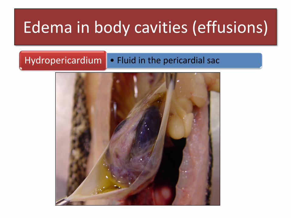
• Fluid in the pericardial sacHydropericardium
Edema in body cavities (effusions)
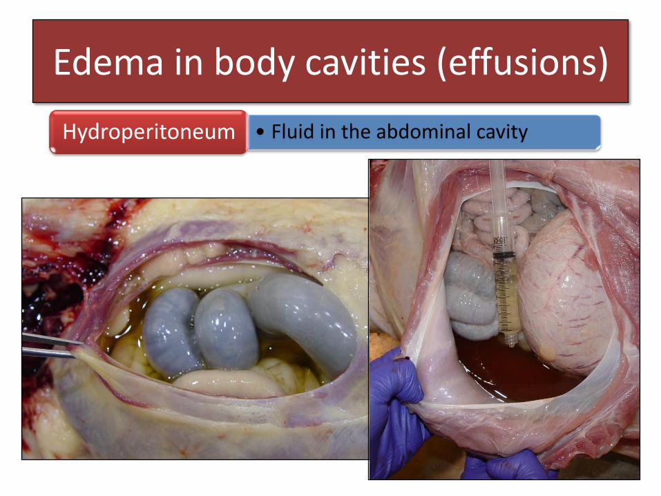
• Fluid in the abdominal cavityHydroperitoneum

Edema – Example 3
What do you see?
Facial swelling
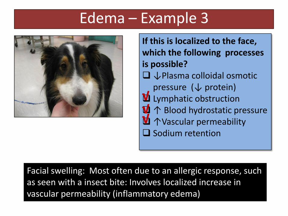
Edema – Example 3
If this is localized to the face, which the following processes is possible?↓Plasma colloidal osmotic
pressure (↓ protein) Lymphatic obstruction↑ Blood hydrostatic pressure↑Vascular permeability Sodium retention
√√√
Facial swelling: Most often due to an allergic response, such as seen with a insect bite: Involves localized increase in vascular permeability (inflammatory edema)
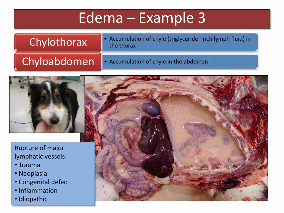
Edema – Example 3• Accumulation of chyle (triglyceride –rich lymph fluid) in
the thoraxChylothorax
• Accumulation of chyle in the abdomenChyloabdomen
Rupture of major lymphatic vessels: • Trauma• Neoplasia• Congenital defect• Inflammation• Idiopathic

Hyperemia and Congestion
• Acute (rapid)
• Chronic (prolonged)Duration
• Localized
• GeneralizedExtent
• Active = ↑ arteriolar flow
• Passive = ↓ venous drainage)Mechanism
An excessive amount of blood in an organ (refers to both volume and flow) *Hyperemia ≠ Hemorrhage
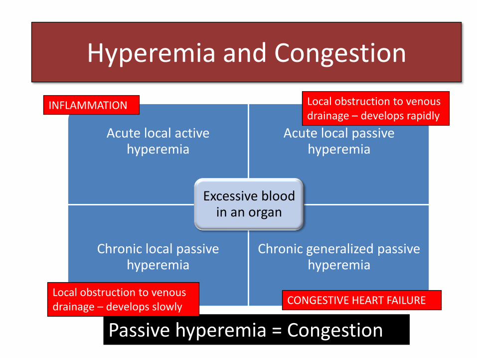
Passive hyperemia = Congestion
Acute local active hyperemiaHyperemia and Congestion
Acute local active hyperemia
Acute local passive hyperemia
Chronic local passive hyperemia
Chronic generalized passive hyperemia
Excessive blood in an organ
INFLAMMATION
CONGESTIVE HEART FAILURE
Local obstruction to venous drainage – develops rapidly
Local obstruction to venous drainage – develops slowly

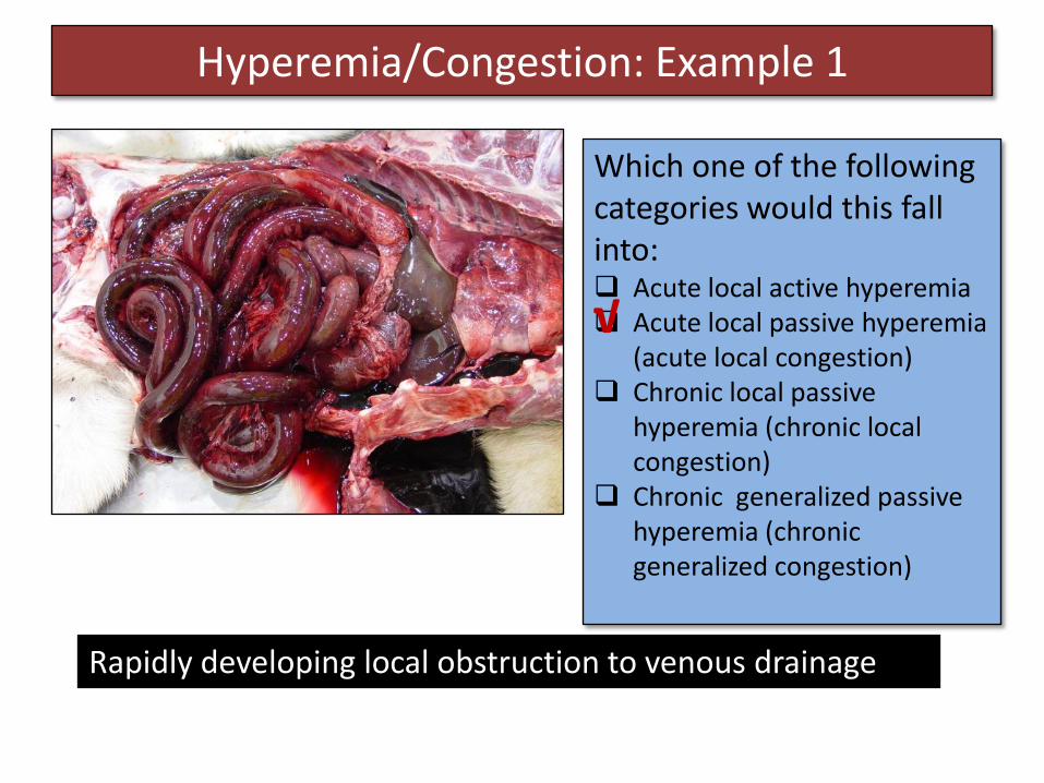
Hyperemia/Congestion: Example 1
What do you see?
4 year old dog: Dog seemed more quite than usual, collapsed and died
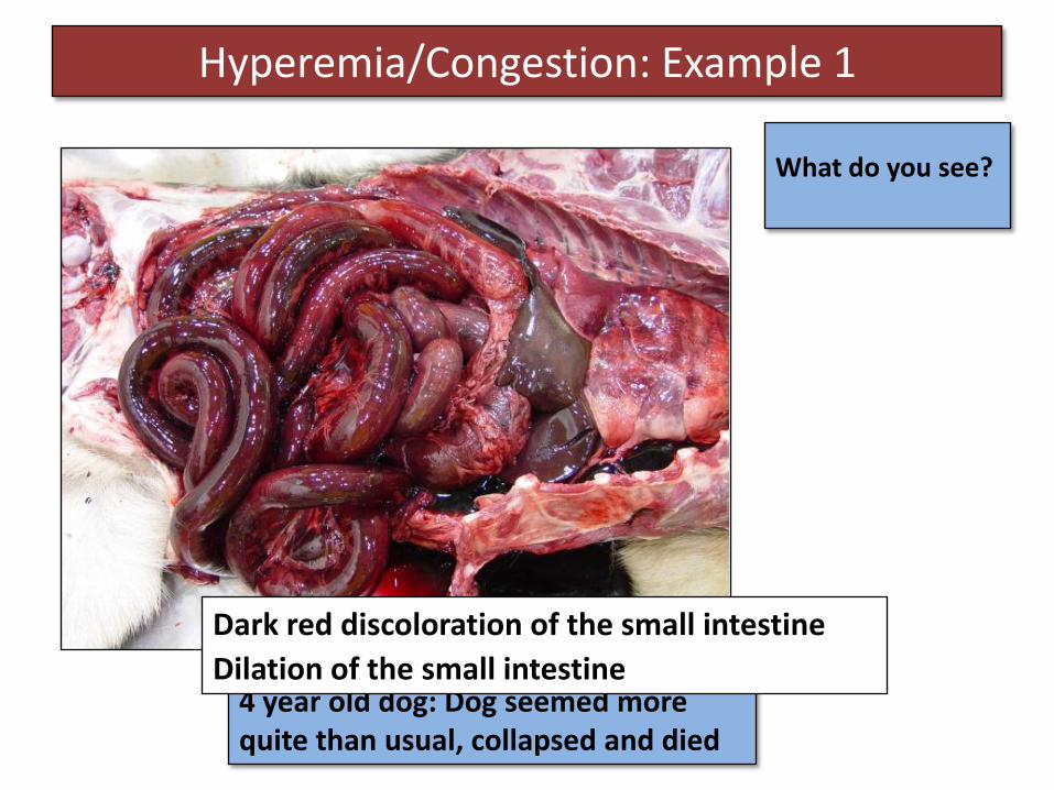
Hyperemia/Congestion: Example 1
What do you see?
4 year old dog: Dog seemed more quite than usual, collapsed and died
Dark red discoloration of the small intestine
Dilation of the small intestine

Hyperemia/Congestion: Example 1
What do you see?
Torsion at the root of the mesentery
Hyperemia/Congestion: Example 1
Which one of the following categories would this fall into: Acute local active hyperemia Acute local passive hyperemia
(acute local congestion) Chronic local passive
hyperemia (chronic local congestion)
Chronic generalized passive hyperemia (chronic generalized congestion)
Rapidly developing local obstruction to venous drainage
√
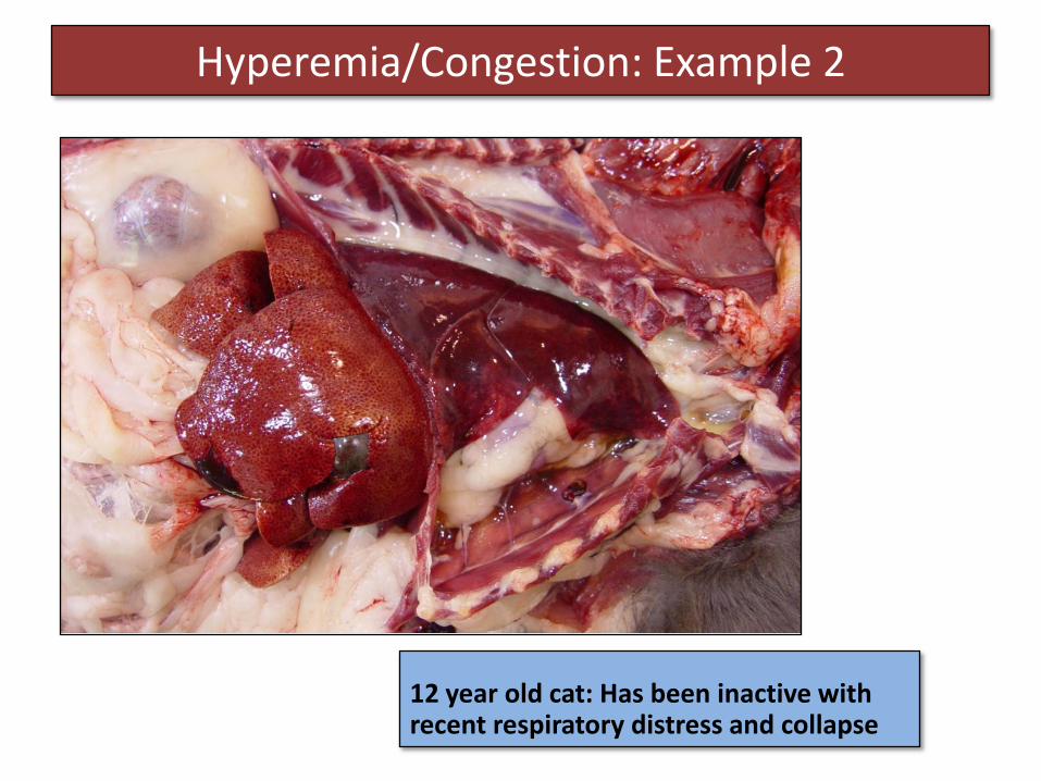
Hyperemia/Congestion: Example 2
12 year old cat: Has been inactive with recent respiratory distress and collapse
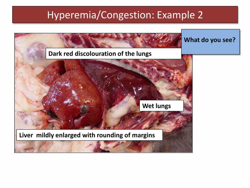
Hyperemia/Congestion: Example 2
What do you see?
Dark red discolouration of the lungs
Wet lungs
Liver mildly enlarged with rounding of margins
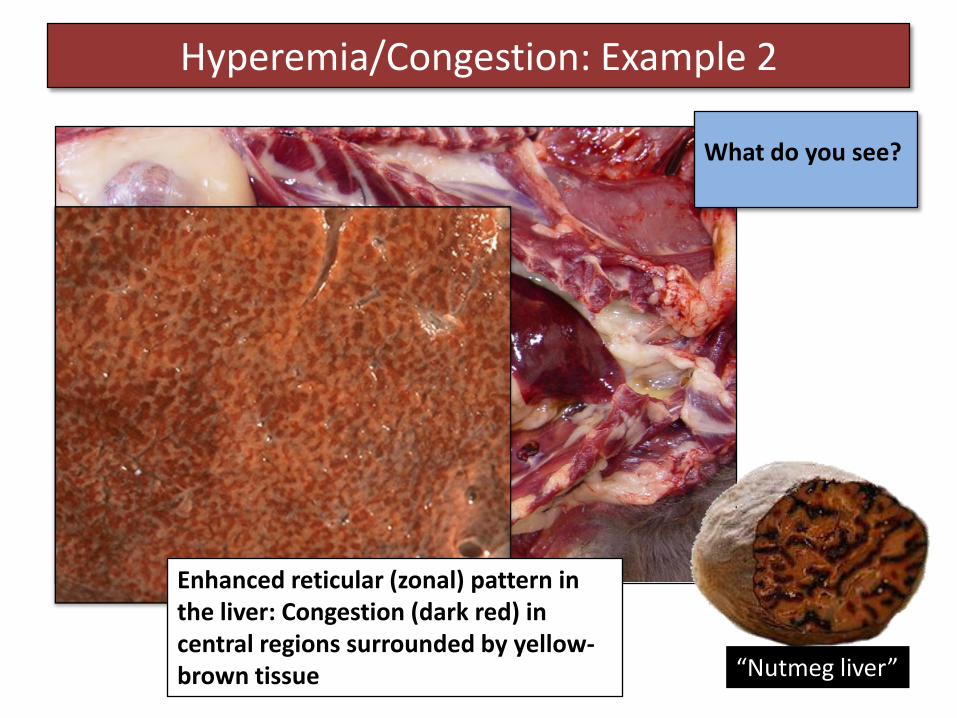
Hyperemia/Congestion: Example 2
What do you see?
Enhanced reticular (zonal) pattern in the liver: Congestion (dark red) in central regions surrounded by yellow-brown tissue “Nutmeg liver”
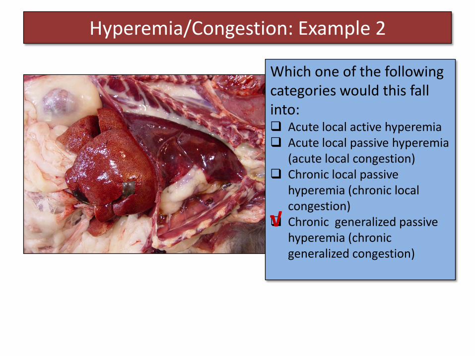
Hyperemia/Congestion: Example 2
Which one of the following categories would this fall into: Acute local active hyperemia Acute local passive hyperemia
(acute local congestion) Chronic local passive
hyperemia (chronic local congestion)
Chronic generalized passive hyperemia (chronic generalized congestion)
√
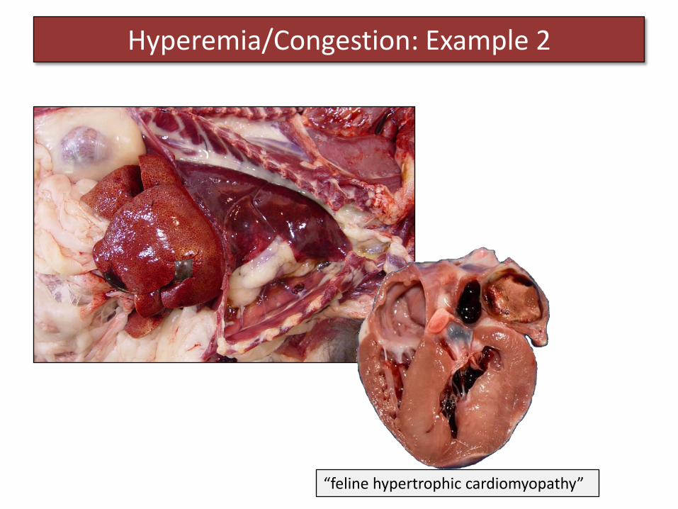
Hyperemia/Congestion: Example 2
“feline hypertrophic cardiomyopathy”
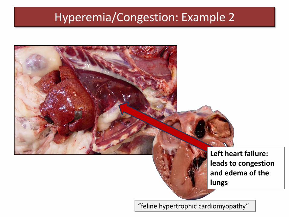
Hyperemia/Congestion: Example 2
“feline hypertrophic cardiomyopathy”
Left heart failure: leads to congestion and edema of the lungs

Hyperemia/Congestion: Example 2
“feline hypertrophic cardiomyopathy”
Right heart failure: leads to congestion of the liver and generalized edema

Hyperemia/Congestion: Example 3
What do you see?
Bright red discolouration of the skin of the ears and snout

Hyperemia/Congestion: Example 3
Which one of the following categories would this fall into: Acute local active hyperemia Acute local passive hyperemia
(acute local congestion) Chronic local passive
hyperemia (chronic local congestion)
Chronic generalized passive hyperemia (chronic generalized congestion)
√
This is active hyperemia – hyperemia of inflammation. Hyperemia of the skin is a common finding in pigs with sepsis.