distribution of glycine-immunoreactive profiles in the monkey spinal cord: a light microscopic and...
TRANSCRIPT
THE JOURNAL OF COMPARATIVE NEUROLOGY 371~589-602 (1996)
Distribution of Glycine-Immunoreactive Profiles in the Monkey Spinal Cord:
A Light Microscopic and Ultrastructural Study
SUSAN M. CARLTON, GREGORY L. HARGETT, AND RICHARD E. COGGESHALL Department of Anatomy and Neuroscience, Marine Biomedical Institute,
University of Texas Medical Branch, Galveston, Texas 77555-1069
ABSTRACT The present study analyzed the relationships of glycine (GLY)-immunoreactive (-IR) and
unlabeled profiles in the primate spinal cord. Light microscopic analysis demonstrated GLY-IR profiles in laminae 111-VII, with fewer labeled profiles in laminae I, 11, VIII, IX and X. The dorsal part of the lateral funiculus and the dorsal funiculus contained few labeled axons, in contrast to all other areas of white matter, which were heavily labeled. At the electron microscopic level, GLY-IR terminals in monkeys contained mainly round, with occasional pleomorphic, clear vesicles; however, F-type GLY-IR terminals synapsing on motoneurons contained pleomorphic vesicles. This seems to be an important species difference because vesicles in GLY-IR terminals in rat and cat are predominantly oval or elliptical.
GLY-IR terminals synapsed on unlabeled as well as GLY-IR cell bodies and dendrites. This is morphological evidence that GLY may be both an inhibitor (GLY-IR terminals syn- apse on and presumably inhibit non-GLY cell bodies and dendrites) and a disinhibitor (GLY-IR terminals synapse on and presumably inhibit other GLY elements) of spinal activity. Also noteworthy was the conspicuous absence of axoaxonic interactions involving GLY-IR terminals. A related finding was that GLY profiles were always postsynaptic, never presynaptic, to glomerular primary afferent terminals. The functional implications would seem to be that primary afferent input can activate the spinal GLY system but that there is little GLY presynaptic control of afferent input in monkeys. This is in contrast to rats and cats, in which axoaxonic interactions involving GLY-IR terminals have been observed and where it is common to find GLY-IR terminals presynaptic to glomerular primary afferent terminals. D 1996 Wiley-Liss, Inc.
Indexing terms: inhibition, primary afferent, disinhibition
Glycine (GLY) is an important inhibitory transmitter in the mammalian central nervous system (Young and Macdon- ald, 1983; Aprison, 1990). The distribution and neurocir- cuitry of GLY in the spinal cord is of particular interest in relation to the modulation of sensory input. Previous studies from this laboratory elucidated the neurocircuitry of another important inhibitory transmitter in the primate spinal cord, gamma aminobutyric acid (GABA, Carlton and Hayes, 1990a,b; Hayes and Carlton, 1992; Carlton et al., 1992; Carlton, 1994). One conclusion from our earlier studies was that there was a paucity of anatomical data showing direct GABAergic control of a subpopulation of glomerular primary afferent terminals (Carlton and Hayes, 1990a; Hayes and Carlton, 1992; Carlton, 1994). This finding represented an important species difference because GABA-immunoreactive (-IR) axons are reported to be pre-
synaptic to other axons, including central terminals in glomeruli, in rat and cat (Barber et al., 1978; Basbaum et al., 1986; Maxwell and Noble, 1987). Furthermore, the observation that numerous GABAergic dendrites in the dorsal horn contained vesicles and participated in synapses indicated that, in addition to classical axonal interactions, the GABAergic system could “multiply” its inhibitory influence through dendrodendritic, dendroaxonic and den- drosomatic interactions. Therefore, in the present study, particular attention was paid to the arrangement of glycin- ergic profiles in relation to primary afferent terminals and to the synaptology of glycinergic dendrites. Similarities in
Accepted March 5, 1996. Address reprint requests to Dr. Susan M. Carlton, Marine Biomedical
Institute, University of Texas Medical Branch, Galveston, TX 77555-1069.
O 1996 WILEY-LISS, INC.
590 S.M. CARLTON ET AL.
above, cut into small blocks and postfixed for 4-6 hours at 4°C in PB. Blocks were sectioned with a vibratome (Lancer Series 1000) at 25 pm and collected in PB. The sections were incubated in 1% sodium borohydride for 0.5 hour and then rinsed in PB. Two different techniques were then used to label GLY profiles at the EM level:
neurocircuitry would suggest that the activation of GABAer- gic and glycinergic systems might have similar functional consequences. Because the primate spinal GLY system has not been previously examined, an additional aim of this study was to compare the arrangements of GLY-IR profiles in the primate to previous reports in rat and cat.
MATERIALS AND METHODS The animals in this study had been used for acute
electrophysiological studies in which pharmacological agents were delivered through a microdialysis fiber implanted in the L7 segment while recording responses from L7 spinotha- lamic tract cells. Cord segments involved in physiological recording, dialysis or exposure to pharmacological agents were not used for this anatomical study. Thus, segments from the cervical enlargement and rostra1 lumbar spinal cord (L1 and L2) were taken from adult monkeys of both sexes weighing 1.5-2.5 kg. Anesthesia was established in these animals with a gaseous mixture of halothane, nitrous oxide and oxygen followed by a dose of a-chloralose (60 mg/kg, i.v.). Anesthesia was maintained during the physi- ological experiment by intravenous infusion of sodium pentobarbital (5.0 mg/kg/hour). The animals were per- fused at the conclusion of the electrophysiological studies after being deeply anesthetized with sodium pentobarbital (70 mg/kg, i.v.).
Light microscopic (LM) analyses Animals (n,= 5) were perfused through the aorta with
heparinized saline at room temperature, followed by a solution of 4% paraformaldehyde and 0.5% glutaraldehyde in 0.1 M phosphate buffer (PB, pH 7.4) at 4°C. Tissue from the cervical enlargement and lumbar cord was removed, cut into small blocks and postfixed at 4°C for 6-8 hours in fresh fixative. For cryoprotection, the segments were stored overnight at 4°C in phosphate buffer (PB) containing 30% sucrose. Sections (25-pm-thick) were cut on a freezing microtome, rinsed with 1% sodium borohydride in PB for 0.5 hour, followed by several rinses in PB. All immunore- agents contained 0.02% Triton X-100 to increase antibody penetration. The tissue was immunostained with a modi- fied version of the Sternberger (1979) peroxidase anti- peroxidase (PAP) technique. Briefly, the sections were placed in GLY antibody (1:500-1,000, Chemicon) in PB for 48 hours at 4°C. Following washing in 1% and 3% normal goat serum (NGS) for 0.5 hour each, the sections were incubated in goat anti-rabbit immunoglobin (IgG; 150) in PB for 1 hour. Following washing in 1% and 3% NGS for 0.5 hour each, the sections were placed in a,solution of PAP (Sternberger-Meyer, 1 : l O O ) in PB for 1 hour. Excess PAP was rinsed away in PB and the tissue reacted with diamino- benzidine (DAB, 0.05%) and Hz02 in PB for 6-12 minutes. Observation of wet sections through a microscope helped to determine when the DAB reactions were maximal, at which time the reaction was terminated. Following several rinses in PB, the sections were mounted on subbed slides, air- dried, cleared in alcohols and xylene and coverslipped.
Electron microscopic (EM) analysis Animals (n = 2) were perfused with 3% paraformalde-
hyde, 3% glutaraldehyde and 0.1% picric acid in PB. Cervical and lumbar segments were removed as described
1.
2.
PAP Technique: Following a previously used technique (Carlton and Hayes, 1990a; Hayes and Carlton, 19921, the sections were first exposed to a graded series of ethyl alcohols (lo%, 25%, 40%, 25%, 10%) in PB for 5 minutes each to increase antibody penetration. The tissue was then immunostained as above except that Triton X-100 was omitted. Following immunostaining, the tissue was placed in 1.0% osmic acid in PB for 1 hour, then stained with 1% uranyl acetate en bloc for 1 hour, dehydrated and flat-embedded in a mixture of Epon-Araldite. Ultra- thin semiserial sections were collected on Formvar- coated slot grids. Immunogold staining technique: Following a previously reported protocol (Hayes and Carlton, 1992), several groups of ultrathin sections were collected on formvar- coated nickel slot grids. The grids were then etched on drops of 0.68 M NaI04 for 1 hour and NaBH4 (10 minutes), washed with dHzO and incubated on drops of 3% NGS for 30 minutes. Following incubation in GLY antiserum (1500-1,000) overnight at 4"C, the grids were thoroughly washed and incubated on drops of goat anti-rabbit I&-gold (15 nm, Janssen). After thorough washing, the grids were poststained with uranyl acetate and lead citrate prior to analysis.
Controls GLY was conjugated to hemocyanin according to the
protocol of Hepler et al. (1988). The conjugate was then incubated with GLY antibody. A complete absence of specific GLY immunostaining was observed in monkey spinal cord tissue when GLY antiserum (1500) was preincu- bated with GLY-hemocyanin conjugate used at a concentra- tion of 100 pg/ml. Unconjugated forms of GLY, GABA, L-glutamate, beta-alanine, L-alanine, L-aspartic acid, and taurine processed with GLY antiserum at 1500 resulted in no reaction product using the immunoblot technique (Lars- son, 1981). There was also no specific immunostaining in sections incubated in solutions lacking GLY primary antise- rum.
RESULTS Light microscopic (LM) observations
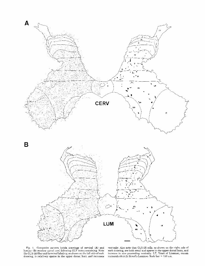
GLY-immunoreactive (GLY-IR) profiles were observed in both the cervical and lumbar segments. Camera lucida drawings of GLY-IR fibers and terminals (on the left) and cell bodies (on the right) are shown at each level in Figure 1. Note that although labeled cell bodies are observed in every lamina at both spinal levels, the majority are concentrated in laminae 111-VII with only a small number of GLY-IR neurons in laminae I, 11, VIII, IX and X. The distribution of GLY-IR myelinated fibers in the white matter is illustrated in Figure 2. Note the heavy concentration of GLY-IR fibers adjacent to the gray matter. The labeled fibers are not restricted to the area around the gray matter, in that, they are found throughout the ventral and lateral funiculi,
B
Fig. 1. Composite camera lucida drawings of cervical (A) and lumbar (B) monkey spinal cord following GLY immunostaining. Note the GLY-IR fiber and terminal labeling, as shown on the left side of each drawing, is relatively sparse in the upper dorsal horn and increases
ventrally. Also note that GLY-IR cells, as shown on the right side of each drawing, are both small and sparse in the upper dorsal horn, and increase in size proceeding ventrally. LT, Tract of Lissauer; roman numerals identify Rexed’s Laminae. Scale bar = 150 pm.

592 S.M. CARLTON ET AL.
although they are concentrated medially. The ventral funicu- lus was essentially filled with labeled fibers, whereas the lateral funiculus, although containing many labeled ele- ments, had a conspicuous absence of staining in the region of the dorsolateral funiculus. The lack of GLY labeling in this region was especially prominent in the cervical enlarge- ment. At both cord levels, the dorsal funiculus showed the fewest GLY-IR fibers, and these followed the contour of the gray-white border, with many labeled in the tract of Lissauer (Figs. 2, 3D) and some fibers present in the fasciculus interfascicularis, especially at cervical levels (Fig. 2A).
Both the nucleus and cytoplasm were immunostained in GLY-IR neurons (Fig. 3B). In the superficial laminae, GLY-IR neurons were of small diameter and the cell bodies were either round or ovoid. In the cervical segments, the average diameter of GLY-IR cells was 14 2 3 pm, and in the lumbar segments it was 13 c 3 pm. The deeper laminae (Iv-VI) contained GLY-IR neurons with intermediate diam- eters (cervical: 21 ? 5 pm; lumbar: 20 ? 4 pm) and robust processes. GLY-IR neurons with the largest diameters were present in laminae VII-IX (cervical and lumbar: 26 ? 6
All laminae contained many GLY-IR fibers and terminals (Figs. 1, 3A) except for lamina I1 which contained few labeled elements (Fig. 3C). Large myelinated fibers immu- nostained for GLY were distributed throughout the gray matter, but were concentrated ventral to lamina I11 (Figs. 1, 3A). Many GLY-IR profiles congregated adjacent to the gray-white border and extended into the white matter, particularly in the dorsal and intermediate horn and in the tract of Lissauer (Figs. 1,2,3D).
A striking finding appeared to be many large GLY-IR terminals in close apposition to GLY-IR cell bodies. The presumed terminals were round or ovoid immunoreactive profiles that followed the contour of, or indented, the postsynaptic cell membrane (Figs. 3A,B; 4; 5A). No space could be observed between the GLY-IR profiles and the postsynaptic GLY cell bodies or proximal dendrite at the LM level and therefore the labeled elements were presumed to be synaptic appositions. Large motoneurons were never GLY-IR; however, they often appeared to be en- crusted with presumed GLY-IR terminals (Fig. 5B and C).
pm).
Electron microscopic (EM) observations In PAP-immunostained tissue, the GLY-IR terminals
contained mainly round, clear vesicles, although occasional flattened or pleomorphic vesicles could also be seen (Fig. 6A and D). The synaptic thickenings associated with the labeled terminals were symmetric (Figs. 6A and D, 7A). Although not illustrated here, these terminals often con- tained one or more dense-core vesicles. Frequently GLY-IR terminals were presynaptic to GLY-IR profiles (Figs. 6D, 7A), confirming observations made at the LM level of GLY-GLY interactions.
Many GLY-IR dendrites could be found throughout the neuropil. These were contacted most frequently by un- stained terminals which contained round clear vesicles and had asymmetric synaptic thickenings (Figs. 6C, 7A). Many of the GLY-IR dendrites contained small clusters of round and/or pleomorphic clear vesicles and participated in den- drodendritic and dendroaxonic synaptic interactions (Figs. 6B and C, 7B).
B
CERV
Fig. 2. Composite camera lucida drawings of the cervical (A) and lumbar (B) monkey spinal cord showing GLY-IR axons in the white matter. Note the high concentration of labeled fibers at the grayiwhite matter border. Much of the lateral and all of the ventral funiculi contain labeled fibers. Note the relative absence of staining in the dorsal funiculi except for the fasciculus interfasicularis, and the dorsolateral part of the lateral funiculi. Scale bars = 500 km.
The interactions between GLY-IR terminals and motoneu- rons were investigated using postembedding immunogold labeling. Positively labeled profiles were identified as hav- ing at least three times as many gold particles per unit area as the surrounding unlabeled neuropil (Maxwell et al., 1990; Hayes and Carlton, 1992). Two categories of GLY-IR terminals were observed presynaptic to motoneurons: S- type terminals, characterized by a concentration of small, round clear vesicles and many mitochondria with several Cytoplasmic thickenings of various lengths (Conradi, 1970; Fig. 8A) and F-type terminals, which were smaller and contained a higher proportion of flat or pleomorphic synap- tic vesicles and symmetrical cytoplasmic thickenings (Con- radi, 1970; Fig. 8B).
Particular attention was focused on the relationship of GLY-IR profiles to primary afferent terminals in glomeruli. The two major findings were that: (1) the central elements

GLYCINERGIC PROFILES IN THE MONKEY SPINAL CORD 593
Fig. 3. Demonstration of GLY-IR profiles observed at the light microscope (LM) level. A GLY-IR cells (black arrows), fibers (arrow- heads), and presumed terminals (open arrows) in the superficial dorsal horn. B: Higher magnification of a GLY-IR cell body (G) contacted by presumed GLY-IR terminals (arrowheads). C: Photomicrograph illus-
trating the absence of GLY-labeled elements in lamina I1 (outlined by dots). D: Illustration of the large numbers of GLY-IR elements in the tract of Lissauer (LT, arrowheads). The dorsal column (DC) and the unmyelinated fibers in the dorsal part of the lateral funiculus (DLF) are also seen. Scale bars = 50 km (A,C,D); 10 pm (B).
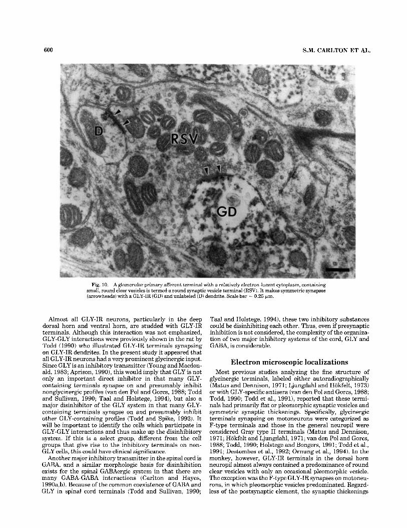
in all three types of glomeruli (large dense-core vesicle, LDCV; dense sinusoidal axon, DSA; round synaptic vesicle, RSV; Ralston, 1979; Knyihar-Csillik et al., 1982) synapsed on GLY-IR dendrites, and (2) no GLY-IR terminals were observed presynaptic to primary afferent terminals in glomeruli. Figure 9A shows a LDCV terminal synapsing on both a GLY-labeled (GD) dendrite and an unlabelled (D) dendrite. Figure 9B shows a DSA terminal synapsing on two GLY-IR (GD) dendrites and one unlabeled (D) dendrite. Figure 10 demonstrates an RSV terminal synapsing on a
GLY-IR (GD) dendrite and an unlabeled dendrite (D). An extensive search was made for axoaxonic or dendroaxonic synapses where either element was labeled for GLY. The only example found was the dendroaxonic synapse illus- trated in Figure 6C, in which a GLY-IR dendrite formed a symmetrical synapse with an unlabeled axon.
Finally, an analysis of the immunostained elements at the graylwhite matter border revealed that both myelin- ated and unmyelinated axons were labeled (Fig. 11). Analy- sis of fiber diameter indicated that the mean diameter of

594 S.M. CARLTON ET AL.
Fig. 4. A and B: GLY-IR neurons (G) from lamina VII showing robust dendrites. Presumed GLY-IR terminals contact the cell bodies and dendrites (arrowheads). Scale bar = 20 pm.
myelinated GLY-IR axons sampled in the white matter was 1.4 ? 0.7 Fm, while the mean diameter of unmyelinated GLY-IR axons was 0.4 2 0.1 km.
DISCUSSION Light microscopic localizations
The LM distribution of GLY-IR cells in the monkey is similar to that previously described in rat (Todd, 1990; Proudlock et al., 1993; Todd and Spike, 1993) and cat (Aprison and Werman, 1965; Graham et al., 1967). For example, labeled neurons are numerous in laminae 111-VII with fewer neurons in laminae I, I1 (the substantia gelati- nosa), VIII, IX and X. The axon and terminal labeling in the gray matter of the monkey also generally parallels what is found in the other animals, in that the numbers of labeled cells are roughly proportional to numbers of labeled axons and terminals, with the exception that axon and terminal labeling is heavy in lamina IX, even though few cells are labeled here. A particular point of earlier studies is that the density of GLY-IR elements is least in dorsal areas of the spinal gray matter and increases ventrally (Todd, 1990; Todd and Spike, 1993), and this is the case in the monkey. The correspondence between the density of GLY-IR cells and the amount of GLY-IR axons and terminals (Campis- tron et al., 1986; Ottersen et al., 1988; van den Pol and Gorcs, 1988; Todd, 1990; Todd and Sullivan, 19901, and the finding that interneurons whose axons aborize locally (e.g.,
islet cells) are labeled for GLY (Todd and Sullivan, 1990; Powell and Todd, 1992; Spike and Todd, 1992), led to the suggestion that the GLY system arises in large part from locally arborizing interneurons. Supporting this hypothesis in the monkey is the observation that GLY-IR axons in the white matter congregate near the graylwhite matter border where propriospinal fibers with local destinations are known to concentrate (Nathan and Smith, 1959). This is seen most clearly in the primate dorsal columns where essentially the only labeling is in fibers directly apposed to the dorsal horn. This finding does not imply that all spinal GLY axons arise from local interneurons, however, in that axons are labeled more laterally in the monkey ventral and lateral funiculi, and there is evidence for long descending GLY systems from the ventromedial brainstem (Holstege and Bongers, 1991) and medial vestibular nuclei (Wilson and Yoshida, 1969).
In monkey, there is a dramatic labeling of fibers outlining the gray matter of the dorsal horn, which has been reported in rat (van den Pol and Gorcs, 19881, but in particular there is intense labeling in the tract of Lissauer. This may be more a matter of emphasis than a true species difference, because previous studies were concerned less with the organization of the white matter than the gray matter. Since GLY has not been reported in primary afferents, the GLY fibers in the tract of Lissauer are presumably a component of the fine propriospinal fibers that characterize this path in rat, cat and monkey (Chung and Coggeshall,

GLYCINERGIC PROFILES IN THE MONKEY SPINAL CORD 595
Flg. 5. A GLY-IR cell (G) in the ventral horn with many GLY-IR terminals (arrowheads) along the cell membrane. B: Unstained moto- neuron (MN) in the ventral horn studded with many large presumed
GLY-IR terminals (arrowheads). C: GLY-IR interneuron (G) and unlabeled motoneurons (MN) showing prominent GLY innervation (arrowheads). Scale bar = 10 pm (A,B); 20 pm (C) .

596 S.M. CARLTON ET AL.
Fig. 6. Synaptic arrangements of GLY-IR profiles in the spinal cord. A GLY-IR axon terminal (GA) making synaptic contact with two unlabeled dendrites (D) via symmetrical thickenings (arrowheads). B GLY-IR dendrite containing vesicles (GD), is identified by numerous microtubules and the small collection of synaptic vesicles which occupy less than half of the cross-sectional area of the dendrite. Note that the vesicles in the dendrite are clear and pleomorphic and that the synaptic thickenings are symmetrical. This profile is in contact with an unla-
beled dendrite (D). C: A GLY-IR dendrite (GD, left) is postsynaptic to an unlabeled axon (A) via an asymmetric thickening. By contrast, the larger GLY-IR dendrite containing vesicles (GD, right) participates in symmetrical synapse with what appears to be an axon terminal (A). D A GLY-IR axon terminal (GA) in synaptic contact via a symmetrical synaptic thickening (arrowheads) with a GLY-IR dendrite (GD). Scale bars = 0.25 pm.
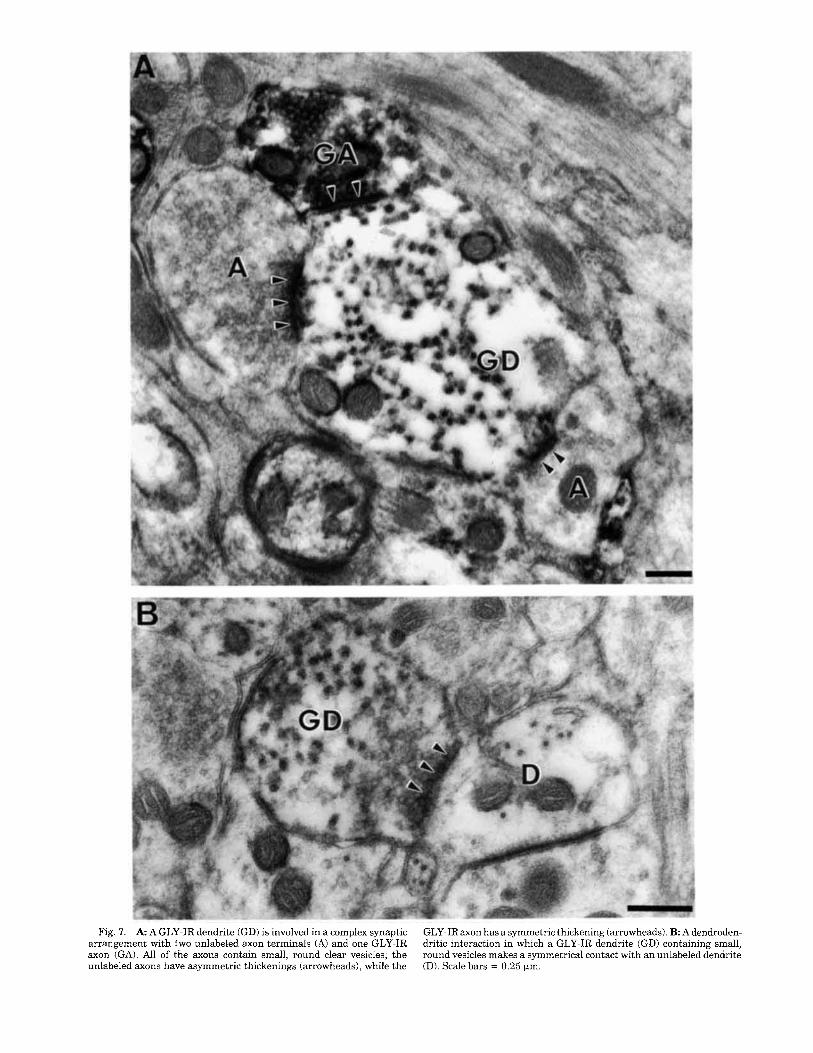
Fig. 7. A A GLY-IR dendrite (GD) is involved in a complex synaptic arrangement with two unlabeled axon terminals (A) and one GLY-IR axon (GA). All of the axons contain small, round clear vesicles; the unlabeled axons have asymmetric thickenings (arrowheads), while the
GLY-IRaxon has a symmetric thickening (arrowheads). B Adendroden- dritic interaction in which a GLY-IR dendrite (GD) containing small, round vesicles makes a symmetrical contact with an unlabeled dendrite (D). Scale bars = 0.25 pm.

598 S.M. CARLTON ET AL.
Fig. 8. A An “S” type terminal ( S ) , characterized by numerous small, round clear synaptic vesicles and several symmetric thickenings (arrowheads) is labeled with immunogold (arrows) for GLY. B: An “F” type terminal (F) characterized by flat vesicles (closed arrows) and
symmetrical synaptic thickenings (arrowheads) is labeled with immuno- gold (open arrows) for GLY. Both of these axon terminals were in synaptic contact with ventral horn motoneurons. Scale bars = 0.5 pm.
1979, 1982, 1985). Another finding is the dramatic absence of GLY-IR axons in the dorsolateral part of the lateral funiculus. Again, because GLY is not found in primary
afferents, this absence of stainingis presumably areflection of the large numbers of fine primary afferent fibers that travel in this region (Chung and Coggeshall, 1983,1985).
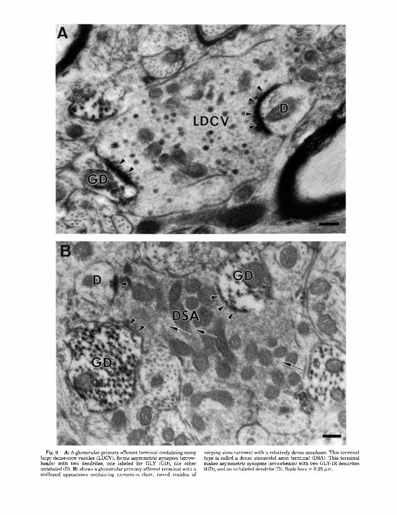
Fig. 9. A A glomerular primary afferent terminal containing many large dense-core vesicles (LDCV), forms asymmetric synapses (arrow- heads) with two dendrites, one labeled for GLY (GD), the other unlabeled (D). B: shows a glomerular primary afferent terminal with a scalloped appearance containing numerous clear, round vesicles of
varying sizes (arrows) with a relatively dense axoplasm. This terminal type is called a dense sinusoidal axon terminal (DSA). This terminal makes asymmetric synapses (arrowheads) with two GLY-IR dendrites (GD), and an unlabeled dendrite (D). Scale bars = 0.25 pm.
600 S.M. CARLTON ET AL.
Fig. 10. A glomerular primary afferent terminal with a relatively electron-lucent cytoplasm, containing small, round clear vesicles is termed a round synaptic vesicle terminal (RSV). It makes symmetric synapses (arrowheads) with a GLY-IR (GD) and unlabeled (D) dendrite. Scale bar = 0.25 fim.
Almost all GLY-IR neurons, particularly in the deep dorsal horn and ventral horn, are studded with GLY-IR terminals. Although this interaction was not emphasized, GLY-GLY interactions were previously shown in the rat by Todd (1990) who illustrated GLY-IR terminals synapsing on GLY-IR dendrites. In the present study it appeared that all GLY-IR neurons had a very prominent glycinergic input. Since GLY is an inhibitory transmitter (Young and Macdon- ald, 1983; Aprison, 1990), this would imply that GLY is not only an important direct inhibitor in that many GLY- containing terminals synapse on and presumably inhibit nonglycinergic profiles (van den Pol and Gorcs, 1988; Todd and Sullivan, 1990; Taal and Holstege, 19941, but also a major disinhibitor of the GLY system in that many GLY- containing terminals synapse on and presumably inhibit other GLY-containing profiles (Todd and Spike, 1993). It will be important to identify the cells which participate in GLY-GLY interactions and thus make up the disinhibitory system. If this is a select group, different from the cell groups that give rise to the inhibitory terminals on non- GLY cells, this could have clinical significance.
Another major inhibitory transmitter in the spinal cord is GABA, and a similar morphologic basis for disinhibition exists for the spinal GABAergic system in that there are many GABA-GABA interactions (Carlton and Hayes, 1990a,b). Because of the common coexistence of GABA and GLY in spinal cord terminals (Todd and Sullivan, 1990;
T a d and Holstege, 19941, these two inhibitory substances could be disinhibiting each other. Thus, even if presynaptic inhibition is not considered, the complexity of the organiza- tion of two major inhibitory systems of the cord, GLY and GABA, is considerable.
Electron microscopic localizations Most previous studies analyzing the fine structure of
glycinergic terminals, labeled either autoradiographically (Matus and Dennison, 1971; Ljungdahl and Hokfelt, 1973) or with GLY-specific antisera (van den Pol and Gorcs, 1988; Todd, 1990; Todd et al., 1991), reported that these termi- nals had primarily flat or pleomorphic synaptic vesicles and symmetric synaptic thickenings. Specifically, glycinergic terminals synapsing on motoneurons were categorized as F-type terminals and those in the general neuropil were considered Gray type I1 terminals (Matus and Dennison, 1971; Hokfelt and Ljungdahl, 1971; van den Pol and Gorcs, 1988; Todd, 1990; Holstege and Bongers, 1991; Todd et al., 1991; Destombes et al., 1992; Ornung et al., 1994). In the monkey, however, GLY-IR terminals in the dorsal horn neuropil almost always contained a predominance of round clear vesicles with only an occasional pleomorphic vesicle. The exception was the F-type GLY-IR synapses on motoneu- rons, in which pleomorphic vesicles predominated. Regard- less of the postsynaptic element, the synaptic thickenings

GLYCINERGIC PROFILES IN THE MONKEY SPINAL CORD 601
Fig. 11. GLY-IR myelinated (M) and unmyelinated (arrows) axons in the spinal cord white matter. Scale bar = 0.5 Fm
associated with GLY terminals were symmetrical. I t is possible that the differences in vesicle populations are due to differences in fixation procedures. In a similar study analyzing GABAergic terminals in the monkey spinal cord, however, few pleomorphic vesicles were observed and round clear vesicles predominated, suggesting that these observa- tions may represent a true species difference. Undoubtedly GLY and GABA are inhibitory transmitters in the primate spinal cord, even though both exhibit vesicle morphology usually associated with excitatory synapses. This morpho- logic difference suggests that a precise comparison of GLY (and GABA) transmission in spinal cord of rat or cat as opposed to monkey might reveal physiological differences as well.
Analysis of the interaction of GLY-IR profiles with primary afferent terminals in the dorsal horn demonstrated some species differences. Many primary afferent axons end in glomeruli in the dorsal horn (Willis and Coggeshall, 1991). Each of these glomeruli consists of a central primary afferent ending which is in synaptic contact with several peripheral dendrites and axon terminals, the whole struc- ture being at least partially set off from the rest of the neuropil by glial processes. The glomeruli are regarded as particularly important because they provide a more com- plex anatomical substrate for the modulation of primary afferent input than the more common simple axodendritic and axosomatic synapses made by primary afferents (Willis and Coggeshall, 1991). There are three types of central primary afferent endings in glomeruli in monkey (Ralston, 1979; Knyihar-Csillik et al., 1982): (1) large dense-core vesicle terminals (LDCV) which are especially prominent in monkey, (2) dense sinusoid axon terminals (DSA), which
are the most common terminals in lamina 11, and (3 ) regular synaptic vesicle terminals (RSV). All of these terminal types form synaptic endings on GLY-IR dendrites in the monkey spinal cord, but despite considerable search- ing, GLY-IR profiles were never observed presynaptic to glomerular central afferent terminals. Thus, in the mon- key, the three types of glomerular primary afferent termi- nals can apparently excite the spinal GLY inhibitory sys- tem, but GLY does not directly (by axoaxonic or dendroaxonic synapses) inhibit glomerular primary affer- ents. This finding corroborates physiological data which indicate that GLY plays no role in presynaptic mechanisms (Eccles et al., 1962), but exerts its control via postsynaptic pathways. These findings differ from the rat in two ways. First, in the rat only the type I1 glomerular afferents, which are thought to arise from large myelinated primary afferent axons (Ribeiro-DaSilva and Coimbra, 1982) and so are equivalent to the RSV terminals of the monkey, interact with GLY elements. Second, axoaxonic contacts, in which the GLY terminal is presynaptic, are common in both the general neuropil and on the type I1 glomeruli in the rat (Todd, 1990). As stated above, these arrangements in the monkey could not be found. This might imply a greater inhibitory control by GLY of primary afferent input in the rodent as compared to the primate.
Finally, in monkey, as in rat (Todd, 1990), many GLY dendrites contain a population of vesicles and unite via synaptic thickenings to other dendrites and occasional cell bodies in the dorsal horn. These dendrodendritic and dendrosomatic interactions may provide a type of local inhibitory control (Ralston, 19791, as previously suggested for GABA dendrites that had similar synaptic interactions

602 S.M. CARLTON ET AL.
(Carlton and Hayes, 1990a,b). Thus, vesicle-containing GLY-IR dendrites could easily “multiply” the inhibitory influence of the glycinergic system in the dorsal horn.
ACKNOWLEDGMENTS This work was supported by NS11255, NS27910 (S.M.C.)
and NS10161 (R.E.C.). We thank Brenda Kenworthy for her excellent secretarial support.
LITERATURE CITED Aprison, M.H. (1990) The discovery of the neurotransmitter role of glycine.
In O.P. Ottersen and J. Storm-Mathisen (eds): Glycine Neurotransmis- sion. New York: John Wiley & Sons, Inc. pp. 2-23.
Aprison, M.H., and R. Werman (1965) The distribution of glycine in cat spinal cord and roots. Life Sci. 42075-2083.
Barber, R.P., J.E. Vaughn, K. Saito, B.J. McLaughlin, and E. Roberts (1978) GABAergic terminals are presynaptic to primary afferent terminals in the substantia gelatinosa of the rat spinal cord. Brain Res. 141:35-55.
Basbaum, A.I., E.J. Glazer, and W. Oertel (1986) Immunoreactive glutamic acid decarboxylase in the trigeminal nucleus caudalis of the c a t A light- and electron-microscopic analysis. Somato. Res. 4t77-94.
Campistron, G., R.M. Buijs, and M. Geffard (1986) Glycine neurons in the brain and spinal cord. Antibody production and immunocytochemieal localization. Brain Res. 376t400-405.
Carlton, S.M. (1994) Presynaptic control of thin primary afferents: an ultrastructural analysis. In J.M. Besson, G. Guilbaud, and H. Ollat (eds): Peripheral Neurons in Nociception. Paris: John Libbey Eurotext, pp. 185-200.
Carlton, S.M., and E.S. Hayes (1990a) Light microscopic and ultrastructural analysis of GABA-immunoreactive profiles in the monkey spinal cord. J. Comp. Neurol. 3OOt162-182.
Carlton, S.M., and E.S. Hayes (199Ob) GABA vesicle-containing dendrites: A critical element in processing sensory input in the primate dorsal horn. Neurosci. Lett. 121:4042.
Carlton, S.M., K.N. Westlund, D. Zhang, and W.D. Willis (1992) GABA- immunoreactive terminals synapse on primate spinothalamic tract cells. J. Comp. Neurol. 322:52%537.
Chung, K., and R.E. Coggeshall(1979) Primary afferent axons in the tract of Lissauer in the cat. J. Comp. Neurol. 186t451464.
Chung, K., and R.E. Coggeshall(1982) Quantitation of propriospinal fibers in the tract of Lissauer of the rat. J. Comp. Neurol. 211:41%426.
Chung, K., and R.E. Coggeshall (1983) Numbers of axons in lateral and ventral funiculi of rat sacral spinal cord. J. Comp. Neurol. 214:72-78.
Chung, K., and R.E. Coggeshall(1985) Unmyelinated primary afferent fibers in dorsal funiculi of cat sacral spinal cord. J. Comp. Neurol. 238:365- 369.
Conradi, S. (1970) Ultrastructure and distribution of neuronal and glial elements on the motoneuron surface in the lumbosacral spinal cord of the adult cat. Acta Phys. Scand. 3325-48.
Destombes, J., G. Horcholle-Bossavit, and D. Thiesson (1992) Distribution of glycinergic terminals on lumbar motoneurons of the adult cat: An ultrastructural study. Brain Res. 599:353-360.
Eccles, J.C., P.G. Kostyuk, and R.F. Schmidt (1962) Central pathways responsible for depolarization of primary afferent fibres. J. Physiol. 161:237-257.
Graham, L.T., Jr., R.P. Shank, R. Werman, and M.H. Aprison (1967) Distribution of some synaptic transmitter suspects in cat spinal cord: Glutamic acid, aspartic acid, yaminobutyric acid, glycine, and gluta- mine. J. Neurochem. 14t465-472.
Hayes, E.S., and S.M. Carlton (1992) Primary afferent interactions: Analysis of calcitonin gene-related peptide-immunoreactive terminals in contact with unlabeled and GABA-immunoreactive profiles in the monkey dorsal horn. Neuroscience 472473-896.
Hepler, J.R., C.S. Toomim, K.D. McCarthy, F. Conti, G. Battaglia, A. Rustioni, and P. Petrusz (1988) Characterization of antisera to gluta- mate and aspartate. J. Histochem. Cytochem. 36t13-22.
Holstege, J.C., and C.M.H. Bongers (1991) A glycinergic projection from the ventromedial lower brainstem to spinal motoneurons. An ultrastruc- tural double labeling study in rat. Brain Res. 566:308-315.
Hokfelt, T., and A. Ljungdahl (1971) Light and electron microscopic autoradiography on spinal cord slices after incubation with labeled glycine. Brain Res. 32189-194.
Knyihar-Csillik, E., B. Csillik, and P. Rakic (1982) Periterminal synaptology of dorsal root glomerular terminals in the substantia gelatinosa of the spinal cord in the Rhesus monkey. J. Comp. Neurol. 210:376-399.
Larsson, L.-I. (1981) A novel immunocytochemical model system for specific- ity and sensitivity screening of antisera against multiple antigens. J. Histochem. Cytochem. 29t408-410.
Ljungdahl, A., and T. Hokfeit (1973) Autoradiographic uptake patterns of PHIGABA and [3H]glycine in central nervous tissues with special reference to the cat spinal cord. Brain Res. 62587-595.
Matus, A.I., and M.E. Dennison (1971) Autoradiogaphic localization of tritiated glycine at “flat-vesicle’’ synapses in spinal cord. Brain Res. 32: 195-197.
Maxwell, D.J., and R. Noble (1987) Relationships between hair-follicle afferent terminations and glutamic acid decarboxylase-containing bou- tons in the cat’s spinal cord. Brain Res. 408:308-312.
Maxwell, D.J., W.M. Christie, A.D. Short, J. Strom-Mathisen, and O.P. Ottersen (1990) Central boutons of glomeruli in the spinal cord of the cat are enriched with L-glutamate-like immunoreactivity. Neuroscience 36:83-104.
Nathan, P.W., and M.C. Smith (1959) Fasciculi proprii of the spinal cord in man: Review of present knowledge. Brain 82t610-668.
Ornung, G., 0. Shupliakov, O.P. Ottersen, J. Storm-Mathisen, and S. Cullheim (1994) Immunohistochemical evidence for coexistence of gly- cine and GABA in nerve terminals on cat spinal motoneurones: An ultrastructural study. NeuroReport 5889-892.
Ottersen, O.P., J. Storm-Mathisen, and P. Somogyi (1988) Colocalization of glycine-like and GABA-like immunoreactivities in Goigi cell terminals in the rat cerebellum: A postembedding light and electron microscopic study. Brain Res. 450:342-353.
Powell, J.J., and A.J. Todd (1992) A light and electron microscope study of GABA-immunoreactive neurons in lamina 111 or rat spinal cord. J. Comp. Neurol. 315t125-136.
Proudlock, F., R.C. Spike, and A.J. Todd (1993) Immunocytochemical study of somatostatin, neurotensin, GABA, and glycine in rat spinal dorsal horn. J. Comp. Neurol. 327,289-297.
Ralston, H.J. (1979) The fine structure of laminae I, 11, and I11 of the macaque spinal cord. J. Comp. NeuroI. 184r619442.
Ribeiro-DaSilva, A., and A. Coimbra (1982) Two types of synaptic glomeruli and their distribution in laminae 1-111 of the rat spinal cord. J. Comp. Neurol. 209:176-186.
Spike, R.C., and A.J. Todd (1992) An ultrastructural and immunocytochemi- cal study of lamina I1 islet cells in rat spinal dorsal horn. J. Comp. Neurol. 323t359-369.
Sternberger, L.A. (1979) Immunocytochemistry. New York John Wiley & Sons, Inc.
Taal, W., and J.C. Holstege (1994) GABAand glycine frequently colocalize in terminals on cat spinal motoneurons. NeuroReport 5.2225-2228.
Todd, A.J. (1990) An electron microscope study of glycine-like immunoreac- tivity in laminae 1-111 of the spinal dorsal horn of the rat. Neuroscience 39:387-394.
Todd, A.J., and R.C. Spike (1993) The localization of classical transmitters and neuropeptides within neurons in laminae 1-111 of the mammalian spinal dorsal horn. Prog. Neurobiol. 41t609-645.
Todd, A.J., and A.C. Sullivan (1990) Light microscope study of the coexist- ence of GABA-like and glycine-like immunoreactivities in the spinal cord of the rat. J. Comp. Neurol. 296,496605.
Todd, A.J., D.J. Maxwell, and A.G. Brown (1991) Relationships between hair-follicle afferent axons and glycine-immunoreactive profiles in cat spinal dorsal horn. Brain Res. 564:132-137.
van den Pol, A.N., and T. Gorcs (1988) Glycine and glycine receptor immunoreactivity in brain and spinal cord. J. Neurosci. 8t472-492.
Willis, W.D., and R.E. Coggeshall(1991) Sensory Mechanisms of the Spinal Cord. New York: Plenum Press.
Wilson, V.J., and M. Yoshida (1969) Monosynaptic inhibition of neck motoneurons by the medial vestibular nucleus. Exp. Brain Res. 9t365- 380.
Young, A.B., and R.L. Macdonald (1983) Glycine as a spinal cord neurotrans- mitter. In R.A. Davidoff (ed): Handbook of the Spinal Cord. New York, Ny: Marcel Dekker, Inc. pp. 1 4 3 .