distinct infrared spectral signatures of the 1,2- and 1,4...
TRANSCRIPT
rXXXX American Chemical Society 1307 DOI: 10.1021/jz100143z |J. Phys. Chem. Lett. 2010, 1, 1307–1311
pubs.acs.org/JPCL
Distinct Infrared Spectral Signatures of the 1,2- and1,4-Fluorinated Single-Walled Carbon Nanotubes:A Molecular Dynamics StudyAkira Ueta,† Yoshitaka Tanimura,† and Oleg V. Prezhdo*,‡
†Department of Chemistry, Graduate School of Science, Kyoto University, Kitashirakawa, Sakyoku, Kyoto 606-8502, Japan, and‡Department of Chemistry, University of Washington, Seattle, Washington 98195-1700
ABSTRACT Fluorinated single-walled carbon nanotubes (F-SWNTs) form impor-tant intermediates in SWNT sidewall functionalization, leading to a variety ofmaterials and biological applications. By simulating the infrared (IR) signals forthe 1,2- and 1,4-addition structures, in which fluorine atoms are arranged in orthoor para positions, respectively, on the aromatic skeleton of the (10,10) SWNTsurface, we identify peaks that are unique to each structure. Our full moleculardynamics simulations show that the [-C(sp3)-C(sp3)-] collective vibrational peakat 400 cm-1 is optically active only in the 1,2-isomer, while the 1300 cm-1 bandarising due to the F-C(sp3) stretchingmotion coupled with the neighboring C(sp2)atoms is seen in the IR spectrum of only the 1,4-isomer. The reported resultssuggest simple and clear experimental means for distinguishing between the twofluorinated structures and provide a valuable tool for controlled SWNT sidewallfunctionalization.
SECTION Nanoparticles and Nanostructures
T he discovery of carbon nanotubes (CNTs)1 has led toan explosionof studies focusing onunderstanding andcontrolling CNT atomic and electronic properties, as
motivated by a variety of electronics,2-13 biological,14-17
materials,18-24 energy,25-27 and other applications. Chemi-cal functionalization of CNT sidewalls provides one of themost efficient routes to the desired property control. Exam-ples are abundant. Addition of fluorinatedolefins and chlorineatoms represents an effective approach toward convertingcommercial mixtures of metallic and semiconducting CNTsinto high-mobility semiconducting tubes.2,3 Functionalizationwith carboxylic acid, nitroso, and maleic anhydride groupsallows one to control CNTcharging.5,6 TheCNToptical proper-ties can be selectively modified by fluorination.7 Functionali-zation and bioconjugation of CNTs have led to multipleprotocols for biomedical applications, including biologicalimaging, labeling, sensing, and drug delivery.14-16 Key biolo-gical advantages of functionalized carbon nanotubes includetheir excellent ability to translocate through membraneswhile retaining low toxicity.17 Fluoride atoms and othersubstituents on the CNT surface can be used to transformCNT films between the superhydrophobic and nearly hydro-philic states.18,19 Control of surface adsorption propertiesby covalent and noncovalent CNT functionalization20 leadsto superior CNT-polymer composites,21 materials with im-proved friction properties,22 and strongly interconnectedCNTblocks.24 Fluorination and defluorination reactions form thebasis for CNT applications in hydrogen storage26 and Li ionbatteries.25 Finally, complexation of CNTs with organic sensi-tizers leads to promising photovoltaic materials.27
Generally, CNTs are chemically nonreactive and are hard tofunctionalize due to the efficient carbon-carbon bonding. Thestrong reactivity of fluorine atoms makes fluorination one ofthe most effective methods to modify and control physical-chemical properties of carbon materials.28,29 Using techno-logy developed for the fluorination of graphite,30 Mickelsonet al. produced fluorinated single-walled carbon nanotubes(F-SWNTs), which serve as a staging point of chemical modi-fication for a wide variety of sidewall functionalizations.31
The structures of F-SWNT were investigated by variousmethods involving infrared (IR) and Raman spectro-scopies,29,32,33 nuclear magnetic resonance (NMR),33,34
transmission electronmicroscopy (TEM),29 scanning tunnel-ing microscopy (STM),35 electron energy loss spectroscopy(EELS),36 and X-ray photoemission spectroscopy (XPS).37,38
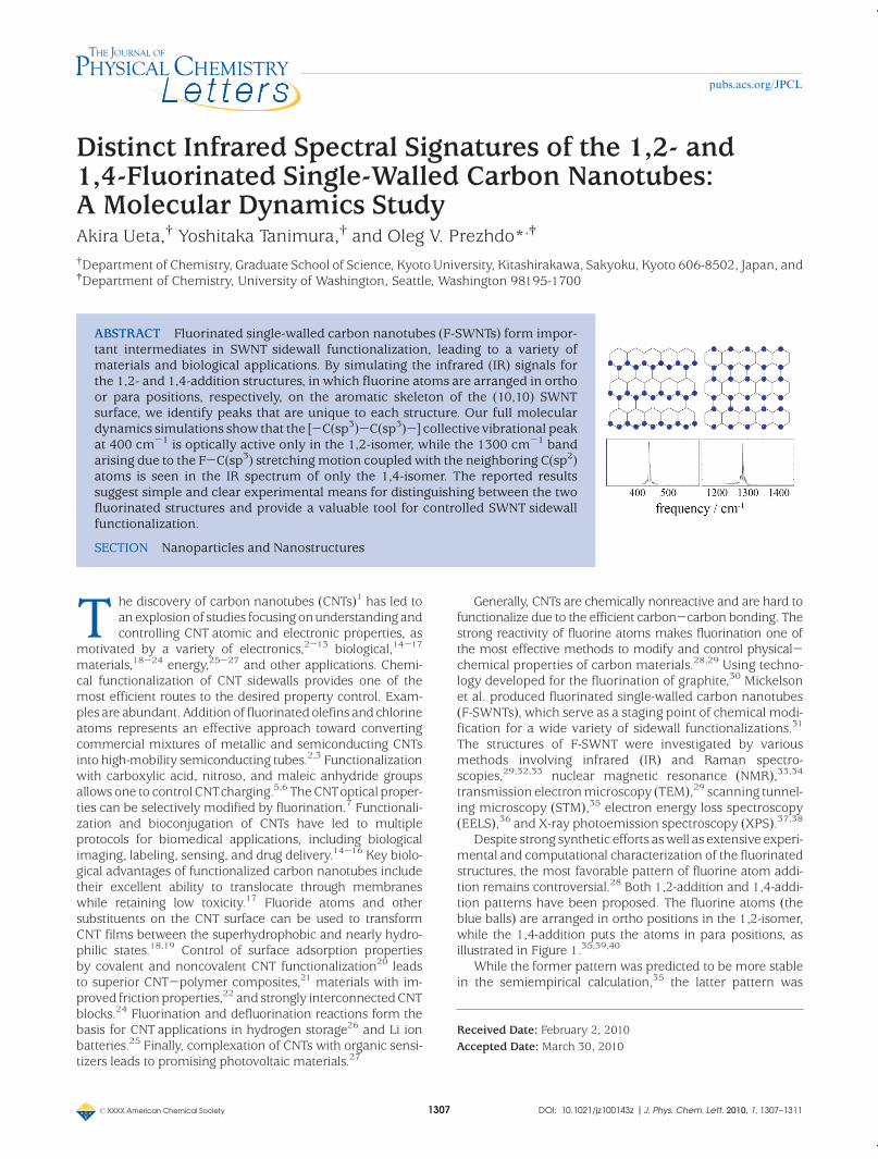
Despite strong synthetic efforts aswell as extensive experi-mental and computational characterization of the fluorinatedstructures, the most favorable pattern of fluorine atom addi-tion remains controversial.28 Both 1,2-addition and 1,4-addi-tion patterns have been proposed. The fluorine atoms (theblue balls) are arranged in ortho positions in the 1,2-isomer,while the 1,4-addition puts the atoms in para positions, asillustrated in Figure 1.35,39,40
While the former pattern was predicted to be more stablein the semiempirical calculation,35 the latter pattern was
Received Date: February 2, 2010Accepted Date: March 30, 2010

rXXXX American Chemical Society 1308 DOI: 10.1021/jz100143z |J. Phys. Chem. Lett. 2010, 1, 1307–1311
pubs.acs.org/JPCL
found to bemore favorable in the DFTcalculations.41 In eachcase, the energy difference between the two patterns wasquite small, implying that both types of fluorinated materialsprobably coexist.28
The purpose of this study is to distinguish between the1,2- and 1,4-addition structures of F-SWNTs by means of IRspectroscopy. In order to explore this prospect, we employclassicalmoleculardynamics (MD)42-45 anddirectly calculatethe IR response function.44-47 Our study shows that eachstructure can be identified by distinct IR peaks. In particular,an optically active band appears at 400 cm-1 in the 1,2-addition product due to collective vibrationalmotions involvingthe [-C(sp3)-C(sp3)-] bond created by the 1,2-addition. Noequivalent band is seen in the IR spectrum of the 1,4-product.On the other hand, a 1300 cm-1 band originating from theF-C(sp3) stretching motion is seen in the IR spectrum of the1,4-isomer but not that of the 1,2-isomer.
In the following, we explain the methodology of the IRresponse analysis by means of MD simulation. Then, wepresent and discuss the calculated IR spectra for the 1,2-and 1,4-F-SWNTaddition structures.
Pristine CNTs have 15 or 16 vibrational modes that areactive in theRaman spectrum.48 The corresponding IR signal isvery weak since the first-order IR optical response depends onthe transition dipole moment, which is practically nonexistentin ideal CNTs. Fluorine atoms break the CNTsymmetry, createlocal dipoles, and generate an IR signal. The fluorine atoms andthe sp3 carbons generated by the fluorine addition affect theCNTs modes. The vibrational frequencies of F-SWNTs aregenerally higher than those of pristine CNTs. The opticalresponse function calculated within the MD approach allowsus to obtain the entire IR spectrum of F-SWNTs. In order tocompare and distinguish between the two isomers, we con-centrate on the C-F vibrational modes. Although fluorinationchanges the properties of the modes associated with theoriginal C(sp2) atoms, these changes are quite complicatedand will not be discussed here since the analysis of theC(sp3)-F motions already gives the desired result.
We consider the dipole correlation function Æμ(t)μ(0)æ thatis obtained from the MD simulation, where μ(t) is the dipolemoment of the F-SWNTsystem. The Fourier transform of thecorrelation function is defined as
IMDðωÞ ¼ 12π
Z ¥
-¥dt e- iωtÆμðtÞμð0Þæ ð1Þ
While the IR spectrumR(ω) is determinedmore rigorously viathe quantummechanical commutator Æ[ μ(t),μ(0)]æ, the spec-trum is well-approximated using the classical result IMD(ω)and the harmonic assumption by
RðωÞ�ω tanhpω
2kBT
� �IMDðωÞ ð2Þ
Here, kB is the Boltzmann constant, T is temperature, and p isPlanck's constant.46,49
The F-SWNT MD was generated using the Amber poten-tials for the intramolecular C-C, C-F, and F-F interactions.In particular, the C(sp2) atoms were assigned the “CA”Amberatom type, while C(sp3) atoms were described using theAmber “CT” atom type.50 Previously, the Amber force fieldwas used in order to describe the IR spectra of peptides44,45
andmanyother systems. The interactions between the atomsthatwere not directly bonded to eachotherwere describedbythe Coulomb and van derWaals (VDW) terms. To simplify thecalculation, the VDW interactions between atoms separatedby more than 5 Å were disregarded. The velocity Verletalgorithm was adopted to integrate the equation of motionwith a 0.1 fs time step. The simulations were performed at300 K.
Note that classical MD does not necessarily give a veryprecise description of the structure and dynamics of F-SWNTdue to the phenomenological interaction potential. However,the approach is sufficiently accurate to establish the differ-ences in the IR signals for the two distinct F-SWNT isomers.
The study was performed using the (10,10) metallic arm-chair SWNT. Periodic boundary conditions along the tube axiswere used. The simulation cell was 2.4 nm long, containing10 benzene rings in the periodic direction. The fluorine atomswere arranged to form the 1,2- and 1,4-addition structuresshown in Figure 1a and b. In both structures, all possibleaddition sites of the outside surface of the tubewere occupiedby fluorine atoms.51We ranMD simulations to equilibrate thesystem under the given conditions and checked the accuracyof the simulated structures.
In order to calculate the IR signal, the dipole momentsassociatedwith theC-Fbondsweredetermined. The chargeson the fluorine and carbon atoms were calculated quantummechanically for a single graphene sheet with the fluorineatoms arranged as in Figure 1. The ab initio density functionaltheory (DFT) calculations were done using the GAMESSpackage, the B3LYP exchange-correlation function, and theSVP basis set. The charges were computed using the CHELPGmethod.We found that thebonded fluorine and carbonatomshad nearly opposite charges of about-0.2e andþ0.2e, whilethe remaining carbon atomswere almost neutral. Further, wecalculated the changes in the point charges associated withthe thermal fluctuations of the C-F bond. In our MD trajec-tory, the bond length fluctuated within 0.1 Å of its equilibriumvalue, and the corresponding changes in the atomic chargescalculated by DFTwere small. These results indicated that thedipolemoments associatedwith theC-Fmodesweredirectlyproportional to the corresponding bond lengths.
Two independent polarizations of light were considered indetail, corresponding to the directions along the tube axis and
Figure 1. Two addition structures of fluorinated carbon nano-tubes. (a) The 1,2-addition; (b) 1,4-addition. In each structure, blueballs depict fluorine atoms.

rXXXX American Chemical Society 1309 DOI: 10.1021/jz100143z |J. Phys. Chem. Lett. 2010, 1, 1307–1311
pubs.acs.org/JPCL
its radius. Circularly polarized light was considered as well.Each IR response function, eqs 1 and 2, was computed byaveraging over an ensemble of 106 initial configurations.Figures 2 and 3 display the IR spectra of the F-SWNT for lightpolarized along the tube axis and its radius, respectively.
Significant differences between the 1,2- and 1,4-additionstructures are seen at 400 and 1300 cm-1. The peak at400 cm-1 is optically active only in the 1,2-addition product,whereas the peak at 1300 cm-1 appears only in the 1,4-isomer. In order to elucidate the atomistic origin of these andother peaks, we performed a normal-mode analysis of theF-SWNTsystems. The results showed that peakswith frequen-cies less than 500 cm-1 arose from collective vibrationalmotions of the SWNT, while peaks with frequencies above900 cm-1 were derived from local modes. The peaks corre-sponding to the local C-C andC-F bondvibrations producedby the Amber force field emergewithin the 1100-1500 cm-1
frequency range. In particular, the C-F bond stretchingmodes appear at about 1200-1300 cm-1.
In order to analyze the positions and origins of the peaks inthe IR spectra, we repeated the calculations using variousconstraints. We fixed the positions of all sp2 carbon atoms,neglected electrostatic and VDW interactions between fluor-ine atoms, and so forth. We found that the peak at around200 cm-1 originated from the VDW interactions between thecarbon and fluorine atoms. This peak is optically active forlight polarized along both the tube axis and its radius. Since itdid not depend on the electrostatic interactions involvingfluorine atoms, the peak appeared in both 1,2- and 1,4-structures. Other spectral features in the 100-300 cm-1
range were due to CNT radial breathing modes (RBM). As aresult, theywere optically active only for the radially polarizedlight.
The peaks in the 350-400 cm-1 frequency range app-eared in both fluorine addition structures for the radiallypolarized light. At the same time, they were optically activeonly in the 1,2-isomer for light polarized along the tube axis.These peaks originated fromvibrationalmotions of the C(sp2)and C(sp3) atoms along and perpendicular to the tube axis. Inparticular, the peaks in the radially polarized spectra for bothF-SWNT isomers were from the [-C(sp3)-C(sp2)-] vibra-tional motions, whereas the 350-400 cm-1 features in thespectrumof the1,2-addition structure for light polarizedalongthe tube axis were attributed to the [-C(sp3)-C(sp3)-]vibrationalmodes. The correspondingmotions along the tubeaxis in the 1, 4-structure arose from the [-C(sp3)-C(sp2)-C(sp2)-C(sp3)-] fragments. They had lower frequencies anddid not appear in the 350-400 cm-1 range.
The peaks due to F-C(sp3) stretching peaks were in the1200-1300 cm-1 frequency range. For the light polarizedalong the tube axis, the distinct peak at 1300 cm-1 wasobserved only in the 1,4-isomer. For radially polarized light,small peaks in the 1200-1300 cm-1 frequency range wereobserved inboth structures. The remaining900and1100cm-1
peaks detectable in the IR spectrum arose from the waggingmotions of the C-F bonds and the stretching motions of theC(sp3)-C(sp2) bonds, respectively.
In addition to light polarized along the tube axis andradially polarized light, we computed the IR spectra forcircularly polarized light. The results were very similar to thespectra shown in Figures 2 and 3, and therefore, they are notdiscussed here. No vibrationalmodes that were specific to thecircularly polarized light were detected, at least for the (10,10)F-SWNTs.
It is instructive to comment on the relaxation mechanismfor the C-F vibrational energy deposited by the IR radiation.Since the (10,10) SWNT ismetallic, onemayexpect relaxationthat is similar to the relaxation of vibrationalmodes of speciesadsorbed on bulk metal surfaces. The presence of a highlypolarizable electroncloud ina bulkmetal leads to the so-called“dynamic dipole mechanism”
52, in which the fluctuations inthe dipole moment of the vibrational mode, in our case theC-F bond, couple to the electronic degrees of freedom of thesubstrate. Our calculations indicate that the dynamic dipolemechanism plays little role in SWNTs. The charges on thecarbon atoms that are not directly bound to fluorines remainclose to zero during the C-F bond oscillation, as determinedby the DFT calculations. This lack of electronic polarizationcan be explained by the quasi one-dimensional structure ofSWNTs, together with the fact that SWNTwalls are made of asingle atomic layer. The C-F vibrations excited by the IRradiation relax through valence bonding between the fluorineand carbon atoms. The corresponding bonding interactionsare significantly weaker between molecules and bulk metalsurfaces.
Finally, we should mention the future prospects andlimitation of the present studies. Our analysis was based onclassical MD with the AMBER potential. Changes in theelectronic structure of the sp2 carbons due to their long-rangeinteractions with the fluorine atoms were not fully takeninto account. Such changes may affect the dynamics andpolarization of the system. In principle, they can be included
Figure 2. IR spectra of a 1,2-fluorinated carbon nanotube ob-tained from full MD simulations. The red solid line represents lightpolarization along the tube axis, whereas the green dotted linecorresponds to radially polarized light.
Figure 3. Same as Figure 2, but for the 1,4-fluorinated CNT.
rXXXX American Chemical Society 1310 DOI: 10.1021/jz100143z |J. Phys. Chem. Lett. 2010, 1, 1307–1311
pubs.acs.org/JPCL
by first-principles MD; however, this approach is computa-tionally too expensive for the calculation of the IR spectra. Thecurrent analysis focused on the linear IR response function. Itallowed us to successfully distinguish the 1,2- and 1,4-stru-cures. Further information, including coupling between theC-F bonds, structural defects, and locations of boundariesbetween the 1,2- and 1,4-isomer domains, may be obtainedby analysis of nonlinear response functions that are relevantfor multidimensional vibrational spectroscopies. 53-56 Weleave this for future studies.
In conclusion, our calculations show that the [-C(sp3)-C(sp3)-] collective vibrational peak is observed at 400 cm-1
only in the IR spectrum of the 1,2-fluorine addition structure,whereas the F-C(sp3) stretching motion coupled with neigh-boring C(sp2) atoms is observed at 1300 cm-1 only in the1,4-addition structure for light polarized along the tube axis.These predicted spectral differences create a possibility todistinguish between the different F-SWNT isomers using IRmeasurements. Furthermore, if the 1,2- and 1,4-additionstructures coexist, their ratio can be determined by therelative intensities of the spectral peaks associated with eachstructure.
AUTHOR INFORMATION
Corresponding Author:*To whom correspondence should be addressed.
ACKNOWLEDGMENT The authors thank Tammie Nelson forcomments on the manuscript. A.U. is supported by the researchfellowship of the Global COE Program, International Center forIntegrated Research, and Advanced Education in Material Science,Kyoto University. Y.T. is grateful for the financial support in the formof a Grant-in-Aid for Scientific Research B19350011 from the JapanSociety for the Promotion of Science. O.V.P. acknowledges thesupport of the U.S. Department of Energy, Grant #DE-FG02-05ER15755. O.V.P. is a Foreign Scholar of the Japan Society forthe Promotion of Science.
REFERENCES
(1) Iijima, S. Helical Microtubules of Graphitic Carbon. Nature1991, 354, 56–58.
(2) Kanungo, M.; Lu, H.; Malliaras, G. G.; et al. Suppression ofMetallic Conductivity of Single-Walled Carbon Nanotubes byCycloaddition Reactions. Science 2009, 323, 234–237.
(3) Lu, J.; Wang, D.; Nagase, S.; et al. Evolution of the ElectronicProperties of Metallic Single-Walled Carbon Nanotubes withthe Degree of CCl2 Covalent Functionalization. J. Phys. Chem.B 2006, 110, 5655–5658.
(4) Bachilo, S. M.; Strano, M. S.; Kittrell, C.; et al. Structure-Assigned Optical Spectra of Single-Walled Carbon Nano-tubes. Science 2002, 298, 2361–2366.
(5) Strano, M. S.; Dyke, C. A.; Usrey, M. L.; et al. ElectronicStructure Control of Single-Walled Carbon Nanotube Functio-nalization. Science 2003, 301, 1519–1522.
(6) McPhail, M. R.; Sells, J. A.; He, Z.; et al. Charging Nanowalls:Adjusting the Carbon Nanotube Isoelectric Point via Sur-face Functionalization. J. Phys. Chem. C 2009, 113, 14102–14109.
(7) Hayashi, T.; Shimamoto, D.; Kim, Y. A.; et al. SelectiveOptical Property Modification of Double-Walled Carbon Nano-tubes by Fluorination. ACS Nano 2008, 2, 485–488.
(8) Habenicht, B. F.; Craig, C. F.; Prezhdo, O. V. Time-Domain AbInitio Simulation of Electron and Hole Relaxation Dynamicsin a Single-Wall Semiconducting Carbon Nanotube. Phys. Rev.Lett. 2006, 96, 187401.
(9) Habenicht, B. F.; Prezhdo, O. V. Nonradiative Quenching ofFluorescence in a Semiconducting Ab Initio Study. Phys. Rev.Lett. 2008, 100, 197402.
(10) Habenicht, B. F.; Kamisaka, H.; Yamashita, K.; et al. Ab InitioStudy of Vibrational Dephasing of Electronic Excitations inSemiconducting Carbon Nanotubes. Nano Lett. 2007, 7,3260–3265.
(11) Yarotski, D. A.; Kilina, S. V.; Talin, A. A.; et al. ScanningTunneling Microscopy of DNA-Wrapped Carbon Nanotubes.Nano Lett. 2009, 9, 12–17.
(12) Murakoshi, K.; Okazaki, K. Electrochemical Potential Controlof Isolated Single-Walled Carbon Nanotubes on Gold Elec-trobe. Electrochim. Acta 2005, 50, 3069–3075.
(13) Takeda, N.; Murakoshi, K. Characteristics of the RamanSpectra of Single-Walled Carbon Nanotube Bundles underElectrochemical Potential Control.Anal. Bioanal. Chem. 2007,388, 103–108.
(14) Liu, Z.; Tabakman, S. M.; Chen, Z.; et al. Preparation ofCarbonNanotubeBioconjugates for Biomedical Applications.Nature Protocols 2009, 4, 1372–1382.
(15) Satishkumar, B. C.; Brown, L. O.; Gao, Y.; et al. ReversibleFluorescence Quenching in Carbon Nanotubes for Biomole-cular Sensing. Nat. Nanotechnol. 2007, 2, 560–564.
(16) Barone, P. W.; Baik, S.; Heller, D. A.; et al. Near-InfraredOptical Sensors Based on Single-Walled Carbon Nanotubes.Nat. Mater. 2005, 4, 86–92.
(17) Li, S. S.; He, H.; Jiao, Q. C.; et al. Applications of CarbonNanotubes in Drug and Gene Delivery. Prog. Chem. 2008, 20,1798–1803.
(18) Zhang, Y. Y.; Stan, L.; Xu, P.; et al. A Double-Layered CarbonNanotube Array with Super-Hydrophobicity. Carbon 2009,47, 3332–3336.
(19) Kakade, B. A.; Pillai, V. K. Tuning the Wetting Properties ofMultiwalled Carbon Nanotubes by Surface Functionalization.J. Phys. Chem. C 2008, 112, 3183–3186.
(20) Usrey, M. L.; Strano, M. S. Controlling Single-Walled CarbonNanotube Surface Adsorption with Covalent and Noncova-lent Functionalization. J. Phys. Chem. C 2009, 113, 12443–12453.
(21) Pulikkathara, M. X.; Kuznetsov, O. V.; Peralta, I. R. G..; et al.Medium Density Polyethylene Composites with Func-tionalized Carbon Nanotubes. Nanotechnology 2009, 20,195602.
(22) Thomas, P.; Himmel, D.; Mansot, J. L.; et al. TribologicalProperties of Fluorinated Carbon Nanofibres. Tribology Lett.2009, 34, 49–59.
(23) Sato, Y.; Ootsubo, M.; Yamamoto, G.; et al. Super-Robust,Lightweight, Conducting Carbon Nanotube Blocks Cross-Linked by De-Fluorination. Acs Nano 2008, 2, 348–356.
(24) Kalugin, O. N.; Chaban, V. V.; Loskutov, V. V.; et al. UniformDiffusion of Acetonitrile Inside Carbon Nanotubes FavorsSupercapacitor Performance. Nano Lett. 2008, 8, 2126–2130.
(25) Groult, H.; Nakajima, T.; Tressaud, A.; et al. OpeningMechan-ism of Closed Graphitized Tips via Low-Temperature SurfaceFluorination. Electrochem. Solid State Lett. 2007, 10, A212–A215.

rXXXX American Chemical Society 1311 DOI: 10.1021/jz100143z |J. Phys. Chem. Lett. 2010, 1, 1307–1311
pubs.acs.org/JPCL
(26) Hattori, Y.; Noguchi, N.; Okino, F.; et al. Defluorination-Enhanced Hydrogen Adsorptivity of Activated Carbon Fibers.Carbon 2007, 45, 1391–1395.
(27) Casey, J. P.; Bachilo, S. M.; Weisman, R. B. Efficient Photo-sensitized Energy Transfer and Near-IR Fluorescence fromPorphyrin-SWNT Complexes. J. Mater. Chem. 2008, 18,1510–1516.
(28) Tasis, D.; Tagmatarchis, N.; Bianco, A.; et al. Chemistry ofCarbon Nanotubes. Chem. Rev. 2006, 106, 1105–1136.
(29) Lee, Y. S. Syntheses and Properties of Fluorinated CarbonMaterials. J.Fluorine Chem. 2007, 128, 392–403.
(30) Nakajima, T. Fluorine-Carbon and Fluoride-Carbon Materi-als; Marcel Dekker, Inc.: New York, 1995.
(31) Mickelson, E. T.; Huffman, C. B.; Rinzler, A. G.; et al. Fluorina-tion of Single-Wall CarbonNanotubes.Chem. Phys. Lett.1998,296, 188–194.
(32) da Silva, A. M.; Junqueira, G. M. A.; Anconi, C. P. A.; et al. NewInsights on Chemical Oxidation of Single-Wall Carbon Nano-tubes: ATheoretical Study. J. Phys. Chem. C 2009, 113, 10079–10084.
(33) Alemany, L. B.; Zhang, L.; Zeng, L. L.; et al. Solid-State NMRAnalysis of Fluorinated Single-Walled Carbon Nanotubes: Asses-sing the Extent of Fluorination. Chem.Mater. 2007, 19, 735–744.
(34) Zurek, E.; Pickard, C. J.; Autschbach, J. A Density FunctionalStudy of the C-13 NMR Chemical Shifts in Fluorinated Single-Walled Carbon Nanotubes. J. Phys. Chem. A 2009, 113, 4117–4124.
(35) Kelly, K. F.; Chiang, I.W.;Mickelson, E. T.; et al. Insight into theMechanism of Sidewall Functionalization of Single-WalledNanotubes: An STM Study. Chem. Phys. Lett. 1999, 313, 445–450.
(36) Hayashi, T.; Terrones, M.; Scheu, C.; et al. Nano Teflons:Structure and EELS Characterization of Fluorinated CarbonNanotubes and Nanofibers. Nano Lett. 2002, 2, 491–496.
(37) An, K. H.; Heo, J. G.; Jeon, K. G.; et al. X-ray PhotoemissionSpectroscopy Study of Fluorinated Single-Walled CarbonNanotubes. Appl. Phys. Lett. 2002, 80, 4235–4237.
(38) Plank, N. O. V.; Cheung, R. Functionalisation of CarbonNanotubes for Molecular Electronics. Microelecton. Eng.2004, 73-74, 578–582.
(39) Bettingner, H. F. Experimental and Computational Investiga-tions of the Properties of Fluorinated Single-Walled CarbonNanotubes. ChemPhysChem 2003, 4, 1283–1289.
(40) Ewels, C. P.; Van Lier, G.; Charlier, J. C.; et al. Pattern Forma-tion on Carbon Nanotube Surfaces. Phys. Rev. Lett. 2006, 96,216103.
(41) Kudin, K. N.; Bettingner, H. F.; Scuseria, G. E. FluorinatedSingle-Wall Carbon Nanotubes. Phys. Rev. B 2001, 63,045413.
(42) Shiomi, J.; Maruyama, S. Non-Fourier Heat Conduction in aSingle-Walled Carbon Nanotube: Classical Molecular Dy-namics Simulations. Phys. Rev. B 2006, 73, 205420.
(43) Shiomi, J.; Maruyama, S. Molecular Dynamics of Diffusive-Ballistic Heat Conduction in Single-Walled Carbon Nano-tubes. Jpn.J.Appl.Phys. 2008, 47, 2005–2009.
(44) Yang, S.; Cho, M. Thermal Denaturation of PolyalaninePeptide in Water by Molecular Dynamics Simulations andTheoretical Prediction of Infrared Spectra: Helix-Coil Transi-tion Kinetics. J. Phys. Chem. B 2007, 111 (3), 605–617.
(45) Kwac, K.; Lee, K. K.; Han, J. B.; et al. Classical and QuantumMechanical/Molecular Mechanical Molecular Dynamics Si-mulations of Alanine Dipeptide in Water: Comparisions withIR and Vibrational Circular Dichroism Spectra. J. Chem. Phys.2008, 128, 105106.
(46) Bursulaya, B. D.; Kim, H. J. Spectroscopic and DielectricProperties of LiquidWater: AMolecular Dynamics SimulationStudy. J. Chem. Phys. 1998, 109, 4911–4919.
(47) Saito, S.; Ohmine, I. Third Order Nonlinear Response ofLiquid Water. J. Chem. Phys. 1997, 106, 4889–4893.
(48) Rao, A. M.; Richter, E.; Bandow, S.; et al. Diameter-SelectiveRaman Scattering from Vibrational Modes in Carbon Nano-tubes. Science 1997, 275, 187–191.
(49) Mukamel, S. Principles of Nonlinear Optical Spectroscopy;Oxford University Press: New York, 1995.
(50) Cornell, W. D.; Cieplak, P.; Bayly, Cl.; et al. A Second Genera-tion Force Field for the Simulation of Proteins, Nucleic Acids,and Organic Molecules. J. Am. Chem. Soc. 1995, 117, 5179–5197.
(51) Claves, D.; Rossignol, J. Fluorine Addition to Single-WallCarbon Nanotubes Revisited. Chem. Phys. Lett. 2009, 468,231–233.
(52) Tully, J. C.; Gomez, M.; Headgordon, M. Electronic andPhonon Mechanisms of Vibrational-Relaxation ; CO onCu(100). J. Vac. Sci. Technol. A 1993, 11, 1914–1920.
(53) Tanimura, Y.; Mukamel, S. Two-Dimensional FemtosecondVibrational Spectroscopy of Liquids. J. Chem. Phys. 1993, 99(12), 9496–9511.
(54) Nagata, Y.; Tanimura, Y.; Mukamel, S. Two-DimensionalInfrared Surface Spectroscopy for CO on Cu(100): Detectionof Intermolecular Coupling of Adsorbates. J. Chem. Phys.2007, 126, 204703.
(55) Hasegawa, T.; Tanimura, Y. Nonequilibrium Molecular Dy-namics Simulations with a Backward-Forward TrajectoriesSampling for Multidimensional Infrared Spectroscopy ofMolecular Vibrational Modes. J. Chem. Phys. 2008, 128,064511.
(56) Tanimura, Y.; Ishizaki, A. Modeling, Calculating, and Analyz-ing Multidimensional Vibrational Spectroscopies. Acc. Chem.Res. 2009, 42, 1270–1279.