disruption of stromal hedgehog signaling initiates rnf5 ... · a paracrine manner leading primarily...
TRANSCRIPT
Research Article
Disruption of stromal hedgehog signaling initiates RNF5-mediated proteasomal degradation of PTEN andaccelerates pancreatic tumor growthJason R Pitarresi2,16,*, Xin Liu2,17,*, Alex Avendano3, Katie A Thies1, Gina M Sizemore4, Anisha M Hammer2,Blake E Hildreth III1 , David J Wang5, Sarah A Steck4, Sydney Donohue6, Maria C Cuitiño1,2, Raleigh D Kladney6,Thomas A Mace7, Jonathan J Chang3, Christina S Ennis3, Huiqing Li8, Roger H Reeves8 , Seth Blackshaw9,Jianying Zhang10, Lianbo Yu10, Soledad A Fernandez10, Wendy L Frankel11, Mark Bloomston12, Thomas J Rosol13,Gregory B Lesinski14, Stephen F Konieczny15, Denis C Guttridge2,5, Anil K Rustgi16, Gustavo Leone1,2, Jonathan W Song3 ,Jinghai Wu6, Michael C Ostrowski1,2
The contribution of the tumor microenvironment to pancreaticductal adenocarcinoma (PDAC) development is currently unclear. Wetherefore examined the consequences of disrupting paracrineHedgehog (HH) signaling in PDAC stroma. Herein, we show that ab-lation of the key HH signaling gene Smoothened (Smo) in stromalfibroblasts led to increased proliferation of pancreatic tumor cells.Furthermore, Smo deletion resulted in proteasomal degradation ofthe tumor suppressor PTEN and activation of oncogenic protein ki-nase B (AKT) in fibroblasts. An unbiased proteomic screen identifiedRNF5 as a novel E3 ubiquitin ligase responsible for degradation ofphosphatase and tensin homolog (PTEN) in Smo-nullfibroblasts.RingFinger Protein 5 (Rnf5) knockdown or pharmacological inhibition ofglycogen synthase kinase 3β (GSKβ), the kinase that marks PTEN forubiquitination, rescued PTEN levels and reversed the oncogenicphenotype, identifying a new node of PTEN regulation. In PDAC pa-tients, low stromal PTEN correlated with reduced overall survival.Mechanistically, PTEN loss decreased hydraulic permeability of theextracellular matrix, whichwas reversed by hyaluronidase treatment.These results define non-cell autonomous tumor-promoting mech-anisms activated by disruption of the HH/PTEN axis and identifiesnew targets for restoring stromal tumor-suppressive functions.
DOI 10.26508/lsa.201800190 | Received 10 September 2018 | Revised 16October 2018 | Accepted 17 October 2018 | Published online 26 October 2018
Introduction
The most prominent histopathological hallmark of pancreaticcancer is its uniquely dense tumor stroma, comprised activatedfibroblasts, immune cell infiltrates, abnormal angiogenesis, andextracellular matrix (ECM) (Feig et al, 2012). The stroma undergoesa dramatic expansion in concert with the step-wise development ofpancreatic ductal carcinoma (PDAC), suggesting that the stroma isan active partner in PDAC initiation and progression (Feig et al,2012). In support of this view, a cohort of patients with tumorsexhibiting a higher content of smooth-muscle actin (SMA)–positivecells had significantly reduced overall survival compared withindividuals with fewer of these cells (Fujita et al, 2010). However,recent results have challenged this interpretation, demonstratingin mouse models that ablation of fibroblasts in the pancreatictumor stroma increases tumor growth and, contrary to previousstudies, an independent cohort of patients with tumors havingfewer SMA-positive cells had poorer overall survival than thosemore enriched for SMA-positive cells (Ozdemir et al, 2014).
Molecular and genomic analysis of human pancreatic tumorsidentified Hedgehog (HH) signaling as a core pathway contributingto tumor malignancy (Jones et al, 2008; Tian et al, 2009). A prevailinghypothesis is that HH signaling in pancreatic cancer occurs in
1Hollings Cancer Center and Department of Biochemistry & Molecular Biology, Medical University of South Carolina, Charleston, SC, USA 2Ohio State BiochemistryGraduate Program, The Ohio State University Columbus, Columbus, OH, USA 3Department of Mechanical and Aerospace Engineering and Ohio State ComprehensiveCancer Center, The Ohio State University, Columbus, OH, USA 4Department of Radiation Oncology and Ohio State Comprehensive Cancer Center, The Ohio StateUniversity, Columbus, OH, USA 5Hollings Cancer Center and the Darby Children’s Research Institute, Medical University of South Carolina, Charleston, SC, USA 6CancerBiology & Genetics Department and Ohio State Comprehensive Cancer Center, The Ohio State University, Columbus, OH, USA 7Department of Internal Medicine, The OhioState University, Columbus, OH, USA 8Department of Physiology and McKusick-Nathans Institute for Genetic Medicine, Johns Hopkins University School of Medicine,Baltimore, MD, USA 9Solomon H. Snyder Department of Neuroscience, Johns Hopkins University School of Medicine, Baltimore, MD, USA 10Department of BiomedicalInformatics’ and Center for Biostatistics, The Ohio State University, Columbus, OH, USA 11Department of Pathology, The Ohio State University, Columbus, OH, USA12Department of Surgery, The Ohio State University, Columbus, OH, USA 13Department of Biomedical Sciences, Ohio University, Athens, OH, USA 14Department ofHematology &Medical Oncology andWinship Cancer Institute, Emory University, Atlanta, GA, USA 15Department of Biological Sciences, Purdue Center for Cancer Researchand Bindley Bioscience Center, Purdue University, West Lafayette, IN, USA 16Division of Gastroenterology, Department of Medicine and Abramson Cancer Center,University of Pennsylvania, Philadelphia, PA, USA 17Department of Surgery, Stanford University, Stanford, CA, USA
Correspondence: [email protected]*Jason R Pitarresi and Xin Liu contributed equally to this work.
© 2018 Pitarresi et al. https://doi.org/10.26508/lsa.201800190 vol 1 | no 5 | e201800190 1 of 12
on 6 June, 2019life-science-alliance.org Downloaded from http://doi.org/10.26508/lsa.201800190Published Online: 26 October, 2018 | Supp Info:
a paracrine manner leading primarily to activation of the pathwayin stromal fibroblasts (Bailey et al, 2009; Tian et al, 2009). Tumor-derived HH ligands, such as sonic hedgehog (SHH), bind to theircognate receptor Patched1 (PTCH1) on stromal fibroblasts, re-leasing its repression of Smoothened (SMO) and allowing for ac-tivation of downstream glioma-associated oncogene homolog (GLI)transcription factors. Pre-clinical studies suggested that inhibitionof canonical stromal HH signaling might enhance anti-tumorchemotherapy (Olive et al, 2009). Thus, disruption of paracrineSHH-SMO signaling through inhibition of stromal SMO emerged asa promising pre-clinical target. However, subsequent clinical trialsbased on these observations failed in pancreatic cancer patients(Ruch & Kim, 2013). More recently, ablation of SHH ligand in tumorcells was shown to decrease stromal activation and increase tumorcell growth (Lee et al, 2014; Rhim et al, 2014). Consistent with theseresults, work from our group demonstrated that deletion of the keyHH signaling effector Smoothened (Smo) from SMA-positive fi-broblasts led to an increase in pre-neoplastic acinar-to-ductalmetaplasia (ADM) (Liu et al, 2016). The mechanism involved acti-vation of a non-canonical AKT/GLI2 oncogenic pathway, productionof TGF-α by fibroblasts, and activation of epidermal growth factorreceptor signaling in epithelial cells (Liu et al, 2016).
In the present work, we provide mechanistic details upstream ofAKT activation in Smo-deficient fibroblasts. We demonstrate that lossof SMO results in reduced phosphatase and tensin homolog (PTEN)protein stability that is linked to increased GSK3β activity. We identifyRNF5 as the E3 ubiquitin ligase targeting PTEN for proteasome-dependent degradation in the absence of SMO. These results in-dicate that PTEN is a molecular switch that can determine whetherstromal fibroblasts act in a suppressive or oncogenic fashion.
Results
Disruption of SMO signaling in pancreatic fibroblasts increasesPDAC tumor cell growth and decreases stability of PTEN
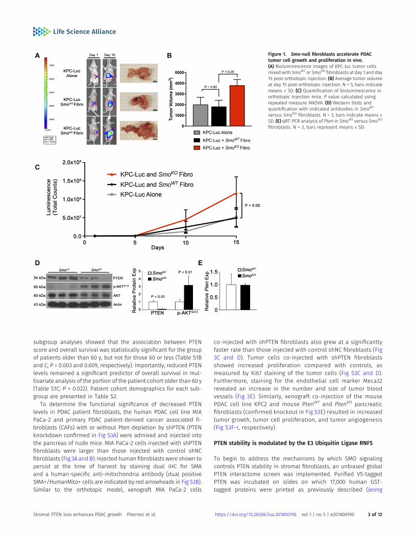
Whether canonical HH signaling through SMO in pancreatic fi-broblasts suppresses or promotes tumor cell growth remainscontroversial (Olive et al, 2009; Rhim et al, 2014). To directly addressthis question, we co-injected pancreatic fibroblasts, in which Smowas deleted by Cre/loxP technology, with a luciferase-taggedmouse KPC-luc tumor cell line (derived from LSL-KrasG12D/+;TP53loxP/loxP; Pdx1-Cre mice [Hwang et al, 2008]) directly into thepancreas of nude mice (see the Materials and Methods section fordescription of cell lines used). Decreased SMO expression in thepancreatic fibroblasts and expression of Shh in KPC-luc tumor cellswas confirmed before injection (Fig S1A and B). After injection, KPC-luc tumor cells were visualized over time via bioluminescenceimaging, revealing that KPC-luc tumor cells injected alone or mixedwith SmoWT fibroblasts produced tumors of the same size after15 d (Fig 1A–C). By contrast, KPC-luc cells injected with fibroblastslacking SMO (SmoKO) formed tumors that were significantly largerthan controls (Fig 1A and B). Tumor cell growth, as measuredby bioluminescence, was significantly increased in KPC-luc cellsco-injected with SmoKO fibroblasts relative to SmoWT fibroblasts
(Fig 1C). To confirm these results in a related assay, we co-injected thesame fibroblasts with a different mouse tumor cell line, KPC2 (fromLSL-KrasG12D/+; TP53R172H/+; Elas-CreER mice), into the flanks of nudemice. Shh expression was confirmed in KPC2 cells before injection(Fig S1B). KPC2 tumor cells injected alone or mixed with SmoWT
fibroblasts produced tumors of the same size after 5 wk (Fig S1C andD). Similar to orthotopic injection, flank KPC2 cells co-injected withSmoKO fibroblasts formed tumors that were significantly larger thancontrols (Fig S1C and D). Further analysis demonstrated an increasein Ki67-positive, proliferating tumor cells upon co-injection withSmoKO fibroblasts relative to SmoWT fibroblasts (Fig S1E and F).
Our previous work demonstrated that activation of AKT upongenetic deletion of Smo in pancreatic fibroblasts accelerated ADMand epithelial cell proliferation (Liu et al, 2016). Whether loss ofPTEN expression contributed to the activation of the AKT pathwaywas studied further. Western blot analysis revealed that PTENprotein was lost and AKT phosphorylation at Ser-473 was in-creased in SmoKO fibroblasts (Fig 1D). Surprisingly, Pten mRNAlevels remained unchanged between SmoWT and SmoKO fibro-blasts (Fig 1E).
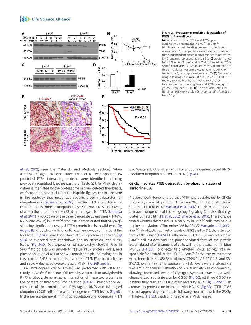
To address the mechanism by which PTEN protein levels weredown-regulated in the absence of Smo, we treated pancreatic fi-broblasts with cycloheximide and measured PTEN protein levelsover time. PTEN protein was highly stable in control SmoWT fi-broblasts and remained unchanged over the 24-h period of cy-cloheximide treatment (Fig 2A and B, lanes 1–6). Strikingly, PTENprotein levels, even when twice the amount of total protein wasloaded on the gel, were dramatically reduced in Smo-deleted fi-broblasts by 8–16 h of cycloheximide treatment (Fig 2A and B, lanes8–13). Cycloheximide treatment led to the expected reduction inTP53 in both SmoWT and SmoKO fibroblasts (Fig 2A). To determine ifPTEN degradation was proteasome-dependent, fibroblasts weretreated with MG132, a potent proteasome inhibitor. MG132 treat-ment of SmoKO cells restored PTEN protein to wild-type levels (Fig2C and D, lanes 5–8), but had no obvious effect on control cellswhere PTEN protein was already very stable (Fig 2C and D, lanes 1–4).
PTEN loss in tumor-associated fibroblasts correlates withreduced overall survival in human PDAC patient samples
To test the hypothesis that loss of fibroblast PTEN is driving diseaseprogression, the Vectra multispectral imaging platform was used toanalyze PTEN levels in SMA-positive pancreatic fibroblasts ina patient tissue microarray (TMA; representative images in Figs 2Eand S2A). In support of using the dual immunohistochemistry (IHC)methodology, the same results were obtained for PTEN/SMAstaining with dual IHC compared with dual immunofluorescence(IF) staining (Fig S2B–E). We examined the relationship betweenpatient outcome and reduced PTEN expression in SMA-positivefibroblasts. Patient samples with PTEN expression scores in thelower quartile had significantly poorer overall survival, with mediansurvival of 8.8 mo for the low PTEN group versus 16.9 mo for thegroup with higher PTEN scores (Fig 2F and Table S1A; log-rank test;P = 0.017). Multivariate Cox regression analysis revealed that lowPTEN scores trended toward statistical significance as a predictor ofpatient survival (Table S1A; P = 0.052). Althoughmultivariate analysisdid not reach statistical significance in the entire cohort, univariate
Stromal PTEN loss enhances PDAC growth Pitarresi et al. https://doi.org/10.26508/lsa.201800190 vol 1 | no 5 | e201800190 2 of 12
subgroup analyses showed that the association between PTENscore and overall survival was statistically significant for the groupof patients older than 60 y, but not for those 60 or less (Table S1Band C; P = 0.003 and 0.609, respectively). Importantly, reduced PTENlevels remained a significant predictor of overall survival in mul-tivariate analysis of the portion of the patient cohort older than 60 y(Table S1C; P = 0.022). Patient cohort demographics for each sub-group are presented in Table S2.
To determine the functional significance of decreased PTENlevels in PDAC patient fibroblasts, the human PDAC cell line MIAPaCa-2 and primary PDAC patient-derived cancer associated fi-broblasts (CAFs) with or without Pten depletion by shPTEN (PTENknockdown confirmed in Fig S3A) were admixed and injected intothe pancreas of nude mice. MIA PaCa-2 cells injected with shPTENfibroblasts were larger than those injected with control shNCfibroblasts (Fig 3A and B). Injected human fibroblasts were shown topersist at the time of harvest by staining dual IHC for SMAand a human-specific anti-mitochondria antibody (dual positiveSMA+/HumanMito+ cells are indicated by red arrowheads in Fig S3B).Similar to the orthotopic model, xenograft MIA PaCa-2 cells
co-injected with shPTEN fibroblasts also grew at a significantlyfaster rate than those injected with control shNC fibroblasts (Fig3C and D). Tumor cells co-injected with shPTEN fibroblastsshowed increased proliferation compared with controls, asmeasured by Ki67 staining of the tumor cells (Fig S3C and D).Furthermore, staining for the endothelial cell marker Meca32revealed an increase in the number and size of tumor bloodvessels (Fig 3E). Similarly, xenograft co-injection of the mousePDAC cell line KPC2 and mouse PtenWT and PtenKO pancreaticfibroblasts (confirmed knockout in Fig S3E) resulted in increasedtumor growth, tumor cell proliferation, and tumor angiogenesis(Fig S3F–I, respectively).
PTEN stability is modulated by the E3 Ubiquitin Ligase RNF5
To begin to address the mechanisms by which SMO signalingcontrols PTEN stability in stromal fibroblasts, an unbiased globalPTEN interactome screen was implemented. Purified V5-taggedPTEN was incubated on slides on which 17,000 human GST-tagged proteins were printed as previously described (Jeong
Figure 1. Smo-null fibroblasts accelerate PDACtumor cell growth and proliferation in vivo.(A) Bioluminescence images of KPC-luc tumor cellsmixed with SmoWT or SmoKO fibroblasts at day 1 and day15 post-orthotopic injection. (B) Average tumor volumeat day 15 post-orthotopic injection. N = 5, bars indicatemeans ± SD. (C) Quantification of bioluminescence inorthotopic injection mice. P value calculated usingrepeated measure ANOVA. (D) Western blots andquantification with indicated antibodies in SmoWT
versus SmoKO fibroblasts. N = 3, bars indicate means ±SD. (E) qRT-PCR analysis of Pten in SmoWT versus SmoKO
fibroblasts. N = 3, bars represent means ± SD.
Stromal PTEN loss enhances PDAC growth Pitarresi et al. https://doi.org/10.26508/lsa.201800190 vol 1 | no 5 | e201800190 3 of 12
et al, 2012) (see the Materials and Methods section). Whena stringent signal-to-noise cutoff ratio of 8.0 was applied, 374predicted PTEN interacting proteins were identified, includingpreviously identified binding partners (Table S3). As PTEN degra-dation is mediated by the proteasome in Smo-deleted fibroblasts,we focused on potential PTEN E3 ubiquitin ligases, the key enzymein the pathway that recognizes specific protein substrates forubiquitination (Lecker et al, 2006). The 374 PTEN interactome listcontained only three E3 ubiquitin ligases: TRIM44, RNF5, and WWP2,of which the latter is a known E3 ubiquitin ligase for PTEN (Maddikaet al, 2011). Knockdown of the three candidate E3 enzymes (TRIM44,RNF5, and WWP2) in SmoKO fibroblasts demonstrated that only Rnf5silencing significantly rescued PTEN protein levels to wild type (Fig4A and B). Knockdown efficiency for each gene was confirmed at theRNA level (Fig S4A), and knockdown of RNF5 protein confirmed (FigS4B). As expected, Rnf5 knockdown had no effect on Pten mRNAlevels (Fig S4C). Overexpression of supra-physiological Pten inSmoKO fibroblasts was unable to rescue PTEN protein levels, andphosphorylation of AKT at Ser-473 remained high, indicating that, inthis context, RNF5 in these cells is a potent PTEN E3 ubiquitin ligaseand rapidly degrades overexpressed PTEN (Fig S4D and E).
Co-immunoprecipitation (co-IP) was performed with PTEN an-tibody in SmoKO fibroblasts, followed by Western blot analysis withRNF5 antibody, demonstrating interaction of these two proteins inthe context of fibroblast Smo deletion (Fig 4C). Remarkably, ex-pression of the combination of V5-tagged RNF5 and HA-taggedubiquitin in 293T cells decreased endogenous PTEN levels (Fig 4D).In the same experiment, immunoprecipitation of endogenous PTEN
and Western blot analysis with HA-antibody demonstrated RNF5-mediated ubiquitin transfer to PTEN (Fig 4E).
GSK3β mediates PTEN degradation by phosphorylation ofThreonine-366
Previous work demonstrated that PTEN was destabilized by GSK3βphosphorylation at position Threonine-366 in the unstructuredC-terminal tail of PTEN (Maccario et al, 2007). Furthermore, GSK3β isa known component of the Hedgehog Signaling Complex that reg-ulates Gli1 stability (Jia et al, 2002; Sharpe et al, 2015). Therefore, wetested whether decreased PTEN stability in SmoKO cells may be dueto phosphorylation of Threonine-366 by GSK3β (Maccario et al, 2007).SmoKO fibroblasts had higher levels of GSK3β-pTyr-216, the activatedform of the kinase (Fig 5A). Furthermore, PTEN-pT366 was detected inSmoKO cell extracts and the phosphorylated form of the proteinaccumulated after treatment of cells with the proteasome inhibitorMG-132 (Fig 5B). To directly test whether GSK3β activity was re-sponsible for destabilization of PTEN, SmoKO fibroblasts were treatedwith three different GSK3β inhibitors (CT99021, AR-A014418, and SB-216763) over a 48-h time course and PTEN levels were analyzed viaWestern blot analysis. Inhibition of GSK3β activity was confirmed byshowing decreased levels of Glycogen Synthase pSer-614, a well-characterized substrate site for GSK3β (Fig 5C). All three GSK3β in-hibitors fully rescued PTEN protein levels by 48 h (Fig 5C and D). Incontrast to proteasome inhibition with MG-132 (Fig 5B), PTEN-pT366did not appreciably accumulate following treatment with the GSK3βinhibitors (Fig 5C), validating its role as a PTEN kinase.
Figure 2. Proteasome-mediated degradation ofPTEN in Smo-null cells.(A) Western blots for PTEN and TP53 uponcycloheximide treatment in SmoWT or SmoKO
fibroblasts. Protein loading amount (μg) indicatedabove lane. (B) The graph represents quantification ofthree independent Western blots relative to untreated.N = 3, squares represent means ± SD. (C) Western blotsfor PTEN in DMSO- (Vehicle) or MG132-treated SmoWT orSmoKO fibroblasts. (D) Graph represents quantitation ofthree individual Western blots relative to vehicle-treated. N = 3, bars represent means ± SD. (E) Compositeimages (1 image per core) of dual color IHC (PTENBrown, SMA Red) of human PDAC TMA and co-localization map showing SMA and PTEN overlap inyellow. Scale bar 50 μm. (F) Kaplan–Meier plots forfibroblast PTEN expression (H-score cutoff of 22) Scalebars, 50 μm.
Stromal PTEN loss enhances PDAC growth Pitarresi et al. https://doi.org/10.26508/lsa.201800190 vol 1 | no 5 | e201800190 4 of 12

Pharmacologic inhibition of SMO destabilizes PTEN in fibroblastsleading to decreased hydraulic permeability
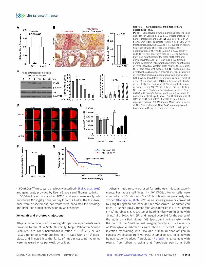
SMO antagonists were promising drugs for pancreatic cancer in pre-clinical studies; however, clinical trials with SMO antagonists in com-binationwith chemotherapy failed (Ruch& Kim, 2013). We hypothesizedthat the down regulation of stromal PTEN levels might contribute tofailure of SMO inhibitors such as GDC-0449. To determine if inhibition ofSMOwith this smallmolecule inhibitor couldmimic the genetic deletionof SMO, LSL-KrasG12D/+;LSL-Trp53R270H/+;Pdx-1-Cre;Brca1loxP/loxP (KPC-BRCA1CKO) mice were treated with GDC-0449 (Shakya et al 2011, 2013).This genetically engineeredmousemodel (GEMM)was chosenas it is anaggressive stroma-rich GEMMof PDAC. KPC-BRCA1CKOmice were treatedwith GDC-0449 at 5 wk of age, when invasive ductal adenocarcinoma isdetected. Short-term treatment of mice with GDC-0449 was performedas a proof-of-principle experiment to show in an autochthonous GEMMof PDAC that SMO inhibition could lead to decreased PTEN levels in vivo.In addition, long-term treatment with GDC-0449 proved toxic in KPC-BRCA1 mice. Analysis of Gli1 and Ptch1mRNA expression in the tumorsvalidated the efficacy of SMO inhibition (Fig 6A). A significant decreasein the PTEN protein levels in SMA-positive fibroblasts was observed inthe GDC-0449 group compared with controls as quantified using multi-spectral microscopy (Figs 6B and S5A).
PDAC patient-derived primary pancreatic CAF were treated withGDC-0449 and Western blot analysis demonstrated a rapid de-crease in PTEN protein and an increase in phosphorylated, acti-vated AKT in response to drug treatment (Figs 6C and S5B). Thesame experiment was repeated in SmoWT mouse pancreatic fi-broblasts. GDC-0449 treatment effectively decreased PTEN ex-pression and activated AKT in these mouse fibroblasts (Fig S5C). Ofnote, human fibroblasts required a slightly higher dose of GDC-0449to reduce PTEN levels, suggesting that human PTEN may be morestable than mouse PTEN.
Fibroblasts are major contributors to the distinct desmoplasticreaction in PDAC that alter the architecture and mechanics of theECM (Provenzano et al, 2012; Stylianopoulos et al, 2013). Wedesigned an in vitro assay using a microfluidic device (Hammeret al, 2017) to test whether PTEN in tumor fibroblasts affects thehydraulic permeability (K), which is a characteristic of the ECM thatrelates interstitial fluid velocity to the fluid pressure gradient (Wiig& Swartz, 2012) (Fig S6A–D). This in vitro assay demonstrated thathuman pancreatic tumor fibroblasts with PTEN knockdown hadincreased resistance to flow (i.e., decreased K) compared withcontrol (Fig 6D and E). Consistent with our results, GDC-0449treatment of control fibroblasts, but not PTEN knockdown fibro-blasts, showed a similar decrease in K relative to untreated cells
Figure 3. Pten-null fibroblasts accelerate PDACtumor cell growth and proliferation in vivo.(A, B) Images and tumor volume quantification oforthotopic MIA PaCa-2 tumor cells co-injected withshNC− (scrambled control) or shPTEN-transducedfibroblasts. N = 5, dots represent means ± SEM. (C, D)Images and tumor volume quantification of xenograftMIA PaCa-2 tumor cells co-injected with shNC-(scrambled control) or shPTEN-transduced fibroblasts.N = 5, dots represent means ± SEM. P-value calculatedusing repeated measure ANOVA. (E) IHC andquantification of Meca32 staining. N = 3, bars representmeans ± SD.
Stromal PTEN loss enhances PDAC growth Pitarresi et al. https://doi.org/10.26508/lsa.201800190 vol 1 | no 5 | e201800190 5 of 12

(Fig 6D and E). Increased hyaluronic acid (HA) in the ECM is known tosignificantly alter the hydraulic permeability in tumors and othertissues (Wiig & Swartz, 2012). Interestingly, treating with hyal-uronidase normalized the decreased hydraulic permeability pro-duced by PTEN-knockdown tumor fibroblasts (Fig 6D and E). HAproduction is regulated by hyaluronan synthase genes HAS1, HAS2,and HAS3 (Itano & Kimata, 2002), of which HAS2 and HAS3 areexpressed by pancreatic fibroblasts. Knockdown of PTEN resulted ina significant increase in HAS3 (Fig 6F), which has previously beenshown to promote tumor growth in pancreatic cancer (Kultti et al,2014). Furthermore, analysis of The Cancer Genome Atlas indicatedthat increased HAS3 expression correlated with decreased survivalin pancreatic cancer patients (Fig 6G).
Discussion
We have identified a mechanism by which stromal fibroblastspromote pancreatic tumor cell growth. Previous studies revealeda potential suppressive function of the tumor microenvironment(Lee et al, 2014; Ozdemir et al, 2014; Rhim et al, 2014). In addition, HHsignaling has been shown to act in a paracrine manner in PDAC,with tumor-secreted SHH activating the HH pathway in pancreaticfibroblasts (Yauch et al, 2008).These studies, however, did not di-rectly address the potential oncogenic functions of the tumormicroenvironment in pancreatic cancer progression. Our studydefinitively illustrates that a set of fibroblasts within the tumormicroenvironment can promote tumor cell growth when paracrinehedgehog signaling is disrupted. Although others have demon-strated that treatment of xenograft tumors with HH antagonistsdelays tumor growth (Yauch et al, 2008), we crucially extend thesefindings to demonstrate that Smo deletion in fibroblasts enhancestumor growth. These seemingly paradoxical results are likely dueto the different origin of fibroblast cultures. Yauch et al (2008)used mouse embryonic fibroblasts with ex vivo cre-mediated re-combination, whereas we used CAFs with in vivo genetic deletion ofSmo. This is in agreement with recent literature demonstrating thatactivated CAFs are inherently different than resident fibroblasts(reviewed in Kalluri [2016]). Notably, we also provide mechanistic
insight into the events that connect the loss of SMO and PTEN,identifying GSK3β and the E3-ligase RNF5 as critical intermediatesin the proteasome-mediated destruction of PTEN (Fig 7). Theseresults extend previous work from our group demonstrating thatloss of Hedgehog signaling in pancreatic stromal fibroblasts causedincreased PI3-kinase/AKT signaling and non-canonical activationof the GLI2 transcription factor, events that led to enhanced TGFαproduction by firoblasts and accelerated ADM and growth ofpancreatic tumor cells via activation of epidermal growth factorreceptor signaling (Fig 7) (Liu et al, 2016).
Analysis of fibroblast PTEN expression in PDAC patient samplesdemonstrated heterogeneous patterns of expression in the stromalfibroblast compartment, with focal areas of intense stainingintermixed with areas ofminimal staining in the same patient tissuesample. Given this observation, we were intrigued to find thatpatients who had decreased stromal PTEN correlated with a worseprognosis (Fig 2E and F). Importantly, we selected multiple areas oftissue for this survival analysis, to obtain a representative andquantifiable measure of the global stromal PTEN expression foreach patient, to control for heterogeneity in staining patterns.Moreover, recent results demonstrating distinct sets of activatedfibroblasts that either produce or respond to IL6 highlight theimportance of stromal heterogeneity in promoting pancreatic tu-mor progression (Ohlund et al, 2017). However, taken together, wecaution the clinical interpretation of results presented in thismanuscript, as analyses of TMA staining is limited with regardto intratumoral heterogeneity. Heterogeneity of tumor cells haslong been appreciated and contributes to tumor recurrence andtherapeutic resistance. Molecular heterogeneity within the stromalfibroblast population may account for, at least in part, the tumor-promoting and tumor-suppressive fibroblast subsets within pan-creatic cancer. The combined results begin to establish the complexheterogeneity of pancreatic CAFs and the potential functionalconsequences of modulating the stroma with targeted therapies.The work presented herein establishes that loss of stromal PTENinfluences tumor growth in a non-cell autonomous fashion.
Structural alterations to the ECMmediated by PTEN expression instromal fibroblasts was assessed by quantifying K, which is a widelyused measurement in interstitial physiology (Wiig & Swartz, 2012).
Figure 4. RNF5 is a novel E3 ubiquitin ligase for PTEN.(A) Western blots for PTEN upon treatment with theindicated siRNA or MG132 in SmoWT or SmoKO
fibroblasts. (B) Graph represents quantitation of threeindividual Western blots relative to vehicle-treated. N =3, bars represent means ± SD. (C)Western blots for RNF5after co-IP with PTEN or IgG antibody in SmoKO
fibroblasts. N = 3. (D) Western blots for PTEN in HEK-293T cells fibroblasts transfected with FLAG-taggedRNF5 or HA-tagged Ubiquitin. N = 3. (E)Western blots forHA-Ubiquitin after co-IP with PTEN antibody in HEK-293T cells fibroblasts transfected with FLAG-taggedRNF5 or HA-tagged Ubiquitin. N = 3.
Stromal PTEN loss enhances PDAC growth Pitarresi et al. https://doi.org/10.26508/lsa.201800190 vol 1 | no 5 | e201800190 6 of 12

Our results suggest that loss of PTEN in fibroblasts correlates withincreased HA synthesis that results in decreased hydraulic per-meability and is a subsequent physical barrier to interstitial drugtransport in tumors. Hingorani et al, have demonstrated that tar-geting HA with PEGylated, recombinant human hyaluronidase in-creases the effectiveness of chemotherapy in pre-clinical PDACmodels and in PDAC patients (Provenzano et al, 2012; Hingoraniet al, 2016). Furthermore, the original study by Olive et al, dem-onstrated that treatment with SMO inhibitors led to increasedintratumoral vasculature (Olive et al, 2009), in agreement with ourresult that PTEN silencing in fibroblasts enhanced angiogenesis.This result is intriguing and suggests that fibroblasts are integralcomponents of the microenvironment that regulate angiogenesisand the vascular network, and that clinical interventions targetingthe stroma should consider potential effects this may have ontumor vasculature.
Importantly, the identification of RNF5 as a new E3 ubiquitinligase for PTEN shows the cell-type specificity of proteasome-mediated degradation machinery. Several E3 ubiquitin ligases thatmediate PTEN degradation have been previously identified, in-cluding NEDD4-1, WWP2, and XIAP (Wang et al, 2007; Van Themscheet al, 2009; Maddika et al, 2011). However, of these three, only WWP2was identified in our initial PTEN interactome screen and sub-sequent experiments indicated that WWP2 is not responsible fordegradation of PTEN in pancreatic stromal fibroblasts. Given thisresult, future studies will be required to determine if RNF5 acts as
an E3 ubiquitin ligase in other cell types and cancers. Breast cancer,in particular, has been previously shown to over-express RNF5(Bromberg et al, 2007), whereas other cancers have yet to be ex-plored. Of note, 75% of prostate cancer patients with reduced PTENprotein lack a corresponding reduction in mRNA, emphasizingthat PTEN protein decay mechanisms may have a broader contextwithin human cancers (Chen et al, 2011). Therefore, targeting PTENdestabilizers such as RNF5 may provide a unique approach torestore PTEN function in both tumor cells and tumor stromalfibroblasts.
Materials and Methods
Animal strains and maintenance
Control (SmoloxP/−, herein referred to as SmoWT) and experimental(FspCre;SmoloxP/−, herein referred to as SmoKO) animals weregenerated by crossing the previously described SmoloxP, Smo− (Longet al, 2001) and FspCre (Trimboli et al, 2008, 2009) strains withtumor-bearing Mist1-KrasG12D animals, as previously described byour group (Liu et al, 2016; Pitarresi et al, 2016). Pancreatic fibroblastswere generated from age-matched littermate SmoWT and SmoKO
mice from a mixed C57BL/6; 129/Sv and FVBN genetic background;all cells were passaged the same number of times for eachexperiment. LSL-KrasG12D/+;LSL-Trp53R270H/+;Pdx-1-Cre;Brca1loxP/loxP
Figure 5. GSK3β inhibition blocks PTEN degradation.(A) Western blots and quantification for total and phosphorylated GSK3β Tyr-216 SmoKO or SmoKO fibroblasts. Graph represents quantitation of three individualWestern blots relative to SmoWT. N = 3, bars represent means ± SD. (B) Western blots of total and phosphorylated PTEN Thr-366 upon treatment with vehicle or MG132in SmoKO fibroblasts. N = 3. (C, D) Western blots and quantification for total and phosphorylated PTEN Thr-366 and phosphorylated glycogen synthase (GS) Ser-614in SmoKO or SmoKO fibroblasts at the indicated time points. Graph represents quantitation of three individual Western blots relative to SmoWT. N = 3, dotsrepresent means ± SD.
Stromal PTEN loss enhances PDAC growth Pitarresi et al. https://doi.org/10.26508/lsa.201800190 vol 1 | no 5 | e201800190 7 of 12
(KPC-BRCA1CKO) mice were previously described (Shakya et al, 2013)and generously provided by Reena Shakya and Thomas Ludwig.
GDC-0449 was dissolved in DMSO and mice were orally ad-ministered 100 mg/kg once per day for 4 d. 4 h after the last dose,mice were dissected and pancreata were harvested for histologyand immunohistochemistry staining as described.
Xenograft and orthotopic injections
Athymic nude mice used for xenograft injection experiments wereprovided by the Ohio State University Target Validation SharedResource Core. For subcutaneous injection, 5 × 10⁵ KPC2 or MIAPaCa-2 tumor cells were admixed in a 1:1 ratio with 5 × 10⁵ fibro-blasts and injected into the flanks of nude mice; tumor volumeswere measured once per week by caliper.
Athymic nude mice were used for orthotopic injection experi-ments. For mouse cell lines, 1 × 10⁵ KPC-luc tumor cells wereadmixed in a 1:5 ratio with 5 × 10⁵ fibroblasts, as previously de-scribed (Hwang et al, 2008). KPC-luc cells were generously providedby Craig D. Logsdon and Zobeida Cruz-Monserrate. For human celllines, 1 × 10⁶ MIA PaCa-2 tumor cells were admixed in a 1:5 ratio with5 × 106 fibroblasts. KPC-luc tumor-bearing mice were injected with15 mg/ml of D-luciferin (IP) and imaged every 5 d for the course ofthe study on a PerkinElmer IVIS Spectrum imaging system withthe help of the Small Animal Imaging Facility at the Universityof Pennsylvania. Fibroblasts were shown to persist 6-wk post-injection by staining with SMA and human nuclear antigen inconsecutive sections from MIA PaCa-2 tumor cells co-injected withhuman patient–derived fibroblasts (Fig S3E), in agreement withresults from others showing that fibroblasts persist in both
Figure 6. Pharmacological Inhibition of SMOdestabilizes PTEN.(A) qRT-PCR analysis of whole pancreas tissue for Gli1and Ptch1 in vehicle or GDC-0449 treated mice. N = 3,bars represent means ± SD. (B) Dual color IHC (PTENbrown, SMA red) of pancreata from vehicle or GDC-0449treated mice, showing SMA and PTEN overlap in yellow.Scale bar, 50 μm. The H-score represents thequantification of the PTEN staining in SMA-positivecells. N = 3, bars represent means ± SD. (C) Westernblots and quantification for total PTEN, total andphosphorylated AKT Ser-473 in GDC-0449–treatedhuman pancreatic CAFs. Graph represents quantitationof three individual Western blots relative to untreated.N = 3, bars represent means ± SD. (D) Rhodamine-BSAdye flow through collagen channel after 48-h culturingof indicated fibroblast populations with and withoutGDC-0449. Yellow dotted line indicates displacement ofdye at 60 s relative to 0 s. (E) Quantification of hydraulicpermeability (rate shown in E). Statistical testing wasperformed using ANOVA with Tukey’s HSD post-testing.N ≥ 4 for each condition. Bars indicate means ± SEM;ANOVA with Tukey’s–Cramer post-testing was used toanalyze statistical significance. (F) qRT-PCR analysis ofHAS3 in shNT and shPTEN fibroblasts. N = 3, barsrepresent means ± SD. (G) Kaplan–Meier survival curveof The Cancer Genome Atlas-PAAD data segregatedbased on HAS3 high or low expression.
Stromal PTEN loss enhances PDAC growth Pitarresi et al. https://doi.org/10.26508/lsa.201800190 vol 1 | no 5 | e201800190 8 of 12
xenograft (Mathew et al, 2014) and orthotopic (Vonlaufen et al,2008) injection model systems. To confirm that fibroblasts were nottransformed, 5.0 × 10⁵ fibroblasts were injected into the flanks ofnude mice and followed for 6 wk; no tumors formed.
Multispectral vectra IHC analysis
Dual stained samples were imaged using the PerkinElmer’s Vectramultispectral slide analysis system. For the mouse samples, at leastthree multispectral images per animal for at least three mice pergenotype (unless otherwise noted) were manually taken. For thehuman PDAC TMA, one field of interest per core was automaticallyacquired. The image acquisitionworkflowconsistedof the following: (1)monochrome imaging of the entire slide; (2) RGB low power imaging ofthe tumor tissue using an inForm tissue finding algorithm; and (3)multispectral high-power imaging of one field containing tumor epi-thelium and stroma by means of an inForm HPF finding algorithm.
For visualization of the component images (PTEN/SMO DAB, SMARed), the multispectral images were spectrally unmixed usingNuance software. Nuance co-localization tool was used to createthe co-localization maps displaying the PTEN/SMO–positive cells inthe SMA-positive cellular compartment overlap in yellow.
For quantification of the PTEN and SMO staining, the multi-spectral images were reviewed and analyzed using inForm TissueFinder software. A pattern recognition algorithm was used forprocessing as follows: (1) trainable tissue segmentation to segmentthe SMA-positive regions from the tumor epithelium; (2) cell seg-mentation of the SMA-positive tissue category to locate the subcellularcompartments; and (3) scoring to bin the spectrally unmixed DABsignal into four categories depending on the staining intensity (0+,1+, 2+ and 3+), providing data in percent. The H-Score, which rangesfrom 0 to 300, was calculated using the following formula: [1 × (%cells 1+) + 2 × (% cells 2+) + 3 × (% cells 3+)]. Thus, H-score measuresstaining intensity and percentage of positive cells in a given cellularcompartment. Comparison of co-IF with dual-color IHC was per-formed on serial sections of the same tissue to show that IHC is ableto recapitulate conventional staining procedures in a quantitativemanner (Fig S2A–B). The percentage of SMA/PTEN dual-positivecells by co-IF was quantified (Fig S2C) and compared with H-score(Fig S2D) for the same mice presented in Fig 3C. Of note, the overlapin IHC staining is a direct quantitative measure of staining for oneantigen (in the case of this example, PTEN) within a distinct cellpopulation defined by a second antigen (in this case, SMA), and isnot simply the overlap in signal between the two stains.
IHC and IF staining
Dissected mouse pancreas tissues were fixed in 10% neutral-buffered formalin solution for 48 h and transferred to 70% etha-nol. Tissues were processed, embedded in paraffin, cut into 5-μmsections on positively charged slides, de-paraffinized, rehydrated,and stained with H&E.
For immunohistochemistry, all sections were stained usinga Bond Rx autostainer (Leica) or Roche Discovery ULTRA autos-tainer, unless otherwise noted, according to the manufacturersrecommendations. Antibodies for the following markers were di-luted in Antibody diluent (Leica): αSMA (1:4,000; Abcam), Ki67 (1:200;Abcam), PTEN (1:150 Cell Signaling), SMO (1:200; Bioss), Meca-32 (1:50;BD Pharmingen), and human anti-mitochondria (1:500; Abcam).
Cell culture and treatments
Primary pancreatic fibroblasts were purified as previously out-lined in the literature (Liu et al, 2016; Pitarresi et al, 2016). SmoWT
and SmoKO pancreatic fibroblast cultures were established fromMist1KrasG12D mice at the PanIN stage that carried SmoLoxP andFspCre;SmoLoxP alleles, respectively. All fibroblast cultures wereshown to be SMA-positive in our previous work {Liu et al, 2016 #11;Pitarresi et al, 2016 #32}. Primary human PDAC CAFs were isolatedas previously described {Liu et al, 2016 #11}. For isolation of murineand human primary fibroblasts, tumor tissue was minced anddigested with collagenase while shaking for 1 h at 37°C. Digestedtissue was gravity purified for 10 min in media, and subsequentpellets were subjected to two more gravity purifications, and thenseeded on tissue culture dishes. MIA PaCa-2 tumor cell line wasobtained from the ATCC. The KPC2 tumor cell line was a generousgift from Stephen F. Konieczny and established from tumor-bearing Elas-CreER; LSL-KrasG12D/+; TP53R172H/+ mice.
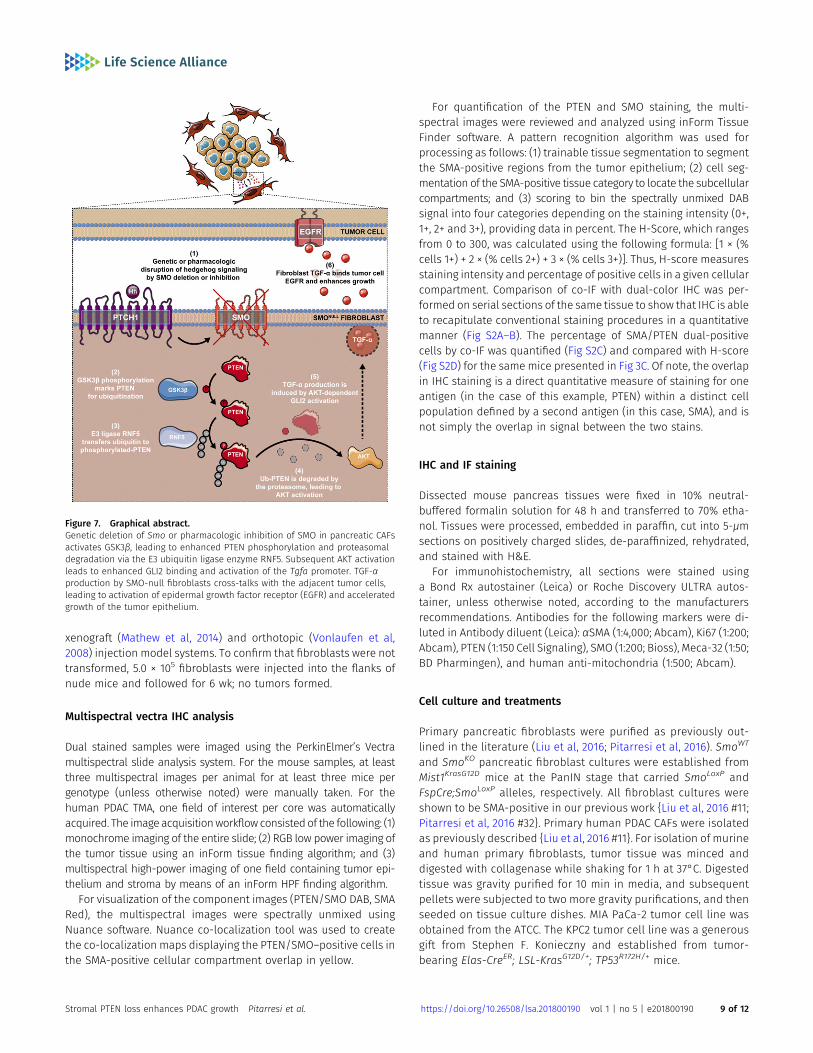
Figure 7. Graphical abstract.Genetic deletion of Smo or pharmacologic inhibition of SMO in pancreatic CAFsactivates GSK3β, leading to enhanced PTEN phosphorylation and proteasomaldegradation via the E3 ubiquitin ligase enzyme RNF5. Subsequent AKT activationleads to enhanced GLI2 binding and activation of the Tgfa promoter. TGF-αproduction by SMO-null fibroblasts cross-talks with the adjacent tumor cells,leading to activation of epidermal growth factor receptor (EGFR) and acceleratedgrowth of the tumor epithelium.
Stromal PTEN loss enhances PDAC growth Pitarresi et al. https://doi.org/10.26508/lsa.201800190 vol 1 | no 5 | e201800190 9 of 12

For cycloheximide assays, a final concentration of 10 μg/ml wasused. Cellular lysates were collected with RIPA buffer at the in-dicated time points for standard Western blotting analysis.
Proteasome inhibitor MG132 treatment was performed at a finalconcentration of 10 μM for the indicated time points.
Three GSK3β inhibitors were used: SB-216763 (#sc-200646; SantaCruz), CT99021 (#CHIR99021; Sigma-Aldrich), and AR-A014418 (#ALX-270-468-M001; Enzo Lifesciences). Cells were grown to 70% con-fluency and treated with 5 μM SB-216763, 5 µM CT99021, or 10 μMAR-A014418 for the indicated time points. After treatment, cellswere harvested for Western blot analysis in RIPA buffer.
co-IP and ubiquitin assays
Cells were harvested by trypsinization and lysed in non-denaturingextraction buffer (CST 9803), and immunoprecipitation with anti-body against PTEN (1:100 CST). After incubation, PRO-A magneticbeads (LSKMAGA02; Millipore) were added. Samples were washedand Laemmli buffer added for Western blot analysis.
siRNA knockdown
Dharmacon ON-TARGETplus SMARTpool siRNA system was used forknockdown. Briefly, ~60% confluent pancreatic fibroblast cultureswere treated with 200 pM of pooled siRNA and 10 μl of lipofectamine2000 in 600 μl of serum-free OPTI-MEM media for 8 h. The mediumwas changed to 10% FBS-DMEM and allowed to sit for 24–48 h, atwhich time the cells were harvested.
Microfluidic device fabrication
Hydraulic permeability measurements were acquired using micro-fluidic devices fabricated out of polydimethylsiloxane using soft li-thography techniques. The polydimethylsiloxane devices consistedof a single straight channel (5 mm long, 500 μm wide, and 1 mm tall)with 4 mm inlet/outlet ports and irreversibly sealed to a glass slideusing plasma oxidation (Harrick, 90 s). For application of flow, a pipettip (Redi-Tip) was inserted at one of the ports. Tips were cut to~2–2.4 cm to ensure a tight fit at the port. The microdevices weresterilized with 30 min of UV treatment.
Fibroblast cell preparation for hydraulic permeabilitymeasurements
Primary human pancreatic tumor fibroblasts were maintained inhigh glucose DMEM supplemented with 10% fetal bovine serum, 1%penn-steptomycin, 0.2% plasmocin, and 0.1% fungin. Rat tail type Icollagen gel (Corning Life Science, 6 mg/ml, pH = 7.4), containingfibroblasts at 1.8 × 10⁶ cells/ml, was introduced into themicrofluidicdevice at 4°C and polymerized at 37°C for 24 h. The collagenconcentration was selected to minimize collagen contraction byfibroblasts. In addition, the devices were incubated with fibronectin(100 μg/ml, 30 min) before injection to improve hydrogel adhesionto the channel walls. For GDC treatment conditions, the fibro-blasts were resuspended in medium containing GDC at 80 µMGDC before mixing with collagen gel solution and cultured for
48 h with additional GDC containing medium after injection intothe microdevices.
The following experimental conditions were conducted in themicrofluidic devices: (1) acellular collagen; (2) collagen containingcontrol tumor fibroblasts (shNC); (3) collagen containing tumorfibroblasts with PTEN knocked down by shRNA (shPTEN); (4) col-lagen containing shnc fibroblasts treated with GDC (shNC + GDC); (5)collagen containing shPTEN fibroblasts treated with GDC (shPTEN +GDC). All devices were cultured for 48 h before measurements byplacing approximately 400 μl of medium at the ports and devicesurface, with the medium being renewed every 24 h.
Microfluidic hydraulic permeability measurements
To measure hydraulic permeability, a rhodamine-bovine serumalbumin (rhodamine-BSA) (Molecular Probes) was flowed throughthe microfluidic device. Flow was established by applying a fluidicheight difference (2–2.4 cm) between the ports using cell culturemedium, measured for each sample. After establishing flow, 4–8μLof rhodamine-BSA was injected into the tip and was transported bythe flow through the microdevice. Timelapse microscopy experi-ments were recorded with an epifluorescence Nikon TS-100F mi-croscope equipped with a Q-Imaging QIClick camera. Images wereacquired every 15 s for 20–30 min. These images were then used toquantify the average velocity of the dye through the collagen/fibroblast matrix by tracking the position of the bulk of the dye as itflowed through the microdevice using FIJI. Hydraulic permeabilitywas then calculated by using Darcy’s Law for flow through porousmedium as follows:
K = μvΔLΔP ; (1)
where μ is the viscosity of the cell culture medium (approximatedusing water), v is the average fluid velocity, ΔL is the length of thechannel, and ΔP is the pressure difference across the channel due tothe fluidic height difference and is given by the following equation:
ΔP = ρgh; (2)
where ρ is the density of the cell culture medium (approximatedusing water), g is the acceleration due to gravity (9.81 m/s2), and h isthe fluidic height difference between ports.
Statistics
Pearson’s correlation, Wilcoxon rank-sum test, ANOVA and t testwere calculated using R 3.0.1 or Prism. The P values from t tests arelisted unless otherwise specified. In all graphs, median, means(bar), and standard deviations or standard error of the means(lines) are denoted. Microarray data were processed by RobustMulti-array Average (RMA) method and analyzed using themoderated t test approach (Yu et al, 2011). For survival analysis,the Kaplan–Meier method and log-rank test were applied tounivariate analysis and Cox regression models were used formultivariate analysis. Comparison wise P-value of 0.05 wasconsidered significant.
Stromal PTEN loss enhances PDAC growth Pitarresi et al. https://doi.org/10.26508/lsa.201800190 vol 1 | no 5 | e201800190 10 of 12

Study approval
The use of animals was in compliance with federal and Ohio StateUniversity Laboratory Animal Resources regulations.
Supplementary Information
Supplementary Information is available at https://doi.org/10.26508/lsa.201800190.
Acknowledgments
We acknowledge the Solid Tumor Biology Histology Core Microscopy,Transgenic/Knockout, Target Validation, Analytic Cytometry, Bioinformatics,and Biostatistics Shared Resources. This study was supported by NationalInstitutes of Health grants PO1 CA097189 (MC Ostrowski and G Leone), NRSAF31 CA189757 (JR Pitarresi), NRSA F32 CA221094 (JR Pitarresi), American CancerSociety IRG-67-003-50 (JW Song), American Heart Association 15SDG25480000(JW Song), and R01 CA124586 (SF Konieczny). This work was also supported bythe Department of Defense (W81XWH-14-1-0040, GM Sizemore), PelotoniaFellowship Program (JR Pitarresi, GM Sizemore, A Avendano, JJ Chang, and CSEnnis), and the OSU Institute for Materials Research. Any opinions, findings,and conclusions expressed in this material are those of the author(s) and donot necessarily reflect those of the Pelotonia Fellowship Program.
Author Contributions
JR Pitarresi: conceptualization, data curation, formal analysis,funding acquisition, investigation, visualization, methodology, andwriting—original draft, review, and editing.X Liu: data curation, formal analysis, and investigation.A Avendano: data curation, formal analysis, investigation.KA Thies: data curation and formal analysis.GM Sizemore: data curation and formal analysis.AM Hammer: data curation and formal analysis.BE Hildreth III: data curation and formal analysis.DJ Wang: methodology.SA Steck: data curation and formal analysis.S Donohue: data curation and formal analysis.MC Cuitiño: data curation and formal analysis.RD Kladney: data curation and formal analysis.TA Mace: data curation and formal analysis.JJ Chang: data curation and formal analysis.CS Ennis: data curation.H Li: data curation.RH Reeves: data curation and methodology.S Blackshaw: data curation and methodology.J Zhang: formal analysis.L Yu: formal analysis.SA Fernandez: formal analysis.WL Frankel: formal analysis.M Bloomston: data curation.TJ Rosol: formal analysis.GB Lesinski: formal analysis.SF Konieczny: formal analysis.DC Guttridge: formal analysis.
AK Rustgi: formal analysis.G Leone: formal analysis and project administration.JW Song: formal analysis and project administration.J Wu: formal analysis and project administration.MC Ostrowski: conceptualization, formal analysis, funding acqui-sition, investigation, methodology, project administration, andwriting—original draft, review, and editing.
Conflict of Interest Statement
The authors declare that they have no conflict of interest.
References
Bailey JM, Mohr AM, Hollingsworth MA (2009) Sonic hedgehog paracrinesignaling regulates metastasis and lymphangiogenesis in pancreaticcancer. Oncogene 28: 3513–3525. doi:10.1038/onc.2009.220
Bromberg KD, Kluger HM, Delaunay A, Abbas S, DiVito KA, Krajewski S, Ronai Z(2007) Increased expression of the E3 ubiquitin ligase RNF5 isassociated with decreased survival in breast cancer. Cancer Res 67:8172–8179. doi:10.1158/0008-5472.can-07-0045
Chen M, Pratt CP, Zeeman ME, Schultz N, Taylor BS, O’Neill A, Castillo-MartinM, Nowak DG, Naguib A, Grace DM, et al (2011) Identification of PHLPP1as a tumor suppressor reveals the role of feedback activation inPTEN-mutant prostate cancer progression. Cancer Cell 20: 173–186.doi:10.1016/j.ccr.2011.07.013
Feig C, Gopinathan A, Neesse A, Chan DS, Cook N, Tuveson DA (2012) Thepancreas cancer microenvironment. Clin Cancer Res 18: 4266–4276.doi:10.1158/1078-0432.ccr-11-3114
Fujita H, Ohuchida K, Mizumoto K, Nakata K, Yu J, Kayashima T, Cui L, Manabe T,Ohtsuka T, Tanaka M (2010) Alpha-smooth muscle actin expressingstroma promotes an aggressive tumor biology in pancreatic ductaladenocarcinoma. Pancreas 39: 1254–1262. doi:10.1097/MPA.0b013e3181dbf647
Hammer AM, Sizemore GM, Shukla VC, Avendano A, Sizemore ST, Chang JJ,Kladney RD, Cuitino MC, Thies KA, Verfurth Q, et al (2017) StromalPDGFR-alpha activation enhances matrix stiffness, impedesmammary ductal development, and accelerates tumor growth.Neoplasia 19: 496–508. doi:10.1016/j.neo.2017.04.004
Hingorani SR, Harris WP, Beck JT, Berdov BA, Wagner SA, Pshevlotsky EM,Tjulandin SA, Gladkov OA, Holcombe RF, Korn R, et al (2016) Phase ibstudy of PEGylated recombinant human hyaluronidase andgemcitabine in patients with advanced pancreatic cancer. Clin CancerRes 22: 2848–2854. doi:10.1158/1078-0432.ccr-15-2010
Hwang RF, Moore T, Arumugam T, Ramachandran V, Amos KD, Rivera A, Ji B,Evans DB, Logsdon CD (2008) Cancer-associated stromal fibroblastspromote pancreatic tumor progression. Cancer Res 68: 918–926.doi:10.1158/0008-5472.can-07-5714
Itano N, Kimata K (2002) Mammalian hyaluronan synthases. IUBMB Life 54:195–199. doi:10.1080/15216540214929
Jeong JS, Jiang L, Albino E, Marrero J, Rho HS, Hu J, Hu S, Vera C, Bayron-Poueymiroy D, Rivera-Pacheco ZA, et al (2012) Rapid identification ofmonospecific monoclonal antibodies using a human proteomemicroarray. Mol Cell Proteomics 11: O111 016253. doi:10.1074/mcp.o111.016253
Jia J, Amanai K, Wang G, Tang J, Wang B, Jiang J (2002) Shaggy/GSK3antagonizes Hedgehog signalling by regulating Cubitus interruptus.Nature 416: 548–552. doi:10.1038/nature733
Jones S, Zhang X, Parsons DW, Lin JC, Leary RJ, Angenendt P, Mankoo P, CarterH, Kamiyama H, Jimeno A, et al (2008) Core signaling pathways in
Stromal PTEN loss enhances PDAC growth Pitarresi et al. https://doi.org/10.26508/lsa.201800190 vol 1 | no 5 | e201800190 11 of 12
human pancreatic cancers revealed by global genomic analyses.Science 321: 1801–1806. doi:10.1126/science.1164368
Kalluri R (2016) The biology and function of fibroblasts in cancer. Nat RevCancer 16: 582–598. doi:10.1038/nrc.2016.73
Kultti A, Zhao C, Singha NC, Zimmerman S, Osgood RJ, Symons R, Jiang P, Li X,Thompson CB, Infante JR, et al (2014) Accumulation of extracellularhyaluronan by hyaluronan synthase 3 promotes tumor growth andmodulates the pancreatic cancer microenvironment. Biomed Res Int2014: 817613. doi:10.1155/2014/817613
Lecker SH, Goldberg AL, Mitch WE (2006) Protein degradation by theubiquitin-proteasome pathway in normal and disease states. J AmSoc Nephrol 17: 1807–1819. doi:10.1681/asn.2006010083
Lee JJ, Perera RM, Wang H, Wu DC, Liu XS, Han S, Fitamant J, Jones PD, GhantaKS, Kawano S, et al (2014) Stromal response to Hedgehog signalingrestrains pancreatic cancer progression. Proc Natl Acad Sci U S A 111:E3091–E3100. doi:10.1073/pnas.1411679111
Liu X, Pitarresi JR, Cuitino MC, Kladney RD, Woelke SA, Sizemore GM, Nayak SG,Egriboz O, Schweickert PG, Yu L, et al (2016) Genetic ablation ofSmoothened in pancreatic fibroblasts increases acinar-ductalmetaplasia. Genes Dev 30: 1943–1955. doi:10.1101/gad.283499.116
Long F, Zhang XM, Karp S, Yang Y, McMahon AP (2001) Genetic manipulation ofhedgehog signaling in the endochondral skeleton reveals a directrole in the regulation of chondrocyte proliferation. Development 128:5099–5108.
Maccario H, Perera NM, Davidson L, Downes CP, Leslie NR (2007) PTEN isdestabilized by phosphorylation on Thr366. Biochem J 405: 439–444.doi:10.1042/bj20061837
Maddika S, Kavela S, Rani N, Palicharla VR, Pokorny JL, Sarkaria JN, Chen J(2011) WWP2 is an E3 ubiquitin ligase for PTEN. Nat Cell Biol 13: 728–733.doi:10.1038/ncb2240
Mathew E, Zhang Y, Holtz AM, Kane KT, Song JY, Allen BL, Pasca di Magliano M(2014) Dosage-dependent regulation of pancreatic cancer growth andangiogenesis by hedgehog signaling. Cell Rep 9: 484–494. doi:10.1016/j.celrep.2014.09.010
Ohlund D, Handly-Santana A, Biffi G, Elyada E, Almeida AS, Ponz-Sarvise M,Corbo V, Oni TE, Hearn SA, Lee EJ, et al (2017) Distinct populations ofinflammatory fibroblasts and myofibroblasts in pancreatic cancer.J Exp Med 214: 579–596. doi:10.1084/jem.20162024
Olive KP, Jacobetz MA, Davidson CJ, Gopinathan A, McIntyre D, Honess D,Madhu B, Goldgraben MA, Caldwell ME, Allard D, et al (2009) Inhibitionof Hedgehog signaling enhances delivery of chemotherapy ina mouse model of pancreatic cancer. Science 324: 1457–1461.doi:10.1126/science.1171362
Ozdemir BC, Pentcheva-Hoang T, Carstens JL, Zheng X, Wu CC, Simpson TR,Laklai H, Sugimoto H, Kahlert C, Novitskiy SV, et al (2014) Depletion ofcarcinoma-associated fibroblasts and fibrosis inducesimmunosuppression and accelerates pancreas cancer with reducedsurvival. Cancer Cell 25: 719–734. doi:10.1016/j.ccr.2014.04.005
Pitarresi JR, Liu X, Sharma SM, Cuitino MC, Kladney RD, Mace TA, Donohue S,Nayak SG, Qu C, Lee J, et al (2016) Stromal ETS2 regulates chemokineproduction and immune cell recruitment during acinar-to-ductalmetaplasia. Neoplasia 18: 541–552. doi:10.1016/j.neo.2016.07.006
Provenzano PP, Cuevas C, Chang AE, Goel VK, Von Hoff DD, Hingorani SR (2012)Enzymatic targeting of the stroma ablates physical barriers totreatment of pancreatic ductal adenocarcinoma. Cancer Cell 21:418–429. doi:10.1016/j.ccr.2012.01.007
Rhim AD, Oberstein PE, Thomas DH, Mirek ET, Palermo CF, Sastra SA, DeklevaEN, Saunders T, Becerra CP, Tattersall IW, et al (2014) Stromal elements
act to restrain, rather than support, pancreatic ductaladenocarcinoma. Cancer Cell 25: 735–747. doi:10.1016/j.ccr.2014.04.021
Ruch JM, Kim EJ (2013) Hedgehog signaling pathway and cancer therapeutics:Progress to date. Drugs 73: 613–623. doi:10.1007/s40265-013-0045-z
Shakya R, Gonda T, Quante M, Salas M, Kim S, Brooks J, Hirsch S, Davies J, CulloA, Olive K, et al (2013) Hypomethylating therapy in an aggressivestroma-rich model of pancreatic carcinoma. Cancer Res 73: 885–896.doi:10.1158/0008-5472.can-12-1880
Shakya R, Reid LJ, Reczek CR, Cole F, Egli D, Lin CS, deRooij DG, Hirsch S, Ravi K,Hicks JB, et al (2011) BRCA1 tumor suppression depends on BRCTphosphoprotein binding, but not its E3 ligase activity. Science 334:525–528. doi:10.1126/science.1209909
Sharpe HJ, Wang W, Hannoush RN, de Sauvage FJ (2015) Regulation of theoncoprotein Smoothened by small molecules. Nat Chem Biol 11:246–255. doi:10.1038/nchembio.1776
Stylianopoulos T, Martin JD, Snuderl M, Mpekris F, Jain SR, Jain RK (2013)Coevolution of solid stress and interstitial fluid pressure in tumorsduring progression: Implications for vascular collapse. Cancer Res 73:3833–3841. doi:10.1158/0008-5472.can-12-4521
Tian H, Callahan CA, DuPree KJ, Darbonne WC, Ahn CP, Scales SJ, de Sauvage FJ(2009) Hedgehog signaling is restricted to the stromal compartmentduring pancreatic carcinogenesis. Proc Natl Acad Sci U S A 106:4254–4259. doi:10.1073/pnas.0813203106
Trimboli AJ, Cantemir-Stone CZ, Li F, Wallace JA, Merchant A, Creasap N,Thompson JC, Caserta E, Wang H, Chong JL, et al (2009) Pten in stromalfibroblasts suppresses mammary epithelial tumours. Nature 461:1084–1091. doi:10.1038/nature08486
Trimboli AJ, Fukino K, de Bruin A, Wei G, Shen L, Tanner SM, Creasap N, RosolTJ, Robinson ML, Eng C, et al (2008) Direct evidence for epithelial-mesenchymal transitions in breast cancer. Cancer Res 68: 937–945.doi:10.1158/0008-5472.can-07-2148
Van Themsche C, Leblanc V, Parent S, Asselin E (2009) X-linked inhibitor ofapoptosis protein (XIAP) regulates PTEN ubiquitination, content, andcompartmentalization. J Biol Chem 284: 20462–20466. doi:10.1074/jbc.c109.009522
Vonlaufen A, Joshi S, Qu C, Phillips PA, Xu Z, Parker NR, Toi CS, Pirola RC, WilsonJS, Goldstein D, et al (2008) Pancreatic stellate cells: Partners in crimewith pancreatic cancer cells. Cancer Res 68: 2085–2093. doi:10.1158/0008-5472.can-07-2477
Wang X, Trotman LC, Koppie T, Alimonti A, Chen Z, Gao Z, Wang J, Erdjument-Bromage H, Tempst P, Cordon-Cardo C, et al (2007) NEDD4-1 is a proto-oncogenic ubiquitin ligase for PTEN. Cell 128: 129–139. doi:10.1016/j.cell.2006.11.039
Wiig H, Swartz MA (2012) Interstitial fluid and lymph formation and transport:Physiological regulation and roles in inflammation and cancer.Physiol Rev 92: 1005–1060. doi:10.1152/physrev.00037.2011
Yauch RL, Gould SE, Scales SJ, Tang T, Tian H, Ahn CP, Marshall D, Fu L, JanuarioT, Kallop D, et al (2008) A paracrine requirement for hedgehogsignalling in cancer. Nature 455: 406–410. doi:10.1038/nature07275
Yu L, Gulati P, Fernandez S, Pennell M, Kirschner L, Jarjoura D (2011) Fullymoderated T-statistic for small sample size gene expression arrays.Stat Appl Genet Mol Biol 10: 42. doi:10.2202/1544-6115.1701
License: This article is available under a CreativeCommons License (Attribution 4.0 International, asdescribed at https://creativecommons.org/licenses/by/4.0/).
Stromal PTEN loss enhances PDAC growth Pitarresi et al. https://doi.org/10.26508/lsa.201800190 vol 1 | no 5 | e201800190 12 of 12