disruption of endoplasmic reticulum structure and integrity in lipotoxic cell death
TRANSCRIPT

Disruption of endoplasmic reticulum structure and
integrity in lipotoxic cell death
Nica M. Borradaile,* Xianlin Han,† Jeffrey D. Harp,* Sarah E. Gale,* Daniel S. Ory,*
and Jean E. Schaffer1,*
Center for Cardiovascular Research, Division of Cardiology,* and Division of Bioorganic Chemistry,†
Department of Internal Medicine, Washington University School of Medicine, St. Louis, MO 63110
Abstract Cell dysfunction and death induced by lipidaccumulation in nonadipose tissues, or lipotoxicity, maycontribute to the pathogenesis of obesity and type 2 dia-betes. However, the mechanisms leading to lipotoxic celldeath are poorly understood. We recently reported that, inChinese hamster ovary (CHO) cells and in H9c2 cardiomyo-blasts, lipid overload induced by incubation with 500 mMpalmitate leads to intracellular accumulation of reactive oxy-gen species, which subsequently induce endoplasmic re-ticulum (ER) stress and cell death. Here, we show thatpalmitate also impairs ER function through a more directmechanism. Palmitate was rapidly incorporated into satu-rated phospholipid and triglyceride species in microsomalmembranes of CHO cells. The resulting membrane re-modeling was associated with dramatic dilatation of the ERand redistribution of protein-folding chaperones to the cy-tosol within 5 h, indicating compromised ER membraneintegrity. Increasing b-oxidation, through the activation ofAMP-activated protein kinase, decreased palmitate incor-poration into microsomes, decreased the escape of chap-erones to the cytosol, and decreased subsequent caspaseactivation and cell death. Thus, palmitate rapidly increasesthe saturated lipid content of the ER, leading to compro-mised ER morphology and integrity, suggesting thatimpairment of the structure and function of this organelleis involved in the cellular response to fatty acid overload.—Borradaile, N. M., X. Han, J. D. Harp, S. E. Gale, D. S. Ory,and J. E. Schaffer. Disruption of endoplasmic reticulumstructure and integrity in lipotoxic cell death. J. Lipid Res.2006. 47: 2726–2737.
Supplementary key words palmitate & fatty acid & lipotoxicity &lipid synthesis
Increased serum triacylglycerol (TAG) and NEFA levels,associated with obesity and type 2 diabetes, contribute tolipid accumulation in many nonadipose tissues. Throughthe process of lipotoxicity, this inappropriate accumula-tion of excess lipid can lead to cellular dysfunction and celldeath (1). For example, evidence from rodent models
strongly implicates cardiac accumulation of lipid in thegenesis of heart failure in diabetes. TAG accumulation incardiomyocytes of leptin- or leptin receptor-deficient obesediabetic animal models is associated with cardiomyocyteapoptosis (2) and contractile dysfunction (2–4). Consistentwith this apparent cardiac lipotoxicity, cardiomyocyte-specific increases in FA uptake in mice with cardiac-restricted overexpression of long-chain acyl-CoA synthetase1, lipoprotein lipase, or fatty acid transport protein 1 aresufficient to cause cardiomyocyte dysfunction and/or deaththat lead to left ventricular dysfunction (5–7).
Studies using cultured cells to model the lipotoxicresponse have helped elucidate the mechanisms involvedin the response to FA overload. Long-chain saturated fattyacids, such as palmitate, induce cell death in a varietyof cell types, including cardiomyocytes (8). In general,palmitate-induced cell death is characterized by markersof apoptosis, including cytochrome c release, caspaseactivation, and DNA fragmentation. Although relativelyfew studies have focused on mechanisms of palmitate-induced cell death in cardiomyocytes, recent evidenceobtained using primary cardiomyocyte cultures fromembryonic chicks and neonatal rats suggests that incuba-tion with palmitate is associated with the loss of mito-chondrial membrane potential, mitochondrial swelling,and cytochrome c release (9–11). These events may beinitiated via several mechanisms, including decreasedsynthesis of the signature mitochondrial membrane phos-pholipid, cardiolipin (11), increased ceramide synthesis (9,12), and increased generation of reactive oxygen species(ROS) (13, 14). However, the induction of apoptosis by bothceramide and oxidative stress requires a flux of calcium ionsfrom the endoplasmic reticulum (ER) to the mitochondria(15, 16), and depletion of these calcium stores can impairnormal protein-folding functions, leading to ER stress (17,
Manuscript received 10 July 2006 and in revised form 25 August 2006.
Published, JLR Papers in Press, September 7, 2006.DOI 10.1194/jlr.M600299-JLR200
Abbreviations: AICAr, 5-aminoimidazole-4-carboxamide-1-b-4-ribofuranoside; AMPK, AMP-activated protein kinase; ER, endoplasmicreticulum; GRP78, glucose-regulated protein 78; PC, phosphatidylcho-line; PDI, protein disulfide isomerase; ROS, reactive oxygen species;SCD1, stearoyl-coenzyme A desaturase 1; TAG, triacylglycerol.
1 To whom correspondence should be addressed.e-mail: [email protected]
Copyright D 2006 by the American Society for Biochemistry and Molecular Biology, Inc.
This article is available online at http://www.jlr.org2726 Journal of Lipid Research Volume 47, 2006
by guest, on Novem
ber 28, 2018w
ww
.jlr.orgD
ownloaded from

18). Consistent with this concept, we and others recentlyshowed that palmitate overload rapidly induces ER stress inpancreatic b-cells (19), hepatocytes (20), and cardiomyo-blasts (14). Furthermore, our studies revealed that palmi-tate-induced ER stress was mediated, in part, through thegeneration of ROS (14).
Several observations indicate that palmitate may also actmore directly at the level of the ER to initiate a lipotoxicresponse. In vitro evidence suggests that palmitoyl-CoAfacilitates ER fission (21) and that the acyl chains of lipidsdirectly affect the fusion/fission events of membranes (22,23). Furthermore, incorporation of saturated fatty acylchains into membrane phospholipids can induce detrimen-tal stiffening of cellular membranes (24–26). Here, we dem-onstrate that, in CHO cells and H9c2 cardiomyoblasts, therapid induction of ER stress in the presence of palmitate isassociated with the rapid incorporation of this fatty acid intolipid components of the rough ER and subsequentcompromise of rough ER structure and integrity. Althoughprevious studies indicate that palmitate-induced intracellu-lar responses converge on the mitochondria, eventuallyresulting in the release of cytochrome c into the cytosol andapoptotic cell death, our studies suggest that the ER mayplay an important proximal role in FA-induced cytotoxicity.
MATERIALS AND METHODS
Cell culture and chemicals
CHO-K1 (CHO) cells (American Type Culture Collection)and stearoyl-coenzyme A desaturase 1 (SCD1)-overexpressingCHO cells (27) were maintained in high-glucose (4.5 mg/ml)DMEM and Ham’s F-12 nutrient mixture (1:1), with 5% FBS, asdescribed (27). H9c2 rat cardiomyoblasts (American TypeCulture Collection) were maintained in high-glucose DMEMwith 10% FBS, as described (14). For experiments, all CHO andH9c2 cell lines (90% confluent) were incubated in CHO cellgrowth medium supplemented with palmitate (500 mM) oroleate (500 mM) (Nu-Chek Prep) complexed to BSA at a 2:1molar ratio, prepared as described previously (13). 5-Aminoim-idazole-4-carboxamide-1-b-4-ribofuranoside (AICAr) was fromCalbiochem; etomoxir, H2O2, a-tocopherol (vitamin E), thapsi-gargin, and DMSO were from Sigma.
Subcellular distribution of radiolabeled palmitate
CHO cells were incubated for 1 h with [9,10-3H]palmitate(Perkin-Elmer), [9,10-3H]oleate (Perkin-Elmer), or [9,10-3H]2-bromopalmitate (American Radiochemicals) at a specific activityof 10 mCi/mmol. Crude mitochondria, cytosol, smooth micro-somes, and rough microsomes were isolated by homogenizationand sequential centrifugation, as described previously (28). Ra-dioactivity in each fraction was measured using a Beckman LS6000IC scintillation counter and normalized to the total proteinin each fraction (BCA Protein Assay; Pierce). The relative purityof the isolated fractions was assessed by immunoblotting usingrabbit polyclonal antibodies against histone H1 (Stressgen),long-chain acyl-CoA dehydrogenase [a gift from A. Strauss (29)],and p63 [a gift from J. Rohrer (30)].
Lipid composition of rough microsomes
CHO cells were incubated for 1 h in the absence or presenceof 500 mM [7,7,8,8-2H]palmitate (Cambridge Isotope). Rough
microsomes were isolated as described above. Lipids were extractedand lipid species were identified and quantitated by electrosprayionization mass spectrometry (31, 32). The mass levels of phos-phatidylethanolamine were not determined in this study.
Electron microscopy
CHO cells were harvested, fixed with 2.5% glutaraldehyde in0.1 M sodium cacodylate buffer, postfixed in 1.25% osmiumtetroxide, and stained with 4% aqueous uranyl acetate. Embed-ded tissue was then thin-sectioned and viewed on a Zeiss 902 elec-tron microscope. Glutaraldehyde, osmium tetroxide, and uranylacetate were from Electron Microscopy Sciences.
Subcellular fractionation and immunoblotting
Crude cytosolic and membrane/organelle fractions wereisolated from CHO cells by sequential detergent extractionusing ProteoExtract reagents from Calbiochem (33). Based on ourpreliminary assessment of the distribution of marker proteins, analternative method of homogenization and sequential centrifuga-tion (34) was required to isolate cytosolic and crude microsomalfractions from H9c2 cells. Glucose-regulated protein 78 (GRP78)and protein disulfide isomerase (PDI) in 7.5–20 mg of proteinfrom each subcellular fraction were detected using rabbitpolyclonal antibodies (Stressgen). Cytochrome c in 40–80 mg ofprotein from crude mitochondrial and cytosolic fractions, isolatedby sequential centrifugation, was detected using a monoclonalantibody (BD Biosciences).
ER calcium depletion
Palmitate-induced depletion of thapsigargin-sensitive calciumstores was assessed using the Fluo-4 NW calcium assay kit (Molec-ular Probes, Invitrogen) in a 96-well plate format, according tothe manufacturer’s protocol. After incubation with palmitate,cells were loaded with Fluo-4 AM in the presence of 2.5 mMprobenicid. Thapsigargin (1 mM) or vehicle (DMSO) was addedimmediately, and fluorescence at 2 min was measured using aHidex plate reader (excitation at 485 nm and emission at 535 nm).Data were expressed as fluorescent increments (change in fluo-rescence) upon addition of thapsigargin.
Mitochondrial staining
Depolarization of mitochondria was assessed using thepotential-dependent dye, JC-1 (Molecular Probes, Invitrogen).CHO cells incubated for up to 5 h with 500 mM palmitate or2.5 mM H2O2 were stained with 7.5 mM JC-1 at 378C, according tothe manufacturer’s protocol. Mean red and green fluorescencewere determined by flow cytometry (104 cells/sample) for sub-sequent calculation of mean FL2/FL1 ratios.
The presence of intact mitochondria was assessed usingMitoTracker Green FM (Molecular Probes, Invitrogen). CHOcells incubated for up to 18 h with 500 mM palmitate or 2.5 mMH2O2 were stained for 30 min with 20 nM MitoTracker Green FMat 378C, according to the manufacturer’s protocol. Mean fluo-rescence was determined by flow cytometry (104 cells/sample).
Caspase activation and cell death
Activation of caspases-3 and -7 was determined by immuno-blotting of cytosolic (40 mg of protein) and microsomal (70 mg ofprotein) fractions from H9c2 cells incubated for up to 24 h withvarious treatments. Rabbit polyclonal antibodies were used tosimultaneously detect both pro- (inactive) and cleaved (active)forms of each caspase (Cell Signaling Technologies). Cell deathwas assessed by membrane permeability to propidium iodide, as
Lipotoxicity and the ER 2727
by guest, on Novem
ber 28, 2018w
ww
.jlr.orgD
ownloaded from

described previously (13). Briefly, CHO and H9c2 cells incubatedfor 24–48 h with various treatments were harvested by trypsiniza-tion and stained with 1 mM propidium iodide. The percentage ofpropidium iodide-positive cells was determined by flow cytometry(104 cells/sample).
RESULTS
Palmitate is rapidly incorporated into saturatedphospholipid and triglyceride species in the rough ER
Previous studies have identified the ER as a target ofpalmitate-induced lipotoxicity downstream of the genera-tion of ROS (14). To test the hypothesis that palmitate mayalso impair ER function more directly through its rapidincorporation into the ER membrane, we determined thesubcellular distribution of palmitate within 1 h of expo-sure to a lipotoxic dose. CHO cells were incubated with500 mM palmitate containing a trace amount of [3H]pal-mitate. Subsequent subcellular fractionation revealed thatthe bulk of the label was distributed between the crudemitochondrial fraction (58.00 6 0.03%) and the roughmicrosomal fraction (22.90 6 0.03%) (Fig. 1A). Althougha significant proportion of [3H]palmitate was associatedwith the relatively pure rough microsomal fraction, com-posed predominantly of rough ER, the crude mitochon-dria were contaminated with rough microsomes (Fig. 1C),indicating that the calculated percentage distribution actu-ally underestimates the incorporation of palmitate intothe rough ER. Similar distributions were observed uponlabeling of CHO cells with oleate and upon labelingof CHO cells overexpressing SCD1 with palmitate. TheseSCD1-overexpressing cells have an increased capacity tointroduce double bonds into palmitate by virtue of in-
creased SCD1 activity (27). Thus, exogenous saturated andunsaturated FAs are channeled quickly to the ER as well asto the mitochondria. This distribution is independent oflipotoxicity, which occurs in CHO cells treated with pal-mitate but not in CHO cells treated with oleate or SCD1-overexpressing cells treated with palmitate (27).
To determine whether modulating b-oxidation couldalter the distribution observed with 500 mM palmitate, thesame experiment was conducted in the presence of eitheretomoxir (an inhibitor of carnitine palmitoyl transferase 1)or AICAr [an activator of AMP-activated protein kinase(AMPK)] (Fig. 1A). These compounds were used at dosesestablished previously to effectively inhibit or increase b-oxidation (35, 36). Etomoxir did not alter the subcellulardistribution of [3H]palmitate within 1 h, whereas AICArreduced the incorporation of palmitate into rough micro-somes by z50%. Palmitate incorporation into mitochon-dria was also reduced in the presence of AICAr by z30%.The latter likely reflects a combination of decreased pal-mitate incorporation into rough microsomal membranes(which contaminate the crude mitochondrial fraction)and increased mitochondrial oxidation of palmitate.
The distribution of palmitate observed with a nontoxicconcentration of palmitate (5 mM) was nearly identical tothat observed with 500 mM (Fig. 1B), suggesting that theinitial trafficking of this fatty acid to various subcellularlocations is not concentration-dependent. In contrast, therelative distribution of 2-bromopalmitate, used as a nonli-potoxic control, was markedly different, with the bulk ofthe label remaining in the cytosol (Fig. 1B). This modifiedfatty acid is not as good a substrate for acyl-CoA synthetases(37), resulting in limited uptake and toxicity comparedwith palmitate (4.4 6 0.8% and 5.2 6 0.4% cell death at 24 hfor control and 500 mM 2-bromopalmitate, respectively).
Fig. 1. Subcellular distribution of tritiated palmitate,oleate, and 2-bromopalmitate in wild-type and stearoyl-coenzyme A desaturase 1 (SCD1)-overexpressing CHOcells. A, B: Wild-type CHO (solid bars) or SCD1-overexpressing CHO (cross-hatched bars) cells wereincubated for 1 h with 10 mCi/mmol [9,10-3H]palmi-tate, [9,10-
3
H]oleate, or [9,10-3H]2-bromopalmitate, atthe indicated concentrations of palmitate (PA), oleate(OA), or 2-bromopalmitate (BrPA). Graphs showradiolabel distribution in fractions isolated by sequen-tial centrifugation. For experiments including etomoxir(Eto; 200 mM) or 5-aminoimidazole-4-carboxamide-1-b-4-ribofuranoside (AICAr; 500 mM), cells were incubatedfor 30 min with either compound before the addition ofradiolabel and both etomoxir and AICAr were includedfor the subsequent incubation. Values are means 6 SEM(n 5 4). * P , 0.05. C: Immunoblotting of subcellularfractions for marker proteins. Histone H1 (his H1) wasused as a marker for crude nuclei (nuc), long-chain acyl-CoA dehydrogenase (LCAD) was used for crudemitochondria (mito), and p63 was used for rough en-doplasmic reticulum (ER) membranes (rm). cyt, cytosol;sm, smooth microsomes.
2728 Journal of Lipid Research Volume 47, 2006
by guest, on Novem
ber 28, 2018w
ww
.jlr.orgD
ownloaded from

To assess the consequences of palmitate incorporationinto rough microsomes on the composition of these mem-branes, we analyzed both newly synthesized and total lipidsin this fraction after a 1 h incubation with 500 mM[2H]palmitate. The largest percentage of exogenous pal-mitate was incorporated into phosphatidylcholine (PC)species. However, significant proportions also remained asFFA or were incorporated into TAG species (Table 1,column 4). Overall, rough microsomes were composedprimarily of PC and FFA, with very limited TAG content,corresponding to 50, 27, and 5% of the examined lipidmass content, respectively (Table 1, column 2). Thus, thelabeled proportions of the rough microsomal pools of PCand FFA were relatively small, whereas the labeled pro-portion of the rough microsomal pool of TAG was sig-nificantly larger (Table 1, column 5). Unsaturated FAsmake up the vast majority (81%) of acyl chain substituentsin CHO cells under basal conditions (Table 2). Strikingly,the proportions of saturated PC and TAG in these mem-branes were increased by 1.5-fold (from 1.56% to 2.40%)and 3.0-fold (from 6.39% to 19.59%), respectively, with nosignificant change in total content of lipid species (Table 2).Thus, the remodeling of PC and TAG species accounted
for a significant 1.3-fold increase (from 18.55% to 24.91%)in the saturated lipid content of rough microsomes fromCHO cells incubated for 1 h with 500 mM palmitate.
Palmitate induces dramatic changesin ER structure and integrity
Increased saturation of lipid species is associated with astiffening of cellular membranes (24–26). Based on ourobservations of increased saturation of PC and TAG speciesin rough microsomes from palmitate-treated CHO cells, westudied the effect of this treatment on the morphology ofthe ER. In electron micrographs of untreated cells, the ERappeared as normal, tubular cisternae delimited byelectron-dense dots corresponding to ribosomes (Fig. 2A).In contrast, cells treated for 5 h with 500 mM palmitatecontained numerous distended structures delimited byelectron-dense ribosomes (Fig. 2B–D), a morphology con-sistent with the presence of markedly dilated rough ER(38). To determine whether this dramatic change in ERstructure could be the result of palmitate-induced ROSgeneration, we compared the morphology of palmitate-treated cells with that of cells treated for 5 h with 2.5 mMH2O2. These conditions induced approximately the same
TABLE 1. Distribution of deuterated palmitate in rough microsomes from CHO cells
Lipid Species Total Lipid Deuterated Lipid Distribution of Label Deuterated Lipid/Total Lipid
nmol/mg protein %
PC 58.01 6 6.49 4.15 6 0.65 42.09 6 5.47 7.12 6 0.48PA 2.02 6 0.21 0.00 6 0.00 0.00 6 0.00 0.00 6 0.00PG 2.83 6 0.33 0.00 6 0.00 0.00 6 0.00 0.00 6 0.00PI 6.97 6 0.24 0.22 6 0.03 2.26 6 0.27 3.19 6 0.38LPC 1.39 6 0.25 0.21 6 0.02 2.13 6 0.09 15.71 6 1.61SM 7.09 6 1.37 0.12 6 0.06 1.16 6 0.58 1.46 6 0.50Cer 0.34 6 0.06 0.06 6 0.01 0.65 6 0.08 19.06 6 1.04FFA 31.06 6 0.24 2.93 6 0.52 30.42 6 6.62 9.42 6 1.61TAG 5.60 6 1.05 2.12 6 0.60 21.29 6 5.67 36.37 6 4.13Total 115.29 6 8.29 9.82 6 0.42 100 8.59 6 0.61
CHO cells were incubated for 1 h with 500 mM [7,7,8,8-2H]palmitate complexed to BSA at a molar ratio of 2:1.Rough microsomes were isolated by sequential centrifugation. Lipids were extracted and quantitated byelectrospray ionization mass spectrometry. PC, phosphatidylcholine; PA, phosphatidic acid; PG, phosphatidylglyc-erol; PI, phosphatidylinositol; LPC, lysophosphatidylcholine; SM, sphingomyelin; Cer, ceramide; TAG,triacylglycerol. Values are means 6 SEM.
TABLE 2. Lipid composition of rough microsomes from CHO cells
Control Palmitate-Treated
Lipid Species Total Lipid (TL) Saturated Lipid (SL) SL/TL Total Lipid (TL) Saturated Lipid (SL) SL/TL
nmol/mg protein % nmol/mg protein %
PC 80.69 6 11.73 1.22 6 0.10 1.56 6 0.15 58.01 6 6.49 1.43 6 0.31 2.40 6 0.27a
PA 2.27 6 0.12 0.00 6 0.00 0.00 6 0.00 2.02 6 0.21 0.00 6 0.00 0.00 6 0.00PG 3.63 6 0.26 0.29 6 0.02 7.90 6 0.19 2.83 6 0.33 0.23 6 0.05 8.06 6 0.87PI 10.79 6 1.55 0.00 6 0.00 0.00 6 0.00 6.97 6 0.24 0.00 6 0.00 0.00 6 0.00LPC 2.48 6 0.53 1.46 6 0.35 58.07 6 1.90 1.39 6 0.25 0.83 6 0.13 60.70 6 2.41SM 8.28 6 1.28 5.84 6 1.09 69.63 6 3.24 7.09 6 1.37 4.98 6 0.88 70.72 6 1.52Cer 0.66 6 0.12 0.49 6 0.10 73.56 6 3.70 0.34 6 0.06 0.24 6 0.05 68.68 6 2.86FFA 29.29 6 1.02 16.91 6 0.94 57.65 6 1.51 31.06 6 0.24 19.69 6 0.29a 63.40 6 0.81TAG 5.96 6 0.13 0.38 6 0.00 6.39 6 0.14 5.60 6 1.05 1.16 6 0.37 19.59 6 3.09a
Total 144.04 6 16.41 26.58 6 2.51 18.55 6 0.46 115.29 6 8.29 28.56 6 1.08 24.91 6 1.10a
CHO cells were incubated for 1 h with 500 mM [7,7,8,8-2H]palmitate complexed to BSA at a molar ratio of 2:1. Rough microsomes were isolatedby sequential centrifugation. Lipids were extracted and quantitated by electrospray ionization mass spectrometry. Values are means 6 SEM.
a P , 0.05 for control versus palmitate-treated.
Lipotoxicity and the ER 2729
by guest, on Novem
ber 28, 2018w
ww
.jlr.orgD
ownloaded from

amount of cell death within 24 h (16.8 6 2.9% and 21.1 6
1.5% cell death for 500 mM palmitate and 2.5 mM H2O2,respectively). In contrast to cells treated with palmitate,those treated with H2O2 contained normal ER cisternae(Fig. 2E). However, the mitochondria in H2O2-treated cellsappeared compromised compared with both control andpalmitate-treated cells (Fig. 2F vs. 2A, D).
We next assessed whether the changes in ER structureobserved in response to palmitate were associated withevidence of compromised ER integrity, and further, whetherany changes in ER integrity were dependent on theinduction of oxidative stress. Isolation and immunoblottingof cytosol and crude membrane/organelle fractions fromCHO cells incubated for 5 h with 500 mM palmitate resultedin the escape of protein-folding chaperones, GRP78(78 kDa) (Fig. 3A) and PDI (58 kDa) (Fig. 3B), from theER to the cytosol. These observations are broadlyconsistent with those recently observed in palmitate-treated pancreatic b-cells (39). Levels of the integral ERmembrane protein, p63, were unaltered and indicate therelative purity of our cytosolic and membrane fractions.Although a-tocopherol (vitamin E) prevents palmitate-induced ROS accumulation within 5 h (14), incubationwith palmitate in the presence of 200 mM vitamin E did notprevent the redistribution of normally ER lumenal pro-teins. Furthermore, these effects were not observed in cellsincubated for 5 h with 2.5 mM H2O2. Conditions that result
in the incorporation of unsaturated FA into ER mem-branes, including incubation of wild-type CHO cells with500 mM oleate and SCD1-overexpressing cells with 500 mMpalmitate, did not alter the distribution of ER chaperoneproteins (Fig. 3). Together with our observations ofdramatically altered ER morphology (Fig. 2), these datasuggest that the detrimental effects of palmitate on ERstructure and integrity are distinct from changes induced bysevere oxidative stress.
Palmitate induces changes in mitochondrial function
Both palmitate-induced alterations of ER structure andintegrity and palmitate-induced oxidative stress couldinitiate the flux of calcium from the ER to the mitochon-dria and lead to the loss of mitochondrial membranepotential. Thus, we determined whether incubation withpalmitate resulted in the rapid depletion of ER calcium.Assays of thapsigargin-sensitive calcium revealed thatER stores were significantly reduced after 15 min and byup to 25% after 30 min with 500 mM palmitate, but notwith the nontoxic, unsaturated fatty acid, oleate (Fig. 4A).This depletion of ER calcium was followed by the gradualescape of protein-folding chaperones, GRP78 (Fig. 4B)and PDI (Fig. 4C), from the ER to the cytosol over thecourse of 5 h.
Previous studies have demonstrated an impairment ofmitochondrial function late in the response to lipotoxic
Fig. 2. Incubation of CHO cells with palmitate, but not H2O2, results in dilatation of the ER. CHO cells were incubated for 5 h in theabsence (A) or presence of either 500 mM palmitate (B–D) or 2.5 mM H2O2 (E, F). Morphological changes were observed by transmissionelectron microscopy. C and F show enlarged views of boxed areas in B and E, respectively. Arrowheads indicate rough ER cisternaedelimited by electron-dense ribosomes. m, mitochondria; N, nuclei. Bars 5 275 nm.
2730 Journal of Lipid Research Volume 47, 2006
by guest, on Novem
ber 28, 2018w
ww
.jlr.orgD
ownloaded from

stress (9). Thus, we determined whether the rapid de-pletion of ER calcium in response to palmitate is accom-panied by changes in mitochondrial function. Isolation andimmunoblotting of crude mitochondrial and cytosolicfractions revealed that 30 min of incubation with 500 mMpalmitate reduced mitochondrial cytochrome c (15 kDa)content by 40% (Fig. 4D). However, no change in cyto-chrome c content was detected after 15 min with palmitate,and the protein was not detected in the cytosol over theentire course of palmitate treatment (Fig. 4D). Mitochon-drial long-chain acyl-CoA dehydrogenase content was notaffected by palmitate (data not shown). Assessment ofmitochondrial membrane potential, by JC-1 staining andflow cytometry, revealed significant reductions in the ratiosof red (FL2) to green (FL1) fluorescence, indicative ofmitochondrial depolarization, only after extended (5 h)incubations with either 500 mM palmitate or 2.5 mM H2O2
(Fig. 4E). The relative abundance of intact mitochondria,assessed by staining with MitoTracker Green FM and flowcytometry, was not significantly reduced after 5 h with500 mM palmitate, as shown by the lack of change in meanfluorescence (Fig. 4F). However, consistent with ourmorphological analyses (Fig. 2), incubation for 5 h with2.5 mM H2O2 reduced mean fluorescence by 37%. Incu-bation for 18 h with 500 mM palmitate reduced meanfluorescence by 46%. Together, these data suggest thatpalmitate-induced changes in ER calcium content mayprecede the gross impairment of mitochondrial function.
Palmitate-induced changes in ER integrity are reducedby increasing b-oxidation
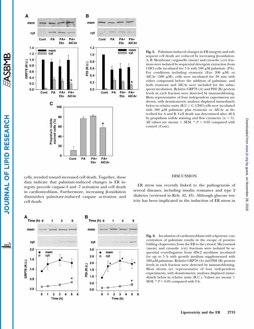
Based on our observations of the subcellular distribu-tion of [3H]palmitate in the presence of AICAr (Fig. 1A),we hypothesized that channeling palmitate toward b-oxidation would prevent the detrimental changes in ERintegrity associated with its incorporation into ER mem-brane PC and TAG (Tables 1, 2). Consistent with thishypothesis, AICAr decreased the palmitate-induced es-cape of GRP78 (Fig. 5A) and PDI (Fig. 5B) from the ERto the cytosol by 42% and 50%, respectively (Fig. 5C),and reduced eventual cell death by 30% (Fig. 5C). Con-versely, incubation with etomoxir, at a concentrationthat inhibits b-oxidation, decreased neither chaperoneescape nor cell death. In fact, etomoxir treatmenttended to increase cell death at 48 h. Thus, increasingb-oxidation diminishes palmitate-induced changes in ERintegrity and cell death.
Palmitate-induced changes in ER integrity, followed bycaspase activation and cell death, occur in cardiomyoblasts
To extend these findings to a cell type more relevant tolipotoxic disease, we determined the distribution of ERprotein-folding chaperones in response to palmitate inH9c2 rat cardiomyoblasts. In experiments similar to thoseperformed in CHO cells, H9c2 cells were incubated for upto 5 h with 500 mM palmitate. Subsequent isolation andimmunoblotting of cytosol and crude membrane/organ-elle fractions revealed that, as was observed in CHO cells,
Fig. 3. Incubation of CHO cells with palmitate results in the escapeof protein-folding chaperones from the ER to the cytosol.Membrane/organelle (mem) and cytosolic (cyt) fractions wereisolated by sequential detergent extraction from wild-type CHO andSCD1-overexpressing CHO (SCD) cells incubated for 5 h in theabsence (Cont) or presence of 500 mM palmitate (PA), 500 mMoleate (OA), or 2.5 mM H2O2. For conditions including a-tocopherol (VitE; 200 mM), cells were incubated for 30 min withthe antioxidant before the addition of palmitate, and a-tocopherolwas included for the subsequent incubation. Relative glucose-regulated protein 78 (GRP78; A), protein disulfide isomerase(PDI; B), and p63 (C) protein levels in each fraction were detectedby immunoblotting. Blots representative of four independentexperiments are shown, with densitometric analyses displayedimmediately below in relative units (R.U.). Values are means 6
SEM (n 5 4). * P , 0.05 compared with control fractions.
Lipotoxicity and the ER 2731
by guest, on Novem
ber 28, 2018w
ww
.jlr.orgD
ownloaded from

incubation with palmitate resulted in the appearance ofGRP78 (Fig. 6A) and PDI (Fig. 6B) in the cytosol.
Although palmitate-induced cell death is characterizedby markers of apoptosis, including cytochrome c release,caspase-3 activation, and DNA fragmentation, we previ-ously observed that caspase-3 activation in response to pal-mitate is relatively modest compared with other inducersof apoptosis (14), raising the possibility that other cas-pases may play an important role. Based on the dramaticeffects observed on ER structure and integrity, we de-termined whether caspase-7, which is activated in responseto ER stress and is the only effector caspase known tolocalize to the ER upon activation (40, 41), could alsobe activated in response to palmitate. H9c2 cells were
incubated for up to 18 h with 500 mM palmitate. Sub-sequent subcellular fractionation and immunoblottingrevealed that, in accordance with previous studies (13),cleaved (active) caspase-3 began to accumulate in thecytosol within 9 h of palmitate treatment (Fig. 7A). Withinthe same time frame, cleaved caspase-7 accumulatedin the microsomal fraction of palmitate-treated cells,with a corresponding loss of pro-caspase-7 from the cytosol(Fig. 7B). Furthermore, increasing b-oxidation with theAMPK activator, AICAr, decreased palmitate-inducedactivation of caspase-3 and -7 by 46% and 49%, respectively(Fig. 7C), and reduced eventual cell death by 23% (Fig. 7D).In contrast, inhibition of b-oxidation with etomoxir in-creased caspase activation and, as was observed in CHO
Fig. 4. Palmitate-induced changes in ER calcium and chaperone content occur early and may precede grossimpairment of mitochondrial function. Wild-type CHO cells were incubated for the indicated times with500 mM palmitate (PA), 500 mM oleate (OA), or 2.5 mM H2O2. A: Palmitate-induced depletion ofthapsigargin-sensitive ER calcium stores was assessed using Fluo-4 AM. Values represent relative incrementin fluorescence (D fluorescence) upon addition of thapsigargin (1 mM; n 5 4). B, C: Membrane/organelle(mem) and cytosolic (cyt) fractions were isolated by sequential detergent extraction from CHO cellsincubated for up to 5 h with 500 mM palmitate. Relative GRP78 (B) and PDI (C) protein levels in eachfraction were detected by immunoblotting. Blots representative of four independent experiments areshown, with densitometric analyses displayed immediately below in relative units (R.U.). D: Crude mito-chondrial (mito) and cytosolic (cyt) fractions were isolated by sequential centrifugation from CHO cellsincubated for up to 60 min with 500 mM palmitate. Relative cytochrome c protein levels in each fraction weredetected by immunoblotting and analyzed as described for B and C. E: Mitochondrial depolarization wasassessed in CHO cells incubated for up to 5 h with BSA (control), 500 mM palmitate, 500 mM oleate, or2.5 mM H2O2 by staining with JC-1. Mean red and green fluorescence were determined by flow cytometry forsubsequent calculation of FL2/FL1 ratios (n 5 5). F: The presence of intact mitochondria was assessed inCHO cells incubated for up to 18 h with either 500 mM palmitate or 2.5 mM H2O2 by staining withMitoTracker Green FM. Mean fluorescence was determined by flow cytometry (n 5 3). All values are means6 SEM. * P , 0.05 compared with 0 h.
2732 Journal of Lipid Research Volume 47, 2006
by guest, on Novem
ber 28, 2018w
ww
.jlr.orgD
ownloaded from
cells, trended toward increased cell death. Together, thesedata indicate that palmitate-induced changes in ER in-tegrity precede caspase-3 and -7 activation and cell deathin cardiomyoblasts. Furthermore, increasing b-oxidationdiminishes palmitate-induced caspase activation andcell death.
DISCUSSION
ER stress was recently linked to the pathogenesis ofseveral diseases, including insulin resistance and type 2diabetes (reviewed in Refs. 42, 43). Although glucose tox-icity has been implicated in the induction of ER stress in
Fig. 5. Palmitate-induced changes in ER integrity and sub-sequent cell death are reduced by increasing b-oxidation.A, B: Membrane/organelle (mem) and cytosolic (cyt) frac-tions were isolated by sequential detergent extraction fromCHO cells incubated for 5 h with 500 mM palmitate (PA).For conditions including etomoxir (Eto; 200 mM) orAICAr (500 mM), cells were incubated for 30 min witheither compound before the addition of palmitate, andboth etomoxir and AICAr were included for the subse-quent incubation. Relative GRP78 (A) and PDI (B) proteinlevels in each fraction were detected by immunoblotting.Blots representative of four independent experiments areshown, with densitometric analyses displayed immediatelybelow in relative units (R.U.). C: CHO cells were incubatedwith 500 mM palmitate plus etomoxir or AICAr as de-scribed for A and B. Cell death was determined after 48 hby propidium iodide staining and flow cytometry (n 5 3).All values are means 6 SEM. * P , 0.05 compared withcontrol (Cont).
Fig. 6. Incubation of cardiomyoblasts with a lipotoxic con-centration of palmitate results in the escape of protein-folding chaperones from the ER to the cytosol. Microsomal(mem) and cytosolic (cyt) fractions were isolated by se-quential centrifugation from H9c2 myoblasts incubatedfor up to 5 h with growth medium supplemented with500 mM palmitate. Relative GRP78 (A) and PDI (B) proteinlevels in each fraction were detected by immunoblotting.Blots shown are representative of four independentexperiments, with densitometric analyses displayed imme-diately below in relative units (R.U.). Values are means 6SEM. * P, 0.05 compared with 0 h.
Lipotoxicity and the ER 2733
by guest, on Novem
ber 28, 2018w
ww
.jlr.orgD
ownloaded from

type 2 diabetes (reviewed in Ref. 44), this disease is char-acterized by pleiotropic metabolic abnormalities, includ-ing increased serum TAG and FA levels, that may also bedetrimental. In fact, recent studies in cultured pancreaticb-cells (19), hepatocytes (20), and cardiomyoblasts (14)indicate that FA overload also induces ER stress, leadingto apoptotic cell death. Furthermore, our studies in car-diomyoblasts suggested that the mechanism whereby pal-mitate overload rapidly induced ER stress involved thegeneration of ROS (14). In addition, our observations inMHC-ACS mice, a model of cardiac-specific lipotoxicity,revealed that increased FA uptake in vivo is associated withoxidative and ER stress and cardiomyocyte death (5, 14).
In this study, we show that palmitate overload rapidlyincreases the saturation of PC and TAG in ER membranes,which is associated with the subsequent compromise of ERstructure and integrity. The effect on ER integrityobserved in wild-type CHO cells treated with palmitate isnot observed in CHO cells treated with oleate or in SCD1-overexpressing CHO cells treated with palmitate. In allthree conditions, the relative distributions of exogenousFA between the ER and mitochondria are comparable, butin the latter two conditions, exogenous FAs are eitherunsaturated or can be efficiently desaturated at the ER. Wehave shown previously that the remodeling of TAG speciesobserved in SCD1-overexpressing CHO cells treated with
palmitate is not as extensive as that observed in wild-typeCHO cells treated with palmitate (27). The effects ofpalmitate on ER structure and integrity were distinct fromthe changes induced by severe oxidative stress, and theeffects of palmitate on ER integrity were not prevented byvitamin E. In light of our previous (14) and current ob-servations, we suggest that the deleterious effect of pal-mitate on the ER is twofold: i) palmitate induces thegeneration of ROS, leading to ER stress, activation of theunfolded protein response, and subsequent inductionof apoptosis (14); and ii) palmitate alters ER membranecomposition, leading to dramatic changes in ER structureand integrity. The latter may also contribute to the initiationof ER stress. Consistent with this concept, recent studies inrodent models of hepatic steatosis, characterized by anincrease in saturated microsomal membrane phospholipidcontent, demonstrated hepatocyte ER stress precedingapoptosis (45). Thus, impairment of the structure and func-tion of this organelle appears to play an early and importantrole in the cellular response to fatty acid overload.
Palmitate-induced cell death is characterized by markersof mitochondria-mediated apoptosis, including the loss ofmitochondrial membrane potential, mitochondrial swell-ing, and cytochrome c release into the cytosol (reviewed inRef. 8). Previous studies suggest that these events occurrelatively late in the process of lipotoxic cell death (9) and
Fig. 7. Palmitate-induced activation of caspase-3 and -7, and subsequent cell death, are reduced by in-creasing b-oxidation in H9c2 cardiomyoblasts. A, B: Cytosolic and microsomal fractions were isolated bysequential centrifugation from undifferentiated H9c2 cells incubated for up to 18 h with 500 mM palmitate.Pro-caspase-3 (pro-C-3) and active caspase-3 (C-3) (A) were detected by immunoblotting cytosolic fractions.Pro-caspase-7 (pro-C-7) and active caspase-7 (C-7) (B) were detected by immunoblotting cytosolic andmicrosomal fractions, respectively. C: Active caspase-3 and -7 were detected in whole cell lysates from cellsincubated for 14 h with 500 mM palmitate (PA). For conditions including etomoxir (Eto; 200 mM) or AICAr(500 mM), cells were incubated for 30 min with either compound before the addition of palmitate, and bothetomoxir and AICAr were included for the subsequent incubation. Mean relative densitometric values, inrelative units (R.U.), for four independent experiments are given below each band. D: Cells were incubatedwith 500 mM palmitate plus etomoxir or AICAr as described for C. Cell death was determined after 24 h bypropidium iodide staining and flow cytometry (n 5 4). All values are means 6 SEM. * P , 0.05 comparedwith palmitate-treated cells. Cont, control.
2734 Journal of Lipid Research Volume 47, 2006
by guest, on Novem
ber 28, 2018w
ww
.jlr.orgD
ownloaded from

may be initiated by decreased cardiolipin synthesis (11),increased ceramide synthesis (9, 46), JNK activation (47),and increased ROS generation (13), which have beendocumented at .5 h after incubation with palmitate. Thedepletion of thapsigargin-sensitive ER calcium stores weobserved after 15 min of palmitate overload is consistentwith both the onset of oxidative stress (14) and the re-modeling of ER membrane lipids leading to impairedorganelle structure and integrity. Similar depletion ofcalcium stores, attributed to the disrupted function ofsarcoplasmic ER calcium ATPase, has been observed dur-ing macrophage foam cell formation, a condition thatresults in reduced fluidity of ER membranes as a result ofenrichment with free cholesterol (48, 49). It is establishedthat calcium flux from the ER to the mitochondria cantrigger mitochondrial permeability transition and initiatemitochondrial pathways of apoptosis (15, 16, 50). Becausethe palmitate-induced changes we observed in ER calciumcontent preceded the impairment of mitochondrial func-tion, as assessed by measurements of cytochrome c de-pletion and membrane depolarization, our studies suggestthat the ER may play a proximal role in lipotoxic celldeath. However, it is not possible to exclude concomitantdirect effects of palmitate on the mitochondria.
Our study also suggests that the channeling of excessFA toward b-oxidation and oxidative phosphorylation isnot detrimental. First, similar relative distributions of FAto the mitochondria are observed in oleate-supplementedwild-type CHO cells, palmitate-supplemented SCD1-over-expressing CHO cells, and palmitate-supplemented wild-type CHO cells. Yet, only the latter condition is associatedwith lipotoxic cell death. Second, consistent with previousstudies (35, 36, 47), we show that increasing b-oxidation,through stimulation of AMPK with AICAr, diminisheslipotoxic cell death. We extend these observations byshowing that AICAr reduces the incorporation of palmi-tate into ER membranes, thereby preserving ER integrity.And third, decreasing b-oxidation, through inhibition ofcarnitine palmitoyl transferase 1 with etomoxir, leads tofurther increases in caspase activation and trends towardincreased cell death, rather than decreased lipotoxicity.
Although palmitate-induced cell death is characterizedby markers of apoptosis, including cytochrome c release,caspase-3 activation, and DNA fragmentation, we previouslyobserved that caspase-3 activation in response to palmitate isrelatively modest compared with other inducers of apoptosis(14). Here, we demonstrate that the effector caspase-7 isactivated in response to palmitate overload within the sametime frame as caspase-3. This caspase has been implicatedpreviously in palmitate-induced apoptosis in a caspase-3-deficient breast cancer cell line (51). Because caspase-7 isactivated in response to ER stress and is the only effectorcaspase known to localize to the ER upon activation (40, 41),its activation upon incubation with palmitate further sup-ports an important role for the ER in the lipotoxic response.
The composition of lipid membranes has dramaticeffects on membrane properties (52). Although the sta-bility of all membrane proteins is sensitive to membranecomposition, the activity of transport proteins is particu-
larly sensitive (49, 53, 54). In addition, the acyl chains oflipids directly affect the fusion/fission events of mem-branes (22, 23). Therefore, it is likely that the changes inER lipid composition we have observed in response topalmitate overload broadly influence ER membranefunctions, including transport and membrane dynamics.Based on our current and previous (14) studies, we suggestthe following model for the role of the ER in palmitate-induced cell death. Saturated FA overload in nonadiposetissues overwhelms the cellular capacity to store FAs astriglycerides or to use them for energy. This FA overload canlead to the production of ROS from several potentialsources, which in turn can induce ER stress (14). As dem-onstrated here, palmitate can also be incorporated rapidlyinto complex lipids in the ER membrane. Increasedsaturation of ER membrane PC and TAG may result indramatic impairment of the structure and integrity of theorganelle and may contribute to ER stress. Both oxidativestress and altered ER composition and integrity could resultin the release of ER calcium stores, triggering apoptotic celldeath via the mitochondria (Fig. 8). Together with theobservation that ER stress is central to cholesterol-inducedapoptosis in macrophages (48, 49, 55), our results may beconsistent with a more general paradigm in which perturba-tions of cellular lipid metabolism can result in a deathresponse initiated by events occurring at the ER. Thesestudies suggest that ER may be a proximal target for thera-pies aimed at improving cellular function in the setting oflipid metabolic disorders.
The authors thank Marilyn Levy of the Morphology Core at theWashington University School of Medicine for the expert
Fig. 8. Model for the role of the ER in palmitate-induced celldeath. Under conditions of saturated FA overload in nonadiposetissues, the cellular capacity to store these FAs as triglycerides or tooxidize them for energy (box) is overwhelmed. This FA overloadcan lead to the production of reactive oxygen species (ROS), fromseveral potential sources, which can in turn induce ER stress.Palmitate can also be rapidly incorporated into complex lipids inthe ER membrane. Increased saturation of ER membrane lipids isassociated with dramatic impairment of the structure and integrityof the organelle. Both oxidative stress and altered ER compositionand integrity could result in the release of ER calcium (Ca21)stores, triggering apoptotic cell death via the mitochondria.
Lipotoxicity and the ER 2735
by guest, on Novem
ber 28, 2018w
ww
.jlr.orgD
ownloaded from

preparation of thin sections and electron micrographs and Dr.Paul Schlesinger from the Department of Physiology and CellBiology at the Washington University School of Medicine forcritical reading of the manuscript. This work was supported bygrants from the National Institutes of Health (DK-064989 toJ.E.S.; HL-57278 to X.H.). N.M.B. is supported by a ResearchFellowship from the Heart and Stroke Foundation of Canada.
REFERENCES
1. Unger, R. H. 2003. Lipid overload and overflow: metabolic traumaand the metabolic syndrome. Trends Endocrinol. Metab. 14: 398–403.
2. Zhou, Y. T., P. Grayburn, A. Karim, M. Shimabukuro, M. Higa, D.Baetens, L. Orci, and R. H. Unger. 2000. Lipotoxic heart disease inobese rats: implications for human obesity. Proc. Natl. Acad. Sci.USA. 97: 1784–1789.
3. Aasum, E., D. D. Belke, D. L. Severson, R. A. Riemersma, M.Cooper, M. Andreassen, and T. S. Larsen. 2002. Cardiac functionand metabolism in type 2 diabetic mice after treatment with BM17.0744, a novel PPAR-alpha activator. Am. J. Physiol. Heart Circ.Physiol. 283: H949–H957.
4. Christoffersen, C., E. Bollano, M. L. Lindegaard, E. D. Bartels, J. P.Goetze, C. B. Andersen, and L. B. Nielsen. 2003. Cardiac lipidaccumulation associated with diastolic dysfunction in obese mice.Endocrinology. 144: 3483–3490.
5. Chiu, H. C., A. Kovacs, D. A. Ford, F. F. Hsu, R. Garcia, P. Herrero,J. E. Saffitz, and J. E. Schaffer. 2001. A novel mouse model oflipotoxic cardiomyopathy. J. Clin. Invest. 107: 813–822.
6. Yagyu, H., G. Chen, M. Yokoyama, K. Hirata, A. Augustus, Y. Kako,T. Seo, Y. Hu, E. P. Lutz, M. Merkel, et al. 2003. Lipoprotein lipase(LpL) on the surface of cardiomyocytes increases lipid uptake andproduces a cardiomyopathy. J. Clin. Invest. 111: 419–426.
7. Chiu, H. C., A. Kovacs, R. M. Blanton, X. Han, M. Courtois, C. J.Weinheimer, K. A. Yamada, S. Brunet, H. Xu, J. M. Nerbonne, et al.2005. Transgenic expression of fatty acid transport protein 1 in theheart causes lipotoxic cardiomyopathy. Circ. Res. 96: 225–233.
8. Borradaile, N. M., and J. E. Schaffer. 2005. Lipotoxicity in the heart.Curr. Hypertens. Rep. 7: 412–417.
9. Sparagna, G. C., D. L. Hickson-Bick, L. M. Buja, and J. B. McMillin.2000. A metabolic role for mitochondria in palmitate-inducedcardiac myocyte apoptosis. Am. J. Physiol. Heart Circ. Physiol. 279:H2124–H2132.
10. Kong, J. Y., and S. W. Rabkin. 2000. Palmitate-induced apoptosis incardiomyocytes is mediated through alterations in mitochondria:prevention by cyclosporin A. Biochim. Biophys. Acta. 1485: 45–55.
11. Ostrander, D. B., G. C. Sparagna, A. A. Amoscato, J. B. McMillin,and W. Dowhan. 2001. Decreased cardiolipin synthesis correspondswith cytochrome c release in palmitate-induced cardiomyocyteapoptosis. J. Biol. Chem. 276: 38061–38067.
12. Dyntar, D., M. Eppenberger-Eberhardt, K. Maedler, M. Pruschy,H. M. Eppenberger, G. A. Spinas, and M. Y. Donath. 2001. Glucoseand palmitic acid induce degeneration of myofibrils and modulateapoptosis in rat adult cardiomyocytes. Diabetes. 50: 2105–2113.
13. Listenberger, L. L., D. S. Ory, and J. E. Schaffer. 2001. Palmitate-induced apoptosis can occur through a ceramide-independentpathway. J. Biol. Chem. 276: 14890–14895.
14. Borradaile, N. M., K. K. Buhman, L. L. Listenberger, C. J. Magee,E. T. Morimoto, D. S. Ory, and J. E. Schaffer. 2006. A critical role foreukaryotic elongation factor 1A-1 in lipotoxic cell death. Mol. Biol.Cell. 17: 770–778.
15. Demaurex, N., and C. Distelhorst. 2003. Cell biology. Apoptosis—the calcium connection. Science. 300: 65–67.
16. Scorrano, L., S. A. Oakes, J. T. Opferman, E. H. Cheng, M. D.Sorcinelli, T. Pozzan, and S. J. Korsmeyer. 2003. BAX and BAKregulation of endoplasmic reticulum Ca21: a control point forapoptosis. Science. 300: 135–139.
17. Rao, R. V., H. M. Ellerby, and D. E. Bredesen. 2004. Couplingendoplasmic reticulum stress to the cell death program. Cell DeathDiffer. 11: 372–380.
18. Rutkowski, D. T., and R. J. Kaufman. 2004. A trip to the ER: copingwith stress. Trends Cell Biol. 14: 20–28.
19. Kharroubi, I., L. Ladriere, A. K. Cardozo, Z. Dogusan, M. Cnop,and D. L. Eizirik. 2004. Free fatty acids and cytokines induce pan-
creatic beta-cell apoptosis by different mechanisms: role of nuclearfactor-kappaB and endoplasmic reticulum stress. Endocrinology. 145:5087–5096.
20. Wei, Y., D. Wang, F. Topczewski, and M. J. Pagliassotti. 2006.Saturated fatty acids induce endoplasmic reticulum stress andapoptosis independently of ceramide in liver cells. Am. J. Physiol.Endocrinol. Metab. 291: E275–E281.
21. Turner, M. D. 2004. Fatty acyl CoA-mediated inhibition ofendoplasmic reticulum assembly. Biochim. Biophys. Acta. 1693: 1–4.
22. Kozlovsky, Y., L. V. Chernomordik, and M. M. Kozlov. 2002. Lipidintermediates in membrane fusion: formation, structure, and decayof hemifusion diaphragm. Biophys. J. 83: 2634–2651.
23. Haque, M. E., and B. R. Lentz. 2004. Roles of curvature andhydrophobic interstice energy in fusion: studies of lipid perturbanteffects. Biochemistry. 43: 3507–3517.
24. Rintoul, D. A., L. A. Sklar, and R. D. Simoni. 1978. Membrane lipidmodification of Chinese hamster ovary cells. Thermal properties ofmembrane phospholipids. J. Biol. Chem. 253: 7447–7452.
25. Schroeder, F., and E. H. Goh. 1980. Effect of fatty acids on physicalproperties of microsomes from isolated perfused rat liver. Chem.Phys. Lipids. 26: 207–224.
26. Spector, A. A., and M. A. Yorek. 1985. Membrane lipid compositionand cellular function. J. Lipid Res. 26: 1015–1035.
27. Listenberger, L. L., X. Han, S. E. Lewis, S. Cases, R. V. Farese, Jr.,D. S. Ory, and J. E. Schaffer. 2003. Triglyceride accumulation pro-tects against fatty acid-induced lipotoxicity. Proc. Natl. Acad. Sci.USA. 100: 3077–3082.
28. Nigam, S. K., and G. Blobel. 1989. Cyclic AMP-dependent proteinkinase in canine pancreatic rough endoplasmic reticulum. J. Biol.Chem. 264: 16927–16932.
29. Hainline, B. E., D. J. Kahlenbeck, J. Grant, and A. W. Strauss. 1993.Tissue specific and developmental expression of rat long- andmedium-chain acyl-CoA dehydrogenases. Biochim. Biophys. Acta.1216: 460–468.
30. Schweizer, A., J. Rohrer, J. W. Slot, H. J. Geuze, and S. Kornfeld.1995. Reassessment of the subcellular localization of p63. J. Cell Sci.108: 2477–2485.
31. Han, X., and R. W. Gross. 2005. Shotgun lipidomics: multidimen-sional MS analysis of cellular lipidomes. Expert Rev. Proteomics. 2:253–264.
32. Han, X., and R. W. Gross. 2005. Shotgun lipidomics: electrosprayionization mass spectrometric analysis and quantitation of cellularlipidomes directly from crude extracts of biological samples. MassSpectrom. Rev. 24: 367–412.
33. Abdolzade-Bavil, A., S. Hayes, L. Goretzki, M. Kroger, J. Anders, andR. Hendriks. 2004. Convenient and versatile subcellular extractionprocedure, that facilitates classical protein expression profiling andfunctional protein analysis. Proteomics. 4: 1397–1405.
34. Nigam, S. K., A. L. Goldberg, S. Ho, M. F. Rohde, K. T. Bush, and M.Sherman. 1994. A set of endoplasmic reticulum proteins possessingproperties of molecular chaperones includes Ca(2+)-bindingproteins and members of the thioredoxin superfamily. J. Biol.Chem. 269: 1744–1749.
35. El-Assaad, W., J. Buteau, M. L. Peyot, C. Nolan, R. Roduit, S. Hardy,E. Joly, G. Dbaibo, L. Rosenberg, and M. Prentki. 2003. Saturatedfatty acids synergize with elevated glucose to cause pancreatic beta-cell death. Endocrinology. 144: 4154–4163.
36. Mishra, R., and M. S. Simonson. 2005. Saturated free fatty acids andapoptosis in microvascular mesangial cells: palmitate activates pro-apoptotic signaling involving caspase 9 and mitochondrial releaseof endonuclease G. Cardiovasc. Diabetol. 4: 2.
37. Oakes, N. D., A. Kjellstedt, G. B. Forsberg, T. Clementz, G.Camejo, S. M. Furler, E. W. Kraegen, M. Olwegard-Halvarsson,A. B. Jenkins, and B. Ljung. 1999. Development and initial eval-uation of a novel method for assessing tissue-specific plasma freefatty acid utilization in vivo using (R)-2-bromopalmitate tracer. J.Lipid Res. 40: 1155–1169.
38. Xue, X., J. H. Piao, A. Nakajima, S. Sakon-Komazawa, Y. Kojima, K.Mori, H. Yagita, K. Okumura, H. Harding, and H. Nakano. 2005.Tumor necrosis factor alpha (TNFalpha) induces the unfoldedprotein response (UPR) in a reactive oxygen species (ROS)-dependent fashion, and the UPR counteracts ROS accumulation byTNFalpha. J. Biol. Chem. 280: 33917–33925.
39. Karaskov, E., C. Scott, L. Zhang, T. Teodoro, M. Ravazzola, and A.Volchuk. 2006. Chronic palmitate but not oleate exposure inducesendoplasmic reticulum stress, which may contribute to INS-1pancreatic beta-cell apoptosis. Endocrinology. 147: 3398–3407.
2736 Journal of Lipid Research Volume 47, 2006
by guest, on Novem
ber 28, 2018w
ww
.jlr.orgD
ownloaded from

40. Zhivotovsky, B., A. Samali, A. Gahm, and S. Orrenius. 1999.Caspases: their intracellular localization and translocation duringapoptosis. Cell Death Differ. 6: 644–651.
41. Rao, R. V., E. Hermel, S. Castro-Obregon, G. del Rio, L. M. Ellerby,H. M. Ellerby, and D. E. Bredesen. 2001. Coupling endoplasmicreticulum stress to the cell death program. Mechanism of caspaseactivation. J. Biol. Chem. 276: 33869–33874.
42. Xu, C., B. Bailly-Maitre, and J. C. Reed. 2005. Endoplasmicreticulum stress: cell life and death decisions. J. Clin. Invest. 115:2656–2664.
43. Hotamisligil, G. S. 2005. Role of endoplasmic reticulum stress andc-Jun NH2-terminal kinase pathways in inflammation and origin ofobesity and diabetes. Diabetes. 54 (Suppl.): 73–78.
44. Kaneto, H., T. A. Matsuoka, Y. Nakatani, D. Kawamori, T.Miyatsuka, M. Matsuhisa, and Y. Yamasaki. 2005. Oxidative stress,ER stress, and the JNK pathway in type 2 diabetes. J. Mol. Med. 83:429–439.
45. Wang, D., Y. Wei, and M. J. Pagliassotti. 2006. Saturated fatty acidspromote endoplasmic reticulum stress and liver injury in rats withhepatic steatosis. Endocrinology. 147: 943–951.
46. Hickson-Bick, D. L., M. L. Buja, and J. B. McMillin. 2000. Palmitate-mediated alterations in the fatty acid metabolism of rat neonatalcardiac myocytes. J. Mol. Cell. Cardiol. 32: 511–519.
47. Miller, T. A., N. K. LeBrasseur, G. M. Cote, M. P. Trucillo, D. R.Pimentel, Y. Ido, N. B. Ruderman, and D. B. Sawyer. 2005. Oleateprevents palmitate-induced cytotoxic stress in cardiac myocytes.Biochem. Biophys. Res. Commun. 336: 309–315.
48. Feng, B., P. M. Yao, Y. Li, C. M. Devlin, D. Zhang, H. P. Harding, M.
Sweeney, J. X. Rong, G. Kuriakose, E. A. Fisher, et al. 2003. Theendoplasmic reticulum is the site of cholesterol-induced cytotox-icity in macrophages. Nat. Cell Biol. 5: 781–792.
49. Li, Y., M. Ge, L. Ciani, G. Kuriakose, E. J. Westover, M. Dura, D. F.Covey, J. H. Freed, F. R. Maxfield, J. Lytton, et al. 2004. Enrichmentof endoplasmic reticulum with cholesterol inhibits sarcoplasmic-endoplasmic reticulum calcium ATPase-2b activity in parallel withincreased order of membrane lipids: implications for depletion ofendoplasmic reticulum calcium stores and apoptosis in cholesterol-loaded macrophages. J. Biol. Chem. 279: 37030–37039.
50. Newmeyer, D. D., and S. Ferguson-Miller. 2003. Mitochondria:releasing power for life and unleashing the machineries of death.Cell. 112: 481–490.
51. Semenov, D. V., P. A. Aronov, E. V. Kuligina, M. O. Potapenko, andV. A. Richter. 2004. Oligonucleosome DNA fragmentation ofcaspase 3 deficient MCF-7 cells in palmitate-induced apoptosis.Nucleosides Nucleotides Nucleic Acids. 23: 831–836.
52. Dowhan, W. 1997. Molecular basis for membrane phospholipid di-versity: why are there so many lipids? Annu. Rev. Biochem. 66: 199–232.
53. Cornelius, F. 2001. Modulation of Na,K-ATPase and Na-ATPaseactivity by phospholipids and cholesterol. I. Steady-state kinetics.Biochemistry. 40: 8842–8851.
54. Allende, D., A. Vidal, and T. J. McIntosh. 2004. Jumping to rafts:gatekeeper role of bilayer elasticity. Trends Biochem. Sci. 29: 325–330.
55. Devries-Seimon, T., Y. Li, P. M. Yao, E. Stone, Y. Wang, R. J. Davis, R.Flavell, and I. Tabas. 2005. Cholesterol-induced macrophageapoptosis requires ER stress pathways and engagement of thetype A scavenger receptor. J. Cell Biol. 171: 61–73.
Lipotoxicity and the ER 2737
by guest, on Novem
ber 28, 2018w
ww
.jlr.orgD
ownloaded from