disorders with complex genetics dcgs. signs & symptoms: memory loss for recent events progresses...
TRANSCRIPT

Disorders with Complex Genetics
DCGs

Signs & Symptoms:
• Memory loss for recent events• Progresses into dementia almost total memory loss• Inability to converse, loss of language ability• Affective/personality disturbance (fatuous, hostile)• Death from opportunistic infections, etc.
Confirmation of Diagnosis:• Neuronal (amyloid, b amyloid, Ab amyloid,
A 42b ) plaques• Neurofibrillary tangles• Brain Atrophy
Alzheimer’s Disease (AD)

Neuronal (Ab 42) Plaques in Alzheimer’s Disease
From http://www.rnw.nl/health/html/brain.html
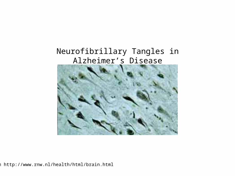
Neurofibrillary Tangles in Alzheimer’s Disease
From http://www.rnw.nl/health/html/brain.html
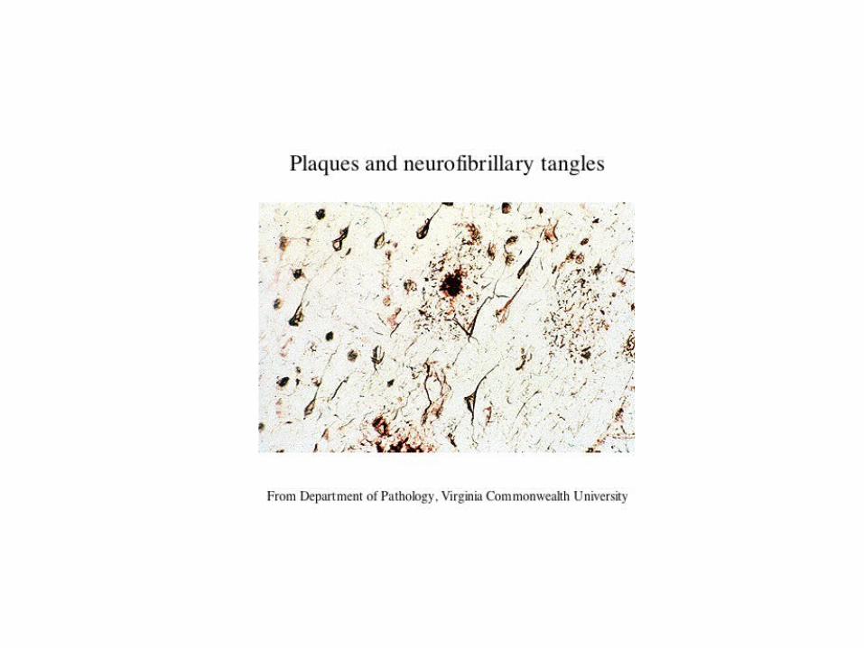
Plaques and neurofibrillary tangles
From Department of Pathology, Virginia Commonwealth University

http://www.hosppract.com/genetics/9707gen.htm

Following are from the NIA, Alzheimer’s DiseaseEducation and Referral Center, Alzheimer’s Disease: Unraveling the Mystery (www.niapublications.org/pubs/unraveling/01.htm ff.)

Amyloid precursor protein (APP) is membrane protein that sits in the membrane and extends outward. It is though tobe important for neuronal growth, survival, and repair.
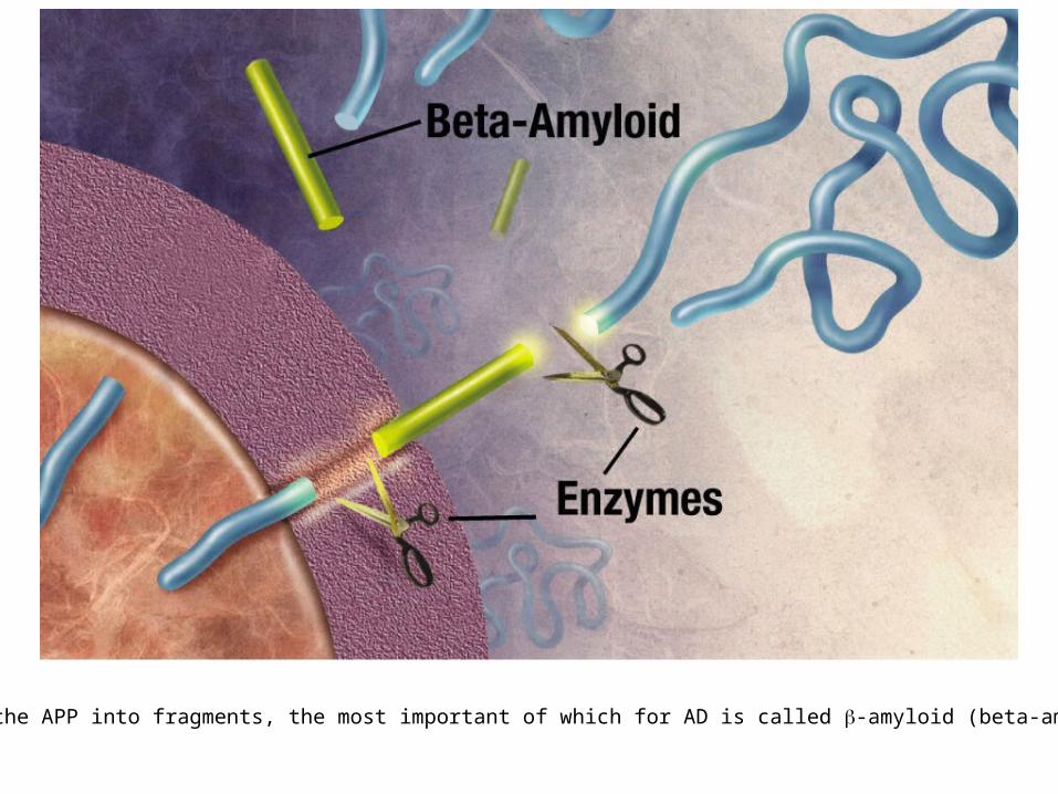
Enzymes cut the APP into fragments, the most important of which for AD is called b-amyloid (beta-amyloid) orAb.
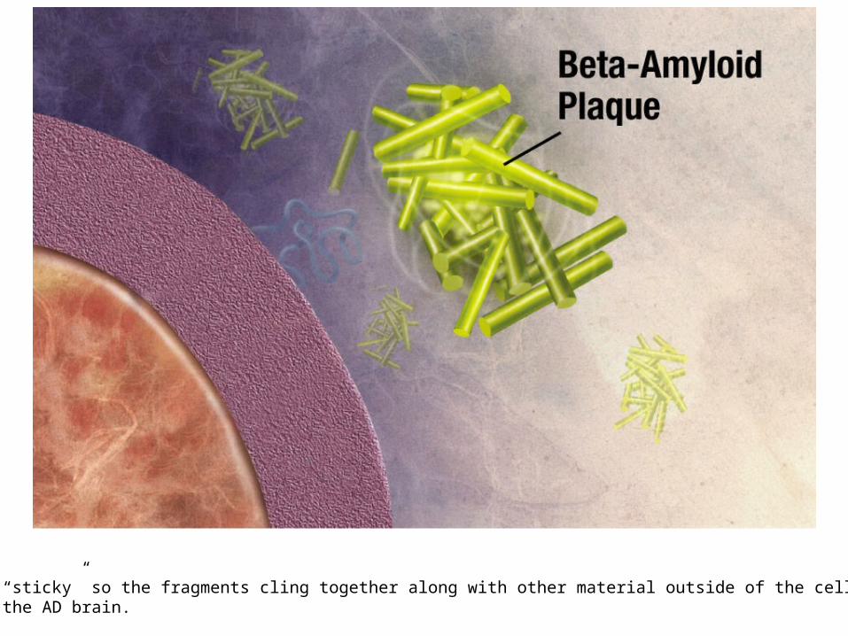
Beta-amyloid is “sticky” so the fragments cling together along with other material outside of the cell, forming theplaques seen in the AD brain.

Microtubules are like railroad tracks that transport nutrition and other molecules. Tau-proteins act as “ties” that stabilize the structure of the microtubules. In AD, tau proteins become tangled, unstabilizing the structure of themicrotubule.

http://abdellab.sunderland.ac.uk/lectures/Neurodegeneration/References/Brain_Neurons_AD_Normal.html
WRONG!
Brain Atrophy in AD

Classification:
(1) FAD v SAD: Familial AD versus Sporadic AD
• No complete consensus• Usually FAD = at least 1 first degree relative affected• Sometimes 2 second degree relatives
(2) Early v Late Onset:
• Early onset (EOAD) = usually before 65• Early onset correlated with FAD• LOAD = late onset AD

Breakthrough:
(1) Down’s Syndrome
• Have AD brain pathology in later life
(2) Pedigrees with dominant-like transmission:
• Studied these first• Concentrated on chromosome 21

Alzheimer’s Disease, Type 1:
• Several mutations in AAP gene on chromosome 21
• Most common = Val717Iso
• Produce abnormal beta amyloid fragment
• 15%-20% of early onset, familial AD
• Autosomal dominant
http://ghr.nlm.nih.gov/condition=alzheimerdisease

http://perso.wanadoo.fr/alzheimer.lille/APP/APPmutations.html

Alzheimer’s Disease, Type 3:
• Mutations (> 130) in the presenilin1 gene on chromosome 14
• Most mutations lead to amino acid substitution
• Overproduction of the beta amyloid fragment
• 30% - 70% of early onset, familial AD
• Autosomal dominant
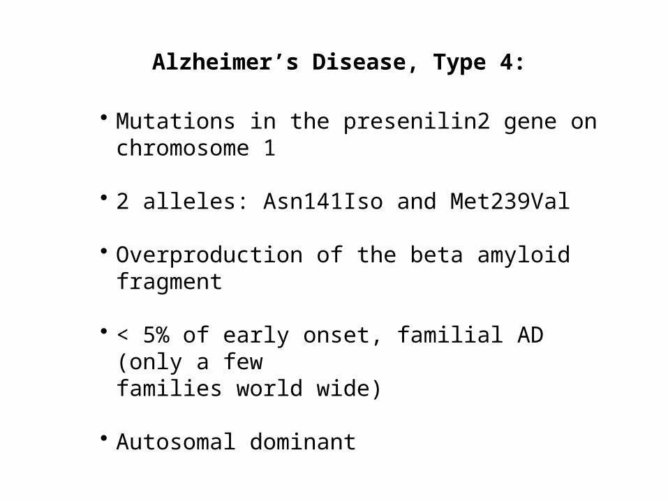
Alzheimer’s Disease, Type 4:
• Mutations in the presenilin2 gene on chromosome 1
• 2 alleles: Asn141Iso and Met239Val
• Overproduction of the beta amyloid fragment
• < 5% of early onset, familial AD (only a fewfamilies world wide)
• Autosomal dominant
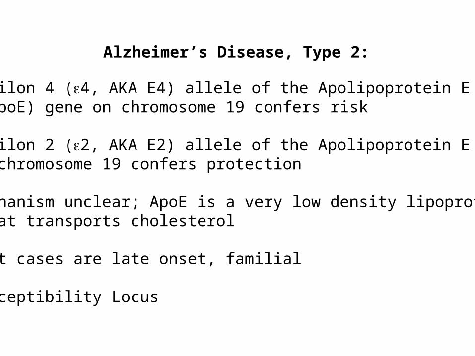
Alzheimer’s Disease, Type 2:
• Epsilon 4 (e4, AKA E4) allele of the Apolipoprotein E (ApoE) gene on chromosome 19 confers risk
• Epsilon 2 (e2, AKA E2) allele of the Apolipoprotein E geneon chromosome 19 confers protection
• Mechanism unclear; ApoE is a very low density lipoprotein that transports cholesterol
• Most cases are late onset, familial
• Susceptibility Locus

Prevalence of APOE genotypes in Alzheimer’s disease (AD) and controls.
Genotype: Controls AD
E2/E2 1.3% 0%
E2/E3 12.5% 3.4%
E2/E4 4.9% 4.3%
E3/E3 59.9% 38.2%
E3/E4 20.7% 41.2%
E4/E4 0.7% 12.9%
Jarvik G, Larson EB, Goddard K, Schellenberg GD, Wijsman EM (1996) Influence of apolipoprotein E genotype on the transmission of Alzheimer disease in a community-based sample. Am J Hum Genet 58:191-200
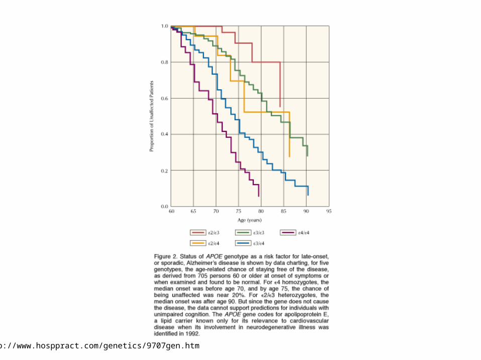
http://www.hosppract.com/genetics/9707gen.htm

AD: The Great Unknown:What is causing the majority of AD cases?
1. Unknown Mendelian forms (probably not many)
2. Unknown major loci (probably not)
3. Heterogeneity (probably polygenic)
4. Phencopies
5. Multifactorial-threshold
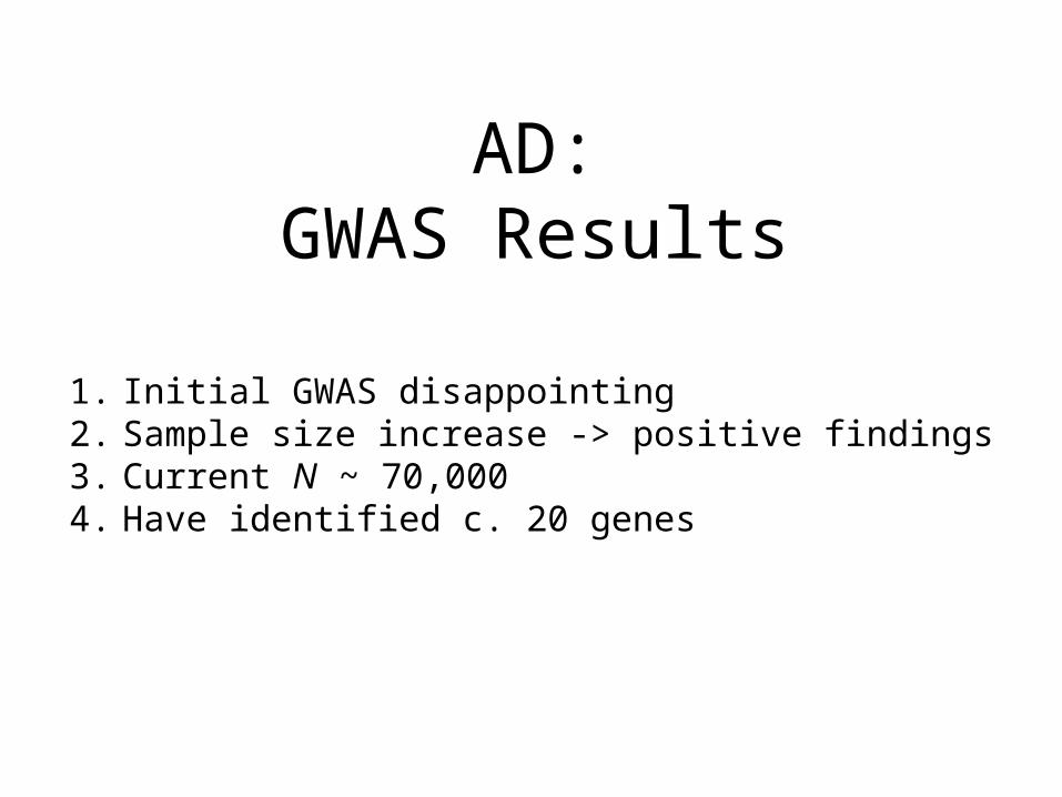
AD:GWAS Results
1. Initial GWAS disappointing2. Sample size increase -> positive findings3. Current N ~ 70,0004. Have identified c. 20 genes

Nature Genetics 41, 1088 - 1093 (2009)

From Medway & Morgan (2014), Neuropathology and Applied Neurobiology, 40:97-105

Current theory: Multifactorial, involvingseveral pathways.
• Protein accumulation: placques & tangles
• Inflammation: Unregulated activation of glia
• Lipid distribution: Lipid membrane site of APP cleavage.
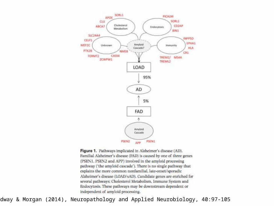
From Medway & Morgan (2014), Neuropathology and Applied Neurobiology, 40:97-105

From Sleegers et al. (2010) Trends in Genetics, 26, 84-94, p. 87
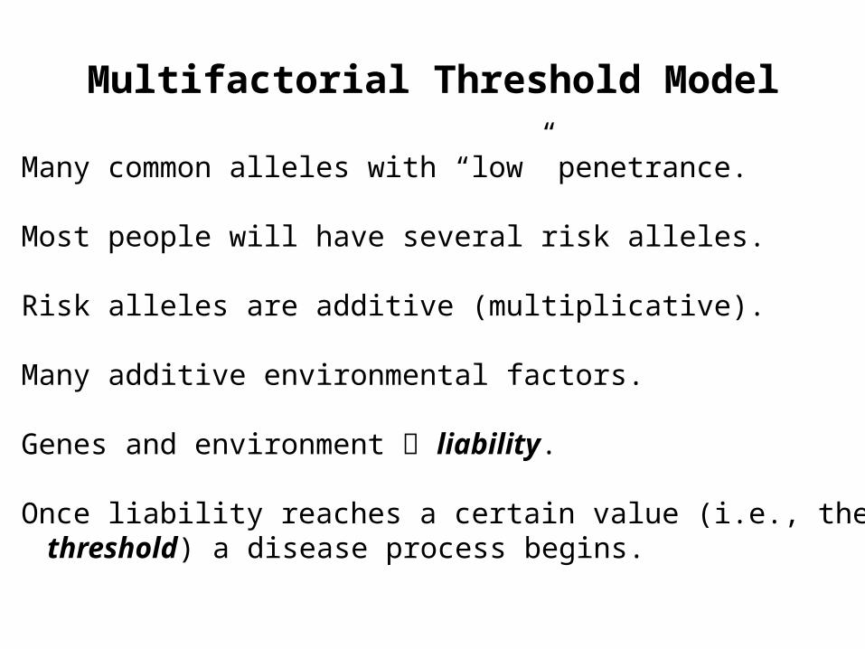
Multifactorial Threshold Model
• Many common alleles with “low” penetrance.
• Most people will have several risk alleles.
• Risk alleles are additive (multiplicative).
• Many additive environmental factors.
• Genes and environment liability.
• Once liability reaches a certain value (i.e., thethreshold) a disease process begins.

HGSS: Carey: Figure 6.1

Mice gratia http://www.kidscolorpages.com/mouse.htm
Human APPgene
Human ApoEgene
Human Presenilingene
Animal Models
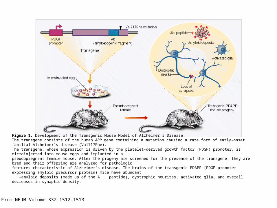
Figure 1. Development of the Transgenic Mouse Model of Alzheimer's Disease. The transgene consists of the human APP gene containing a mutation causing a rare form of early-onset familial Alzheimer's disease (Val717Phe). The transgene, whose expression is driven by the platelet-derived growth factor (PDGF) promoter, is microinjected into mouse eggs and implanted in a pseudopregnant female mouse. After the progeny are screened for the presence of the transgene, they are bred and their offspring are analyzed for pathologic features characteristic of Alzheimer's disease. The brains of the transgenic PDAPP (PDGF promoter expressing amyloid precursor protein) mice have abundant -amyloid deposits (made up of the A peptide), dystrophic neurites, activated glia, and overall decreases in synaptic density.
From NEJM Volume 332:1512-1513
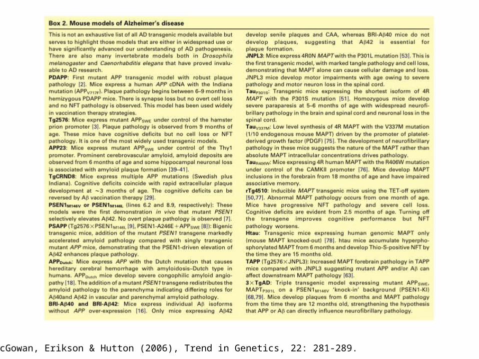
From McGowan, Erikson & Hutton (2006), Trend in Genetics, 22: 281-289.