disorders of endocrine control of growth & … · disorders of endocrine control of growth...
TRANSCRIPT

DISORDERS OF ENDOCRINE CONTROL OF GROWTH & METABOLISM
CHAPTER 42

THYROID HORMONE
T3- Triiodothyronine- likely the active form.T4- thyroxine.Both are released from the thyroid, T4 is converted to T3 in the periphery.Hypothalamus → TRH → anterior pituitary →release of TSH → thyroid → release of T3 & T4 from the follicular cells.Control is via negative feedback (inhibition) to both the hypothalamus and the pituitary.
FUNCTIONS OF THYROID HORMONE
2 MAJOR FUNCTIONS1) Increases metabolism and protein synthesis.2) Necessary for growth in children.
EFFECT ON METABOLISMIncreased use of glucose, fat, and protein.Lipids mobilized from adipose, cholesterol catabolized in the liver.Muscle protein broken down, vitamins consumed.
FUNCTIONS OF THYROID HORMONE
EFFECT ON METABOLISMCardiovascular and gastrointestinal function is increased.Increased activity of the sympathetic nervous system.
CONGENITAL HYPOTHYROIDISM
CRETINISM- term that describes untreated congenital hypothyroidism.Thyroid hormone- essential for normal brain development & growth.Untreated → mental retardation, impaired growth.Mandatory neonatal screening in all states.
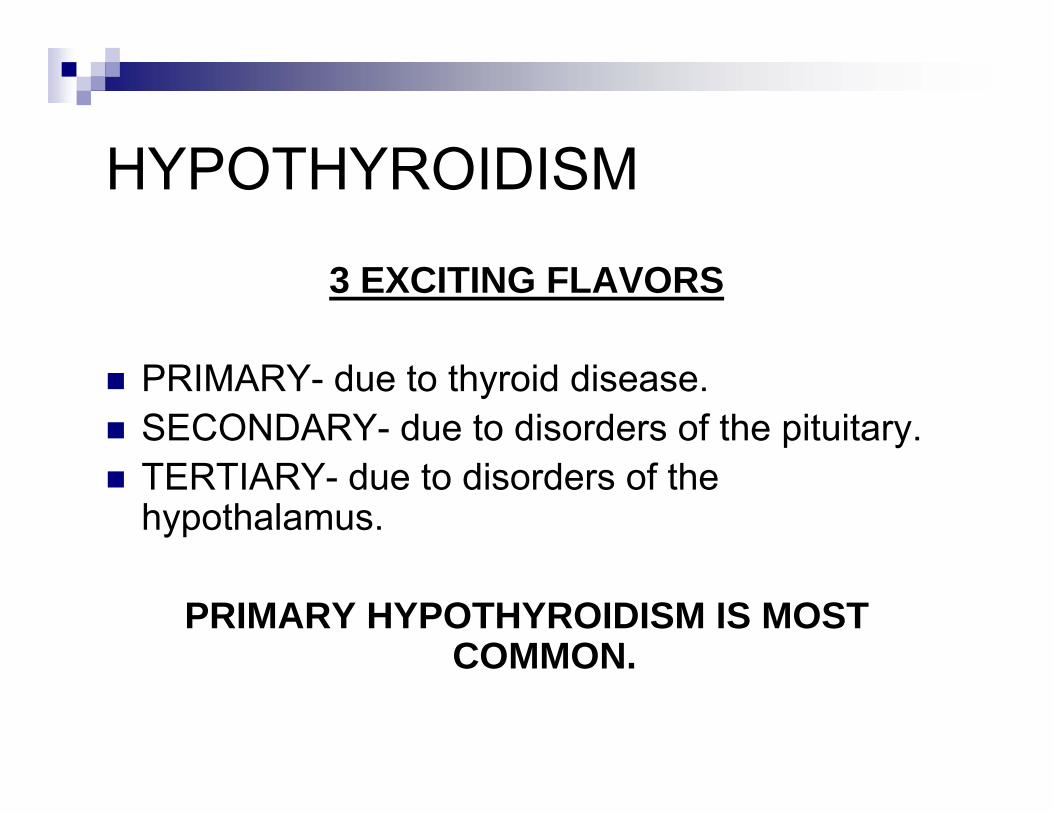
HYPOTHYROIDISM
3 EXCITING FLAVORS
PRIMARY- due to thyroid disease.SECONDARY- due to disorders of the pituitary.TERTIARY- due to disorders of the hypothalamus.
PRIMARY HYPOTHYROIDISM IS MOST COMMON.

PRIMARY HYPOTHYROIDISM
Most common cause is Hashimoto’s Thyroiditis.Other causes: thyroidectomy; “goitrogenic”agents: lithium, antithyroid drugs (PTU); iodine deficiency- rare in the U.S.Also results from treated or “burned-out”Grave’s Disease.

PRIMARY HYPOTHYROIDISM
HASHIMOTO’S THYROIDITISAn autoimmune disorder. Thyroid gland is destroyed by anti-thyroid antibodies. Results in hypothyroidism, although a transient hyperthyroid state is seen as damaged cells release thyroid hormone.Female predominance, 5:1.Begins w/ a goiter → hypothyroidism.
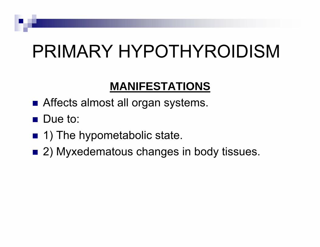
PRIMARY HYPOTHYROIDISM
MANIFESTATIONSAffects almost all organ systems.Due to:1) The hypometabolic state.2) Myxedematous changes in body tissues.
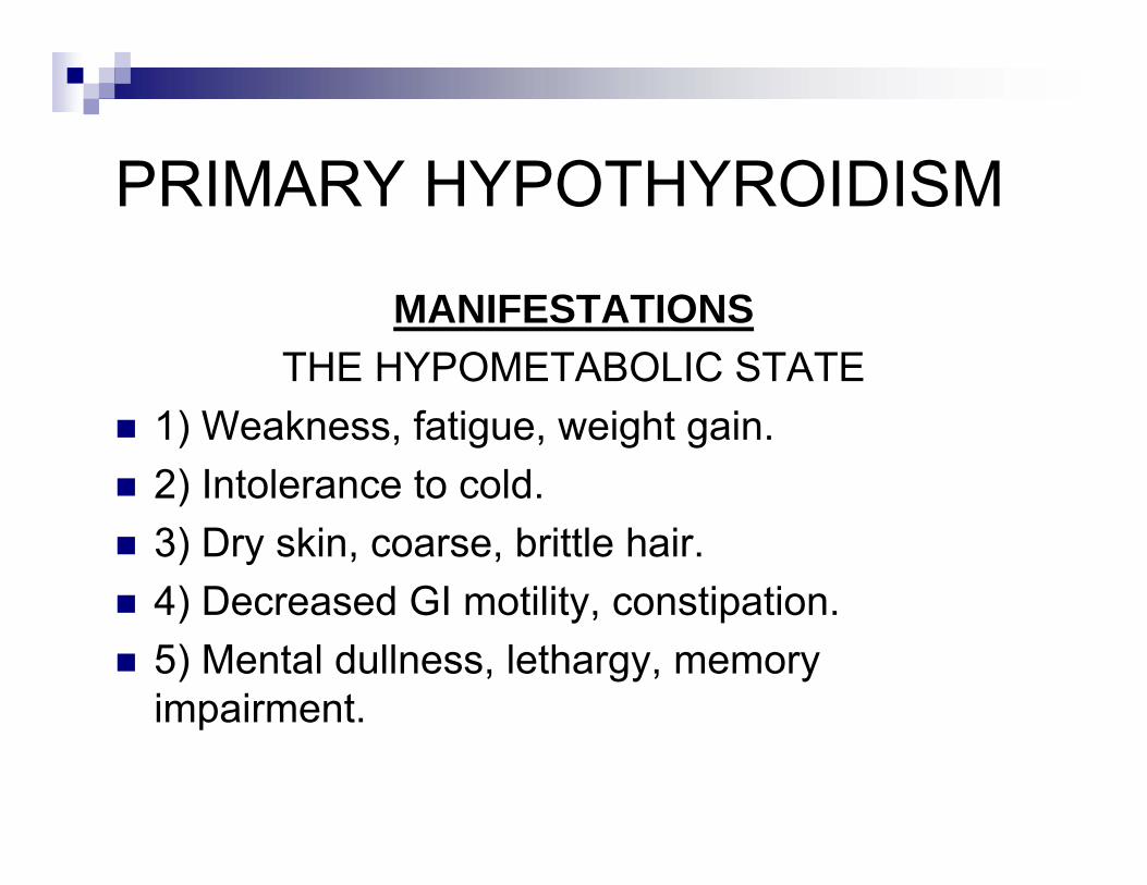
PRIMARY HYPOTHYROIDISM
MANIFESTATIONSTHE HYPOMETABOLIC STATE
1) Weakness, fatigue, weight gain.2) Intolerance to cold.3) Dry skin, coarse, brittle hair.4) Decreased GI motility, constipation.5) Mental dullness, lethargy, memory impairment.
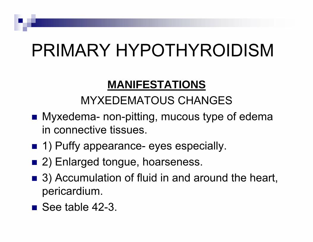
PRIMARY HYPOTHYROIDISM
MANIFESTATIONSMYXEDEMATOUS CHANGES
Myxedema- non-pitting, mucous type of edema in connective tissues.1) Puffy appearance- eyes especially.2) Enlarged tongue, hoarseness.3) Accumulation of fluid in and around the heart, pericardium.See table 42-3.
PRIMARY HYPOTHYROIDISM
MANIFESTATIONSMYXEDEMATOUS COMA
Potentially fatal, end-stage hypothroidism.“Myxedema madness”- the psychosis that preceded a myxedematous come.See text, Pg 975.
HYPERTHYROIDISM
Excess production of thyroid hormone.
CAUSES
1) GRAVE’S DISEASE- most common cause.2) MULTINODULAR GOITER.3) BENIGN ADENOMA.4) IODINE.
HYPERTHYROIDISM
MANIFESTATIONSExcept for Grave’s Disease, result from the hypermetabolic state and increased sympathetic activity.Nervousness, irritability, fatigue, weight loss.Tachycardia, palpitations, SOB, sweating, muscle cramps, heat intolerance.Tremor, restlessness.Goiter, exophthalmos.
GRAVE’S DISEASE
Hyperthyroidism, goiter, exophthalmos.An autoimmune disorder, anti-thyroid antibodies.Antibodies act through TSH receptors.Treatment results in hypothyroidism, as can “burned-out” Grave’s Disease.
DISORDERS OF THE ADRENAL CORTEX
HORMONES OF THE ADRENAL CORTEX:
1) MINERALOCORTICOIDS- aldosterone.2) GLUCOCORTICOIDS- cortisol.3) SEX STEROIDS- testosterone, progesterone, DHEAS- see figure 42-10, Pg 978. Really.SALT.SUGAR.SEX.
DISORDERS OF THE ADRENAL CORTEX
HORMONES OF THE ADRENAL CORTEX:1) MINERALOCORTICOIDS- promote tubular reabsorption of Na+ & H2O, excretion of K+.2) GLUCOCORTICOIDS- stimulates glucose production by the liver, breakdown of proteins, mobilization of fatty acids.3) SEX STEROIDS- not essential in males; development at puberty of secondary sex characteristics in the female. DHEA, pregnancy.
DISORDERS OF THE ADRENAL CORTEX
Adrenal cortex controlled by ACTH.Total failure of the adrenal cortex is fatal in 4 to 14 days if untreated.
DISORDERS:1) CONGENITAL ADRENAL HYPERPLASIA.2) ADDISON’S DISEASE (adrenal failure).3) CUSHING’S DISEASE (corticoid excess).
CONGENITAL ADRENAL HYPERPLASIA
A disorder caused by a deficiency of one of the various enzymes responsible for adrenocorticoidsynthesis (21 hydroxylase, 11-β hydroxylase).See figure42-10, Pg 978. Really.Results in: 1) Excess androgen production, 2) Decreased glucocorticoids, 3) Increased OR decreased mineralocorticoids, depending on location of enzyme defect.
CONGENITAL ADRENAL HYPERPLASIA
Decreased cortisol → Increased ACTH →adrenal hyperplasia → continued / increase in mineralocorticoid production (not always; again, depending on the location of the enzyme defect)→ excess Na+ and water reabsorption→ hypernatremia and hypertension, and excess excretion in the tubule of K+ → hypokalemiaDecreased cortisol → Increased ACTH →continued / increase production of androgens →virilization.
CONGENITAL ADRENAL HYPERPLASIA
Males: Dx’d if enlarged genitalia, “salt-losing,” adrenal crisis from lack of cortisol.Females: virilization- ambiguous genitalia, enlarged clitoris, fused labia.The “salt-losing” form is a rarer form in which the enzyme defect results in deficient rather than increased production of mineralocorticoid, causing loss of sodium → hyponatremia, hyperkalemia.
ADRENOCORTICAL INSUFFICIENCY
PRIMARY- ADDISON’S DISEASE-destruction of the adrenal gland.SECONDARY- abnormalities of the pituitary.TERTIARY- abnormalities of the hypothalamus.
ADDISON’S DISEASEPrimary adrenocortical failure from destruction of the adrenal cortex (all layers) by:1) Autoimmune destruction.2) OTHERS: TB, metastatic carcinoma, fungi, amyloid, hemochromatosis, trauma.Not clinically manifest until 90% of the adrenal cortex is destroyed.Manifestations are those of deficiencies of mineralocorticoids and glucocorticoids, and excess ACTH.
ADDISON’S DISEASE
MINERALOCORTICOID DEFICIENCYIncreased urinary loss of Na+ (and water).Decreased excretion of K+.↓ Na+ → loss of extracellular fluid, C.O.As such → Hyponatremia and Hyperkalemia.CAUSING:Dehydration, weakness, fatigue.Cardiovascular collapse, shock.
ADDISON’S DISEASE
GLUCOCORTICOID DEFICIENCYDecreased response to stress.Hypoglycemia, weakness, lethargy, fever.Anorexia, nausea, vomiting, weight loss.
ADDISON’S DISEASE
EXCESS ACTHAmino acid sequence of ACTH is similar to MSH, resulting in hyperpigmentation.Helps to distinguish primary adrenal failure (adrenal) from secondary (pituitary) and tertiary (hypothalamic).
SECONDARY ADRENOCORTICAL INSUFFICIENCY
From hypopituitarism: surgical removal, tumor, etc.“TERTIARY” INSUFFICIENCY- from a hypothalamic defect. Most common cause is long-term administration of corticosteroids w/ abrupt withdrawal / cessation (prednisone, hydrocortisone).
CUSHING’S SYNDROMEExcess cortisol production from:1) PITUITARY- ACTH-producing tumor of the pituitary. 1st type described, is known as Cushing’s Disease.2) ADRENAL- cortisol-producing tumor of the adrenal; benign or malignant.3) ECTOPIC ACTH- ACTH production by non-pituitary tumors, mainly lung.4) IATROGENIC- long-term administration of corticosteroids.
CUSHING’S SYNDROME
MANIFESTATIONSRelated to cortisol excess. See table 42-4.1) Altered fat metabolism- protruding abdomen, buffalo hump, moon face.2) Thin skin- striae on abdomen, thighs breasts.3) Protein catabolism- thin legs and arms.4) Osteoporosis.5) Immune dysfunction, infection.
CUSHING’S SYNDROME
MANIFESTATIONS1) Derangements of glucose metabolism, diabetes from effects of cortisol.2) Hypokalemia, hypernatremia, water retention, hypertension, from mineralocorticoid properties of cortisol.3) Hyperandrogenism.
CUSHING’S SYNDROME
DIAGNOSIS1) Measurement of cortisol & ACTH. Diurnal variation, ACTH suppression. See text.2) MRI, CT.3) Need to determine source of the cortisolexcess- pituitary vs. adrenal.
CONN’S SYNDROME
Aldosterone excess, usually from a benign adenoma in one adrenal cortex.Causes approx 1% of cases of secondary hypertension.Excess aldosterone → excess reabsorption of Na+ and H20 → hypertension.Also excess excretion of K+ → hypokalemia.
DIABETES MELLITUS AND METABOLIC SYNDROMECHAPTER 43

DIABETES
Review metabolism of glucose.Insulin, catecholamines, growth hormone, glucocorticoids, glucagon, cortisol. Pg 988-992.Insulin receptors.
DIABETES
Due to an imbalance between insulin need and insulin availability.Not one disease. See table 43-2.Most cases are Type I Diabetes- 90-95%.Most of the rest are Type II.There’s also Type Ia, Ib, IIa, IIb.See table 43-1 for diagnostic criteria.
TYPE I DIABETES
Destruction of the pancreatic beta cells.Absolute lack of insulin.TYPE 1A- autoimmune. 90%. Formerly juvenile.TYPE 1B- idiopathic. 10%.All require insulin replacement.Prone to diabetic ketoacidosis (DKA).
TYPE II DIABETES
A heterogeneous disorder associated with :1) Disordered beta cell function- diminished insulin levels, and / or2) Disordered insulin function- insulin resistance.3) Increased hepatic production of glucose.Can be associated with high, low, or normal levels of insulin.
TYPE II DIABETES
Most patients are older and obese.Can progress to type I diabetes.
See text Re Metabolic Syndrome / Syndrome X.
DIABETES
CLINICAL MANIFESTATIONS1) Polyuria- osmotic diuresis.2) Polydipsia.3) Polyphagia.
Type II- obesity.Type I- weight loss due to utilization of fat and protein for metabolic energy.
DIABETES
CLINICAL MANIFESTATIONSOther:1) Blurred vision- from hyperosmolar changes of the lens and retina.2) Chronic skin infections.3) Yeast infections- vaginal, foreskin.

COMPLICATIONS OF DIABETES
ACUTE1) DKA.2) HYPEROSMOLAR HYPERGLYCEMIC STATE.3) HYPOGLYCEMIA.
DKA
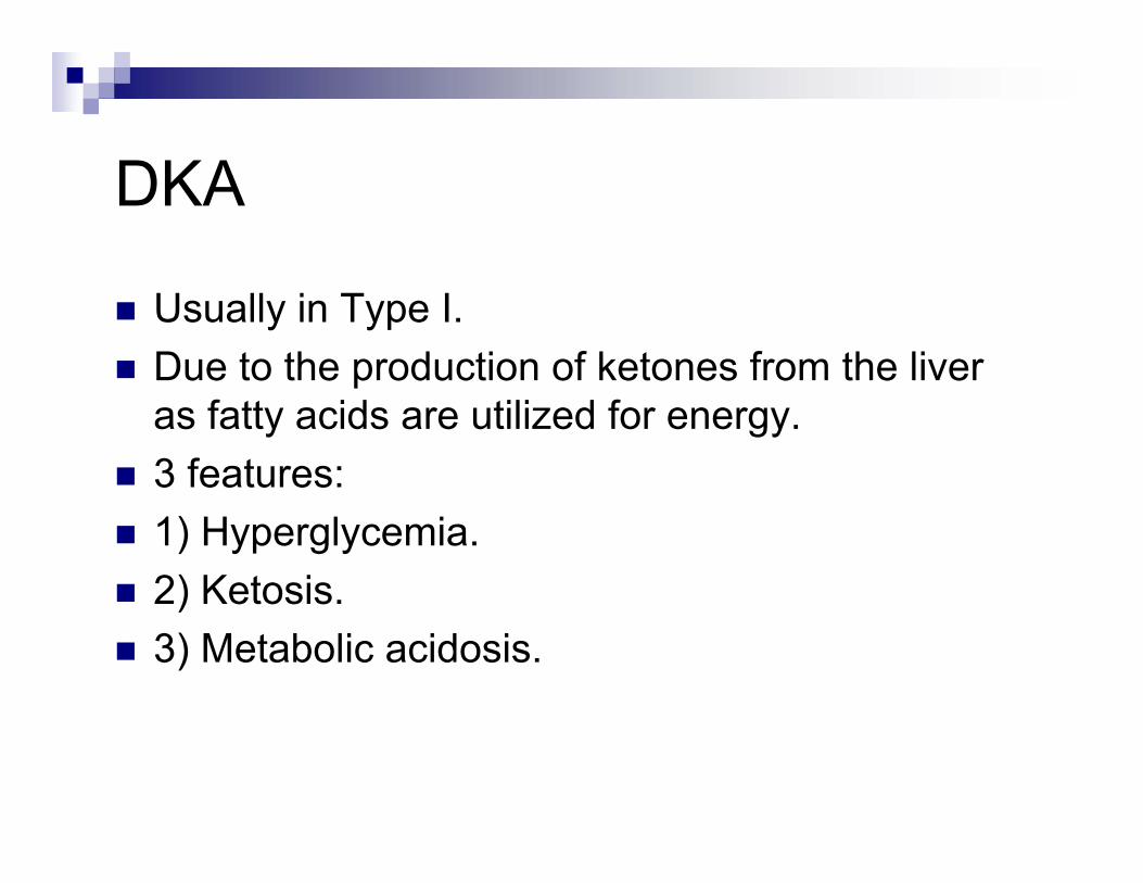
Usually in Type I.Due to the production of ketones from the liver as fatty acids are utilized for energy.3 features:1) Hyperglycemia.2) Ketosis.3) Metabolic acidosis.
DKA
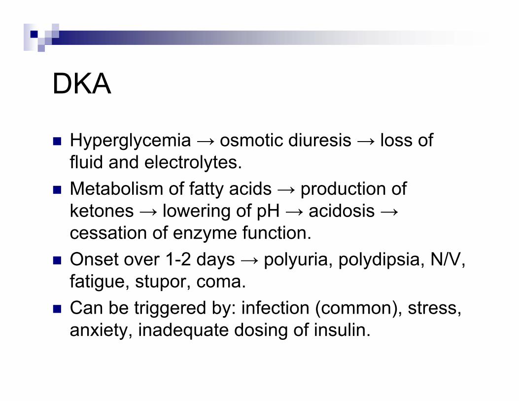
Hyperglycemia → osmotic diuresis → loss of fluid and electrolytes.Metabolism of fatty acids → production of ketones → lowering of pH → acidosis →cessation of enzyme function.Onset over 1-2 days → polyuria, polydipsia, N/V, fatigue, stupor, coma.Can be triggered by: infection (common), stress, anxiety, inadequate dosing of insulin.
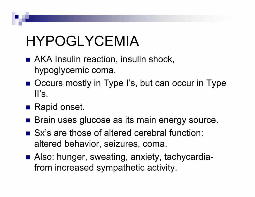
HYPOGLYCEMIAAKA Insulin reaction, insulin shock, hypoglycemic coma.Occurs mostly in Type I’s, but can occur in Type II’s.Rapid onset.Brain uses glucose as its main energy source.Sx’s are those of altered cerebral function: altered behavior, seizures, coma.Also: hunger, sweating, anxiety, tachycardia-from increased sympathetic activity.
HYPOGLYCEMIA
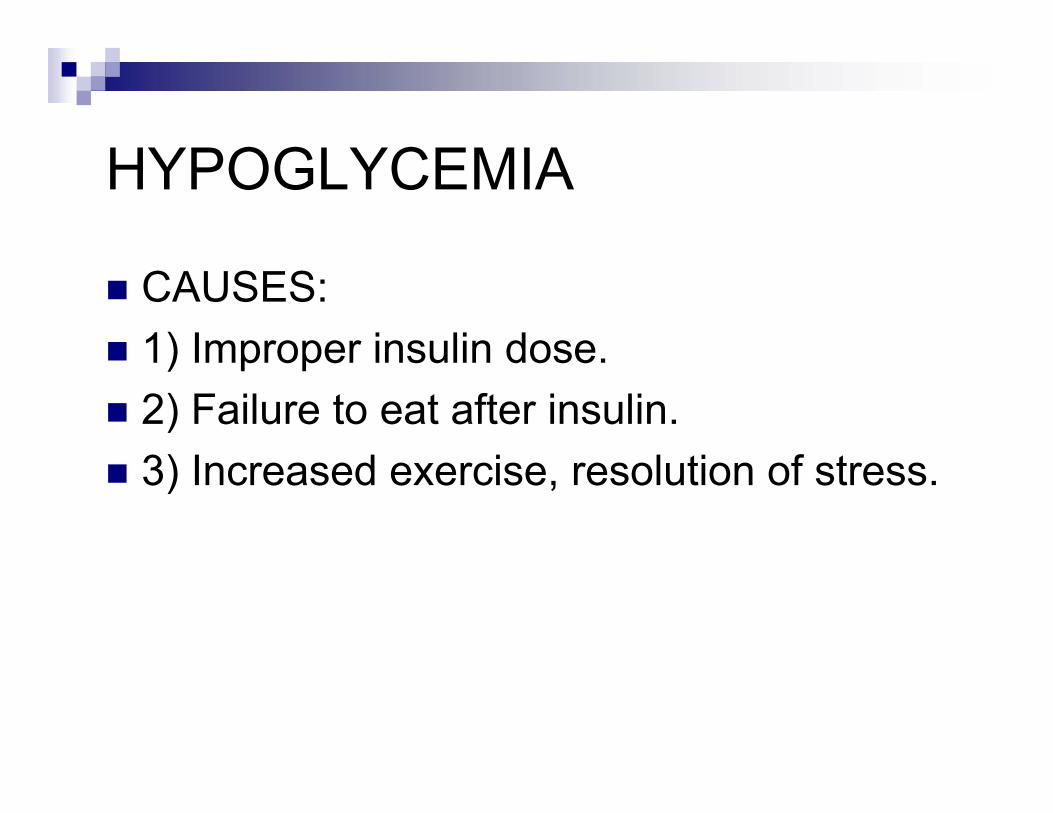
CAUSES:1) Improper insulin dose.2) Failure to eat after insulin.3) Increased exercise, resolution of stress.
COMPLICATIONS OF DIABETES
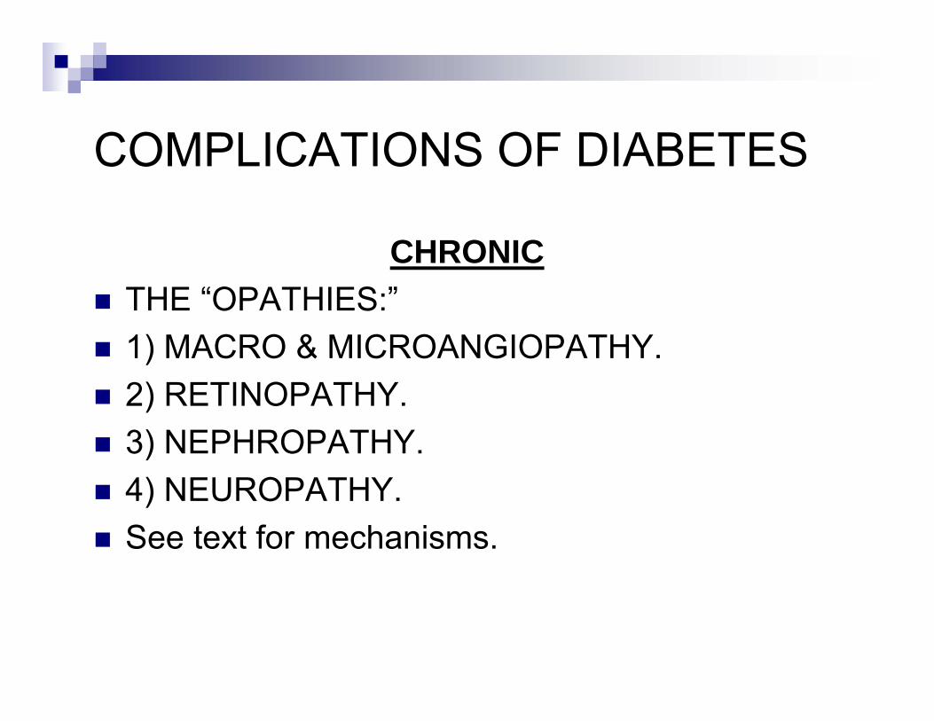
CHRONICTHE “OPATHIES:”1) MACRO & MICROANGIOPATHY.2) RETINOPATHY.3) NEPHROPATHY.4) NEUROPATHY.See text for mechanisms.
COMPLICATIONS OF DIABETES
CHRONIC
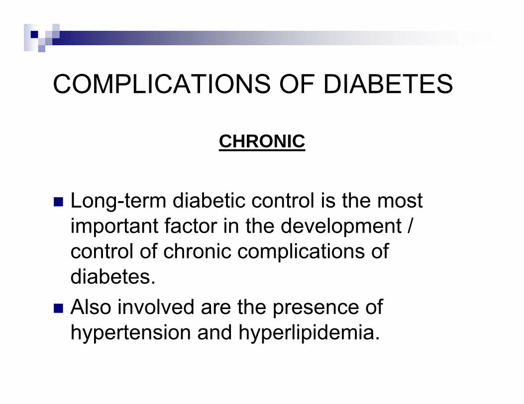
Long-term diabetic control is the most important factor in the development / control of chronic complications of diabetes.Also involved are the presence of hypertension and hyperlipidemia.
COMPLICATIONS OF DIABETES
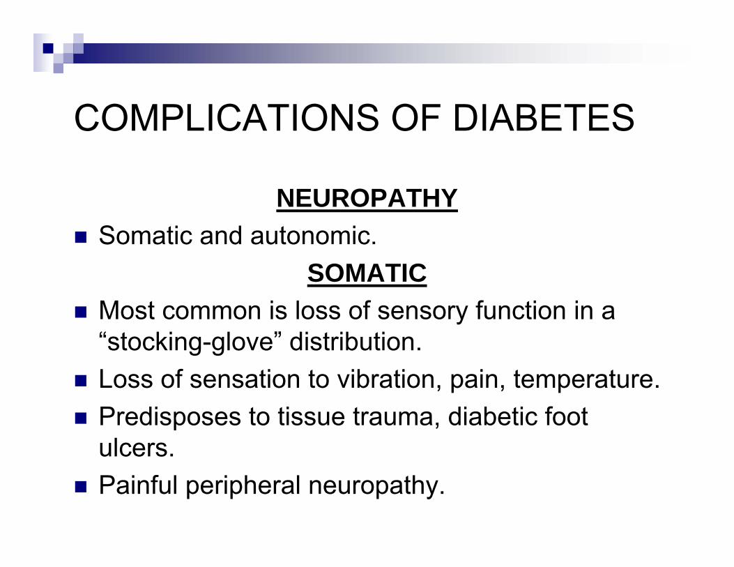
NEUROPATHYSomatic and autonomic.
SOMATICMost common is loss of sensory function in a “stocking-glove” distribution.Loss of sensation to vibration, pain, temperature.Predisposes to tissue trauma, diabetic foot ulcers.Painful peripheral neuropathy.
COMPLICATIONS OF DIABETES
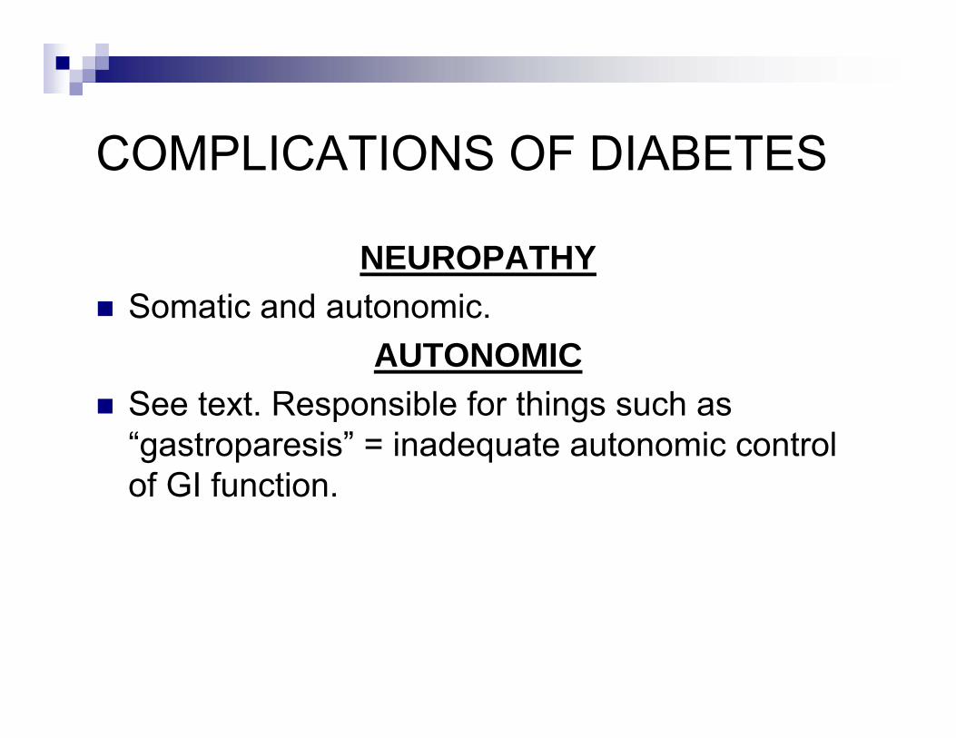
NEUROPATHYSomatic and autonomic.
AUTONOMICSee text. Responsible for things such as “gastroparesis” = inadequate autonomic control of GI function.
COMPLICATIONS OF DIABETES
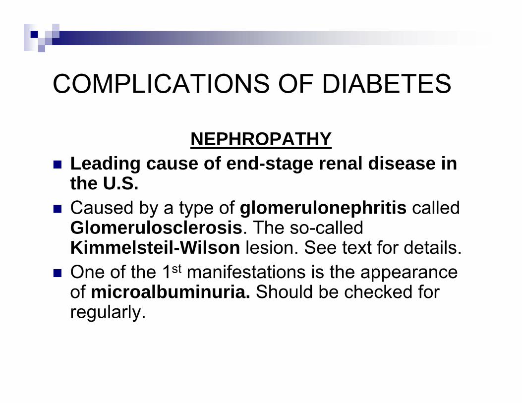
NEPHROPATHYLeading cause of end-stage renal disease in the U.S.Caused by a type of glomerulonephritis called Glomerulosclerosis. The so-called Kimmelsteil-Wilson lesion. See text for details.One of the 1st manifestations is the appearance of microalbuminuria. Should be checked for regularly.
COMPLICATIONS OF DIABETES
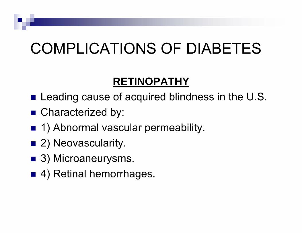
RETINOPATHYLeading cause of acquired blindness in the U.S.Characterized by:1) Abnormal vascular permeability.2) Neovascularity.3) Microaneurysms.4) Retinal hemorrhages.
COMPLICATIONS OF DIABETES
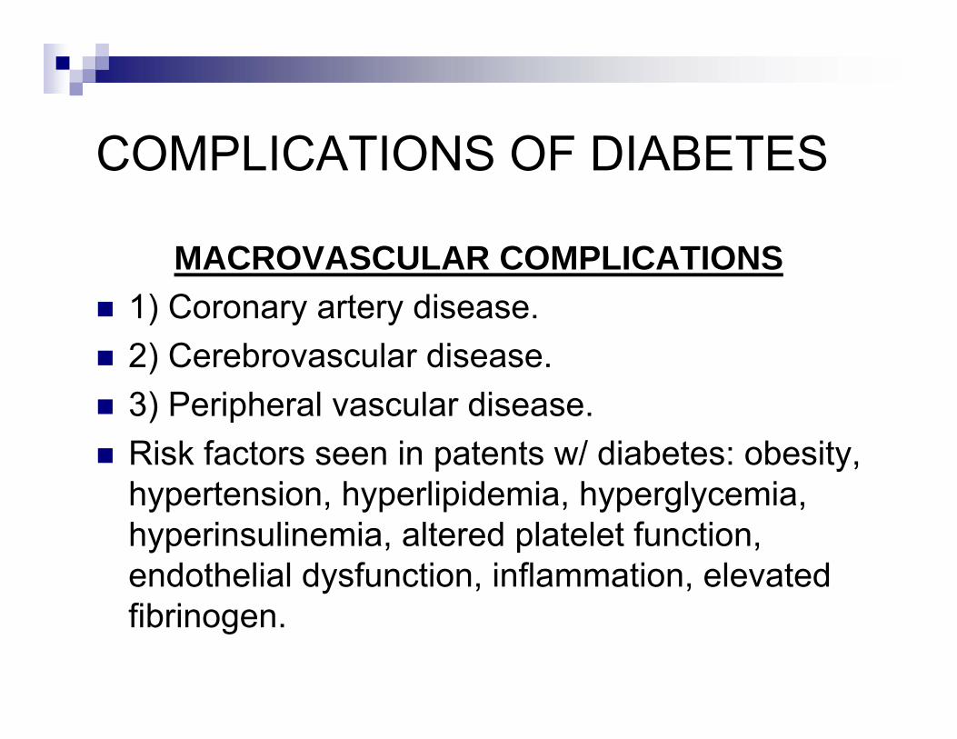
MACROVASCULAR COMPLICATIONS1) Coronary artery disease.2) Cerebrovascular disease.3) Peripheral vascular disease.Risk factors seen in patents w/ diabetes: obesity, hypertension, hyperlipidemia, hyperglycemia, hyperinsulinemia, altered platelet function, endothelial dysfunction, inflammation, elevated fibrinogen.
COMPLICATIONS OF DIABETES
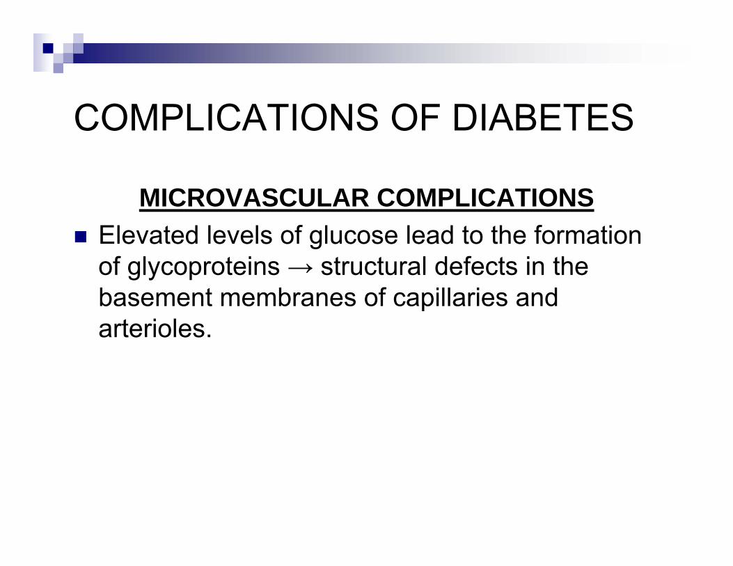
MICROVASCULAR COMPLICATIONSElevated levels of glucose lead to the formation of glycoproteins → structural defects in the basement membranes of capillaries and arterioles.
COMPLICATIONS OF DIABETES
DIABETIC FOOT ULCERSCommon. Can lead to infection and amputation.Due to micro and macrovascular complications and from peripheral sensory neuropathy.
GESTATIONAL DIABETES
Glucose intolerance 1st detected during pregnancy, as opposed to preexisting DM.
RISK FACTORSFamily Hx DM, advanced maternal age, obesity.Hx of prior still birth, spontaneous abortion, congenital anomaly, LGA.5 or more pregnancies.
GESTATIONAL DIABETES
COMPLICATIONSIncreased risk of:Placental insufficiency → impaired growth, stillbirth.Fetal macrosomia from excess glucose.Toxemia / pregnancy-induced hypertension.Fetal anomalies.
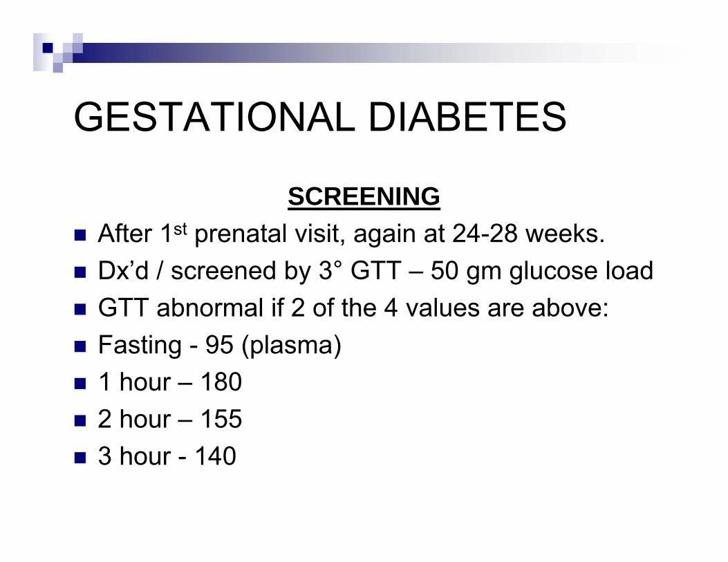
GESTATIONAL DIABETES
SCREENINGAfter 1st prenatal visit, again at 24-28 weeks.Dx’d / screened by 3° GTT – 50 gm glucose loadGTT abnormal if 2 of the 4 values are above:Fasting - 95 (plasma)1 hour – 1802 hour – 1553 hour - 140
GESTATIONAL DIABETES
Treated with dietary modification and insulin if needed.Oral hypoglycemics are not used due to risk of birth defects.
THE MALE GENITOURINARY SYSTEMCHAPTER 44

SPERMATOGENESIS
Occurs from the Sertoli Cells within the seminiferous tubules of the testes.Spermatogonia → mitosis → primary spermatocyte (46) → meiosis → 2 secondary spermatocytes (23) → 4 spermatids →spermatozoa.Maturation of the spermatozoa requires testosterone, produced in the interstitial LeydigCells.
HORMONAL CONTROL OF SPERMATOGENESIS
Hypothalamus → GnRh → pituitary → FSH and LH.FSH → Sertoli Cells → spermatogenesis and production of Inhibin.Inhibin → pituitary → suppresses release of FSHLH → Leydig Cells → testosterone → negative feedback to inhibit LH at both the hypothalamus and pituitary.
ANDROGENS
1) TESTOSTERONE.2) DIHYDROTESTOSTERONE.3) ANDROSTENEDIONE.Testosterone is most abundant.Adrenal cortex produces androgens - < 5%.Testes also produce small amounts of estradiol(E2) and estrone (E1).
ANDROGENSEFFECTS
Anabolic → increase in protein synthesis.1) Differentiation of the internal and external genitalia.2) Development of primary and secondary sex characteristics.3) Pubic and facial hair, muscle mass.4) Deepening of the voice, activity of sebaceous glands.

THE MALE GENITOURINARY SYSTEM
SEE TEXT RE: TESTICULAR CANCERPROSTATITISBPHPROSTATE CANCER