diseases of placenta - gmch.gov.in lectures/pathology/diseases of placenta.pdf · choriocarcinoma...
TRANSCRIPT

DISORDERS OF PLACENTA

GESTATIONAL TROPHOBLASTIC DISEASE
• Gestational trophoblastic disease constitutes a spectrum of tumors and tumor‐like conditions characterized by proliferation of placental tissue, either villous or trophoblastic.
• The lesions include:– hydatidiform mole (complete and partial)
– invasive mole
– malignant choriocarcinoma and placental‐site trophoblastic tumor

HYDATIDIFORM MOLE• Hydatidiform mole is characterized histologically by cystic swelling of the chorionic villi, accompanied by variable trophoblastic proliferation.
• In the past, most patients presented in the fourth or fifth month of pregnancy with vaginal bleeding.
• Molar pregnancy can develop at any age, but the risk is higher at the far ends of reproductive life: in teens and between the ages of 40 and 50 years.

• Two types of benign, noninvasive moles—complete and partial—can be identified by cytogenetic and histologic studies.
• The most important reason for the correct recognition of moles is that they are associated with an increased risk of persistent trophoblastic disease (invasive mole) or choriocarcinoma.
Complete Mole
• Complete mole results from fertilization of an egg that has lost its chromosomes, and the genetic material is completely paternally derived
• Histologically, in complete mole all or most of the villiare enlarged and edematous, and there is diffuse trophoblast hyperplasia.
• Fetal vessels and fetal parts are rare in complete moles since the embryo dies very early in development.
• Patients have 2.5% risk of subsequent choriocarcinoma.


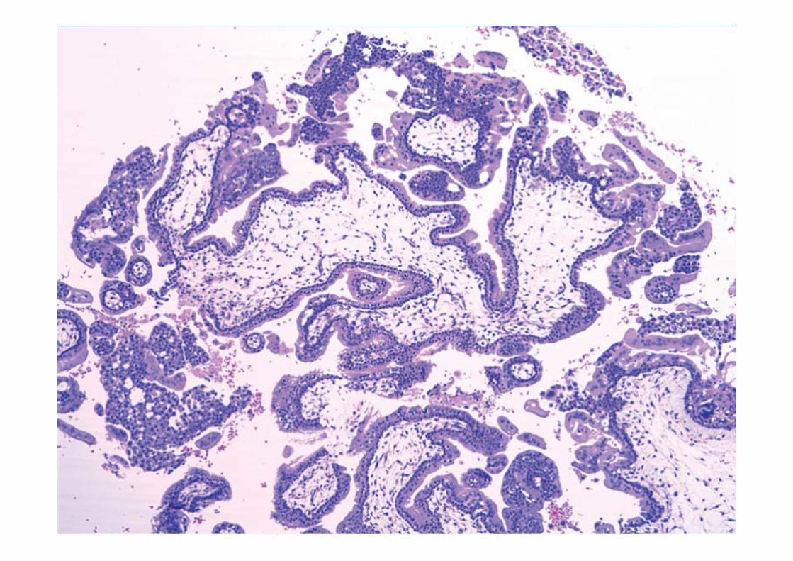
Complete Mole ‐ Pathology
• Gross appearance : a delicate, friable mass of thin‐walled, translucent, cystic, grape‐like structures consisting of swollen edematous (hydropic) villi.
• On microscopy, complete moles show abnormalities that involve all or most of the villous tissue.
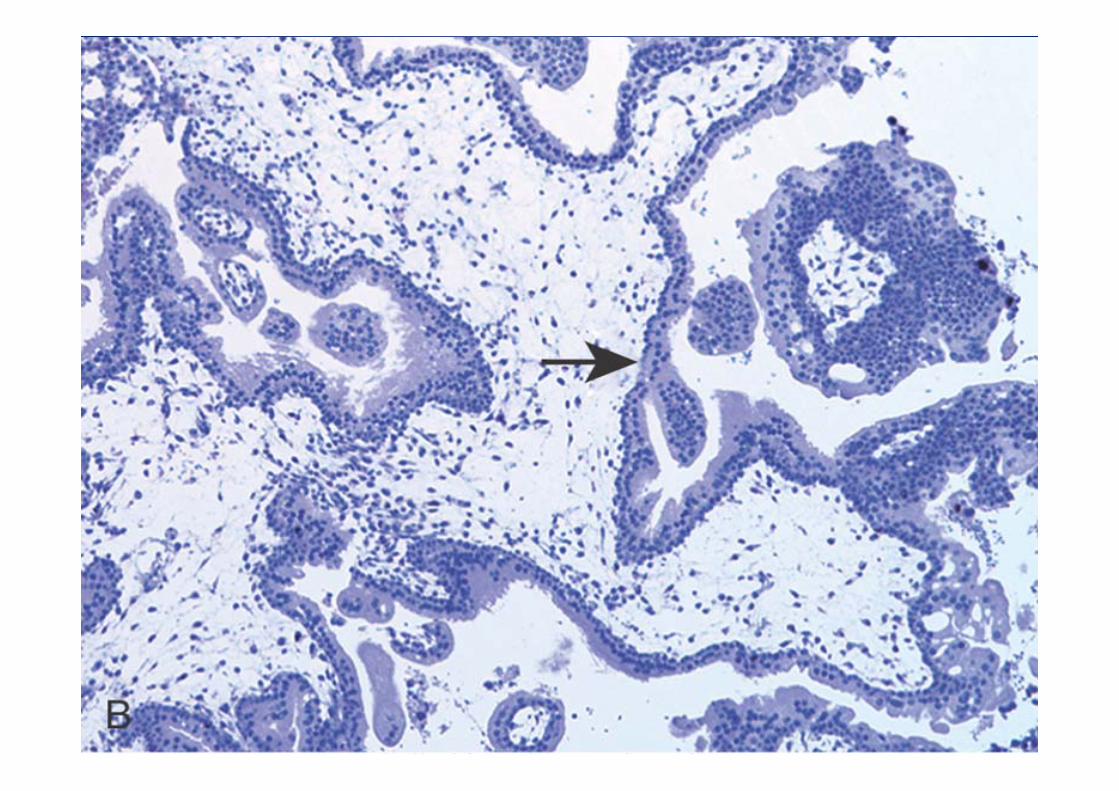
• The chorionic villi are enlarged, scalloped in shape with central cavitation (cisterns), and lack adequately developed vessels.
• An extensive trophoblast proliferation that involves the entire circumference of the villi, in addition to “extravillous” islands of trophoblast proliferation.


Partial mole
• Partial mole results from fertilization of an egg by two sperms. In these moles the karyotypeis triploid (e.g., 69XXY) or even occasionally tetraploid (92XXXY).
• Although partial moles have an increased risk of persistent molar disease, they are not considered to have an increased risk for choriocarcinoma.

Partial Mole ‐ Pathology
• Grossly, fetal parts are more commonly present than in complete moles.
• Some of the villi are edematous, and other villishow only minor changes; the trophoblasticproliferation is focal and less marked.
• The trophoblastic proliferation is moderate but still may be circumferential.

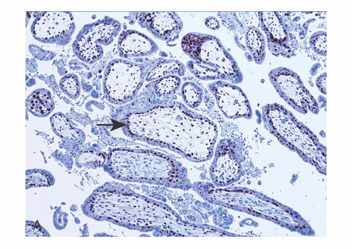
Hydatidiform Mole ‐ Genetics• Histologic distinction of complete mole from partial molar
gestations is important.
• In equivocal cases immunostaining for p57, a cell cycle inhibitor, may aid the diagnosis.
• The p57KIP2 gene is maternally transcribed but paternally imprinted, and shows expression in maternal decidual tissue as well as cytotrophoblast and stromal cells of the villi, when maternal genetic material is present in the conceptus .
• In contrast, since both the X chromosomes in complete moles are derived from the father, there is no expression of p57 protein in the cytotrophoblast or stromal cells of the villi in complete moles.

Clinical aspect• Spontaneous pregnancy loss or removed by thorough curettage.
• In complete moles, beta‐HCG levels are markedly raised.
• Monitoring serum concentrations of HCG is necessary to determine
the early development of persistent trophoblastic disease, since up
to 10% of moles develop into persistent or invasive moles.
• 2.5% of complete moles evolve into gestational choriocarcinoma.
Therefore, serum HCG levels are followed until they fall to and
remain at zero for 6 months to a year.

INVASIVE MOLE• This is defined as a mole that penetrates or even perforates the uterine wall.
• There is invasion of the myometrium by hydropicchorionic villi, accompanied by proliferation of both cytotrophoblast and syncytiotrophoblast.
• The tumor is locally destructive and may invade parametrial tissue and blood vessels.
• Hydropic villi may embolize to distant sites, such as lungs and brain, but do not grow in these organs as true metastases, and even without chemotherapy they eventually regress

• The tumor is manifested clinically by vaginal bleeding and irregular uterine enlargement.
• It is always associated with a persistently elevated serum HCG and varying degrees of luteinization of the ovaries.
• The tumor responds well to chemotherapy but may result in uterine rupture and necessitate hysterectomy.


CHORIOCARCINOMA • Gestational choriocarcinoma is a malignant neoplasm of
trophoblastic cells derived from a previously normal or abnormal pregnancy.
• Choriocarcinoma is rapidly invasive and metastasizes widely, but once identified responds well to chemotherapy.
It is preceded by :
• 50% arise in hydatidiform moles
• 25% in previous abortions
• 22% in normal pregnancies
• Rest in ectopic pregnancies
• Very rarely, a nongestational choriocarcinoma may develop from germ cells in the ovaries or the mediastinum.

Choriocarcinoma ‐ Pathology• Grossly : a soft, fleshy, yellow‐white tumor with a marked
tendency to form large pale areas of ischemic necrosis, foci of cystic softening, and extensive hemorrhage.
• Histologically, it does not produce chorionic villi and consists entirely of a mixed proliferation of syncytiotrophoblasts and cytotrophoblasts.
• Mitoses are abundant and sometimes abnormal.
• The tumor invades the underlying myometrium, frequently penetrates blood vessels and lymphatics, and in some cases extends out onto the uterine serosa and into adjacent structures.


• Due to rapid growth it is subject to hemorrhage, ischemic necrosis, and secondary inflammation.
• Metastases are found in the lungs, brain, bone marrow, liver, and other organs.
• On occasion, metastatic choriocarcinoma is discovered without a detectable primary in the uterus (or ovary), presumably because the primary has undergone complete necrosis.
Clinical features
• Uterine choriocarcinoma usually does not produce a large, bulky mass, but it manifests as irregular vaginal spotting of a bloody, brown fluid.
• Usually, by the time the tumor is discovered, radiographs of the chest and bones already disclose the presence of metastatic lesions.
• The titers of HCG are elevated to levels above those encountered in hydatidiform moles.

• The results of chemotherapy for gestational choriocarcinoma are spectacular and result in nearly 100% remission and a high rate of cures.
• By contrast, nongestational choriocarcinomasare much more resistant to therapy.

PLACENTAL‐SITE TROPHOBLASTIC TUMOR (PSTT)
• PSTTs represent neoplastic proliferation of extravillous
trophoblast, also called intermediate trophoblast.
• In normal pregnancy, extravillous (intermediate) trophoblast is
found in nonvillous sites such as the implantation site, islands of
cells within the placental parenchyma, chorionic plate, and in
the placental membranes.
• They are polygonal mononuclear cells that have abundant
cytoplasm and produce human placental lactogen.
• Malignant transformation of extravillous trophoblast gives rise to
PSTT which is composed of malignant trophoblastic cells diffusely
infiltrating the endomyometrium.


• PSTTs may be preceded by a normal pregnancy, spontaneous abortion or hydatidiform mole.
• presents as a uterine mass, either abnormal uterine bleeding or amenorrhea and moderate elevation of β‐HCG.
• Patients with localized disease or a less than 2‐year interval from the prior pregnancy to diagnosis have an excellent prognosis.
• Tumors diagnosed at advanced stage, or diagnosed 2 or more years following pregnancy, have a poor prognosis.

ENDOMETRIAL CARCINOMA
• Carcinoma of the endometrium shows a peak incidence in 55‐ to 65‐year‐old women.
• Clinicopathologic studies and molecular analyses support the classification of endometrial carcinoma into two broad categories, referred to as type I and type II.
Endometrial Carcinoma ‐ Type I
• Most common type >80% of all cases.
• Endometrioid carcinoma.
• Typically arise in the setting of endometrial hyperplasia
• Indolent
• Spread via lymphatics


Pathology• Grossly, endometrial carcinoma can be either a localized
polypoid tumor or a diffuse tumor involving the endometrial surface.
• Spread generally occurs by direct myometrial invasion with eventual extension to the peri‐uterine structures by direct continuity.
• Spread into the broad ligaments may create a palpable mass.
• Dissemination to the regional lymph nodes eventually occurs, and in the late stages, the tumor may metastasize to the lungs, liver, bones, and other organs.

• On histologic examination, endometrioid adenocarcinomascharacterized by gland patterns resembling normal endometrial epithelium.
• Up to 20% of endometrioid carcinomas contain foci of squamous differentiation.
A three‐step grading system is applied :
• G1. Well‐differentiated adenocarcinoma, less than 5% solid growth
• G2. Moderately differentiated adenocarcinoma with partly (less than 50%) solid growth
• G3. Poorly differentiated adenocarcinoma with predominantly solid growth (greater than 50%)


Endometrial Carcinoma ‐ Type II• These generally occur in women a decade later than type I carcinoma
• arise in the setting of endometrial atrophy. • Type II tumors are by definition poorly differentiated (grade 3) tumors and account for approximately 15% of cases of endometrial carcinoma.
• The most common subtype is serous carcinoma. • Less common histologic subtypes (clear cell carcinoma and malignant mixed müllerian tumor) within this category.
• Mutation of the p53 tumor suppressor gene.


• Precursor of serous carcinoma, endometrial intraepithelial carcinoma (EIC), consists of cells identical to those of serous carcinoma but lacks identifiable stromal invasion.
• Thus, serous carcinoma presumably begins as a surface epithelial neoplasm that extends into adjacent gland structures and later invades endometrial stroma.
• Generally poorer prognosis.
• Intraperitoneal and lymphatic spread


Staging of types I and II of endometrial adenocarcinomais as follows:
• Stage I. Carcinoma is confined to the corpus uteri itself.
• Stage II. Carcinoma involves the corpus and the cervix.
• Stage III. Carcinoma extends outside the uterus but not outside the true pelvis.
• Stage IV. Carcinoma extends outside the true pelvis or involves the mucosa of the bladder or the rectum.
Staging of Endometrial Carcinoma

DISORDERS OF MYOMETRIUM

LEIOMYOMAS
• Uterine leiomyomas (fibroids) are the most common tumor in women.
• They are benign smooth muscle neoplasms that may occur singly, but most often are multiple.
• Most leiomyomas have normal karyotypes, but approximately 40% have a simple chromosomal abnormality. Several cytogenetic subgroups have been recognized: balanced translocation between chromosomes 12 and 14 – t (12;14).

Leiomyomas ‐ Pathology• Leiomyomas are sharply circumscribed, discrete, round,
firm, gray‐white tumors varying in size.
• They can occur within the myometrium (intramural), just beneath the endometrium (submucosal), or beneath the serosa (subserosal). Rarely, they involve the uterine ligaments, lower uterine segment, or cervix.
• Characteristic whorled pattern of smooth muscle bundles on cut section usually makes these lesions readily identifiable on gross inspection.
• Large tumors may develop areas of yellow‐brown to red softening (red degeneration).


• Microscopy : whorled bundles of smooth muscle cells that
resemble the uninvolved myometrium and are uniform in size
and shape with the characteristic oval nucleus and long, slender
bipolar cytoplasmic processes. Mitotic figures are scarce.
BENIGN VARIANTS:
• atypical or bizarre (symplastic) tumors with nuclear atypia and
giant cells, and cellular leiomyomas. They have a low mitotic
index.
• benign metastasizing leiomyoma, extends into vessels and
migrates to other sites, most commonly the lung.
• disseminated peritoneal leiomyomatosis, presents as multiple
small nodules on the peritoneum.
Leiomyoma – Clinical Features• Leiomyomas of the uterus, even when they are extensive, may be asymptomatic.
• The most important symptoms are abnormal bleeding, urinary frequency, sudden pain if disruption of blood supply occurs, and impaired fertility.
• Myomas in pregnant women increase the frequency of spontaneous abortion, fetal malpresentation, uterine inertia, and postpartum hemorrhage.
• Malignant transformation (leiomyosarcoma) within a leiomyoma is extremely rare.

LEIOMYOSARCOMA • These uncommon malignant neoplasms arise de novo from
the myometrium or endometrial stromal precursor cells.
• In contrast to leiomyomas, leiomyosarcomas have complex, highly variable karyotypes that frequently include deletions.
• Leiomyosarcomas grow within the uterus in two somewhat distinctive patterns: bulky, fleshy masses that invade the uterine wall, or polypoid masses that project into the uterine lumen .
• On histologic examination, they contain a wide range of atypia, from those that are extremely well differentiated to highly anaplastic, pleomorphic lesions .


• The distinction from leiomyomas is based on nuclear atypia, mitotic index, and zonal necrosis.
• The presence of 10 or more mitoses per 10 high‐power (400×) fields indicates malignancy, particularly if accompanied by cytologic atypiaand/or necrosis.
• A proportion of smooth muscle neoplasms may be impossible to classify and are called smooth muscle tumors of “uncertain malignant potential” (STUMP)

Thank You