diseases of date palms (phoenix dactylifera l · basrah journal for date palm researches vol.9 no.2...
TRANSCRIPT

Vol.9 No.2 Year 2010Basrah Journal for Date Palm Researches
١
Diseases of date palms (phoenix dactylifera L.)Samir K.Abdullah, L.V.Lopez Lorca and H.B.Jansson.
*Biology Department, College of Science, University of Zakho, Iraq
Marine and Applied Biology Department, University of Alicante, Spain
Summary
Date palm (Phoenix dactylifera L.) is one of the most important fruit trees
growing in the Arabian world and some neighboring countries and
represents a good cash crop for many farmers. Palm diseases are among the
major factors that affecting the products. Fungi and Phytoplasma are
known as the most causal pathogens on date palm trees. The present study
is an attempt to provide an update informations on the previously known
as well as the recently reported pathogens on date palm trees. The causal
pathogens, their associated symptoms, distribution, known epidemiology
and possible control strategies are discussed.
Keywords: date palm, palm diseases, fungi, phytoplasma.
Correspondence: Samir K.Abdullah
Email [email protected]
Tel. +964 7704574841
1-Introduction

Vol.9 No.2 Year 2010Basrah Journal for Date Palm Researches
٢
Date palm( Phoenix dactylifera L.) is of economic importance and
represents a source of income to many farmers in large parts of Iraq, Iran,
Arabian Peninsula and countries in North Africa. In addition date palm
groves create a favorable conditions to oases farmers for cultivation other
crops in between date palm trees, like alfalfa ,barley and different
vegetables. In middle of Iraq, date palm plantations provide good
conditions for raising several fruit trees including orange, lemon, figs,
grapes and some stone fruits. The date palm disease have been reviewed by
several authors, Klotz (1931), Fawcett &Klotz (1932), Calcat (1959),
Carpenter&Elmer (1987) and Djerbi(1983).
The most recent comprehensive review was published by Zaid et al.(2002).
New accumulating data on date palm diseases have been appeared during
the last few years.
This paper is an attempt to provide an update information's on the most
important diseases that have been reported on date palm (Phoenix
dactylifera L.).
2- Diseases caused by Fusarium species.
2-1. Bayoud disease
The causal pathogen is Fusarium oxysporum Schlechtendahl
f.sp.albedinis
(Killian&Maire)W.L.Gordon.
Origin and importance:
Vol.9 No.2 Year 2010Basrah Journal for Date Palm Researches
٣
The pathogen F.oxysporum f.sp.albedinis (FOa) is a soil-bone fungus .
Up to date the disease is only known from the Eastern part of North Africa.
The disease was first known in Morocco since more than a century
ago(Killian&Maire,1930). The disease then spread to neighboring
Algeria(Djerbi,1982). More than twelve million date palm trees in Morocco
and three million in Algeria have been killed since the origin of the disease.
This castrotraphy imposed negative effects on the farmers in the affected
areas by creating social and economical problems due to their leaving their
lands and loosing their main source of income. Beside that , the neglected
lands were subjected to the phenomenon of desertification .Almost all the
Moroccan oases were affected by the disease , while the spread of the
disease was restricted to the
western and central oases in Algeria. Unfortunately , the most commercial
cultivars in North Africa ( Medjool and Deglet Noor) are highly susceptible
to bayoud disease. This resulting in dominating the poor quality cultivars
on the expense of those of high quality(Djerbi,1982).
Biology and epidemiology of the pathogen
Fusarium oxysporum f.sp.albedinis developed chlamydospores in dead
tissues of different organs of the infected plant especially in the roots.
Chlamydospores may released to soil after decaying of such tissues where
they remain dormant and can survive in soil for longer than eight years
(Tantaoui,1989). Under favorable conditions , chlaymydospores germinate

Vol.9 No.2 Year 2010Basrah Journal for Date Palm Researches
٤
and penetrate the roots of the host plant.The mycelium of the fungus enters
the vascular tissue of the infected root and then advanced to the stem. The
fungus develops microconidia in the vessels and carried upwards to reach
the terminal bud. The fungus colonizes the surrounding parenchyma cells
by inter and intracellular mycelium during its upward progression in the
xylem vessels .The tree dies when the fungus and its toxins reach the
terminal bud. The mycelium continue to develop in the dead tissues and
develops numerous chlamydospores in the sclerenchyma cells (
Louvet,1977).
The fungus has been found colonizing roots of several other crops and
vegetables grown as intercrops in palm groves. These symptomless carriers
serve in the persistence and the increase of the pathogen inoculum in date
palm nursery (Djerbi et al.1983).
The spread of the pathogen from the infected areas to non-infected one can
be achieved by planting infecting offshoots or by the transport of dead
palm fragments harboring the fungus ,symptomless hosts, manures,
infected soil and by irrigation water passes through infected fields.
Colonies of the pathogen on potato dextrose agar appear salmon-pink .
Phialides short swollen at the base and pointed at the tip. Microconidia are
mostly unicellular ,hyaline spherical to elongated, 3-15 X 3-5 um.
Macrocoidia are falcate ,usually 3-septate, 20-35 X 3-5 um.
Chlamydospores are spherical, occurring singly or in groups of two to

Vol.9 No.2 Year 2010Basrah Journal for Date Palm Researches
٥
three, intercalary or teminal. Sclerotia are rare in culture, dark blue to
black, 1-2 mm diameter (Bounaga, 1975).
Disease symptoms:
External symptoms: The first external symptom of the disease appears on
one or more leaves of the middle crown. The affected leaves showed a
leaden hue color and then withens from base to top. Pinnae or spines
stunted on one side of the leaf , become white and then the disease progress
from the base to the apex. After one side has been affected ,the whitening
begins on the other side , progressing this time from the top of the leaf to
the base , until the whole leaf dies. Corresponding to the passage of
mycelium in the vessels of the rachis , a brown stain appears lengthwise on
the dorsal side of the rachis and advances from the base to the tip of the
frond. Afterwards , the leaf appears arched , resembling a wet feather and
hangs down along the trunk . The whitening and dying process of the
pinnae may take a few days to several weeks. Similar symptoms then begin
to appear on adjacent leaves . The palm dies when the terminal bud is
affected. Death of the palm can take place from 6 weeks to 2 years after the
appearance of the first symptoms depending on the cultivar and the
planting conditions . Finally offshoots at the base of the palm tree are
attacked ( Built et al.1967, Louvet et al.1970, Djerbi,1982).
Internal symptoms: When the affected palm is uprooted , a small number
of infected roots showed reddish color. Toward the stipe base, the colored

Vol.9 No.2 Year 2010Basrah Journal for Date Palm Researches
٦
areas are large and numerous in numbers. Higher up , the colored vascular
bundles separate from the healthy tissue. Palm fronds manifesting external
symptoms exhibit a reddish brown
color when cut, showing highly colored vascular bundles . Therefore, a
continuing of vascular symptoms is existing from roots of the palm to the
tips of the fronds (Zaid et al.2002).
Diagnosis and detection of the pathogen:
Preliminary diagnosis are verified by isolation and identification of the
fungus from infected plant materials , symptomless carriers and soil.
Pathogenicity test should be applied by artificially inoculation of fungal
isolates to the roots of young date palm seedlings at the two leaf stage.
Confirmation of the pathogenicity is recognized by the death of the plants
after 1-2 months (Watson,1974). However, applying inoculation test to
asses pathogenicity of F. oxysporum fsp.albedinis remains difficult mainly
because of time consuming. Studies at the molecular level showed that
isolates of F. oxysporum f.sp. albedinis are genetically closely related and
assigned to a single clonal lineage (Tantaoui et al.1996). In a recent stydy,
Fernandez et al.(1998) able to develop a specific oligonucleotides to use as
primer in polymerase chain reaction (PCR) assay for rapid identification of
the pathogen. It has been well documented now that PCR has identified the
presence of many forma speciales of F. oxysporum ( Plyler et al.1999,
Fernandez et al.1998, Tantaoui et al.1997).
Vol.9 No.2 Year 2010Basrah Journal for Date Palm Researches
٧
Control of Bayoud disease:
Since the pathogen is a soil-borne fungus, control of the disease by using
chemical materials is uneconomic, except for a limited site of infection in a
disease-free areae. Soil fumigation by methyl bromide has been used as a
control measure in Algeria (Frederix &Den Brader,1989). The practical
way for controlling the disease is by selecting resistant high quality
cultivars. In Morroco, this was achieved by the results obtained in field and
laboratory (Djerbi et al.1986).
A collaboration between Moroccan and French scientists led to the
development of a rapid and efficient selection of bayoud resistant
individuals from the large number of date palm trees obtained by natural
crosses which display good date quality. The diagnostic tool based on the
presence or absence of two plasmids-like DNA ( the S and R plasmid) in
mitochondria as a reliable molecular marker of resistance or susceptibility
to bayoud disease caused by the fungus F.oxysporum f.sp.albedinis
(Quenzer et al.2001). By using in vitro propagation it would be possible to
select hundreds of bayoud-resistant genotypes to rehabilate the Moroccan
and Algerian palm groves that have been destroyed by bayoud (Zaid et
al.2002).
Other attempts used as a control measure of the pathogen of bayoud disease
were including inducing resistance and using biocontrol agents. Inducing of
host resistant in the date palm in response to FOa expressed different

Vol.9 No.2 Year 2010Basrah Journal for Date Palm Researches
٨
mechanisms such as the induction of phytoalexins biosynthesis , the
accumulation of cell wall-bound phenolic, the intensification of
liqnification and the increase of accumulation of caffeoylshikimic acid. The
induction of these mechanisms is always early and intense in the resistant
cultivars , whereas, , it is late and weak in susceptible cultivars (El Modafer
& El Bustani,2002). Pretreatment of date palm seedlings with an
hypoaggressive isolate of FOa , protected them partially from further
infection with FOa , the bayoud disease pathogen. Such protection involved
biochemical interaction between the host plant and the Bayoud pathogen.
Plants treated with the hypoaggressive isolate accumulated higher amount
of phenolic mainly non-constitutive hydroxycinnamic acid derivatives
along with constitutive caffeoylshikimic acid.(El Hassne et al 2004b).
These compounds thought to play a role in date palm defense against FOa
as previously showed by Daayf et al(.2003) and EL Modafar et al.(.2000).
El Hassni et al.(2004a) investigated the effect of chitosan on the growth
and morphology of FOa and its ability to elicit a defense reaction against
the pathogen in date palm roots. Chitosan at 1 mg .ml reduced the growth
of FOa on potato dextrose agar medium by an average of 75% and caused
morphological changes in the fungal mycelium , while mycelial growth
was totally inhibited in a liquid medium. In addition chitosan injected into
roots at three concentrations (0.1, 0.5 and 1mg.ml) elicited peroxidase and
polyphenol oxidase activity and particularly at the concentration of
Vol.9 No.2 Year 2010Basrah Journal for Date Palm Researches
٩
1mg.ml, increased the level of phenolic compounds. Chitosan led to the
accumulation of non-constitutive hydroxycinnamic acid derivatives known
to be of great importance role in date palm resistance to FOa (El Hadrami
et al.1996).
In a recent study, El Hasseni et al.(2007) tested twenty one isolates of
microorganisms (Bacteria and Fungi) to determine their effect on the
mycelial growth and sporulation of FOa and the potential of these
antagonists in the induction of defense reactions in date palm seedlings.
Four bacterial isolates viz. Bacillus pumiius WI, Rahnella aquatica W2,
B.cereus X16 and undetermined isolate have exhibited a high inhibition
toward mycelial growth of FOa (70-77%) or its sporulation ( 80-95%) of
the control. Application of these antagonists to date palm seedlings has led
to trigger defense reactions with an accumulation of non-constitutive
hydroxycinnamic acid derivatives , known to play a crucial role in
resistance of date palm to FOa. The reaction was more clear in resistant
cultivars than in susceptible.
An actinomycete strain assigned to the genus Kitasatosporia isolated
from date palm rhizosphere soil sample collected in Marrakesh showed
strong antifungal activity against FOa and appears of high potential interest
for the biocontrol of the disease (Oubdouch et al.1996).
2-2. Wilt diseases caused by other Fusarium species.

Vol.9 No.2 Year 2010Basrah Journal for Date Palm Researches
١٠
In recent years several reports on the isolation of Fusarium species from
roots, leaves and trunks of date palm trees showed wilt and decline.
Abdalla et al.(2000) during their investigation on the incidence of date
palm disease in Saudia Arabia and in particular in Al Qassim and Medina
Al Monawara regions, several trees showed symptoms of wilt and dieback
very similar to those caused by FOa. Three Fusarium species were isolated
from the infected leaves and roots of the date palm trees. These identified
as F. proliferatum, F. solani and F. oxysporum. Pathogenicity test on the
date palm seedlings showed that F. proliferatum should be regarded as a
potentially dangerous pathogen of date palm in Saudia Arabia , since the
species was the most frequently isolated one from palms showing disease
symptoms . Although, F. solani was highly pathogenic on seedlings of date
palm, but it was considered less important than F. proliferatum in the
regions since it was isolated rarely.In contrast , the F.oxysporum strains
tested showed low virulence on the date palm seedlings (Abdalla et
al.2000). More recently, Mansoori and Kord (2006) reported a serious
disease of date palm caused by F. solani associated with yellowing and
death of the fronds. The disease occurred in date palm groves in Kazeron
district, west of Fars province in Iran. The causal pathogen was isolated
from the crown and xylem rays sampled from the trunk 1.5 m above soil
level. Pathogenicity test was performed by planting 1-year old date palm
seedlings in artificially infested soil with an isolate from the trunk of

Vol.9 No.2 Year 2010Basrah Journal for Date Palm Researches
١١
diseased palm tree as well as seedlings planted in naturally infested soil.
Similar symptoms were obtained in both procedures, distal portions of the
roots and crown were affected.
The pathogen was re-isolated from the crown and leaf bases of the
inoculated seedlings. In Iraq, a similar disease symptoms caused by F.
solani have been reported recently (Al Yaseri et al.2006). The wilt
symptoms appeared with gradual yellowing that reached the palm tip
followed by quick death.
2-3 Fusarium species associated with date palm decline.
Fusarium monliforme and F. solani were found associated with declined
date palm trees in Egypt (Rashed & El Hafez, 2001). Symptoms appeared
on th leaves, fruit stalks and the heart of palm tree. The symptoms on the
leaves appeared as yellowish brown streaks on rachis, then turn to brown
and eventually became malformed and dried. The symptoms on fruit stalks
appeared as brown necrosis and stunting of new fruit stalks. On the heart of
palm tree , the new leaves exhibited yellow to brown color. Pathogenicity
test proved a relation between the infection by F. moniliforme and F.solani
and the decline of date palm.
Fusarium oxysporum and F.solani were the most frequent and most
abundant in the roots of date palm trees showing decline in middle of
Iraq.(Sarhan,2001).
3- Inflorescence rot of date palm

Vol.9 No.2 Year 2010Basrah Journal for Date Palm Researches
١٢
Origin and importance: Inflorescence rot disease also called Khamedj in
North Africa caused by Mauginiella scaettae Cav. was reported for the first
time by Cavara (1925) in Libya. The disease was reported subsequently in
other North African countries
(Cabrolin,1938;Muneer,1955;Calcat;1959;Michael&Sabet;1970,Taxana&L
arous ,2003) and has also been reported from Arabian Peninsula (Abu Yam
& Abu Blam,1971;Djerbi;1982; Al Shridia & Al Shahwan,2003) and from
Iraq (Allison,1952;Hussain,1958,Al Ani et al.1971,Abdullah et al.2006).
Recently the disease has been reported in Elx, SE Spain (Abdullah et
al.2005).
The disease is considered as the second most dangerous pathogen causing
losses to date palm, next to FOa, the bayoud pathogen. The disease is
considered to be of major economic importance in Iraq and Suadia Arabia .
Severe outbreaks occurred in Basrah, Iraq in 1948-1949 and 1977-1978,
causing 80% loss of the annual harvest ( Al Hassan & Waleed). Losses up
to 70% of the crop occurred in 1983 in the Katif province, Suadia Arabia (
Zaid et al.2002).
Disease symptoms:
Infected spathes first showed rot symptoms when they begin to emerge
in early spring . These symptoms were observed on the external surface of
unopened spathes as brownish or rusty-colored lesions . The side of the
spathe facing the infected flowers showed similar but milder symptoms.
Vol.9 No.2 Year 2010Basrah Journal for Date Palm Researches
١٣
When the infected spathes split, symptoms appeared mostly nearly the top
of the spathe and thereafter, a complete destruction of the flowers and
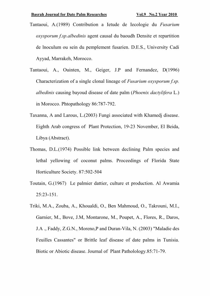
strands occurred. (Fig.1:a,c). Severely affected spathes at an early stage
remain unopened and became dry (Fig1:b) (Al Ani et
al.1971;Djerbi,1983;Abdullah et al.2005).
Diagnosis and detection of the pathogen:
The major cause of inflorescence rot is considered to be the fungus
Mauginiella scaettae Cav. (Cavara,1925; Hussain,1985;Al Ani et
al.1971;Djerbi,1983;Abdullah et al.2005). However, other fungi such as
F.oxysporum , F.moniliforme, F.solani, Trichothecium roseum, Botrytis
aclada, Thielaviopsis paradoxa, Acremonium strictum and Memmoniella
sp., have also been found associated with date palm rotted inflorescences
and considered of minor importance (Brown & Butler,1938; El Behadli et
al.1977; Rattan & A l Dboon,1980; Al Roubaie et al.1987; Al Shraridia &
Shahwan,2003; Taxana & Larous,2003; Abdulah et al.2005).
Mauginiella scaettae can be easily isolated from rotted inflorescence after
surface disinfection of small pieces with 5% sodium hypochlorite solution
and plated on suitable culture media such as malt extract agar, potato
dextrose agar or potato carrot agar. Isolation can be achieved also after
incubation of disinfected pieces in moist chambers and then picking up
conidia which developed abundantly and streak them on a suitable medium.
Inoculated plates should be incubated at 25 C. The fungus grows as white

Vol.9 No.2 Year 2010Basrah Journal for Date Palm Researches
١٤
colonies with immersed and superficial mycelia. Mycelium is composed of
branched hyaline septate hyphae. Colony reverse at first creamy to pale
brown, becoming black in some isolates on potato dextrose agar.
Sporulation are abundant showing powdery appearance . Immersed hyphae
are 2-2.5 um wide ,aerial hyphae measuring 3-4 um wide. Arthroconidia
produced by segmentation of the aerial hyphae ,unicellular, or
multicellular, hyaline , glistening white in mass, non-septate conidia, 6-
8X2.5-4um, septate conidia 6-14 X 3-4um ,2-eptate conidia 16-22 X 3.5-
4um, 3-septate condia 12-26 X 3.5-5um 4-septate condia,24-26 X 3.5-
4.5um and 6-septate conidia up to 35 um long (Fig.1:d) ( Cavara, 1925;
Abdullah et al.2005).
Pathgenicity test can be performed on detached inflorecence free of
disease. Inoculation with spore suspension of the pathogen developed
typical symptoms after 4 days.
Biology and Epidemeology:
The ultrastructure of the cell wall and the hyphal septa, together with the
diazonium blue B test have shown that M. scaettae represents an anamorph
of an unknown ascomycete ( Walt, Van der and Hopsu-Hava, 1976; Arx,
Von et al.1982). Recently Abdullah et al.(2005) showed that sequncing of
the Internal transcribed spacer (ITS) region of this fungus demonstrated
that it is closely related to Phaeosphaeria I.Miyake clade B and in
particular to P. triglochinicola which belongs to subclade B4 according to

Vol.9 No.2 Year 2010Basrah Journal for Date Palm Researches
١٥
Camara et al.(2002). The majority of species of Phaeosphaeria form
pseudoparenchymatous ascomata with bitunicate asci which mainly
occurred on monocotyledonous plants ( Barr,1987; shoemaker &
Babcock,1989).
Al Ani et al. (1971)demonstrated that the pathogen is mainly preserved
as mycelium in infected inflorescence remaining on palms from the
previous season or remain within the infected leaf bases. Al Roubaie et
al.(1987) suggested that the primary infection by M. scaettae probably
occurred during the early stage of floral bud formation and prior to the
envelope development of the spathes and their hardening. The availability
of rain prior to the stage of flower bud formation and during the early stage
of bud formation is probably responsible for creating favorable conditions
for fungal growth ,when hyphae hidden between the leaf bases can grow
and infect newly developed inflorescence (Abdullah et al.2005).
The disease is more serious in hot and humid regions or in areas with
prolonged periods of heavy rains. Hussain and El Baldawy (1977) indicated
that up to 52% of palms might be affected in Al Fao town in Basrah
province, southern Iraq, where high humidity is prevailed , whereas
proportions of the affected trees in the middle Iraq was ranging between
10-20%.
Abdullah et al.(2006) demonstrated that conidia of M.scaettae
germinated best at high % r.h. Maximum percentage of conidial

Vol.9 No.2 Year 2010Basrah Journal for Date Palm Researches
١٦
germination (80.7%) occurred at 95% r.h. and declined sharply (20.8%) at
relative humidity below 95% r.h. and no germination occurred below 80%
r.h. Moreover, obvious increase in sporulation occurred according to the
increase in relative humidity. The highest is being at 100% r.h. and the
lowest occurred at 70% r.h( Abdullah et.al.2006)
It is generally assumed that conidia of M. scaettae are very short lived
and do not persist through the winter. Primary infections are thought to
arise from mycelium (Al Ani et al.1971; Al Hassan & Waleed,1979;
Djerbi,1983). However, Abdullah et al.(2006) have showed in a recent
study that conidia of M. scaettae can survive as a saprophyte in infected
dead inflorescences for a period of more than twelve months and therefore,
these conidia may contribute to the new infection.
Eight isolates were tested for their ability to produce extracellular
enzymes on solid media. All isolates showed positive activity with varying
degrees for cellulase, lipase, protease, phenol oxidase, polygalacturonase
and pectate lyase. In contrast, all isolates gave a negative test for
amylase(Al Saadoon et al.2004).
Control measure:
The first step in the control of inflorescence rot disease achieved by
good management such as leaf pruning and collection and burning of all
infected inflorescences. Application of several fungicides including 3%

Vol.9 No.2 Year 2010Basrah Journal for Date Palm Researches
١٧
dichlone spray or 4% thirame spray at the rate of 8 litres per individual
palm ( Al Hassan et al.1977).
4- Diseases caused by Ceratocystis paradoxa and C. radicicola.
Ceratocystis paradoxa (Dade) C.Moreau ( anamorph: Thielaviopsis
paradoxa (de Seynes) Hohn.), and C.radicicola (Bliss) Moreau (
anamorph: T. punctulata (Hennebert) Paulin, Harrington et McNew, are
two pathogens commonly found either alone or in combination associated
with several disease symptoms on palm trees. These fungi can infect any
part of the palm tree, and symptoms are often expressed as black scorched
leaves, trunk rot, neck bending or inflorescence blight.( Suleman et
al.2001; Djerbi,1983, Zaid et al.2001; Abbas & Abdulla,2003; Abbas et
al.1997; El Gariani et al.2007) . These disease have been observed in the
majority of date growing areas of the world ( Abdullah et al. 2009). The
diseases are more likely to occur on stressed trees especially in areas where
drought and salinity are prevailing . In vivo studies also showed that both
C. paradoxa and C. radicicola colonized palm tissues under drought stress
at -2.3 MPa and had relatively larger necrotic lesions then developed into
cankers , death of buds and eventually plant death (Suleman et al.2001).
In severe cases, the pathogen attacks the terminal bud and heart leading
to the mechanical weakness of the tissues in the uppermost portion of the
trunk resulting in the neck bending. Sometimes the crown rotted off ,
leaving a bare trunk ( Abbas & Abdulla,2003). Some palms recover
Vol.9 No.2 Year 2010Basrah Journal for Date Palm Researches
١٨
probably by the development of a lateral bud initiated from the unaffected
meristematic tissues of the terminal bud. The palms set normal growth back
by several years and that is why it is called in Arabic Medjnoon (fool
disease). (Zaid et al.2003).
The anamorphs of the two pathogens produced an abundance of
endoconidia (Phialoconidia) and Chlamydospores ( aleuroconidia) on
media such as potato dextrose agar, malt extract agar and potato carrot
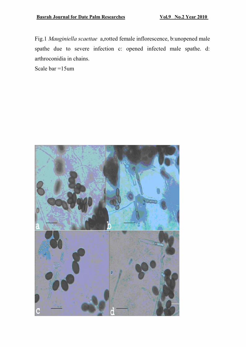
agar. In T. paradoxa anamorph of C.paradoxa , the aleuroconidia borne
terminally in chains from short hyphal branches and are thick-walled ,pale
brown to brownish black, smooth, oval, measuring 10-17 X 5-10um,
phialoconidia are hyaline to pale brown, cylindrical formed endogenously
in uniseriate chain measuring 7-12 X 3-5 um ( Fig.2:a,b).
In T. punctulata anamorph of C. radicicola , the aleuroconidia are borne
singly on a short hyphal branches and are thick-walled, minutely roughened
, pale brown to dark brown, oval, measuring 8-22 X 7-14 um.
Phialoconidia are hyaline to pale brown, cylindrical, formed endogenously
in uniseriate chain measuring 6-12 X 3-5 um ( Fig.2:c,d). The thick-walled
aleuroconidia are likely to play a role as survival propagules of the two
plant pathogens in soil.
Control measure:
The avoidance of wounds on palms grown in the field or nurseries can
limit disease incidence ( Chase & Broschat,1993). The affected fronds,

Vol.9 No.2 Year 2010Basrah Journal for Date Palm Researches
١٩
leaf bases and inflorescencs should be pruned , collected immediately and
burned . The pruning cuts and surrounding tissues should be protected by
spraying with any copper-based fungicides ( Zaid et al.2003).The use of
less saline water for irrigation ( Suleman et al.(2001). In laboratory,
Suleman et al.(2002) assessed the efficacy of the biofungicide Mycostop on
C. radicicola which causes black scorch on date palm in Kuwait. Mycostop
at a rate of 0.35 g /l or greater reduced spore germination , plasmolysed
germlings and reduced sporulations . Roots inoculated with C. radicicola
and then treated with Mycostop were less necrotic than those in untreated
soil.
5- Diplodia leaf-base disease:
The disease is caused by Diplodia phoenicum (Sacc.)H.Facet &
L.J.Klotz. The fungus attacks offshoots while they are still attached to the
mother palm or after their detachment and planted out. The disease was
originally reported from California by Fawcett & Klotz (1932) and then its
distribution covered most of date palm growing regions ( Djerbe, 1983;
Sarhan ,2001; El Deeb et al.2007).
The pathogen may infect the outside leaves of the offshoots while
younger leaves and the buds remain unaffected but finally both of them
killed. Other types of symptoms, started with the infection of the central
young leaves and terminal bud and then gradually infect the outside leaves
and finally leading to the death of the whole plant. On the leaves of the

Vol.9 No.2 Year 2010Basrah Journal for Date Palm Researches
٢٠
older palms, symptoms appeared as yellowish brown streaks ,15 cm to one
meter in length extending along the leaf- base and rachis . The upper part of
the leaf remain unaffected and still appear green. The symptoms appeared
on the ventral surface of the leaf which facing the palm crown. Pycnidial
bodies developed on the dead leaf bases . Pycnispores are at first hyaline
,unicellular becoming dark two-celled with age, measuring 22-24 X 10-12
um. Pycnidia can bee seen after incubation of infected pieces in moist
chamber. Since the infection of the palm takes place through the wounds
made during pruning or cutting when removing the offshoots from the
mother plant, disinfection all tools and cut surface is necessary . In addition
to dipping or spraying the removed offshoots with various fungicides such
as benomyl , bordeaux mixture, or thiram ( Carpenter,1975).
6- Graphiola leaf spot:
The disease is also called false smut on date palm. The causal pathogen
is Graphiola phoenicis (Moug.)Poit. Symptoms of the disease appear as
subepidermal spots on both sides of the pinnae (leaf flat) and on the rachis
with small black sori (fruiting bodies) developing in abundance on old
fronds . The sori are 1-3 mm in diameter, more abundant in the apical
regions of the pinnae . Sori superficially resemble a scale insect but
microscopic examination revealed the presence of powdery yellow spores
on whitish filaments . Spores are spherical to ellipsoidal , 3-6 um in
diameter, with smooth hyaline wall. The disease is widely spread and

Vol.9 No.2 Year 2010Basrah Journal for Date Palm Researches
٢١
occurs whenever the date palm is cultivated under humid conditions but
absent in less humid regions ( Abbas & Abdulla,2004; Djerbe,1983; Zaid et
al.2002; El Deeb et al.2007; El Gariani et al.2007; CAB
international,2003).
Date palm cultivars showed variability in their response to the pathogen.
For example, Barhee, Abdal Rahman, Gizaz showed resistane, while
cultivars Khistawi, Gozi, are tolerance. In contrast cultivars Khisab,
Ashrasi,Maktoom, Zahdi and Bream are very susceptible ( Nixon,1957;
Sinha et al. 1970; zaid et al.2002).
Severe infection reduces tree growth and date production through
premature death of leaves . To ovoid the incidence of the pathogen, leaf
pruning and then burning of the infected leaves should be carried out to
prevent new infection. Spraying the palms after pruning with appropriate
fungicides such as bordeaux mixture, mancozeb, cupric hydroxide and
maneb ( Zaid et al.2002).
7- Belaat disease:
The causal pathogen is phytophthora sp.. The disease is of minor
importance and sporadic. It is known from North African countries (
Calcat,1959; Toatain,1967). Symptoms appear at the crown of the palm.
Young fronds whiten and die ,followed by the infection and death of the
terminal bud and then progression of the infection downwards in the trunk
Vol.9 No.2 Year 2010Basrah Journal for Date Palm Researches
٢٢
as a conical wet heart rot form, releasing an odour of acetic and butyric
fermentation ( Zaid et al.2002).
To ovoid attacks by this fungus , efficient management of date palm
plantation is recommended . To control the disease at its early stage ,
spraying with maneb or bordeaux mixture at the rate of 8 liters /palm is
recommended. Offshoots of the infected palms usually remain free of the
disease( Djerbi,1983).
8- Omphalia root rot:
The disease is caused by two species of Omphalia ( O. tralucida Bliss
and O. pigmentata Bliss). The disease is of minor economic importance to
date palm and it is known from USA (California) and in Muritania (
Fawcett & Klotz,1932; Bliss,1944).
The disease is characterized by the premature death of fronds followed by
the retardation and cessation of the plant growth , and then necrosis and
destruction of the roots . Sachs(1967) recommended the use of brestan or
dexon fungicides at the rate of one spray every two weeks for eight weeks
as a chemical control.
9- Leaf spot diseases:
In general , leaf spot diseases are of minor economic importance .
Different fungal species have been isolated from palm leaves showing leaf
spot symptoms. Leaf spot diseases are very common on date palm trees in
Vol.9 No.2 Year 2010Basrah Journal for Date Palm Researches
٢٣
all date palm growing countries ( Carpenter & Elmer,1978; Fayad &
Mania,2006; El Deeb et al.2007; Livingston et al. 2002).
Generally infection is more severe on the lower whorls and old leaves than
in upper young leaves, and the infection rate and severity is increased with
increasing palm age. Negative correlation between tannin and wax content
in the leaves and severity of infection were recorded ( Fayad &
Mania,2006). Among these diseases , brown leaf spot caused by
Mycosphaerella tassiane ( anamorph: Cladosporium herbarum) is the most
common. Symptoms of the disease occur on the rachis ,pinnae and spines
as dark lesions with well-defined margin on green leaves and on drying
leaves, the margin of the lesion remains reddish brown as the centre
becomes pale.
Other fungi caused leaf spot symptoms on palm trees include Alternaria
alternate, Bipolaris australiensis, Drechslera sp., Helmnthosporium sp.,
Colletotrichum sp., Stemphylium sp., Pestalotiopsis palmarum ,
Chaetosphaeria sp., Phomopsis sp., Phoma spp., ( Livengston et al. 2002;
Fayad and Mania,2006; El Deeb et al.2007, Carpenter & Elmer,1978; El
Gariani et al 2007).
Control measures include annual pruning of old infected leaves and their
immediate burning is recommended ( Zaid et al.2002). At early stage of the
disease, spraying with mancozeb , mancozeb + copper are effectively
control the disease ( Livingston et al.2002).

Vol.9 No.2 Year 2010Basrah Journal for Date Palm Researches
٢٤
10- Disease caused by Phytoplasma.
10.1- Al Wijam
In Arabic Al Wijam means poor fruitful . The disease was observed for
the first time by Nixon (1954) in Al Hassa oasis eastern of Saudia Arabia .
The main symptoms of the disease are leaf stunting with yellow streaking
and a marked reduction in fruit and stalk size. Leaves become choritic and
their life span is reduced. Stunting and yellowing increases with age
leading to the death of the leaves. Diseased spathes are shorter than healthy
one and split open before their complete emergence. Fruits and fruit stalks
showed reduction by 36-40% in the size. Al Hudaib et al.(2007) reported
on the identification and molecular characterization of phytoplasma
associated with Al Wijam in Al Hassa ( Saudia Arabia). The phytoplasma
identified from 28/40 date palm showing typical Al Wijam symptoms
clearly placed in the 16SrI group "Ca.P.asteris" which is supported by the
sequencing and phylogenetic data. Moreover, phylogenetic analysis
showed that the phytoplasma identified in the leafhopper Cicadulina
bipunctata Melichar was 100% identical to that detected from date palm
showing Al Wijam symptoms and accordingly it has been identified as a
putative vector of the disease (Alhudaib et al.2007).
10-2- Leathal yellowing.

Vol.9 No.2 Year 2010Basrah Journal for Date Palm Researches
٢٥
The importance of the disease was first known from USA (Florida) on
coconut palms destroying about 1/2 million coconut palms ( McCoy,1976).
The disease has wide range of hosts including Phoenix dactylifera L., P.
canariensis Hort., and P. reclinata Jacq ( Thomas,1974). Symptoms on
coconut are characterized by early dropping of developing fruits , followed
by formation of new inflorescence which rapidly becomes necrotic, a rapid
and generalized yellowing and eventually the death of the palm. In Kuwait,
Al Awadhi et al.(2002) reported their finding on a phytoplasma associated
with yellowing disease of date palms. The disease displayed similar
symptoms of Al Wijam as expressed on leaves, spathes and bunches of date
palm. In Egypt, Ammar et al. (2005) detected phytoplasma associated with
diseased date palm refereed to it as streaking and yellowing disease.
Harrison et al.(2002) detected phytoplasma belonging to the 16SrIV group,
subgroup D, causing lethal yellowing decline in Canary Island date palm in
Texas.
10-3. White tip die-back.
This is newly recognized disease on young date palms (Phoenix
dactylifera L.). The disease occurs in isolated foci in northern Sudan (
Cronje et al.2000a). Symptoms appeared on 5-8 years old palm trees which
die within 6-12 months of symptoms appearance . Severe chlorosis of the
emerging leaf and at the tip of the pinnae of older fronds which change
quickly from green to dry white without showing yellowing of the crown..

Vol.9 No.2 Year 2010Basrah Journal for Date Palm Researches
٢٦
Using molecular techniques, the causal pathogen has been assigned to
Phytoplasma ( Cronje et al.2000a).
10-4. Slow decline:
The disease is attacking mature date palms along the Nile between
Dongola and Mero-karem , North Sudan. Palm death occurs between 1-2
years after appearance of symptoms and causing losses estimated at 6%.
The symptoms appeared at first as yellowing of the outermost fronds and
progressing towards the young central fronds and newly emerging leaves.
Eventually, all fronds dry white to light brown and are then shed leaving
few young leaves at the top of the trunk which may break off leaving the
trunk alone( Cronje et.al.200b).
The sequence of slow decline Phytoplasma 16S/235rDNA intergenic
spacer showed a very high (99%) homology with comparable sequencies
of Phytoplasma associated with White tip-dieback disease on young date
palm ( Cronje et al.2000b).
11- Brittle leaf disease.
In French the disease is called "Malade des feuilles cassantes". The
disease was first known from southern (desert) parts of Tunisia . According
to Mehani (1958) palms with symptoms of the disease were found since
1960s in Nefta and Tozeur oases. However, only after twenty years the
disease begins to draw attention due to the rapid increase of the effected

Vol.9 No.2 Year 2010Basrah Journal for Date Palm Researches
٢٧
trees particularly in the Nefta oasis ( Tukrouni et al.1988). In Algeria, the
presence of the disease was confirmed in 2006 ( Al saadi et al.2006).
The causal pathogen is not yet determined exactly. The symptoms are
assocated with manganese deficiency and the presence of a small double
strand RNA. However, the effected trees in the field seem to cluster into
foci , suggesting a biotic origin. A possible soil microorganism is
responsible for rendering soil manganese insoluble and unavailable to the
palm trees ( Triki et al.2003).
The early symptoms of the disease appeared on the fronds showing
chlorosis . Leaflets become brittle, twisted, frizzled and shrivled with a
scorched appearance . In severe cases, only frond midribs without leaflets
remain. Affected trees have shorter fronds, stop growing and eventually
die. Four to six years may elapse between the appearance of the first
symptoms and the death of the tree (Tiriki et al.2003).
12- Date Bunch Fading Disorder (DBF).
The date palm bunch fading disease was first reported in 1997 in the
south of Kerman province (Iran). In the last 5 to 6 years, the DBF has been
the most harmful phenomenon on date yields in date palm plantations of
southern Iran. The mean amount of damage at different regions and in
different years has been estimated between 30-50% of the crop (
Karampur,20002).

Vol.9 No.2 Year 2010Basrah Journal for Date Palm Researches
٢٨
Symptoms of this disorder occur at first as light yellow lesions on
peduncles and gradually developing to longitudinal pale brown strips on
the whole peduncle. Date fruits wilt usually from the bottom of the strand
up and then the pedicel, peduncle and whole bunch wilt dry.(
Karampour,1999).
Many fungal species have been isolated from affected date palm trees
showing DBF disorder. These include Alternaria sp., Aspergillus flavus, A.
niger, Penicillium sp., Fusarium sp., Trichoderma sp., and Thielaviopsis
paradoxa. Among the isolated fungi, T. paradoxa had the ability to
increase incidence of DBF disorder on date tree "Mordaseng" under
drought and hot winds stresses in natural climatic conditions of date palm
plantations in Hormozgan province exclusively . Karampour and Pejman
(2007) concluded that the associated fungal agents had no direct and or
primary role in occurrence of BDF disorder
References
Abbas, E.H. and Abdulla, A.S.(2003) First report of neck bending disease
on date palm in Qatar. Plant Pathology 52:790.
Abbas, E.H and Abdulla, A.S.(2004) First report of false smut disease
caused by Graphiola phoenicis on date palm trees in Qatar. New
Disease Reports 1-2.

Vol.9 No.2 Year 2010Basrah Journal for Date Palm Researches
٢٩
Abbas, E.H.,Al Izi, M.J., Aboud, H.M.and Saleh, H.M.(1997) Neck
bending, a new disease affecting date palm in Iraq. Six Arab Congress
of Plant Protection. Beirut, Lebanon (Abstract).
Abdalla, M.Y.,Al Rokiba, A., Moretti, A and Mule, G.(2000) Pathogenicity
of toxigenic Fusarium proliferatum from date palm in Saudia Arabia.
Plant Disease 84:321-324
Abdullah,S.K., Asensio, L., Monfort, E., Gomez- Vidail, S., Salinas, J.,
Lopez- Lorca, L.V., Jansson, H .B.(2009). Incidence of the two date
palm pathogens, Thielavia paradoxa and T. punctulata in soil from
date palm plantations in Elx, South- East Spain. Journal of Plant
Protection Research .49: 276-279.
Abdullah, S.K.,Al Saadoon, A.H.and Al Issa, A.H.(2006) Further
biological study on Mauginiella scaettae , the pathogen of
inflorescence rot disease of date palm. Proceedi ngs of the twelve
Congress of Mediterranean. Phytopathological Union 11-15 June,
Rhodes Island, Greece pp 200-202.
Abdullah, S.K., Asensio, L.,Monfort, E.,Gomez-Vidal, S.,Palma-Guerrero,
J., Salinas.j., Lopez-Lrca, L.V.,Jansson, H-B and Guarro, J.(2005)
Occurrence in Elx ,SE Spain of inflorescence rot disease of date palm
caused by Mauginiella scaettae , Journal of Phytopathology 153:417-
422

Vol.9 No.2 Year 2010Basrah Journal for Date Palm Researches
٣٠
Abu Yaman, I.K.and Abu Blam, H.A.(1971) Major diseases of cultivated
crops in the central province of Saudia Arabia 1:diseases of fruit trees.
Zeitschrift für Pflanzenkrankh 78:607-611.
Al Ani, H.Y., El Behadeli, A.,Majeed, H.A and Majeed, M.(1971) Reaction
of date palm cultivars to inflorescence rot and persistency and
spreading of the disease . Phytopathologia Mediterranea 10:57-62.
Al Awadhi, H.A., Hanif, A., Suleman, P.and Montasser, M.S.(2002)
Molecular and microscopical detection of Phytoplasma associated
with yellowing disease of date palm Phoenix dactlifera L. in Kuwait.
Kuwait Journal of Science & Engineering. 29:87-109.
Al Hassan, K.K.and Waleed, B.K.(1977) Biological study on Mauginiella
scaettae Cav.,the cause of inflorescence rot of date palm in Iraq.
Yearbook of Plant Protection Research, Ministry of Agriculture.
Iraq.1:184-206 (In Arabic)
Al Hassan, K.K.,Abdallah, M.S and Aboud, A.K.(1977) Controlling
inflorescence rot disease of date palm caused by Mauginiella scaettae
Cav., by chemical methods. Yearbook of Plant Protection Research,
Ministry of Agriculture. Iraq. 1:223-236 (In Arabic)
Alhudaib, K.,Arocha.M.,Wilson.M.and Jones,P. (2007) Al-Wijam, a new
Phytoplasma disease of date palm in Saudia Arabia Bulletin of
Insectology 60:285-286
Vol.9 No.2 Year 2010Basrah Journal for Date Palm Researches
٣١
Allison, J.I.(1952) Diseases of economic plants in Iraq. FAO Plant
Protection Bulletin.1:9-11
Al Roubaie, J.J.,Hama, N.N.and Al Hassan, K.k.(1987) Studies on spread
of inflorescence rot and susceptibility of some male palm cultivars to
the disease. Journal of Agriculture and Water Resources. Research.
6:67-79 (In Arabic).
Arx, von,J., Walt, vander J.p.and Liebenberg,N.V.D.w (1982) On
Mauginiella scaettae . Sydowia 34:42-45
Al Saadoon, A.H., Abdullah, S.K.and Al Issa, A.H.(2004) Extracellular
enzymatic activity of Mauginiella scaettae Cav., the causal pathogen
of inflorescence rot disease of date palm. Basrah Journal of Date Palm
Research. 3:1-12
Al Sharidi, A.M and Al Shahwan, I. (2003) Fungi associated with rot
disease of inflorescence and fruits of date palm in Riyadh region ,
Saudia Arabia Eights Arab Congress of Plant Protection. 12-16
October, El Beida, Libya. (Abstract).
Al Yaseri, I.I.,Ismail, A.Z and Mohammed, A.A.(2006) A preliminary
study on spread of date palm pests in IraqNinth Arab Congress of Plant
Protection, 19-23 November, Damascus , Syria (Abstract).
Ammar, M.I.,Amer,M.A and Rashed,M.F.(2005) Detection of Phytoplasma
associated with yellow streak disease of date palms ( Phoenix
dactylifera L.) in Egypt. Egyptian Journal of Virology 2:74-86

Vol.9 No.2 Year 2010Basrah Journal for Date Palm Researches
٣٢
Barr, M.E.(987) Pedramus to class Loculascomycetes . Amherst,
Massachusetts, Hamilton I. Newell.Inc.168pp.
Bliss, D.E.(1944) Omphalia root rot of the date palm. Hilgardia 16:15-124
Bounaga, N.(1975) Germination de microconidies et macroconidies de
Fusarium oxysporum f.sp.albedinis . Bulletin de Ia Sociĕtĕ d’ Histoire
Naturelle d’ Afrique du Nord.60:39-44
Brayford, D.(1992) Fusarium oxysporum f.sp.albedinis IMI Descriptions
of Fungi and Bacteria No.111. CAB International, Wallingford,UK.
Brown, J.G and Butler, K.D.(1938) Inflorescence blight of the date palm.
Journal of .Agriculture.Research.57:313-318
Built, J.,Bouhot,D.,Louvet,J and Toutain,G. (1967) Recherches Sur Ies
fusarioses 1. Travaux sur le bayoudh fusariose vasculaire du palmier
dattier en Afrique du Nord. Annals des Epiphyties 18:213-239
CAB International (2003) Crop protection compendium Wallingford,
CABI Publishing,UK.
Calcat,A.(1959) Diseases and pests of date palm in the Sahara and North
Africa. FAO Plant Protection Bulletin. 8;5-10
Camara, M.P.S.,Palm,M.E, Burkum,P.V and O Neill, N.R.(2002)
Molecular phylogeny of Leptosphaeria and Phaeophaeria . Mycologia
94:630-640

Vol.9 No.2 Year 2010Basrah Journal for Date Palm Researches
٣٣
Carpenter, J.B (1975) Notes on date culture in the Arab Republic of Egypt
and the Peoples Republic of Yemen .Date Growers Institute Report
52:18-24
Carpenter,J.P and Elmer,H.S (1978) Pests and diseases of date palm.
Agriculture Handbook 523 USDA 42pp.
Cavara, F.(1925) Mauginiella scaettae Cav., nuovo ifomicete parassita
della palma da datteri in Cirenacia. Boletin Orto.Botanico Napoli 8:207-
211
Chabrolin, C.(1928) La pourriture de inflorescence du palmier-dattier
Annales des Epiphytica.14:377-414
Chase, A.R and Broschat, T.K.(1993) Diseases and disorders of ornamental
palms. 2nd ed. American Phytopatholgical Society, St. Paul. MN.
Cronje,P.,Dabek,A.J.,Jones,P and Tymon,A.M (2000a) First report of a
Phytoplasma associated with a disease of date palm n North Africa .
New Disease Report 49.801
Cronje,P.,Dabek,A.J.,Jones,P and Tymon,A.M (2000b) Slow decline: a
new disease o mature date palms in North Africa assocatd with a
Phytoplasma. New Disease Report 49:804
Daayf, F.,El Bellaj, M.,El Hassni, M.,Jaiti,F and El Hadrami, I.(2003)
Elicitation of soluble phenolic in date palm (Phoenix dactylifera l.)
callus by Fusarium oxysporum f.sp.albedinis culture medium.
Environmental Experimental Botany. 49:41-47
Vol.9 No.2 Year 2010Basrah Journal for Date Palm Researches
٣٤
Djerbi, M.(1982) Bayoudh disease in North Africa, history, distribution,
diagnosis and control. Date Palm Jounal 1:153-197
Djerbi, M.(1983) Diseases of the date palm (Phoenix dactylifera) Regional
Project for Palm and Dates Research Centre in Near East and North
Africa .Baghdad, Iraq, FAO.
Djerbi, M., El Ghorfi, A.and El Idrissi, A.M.(1985) Etude du
comportement du hennĕ Lawsonia inermis et de la luzerne Medicago
sativa et quelques especes de palmacĕes vis-à-vis Fusarum oxysporum
f.sp. albedinis agent causal du bayoudh. Annales de l Institut National
de la Recherche Agronomique de Tunisie 58:1-11
Djerbi,M., Aouada, L., Filali, H.,Saaidi, M, Chtioui, A., Sedra, M.H.,
Aliaoui, M., Hamdaoui, T and Oubrich, A.(1986) Preliminary results of
selection of high quality bayoudh resistant clones among natural date
Palm population in Morocco. Second Symposium on the date palm , 3-6
March, Saudia Arabia pp 383-399
El Behadili, A.H., Mawlood.,K.A. and Diwan.,M.M.(1977) A new
pathogen causing inflorescence rot of date palm in Iraq. Fourth Iraqi
Biological Society Conference. 20-25 September, Baghdad (Abstract).
El Deeb, H.M., Lashin,S.M and Arab, Y.A.(2007) Distribution and
pathogenesis of date palm fungi in Egypt. Acta Horticulturae. 736:421-
429
Vol.9 No.2 Year 2010Basrah Journal for Date Palm Researches
٣٥
El Gariani, N.k., El Rayani, A.M and Edongali, E.A.(2007) Distribution of
phytopathogenic fungi on the coastal region of Libya and their
relationships with date cultivars. Acta Horticulturae. 736:449-455
El Hadrami, I., Ramos, T and Macheix, J.J.(1996) Caracterisation de
nouveaux derives hydroxycinnamiques amines chez Phoenix dactylifera
L. relation avec le brunissement des tssue et ln resistance des cultivars
au bayoud. Polyphenols communications 2:341 342
El Hassni, M., El Hdrami., A., Daayf, F., Ait Birka, E and El Hadrami,
I.(2004a) Chitosan , antifungal product against Fusarium oxysporum
f.sp.albedinis and elicitor of defense reactions in date palm roots.
Phytopathologia Mediterranea.43:195-204
El Hassni, M., Jaiti.,F., Dihazi.,A., Ait Birka , E., Daayf., F and El
Adrami.I.(2004b)
The enhancement of defense responses against Bayoud disease by
treatment of date palm seedlings with an hypoaggressive Fusarium
oxysporum isolate. Journal of Phytopathology 152:182-189
El Hassni, M.,El Hadrami, A.,Daayf, F.,Cherif, M., Ait Birka,E and El
Hadrami.,I.(2007) Biological control of Bayoud disease in date palm:
Selection of microorganisms inhibiting the causal agent and inducing
defense reactions. Environmental Experimental Botany. 59:224-234
El Modafar, C.and El Boustani, E.S.(2002) Mechanisms of date palm
defense to bayoud disease. Date Palm News 1:1-3
Vol.9 No.2 Year 2010Basrah Journal for Date Palm Researches
٣٦
El Modafar, C., Tantaoui, A and El Boustani, E.(2000) Effect of
cafeoylshikimiques of date palm roots on activity and production of
Fusarium oxysporum f.sp. albedinis cell wall degrading-
enzymes.Journal of Phytopathology 148:101-108
Fawcett, H.S.and Klotz, L.T.(1932) Diseases of date palm. California
Agriculture Expteriental Station Bulletin. 522, 47pp.
Fayad, M.A and Mania, A.O (2006) Study of date palm leaf spot disease in
Basrah, Iraq and the relation of age of palm and wax content with
infection Ninth Arab Congress of Plant Protection. 19-23 December,
Damascus, Syria (Abstract).
Fernandez, D.,Quinten,M., Tantaui, A ., Geiger, J.,Bahoussi,M and
Langin,T.(1998) Fot 1 Insertions in the Fusarium oxysporum
f.sp.albedinis genome provide diagnostic PCR targets for detection of
the date palm pathogen. Applied and Environmental.
Microbiology.64:633-636
Frederix, M.J.J and Den Brader, K.(1989) Resultats des essais de
desinfecton des sols contenant des chantillons de Fusarium oxysprum
f.sp. albedinis FAO/PNUD/RAB/88?024 Ghardaa, Algeria
Harrison, N.A, Womack, M and Carpio, M.L.(2002) Detection and
characterization of lethal yellowing (16SrIV) group Phytoplasma in
Canary Islnd date palms affected by lethal decline in Texas. Plant
Disease 86:676-681

Vol.9 No.2 Year 2010Basrah Journal for Date Palm Researches
٣٧
Hussain, F.(1958) Occurrence of date palm inflorescence rot in Iraq. Plant
Disease Report.42:535
Hussain, F and El Baldawy, A.(1977) studies on inflorescence rot disease
of date palm and is control. Yearbook of Plant Protection Research
Ministry of Agriculture.Iraq.(In Arabic). 1:207-222
Karampour, F.(1999) Report of wilting and defoliation of date fruits in
Boushehr province. Agricultural Research Centre of Boushehr, Iran
22pp.
Karampour, F.(2002) A review on abstracts of plant protection research
results on DBF disorder in Iran, Agricultural Research Centre of
Hormozgan province, Bandar Abbas,Iran 11pp.
Karampour, F and Pejman, H.(2007) Study on possible influence of
pathogenic fungi on date bunch fading disorder in Iran . Acta
Horticulturae.736:431-437.
Killian, C and Maire, R (1930) Le bayoud maladie du dattier. Bulletin of
African Natural History Society..No. 21:89-101
Klotz, L.J (1831) Investigations on date palm diseases. Annals of Date
Growers Institute. 8:14-18
Livengston, S., Al Mufargi, K and Al Suhkeli, M(2002) Chemical control
of leaf spot of date palm (Phoenix dactylifera ) in Sultanate of Oman .
Plant Pathology Journal. 18:163-167.

Vol.9 No.2 Year 2010Basrah Journal for Date Palm Researches
٣٨
Louvet, J.(1977) Observations sur la localisation des chlamydospores de
Fusarium oxysporum das les tissue des plantes parasitees. Travaux
Dĕdiĕs aَG. Viennot Bourgin pp 193-197, INRA Paris France.
Louvet, J.,Burlt, J., Toutain, G and Rieuf, P.(1970) Le bayoudh fusariose
vasculaire du palmier dattie, symptomes et nature de la maladie moyens
de lutte . Al-Awamia 35:161-182.
Mansoori, B and Kord, M.H.(2006) Yellow death: A disease of date palm
in Iran caused by Fusarium solani. Journal of Phytopathology 154:125-
127
Mc Coy, R.E.(1976) Comparative epidemiology of the lethal yellowing.
Kaincop and cadang-cadang disease of coconut palm. Plant Disease
Report. 60:498-502.
Mehani, S (1988) Compte rendu de mission de consultation aupres de la
direction generale de la production vegetale, Ministere de I Agriculture,
Tunis.
Michael, I.F and Sabet, K.A.(1970) Biological control of Mauginiella
scaettae Cav., the pathogen of Khamedj dsease in United Arab
Republic. Annals of Date Growers Institute.47:5-8.
Munier,P (1955) Le Palmier en Mauritanie. Annals Institute Fruits et
Agromes Coloniaux 12:66.
Nixon, R.W (1954) Date culture in Saudia Arabia. Annals of Date Growers
Institute.31:15-20.
Vol.9 No.2 Year 2010Basrah Journal for Date Palm Researches
٣٩
Nixon, R.W (1957) Differences among varieties of the date palm in
tolerance to Graphiola leaf spot. Plant Diseae Report. 41:1026-1028
Ouhdouch, Y., Boussaid, A and Finance, C. (1996) Akitasatosporia strain
with non-polyenic activity against the agent of date palm vascular wilt.
Actinomyetes 7:1-3.
Plyler, T.R., Simone, G.W., Fernandez, D and Kistler, H.C.(1999) Rapid
detection of the Fusarium oxysporum lineage containing the Canary
Island date palm wilt pathogen. Phytopathology 89:407-413.
Quenzan, B., Trifi, M., Bouachrine, B., Hartman, C., Marrakichi, M.,
Benslmani, A.A and Rode, A. (2001) A mitochondrial molecular marker
of resistance to bayoud disease in date palm. TAG Theoretical and
Applied Genetics 103:366-370.
Rashed, M.F and Abdel Hafeez, N.E.(2001) Decline of date palm trees in
Egypt. Second International Conference of Date Palm, 25-27 March Al
Ain, UAE, pp 401-407.
Rattan, S.S and Dboon, A.H.A (1980) Notes on fungi associated with date
palm I. Sydowia 32:246-273.
Saaidi, I, Namsi.,A., Ben Mahmoud, O., Takrouni, M.I.,Zouba, A.,Bove,.M
and Duran-Vila,N. (2006) First report of "Maladie des feuilles
Cassantes" (brittle leaf disease) of date palm in Algeria. New Disease
Reports 13.

Vol.9 No.2 Year 2010Basrah Journal for Date Palm Researches
٤٠
Sachs, G.(1967) Sur la presence d Omphalia sp. Bliss dans une palmeraie
Muritanieene.
Fruits 22:497-501.
Sarhan, A.R.T(2001) A study on the fungi causing decline of date palm
trees in middle of Iraq Second International Conference of Date Palm,
25-27 March, Al Ain, UAE pp 424-430.
Shoemaker, R.A and Babcock, C.E.(1989) Phaeosphaeria . Canadian
Journal of Botany. 67:1500-1599.
Sinha, M.K.,Singh, R and Jeyarajan, R.(1970) Graphiola leaf spot on date
palm (Phoenix dactylifera L.) susceptibility of date varieties effect on
chlorophyll content. Plant Disease Reports. 54:617-619
Suleman, P., Al Musallam, A and Menezes, C.A.(2001) The effect of
solute potential and water stress on black scorch caused by Chalara
paradoxa and Chalara radicicola on date palms. Plant Disease.85:80-83
Suleman, P., Al Musallam, A and Menzes, C.A.(2002) The effect of
biofungicide Mycostop on Ceratocystis radicicola, the causal agent of
black scorch on date palm . Bio Control 47:207-216
Takrouni, L., Rhouma, A.,Khoualdi, O and Allouchi, B.(1988)
Observations sur dues graves maladies d orgine inconnue du palmier
dattier en Tunisie. Annales de I Institut National de la Recherche
Agronomique de Tunisie 61:3-14.
Vol.9 No.2 Year 2010Basrah Journal for Date Palm Researches
٤١
Tantaoui, A.(1989) Contribution a Ietude de Iecologie du Fusarium
oxysporum f.sp.albedinis agent causal du baoudh Densite et repartition
de lnoculum ou sein du pemplement fusarien. D.E.S., University Cadi
Ayyad, Marrakeh, Morocco.
Tantaoui, A., Ouinten, M., Geiger, J.P and Fernandez, D(1996)
Characterization of a single clonal lineage of Fusarium oxysporum f.sp.
albedinis causing bayoud disease of date palm (Phoenix dactylifera L.)
in Morocco. Phtopathology 86:787-792.
Taxanna, A and Larous, L.(2003) Fungi associated with Khamedj disease.
Eighth Arab congress of Plant Protection, 19-23 November, El Beida,
Libya (Abstract).
Thomas, D.L.(1974) Possible link between declining Palm species and
lethal yellowing of coconut palms. Proceedings of Florida State
Horticulture Society. 87:502-504
Toutain, G.(1967) Le palmier dattier, culture et production. Al Awamia
25:23-151.
Triki, M.A., Zouba, A., Khoualdi, O., Ben Mahmoud, O., Takrouni, M.I.,
Garnier, M., Bove, J.M, Montarone, M., Poupet, A., Flores, R., Daros,
J.A ., Faddy, Z.G.N., Moreno,P and Duran-Vila, N. (2003) "Maladie des
Feuilles Cassantes" or Brittle leaf disease of date palms in Tunisia.
Biotic or Abiotic disease. Journal of Plant Patholology.85:71-79.

Vol.9 No.2 Year 2010Basrah Journal for Date Palm Researches
٤٢
Walt, Van der,J.W and Hopsu-Hauva,n V.K. (1976) A color reaction for
differentiation of ascomycetous and basidiomycetous yeasts . Antonie
Van Leuwehoek 42:157-163
Watson, A.G.(1974) Pathogenicity test for identification of Fusarium
oxysporum f.sp.albedinis . Bulletin d Agronomie Saharienne 1:37-38.
Zaid, A., de Wet, P.F., Djerbi, M and Oihabi, A.C.(2002) Diseases and
pests of date palm. In Date Palm Cltivation .FAO plant production and
protection paper 156 (eds.) Zaid, A and Arias-Jimenez, E.pp227-281.
Vol.9 No.2 Year 2010Basrah Journal for Date Palm Researches
٤٣
Fig.1 Mauginiella scaettae a,rotted female inflorescence, b:unopened male
spathe due to severe infection c: opened infected male spathe. d:
arthroconidia in chains.
Scale bar =15um

Vol.9 No.2 Year 2010Basrah Journal for Date Palm Researches
٤٤
Fig.2. Ceratocysts paradoxa ( anamorph: Thielaviopsis paradoxa) a,b:
aleuroconidia(chlamydospores) and phialoconidia (endoconidia)
Ceratocystis radicicola ( anamorph: Thielaviopsis punctulata) c,d.aleuroconidia and phialoconidia. Scale bar =20um