direct identification of hazardous elements in ultra-fine and nanominerals from coal fly ash...
TRANSCRIPT

Science of the Total Environment 470–471 (2014) 444–452
Contents lists available at ScienceDirect
Science of the Total Environment
j ourna l homepage: www.e lsev ie r .com/ locate /sc i totenv
Direct identification of hazardous elements in ultra-fine andnanominerals from coal fly ash produced during diesel co-firing
Kátia Martinello a, Marcos L.S. Oliveira a,b, Fernando A. Molossi b, Claudete G. Ramos b, Elba C. Teixeira c,Rubens M. Kautzmann b, Luis F.O. Silva a,b,⁎a Environmental Science and Nanotechnology Department, Institute of Environmental Research and Human Development, IPADHC, Capivari de Baixo, Santa Catarina, Brazilb Laboratory of Environmental Researches and Nanotechnology Development, Centro Universitário La Salle, Mestrado em Avaliação de Impactos Ambientais, Victor Barreto,2288 Centro 92010–000, Canoas, RS, Brazilc Fundação Estadual de Proteção Ambiental Henrique Luis Roessler/RS, Fundação Estadual de Proteção Ambiental Henrique Luis Roessler-RS, Rua Carlos Chagas 55/802,Centro 90030–020 – Porto Alegre, RS, Brazil
H I G H L I G H T S G R A P H I C A L A B S T R A C T
• Environmental and human health riskassessment was performed.
• Further exposition risk assessment re-quired if future expansion is pursued.
• The advanced methodology has beenapplied to investigate elements occur-rence and nanomineralogy properties.
⁎ Corresponding author at: Laboratory of EnvironImpactos Ambientais, Victor Barreto, 2288 Centro 92
E-mail address: [email protected] (L.F.O. Silv
0048-9697/$ – see front matter © 2013 Elsevier B.V. All rihttp://dx.doi.org/10.1016/j.scitotenv.2013.10.007
a b s t r a c t
a r t i c l e i n f oArticle history:Received 28 August 2013Received in revised form 29 September 2013Accepted 1 October 2013Available online 22 October 2013
Editor: Pavlos Kassomenos
Keywords:Fly ashHazardous elementsNanomineralsHuman health exposure
This study has provided an initial assessment of the environmental impacts and potential health effects associatedwith coal fly ash produced during diesel co-firing.Many hazardous elements that are typically detected bymultifac-eted chemical characterization by XRD, petrology, FE-SEM/EDS, and HR-TEM/SEAD/FFT/EDS in ultra-fine com-pounds and nanominerals from the co-fired coal fly ashes (CFAs). It provided an in-depth understanding of coalash produced during diesel co-firing. Several of the neoformed ultra-fine compounds and nano-minerals found inthe coal ashes are the same as those commonly associated with oxidation/transformation of aluminosilicates,carbonates, sulphides and phosphates.
© 2013 Elsevier B.V. All rights reserved.
mental Researches and Nanotechnology Development, Centro Universitário La Salle, Mestrado em Avaliação de010–000, Canoas, RS, Brazil. Tel./fax: +55 31 655545013.a).
ghts reserved.

445K. Martinello et al. / Science of the Total Environment 470–471 (2014) 444–452
1. Introduction
Approximately 11% of the electricity generated in Brazil is producedby seven coal-fired power plants in the states of Rio Grande do Sul,Santa Catarina, and Paraná (ANEEL, 2006). These plants produce 3 Mtof ashes per year, which consist of 65–85% fly ash and 15–35% bottomash (Levandowski and Kalkreuth, 2009). In addition, the existing coal-fired energy park in Brazil is expected to increase with another fivepower plants. When fully operational, this new scenario will triplicatethe current coal ash production to around 12 Mt per year (Rohde andSilva, 2006).
Ultra-fine particulate matter emitted from coal power plants maycontain potentially harmful constituents that include a wide range ofhazardous elements and metalloids, polycyclic aromatic hydrocar-bons (PAHs), amorphous silica, fluorides, elemental carbon and isknown to impact adversely on environmental quality and humanhealth (Finkelman, 2004; Ando et al., 1998; Quispe et al., 2012;Silva et al., 2011a, 2011b, 2011c, 2011d). Many of these particlesare ultrafine (b100 nm) and nanometric (b50 nm) in size, thuswith an enhanced potential for easy entry into the human body viathe skin, digestive system or respiratory tract (Hochella et al.,2008; Nowack and Buchelli, 2007; Fubini and Fenoglio, 2007; Hoetet al., 2004). Given the growing interest in the toxicology of ultrafineparticles, especially those containing hazardous elements (Ispaset al., 2009; Brian et al., 2009; Meng et al., 2007; Khan et al., 2007;Chen et al., 2006, 2005), this study reports on the direct observationof metal-bearing CFA ultrafine and nano particles using advancedelectron beam.
Coal fly ash is typically polymineralic and chemically complex.The distribution, mode of occurrence and concentration of elementsin the ash particles are crucial in determining their bioreactivity andbiodurability. Potentially toxic trace elements homogeneously dis-seminated as impurities hosted in insoluble minerals pose a lowerhealth risk than occurring as major constituents in individual,metal-rich ultrafine or nanoscale particles. Ultra-fine and nano-particles are known to have a greater reactivity compared tolarger-size particles of the same composition due to an increasedsurface-to-volume ratio (Navrotsky, 2001). For example, in particlessmaller than 20nm in diameter, at least 40% of the atoms are presenton the surface of the nanoparticle (Banfield and Navrotsky, 2001).The effects of this reactivity in ultrafine and nanoscale materialsare more likely as distances between functional groups and facetson nanoparticle surface decrease and quantum effects become im-portant (Auffan et al., 2008). To date, such nanoscale effects andtheir influence on metal absorption or complexation as a functionof particle size have not been studied in detail.
With the growing concern about the impacts of exposure to hazard-ous elements, there is a need for a better understanding of the relation-ship between geochemistry, nanomineralogy and the effects associatedwith both short-term and chronic inhalation exposure to coal ash.Therefore, we report on hazardous elements present in ultrafine andnanoparticles sourced from CFA, with emphasis on Al, Cr, Fe, Ni, Ti, Zr,which are the most abundant in the studied samples. Such metal-richultrafine and nanoparticles are very common in CFA and, given thefact that around 40% of global electricity is coal-generated (IEA, 2005),they are produced in large quantities (Silva et al., 2009a; Ruhl et al.,2009; Hower et al., 2008; Giere et al., 2006; Chen et al., 2004). Anestimated 780 Mt of coal ash are produced every year (WWCCPN,2012). Typically risk assessment methodologies have been widelyapplied for the management of contaminated sites. However, theirapplication to element and coal power plants sites affected by tailingcontamination is more limited (Moreno-Jimenez et al., 2011; Quispeet al., 2012). In a first-tier approach, an ecological risk assessment isbased on geochemical analysis, where concentrations measured in soilare compared to established thresholds. These accepted levels arebased on the worst possible scenarios: toxicity data on the most
sensitive species and the application of the most protective safetyfactors. Under these assumptions, the risk may be overestimated andmay result in unnecessary remediation (Ollson et al., 2009). A riskassessment based on geochemical analyses is highly simplified anddoes not take into account factors such as the bioavailability of a contam-inant or the simultaneous presence of different contaminants, which canaffect toxicity and exposure estimates (De Zwart and Posthuma, 2005;O'Halloran, 2006). These facts are of the utmost importance for toxic haz-ardous elements. The availability of hazardous elements in the soil ishighly dependent on CFA properties. Moreover, hazardous elementscan be present in different geochemical forms with different geochemi-cal and toxicological properties that can be transformed from one intoanother due to environmental changes. To have a more complete im-pression of the degree of pollution and toxicity of a contaminated site,it is recommended to consider alternative approaches that more accu-rately reflect specific site conditions. Direct toxicity assessment, conduct-ed with CFA samples taken at the site, allows the measurement of thetoxicity of complex mixtures of contaminants and can enhance the real-ism and certainty of the risk assessment. Although these techniques havean increasingly important role, they are not generally available inexisting guidelines for ecological risk assessment on contaminatedsites. Therefore, the ecotoxicological risk assessments based on di-rect toxicity assays need to be validated trough field studies performedwith natural samples.
The fact that coal-fired power plants can be a source of airborne par-ticulatematterwith elevated concentrations of toxic hazardous elementsis well established in areas with coal generation plants (e.g. Iordanidiset al., 2008; Bhanarkar et al., 2008). Coal combustion ash has particulesizes ranging from less than 1 μm to 100 micrometers in size (Teixeiraet al., 1992; Blaha et al., 2008), with the inhalable sizes (b10 μm) beingof most direct concern to human health. The most readily bioavailableparticles of allwill be those in the sub-micronic, ultrafine andnanometricsize-ranges, although given the difficulty involved in their study relative-ly few publications have so far specifically characterized such materials.
In thisworkwe aim to provide amulti-analytical approach to under-stand the implications of coal ash materials. A key constituent of thisstudy was the identification and analysis of nano-particles in the differ-ent CFA, an area of researchwith potentially great environmental signif-icance but about which very little is currently known. At the time ofsampling, two of the units were being co-fired with a mixture of fueloil and diesel oil as part of the boiler start-up procedure. The opportuni-ty was, therefore, taken to investigate the influence of oil co-firing onthe resulting coal combustion products. Little attention has been paidto the effects of co-firing with oil during start-up, even though this pro-cess may also have a significant impact on fly ash characteristics(Navarrette et al., 2004).
2. Methods
2.1. Sampling
Fly ash samples were obtained from the largest coal-fired powerplant in Brazil, located in Santa Catarina State, which generates857MW/h (Quispe et al., 2012; Oliveira et al., 2012). The incinerationtemperature in the combustion chamber is ca. 1000–1500 °C, andabout 98.5% of the fly ash is captured in the electrostatic precipitators(ESP).
A total of 7 CFA samples were collected from the ESP over a five-dayperiod. The CFAs from Units 3 and 4 (Samples FA15 and FA16) weretaken while oil was co-fired with coal as a means of starting up therespective boiler systems. Representative samples of around 15–20 kgof fly ash were collected from each unit. Samples of fresh dry fly ashwere also collected from the current station output immediately priorto emplacement at the dry disposal site. The CFA characteristics havechanged little over time due to the essentially constant geologicalsource of the coal supplied (Depoi et al., 2008; Silva et al., 2009a,b).

446 K. Martinello et al. / Science of the Total Environment 470–471 (2014) 444–452
2.2. Analytical procedures
The crystalline mineralogical composition of fly ashes was deter-mined by means of a Siemens model D5005 X-ray diffraction (XRD).Prior to the characterisation of CFAs samples, it is important toknow, not only what kind of information a specific technique canprovide (e.g. size distribution, elemental information, sensitivity,structural information etc.), but also the requirements of the sam-ple (size range, elemental composition etc.) for each method tomake analysis possible and to guarantee meaningful results (Tiedeet al., 2009). Each technique has advantages and disadvantages,but only Field Emission Scanning Electron Microscope (FE-SEM)and high-resolution transmission electron microscope (HR-TEM)currently allow the direct (real space) visualization of nanoparti-cles. In this investigation, morphology, structure, and compositionof ultrafine minerals were investigated using a FE-SEM ZeissModel ULTRA plus with charge compensation for all applicationson conductive as well as non-conductive samples and a 200 keVJEOL-2010F HR-TEM equipped with an Oxford energy dispersiveX-ray detector, and a scanning (STEM) unit (Silva et al., 2009a;Hower et al., 2008). The FE-SEM was equipped with an energy-dispersive X-ray spectrometer (SEM-EDX) and the mineral identifi-cations were made on the basis of morphology and grain composi-tion using both secondary electron and back-scattered electronmodes. Geometrical aberrations were measured by HR-TEM andcontrolled to provide less than a π/4 phase shift of the incomingelectron wave over the probe-defining aperture of 14.5 mrad. Thescanning acquisition was synchronized to the ac electrical powerto minimize 60Hz noise, and a pixel dwell time of 32μs was chosen.EDS spectra were recorded in TEM image mode and then quantifiedusing ES Vision software that uses the thin foil method to convert X-ray counts of each element into atomic or weight percentages. Elec-tron diffraction patterns of the crystalline phases were recorded inSAED (selected area electron diffraction) or MBD (microbeamdiffraction) mode, and the d spacings were compared to theInternational Center for Diffraction Data (ICDD, 2009) inorganiccompound powder diffraction file (PDF) database to identify thecrystalline phases.
Mineralogy sample preparation involved the use of ultrapureacetone and/or hexane to disperse inorganic powders, a commonlyused technique for HR-TEM sample preparation (Chen et al., 2004).Different suspensions, namely hexane, acetone, dichloromethane andmethanol were selected to prevent possible mineralogical changes inindividual solvents. The suspension dissolves this “binder” materialand breaks up aggregates to provide physically separated individualparticles amenable for electron microscopes analysis. The suspensionwas pipetted onto lacycarbon films supported by Cu grids and left toevaporate before inserting the sample into the SEM and TEM. Thismethod may have led to agglomeration but is a widely used standardprocedure because most minerals, including metal sulphates (Giereet al., 2006). Before FE-SEM and STEM analysis, the TEM specimenholder was cleaned with a Advanced Plasma System (Gatan Model950) to minimize contamination. A drift correction system was usedfor the STEM-EDS mapping.
All sampleswere acid digested following a two-step digestionmeth-od devised to retain volatile elements in coal dissolution (Querol et al.,1997); this consists of a HNO3 hot extract followed by a HF: HNO3:HClO4 acid digestion of the residue. The resulting solution was thenanalyzed by Inductively Coupled Plasma Atomic-Emission Spectrome-try (ICP-AES) for major and selected trace elements, and by InductivelyCoupled Plasma Mass Spectrometry (ICP-MS) for most trace elements(Querol et al., 2008). The digestion of international reference materials(SARM-19) and blanks was prepared following the same procedure.Analytical errors were estimated at b3% for most of the elements andaround 10% for Cd, Mo, and P. In addition, Hg analyses were madedirectly on solid samples using a LECO AMA 254 gold amalgam atomic
absorption spectrometer and total organic carbon, S and N contentswere determined using LECO CHNS-932.
3. Results and discussion
3.1. Distribution of elements
Table 1 summarizes the concentration of major and trace elementcontents in the studied fly ashes samples. The high concentrations oftrace hazardous elements reported in this study for the bulk coal ash(Table 1) are expected to magnify, as fine fractions of CFA (which maybe resuspended and deposited in the human respiratory system) areoften enriched in hazardous elements.
The electron beam analysis revealed that the carbon content de-creases with a decrease in the particle size, as seen in the electronbeam analysis. Concomitant with the decrease in carbon is an in-crease in the glass content. The trace elements listed in Table 1 arereported on the ash basis (eliminating the C from the fly ash),with the exception of Hg. Most of the elements that are capturedby the CFA (especially samples CFA6 and CFA7), are associatedwith the carbon/char content. A large number of elements such asAs, Be, Cd, Cr, Cu, Ga, Hg, Mo, Pb, Sb, Sn, Sr, U, W and Zn are notenriched in the ESP fly ash (samples CFA1; CFA2; CFA3; CFA4), butare highly enriched in the baghouse fly ashes (CFA6 AND CFA7).The elements could be present as a condensate on the surface ofthe aluminosilicate fly ash particles. Hower et al. (2008) notedthat As, Hg, Se, among other elements, were present in 5-nmmetal entities within a fullerene-like carbon in a CFA from a pulver-ized fuel-boiler burning an Eastern Kentucky coal, demonstratingthe potential for such associations in CFAs from low- to medium-Cand S coals. In the present study As, Ga, Ge, Hg, Mo, Pb, Se, Sn, Tl,V, W, among other elements and Zn compounds may be emittedto the environment during coal combustion processes, althoughsome compounds are retained in the fly ashes, in different propor-tions depending on the characteristics of the ashes and process con-ditions (Table 1). There is a great potential for improving theoperational conditions which would optimize this retention and assessthe possibility of reusing fly ashes as adsorbents of hazardous elementsand organic compounds with hormones and agrochemicals.
3.2. Nanomineralogy
All CFAs have a high glass andmullite contents. This is known to en-hance concrete workability and durability (Chávez-Valdez et al., 2011)and it is for this reason that these CFAs have been widely used as ahigh-performance substitute for portland cement and as a clinker addi-tion in the manufacturing of portland cement in Brazil. In addition, theoccurrence of great contents ofmullite is particularly promising becauseof its high thermal stability, low thermal expansion, high creep resis-tance in highly oxidative and corrosive environments, high resistanceto crack propagation and high thermal shock resistance (Schneideret al., 2008). However, the present situation is far from full utilizationand large amounts of fly ash are either stored temporarily in stockpilesor disposed of. Therefore further applications need to be developed inorder to recycle them.
Results presented in this work explain the potential elementssuch as As, Cd, Cr, Pb, Mo, and Mn were detected in several ultrafineagglomerates of nanomagnetite, nanoschwertmannite aglomerates,nanoferrihydrite, nanohematite from CFA Brazilian samples. Thispoint improves our understanding of the surface reactivity of ironand salts minerals from a mechanistic point and providing opportu-nities for training young investigators in the latest advances inrelevant techniques, such as HR-TEM and FE-SEM to provides imag-ing capabilities for evaluating changes in particle morphology andestimating particle sizes. This pioneer Brazilian study is the first di-rect observation of these phases as on nanominerals in this coal

Table 1Concentration () of major and trace elements in coal fly ash. Values reported on a dry bulk basis.
CFA 01 CFA 02 CFA 03 CFA 04 CFA 05 CFA 06 CFA 07
%Al 13.00 12.56 15.08 15.90 14.93 13.46 16.51C 0.92 5.24 3.22 1.26 0.53 15.77 15.98Ca 1.51 1.12 1.16 1.27 1.09 0.98 1.12Fe 5.93 3.87 3.60 4.17 4.20 2.88 3.36K 1.92 2.23 2.58 2.53 2.32 2.27 2.74Mg 0.55 0.47 0.54 0.56 0.50 0.48 0.54N b0.003 b0.003 b0.003 b0.003 b0.003 0.11 0.12Na 0.30 0.37 0.41 0.37 0.30 0.31 0.38O 0.40 0.28 0.37 0.25 0.22 1.22 2.03P 0.03 0.03 0.04 0.03 0.04 0.07 0.09S 0.07 0.05 0.11 0.04 0.03 0.54 0.63Ti 0.63 0.63 0.74 0.75 0.75 0.69 0.83
mg/kgAs 2.1 10.0 24.4 15.0 19.0 107.4 139.0Ba 513.5 427.0 485.1 473.9 568.7 586.4 729.1Be 10.3 7.9 9.5 10.1 9.5 10.9 12.6Cd b1 b1 1.2 1.1 1.1 2.8 3.4Ce 177.2 168.1 194.5 197.7 188.2 181.5 194.0Co 19.3 15.0 15.2 16.9 17.9 15.5 17.7Cr 106.4 105.3 130.5 122.8 133.7 146.9 161.1Cu 47.1 38.5 43.2 41.1 42.8 46.6 54.8Dy 13.0 10.6 12.8 12.6 12.8 13.1 14.3Ga 24.0 30.4 42.6 34.5 39.7 81.7 110.9Gd 16.2 14.2 16.6 16.5 16.2 15.9 17.1Ge 16.1 14.7 21.4 18.7 22.5 66.4 85.2Hg 0.00062 0.00044 000071 0.00042 000037 0.0014 0.6817La 67.1 63.6 73.6 73.9 70.8 69.6 75.0Li 85.3 79.1 93.1 90.3 90.9 89.3 104.6Mn 554.2 307.3 314.2 348.5 334.3 281.2 326.5Mo 5.8 5.3 6.9 6.2 7.6 16.7 19.6Nb 112.3 113.2 131.6 124.5 133.4 123.5 126.3Nd 74.7 68.3 79.3 80.0 77.0 74.7 79.5Ni 44.3 35.0 35.9 38.8 37.9 39.2 43.6Pb 34.4 43.2 64.3 51.9 59.1 128.4 161.0Sb 2.5 2.1 4.8 2.3 2.5 5.6 7.1Se 7.6 5.2 5.4 7.0 5.1 21.9 18.6Sr 113.4 132.0 158.7 136.8 141.2 183.6 200.3Th 39.7 33.5 39.9 42.0 38.9 38.0 41.3Tl 3.3 4.2 5.4 4.6 4.8 8.3 9.7U 17.2 14.5 17.5 15.8 16.3 21.7 24.8V 199.5 197.9 238.6 234.7 251.4 315.3 351.3W 21.3 23.1 29.6 26.9 28.7 59.1 71.0Y 92.8 69.0 82.1 83.7 83.1 84.2 92.8Zn 203.4 219.9 320.4 287.4 300.7 700.2 913.0Zr 510.6 530.9 661.4 540.7 610.4 714.7 746.0
447K. Martinello et al. / Science of the Total Environment 470–471 (2014) 444–452
power plant samples and this contribute to immobilization of As, Cd, Pb,Mo, Se, and others faces, the reduced, highly toxic form of arsenic in con-taminated anoxic systems, as well as to putative remediation processes.
Fig. 1. A) Nano-rutile (HR-TEM image)
The natural nanocrystal ferrihydrites formed on samples surfaces are animportant component of many environmental (including soil) systemsand have been implicated as the inorganic core of ferritin in biological
; B) Micro-rutile (FE-SEM image).
448 K. Martinello et al. / Science of the Total Environment 470–471 (2014) 444–452
systems. They strongly influence trace element mobility, bioavailability,and cycling in terrestrial and aquatic systems. In addition, the naturesof Fe-nanominerals surface properties, how surface chemistry changes,transient intermediate phases, and steady-state endpoints are unknown.
In this work, particular attention was paid to ultra-fine and nano-particles containing Al, Cr, Fe, Ni, Ti, and Zr because these were themost commonly identified hazardous elements. Metal-rich ultra-fineandnano-structures have been found to produce toxic effects in living or-ganisms, which were not observed for larger particles (Elder et al., 2007;Franklin et al., 2007; Meng et al., 2007). In the case of Ti, rutile (TiO2)particles were common in the ash samples, chemically homogeneous,and typically only loosely attached to the sample surface. Most of thesetitaniferous nanoparticles are 0.1–5 nm in size (Fig. 1). These particlesare spherical, highly aggregated and can significantly affect the geochem-ical cycling of hazardous elements and the dissolution of redox-sensitivemetal oxides promoting potential environmental impacts. The potential
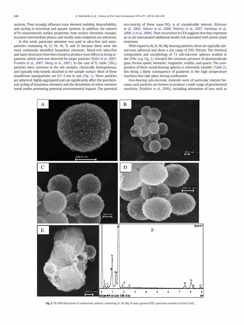
Fig. 2. FE-SEM illustration of submicronic spheres, containing Zr, N
eco-toxicity of these nano-TiO2 is of considerable interest (Rahmanet al., 2002; Adams et al., 2006; Federici et al., 2007; Heinlaan et al.,2008; Li et al., 2008). Their occurrence in CFA suggests that they representan as yet unevaluated additional health risk associated with power plantemissions
With regard to Al, Zr, Ni,Mg-bearing particles, these are typically sub-micronic spherical and show a size range of 250–350nm. The chemicalcomposition and morphology of 73 sub-micronic spheres studied inthe CFAs (e.g. Fig. 2) revealed the common presence of aluminosilicateglass, ferrian spinel, hematite; magnetite, mullite, and quartz. The com-position of these metal-bearing spheres is extremely variable (Table 2),this being a likely consequence of gradients in the high temperaturereactions that take place during combustion.
Iron-bearing sub-micronic minerals were of particular interest be-cause such particles are known to produce a wide range of geochemicalreactivity (Madden et al., 2006), including adsorption of ions such as
i, Mg, Al and a general EDS (spectrum contains Cu from Grid).

Table 2Maximum, minimum and average values concentrations of Al, Mg Ni, Zr bearingnanoparticles measured by EDS.
Element Maximum (%) Minimum (%) Average (%)
C 44.70 24.16 35.52O 40.28 3.73 29.93Mg 3.85 2.45 2.78Al 50.62 41.68 43.81Ni 8.80 5.81 7.24Zr 2.69 0.17 1.36
Fig. 4. HR-TEM and Fourier transformation (FFT) confirm the nanohematite structurewhich are approximately spherical. EDS demonstrated affinity of Cr and Pb for hematite.
449K. Martinello et al. / Science of the Total Environment 470–471 (2014) 444–452
phosphates and arsenates (Waychunas et al., 2005a,b), photochem-ical reduction in aqueous solution (Sherman, 2005), heterogeneouscatalysis (Feng et al., 2004), and acceptance of electrons from micro-bial respiration. Ferruginous sub-micronic particles are ubiquitouswithin all fly ash samples, having been derived from pyrite (and, toa lesser extent, jarosite) present in the parent coal (Silva et al., 2009a,b). The sub-micronic crystalline ferruginous phases observed in thisstudy included jarosite (KFe3+3(OH)6(SO4)2, Fig. 3), hematite (Fe2O3,Fig. 4), magnetite (Fe3O4), pyrrhotite (FeS), ankerite (Ca(Fe,Mg,Mn)(CO3)2), chromite (FeCr2O4), schwertmannite (Fe8O8(OH)6(SO4)),and yavapaiite (KFe(SO4)2). These mineral phases often also incorporatetrace elements, especially Cr, within their structure. Chromium occursin Brazilian coals primarily as Cr3+ in organic association and in illite,
Fig. 3. Cubic jarosite pseudomorph present in coal fly ash; (A) TEM image; (B) FE-SEM image; (C) EDS spectrum of the cubic crystals shown in (A) containing Cr (EDS spectrum containingCu from Grid).
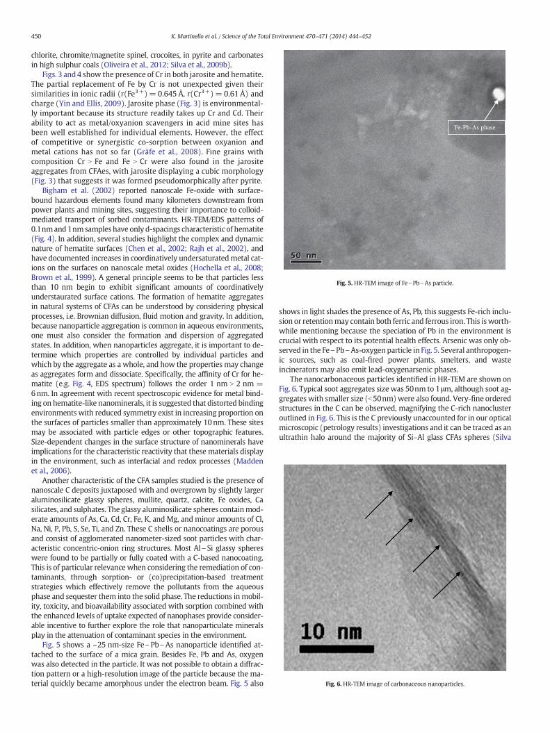
Fe-Pb-As phase
Fig. 5. HR-TEM image of Fe\Pb\As particle.
Fig. 6. HR-TEM image of carbonaceous nanoparticles.
450 K. Martinello et al. / Science of the Total Environment 470–471 (2014) 444–452
chlorite, chromite/magnetite spinel, crocoites, in pyrite and carbonatesin high sulphur coals (Oliveira et al., 2012; Silva et al., 2009b).
Figs. 3 and4 show the presence of Cr in both jarosite and hematite.The partial replacement of Fe by Cr is not unexpected given theirsimilarities in ionic radii (r(Fe3+)= 0.645 Å, r(Cr3+)= 0.61 Å) andcharge (Yin and Ellis, 2009). Jarosite phase (Fig. 3) is environmental-ly important because its structure readily takes up Cr and Cd. Theirability to act as metal/oxyanion scavengers in acid mine sites hasbeen well established for individual elements. However, the effectof competitive or synergistic co-sorption between oxyanion andmetal cations has not so far (Gräfe et al., 2008). Fine grains withcomposition Cr N Fe and Fe N Cr were also found in the jarositeaggregates from CFAes, with jarosite displaying a cubic morphology(Fig. 3) that suggests it was formed pseudomorphically after pyrite.
Bigham et al. (2002) reported nanoscale Fe-oxide with surface-bound hazardous elements found many kilometers downstream frompower plants and mining sites, suggesting their importance to colloid-mediated transport of sorbed contaminants. HR-TEM/EDS patterns of0.1nmand1nmsamples have only d-spacings characteristic of hematite(Fig. 4). In addition, several studies highlight the complex and dynamicnature of hematite surfaces (Chen et al., 2002; Rajh et al., 2002), andhave documented increases in coordinatively undersaturatedmetal cat-ions on the surfaces on nanoscale metal oxides (Hochella et al., 2008;Brown et al., 1999). A general principle seems to be that particles lessthan 10 nm begin to exhibit significant amounts of coordinativelyunderstaurated surface cations. The formation of hematite aggregatesin natural systems of CFAs can be understood by considering physicalprocesses, i.e. Brownian diffusion, fluid motion and gravity. In addition,because nanoparticle aggregation is common in aqueous environments,one must also consider the formation and dispersion of aggregatedstates. In addition, when nanoparticles aggregate, it is important to de-termine which properties are controlled by individual particles andwhich by the aggregate as a whole, and how the properties may changeas aggregates form and dissociate. Specifically, the affinity of Cr for he-matite (e.g. Fig. 4, EDS spectrum) follows the order 1 nm N 2 nm =6nm. In agreement with recent spectroscopic evidence for metal bind-ing on hematite-like nanominerals, it is suggested that distorted bindingenvironments with reduced symmetry exist in increasing proportion onthe surfaces of particles smaller than approximately 10nm. These sitesmay be associated with particle edges or other topographic features.Size-dependent changes in the surface structure of nanominerals haveimplications for the characteristic reactivity that these materials displayin the environment, such as interfacial and redox processes (Maddenet al., 2006).
Another characteristic of the CFA samples studied is the presence ofnanoscale C deposits juxtaposed with and overgrown by slightly largeraluminosilicate glassy spheres, mullite, quartz, calcite, Fe oxides, Casilicates, and sulphates. The glassy aluminosilicate spheres containmod-erate amounts of As, Ca, Cd, Cr, Fe, K, and Mg, and minor amounts of Cl,Na, Ni, P, Pb, S, Se, Ti, and Zn. These C shells or nanocoatings are porousand consist of agglomerated nanometer-sized soot particles with char-acteristic concentric-onion ring structures. Most Al\Si glassy sphereswere found to be partially or fully coated with a C-based nanocoating.This is of particular relevance when considering the remediation of con-taminants, through sorption- or (co)precipitation-based treatmentstrategies which effectively remove the pollutants from the aqueousphase and sequester them into the solid phase. The reductions inmobil-ity, toxicity, and bioavailability associated with sorption combined withthe enhanced levels of uptake expected of nanophases provide consider-able incentive to further explore the role that nanoparticulate mineralsplay in the attenuation of contaminant species in the environment.
Fig. 5 shows a ~25 nm-size Fe\Pb\As nanoparticle identified at-tached to the surface of a mica grain. Besides Fe, Pb and As, oxygenwas also detected in the particle. It was not possible to obtain a diffrac-tion pattern or a high-resolution image of the particle because the ma-terial quickly became amorphous under the electron beam. Fig. 5 also
shows in light shades the presence of As, Pb, this suggests Fe-rich inclu-sion or retentionmay contain both ferric and ferrous iron. This isworth-while mentioning because the speciation of Pb in the environment iscrucial with respect to its potential health effects. Arsenic was only ob-served in the Fe\Pb\As-oxygenparticle in Fig. 5. Several anthropogen-ic sources, such as coal-fired power plants, smelters, and wasteincinerators may also emit lead-oxygenarsenic phases.
The nanocarbonaceous particles identified in HR-TEM are shown onFig. 6. Typical soot aggregates size was 50nm to 1μm, although soot ag-gregates with smaller size (b50nm)were also found. Very-fine orderedstructures in the C can be observed, magnifying the C-rich nanoclusteroutlined in Fig. 6. This is the C previously unaccounted for in our opticalmicroscopic (petrology results) investigations and it can be traced as anultrathin halo around the majority of Si–Al glass CFAs spheres (Silva
451K. Martinello et al. / Science of the Total Environment 470–471 (2014) 444–452
et al., 2009a). Several particles are typically 0.1–30 nm in size, andconcentration stacked graphitic layers area seen in the particles inHR-TEM image (Fig. 6). These nanocarbonaceous particles arenumerous but represent small contribution to the total ash on aweight basis.
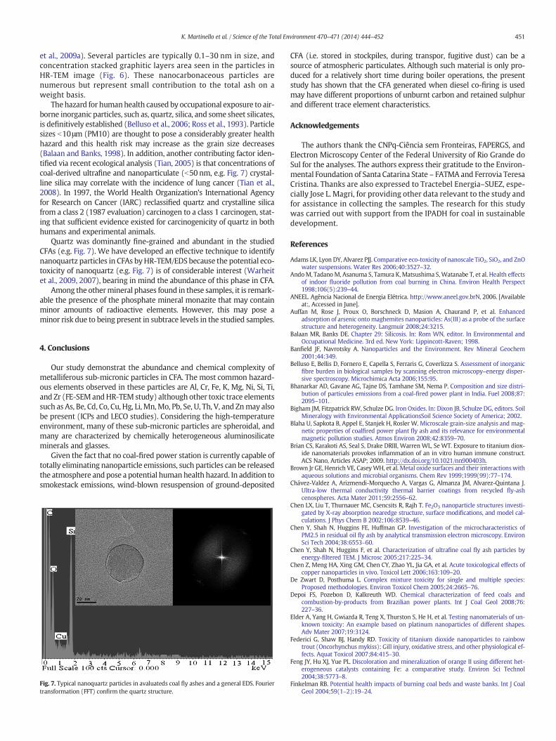
Thehazard for humanhealth caused by occupational exposure to air-borne inorganic particles, such as, quartz, silica, and some sheet silicates,is definitively established (Belluso et al., 2006; Ross et al., 1993). Particlesizes b10μm (PM10) are thought to pose a considerably greater healthhazard and this health risk may increase as the grain size decreases(Balaan and Banks, 1998). In addition, another contributing factor iden-tified via recent ecological analysis (Tian, 2005) is that concentrations ofcoal-derived ultrafine and nanoparticulate (b50 nm, e.g. Fig. 7) crystal-line silica may correlate with the incidence of lung cancer (Tian et al.,2008). In 1997, the World Health Organization's International Agencyfor Research on Cancer (IARC) reclassified quartz and crystalline silicafrom a class 2 (1987 evaluation) carcinogen to a class 1 carcinogen, stat-ing that sufficient evidence existed for carcinogenicity of quartz in bothhumans and experimental animals.
Quartz was dominantly fine-grained and abundant in the studiedCFAs (e.g. Fig. 7). We have developed an effective technique to identifynanoquartz particles in CFAs byHR-TEM/EDS because the potential eco-toxicity of nanoquartz (e.g. Fig. 7) is of considerable interest (Warheitet al., 2009, 2007), bearing in mind the abundance of this phase in CFA.
Among the othermineral phases found in these samples, it is remark-able the presence of the phosphate mineral monazite that may containminor amounts of radioactive elements. However, this may pose aminor risk due to being present in subtrace levels in the studied samples.
4. Conclusions
Our study demonstrat the abundance and chemical complexity ofmetalliferous sub-micronic particles in CFA. The most common hazard-ous elements observed in these particles are Al, Cr, Fe, K, Mg, Ni, Si, Ti,and Zr (FE-SEMandHR-TEM study) although other toxic trace elementssuch as As, Be, Cd, Co, Cu, Hg, Li, Mn,Mo, Pb, Se, U, Th, V, and Znmay alsobe present (ICPs and LECO studies). Considering the high-temperatureenvironment, many of these sub-micronic particles are spheroidal, andmany are characterized by chemically heterogeneous aluminosilicateminerals and glasses.
Given the fact that no coal-fired power station is currently capable oftotally eliminating nanoparticle emissions, such particles can be releasedthe atmosphere and pose a potential humanhealth hazard. In addition tosmokestack emissions, wind-blown resuspension of ground-deposited
Fig. 7. Typical nanoquartz particles in avaluateds coal fly ashes and a general EDS. Fouriertransformation (FFT) confirm the quartz structure.
CFA (i.e. stored in stockpiles, during transpor, fugitive dust) can be asource of atmospheric particulates. Although such material is only pro-duced for a relatively short time during boiler operations, the presentstudy has shown that the CFA generated when diesel co-firing is usedmay have different proportions of unburnt carbon and retained sulphurand different trace element characteristics.
Acknowledgements
The authors thank the CNPq-Ciência sem Fronteiras, FAPERGS, andElectron Microscopy Center of the Federal University of Rio Grande doSul for the analyses. The authors express their gratitude to the Environ-mental Foundation of Santa Catarina State – FATMA and Ferrovia TeresaCristina. Thanks are also expressed to Tractebel Energia–SUEZ, espe-cially Jose L. Magri, for providing other data relevant to the study andfor assistance in collecting the samples. The research for this studywas carried out with support from the IPADH for coal in sustainabledevelopment.
References
Adams LK, Lyon DY, Alvarez PJJ. Comparative eco-toxicity of nanoscale TiO2, SiO2, and ZnOwater suspensions. Water Res 2006;40:3527–32.
AndoM, TadanoM, Asanuma S, Tamura K, Matsushima S,Watanabe T, et al. Health effectsof indoor fluoride pollution from coal burning in China. Environ Health Perspect1998;106(5):239–44.
ANEEL. Agência Nacional de Energia Elétrica. http://www.aneel.gov.brN, 2006. [Availableat:, Accessed in June].
Auffan M, Rose J, Proux O, Borschneck D, Masion A, Chaurand P, et al. Enhancedadsorption of arsenic ontomaghemites nanoparticles: As(III) as a probe of the surfacestructure and heterogeneity. Langmuir 2008;24:3215.
Balaan MR, Banks DE. Chapter 29: Silicosis. In: Rom WN, editor. In Environmental andOccupational Medicine. 3rd ed. New York: Lippincott-Raven; 1998.
Banfield JF, Navrotsky A. Nanoparticles and the Environment. Rev Mineral Geochem2001;44:349.
Belluso E, Bellis D, Fornero E, Capella S, Ferraris G, Coverlizza S. Assessment of inorganicfibre burden in biological samples by scanning electron microscopy–energy disper-sive spectroscopy. Microchimica Acta 2006;155:95.
Bhanarkar AD, Gavane AG, Tajne DS, Tamhane SM, Nema P. Composition and size distri-bution of particules emissions from a coal-fired power plant in India. Fuel 2008;87:2095–101.
Bigham JM, Fitzpatrick RW, Schulze DG. Iron Oxides. In: Dixon JB, Schulze DG, editors. SoilMineralogy with Environmental ApplicationsSoil Science Society of America; 2002.
Blaha U, Sapkota B, Appel E, Stanjek H, Rosler W. Microscale grain-size analysis and mag-netic properties of coalfired power plant fly ash and its relevance for environmentalmagnetic pollution studies. Atmos Environ 2008;42:8359–70.
Brian CS, Karakoti AS, Seal S, Drake DRIII, Warren WL, Se WT. Exposure to titanium diox-ide nanomaterials provokes inflammation of an in vitro human immune construct.ACS Nano, Articles ASAP; 2009. http://dx.doi.org/10.1021/nn900403h.
Brown Jr GE, Henrich VE, CaseyWH, et al. Metal oxide surfaces and their interactionswithaqueous solutions and microbial organisms. Chem Rev 1999;1999(99):77–174.
Chávez-Valdez A, Arizmendi-Morquecho A, Vargas G, Almanza JM, Alvarez-Quintana J.Ultra-low thermal conductivity thermal barrier coatings from recycled fly-ashcenospheres. Acta Mater 2011;59:2556–62.
Chen LX, Liu T, Thurnauer MC, Csencsits R, Rajh T. Fe2O3 nanoparticle structures investi-gated by X-ray absorption nearedge structure, surface modifications, and model cal-culations. J Phys Chem B 2002;106:8539–46.
Chen Y, Shah N, Huggins FE, Huffman GP. Investigation of the microcharacteristics ofPM2.5 in residual oil fly ash by analytical transmission electron microscopy. EnvironSci Tech 2004;38:6553–60.
Chen Y, Shah N, Huggins F, et al. Characterization of ultrafine coal fly ash particles byenergy-filtered TEM. J Microsc 2005;217:225–34.
Chen Z, Meng HA, Xing GM, Chen CY, Zhao YL, Jia GA, et al. Acute toxicological effects ofcopper nanoparticles in vivo. Toxicol Lett 2006;163:109–20.
De Zwart D, Posthuma L. Complex mixture toxicity for single and multiple species:Proposed methodologies. Environ Toxicol Chem 2005;24:2665–76.
Depoi FS, Pozebon D, Kalkreuth WD. Chemical characterization of feed coals andcombustion-by-products from Brazilian power plants. Int J Coal Geol 2008;76:227–36.
Elder A, Yang H, Gwiazda R, Teng X, Thurston S, He H, et al. Testing nanomaterials of un-known toxicity: An example based on platinum nanoparticles of different shapes.Adv Mater 2007;19:3124.
Federici G, Shaw BJ, Handy RD. Toxicity of titanium dioxide nanoparticles to rainbowtrout (Oncorhynchusmykiss): Gill injury, oxidative stress, and other physiological ef-fects. Aquat Toxicol 2007;84:415–30.
Feng JY, Hu XJ, Yue PL. Discoloration and mineralization of orange II using different het-erogeneous catalysts containing Fe: a comparative study. Environ Sci Technol2004;38:5773–8.
Finkelman RB. Potential health impacts of burning coal beds and waste banks. Int J CoalGeol 2004;59(1–2):19–24.

452 K. Martinello et al. / Science of the Total Environment 470–471 (2014) 444–452
Franklin NM, Rogers NJ, Apte SC, Batley GE, Gadd GE, Casey PS. Comparative toxicityof nanoparticulate ZnO, bulk ZnO, and ZnCl2 to a freshwater microalga(Pseudokirchneriella subcapitata): The importance of particle solubility. Envi-ron Sci Technol 2007;41:8484–90.
Fubini B, Fenoglio I. Toxic Potential of Mineral Dusts. Mineral. Soc. Am. 2007;3:407–14.
Giere R, Blackford M, Smith K. TEM Study of PM2.5 Emitted from Coal and Tire Combus-tion in a Thermal Power Station. Environ Sci Technol 2006;40:6235–40.
Gräfe M, Beattie DA, Smith E, Skinner WM, Singh Balwant. Copper and arsenateco-sorption at the mineral–water interfaces of goethite and jarosite. J Colloid Inter-face Sci 2008;322:399–413.
Heinlaan M, Ivask A, et al. Toxicity of nanosized and bulk ZnO, CuO and TiO2 to bacteriaVibrio fischeri and crustaceans Daphnia magna and Thamnocephalus platyurus.Chemosphere 2008;71:1308–16.
Hochella MF, Lower SK, Maurice PA, Penn RL, Sahai N, Sparks DL, et al. Nanominerals,mineral nanoparticles, and Earth systems. Science 2008;319:1631–5.
Hoet P, Bruske-Hohlfeld I, Salata O. NanoparticlessKnown and unknown health risks. JNanobiotechnol 2004;2:12.
Hower JC, Graham UM, Dozier A, Tseng MT, Khatri RA. Association of the Sites of HeavyHazardous elements with Nanoscale Carbon in a Kentucky Electrostatic PrecipitatorFly Ash. Environ Sci Technol 2008;42:8471–7.
International Center For Diffraction Data (ICDD). http://www.icdd.com, 2009. [access: 20July 2009].
International Energy Agency (IEA). Electricity Information 2005. Paris: OECD/IEA; 2005[http://www.worldenergyoutlook.org [access: 30 September 2006]].
Iordanidis A, Buckma J, Triantafyllou AG, Asvesta A. Fly ash-airborne particles fromPtolemais–Kozani area, northern Greece, as determined by ESEM-EDX. Int J CoalGeol 2008;73:63–73.
Ispas C, Andreescu D, Patel A, Goia DV, Andreescu S, Wallace KN. Toxicity and Develop-mental Defects of Different Sizes and Shape Nickel Nanoparticles in Zebrafish. Envi-ron. Sci. Technol. 2009;43:6349–56.
Khan JA, Pillai B, Das TK, Singh Y, Maiti S. Molecular effects of uptake of gold nanoparticlesin HeLa cells. Chembiochem 2007;8:1237–40.
Levandowski J, Kalkreuth W. Chemical and petrographical characterization of feed coal,fly ash and bottom ash from the Figueira Power Plant, Paraná, Brazil. Int J Coal Geol2009;77:269–81.
Li SQ, Zhu RR, Zhu H, Xue M, Sun X, Yao S, et al. Nanotoxicity of TiO2 nanoparticles toerythrocyte in vitro. Food Chem Toxicol 2008;46:3626–31.
Madden AS, Hochella Jr MF, Luxton TP. Insights for size-dependent reactivity of hematitenanomineral surfaces through Cu2+ sorption. Geochim Cosmochim Acta 2006;70:4095–104.
Meng H, Chen Z, Xing GM, Yuan H, Chen CY, Zhao F, et al. Ultrahigh reactivity provokesnanotoxicity: Explanation of oral toxicity of nano-copper particles. Toxicol Lett2007;175:102–10.
Moreno-Jimenez E, Garcia-Gomez C, Oropesa AL, Esteban E, Haro A, Carpena-Ruiz R, et al.Screening risk assessment tools for assessing the environmental impact in an aban-doned pyritic mine in Spain. Sci Total Environ 2011;409:692–703.
Navarrette B, Vilches LF, Canadas L, Salvador L. Influence of start-ups with fuel oil on theoperation of electrostatic precipitators in pulverized coal boilers. Environ Prog2004;23(1):29–38.
Navrotsky A. In nanoparticles and the environment. RevMiner Geochem 2001;44:73–103.Nowack BB, Thomas D. Occurrence, behavior and effects of nanoparticles in the environ-
ment. Environ Pollut 2007;150:5–22.O'Halloran K. Toxicological considerations of contaminants in the terrestrial environment
for ecological risk assessment. Hum Ecol Risk Assess 2006;12:74–83.Oliveira MLS, Ward CR, French D, Hower JC, Querol X, Silva LFO. Mineralogy and leaching
characteristics of beneficiated coal products from Santa Catarina, Brazil. Int J CoalGeol 2012;94:314–25.
Ollson CA, Koch I, Smith P, Knopper LD, Hough C, Reimer KJ. Addressing arsenic bioacces-sibility in ecological risk assessment: a novel approach to avoid overestimating risk.Environ Toxicol Chem 2009;28:668–75.
Querol X, Whateley MKG, Fernandez-Turiel JL, Tuncali E. Geological controls on the min-eralogy and geochemistry of the Beypazari lignite, central Anatolia, Turkey. Int J CoalGeol 1997;33:255–71.
Querol X, Izquierdo M, Monfort E, Alvarez E, Font O, Moreno T, et al. Environmental char-acterization of burnt coal gangue banks at Yangquan, Shanxi Province, China. Int JCoal Geol 2008;75:93–104.
Quispe D, Pérez-López R, Silva LFO, Nieto JM. Changes in mobility of hazardous elementsduring coal combustion in Santa Catarina power plant (Brazil). Fuel 2012;94:495–503.
Rahman Q, Lohani M, Dopp E, Pemsel H, Jonas L, Weiss DG, et al. Evidence that ultrafinetitanium dioxide induces micronuclei and apoptosis in Syrian hamster embryofibroblasts. Environ Health Perspect 2002;110:797–800.
Rajh T, Chen LX, Lukas K, Liu T, Thurnauer MC, Tiede DM. Surface restructuring of nanopar-ticles: an efficient route for ligand–metal oxide crosstalk. J Phys Chem B 2002;106:10543–52.
Rohde GM, Silva NIW. Cinzas de Carvão Fóssil no Brasil Aspectos Técnicos e Ambientais, 1.Porto Alegre: CIENTEC; 2006.
Ross M, Nolan RP, Langer MA, Cooper WC. In: Guthrie Jr GD, Mossman BT, editors. Healtheffects of mineral dusts. Chelsea, Michigan: BookCrafters, Inc.; 1993. p. 361.
Ruhl L, Vengosh A, Dwyer GS, Hsu-kim H, Deonarine A, Bergin M, et al. Survey of thePotential Environmental and Health Impacts in the Immediate Aftermath of theCoal Ash Spill in Kingston, Tennessee. Environ Sci Technol 2009;43:6326–33.
Schneider H, Schreurer J, Hildmann B. Structure and properties of mullite—a review. J EurCeram Soc 2008;28:329.
Sherman DM. Electronic structures of iron(III) and manganese(IV) (hydr)oxide minerals:thermodynamics of photochemical reductive dissolution in aquatic environments.Geochim Cosmochim Acta 2005;69:3249–55.
Silva LFO, Moreno T, Querol X. An introductory TEM study of Fe-nanominerals within coalfly ash. Sci Total Environ 2009a;407:4972–4.
Silva LFO, Oliveira MLS, da Boit KM, Finkelman RB. Characterization of Santa Catarina(Brazil) coal with respect to Human Health and Environmental Concerns. EnvironGeochem Health 2009b;31:475–85.
Silva LFO, Querol X, da Boit KM, Vallejuelo SFOD,Madariaga JM. Brazilian CoalMining Res-idues and Sulphide Oxidation by Fenton's Reaction: an acceleratedweathering proce-dure to evaluate possible environmental impact. J Hazard Mater 2011a;186:516–25.
Silva LFO, Izquierdo M, Querol X, Finkelman RB, Oliveira MLS, Wollenschlager M, et al.Leaching of potential hazardous elements of coal cleaning rejects. Environ MonitAssess 2011b;175:109–26.
Silva LFO, Macias F, Oliveira MLS, da Boit KM, Waanders F. Coal cleaning residues andFe-minerals implications. Environ Monit Assess 2011c;172:367–78.
Silva LFO, Oliveira MLS, Neace ER, O'Keefe JMK, Henke KR, Hower JC. Nanominerals andultrafine particles in sublimates from the RuthMullins coal fire, Perry County, EasternKentucky, USA. Int J Coal Geol 2011d;85:237–45.
Teixeira EC, Samama JC, Brun A. Study of the Concentration of Trace-Elements in Fly-AshResulting from Coal Combustion. Environ Technol 1992;13:995–1000.
Tian L. Coal Combustion Emissions and Lung Cancer in Xuan Wei, China. PhDThesisBerkeley, CA: University of California; 2005.
Tian LW, Dai SF, Wang JF, Huang YC, Ho SC, Zhou Y, et al. Nanoquartz in Late Permian C1coal and the high incidence of female lung cancer in the Pearl River Origin area: aretrospective cohort study. BMC Public Health 2008;8:398.
Tiede K, Hassellöv M, Breitbarthc E, Chaudhryb Q, Boxall ABA. Considerations for environ-mental fate and ecotoxicity testing to support environmental risk assessments forengineered nanoparticles. J Chromatogr A 2009;1216:503–9.
Warheit DB, Webb TR, Reed KL. Pulmonary Toxicity Screening Studies in Male Rats withM5 Respirable Fibers and Particulates. Inhal Toxicol 2007;19:951.
Warheit DB, Reed KL, Sayes CM. A role for nanoparticle surface reactivity in facilitatingpulmonary toxicity and development of a base set of hazard assays as a componentof nanoparticle risk management. Inhal Toxicol 2009;21:61.
Waychunas GA, Kim CS, Banfield JF. Nanoparticulate iron oxide minerals in soils and sed-iments: unique properties and contaminant scavenging mechanisms. J NanoparticleRes 2005a;7:409–33.
Waychunas G, Trainor T, Eng P, Catalano J, Brown G, Davis J, et al. Surface complexationstudied via combined grazing-incidence EXAFS and surface diffraction: arsenate anhematite (0 0 0 1) and (1 0–1 2). Anal Bioanal Chem 2005b;383:12–27.
WWCCPN.World-Wide Coal Combustion Products Network; 2012 [http://www.wwccpn.org].
Yin S, Ellis DE. DFT studies of Cr(VI) complex adsorption on hydroxylated hematite(1102) surfaces. Surf Sci 2009;603:736–46.