direct association of occludin with zo-1 and its possible
TRANSCRIPT
Direct Association of Occludin with ZO-1 and Its Possible Involvement in the Localization of Occludin at Tight Junctions Mikio Furuse,** Masahiko Itoh,* Tetsuaki Hirase,*§ Akira Nagafuchi,* Shigenobu Yonemura,* Sach iko Tsukita,*ll a n d Shoich i ro Tsukita*~¶
* Laboratory of Cell Biology, Department of Information Physiology, National Institute for Physiological SCiences; ¢ Department of Physiological Sciences, School of Life Sciences, The Graduate University of Advanced Studies, Myodaiji-cho, Okazaki, Aichi 444; § First Department of Internal Medicine, Faculty of Medicine, Kobe University, Chuo-ku, Kobe, Hyogo 650; and 1t College of Medical Technology and ¶ Department of Medical Chemistry, Faculty of Medicine, Kyoto University, Sakyo-ku, Kyoto 606-01, Japan
Abstract. Occludin is an integral membrane protein localizing at tight junctions (TJ) with four transmem- brane domains and a long COOH-terminal cytoplas- mic domain (domain E) consisting of 255 amino acids. Immunofluorescence and laser scan microscopy revealed that chick full-length occludin introduced into human and bovine epithelial cells was correctly deliv- ered to and incorporated into preexisting TJ. Further transfection studies with various deletion mutants showed that the domain E, especially its COOH- terminal ,~150 amino acids (domain E358/504), was necessary for the localization of occludin at TJ. Sec- ondly, domain E was expressed in Escherichia coli as a fusion protein with glutathione-S-transferase, and this fusion protein was shown to be specifically bound
to a complex of ZO-1 (220 kD) and ZO-2 (160 kD) among various membrane peripheral proteins. In vitro binding analyses using glutathione-S-transferase fusion proteins of various deletion mutants of domain E nar- rowed down the sequence necessary for the ZO-1/ZO-2 association into the domain E358/504. Furthermore, this region directly associated with the recombinant ZO-1 produced in E. coli. We concluded that occludin itself can localize at TJ and directly associate with ZO-1. The coincidence of the sequence necessary for the ZO-1 association with that for the TJ localization suggests that the association with underlying cyto- skeletons through ZO-1 is required for occludin to be localized at TJ.
T HE establishment of compositionaUy distinct fluid compartments by epithelium and endothelium is cru- cial for the development and function of most organs.
Tight junction (TJ),~ an element of epithelial and en- dothelial junctional complexes, is directly involved in this compartmentation by sealing cells to create the primary bar- rier to the diffusion of solutes through the paracellular path- way (Schneeberger and Lynch, 1992; Gumbiner, 1987, 1993). TJ also functions as a boundary between the apical and basolateral plasma membrane domains, which differ in proteins, lipid composition, and physiological functions, to create and maintain epithelial and endothelial cell polarity (Rodriguez-Boulan and Nelson, 1989). Therefore, TJ has been attracting increasing interest among cell biologists.
Address all correspondence to Prof. Shoichiro Tsukita, National Institute for Physiological Sciences, Myodaiji-cho, Okazaki, Aichi 444, Japan. Phone: 81-564-55-7851; fax: 81-564-55-7853.
1. Abbreviations used in this paper: GST, glutathione-S-transferase; MBP, maltose-binding protein; MDBK, Madin-Darby bovine kidney cells; TJ, tight junction.
Accumulating evidence has shown that some unique pro- teins constitute TJ (Anderson et al., 1993; Citi, 1993). The first protein identified as a TJ constituent was ZO-1 with a molecular mass of 220 kD (Stevenson et al., 1986; Anderson et al., 1988). This protein is a peripheral membrane protein that is localized in the immediate vicinity of the plasma membrane of TJ in epithelial and endothelial cells (Steven- son et al., 1986, 1989), whereas it is colocalized with cadhe- rins in cells lacking TJ, such as fibroblasts and cardiac mus- cle cells (Itoh et al., 1991, 1993; Howarth et al., 1992; Tsukita et al., 1992), with some exceptions (Howarth et al., 1994). As a ZO-l-binding protein, another peripheral pro- tein called ZO-2 with a molecular mass of 160 kD has been identified (Gumbiner et al., 1991). Unlike ZO-1, the distri- bution of this protein is restricted to TJ (Jesaitis and Good- enough, 1994). Both ZO-1 and ZO-2 reportedly show se- quence similarity to the product of lethal (1) discs large-1 (dig), one of the tumor suppressor molecules in Drosophila (Itoh et al., 1993; Tsukita et al., 1993; Willott et al., 1993; Jesaitis and Goodenough, 1994). In addition to ZO-1 and ZO-2, two other TJ-specific peripheral membrane proteins have been so far identified; cingulin and the 7H6 antigen
© The Rockefeller University Press, 0021-9525/94/12/1617/10 $2.00 The Journal of Cell Biology, Volume 127, Number 6, Part 1, December 1~94 1617-1626 1617
Dow
nloaded from http://rupress.org/jcb/article-pdf/127/6/1617/1264377/1617.pdf by guest on 24 M
arch 2022

(Citi et al., 1988; Zhong et al., 1993). They are distributed more distantly from the membrane than ZO-1 (Stevenson et al., 1989; Zhong et al., 1993).
To clarify the structure and function of TJ at the molecular level, an integral membrane protein working at TJ should be identified. However, this integral membrane component re- mained elusive for quite some time. Most recently, using mAbs, we identified an integral membrane protein named occludin that was exclusively localized at TJ both in epi- thelial and endothelial cells (Furuse et al., 1993). The follow- ing structural characteristics of occludin molecules were clarified by eDNA cloning and sequencing (see Fig. 2). (a) In the NH2-terminal half, occludin contains four transmem- brane domains that segment the molecule into five domains (domains A-E). (b) A COOH-terminal half (domain E) con- sisting of '~ 250 amino acid residues resides in cytoplasm. (c) Charged amino acids mostly locate at domain E. (d) The content of tyrosine and glycine residues is very high in the extracellular domains (domains B and D).
Since occludin has been identified and its eDNA has been obtained, the following issues on the structure of TJ require resolution: how the newly synthesized occludin molecules are delivered and localized at'13; how occludin interacts with TJ-specific peripheral proteins such as ZO-1 and ZO-2; and whether or not the TJ strand is composed solely of occludin molecule. While studying these issues, we identified the im- portance of the COOH-terminal cytoplasmic domain (do- main E) of occludin molecules. In this study, we showed that chick occludin introduced into human and bovine epithelial cells was correctly delivered to and localized at TJ, and that domain E of occludin was necessary for the localization of the newly synthesized oceludin at TJ. In vitro binding using glutathione-S-transferase (GST)-domain E fusion protein re- vealed that occludin directly bound to ZO-1, and that domain E was necessary for the occludin-ZO-1 association. Further- more, we narrowed down the sequences necessary for TJ lo- calization and ZO-1 association, and found that both se- quences fell within the same region in domain E. We believe that this type of study will lead to further understanding of the structure and functions of TJ at the molecular level.
Materials and Methods
Cells and Antibodies
Madin-Darby bovine kidney (MDBK) cells and human intestinal epithelial cells (I"84) were obtained from the Japanese Cancer Research Resources Bank (Tokyo, Japan) and the American Type Culture Collection (Rockville, MD), respectively. Human esophagus fibroblast primary culture cells (PF- 7N) were provided by Dr. T. Iwazawa (Osaka University). MDBK and PF- 7N cells were grown in Dulbecco's modified Eagle's medium supplemented with 10% FCS. "['84 cells were grown in a I:I mixture of Dulbecco's modified Eagle's medium and Ham's F-12 medium supplemented with 5% FCS.
Rat anti-chicken occludin mAb (Oc-2) and mouse anti-rat ZO-I mAbs (1'8-754, "1'8-109) were obtained and characterized as described (Itoh et al., 1991; Furuse et al., 1993). Rabbit anti-ZO-2 pAb (R9989) was provided by Dr. D. Goodenough (Harvard Medical School, Boston, MA). Mouse anti-c-myc mAb was purchased from Oncogene Science Inc. (Manhasset, NY). Rabbit anti-bovine brain spectrin pAb was purchased from Chemicon International, Inc. (Temecula, CA).
Occludin Expression Constructs and Mutants
The expression plasmid (pBATOC) of full-length occludin driven by the chick /3-actin promoter was constructed using two plasmids, pX1 and
pBATEM2. To construct pXl, occludin cDNA containing the whole open reading frame was constructed by combining two cDNA fragments, FHI-14 and FH2-9, at the BglII site (Furuse et al., 1993), and then it was cloned into the EcoRI site of pBluescript SK(-). The BglII-SalI fragment of pBATEM2 (E-cadherin expression vector; Nose et al., 1988) that encodes full-length E-cadherin was replaced with the BamHI-SalI !.6-kb fragment obtained from pX1 to construct pBATOC.
An epitope tag of the partial sequence of c-myc (EQKLISEEDL) was linked to the COOH-terminal end of full-length or mutant occludin. For this purpose, we used a plasmid pCMYCB that was constructed by N. Funayama (National Institute for Physiological Sciences, Okazaki, Japan) as follows. An ollgonucleotide encoding EQKLISEEDL followed with two stop cnduns and its complement oligonucleotide were synthesized and annealed. The EcoRI-EcoRV fragment of pBluescript SK(-) was replaced with this DNA fragment to produce pCMYCB.
Expression plasmids for full-length or mutant occlndin with c-myc tag were constructed as follows (see Fig. 2). DNA fragments encoding the en- tire domain E of occludin or its deletion mutants were produced by PCR using appropriate primers. Using pCMYCB, the oligonucleotide encoding c-myc tag was linked to each PCR product. A fragment between BglH (I!e- 255) and SalI (3' noncoding region) sites in pBATOC were replaced with each c-myc-tagged fragment from pCMYCB.
Proteins expressed by these constructs have an additional Glu-Ser before the c-myc tag. Furthermore, rnOc/dN358 and mOc/d(445-474) products have additional Glu-Arg-Ser and Glu-Ser at their deletion sites, respectively. All DNA fragments in the plasmids amplified by PCR were sequenced using the Taq DyeDeoxy TM Terminator cycle Sequencing Kit (Applied Biosystems, Inc., Foster City, CA) to insure that no errors were introduced during PCR amplification.
DNA Transfections
MDBK and PF-7N cells were transfected with DNA using Lipofectin and Lipofectamine, respectively (GIBCO BRL, Gaithersburg, MD). Cells cul- tured on coverslips were washed once with Opti-MEM (GIBCO BRL), and were incubated for 5 h with 1 ml of Opti-MEM containing 1/zg of plasmid DNAs and 10/~1 of the reagents, followed by the addition of 3 ml of normal medium containing FCS. Cells were then cultured until observation.
When TM cells were transfected with DNA, the efficiency was improved by culturing the cells for >48 h on coverslips in Eagle's MEM containing 50/~M Ca 2+ (LCM) in the presence of 5% FCS dialyzed against saline. Transfection was performed using Lipefectamine as described above in LCM and saline-dialyzed FCS instead of Opti-MEM and normal FCS, respectively. 24 h after transfection, the medium was replaced with the nor- mal medium.
Immunofluorescence Microscopy and Laser Scan Microscopy
Indirect immunofluorescence microscopy of transfected cells was per- formed as described previously (Itoh et al., 1991; Tsukita et al., 1989). Briefly, ,,o48 h after transfection, cells were fixed with 1% formaldehyde in PBS for 10 rain, followed by soaking in 0.2% Triton X-100 in PBS for 10 rain. The second antibodies were F1TC-conjngated goat anti-rat IgG (Tago Inc., Burlingame, CA) for Oc-2, rhndamine-conjugated goat anti-mouse IgG (Chemicon International, Inc.) for T8-754, and FITC-conjugated sheep anti-mouse IgG (Amersham International PLC, Bucks, UK) for anti-c-myc mAb. Samples were examined using a fluorescence microscope, an Ax- iophot photomicroscope, or a laser scan microscope LSM310 (Carl Zeiss, Inc., Thornwood, NY).
Generation of Fusion Proteins
Occludin-domaln E full-length or mutant cDNAs obtained by PCR were introduced into pGEX vectors, pGEX-2T or pGEX-3X (Pharmacia Fine Chemicals, Piscataway, N J), to express fusion proteins with GST in Esche- richia coli (see Fig. 5). All constructs, except plasmids for GST-OcE and GST-OcE/dN358, have an additional Glu-Phe-Ile-Val-Thr-Asp derived from pGEX vectors at their COOH-terminal ends of fusion proteins. Fusion pro- teins expressed by a plasmid for GST-OcE/d (445-474) have another addi- tional Glu-Phe at its deletion site. All DNA fragments amplified by PCR were sequenced to insure that no errors were introduced during PCR am- plification. Fusion proteins were produced in E. coil (XL-1/Blne) from these constructs, according to the procedure described by the manufacturer.
The fusion protein of mouse ZO-I with maltose-binding protein (MBP) was produced using an F22 fragment (190-1235 aa) in pMAL-CRI (New
The Journal of Cell Biology, Volume 127, 1994 1618
Dow
nloaded from http://rupress.org/jcb/article-pdf/127/6/1617/1264377/1617.pdf by guest on 24 M
arch 2022

England BioLabs, Beverly, MA), as described previously (Itoh et al., 1993).
In Vitro Binding Assay
In vitro binding assays were performed using a column. Cultures ofE. coli expressing GST-fusion proteins (100 ml) were collected by brief centrifuga- tion and resuspended in 6 ml of solution K (140 mM KCI, 10 mM Hepes [pH 7.5], 1 mM MgCI2, 2 #g/rnl leupeptin, and 1 mM p-amidinophenyl methanesulfonyl fluoride hydroehloride [pAPMSF]). After sonication and centrifugation at 10,000 g for 10 min, the supernatant was applied to a column containing glntathione-Sepharose 4B beads (Pharrnacia Fine Chemicals), which was washed with 20 vol of solution K. Thereafter, the low salt extract of chick junctional fraction or high salt extract of MDBK ceils (see below) was applied onto the column. After washing with 40 vol of solution K, bound proteins were eluted with 50 mM Tris-HCl buffer (pH 8.0) containing 10 mM glutathione. 0.8-ml fractions were collected. Since almost all the bound proteins were eluted within the first five fractions, they were mixed and used for SDS-PAGE and immunoblotting.
To detect ¢-spectrin or MBP-ZO-1 fusion protein, binding assays were performed by means of the batch method. GST-fusion proteins of oecludin were incubated with glutathione beads at 40C for I h. After five washes with 10 vol of solution K by brief centrifugation, the beads were incubated with the high salt extract of MDBK ceils or the extract ofE. coli containing MBP- ZO-I fusion protein at 4°C for 1 h. The beads were then washed five times with solution K, and the excess solution was removed. Bound proteins were released from beads with SDS-PAGE sample buffer.
The low salt extract of chick junctional fraction was prepared as de- scribed previously (Furuse et al., 1993; Tsukita and Tsukita, 1989). The extract from '~40 chicks was used in one experiment. The low salt extract was freeze dried and resolved in 1 ml of solution K, followed by centrifuga- tion at 100,000 g for 1 h. The supernatant was used for the binding assay. The high salt extract of MDBK cells was prepared according to the method for purifying ZO-1 from mouse brain, as described (Itoh et al., 1991). Confluent MDBK cells from two 15-cm dishes were scraped and collected by brief centrifugation. They were homogenized in 1 mM NaHCO3 or in solution K with tight-fitting Dounce homogenizer, followed by centrifuga- tion at 100,000 g for 1 h. The pellet was resuspended by sonication in 1 ml of 10 mM Hepes buffer (pH 7.5) containing 1 M KC1, 1 mM EGTA, 2 #g/ml leupeptin, and 0.5 mM PMSF, and was then incubated on ice for 1 h. After centrifugation at 100,000 g for 1 h, the supernatant was diluted with 10 mM Hepes buffer (pH 7.5) at a final concentration of 140 mM KC1. Aggregated proteins were removed by centrifugation at 10,000 g for 10 min, and the supernatant was used for the binding assay. The extract containing MBP-ZO-1 fusion protein was prepared by the same procedure as that for GST-occludin fusion proteins.
Gel Electrophoresis and Immunoblotting
One-dimensional SDS-PAGE (12.5% gel) was based on the method of Laemmli (1970). Gels were stained with Coomassie brilliant blue R-250 or by using a silver staining kit (Wako Pure Chemical Industries, Osaka, Ja- pan). For immunoblotting, proteins separated by SDS-PAGE were elec- trophoretically transferred to nitrocellulose sheets, which were then in- cubated with the antibodies. The antibodies were detected with a blotting detection kit (Amersham).
Results
Localization of Chick Occludin Expressed by a Full-length cDNA at Tight Junctions in Human and Bovine Epithelial Cells
Chick occludin eDNA clones were isolated and sequenced (Furuse et al., 1993). To construct the expression vector, a 1.6-kb eDNA encoding full-length occludin was assembled from two overlapping clones. This eDNA encodes a 55.9 kD occludin polypeptide (Furuse et al., 1993). The complete eDNA was subcloned into a mammalian expression vector driven by the/3-actin promoter, which was then introduced into cultured cells.
The bovine and human epithelial cell lines, MDBK and
T84, were selected for our transfection studies. Both types of cells bear the typical junctional complex including TJ at the most apical portion of the lateral membranes. To detect the transiently expressed chick occludin by immunofluores- cence microscopy, we used the rat anti-chick occludin mAb Oc-2, which did not recognize human and bovine occludin (Furuse et al., 1993).
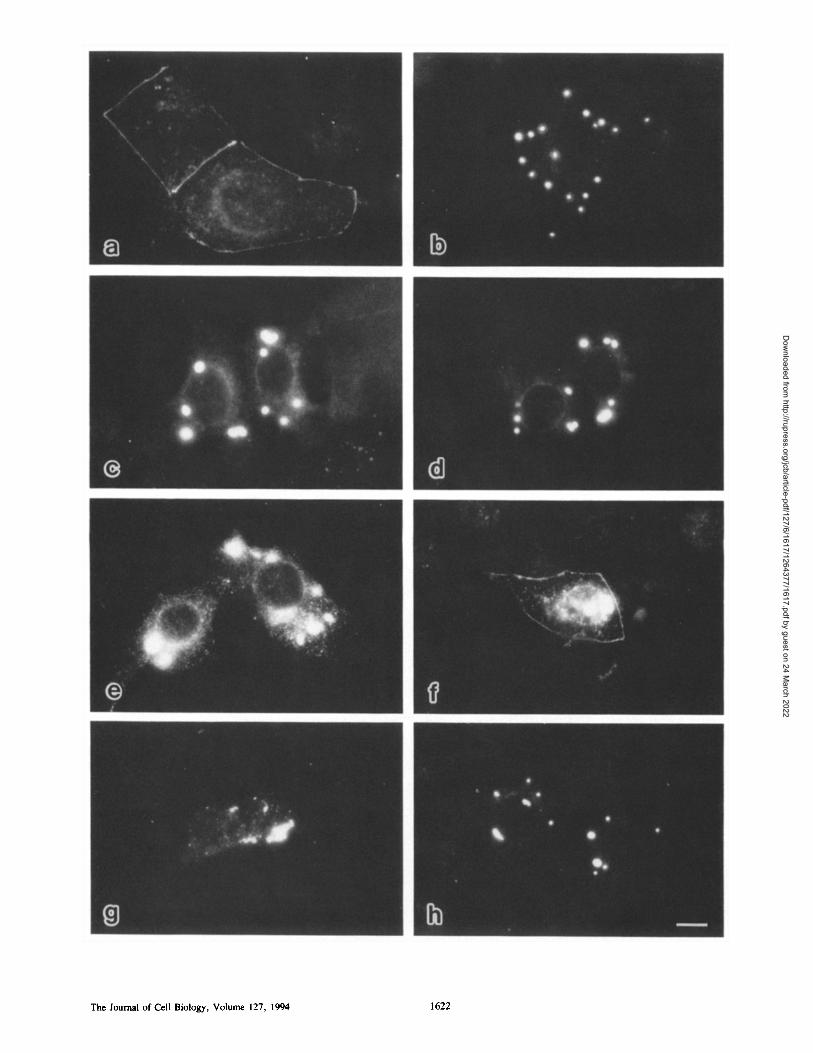
MDBK cells transfected with plasmids encoding full- length occludin displayed a characteristic pattern of fluores- cence. In addition to the diffuse staining at perinuclear cytoplasm, the concentration of expressed chick occludin was detected in a linear fashion at the cell-cell border (Fig. 1 a). When the transfectants were doubly stained with anti- occludin mAb and anti-ZO-1 mAb, ZO-1 appeared to be colocalized with the expressed chick occludin concentrated at the cell-cell border (Fig. 1 b). Therefore, to precisely compare the distribution of occludin with that of ZO-1, we analyzed the doubly stained 1"84 transfectants by laser scan microscopy (Fig. 1, c and d). As shown in Fig. 1, e and f as overlaid computer-generating cross-sectional images, the expressed chick occludin and ZO-1 were precisely colocal- ized at the most apical region of lateral membranes. In our laser scan microscopy system, tight and adherens junctions can be resolved (Yonemura et al., 1994). All these observa- tions together led us to the conclusion that the expressed chick occludin was correctly delivered to and concentrated at TJ in human and bovine epithelial cells.
The question naturally arose as to whether or not occludin expressed in nonepithelial cells lacking TJ were delivered to cell-cell contact sites. Chick occludin was then introduced into the human fibroblast PF-7N. As reported Otoh et al., 1993), ZO-1 was concentrated at cell-cell contact sites in fibroblasts. The introduced occludin was concentrated at some of these ZO-l-enriched cell-cell contact sites in cells expressing a large amount of occludin (Fig. 1, g and h).
Sequences in the COOH-terminal Cytoplasmic Domain of Occludin Necessary for Localization at Tight Junctions
To analyze the role of the COOH-terminal cytoplasmic do- main of occludin molecules (domain E; amino acid residues 250-504) in their localization at TJ, we performed transfec- tions with various domain E deletion mutants. Since the efficiency of transfection was significantly higher in MDBK than in "1"84 cells, we used the former in the following mutant occludin expression studies. We designed an expression vec- tor with two major considerations. Immunofluorescence mi- croscopy would be used and the sequences encoding the anti- genic determinant of the protein should not be lost during the construction of deletion mutants. To accomplish these goals, a 30-bp sequence encoding a portion of c-myc was added to the 3' end of each eDNA construct, allowing us to detect the expressed protein by anti-c-myc mAb.
To narrow down the sequence necessary for the TJ local- ization of occludin from both COOH- and NH2-terminal sides, we constructed several COOH- or NH:-terminal truncations of domain E from full-length occludin with the c-myc epitope on their COOH-terminal end (Fig. 2). As shown in Fig. 3 a, the c-myc-tagged, full-length occludin (mOc) was transiently expressed and localized at TJ in MDBK cells, indicating that the tag peptide did not interfere
Furuse et al. Functions of the Occludin Carboxyl-terminal Domain 1619
Dow
nloaded from http://rupress.org/jcb/article-pdf/127/6/1617/1264377/1617.pdf by guest on 24 M
arch 2022

Figure L Subcellular distribution of chick full-length occludin in transient transfectants. Bovine and human epithelial cells, MDBK (a and b) and 1"84 (c-f), respectively, or human fibroblasts, PF-7N (g and h) were transfected with plasmids encoding full-length occludin~ and were then doubly stained with anti-chick occludin mAb, Oc-2 (a, c, e, and g) and anti-rat ZO-1 mAb, T8-754 (b, d, f, and h).
The Journal of Cell Biology, Volume 127, 1994 1620
Dow
nloaded from http://rupress.org/jcb/article-pdf/127/6/1617/1264377/1617.pdf by guest on 24 M
arch 2022

Figure 2. Full-length and truncated occludin constructs with the c-myc epitope on their COOH-terminal end (m). Occludin is seg- mented into five domains (A-E) by four transmembrane domains, and both COOH- and NH2-terminal truncations of domain E from full-length occludin were constructed.
with the concentration of occludin at TJ. All COOH-terminal truncations constructed here (mOc/dC474, mOc/dC444, mOc/dC414, and mOc/dC357) failed to localize at TJ (Fig. 3, b-e), indicating that the responsible sequence could not be narrowed down from the COOH side. By contrast, the NH2-terminal truncation, mOc/dN358, was clearly concen- trated at TJ, although its localization efficiency was rather lower than that of mOc (Fig. 3 f ) . The further NH2- terminal truncation, mOc/dN387, was by no means localized at TJ (Fig. 3 g).
Taking all these results together, we concluded that amino acid residues 358-504 (domain E358/504) is necessary for the TJ localization of occludin, although detailed analysis of this domain remains to be performed. For example, mOc/d (445-474) was not concentrated at TJ (see Figs. 2 and 3 h).
Association of Itght Junction Peripheral Proteins with the COOH-terminal Cytoplasmic Domain of Occludin
Another possible function of the COOH-terminal cytoplas- mic domain (domain E) of occludin is its association with
TJ peripheral proteins such as ZO-1, ZO-2, etc. To test this association in vitro, domain E was expressed in E. coli as a fusion protein with GST (GST-OcE). E. coli lysate was in- cubated with glutathione-Sepharose beads on a column, and after washing, the low salt alkali extract of junctional frac- tion isolated from chick liver was applied onto the column. After incubation and washing, the proteins associated with GST-OcE coupled to glutathione-Sepharose beads were eluted with a solution containing glutathione, and were then resolved by SDS-PAGE.
As shown in Fig. 4 a, lane 2, this in vitro binding assay revealed two major bands with molecular masses of 220 and 160 kD bound to domain E of occludin. The 220-kD band was specifically recognized by anti-ZO-1 mAb (Fig. 4 a, lane 6). The molecular mass of the 160-kD band indicated that it would be related to ZO-2, a ZO-l-binding protein identified in canines (Gumbiner et al., 1991). Since an mAb recognizing chick ZO-2 was not available, we prepared a high salt extract from the membranes of cultured MDBK cells, from which ZO-2 is recognized by the pAb R9989 pro- duced by Jesaitis and Goodenough (1994). Oecludin-binding proteins were recovered from the extract using the column system described above. Immunoblots of these proteins re- vealed that ZO-2, as well as ZO-1, were bound to domain E of occludin (Fig. 4 b). This suggests that the 160-kD band from the chick junctional fraction is the chick homologue of ZO-2. Furthermore, as shown in Fig. 4 b, a,-spectrin from the high salt extract of MDBK cells was also specifically trapped by the GST-OcE beads.
Sequences in the COOH-terminal Cytoplasmic Domain of Occludin Necessary for Association with ZO-1
The question has naturally arisen whether the association of occludin with ZO-1 is required for TJ localization of oc- cludin. We attempted to narrow down the sequence neces- sary for the association of occludin with ZO-1 from both COOH- and NH2-terminal sides, and to evaluate whether or not this domain is included in or overlapped with the do- main E358/504, which is necessary for TJ localization ofoc- cludin. We expressed several GST-fusion proteins containing COOH- or NH2-terminal truncations of domain E in E. coli (Fig. 5). Using these fusion proteins and the extract of junc- tional fraction, we performed in vitro binding studies in which the amount of ZO-1 molecules bound to a fixed quan- tity of GST-fusion proteins was evaluated by immunoblot- ting.
As shown in Fig. 6, all COOH-terminal truncated fusion proteins (GST-OcE/dC474, GST-OcE/dC444, GST-OcE/ dC414, and GST-OcE/dc357) exhibited no or markedly weaker binding to ZO-1 than GST-OcE, indicating that the sequence necessary for the ZO-1 binding could not be nar-
Oc-2 recognizes chick occludin but neither bovine nor human occludin, whereas "1"8-754 crossreacts with both human and bovine ZO-I. (a and b) Conventional immunofluorescence microscopic images of MDBK cells. The transiently expressed chick occlndin and bovine ZO-1 were coconcentrated at the eel-cell border. The cytoplasmic staining with anti-occludin mAb is specific, and this may be a result of overexpression of chick occludin. (c-f) Laser scan microscopic images of'1"84 cells. Optical sections at the level of the junctional complex (c and d) and computer-generated cross-sectional images (e and f) . The expressed chick occludin and human ZO-1 were precisely colocal- ized at the most apical region of lateral membranes (arrows). ap, the level of apical surface; ba, the level of basal membrane. (g and h) Conventional immunofluorescence microscopic images of fibroblasts. Most of the expressed chick occludin was distributed in the cytoplasm, but some of them was colocalized with ZO-I at cell-cell contact sites. Bar, 10/~m.
Furuse et al. Functions of the Occludin Carboxyl-terminal Domain 1621
Dow
nloaded from http://rupress.org/jcb/article-pdf/127/6/1617/1264377/1617.pdf by guest on 24 M
arch 2022
The Journal of Cell Biology, Volume 127, 1994 1622
Dow
nloaded from http://rupress.org/jcb/article-pdf/127/6/1617/1264377/1617.pdf by guest on 24 M
arch 2022

Figure 4. Association of tight junction peripheral proteins with do- main E of oeeludin. (a) In vitro binding of GST-OcE fusion protein to proteins in the low salt alkali extract of junctional fraction iso- lated from chick liver (IF extract). Domain E of oecludin was ex- pressed in E. coli as a fusion protein with GST (GST-OcE), and E. coli lysate was incubated with glutathione-Sepharose beads on a column. After washing and applieatiun of JF extract onto the column, the proteins associated with GST-OcE were eluted with a solution containing glutathione. Silver-stained gel (lanes 1-4) and accompanying immunoblot with anti-ZO-1 mAb, I'8-754 (lanes 5-8) of glutathione-eluate from GST column incubated with IF ex- tract (lanes I and 5), ghtathione-eluate from GST-OcE eolunm in- cubated with JF extract (lanes 2 and 6), ghtathione-eluate from GST-OcE column without the incubation with IF extract (lanes 3 and 7), and JF extract (lanes 4 and 8). Comparison between lanes 1, 2, and 3 revealed that two major bands of • 220 and 160 kD bound to domain E of occludin (arrowheads), and immunoblot analyses identified the former bands as ZO-1. The mobility of mo- lecular mass markers is shown at the left (200, 116, 97, 66, 45, and 31 kD from the top). (b) In vitro binding of GST-OcE fusion pro- tein to proteins in the high salt extract of MDBK ceils. Immuno- blots with anti-ZO-1 mAb, I"8-754 (lanes I and 2), anti-ZO-2 pAb, R9989 (lanes 3 and 4), and anti-u-spectrin pAb (lanes 5 and 6) of glutathione-eluate from GST beads incubated with MDBK extract (lanes 1, 3, and 5) and glutathione-eluate from GST-OeE beads in- cubatr.d with MDBK extract (lanes 2, 4, and 6). Note that ot-spec- trin and ZO-1/ZO-2 were specifically trapped by domain E of oc- cludin. The mobility of molecular mass markers is shown at the left (200 and 116 kD from the top).
rowed down from the COOH-terminal side. On the other hand, the NH2-terminal truncated fusion proteins, GST- OcE/dN358, strongly bound to ZO-1, whereas the further- truncated fusion proteins, GST-OcE/dN387, showed very poor binding.
Therefore, this bidirectional strategy identified amino acid residues 358-504 (domain E358/504) as the region neces- sary for the association of occludin with ZO-1. The same re- suits were obtained when the high salt extract from MDBK and "184 cells was applied onto the column (data not shown). This conclusion is similar to that of the analysis of sequences
Figure 5. GST-fusion proteins containing normal or truncated do- main E of occludin.
necessary for the TJ localization of occludin, but in sharp contrast, GST-OcE/d (445-474) strongly bound to ZO-1.
Finally, to evaluate whether ZO-1 is associated with oc- cludin directly or indirectly, we performed the in vitro bind- ing studies between various GST-OcE mutant proteins and an MBP-ZO-1 fusion protein produced in E. coli. As shown in Fig. 7, the MBP-ZO-1 fusion protein bound strongly to GST-OcE, GST-OcE/dN358, and GST-OcE/d (445-474), but very weakly to the other GST-OcE mutant proteins. These data indicated that ZO-1 directly associates with the domain E358/504 of occludin.
Discussion
In our previous study, we identified a novel integral mem- brane protein with an apparent molecular mass of •65 kD called occludin, and showed by immunofluorescence and im- munoelectron microscopy that it is localized exclusively at TJ of various types of epithelial and endothelial cells (Furuse et al., 1993). Preembedding electron microscopic immuno- labeling of isolated bile canaliculi with anti-occludin mAb characteristically revealed immunogold particles directly over the points of membrane fusion of TJ, suggesting that oc- cludin is a component of the TJ strand. In this study, we dem- onstrated, by means of transfection, that the newly syn- thesized chick full-length occludin was delivered to and incorporated into preexisting TJ in human and bovine epithe- lial cells. Also in fibroblasts lacking TJ, the introduced chick occludin was occasionally localized at cell-cell contact sites, although it remains to be checked electron microscopically whether or not TJ-like structures are formed there. Further analyses using these transfection systems will lead us to a
Figure 3. Subeellular distribution of c-myc-tagged fnll-length and truncated chick occludin in transient MDBK transfectants. MDBK cells were transfected with plasmids of mOc (a), mOc/dC474 (b), mOetdC444 (c), mOc/dC414 (d), mOc/dC357 (e), mOctdN358 (f) , mOc/dN387 (g), or mOo/d(445-474) (h), and were then immunofluorescently stained with anti-c-myc mAb. Only in a andf intense signal was detected from the cell--cell border. In the other transfectants, the expressed truncated occludin was concentrated at the cytoplasm, and it formed large spheres. Even in the transfectants expressing a large amount of mOc and mOc/dN358 products, the products were concentrated not only in TJ, but also in similar spheres in the cytoplasm (data not shown). Bar, 10/zm.
Furuse et al. Functions of the Occludin Carboxyl-terminal Domain 1623
Dow
nloaded from http://rupress.org/jcb/article-pdf/127/6/1617/1264377/1617.pdf by guest on 24 M
arch 2022
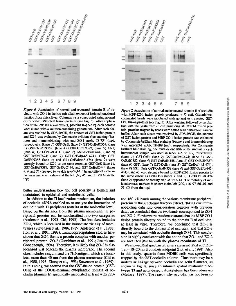
Figure 6. Association of normal and truncated domain E of oc- cludin with ZO-1 in the low salt alkali extract of isolated junctional fraction from chick liver. Columns were constructed using normal or truncated GST-OcE fusion proteins (see Fig. 5). After applica- tion of the low salt alkali extract, proteins trapped by each column were elnted with a solution containing ghitathione. After each elu- ate was resolved by SDS-PAGE, the amount of GST-fusion protein and ZO-1 was evaluated by Coomassie brilliant blue staining (bot- tom) and immunoblotting with anti-ZO-1 mAb, "18-754 (top), respectively. (Lane 1) GST-OcE; (lane 2) GST-OcE/dC357; (lane 3) GST-OcE/dN358; (lane 4) GST-OcE/dN387; (lane 5) GST; (lane 6) GST-OcE/dC414; (lane 7) GST-OcE/dC444; (lane 8) GST-OcE/dC474; (lane 9) GST-OcE/d(445-474). Only GST- OcE/dN358 (lane 3) and GST-OcE/d(445-474) (lane 9) were strongly bound to ZO-1 to the same extent as GST-OcE (lane 1 ). GST-OcE/dN387, GST-OcE/dC414, and GST-OeE/dC444 (lanes 4, 6, and 7) appeared to weakly trap ZO-1. The mobility of molecu- lar mass markers is shown at the left (66, 45, and 31 kD from the top).
better understanding how the cell polarity is formed and maintained in epithelial and endothelial cells.
In addition to the TJ localization mechanism, the isolation of occludin cDNA enabled us to analyze the interaction of occludin with TJ peripheral proteins at the molecular level. Based on the distance from the plasma membrane, TJ pe- ripheral proteins can be subclassified into two categories (Anderson et al., 1993; Citi, 1993). The first class includes ZO-1, which is localized in the immediate vicinity of mem- branes (Stevenson et al., 1986, 1989; Anderson et al., 1988; Itoh et al., 1991, 1993). Immunoprecipitation studies have shown that ZO-1 forms a protein complex with another pe- ripheral protein, ZO-2 (Gumbiner et al., 1991; Jesaitis and Goodenough, 1994). Therefore, it is likely that ZO-2 is also localized just beneath the plasma membrane. The second class includes cingulin and the 7H6 antigen, which are local- ized more than 40 nm from the plasma membrane (Citi et al., 1988, 1993; Zhong et al,, 1993; Stevenson et al., 1989). In this study, we showed that the GST-fusion protein (GST- OcE) of the COOH-terminal cytoplasmic domain of oc- cludin (domain E) specifically associated at least with 220-
Figure 7. Association of normal and truncated domain E of occludin with MBP-ZO-1 fusion protein produced in E. coli. Glutathione- conjugated beads were incubated with normal or truncated GST- OcE fusion proteins (see Fig. 5). After washing followed by incuba- tion with the lysate from E. coil producing MBP-ZO-I fusion pro- tein, proteins trapped by beads were ehited with SDS-PAGE sample buffer. After each eluate was resolved by SDS-PAGE, the amount of GST-fusion protein and MBP-ZO-1 fusion protein was evaluated by Coomassie brilliant blue staining (bottom) and immunoblotting with anti-ZO-1 mAb, I"8-109 (top), respectively. For Coomassie brilliant blue staining, one tenth or one fifth of the amount of each immunoblot sample was used in lanes 1-6 or 7-9, respectively. (Lane 1) GST-OcE; (lane 2) GST-OcE/dC474; (lane 3) GST- OcE/dC357; (lane 4) GST-OcE/dN358; (lane 5) GST-OcE/dN387; (lane 6) GST; (lane 7) GST-OcE; (lane 8) GST-OcE/d(445-474); (lane 9) GST. Only GST-OcE/dN358 (lane 4) and GST-OcE/d(445- 474) (lane 8) were strongly bound to MBP-ZO-1 fusion protein to the same extent as GST-OcE (lanes 1 and 7). GST-OcE/dC474 (lane 2) appeared to weakly trap MBP-ZO-1. The mobility of mo- lecular mass markers is shown at the left (200, 116, 97, 66, 45, and 31 kD from the top).
and 160-kD bands among the various membrane peripheral proteins in the junctional fraction extract. Taking our immu- noblotting data into consideration together with previous data, we concluded that the two bands corresponded to ZO-1 and ZO-2. Furthermore, we demonstrated that the MBP-ZO-1 fusion protein directly bound to the domain E of occludin, at least in vitro. Therefore, we concluded that ZO-1 is directly bound to the domain E of occludin, and that ZO-2 may be associated with occludin through ZO-1. This conclu- sion is highly consistent with the notion that ZO-1 and ZO-2 are localized just beneath the plasma membrane of TJ.
We showed that spectrin tetramers are associated with ZO- 1 at ~10-20 nm from their midpoint (Itoh et al., 1991). Also in this study, spectrin from MDBK cells was specifically trapped by the GST-occludin column. Thus there may be a molecular linkage between occludin and actin filaments, as shown in Fig. 8, since an intimate spatial relationship be- tween TJ and actin-based cytoskeletons has been observed (Madara, 1987). The reason why occludin has not been so
The Journal of Cell Biology, Volume 127, 1994 1624
Dow
nloaded from http://rupress.org/jcb/article-pdf/127/6/1617/1264377/1617.pdf by guest on 24 M
arch 2022
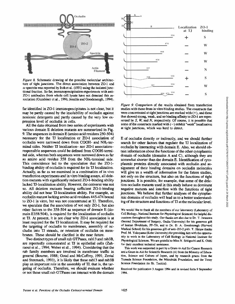
Figure 8. Schematic drawing of the possible molecular architec- ture of tight junctions. The direct association between ZO-1 and a-spectrin was reported by Itoh et al. (1991) using the isolated junc- tional fraction. So far, immunoprecipitation experiments with anti- ZO-I antibodies from whole cell lysate have not detected this as- sociation (Gumbiner et al., 1991; Jesaitis and Goodenough, 1994).
far identified in ZO-1 immunoprecipitates is not clear, but it may be partly caused by the insolubility of occludin against nonionic detergents and partly caused by the very low ex- pression level of occludin in cells.
All the data obtained from two series of experiments with various domain E deletion mutants are summarized in Fig. 9. The sequences in domain E (amino acid residues 250-504) necessary for the TJ localization or ZO-1 association of occludin were narrowed down from COOH- and NH2-ter- minal sides. Neither TJ localization- nor ZO-1 association- responsible sequences could be defined from COOH-termi- nal side, whereas both sequences were narrowed down as far as amino acid residue 358 from the NH2-terminal side. This coincidence led to the speculation that the ZO-1- binding ability of occludin is required for its TJ localization. Actually, as far as we examined in a combination of in vivo transfection experiments and in vitro binding assays, all dele- tion mutants with significantly reduced ZO-l-binding ability lacked TJ-localization ability. However, the converse was not so. All deletion mutants bearing sufficient ZO-l-binding ability did not bear TJ-localization ability. For example, an occludin mutant lacking amino acid residues 445-474 bound to ZO-1 in vitro, but was not concentrated at TJ. Therefore, we speculate that the association of not only ZO-1, but also other factors to the 358-504 aa sequence of domain E (do- main E358/504), is required for the localization of occludin at TJ. At present, it is not clear why ZO-1 association is at least required for the TJ localization. It may be required for the targeting of occludin to membranes, assembly of oc- cludin into TJ strands, or retention of occludin on mem- branes. These should be clarified in the near future.
Two distinct types of small rab GTPases, rabl3 and rab3B, are reportedly concentrated at TJ in epithelial cells (Zah- raoui et al., 1994; Weber et al., 1994). Considering that the rab family members are involved in membrane traffic in general (Bourne, 1988; Goud and McCaffrey, 1991; Zerial and Stenmark, 1993), it is likely that these rabl3 and rab3B play an important role in the assembly of TJ, that is, the tar- geting of occludin. Therefore, we should evaluate whether or not these small rab GTPases can interact with the domain
Figure 9. Comparison of the results obtained from transfection studies with those from in vitro binding studies. The constructs that were concentrated at tight junctions are marked with (+), and those that showed strong, weak, and no binding affinity to ZO-1 are repre- sented by S, W, and N, respectively. Of course, it is possible that some of the constructs marked with ( - ) exhibit "weak ~ localization at tight junctions, which was hard to detect.
E of occludin directly or indirectly, and we should further search for other factors that regulate the TJ localization of occludin by interacting with domain E. Also, we should ob- tain information about the functions of the other cytoplasmic domain of occludin (domains A and C), although they are somewhat shorter than the domain E. Identification of cyto- plasmic proteins directly associated with occludin and as- signment of their binding domains on occludin molecules will give us a wealth of information for the future studies, not only on the structure, but also on the functions of tight junctions. It is possible, for example, that some of the dele- tion occludin mutants used in this study behave as dominant negative mutants and interfere with the functions of tight junctions. We believe that further analyses of the cytoplas- mic domains of occludin will lead us to a better understand- ing of the structure and functions of TJ at the molecular level.
We would like to thank all the members of our laboratory (Laboratory of Cell Biology, National Institute for Physiological Sciences) for helpful dis- cussions throughout this study. Our thanks are also due to Dr. T. Iwazawa (Second Department of Surgery, Osaka University) for his generous gift of human fibroblasts, PF-7N, and to Dr. D. A. Goodenough (Harvard Medical School) for his generous gift of anti-ZO-2 pAb. T. Hirase thanks Prof. M. Yokoyama (Kobe University) for providing him with the opportu- nity to work in the Laboratory of Cell Biology in National Institute for Physiological Sciences. We are grateful to Miss N. Sekiguchi and K. Oishi for their excellent technical assistance.
This work was supported in part by a Grant-in-Aid for Cancer Research and a Grant-in-Aid for Scientific Research (A) from the Ministry of Educa- tion, Science and Culture of Japan, and by research grants from the Yamada Science Foundation, the Mitsubishi Foundation, and the Toray Science Foundation (to Sh. Tsukita).
Received for publication 3 August 1994 and in revised form 9 September 1994.
Furus¢ et al. Functions of the Occludin Carboxyl-terminal Domain 1625
Dow
nloaded from http://rupress.org/jcb/article-pdf/127/6/1617/1264377/1617.pdf by guest on 24 M
arch 2022
References
Anderson, J. M., M. S. Balda, and A. S. Fanning. 1993. The structure and regulation of tight junctions. Curt. Opin. Cell Biol. 5:772-778.
Anderson, J. M., B. R. Stevenson, L. A. Jesaitis, D. A. Goodenough, and M. S. Mooseker. 1988. Characterization of ZO-1, a protein component of the tight junction from mouse liver and Madin-Darby canine kidney cells. J. Cell Biol. 106:1141-1149.
Bourne, H. R. 1988. Do GTPases direct membrane traffic in secretion? Cell. 53:669-671.
Citi, S. 1993. The molecular organization of tight junctions. J. Cell Biol. 121:485-489.
Citi, S., H. Sabanay, R. Jakes, B. Geiger, andJ. Kendrick-Jones. 1988. Cingu- lin, a new peripheral component of tight junctions. Nature (Lond.). 33: 272-276.
Furuse, M., T. Hirase, M. Itoh, A. Nagafuchi, S. Yonemura, Sa. Tsukita, and Sh. Tsukita. 1993. Occludin: a novel integral membrane protein localizing at tight junctions. J. Cell Biol. 123:1777-1788.
Goud, B., and M. Mceaffrey. 1991. Small GTP-binding proteins and their role in transport. Curt. Opin. Cell Biol. 3:626-633.
Gumbiner, B. 1987. Structure, biochemistry and assembly of epithelial tight junctions. Am. J. Physiol. 253:C749-C758.
Gumbiner, B., T. Lowenkopf, and D. Apatira. 1991. Identification of a 160 kDa polypeptide that binds to the tight junction protein ZO-1. Proc. Natl. Acad. Sci. USA. 88:3460-3464.
Gumbiner, B. 1993. Breaking through the tight junction barrier. J. Cell Biol. 123:1631-1633.
Howarth, A. G., M. R. Hughes, and B. R. Stevenson. 1992. Detection of the tight junction-associated protein ZO-1 in astrocytes and other nonepithelial cell types. Am. J. Physiol. 262:C461-469.
Howarth, A. G., K. L. Singer, and B. R. Stevenson. 1994. Analysis of the dis- tribution and phosphorylation state of ZO-I in MDCK and nonepithelial cells. J. Membr. Biol. 137:261-270.
Itoh, M., A. Nagafuchi, S. Yonemura, T. Kitani-Yasuda, Sa. Tsukita, and Sh. Tsukita. 1993. The 220-kD protein colocalizing with cadherins in non- epithelial cells is identical to ZO- 1, a tight junction-associated protein in epi- thelial cells: cDNA cloning and immunoelectron microscopy. J. Cell Biol. 121:491-502.
Itoh, M., S. Yonemura, A. Nagafuchi, Sa. Tsukita, and Sh. Tsukita. 1991. A 220-kD undercoat-constitutive protein: its specific localization at cadheriu- based cell-cell adhesion sites. J. Cell Biol. 115:1449-1462.
Jesaitis, L. A., and D. A. Goodenough. 1994. Molecular characterization and tissue distribution of ZO-2, a tight junction protein homologous to ZO-1 and the Drosophila discs-large tumor suppressor protein. J. Cell Biol. 124: 949-961.
Laemmli, U.K. 1970. Cleavage of structural proteins during the assembly of the head of bacteriophage T4. Nature (Lond.). 227:680-685.
Madara, J. L. 1987. Intestinal absorptive cell tight junctions are linked to cytoskeleton. Am. J. Physiol. 253:C!71-C175.
Nose, A., A. Nagafuchi, and M. Takeichi. 1988. Expressed recombinant cadherins mediate cell sorting in model systems. Cell. 61:147-155.
Rndriguez-Boulan, E., and W. J. Nelson. 1989. Morphogenesis of the polar- ized epithelial cell phenotype. Science (Wash. DC). 245:718-725.
Schneeberger, E. E., and R. D. Lynch. 1992. Structure, function, and regula- tion of cellular tight junctions. Am. J. Physiol. 262:L647-L661.
Stevenson, B. R., M. B. Heintzeiman, J. M. Anderson, S. Citi, and M. S. Mooseker. 1989. ZO-I and cingulin: tight junction proteins with distinct identities and localizations. Am. J. Physiol. 257:C621-C628.
Stevenson, B. R., J. D. Siliciano, M. S. Mooseker, and D. A. Go(xienough. 1986. Identification of ZO- 1: a high molecular weight polypeptide associated with the tight junction (zonula occlndens) in a variety of epithelia. J. Cell Biol. 103:755-766.
Tsukita, Sh., M. Itoh, A. Nagafuchi, S. Yonemura, and Sa. Tsukita. 1993. Sub- membranous junctional plaque proteins include potential tumor suppressor molecules. J. Cell Biol. 123:1049-1053.
Tsukita, Sh., M. Itoh, and Sa. Tsukita. 1989. A new 400 kD protein from iso- lated adherens junctions: its localization at the undercoat of adherens junc- tions and at microfilament bundles such as stress fibers and circumferential bundles. J. Cell Biol. 109:2905-2915.
Tsukita, Sh., and Sa. Tsukita. 1989. Isolation of cell-to-cell adherens junctions from rat liver. J. Cell Biol. 108:31-4.1.
Tsukita, Sh., Sa. Tsukita, A. Nagafuchi, and S. Yonemura. 1992. Molecular linkage between cadherins and actin filaments in cell-to-cell adherens junc- tions. Curr. Opin. Cell Biol. 4:834-839.
Weber, E., G. Berta, A. Tousson, P. St. John, M. W. Green, U. C, opalokrish- nan, T. Jilling, E. J. Sorscher, T. S. Elton, D. R. Abrahamson, and K. L. Kirk. 1994. Expression and polarized targeting ofa Rab3 isoform in epithe- lial cells. J. Cell Biol. 125:583-594.
Willott, E., M. S. Baida, A. S. Fanning, B. Jameson, C. Van Itallie, and J. M. Anderson. 1993. The tight junction protein ZO-! is homologous to the Dro- sophila discs-large tumor suppressor protein of septatejunctions. Proc. Natl. Acad. Sci. USA. 90:7834-7838.
Yonemura, S., M. Itoh, A. Nagafucbi, and Sh. Tsukita. 1994. The cadhe- rin/actin spatial relationship: similarity and difference between nonpolarized fibroblasts and polarized epithelial cells. J. Cell Sci. In press.
Zahraoui, A., G. Joberty, M. Arpin, J. J. Fontaine, R. Hellio, A. Tavitian, and D, Louvard. 1994. A small tab GTPase is distributed in cytoplasmic vesicles in non polarized cells but colocalizes with the tight junction marker ZO-I in polarized epithelial cells. J. Cell Biol. 124:101-115.
Zerial, M., and H. Stenmark. 1993. Rab GTPases in vesicular transport. Curt. Opin. Cell Biol. 5:613-620.
Zhong, Y., T. Saitoh, T. Minas¢, N. Sawada, K. Enomoto, and M. Mori. 1993. Monoclonal antibody 7H6 reacts with a novel tight junction-associated pro- tein distinct from ZO-I, cingulin and ZO-2. J. Cell Biol. 120:477-483.
The Journal of Cell Biology, Volume 127, 1994 1626
Dow
nloaded from http://rupress.org/jcb/article-pdf/127/6/1617/1264377/1617.pdf by guest on 24 M
arch 2022