direct and indirect mutation analysis i: pcr
TRANSCRIPT
Direct and indirect mutation analysis I: PCR
Dr. Rasime Kalkan, Ph.D.
Faculty of Medicine

Correcting Errors During DNA Replication • DNA polymerase I and II have proofreading abilities.
These enzymes recognize and correct errors in newly synthesized strands of DNA. This method repairs about 99% of the mismatch errors that occur during replication.
• Mismatch repair is done by a group of proteins that can recognize and repair deformities in newly synthesized DNA that feature the mispairing of bases.
• Errors that remain after DNA polymerase proofreading or mismatch repair are considered mutations once cell division occurs.

• The ability to prepare and isolate genomic DNA
from a variety of sources is a crucial step in many molecular biology protocols.
• The quality and purity of genomic DNA are some of the most critical factors for downstream applications such as polymerase chain reaction (PCR) and sequencing.
• In order to obtain high-quality (i.e., intact) and high-purity (i.e., free from contaminants or inhibitors) genomic DNA, suitable isolation methods should be applied.

Extraction and Purification of DNA from Biological Samples
• Various sources of genomic DNA include: • –Clinical samples (e.g., blood and stool) – -lymphocytes- • –Cultured cells (e.g., bacteria and yeast) • –Animal tissues (e.g., tail) • –Plant tissues (e.g., leaves and needles) • –Fungal materials (e.g., mycelium)
Extraction and Purification of DNA from Biological Samples
• DNA is a relatively stable molecule; however, the
introduction of nucleases to working solutions should be avoided as these enzymes will degrade DNA.
• Genomic DNA consists of very long DNA molecules, which are fragile and subject to double-strand breaks. To ensure the integrity of genomic DNA, excessive and rough pipetting, mixing and vortexing should be avoided.
• DNA is subject to acid hydrolysis when stored in water, and should therefore be stored in Tris‒EDTA (TE) buffer.

Basic Protocol
• Most DNA extraction protocols consist of two parts
1. A technique to lyse the cells gently and solubilize the DNA
2. Enzymatic or chemical methods to remove contaminating proteins, RNA, or macromolecules

• The successful isolation of genomic DNA has
three basic steps: • –Cell disruption (i.e., disruption of the cellular
organisation to solubilise nucleic acids) chemical, mechanical/physical or enzymatic means
• –Nucleic acid extraction (i.e., separation of nucleic acids from cell debris and insoluble proteins)
• –DNA purification (i.e., separation of DNA from soluble proteins, RNA and inhibitors)
• Chemical disruption generally involves the use
of a lysis buffer containing a detergent, such as sodium dodecyl sulphate (SDS), to break apart the cell and organelle membranes.
• Because of the similar structure of both the lipid molecules and the detergent molecules, the detergent component of the lysis buffer has the function of capturing the lipids constituting the plasma membrane and the nuclear, mitochondrial and chloroplast membranes.
• This allows for the release of genomic DNA.
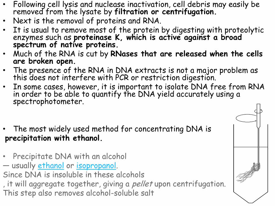
• Following cell lysis and nuclease inactivation, cell debris may easily be removed from the lysate by filtration or centrifugation.
• Next is the removal of proteins and RNA. • It is usual to remove most of the protein by digesting with proteolytic
enzymes such as proteinase K, which is active against a broad spectrum of native proteins.
• Much of the RNA is cut by RNases that are released when the cells are broken open.
• The presence of the RNA in DNA extracts is not a major problem as this does not interfere with PCR or restriction digestion.
• In some cases, however, it is important to isolate DNA free from RNA in order to be able to quantify the DNA yield accurately using a spectrophotometer.
• The most widely used method for concentrating DNA is precipitation with ethanol. • Precipitate DNA with an alcohol — usually ethanol or isopropanol. Since DNA is insoluble in these alcohols , it will aggregate together, giving a pellet upon centrifugation. This step also removes alcohol-soluble salt

• It is essential to inactivate DNases, which will otherwise digest DNA.
• Ethylene diamine tetraacetate (EDTA) is a chelating agent that acts to remove the divalent cations from the solution, thus inactivating DNase I and preventing it from degrading the genomic DNA.

What are the essential components of a DNA extraction Procedure?
1. Maximize DNA recovery
2. Remove inhibitors
3. Remove or inhibit nucleases
4. Maximize the quality of DNA
5. Double strand vs. Single strand (RFLP or PCR)
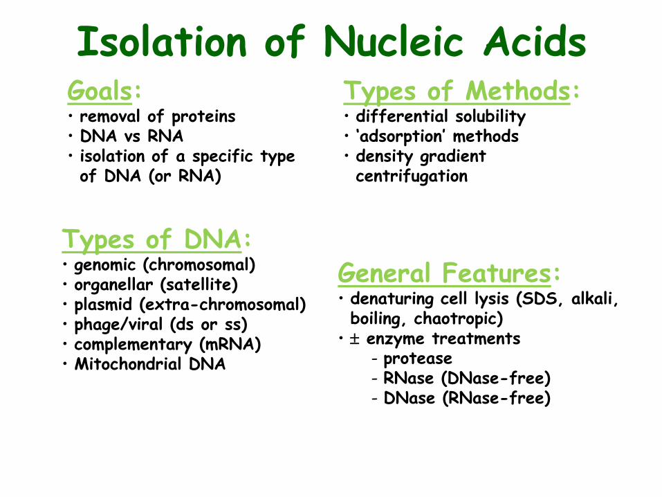
Isolation of Nucleic Acids Goals: • removal of proteins • DNA vs RNA • isolation of a specific type of DNA (or RNA)
Types of Methods: • differential solubility • ‘adsorption’ methods • density gradient centrifugation
Types of DNA: • genomic (chromosomal) • organellar (satellite) • plasmid (extra-chromosomal) • phage/viral (ds or ss) • complementary (mRNA) • Mitochondrial DNA
General Features: • denaturing cell lysis (SDS, alkali, boiling, chaotropic)
• enzyme treatments protease RNase (DNase-free) DNase (RNase-free)

UV‒Visible Absorbance Spectrophotometric
Measurement of DNA Concentration and Purity

DNA Replication in Cells
• DNA replication is copying DNA
• DNA replication is semi-conservative (i.e. one strand of the DNA is used as the template for the growth of a new DNA strand)
• This process occurs with very few errors (on average there is one error per 1 billion nucleotides copied)
• More than a dozen enzymes and proteins participate in DNA replication
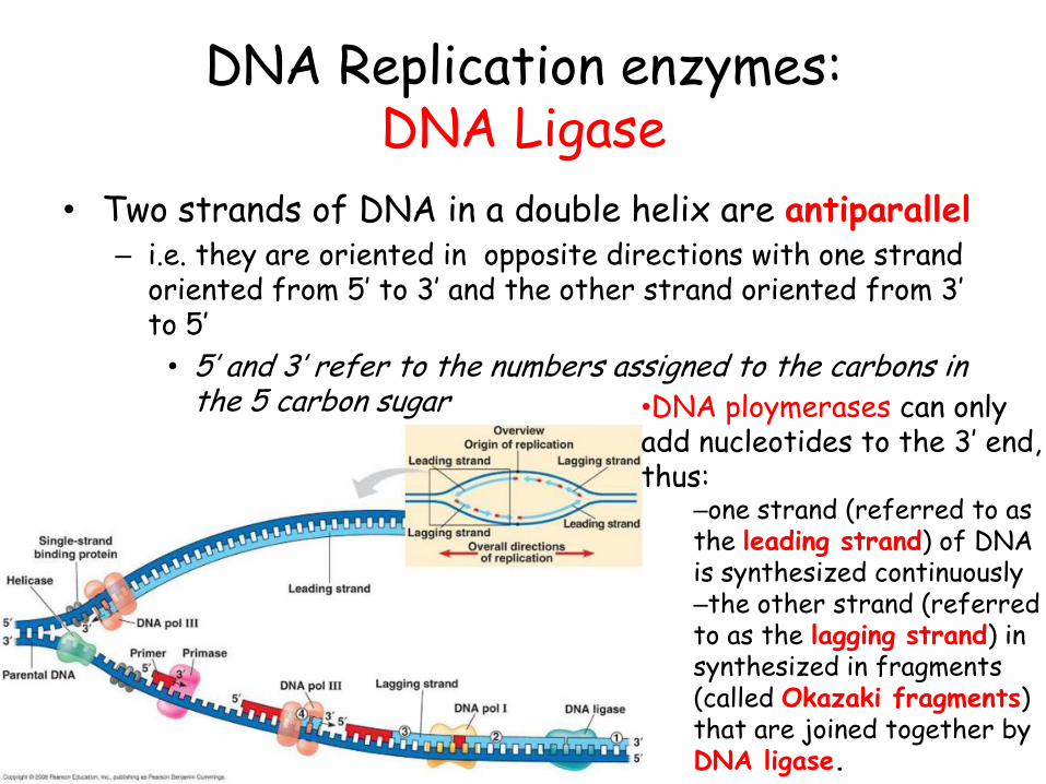
DNA Replication enzymes: DNA Ligase
• Two strands of DNA in a double helix are antiparallel – i.e. they are oriented in opposite directions with one strand
oriented from 5’ to 3’ and the other strand oriented from 3’ to 5’
• 5’ and 3’ refer to the numbers assigned to the carbons in the 5 carbon sugar •DNA ploymerases can only
add nucleotides to the 3’ end, thus:
–one strand (referred to as the leading strand) of DNA is synthesized continuously –the other strand (referred to as the lagging strand) in synthesized in fragments (called Okazaki fragments) that are joined together by DNA ligase.
DNA Replication enzymes: Helicase, Topoisomerase and Single-strand binding protein
• Helicase separates two parallel DNA strands
• Topoisomerase regulate the overwinding or underwinding of DNA and relieves the stress of this twisting
• Single-strand binding protein binds to and stabilizes the unpaired DNA strands

Polymerase chain reaction (PCR)
• in vitro version of DNA Replication
• Multiple copies of specific DNA sequence – ‘Molecular photocopying’

Polymerase chain reaction
• “discovered” in 1983 by Kary Mullis
• 1993: Nobel Prize for Chemistry

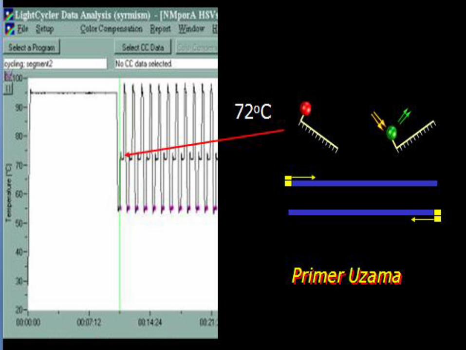
• Denaturation – Heat to separate
double strands – This occurs at 95 ºC – Mimics the function
of helicase in the cell.
3’ 5’
5’ 3’
Stages in PCR
5’ 3’
5’ 3’
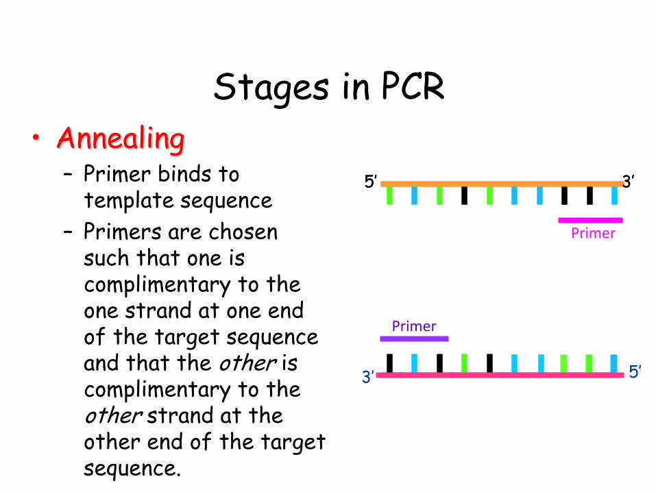
• Annealing – Primer binds to
template sequence
– Primers are chosen such that one is complimentary to the one strand at one end of the target sequence and that the other is complimentary to the other strand at the other end of the target sequence.
5’ 3’
Stages in PCR
3’ 5’
5’ 3’
Primer
Primer
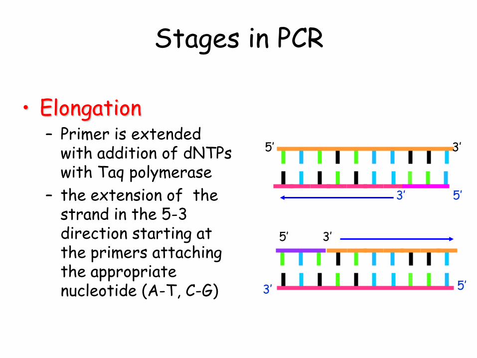
• Elongation – Primer is extended
with addition of dNTPs with Taq polymerase
– the extension of the strand in the 5-3 direction starting at the primers attaching the appropriate nucleotide (A-T, C-G)
Stages in PCR
3’ 5’
5’ 3’
5’ 3’
5’ 3’

Steps of a single PCR cycle involve:
• –DNA denaturation: expose the DNA template to high
temperatures to separate the two DNA strands and allow access by DNA polymerase and PCR primers.
• –Primer annealing: lower the temperature to allow primers to anneal to their complementary sequence.
• –Primer extension: adjust the temperature for optimal thermostable DNA polymerase activity to extend primers.
•PCR uses a thermostable DNA polymerase so that the
DNA polymerase is not heat-inactivated during the DNA denaturation step. Taq DNA polymerase is the most commonly used DNA polymerase for PCR.

The Size of the DNA Fragment Produced in PCR is Dependent on the Primers
• The PCR reaction will amplify the DNA section between the two primers.
• If the DNA sequence is known, primers can be developed to amplify any piece of an organism’s DNA.
Forward primer
Reverse primer
Size of fragment that is amplified

PCR amplification
2n

1st Cylce
2nd Cylce
35th Cylce 235 copies

Genomic DNA

Reagents for PCR
What we need in the laboratory:
• DNA template
• Primers
• DNA polymerase
• Buffer
• dNTPS (bases)
• MgCl2
Reagents for PCR
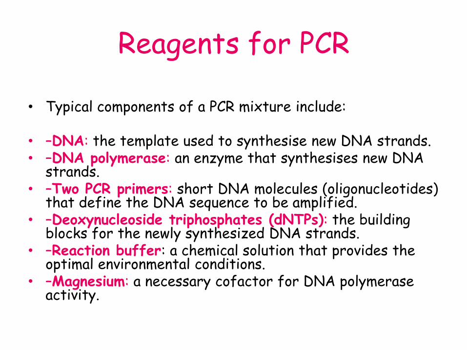
• Typical components of a PCR mixture include:
• –DNA: the template used to synthesise new DNA strands. • –DNA polymerase: an enzyme that synthesises new DNA
strands. • –Two PCR primers: short DNA molecules (oligonucleotides)
that define the DNA sequence to be amplified. • –Deoxynucleoside triphosphates (dNTPs): the building
blocks for the newly synthesized DNA strands. • –Reaction buffer: a chemical solution that provides the
optimal environmental conditions. • –Magnesium: a necessary cofactor for DNA polymerase
activity.

Reagents for PCR
• Primers – ssDNA sequences flanking region of interest – Present in excess – up to 0.5mM
• (too high – primer dimers)
• Primer Design – Homology of primers – Length (18-28bp) – GC content (45-60%) Tm=2X(A+T)+
4X(C+G)
Reagents for PCR • DNA polymerase • Enzyme responsible for copying the sequence starting at the
primer from the single DNA strand by adding nucleoside triphosphates to the 3’ end of the growing strand
• DNA polymerase uses each strand as a template to synthesize new strands of DNA, complementary order of nucleotides.
• This enzyme is heat-tolerant thermally tolerant (survives the melting temperature of DNA denaturation) which also means the process is more specific, higher temperatures result in less mismatch – more specific replication
• Many types available
• Some modified to allow hot start
• Some have long half life / stable at high temperature
• Some have high rate of processivity
Reagents for PCR
• MgCl2
• required for enzyme activation and amplification
• It stabilizes dsDNA and raises the Tm
• Mg2+ concentration controls the specificity of the reaction.

Reagents for PCR
• Buffer - providing a suitable chemical environment for optimum activity and stability of the DNA polymerase
• dNTPs (bases)

PCR - before the thermocycler
8 BORING hours per PCR!
95º C 5 min
35 times
55º C 3 min
72º C 5 min

PCR
THERMOCYCLER PCR tube with all the reagents
•The thermal cycler allows heating and cooling of the reaction tubes to control the temperature required at each reaction step. •Thin-walled reaction tubes permit favorable thermal conductivity to allow for rapid thermal equilibration. •Most thermal cyclers have heated lids to prevent condensation at the top of the reaction tube.

PCR is not usually 100% efficient and the reaction conditions may need to be optimized to increase yield, sensitivity of detection or amplification specificity. •The usual parameters to vary include: –Magnesium concentration –Primer annealing temperature –PCR primer design –DNA quality –DNA quantity •This is very important when trying to amplify a particular target from a population of other sequences (e.g., one gene from genomic DNA).

• Gel electrophoresis
• Fluorescent PCR
• Sequencing
• Blotting
• Short tandem repeat (STR) analysis
How do we analyse the PCR products?

Gel Electrophoresis
• Fragmentation products of differing length are separated
• Separation of DNA fragments • Separation based (mostly) on length
– longer molecules move slower.
• Size and shape
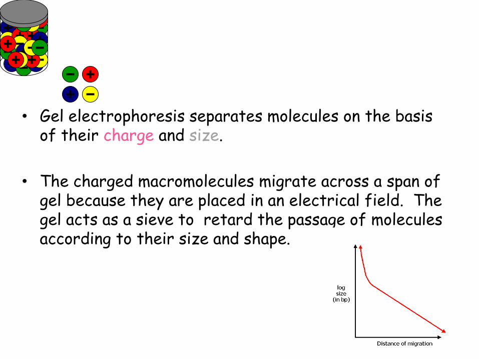
• Gel electrophoresis separates molecules on the basis
of their charge and size.
• The charged macromolecules migrate across a span of gel because they are placed in an electrical field. The gel acts as a sieve to retard the passage of molecules according to their size and shape.
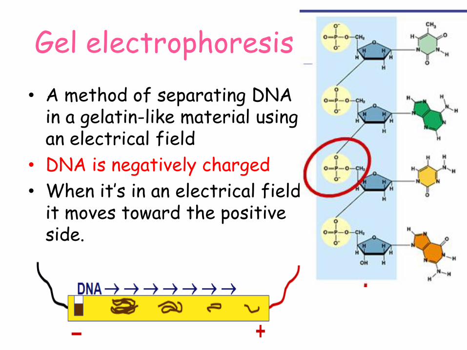
Gel electrophoresis
• A method of separating DNA in a gelatin-like material using an electrical field
• DNA is negatively charged
• When it’s in an electrical field it moves toward the positive side.

DNA (or RNA) samples loaded into wells
Gel Electrophoresis
http://cglab.ca/~morin/teaching/compbio/electro.html

How does gel electrophoresis work?
• DNA is forced by an electrical current through a firm gel – Phosphate group in DNA is negatively charged
so it is moved towards a positive electrode by the current
– Longer fragments have more nucleotides • So have a larger molecular weight • So are bigger in size • So aren’t able to pass through the small holes in the
gel and get hung up at the beginning of the gel
– Shorter fragments are able to pass through and move farther along the gel
– Fragments of intermediate length travel to about the middle of the gel

How does gel electrophoresis work?
• DNA fragments are then visualized in the gel with a special dye
• The number of nucleotides are then estimated by comparing it to a known sample of DNA fragments which is run through the gel at the same time ladder

– Sample of DNA fragments – Known sample of DNA fragments
• DNA ladder
– Gel • Agarose
– Dye to visualize the movement of the sample as it is traveling through the gel • Loading dye
– Dye to visualize DNA after it has traveled to its final spot in the gel • Ethidium bromide
– Buffer
Reagents Needed for gel electrophoresis

• The migrated DNA is visualised under UV light with the help of an intercalating dye, ethidium bromide, which fluoresces when irradiated with UV
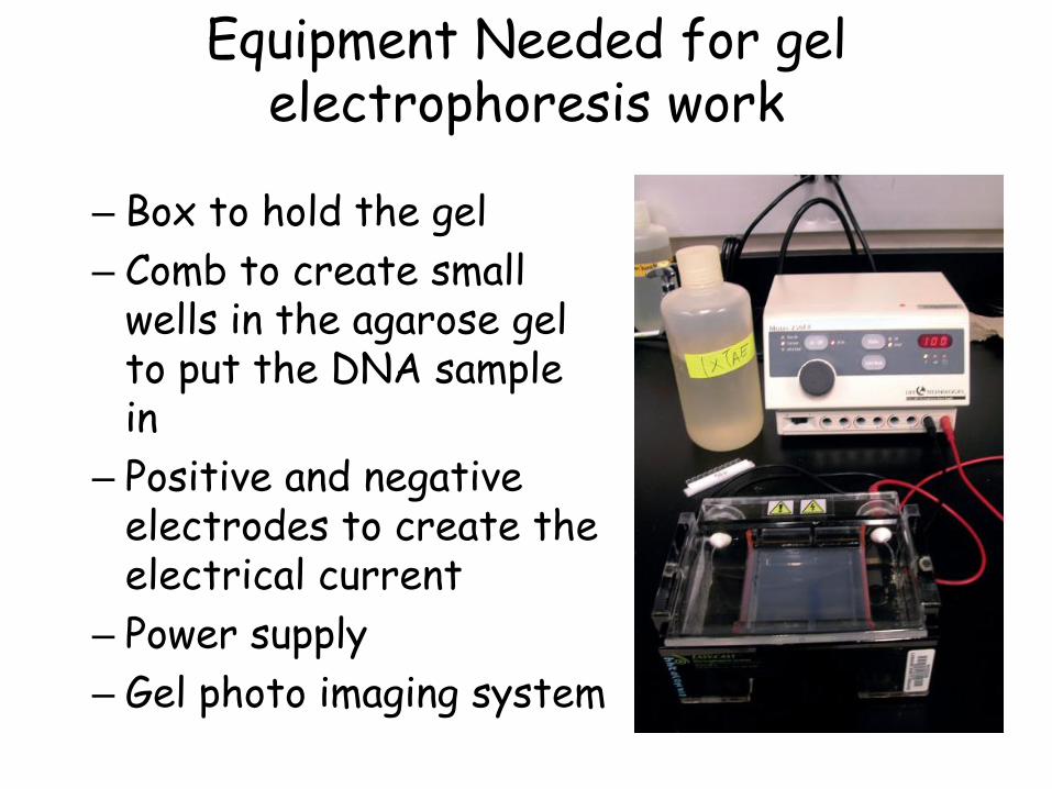
– Box to hold the gel
– Comb to create small wells in the agarose gel to put the DNA sample in
– Positive and negative electrodes to create the electrical current
– Power supply
– Gel photo imaging system
Equipment Needed for gel electrophoresis work
What are the different types of PCRs?
MOLECULAR AMPLIFICATION
TECHNIQUES
• Nucleic acid (NA) amplification methods fall into 3 categories – Target amplification systems
– Probe amplification systems
– Signal amplification
Target Amplification Methods
• PCR – – PCR using specific probes – RT PCR – Nested PCR-increases sensitivity, uses two sets of
amplification primers, one internal to the other – Multiplex PCR-two or more sets of primers specific for
different targets – Arbitrarily Primed PCR/Random Primer PCR
• NASBA - Nucleic Acid Sequence-Based Amplification
• TMA – Transcription Mediated Amplification • SDA - Strand Displacement Amplification

Signal and Probe Amplification Methods
• Signal Amplification – bDNA – Branched DNA probes
– Hybrid Capture – Anti-DNA-RNA hybrid antibody
• Probe Amplification – LCR – Ligase Chain Reaction
– Cleavase Invader – FEN-1 DNA polymerase (cleavase)

PCR Modifications
• Nested PCR
• Multiplex PCR
• Tailed primers
• Sequence-specific PCR
• Reverse-transcriptase PCR
• Long-range PCR
• Whole-genome amplification
• RAPD PCR (AP-PCR)
• Quantitative real-time PCR
RT-PCR
• PCR of cDNA is used to detect specific transcripts in RNA sample.
• In this procedure, known as RT-PCR, reverse transcriptase is used to copy all of the mRNAs in an RNA sample into cDNA.
• Usually, oligo dT molecules, that anneal to the poly A tails of the mRNA, are used as primers.
• This single stranded cDNA can then be amplified by PCR using primers that anneal to a specific transcript sequence.

• The amplified DNA fragments that are produced can be analysed by agarose gel electrophoresis or fluorescent PCR or real time PCR.
• The amount of amplified fragment produced is proportional to the amount of target mRNA in the original RNA sample.
• RT-PCR is extremely sensitive and can be used to detect very rare mRNA species.
RT-PCR

Synthesize cDNA Using RT
Reverse
Transcriptase
mRNA
cDNA

Reverse Transcription • mRNA can be copied to
complementary DNA sequence (cDNA) using reverse transcriptase—a DNA polymerase that uses ssRNA as template.
• Processed mRNA will match protein coding sequence while unprocessed (nuclear) mRNA will contain intron sequences.

What is Real-Time PCR?
The Polymerase Chain Reaction (PCR) is a process for the amplification of specific fragments of DNA.
Real-Time PCR a specialized technique that allows a PCR reaction to be visualized “in real time” as the reaction progresses.
As we will see, Real-Time PCR allows us to measure minute amounts of DNA sequences in a sample!

• Real-Time PCR combines DNA amplification with real time amplified product detection in a single tube.
• More specific then gel or hybridization assays.
• Less time consuming • Quantitative results as opposed to semi-
quantitative or qualitative results. • Human (higher primate) or Y chromosome
specific • Monitors for PCR inhibition
Overview of Real -Time PCR
What is Real-
Time PCR used for?
Real-Time PCR has become a cornerstone of molecular biology:
• Gene expression analysis
– Cancer research
– Drug research
• Disease diagnosis and management
– Viral quantification
• Food testing
– Percent GMO food
• Animal and plant breeding
– Gene copy number

Real-Time PCR in Gene Expression
Analysis
Example: BRCA1 Expression Profiling
BRCA1 is a gene involved in tumor suppression.
BRCA1 controls the expression of other genes.
In order to monitor level of expression of BRCA1, real-time PCR is used.
DNA
mRNA
Protein
BRCA1
Real-Time PCR in
Disease Management
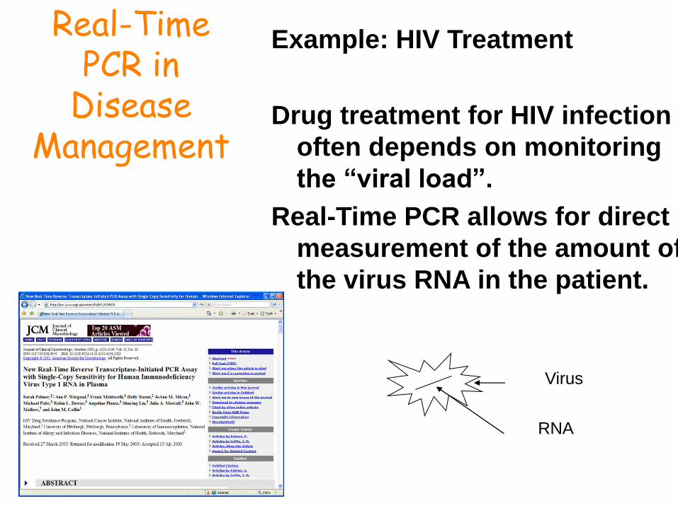
Example: HIV Treatment
Drug treatment for HIV infection
often depends on monitoring
the “viral load”.
Real-Time PCR allows for direct
measurement of the amount of
the virus RNA in the patient.
Virus
RNA
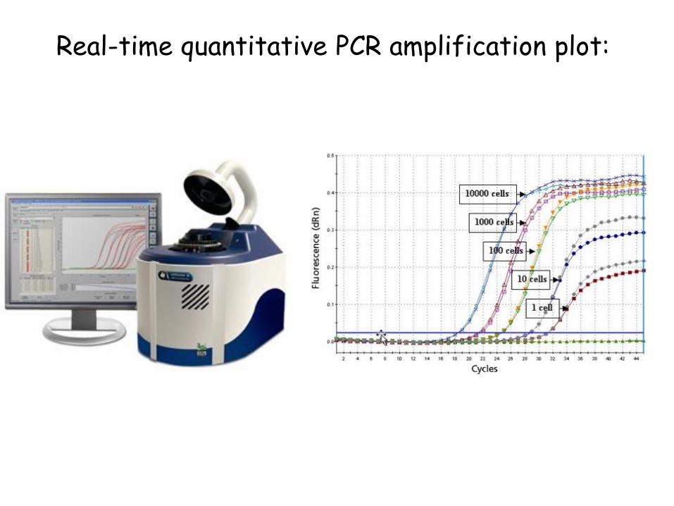
Real-time quantitative PCR amplification plot:

How do We
Measure DNA in a
PCR Reaction?
We use reagents that fluoresce in the presence of amplified DNA!
Ethidium bromide and SYBR Green I dye are two such reagents.
They bind to double-stranded DNA and emit light when illuminated with a specific wavelength.
SYBR Green I dye fluoresces much more brightly than ethidium.
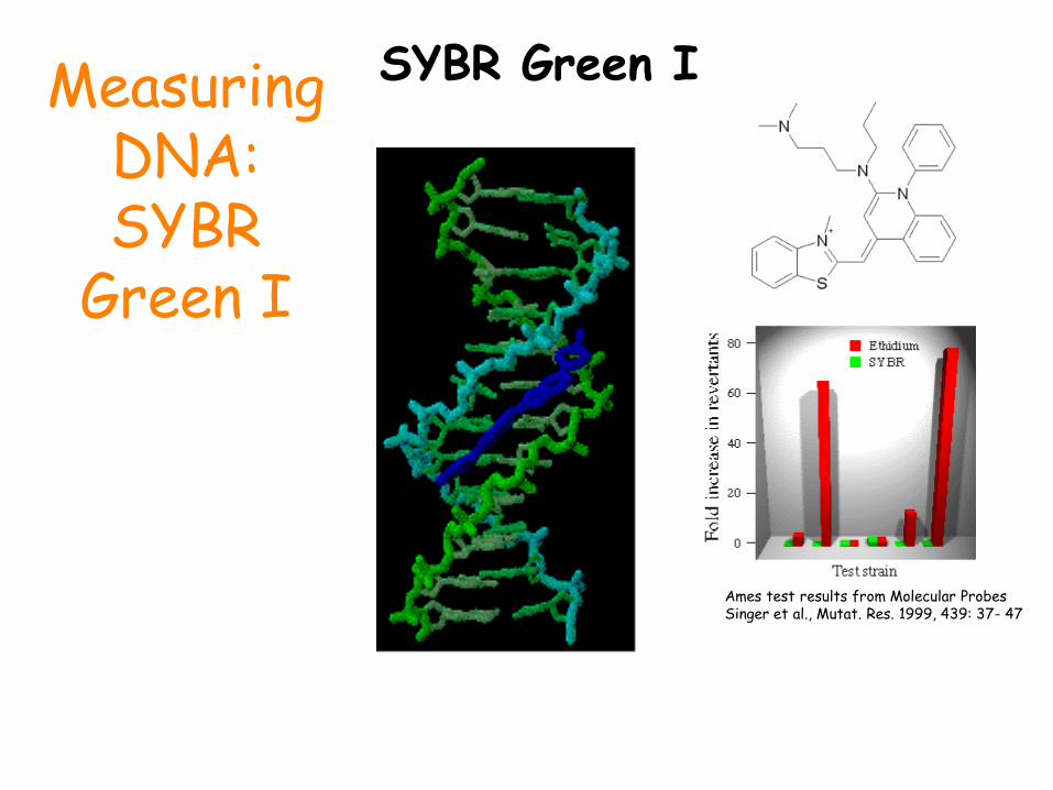
Measuring DNA: SYBR
Green I
SYBR Green I
Ames test results from Molecular Probes Singer et al., Mutat. Res. 1999, 439: 37- 47

What Type of Instruments are used with Real-Time PCR?
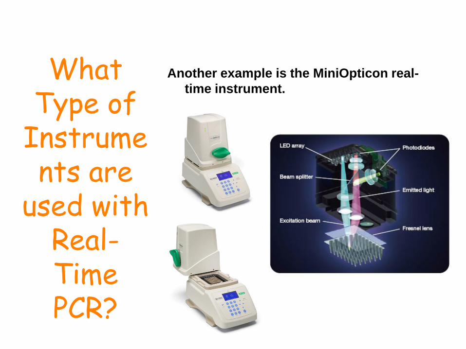
Real-time PCR instruments consist of THREE
main components:
1. Thermal Cycler (PCR machine)
2. Optical Module (to detect fluorescence in the tubes during the run)
3. Computer (to translate the fluorescence data into meaningful results)

What Type of
Instruments are
used with Real-Time PCR?
An example of such an instrument is the Bio-Rad iQ5 real-time PCR instrument.
What Type of
Instruments are
used with Real-Time PCR?
Another example is the MiniOpticon real-
time instrument.

What Type of
Software is used with Real-Time
PCR?
The real-time software converts the fluorescent signals in each well to meaningful data.
1.Set up PCR protocol.
2.Set up plate layout.
3.Collect data.
4.Analyze data.
1 2 3,4

Cystic fibrosis
Figure 1. Examples of specific molecular beacon fluorescence increase during real-time PCR in samples
containing single lymphoblasts homozygous normal for CF (green), heterozygous DF508 (blue), or
homozygous DF508 (red). (A) Fluorescent signal from the molecular beacon detecting the normal allele.
(B) Fluorescent signal from the molecular beacon detecting the DF508 allele. Dashed lines indicate the
threshold of 200 units (~10 SD above baseline readings) used for determining CT values.


Kalkan R . Et al 2015

Allele-specific PCR • Allele-specific PCR is used to identify or utilize single
nucleotide polymorphisms (SNPs: single base differences in DNA).
• It requires prior knowledge of a DNA sequence, including differences between alleles and uses primers whose 3' ends encompass the SNP.
• PCR amplification under stringent conditions is much less efficient in the presence of a mismatch between template and primer, so successful amplification with an SNP-specific primer signals presence of the specific SNP in a sequence.

• Nested PCR increases the specificity of DNA amplification by reducing background due to non-specific amplification of DNA.
• Two sets of primers are being used in two successive PCR.
– In the first reaction, one pair of primers is used to generate DNA products, which besides the intended target, may still consist of non-specifically amplified DNA fragments.
– The product(s) are then used in a second PCR with a set of primers whose binding sites are completely or partially different from and located 3' of each of the primers used in the first reaction.
• Nested PCR is often more successful in specifically amplifying long DNA fragments than conventional PCR, but it requires more detailed knowledge of the target sequences.
Nested PCR

Methylation-specific PCR (MSP): • MSP is used to detect methylation of CpG islands in
genomic DNA. 1- DNA is first treated with sodium bisulfite, which converts
unmethylated cytosine bases to uracil, which is recognized by PCR primers as thymine.
2- Two PCRs are then carried out on the modified DNA, using primer sets identical except at any CpG islands within the primer sequences.
At these points, one primer set recognizes DNA with cytosines to amplify methylated DNA
One set recognizes DNA with uracil or thymine to amplify unmethylated DNA.


Fluorescent PCR
• Primers labelled at 5’terminus with fluorescent molecule
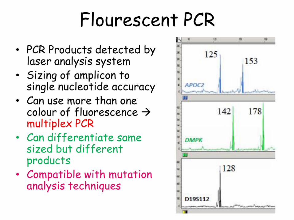
Flourescent PCR
• PCR Products detected by laser analysis system
• Sizing of amplicon to single nucleotide accuracy
• Can use more than one colour of fluorescence multiplex PCR
• Can differentiate same sized but different products
• Compatible with mutation analysis techniques
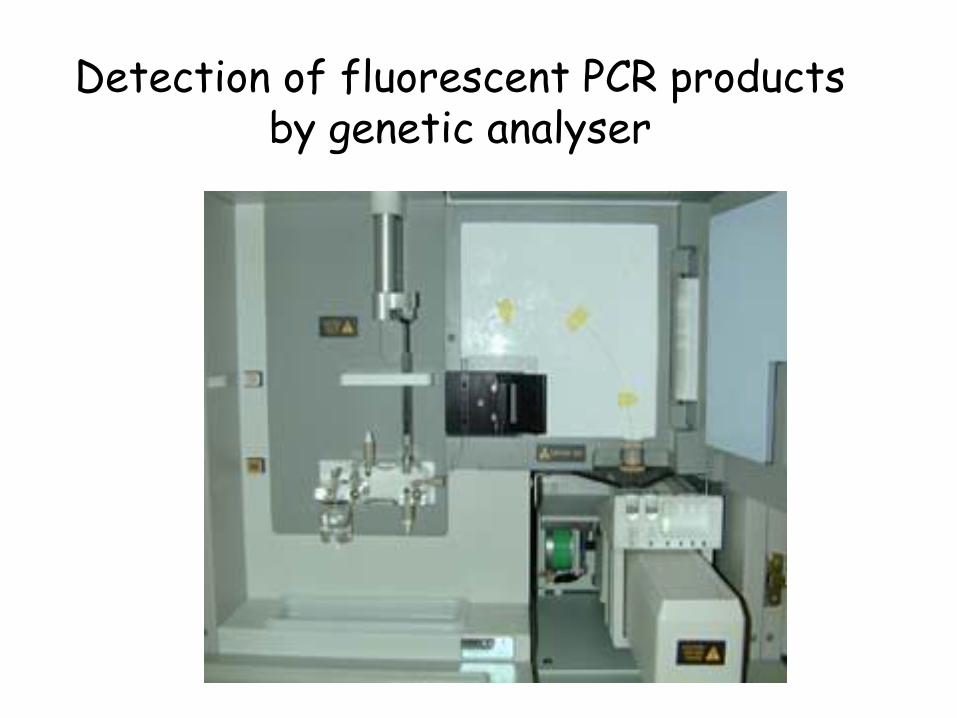
Detection of fluorescent PCR products by genetic analyser

Detection of fluorescent PCR products by genetic analyser
Time / Length Short time/Short fragments Long time/Long fragments
Amount
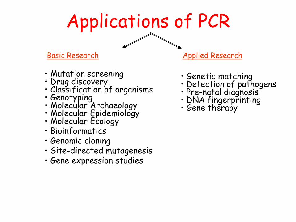
Applications of PCR
Basic Research Applied Research
• Genetic matching • Detection of pathogens • Pre-natal diagnosis • DNA fingerprinting • Gene therapy
• Mutation screening • Drug discovery • Classification of organisms • Genotyping • Molecular Archaeology • Molecular Epidemiology • Molecular Ecology • Bioinformatics • Genomic cloning • Site-directed mutagenesis • Gene expression studies
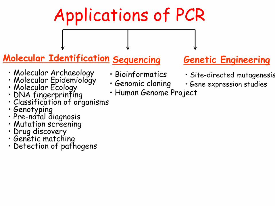
Applications of PCR
Molecular Identification Sequencing Genetic Engineering
• Molecular Archaeology • Molecular Epidemiology • Molecular Ecology • DNA fingerprinting • Classification of organisms • Genotyping • Pre-natal diagnosis • Mutation screening • Drug discovery • Genetic matching • Detection of pathogens
• Bioinformatics • Genomic cloning • Human Genome Project
• Site-directed mutagenesis • Gene expression studies
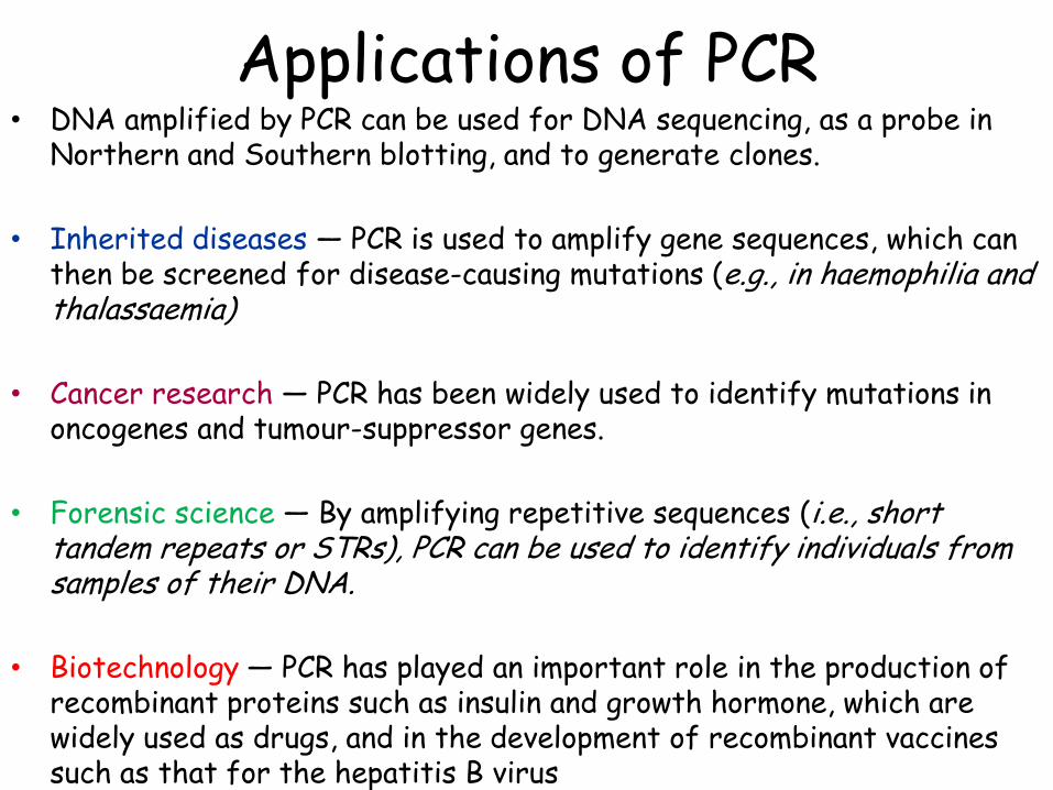
Applications of PCR • DNA amplified by PCR can be used for DNA sequencing, as a probe in
Northern and Southern blotting, and to generate clones.
• Inherited diseases ― PCR is used to amplify gene sequences, which can then be screened for disease-causing mutations (e.g., in haemophilia and thalassaemia)
• Cancer research ― PCR has been widely used to identify mutations in oncogenes and tumour-suppressor genes.
• Forensic science ― By amplifying repetitive sequences (i.e., short tandem repeats or STRs), PCR can be used to identify individuals from samples of their DNA.
• Biotechnology ― PCR has played an important role in the production of recombinant proteins such as insulin and growth hormone, which are widely used as drugs, and in the development of recombinant vaccines such as that for the hepatitis B virus

PCR and Disease
• Primers can be created that will only bind and amplify certain alleles of genes or mutations of genes
• This is the basis of diagnostic tests and genetic counseling
• PCR is used for diagnosis of genetic diseases. • Some diseases that can be diagnosed with the
help of PCR: • Huntington's disease • Cystic fibrosis • Human immunodeficiency virus
• Detection of Pathogenic Organisms in Clinical Samples
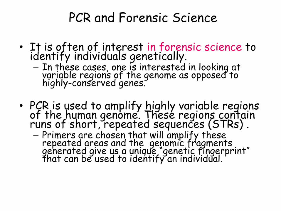
PCR and Forensic Science
• It is often of interest in forensic science to identify individuals genetically. – In these cases, one is interested in looking at
variable regions of the genome as opposed to highly-conserved genes.
• PCR is used to amplify highly variable regions
of the human genome. These regions contain runs of short, repeated sequences (STRs) . – Primers are chosen that will amplify these
repeated areas and the genomic fragments generated give us a unique “genetic fingerprint” that can be used to identify an individual.

DNA Fingerprinting Paternity testing
Maternal DNA
Paternal DNA
Possible
Genotypes of the children

Applications
Diagnosing Disease

Applications
Paternity Testing

Applications Forensics

Crime Scene
Victim
Suspect
Applications

Advantages of PCR • Minute amounts of DNA template may be used from as little as a single cell. • DNA degraded to fragments only a few hundred base pairs in length can serve as effective templates for amplification. • Large numbers of copies of specific DNA sequences can be amplified simultaneously with multiplex PCR reactions. • Contaminant DNA, such as fungal and bacterial sources, will not amplify because human-specific primers are used. • Commercial kits are now available for easy PCR reaction setup and amplification.

Potential Pitfalls of PCR
• The target DNA template may not amplify due to the presence of PCR inhibitors in the extracted DNA
• Amplification may fail due to sequence changes in the primer binding region of the genomic DNA template
• Contamination from other human DNA sources besides the forensic evidence at hand or previously amplified DNA samples is possible without careful laboratory technique and validated protocols

