dinesh gupta structural and computational biology group … · dinesh gupta structural and...
TRANSCRIPT

Insilico drug designing
Dinesh GuptaStructural and Computational Biology GroupICGEB
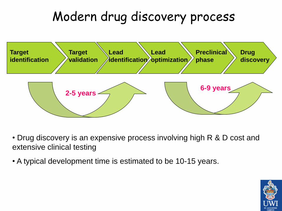
Modern drug discovery process
Target
identification
Target
validation
Lead
identification
Lead
optimization
Preclinical
phase
Drug
discovery
2-5 years
• Drug discovery is an expensive process involving high R & D cost and
extensive clinical testing
• A typical development time is estimated to be 10-15 years.
6-9 years

Drug discovery technologies
• Target identification– Genomics, gene expression profiling and proteomics
• Target Validation– Gene knock-out, inhibition assay
• Lead Identification– High throughput screening, fragment based screening, combinatorial
libraries
• Lead Optimization– Medicinal chemistry driven optimization, X-ray crystallography, QSAR,
ADME profiling (bioavailability)
• Pre Clinical Phase– Pharmacodynamics (PD), Pharmacokinetics (PK), ADME, and toxicity
testing through animals
• Clinical Phase– Human trials


Identify and validate target
Clone gene encoding target
Rational Approach to Drug Discovery
Express target
Synthesize modified lead compounds
Crystal structures/MM of target and target/inhibitor complexes
Preclinical trials
Identify lead compounds
Toxicity & pharmacokinetic studies
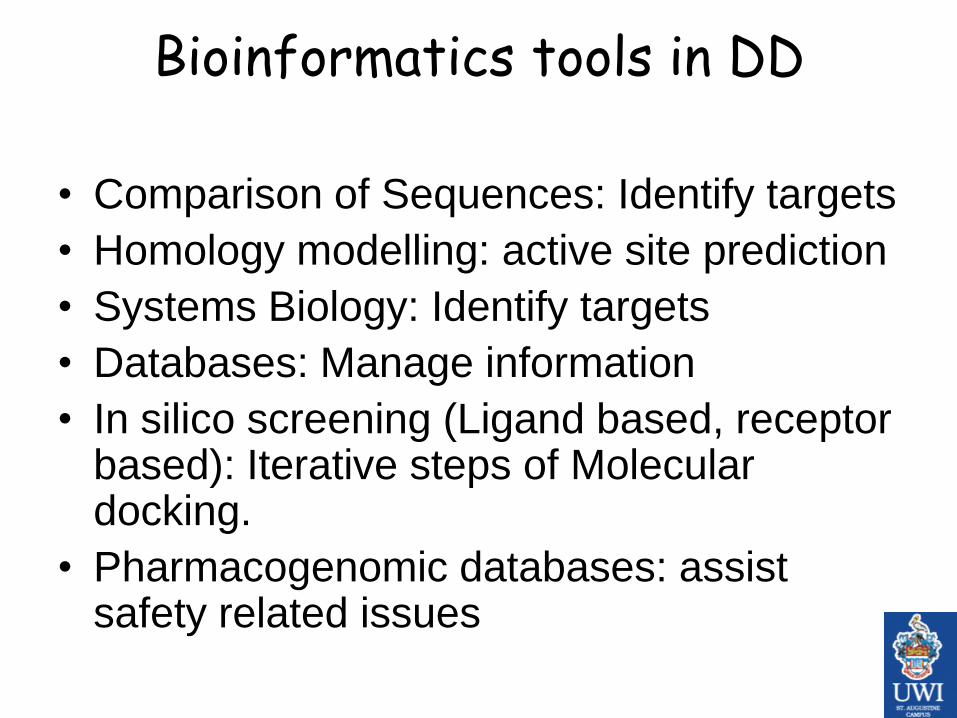
Bioinformatics tools in DD
• Comparison of Sequences: Identify targets
• Homology modelling: active site prediction
• Systems Biology: Identify targets
• Databases: Manage information
• In silico screening (Ligand based, receptor based): Iterative steps of Molecular docking.
• Pharmacogenomic databases: assist safety related issues

Published by AAAS
J. Drews Science 287, 1960 -1964 (2000)
Currently used drug targets
This information is used by bioinformaticians to narrow the search in the groups

Insilico methods in Drug Discovery
• Molecular docking
• Virtual High through put screening.
• QSAR (Quantitative structure-activity relationship)
• Pharmacophore mapping
• Fragment based screening
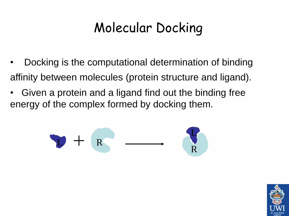
Molecular Docking
RL
• Docking is the computational determination of binding
affinity between molecules (protein structure and ligand).
• Given a protein and a ligand find out the binding free
energy of the complex formed by docking them.
L
R

Molecular Docking: classification
• Docking or Computer aided drug designing can be
broadly classified
– Receptor based methods- make use of the structure of the target
protein.
– Ligand based methods- based on the known inhibitors

Receptor based methods
• Uses the 3D structure of the target receptor to search for the potential candidate compounds that can modulate the target function.
• These involve molecular docking of each compound in the chemical database into the binding site of the target and predicting the electrostatic fit between them.
• The compounds are ranked using an appropriate scoring function such that the scores correlate with the binding affinity.
• Receptor based method has been successfully applied in many targets

Ligand based strategy
• In the absence of the structural information of the target,
ligand based method make use of the information
provided by known inhibitors for the target receptor.
• Structures similar to the known inhibitors are identified
from chemical databases by variety of methods,
• Some of the methods widely used are similarity and
substructure searching, pharmacophore matching or 3D
shape matching.
• Numerous successful applications of ligand based
methods have been reported

Ligand based strategy
Search for similar compounds
database known actives structures found

Binding free energy
• Binding free energy is calculated as the sum of the following energies
- Electrostatic Energy
- Vander waals Energy
- Internal Energy change due to flexible deformations
- Translational and rotational energy
• Lesser the binding free energy of a complex the more stable it is

Basic binding mechanism
Complementarities between the ligand and the
binding site:
• Steric complementarities, i.e. the shape of the
ligand is mirrored in the shape of the binding site.
• Physicochemical complementarities

Components of molecular docking
A) Search algorithm
• To find the best conformation of the ligand
and the protein system.
• Rigid and flexible docking
B) Scoring function
• Rank the ligands according to the interaction energy.
• Based on the energy force-field function.


Success with vHTS
• Dihydrofolate reductase inhibitor (1992)
• HIV-protease (1992)
• Phospholypase A2 (1994)
• Thrombine (1996)
• Carbonic anhydrase inhibitors(2002)

Virtual High Throughput Screening
• Less expensive than High Throughput Screening
• Faster than conventional screening
• Scanning a large number of potential drug like
molecules in very less time.
• HTS itself is a trial and error approach but can be
better complemented by virtual screening.

QSAR
• QSAR is statistical approach that attempts to relate
physical and chemical properties of molecules to their
biological activities.
• Various descriptors like molecular weight, number of
rotatable bonds LogP etc. are commonly used.
• Many QSAR approaches are in practice based on the
data dimensions.
• It ranges from 1D QSAR to 6D QSAR.

Pharmacophore mapping
• It is a 3D description of a pharmacophore, developed by specifying the nature of the key pharmacophoric features and the 3D distance map among all the key features.
• A Pharmacophore map can be generated by superposition of active compounds to identify their common features.
• Based on the pharmacophore map either de novo design or 3D database searching can be carried out.

Modeling and informatics in drug design

Increased application of structure based drug designing is facilitated by:
Growth of targets number
Growth of 3D structures determination (PDB
database)
Growth of computing power
Growth of prediction quality of protein-
compound interactions

Summary: role of Bioinformatics?
• Identification of homologs of functional
proteins (motif, protein families, domains)
• Identification of targets by cross species
examination
• Visualization of molecular models
• Docking, vHTS
• QSAR, Pharmacophore mapping

Example: use of Bioinformatics in Drug discovery
Identification of novel drug targets against human malaria

Malaria – A global problem!
• Malaria causes at least 500 million clinical cases and more than one million deaths each year.
• A child dies of malaria every 30 seconds.
• Out of four Plasmodium species causing human malaria, P.falciparum poses most serious threat: because of its virulence, prevalence and drug resistance.
• Malaria takes an economic toll - cutting economic growth rates by as much as 1.3% in countries with high disease rates.
• There are four types of human malaria:– Plasmodium falciparum
– Plasmodium vivax
– Plasmodium malariae
– Plasmodium ovale.

• Approximately half of the world's population is at risk of malaria, particularly those living in lower-income countries.
• Today, there are 109 malaria affected countries in 4 regions

a) Chloroquine
b) Quinine
c) Artemether
d) Sodium artesunate
e) Dihydroartemisinin
f) Pyrimethamine
g) Sulfadoxine
h) Mefloquine
i) Halofantrine
j) Primaquine
k) Tafenoquine
l) Chlorproguanil
m) Dapsone
Chemical structures of drugs in widely used for treatment of Malaria


http://malaria.who.i
nt/docs/adpolicy_t
g2003.pdf

Problems with the existing drugs
• Drug resistance is most common problem
• Adverse effects (Shock and cardiac arrhythmias
caused by Chloroquine)
• Poor patient compliance (Quinine tastes very
unpleasant, causes dizziness, nausea etc.)
• High cost of production for some effective drugs
(Atovaquine).
• Urgent need for identification of novel drug
targets which are effective and affordable.

Strategies for drug target identification in P. falciparum
• Parasite culture for functional assays are difficult and expensive.
Making computational approaches more relevant.
• Malaria remains a neglected disease- very few stake holders!
• Availability of the genomic data of P.falciparum and H.sapiens has
facilitated the effective application of comparative genomics.
• Comparative genomics helps in the identification and exploitation of
different characteristic features in host and the parasite.
• Identification of specific metabolic pathways in P.
falciparum and targeting the crucial proteins is an attractive approach
of target based drug discovery.

Comparison of proteomes helps in identifying important indispensible parasite proteins
• Out of 5334 predicted
proteins in P. falciparum,
60% didn’t show any
similarity to known proteins.
• Hence assigning a
physiological functional role
to these hypothetical
proteins using
bioinformatics approach still
remains a challenge.
A. gambiae
P. falciparum H. sapiens
Predicted
proteome

Large set of proteins with no/low
similarity
Novel drug target identification in P.falciparum
BlastP
~40% identity threshold for
three-dimensional
modeling
Relational
Database of
homology
models
476 P.falciparum
proteins
Human
proteome
Putative drug
targets in
P.falciparum
Comparative genomics studies
Literature search for all these proteins
Check for physiological and biochemical
functions; etc ..
Proteasome
machinery (ClpQY
and ClpAP) in
P.falciparum


Targets identified by comparison of proteins models
• Identification of two proteasomal proteins
of prokaryotic origin, not present in hosts.
• The protein degradation is an important
process in parasite development inside
host RBCs.

26S proteasome: eukaryotic type
•19S regulatory + 20S proteolytic particle
•Present only in Eukaryotes and archae
•Degrades ubiquitinated proteins
> 20 different proteins involved
20S proteasome
ClpQY system: prokaryotic type
•ClpY cap + ClpQ core particle
•Present only in prokaryotes
•No ubiquitination in prokaryote
•Substrate specificity is not known
•Only two proteins ClpQ & ClpY
Eukaryotic and prokaryotic proteasome machinery
ClpQ
ClpY
ClpYSubstrate protein
Peptides

ATP Dependent Protease Machinery
ClpQY (PfHslUV system)
• The HslUV complex in prokaryotes is composed of an
HslV threonine protease and HslU ATP-dependent
protease, a chaperone of Clp/Hsp100 family.
• HslV (ClpQ) subunits are arranged in form of two-stacked
hexameric rings and are capped by two HslU (ClpY)
hexamers at both ends.
• HslU (ClpY) hexamer recognizes and unfold peptide
substrates with an ATP dependent process, and
translocates them into HslV for degradation.
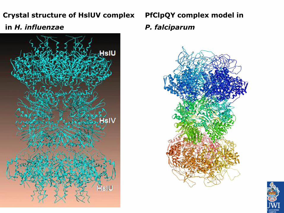
Crystal structure of HslUV complex
in H. influenzae
PfClpQY complex model in
P. falciparum

ATP Dependent Protease
machineries ClpQY (PfHslUV system)
• The HslUV complex in prokaryotes is composed of an HslV threonine protease and ATP-dependent protease HslU, a chaperone of clp/Hsp100 family.
• HslV subunits are arranged in the form of two-stacked hexameric rings and are capped by two HslU hexamers at both ends.
• In an ATP dependent process, HslU hexamer recognizes and unfold peptide substrates and translocate them into HslV for degradation.

MFIRNFVNIIGSQKSITKTIARNYFSDNSKLIIPRHGTTILCVRKNNEVCLIGDGMVSQGTMIVKGNAKKIRRLKDNILMGFAGATADCFTLLDKFETKIDEYPNQL
LRSCVELAKLWRTDRYLRHLEAVLIVADKDILLEVTGNGDVLEPSGNVLGTGSGGPYAMA
AARALYDVENLSAKDIAYKAMNIAADMCCHTNNNFICETL
For full length & matured active protein
Length : 207 aa (170)
Pro domain : 37aa
Important motifs found:
•TT at N terminal in mature protein
•GSGG common chymotrypsin protease signal.
•Lys(28) and Arg(35) are two conserved amino acids play some role in the activity.
PfClpQ component

PK_ClpQ TTILCVRKNNEVCLIGDGMVSQGTMIVKGNAKKIRRLKDNILMGFAGATADCFTLLDKFE
PV_ClpQ TTILCVRKNNEVCLIGDGMVSQGTMIVKGNAKKIRRLKDNILMGFAGATADCFTLLDKFE
PF_ClpQ TTILCVRKNNEVCLIGDGMVSQGTMIVKGNAKKIRRLKDNILMGFAGATADCFTLLDKFE
PY_ClpQ TTILCVRKNNEVCLIGDGMVSQGTMIVKGNAKKIRRLKDNILMGFAGATADCFTLLDKFE
PB_ClpQ TTILCVRKNNEVCLIGDGMVSQGTMIVKGNAKKIRRLKDNILMGFAGATADCFTLLDKFE
************************************************************
PK_ClpQ TKIDEYPDQLLRSCVELAKLWRTDRYLRHLEAVLIVADKDVLLEVTGNGDVLEPSGNVLG
PV_ClpQ TKIDEYPDQLLRSCVELAKLWRTDRYLRHLEAVLIVADKDVLLEVTGNGDVLEPSGNVLG
PF_ClpQ TKIDEYPNQLLRSCVELAKLWRTDRYLRHLEAVLIVADKDILLEVTGNGDVLEPSGNVLG
PY_ClpQ TKIDEYPDQLLRSCVELAKLWRTDRYLRHLEAVLIVADKDTLLEVTGNGDVLEPSGNVLG
PB_ClpQ TKIDEYPDQLLRSCVELAKLWRTDRYLRHLEAVLIVADKDTLLEVTGNGDVLEPSGNVLG
*******:******************************** *******************
PK_ClpQ TGSGGPYAIAAARALYDVENLSAKDIAYKAMNIAADMCCHTNNNFICETL
PV_ClpQ TGSGGPYAIAAARALYDVENLSAKDIAYKAMNIAADMCCHTNNNFICETL
PF_ClpQ TGSGGPYAMAAARALYDVENLSAKDIAYKAMNIAADMCCHTNNNFICETL
PY_ClpQ TGSGGPYAMAAARALYDIENLSAKDIAYKAMNIAADMCCHTNHNFICETL
PB_ClpQ TGSGGPYAIAAARALYDIENLSAKDIAYKAMNIAADMCCHTNHNFICETL
********:********:************************:*******
Homologs of PfClpQ protein in other Plasmodium spp

PfClpQ
1kyi
Conservation of catalytic residues
S125-G45-T1-K33
Homology modeling of PfClpQ
Structural alignment of PfClpQ and HslV
(H.influenzae)

E. coli S. enterica H. influenzae X. campestris W. pipientis P. falciparumT. brucei T. cruzi L. infantum
E. coli S. enterica H. influenzae X. campestris W. pipientis P. falciparumT. brucei T. cruzi L. infantum
E. coli S. enterica H. influenzae X. campestris W. pipientis P. falciparumT. brucei T. cruzi L. infantum
Homology Modeling of PfClpQ
•Most of the conserved residues in different bacterial species
were either identical or similar in PfClpQ

Km =19.18 mM
Cbz-GGL-AMC
Lactacystin
Activity assay for PfClpQ protein
0
50
100
150
1h 2h 3h 4h 5h 6h
Time
Threonine protease likeSubstrate:
Inhibitor:
Biochemical characterization of PfClpQ proteinA
MC
rel
ease
d (
m m
ole
s)
Substrate conc (mM)
Km = 58.22 mM Km =37.79 mM
Chymotrypsin like
Suc-LLVY-AMC
chymostatin
Peptidyl glutamyl hydrolase
Z-LLE-AMC
MG132
0
100
200
300
400
500
30 60 90 120 150 180
Time in minutes
0
50
100
150
1h 2h 3h 4h 5h 6h
Time
AM
C r
elea
sed
(m
mo
les)
AM
C r
elea
sed
(m
mo
les)
Substrate conc (mM) Substrate conc (mM)
Fluorogenic
peptide
substrate
Fluorescence
Protease

Top 100 solutions
Out of top 40 only 10 compounds available for purchase
Drug-like compound
library (1,000,00)
Molecular
docking
Ligand docked into protein’s
active site
Insilico identification of novel inhibitors against PfClpQ , a novel drug target of P.falciparum by high throughput docking
PfclpQ

Phe46
Arg36
Val21
Gly49
Gly48
Ser22
Thr2
Thr50
ClpQ interaction with ligand identified by virtual screening
Crystal structure of
HslV complexed
with a vinyl sulfone
inhibitor
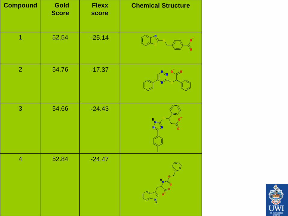
Compound Gold
Score
Flexx
score
Chemical Structure
1 52.54 -25.14
2 54.76 -17.37
3 54.66 -24.43
4 52.84 -24.47
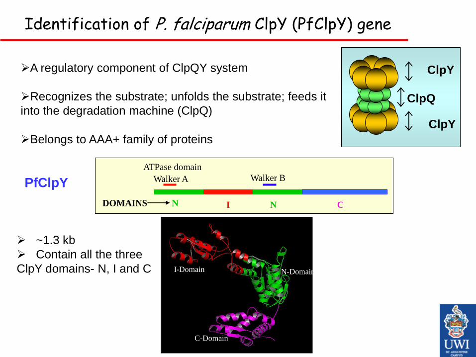
A regulatory component of ClpQY system
Recognizes the substrate; unfolds the substrate; feeds it
into the degradation machine (ClpQ)
Belongs to AAA+ family of proteins
Identification of P. falciparum ClpY (PfClpY) gene
PfClpY
~1.3 kb
Contain all the three
ClpY domains- N, I and C N-Domain
C-Domain
I-Domain
N I CNDOMAINS
Walker A Walker B
ATPase domain
ClpY
ClpY
ClpQ

Variation in I domain:
plays role in recognition of
different substrate
Homology of PfClpY protein with homologs in other organisms

Targeting the ClpQY interaction
Crystal structure of HslUV in H. influenzae Modeled ClpQY interaction in P.falciparum
J Biomol Struct Dyn. 2009 Feb;26(4):473-9

EXTRACTING THE
MICROARRAY DATA FROM
NCBI GEO
NORMALIZATION IF NECESSARY
OTHERWISE PREPARING EXCEL
FILES FOR WGCNA ANALYSIS
EXCEL SHEET OF NORMALIZED DATA AND GENE SIGNIFICANCE
ANALYSING THESE FILES IN R
LANGUAGE AND RUNNING THEM IN
ANOTHER R PACKAGE –”WGCNA”
PRINCIPLE BEHIND CONSTRUCTING NETWORK IS THAT THE GENES
WHICH ARE CO-EXPRESSED, RELATED AND CAN BE CONNECTED
TO MAKE A NETWORK , USING PEARSON CORRELATION
COEFFICIENT
VISUALIZATION OF
NETWORKS BY DIFFERENT
GRAPHS AND SOFTWARE IN R
PACKAGE
FINDING DIFFERENT HUB GENES AND MODULES WHICH CAN BE USED AS
DRUG TARGET BY REFERING TO THESE NETWORKS
IDENTIFICATION OF DRUG TARGETS USING INTERACTION NETWORKS

THESE NETWORKS CAN BE USED FOR FINDING THE DRUG
TARGETS
THESE CAN ALSO BE USED FOR ANNOTATION OF PROTEINS AND
GENES BY COMPARING THEM BY INTERACTOME STUDIES
THESE NETWORKS CAN BE USED FOR PATHWAY ANNOTATION
BETTER THAN OTHER STUDIES AS THEY ARE BASED ON THE
MICROARRAY DATA

Tools used:
• Sequence analysis: Pairwise and multiple
sequence alignments, Pfam.
• Molecular modelling: Modeller
• Docking: Tripos FlexX, GOLD, Arguslab
• PP network: R package and Visant

Molecular docking hands on
• Download and install Arguslab in windows
• Load a PDB file, practice Arguslab tools
• Follow the tutorial at
http://www.arguslab.com/tutorials/tutorial_
docking_1.htm

Molecular Docking using Argus lab: Ex : Benzamidine inhibitor docked into Beta Trypsin


Create a binding site from bound ligand

Setting docking parameters

Analyzing docking results


Polypeptide builder.