digestive system of frog - home - nepeducation
TRANSCRIPT

www.nepeducation.com
Digestive system of FrogDefinition:- The active biological process by which food materials impermeable to the cell membrane is converted in food materials permeable to the cell membrane The group of organs which performs a set of function of digestion constitutes digestive system. Digestive system of frog is differentiated into two parts as follows:
1. Alimentary Canal:- The channel or, pprocess of digestion is called alimentary canal. Alimentary canal of frog is complete and it oesophagous, stomach, Small Intestinecloacal aperture.
2. Digestive glands:- The cell, tissue or, organs which secrete chemicals to mix up with food during the process of digestion is called digestive gland. The digestive gland of frog consists of Liver, Pancreas, gas
1. Alimentary Canal:- The alimentary canal of frog is long and coiled tube of varying diameter extends from mouth to cloacal aperture.i) Mouth:- It is an anterior opening of alimentary canal meant for ingestion.The mouth of frog is a wide opening extends from one end of the snout to other end. It remains guarded by two immovable lips. ii) Bucco – pharyngeal cavity:- Mouth leads into a wide chamber called bucco – pharyngeal cavity. It remains surrounded upper by immovable upper jaw and lower by movable lower jaw. It contains following structures:- A) Tooth:- It remains present only in upper jaw and on vomerjaw is called maxillary teeth and that of vomer bone is called vomerine teeth. The tooth of frog is Acrodont it means it remains attached at the top of jaw bone. The teeth of frog are homodont it means all are similar in struc The teeth of frog are polyphyodont it means frog gets many set of teeth in its life time.Structure of tooth:- The tooth of frog can be differentiated into base and crown.Base:- It is lower part of tooth remains attached with the
Abhay Mishra
Digestive system of Frog The active biological process by which food materials impermeable to the cell
membrane is converted in food materials permeable to the cell membrane is called digestion.
The group of organs which performs a set of function of digestion constitutes digestive
Digestive system of frog is differentiated into two parts as follows:-
The channel or, path through which food material passes during the process of digestion is called alimentary canal.
is complete and it consists of mouth, bucco – pharyngeal cavity, gullet, Intestine (Duodeneum, Ileum), Large intestine or, rectum,
The cell, tissue or, organs which secrete chemicals to mix up with food during the process of digestion is called digestive gland.
The digestive gland of frog consists of Liver, Pancreas, gastric glands and intestinal glands.
The alimentary canal of frog is long and coiled tube of varying diameter extends from mouth to cloacal aperture.
It is an anterior opening of alimentary canal meant for ingestion. The mouth of frog is a wide opening extends from one end of the snout to other end. It remains
Mouth leads into a wide
pharyngeal cavity. It remains r by immovable upper jaw and lower by
It remains present only in upper jaw and on vomer bone. The teeth present on upper jaw is called maxillary teeth and that of vomer bone is called vomerine teeth.
The tooth of frog is Acrodont it means it remains attached at the top of jaw bone. The teeth of frog are homodont it means all are similar in structure and function. The teeth of frog are polyphyodont it means frog gets many set of teeth in its life time.
The tooth of frog can be differentiated into base and crown.
It is lower part of tooth remains attached with the jaw bone. It consists of only dentine.
Abhay Mishra
The active biological process by which food materials impermeable to the cell is called digestion.
The group of organs which performs a set of function of digestion constitutes digestive
passes during the
pharyngeal cavity, gullet, rectum, Cloaca and
The cell, tissue or, organs which secrete chemicals to mix up with food during
tric glands and intestinal glands.
The alimentary canal of frog is long and coiled tube of varying diameter
The mouth of frog is a wide opening extends from one end of the snout to other end. It remains
The teeth present on upper
The teeth of frog are polyphyodont it means frog gets many set of teeth in its life time.
jaw bone. It consists of only dentine.

www.nepeducation.com
Crown:- The visible part of tooth of frog is called crown. of a hard structure called dentine which remains covered by a substance called enamel. Enamel is the hardest of body. It is chemical resist and heat resist. Tooth present in anterior part of jaw is more pointed called pre – maxillary teeth.
Function of tooth of frog:- The function of tooth of frog is to prevent prey from escaping. Fossae:- There are three pits remain present atmedian subroastal fossa and rest two are called lateral fossa.Internal nostrils- There are one pair of opening of external nostrils remain present in anterior part of upper jaw. These openings are called internal nostril. Vomer:- There are two small bones present close to the internal nostril is called vomer.Bulging of eye ball:- There are one pair oval bulges part of eye can be seen in the upper jaw. Such bulges part is called bulging of eye ball. Openings of Eustachian tube:- Eustachian tube is a duct like structure posterior part of bucco – pharyngeal cavity. Eustachian tube of both sides open separately on either side of mid dorsal line. Opening of vocal sac:- In male frog there are two openings of vocal sac one on either side of tongue.Vocal sac of male works as a resonator so, it can produce louder sound.
Tongue:- Its tongue is called bifid tongue. It remains attached anteriorly Pre – lingual elevation:- There are three small pre upper jaw. It fits into fossae while frog closing the mouth.
Glottis:- A longitudinal small opening of glottis.
Abhay Mishra
The visible part of tooth of frog is called crown. It consists of a hard structure called dentine which remains covered by a
Enamel is the hardest substance resist and heat resist.
Tooth present in anterior part of jaw is more pointed
The function of tooth of frog is to prevent
There are three pits remain present at the tip of upper jaw called fossae. Middle one is called median subroastal fossa and rest two are called lateral fossa.
There are one pair of opening of external nostrils remain present in anterior part of openings are called internal nostril.
There are two small bones present close to the internal nostril is called vomer.
There are one pair oval bulges part of eye can be seen in the upper jaw. Such bulges
Eustachian tube is a duct like structure running from middle ear to the pharyngeal cavity. Eustachian tube of both sides open separately on either
In male frog there are two openings of vocal sac one on either side of tongue.Vocal sac of male works as a resonator so, it can produce louder sound.
Its tongue is called bifid tongue. It remains attached anteriorly and posteriorly remains free.
There are three small pre – lingual elevation in lower jaw just against the fossae of upper jaw. It fits into fossae while frog closing the mouth.
A longitudinal small opening of trachea in posterior part of bucco – pharyngeal cavity is called
Abhay Mishra
the tip of upper jaw called fossae. Middle one is called
There are one pair of opening of external nostrils remain present in anterior part of
There are one pair oval bulges part of eye can be seen in the upper jaw. Such bulges
running from middle ear to the pharyngeal cavity. Eustachian tube of both sides open separately on either
In male frog there are two openings of vocal sac one on either side of tongue.
eriorly remains free.
lingual elevation in lower jaw just against the fossae of
pharyngeal cavity is called

www.nepeducation.com
Gullet:- Just ventral to the glottis a horizontal opening of oesophagous called gullet.
Oesophagous:- It is short, highly muscular tube extends from bucco
Stomach:- It is a broad, slightly curved muscular bag like structure which stores the food for period of time. Location:- It remains present on left side of body cavity.External structure:- Externally it is differentiated into two parts. Anterior broader part is called cardiac and posterior narrower part is pyloric. The opening of oesophagous to stomach is guarded by cardiac sphincter and its opening in small intestine is guarded by pyloric sphincter. Externally stomach is marked from small intestine by a constriction called pyloric constriction. Anatomy:- Anatomically stomach of frog consists of a) Peritoneum:- The outermost layer is peritoneum or, serosa formed by flattened epithelial cells or, tissues. b) Muscular layer:- It lies below peritoneum and is formed by an outer longitudinal and inner well developed circular muscles.c) Sub – mucosa:- It is the middle layer consists of areolar connective tissue containing blood vessel and nerves. d) Muscularis mucosa or, inner muscular layer:- On the inner side of the sub – mucosa is this layer formed by an outer longitudinal and an inner layer of circular muscles. This layer remains thinner than muscular layer. e) Mucosa:- This is the innermost layer and includes the glandular tissue which forms gastric glands.
Abhay Mishra
Just ventral to the glottis a horizontal opening of oesophagous called gullet.
It is short, highly muscular tube extends from bucco – pharyngeal cavity to stomach.
curved muscular bag like structure which stores the food for
It remains present on left side of body cavity.
Externally it is differentiated into two parts. Anterior broader part is called cardiac and posterior narrower part is pyloric. The opening of oesophagous to stomach is guarded by cardiac sphincter and its opening in small intestine is
Externally stomach is marked from small intestine by a constriction called pyloric constriction.
Anatomically stomach of frog consists of following layers from outer side:-
The outermost layer is
It lies below peritoneum and is formed by an outer longitudinal and inner well developed circular muscles.
On the inner side of the sub mucosa is this layer formed by an outer
longitudinal and an inner layer of circular
than
This is the innermost layer and includes the glandular tissue which forms gastric glands.
Abhay Mishra
o stomach.
curved muscular bag like structure which stores the food for certain
Externally stomach is marked from small intestine by a constriction called pyloric constriction.
This is the innermost layer and includes the glandular tissue which forms gastric glands.

www.nepeducation.com
Duct of gastric glands is formed by epithelial cell and following secretory cell collectively forms gastric glands: Peptic cell secretes pepsinogen. Oxyntic cell secretes hydrochloric acid (HCl) Mucin cell or, goblet cell secretes mucin or, mucous. Small intestine:- It is long coiled and narrow tube like structure.It consists of following structures or, parts: Duodeneum:- It is uncoiled and anterior part of small intestine. It is marked out from stomach by pyloric constriction. It remains parallel to the stomach and receives the opening of hepato – pancreatic duct.Ileum:- It is the posterior and coiled part of small intestine. Its coiled structure is maintained by a very thin fan like structure called mesentry. Anatomy:- Histologically its wall consists of following layers: a) Peritoneum:- It is the outermost thin layer of flattened epithelial cell. b) Muscular layer:- This layer is made up of two sub layers of muscles i. e. outer of longitudinal muscle and inner of circular muscles. The circular layer is not so developed as in stomach.C) Sub – mucosa:- It is made up of loose connective tissue containg blood vessels, nerves, lymph vessel etc. Its muscularis mucosa is not so developed as in stomach. Even it remains absent in posterior part.d) Mucosa:- It is formed of columnar cell. It remains arranged in a large no of folds secretory cells contained by it is called intestinal glands.
Large intestine:- Ileum open into a broad rectum called large intestine. Cloaca opens outside by cloacal aperture
Abhay Mishra
Duct of gastric glands is formed by epithelial cell and following secretory cell collectively forms gastric
Oxyntic cell secretes hydrochloric acid (HCl) Mucin cell or, goblet cell secretes mucin or, mucous.
It is long coiled and narrow tube like structure. It is about 30 cm long. or, parts:-
It is uncoiled and anterior part of small intestine. It is marked out from stomach by pyloric constriction. It remains parallel to the stomach and
pancreatic duct.
and coiled part of small intestine. Its coiled structure is maintained by a very thin fan like structure called mesentry.
Histologically its wall consists of following layers:
It is the outermost thin layer of flattened
This layer is made up of two sub layers of muscles i. e. outer of longitudinal muscle and inner of
The circular layer is not so developed as in stomach.
onnective tissue containg blood vessels, nerves, lymph vessel etc.
Its muscularis mucosa is not so developed as in stomach. Even it remains absent in posterior part.
It is formed of columnar cell. It remains arranged in a large no of folds called villi. The secretory cells contained by it is called intestinal glands.
Ileum open into a broad rectum which opens into cloaca. Rectum and cloaca are collectively called large intestine. Cloaca opens outside by cloacal aperture or, vent.
Abhay Mishra
Duct of gastric glands is formed by epithelial cell and following secretory cell collectively forms gastric
called villi. The
which opens into cloaca. Rectum and cloaca are collectively

www.nepeducation.com
2. Digestive glands:- Two digestive glands are associated with the alimentary canal of frog i.e. Liver and Pancreas. Liver:- It is the largest gland of vertebrate. In frog it is deep red in colour and situated in anterior part of body cavity. External structure and ducts system: Liver of frog is made up of three lobes i.e. Right lobe (Largest), median lobe and left lobe. Between right lobe and left lobe is a round thin walled bag like structure called gall bladder. Bile which is secreted by liver is stored in it. Bile is collected by many hepatic ducts from liver lobes. These unite to form cystic duct which opens into gall bladder. The duct coming out from gall bladder is called bile duct. The bile duct the pancreas unites with pancreatic duct and forms the common bile pancreatic duct which opens in the duodeneum.
Anatomy of Liver:- Each lobe of liver is made up of many lobules. Each lobules consists of many pola distinct nucleus and the cytoplasm contains protein, fat and glycogen. These cells secrete bile which is stored in gall bladder or transferred to duodeneum through duct system.
Pancreas:- It is a heterocrine gland it means it possesof tissues i.e. exocrine tissue and endocrine tissue. Its exocrine tissue plays an important role in digestion where as endocrine tissue is related with the metabolism of sugar. Location:- It remains located between duodeneum and stomach around the hepato pancreatic duct. Anatomy:- Pancreas is made of a large number of lobules. lobule has a lumen surrounded by a layer of secretory cells called cells of Acini. Lumen of each lobule communicated together by a small duct to pancreatic duct which finally opens into duodeneum by hepato pancreatic duct.
Abhay Mishra
Two digestive glands are associated with the alimentary canal of frog i.e. Liver
It is the largest gland of vertebrate.
In frog it is deep red in colour and situated in
External structure and ducts system:- Liver of frog is made up of three lobes i.e.
Right lobe (Largest), median lobe and left lobe. Between right lobe and left lobe is a round thin walled bag like structure called gall bladder. Bile which is secreted by liver is stored in it.
llected by many hepatic ducts from liver lobes. These unite to form cystic duct which opens into gall bladder. The duct coming out from gall bladder is called bile duct. The bile duct while passing tthe pancreas unites with pancreatic duct and forms the common bile – pancreatic duct or, hepato pancreatic duct which opens in the duodeneum.
is made up of many lobules. Each lobules consists of many polyhedral cells with a distinct nucleus and the cytoplasm contains protein, fat and glycogen. These cells secrete bile which is stored in gall bladder or transferred to duodeneum through duct system.
It is a heterocrine gland it means it posses both types of tissues i.e. exocrine tissue and endocrine tissue.
Its exocrine tissue plays an important role in digestion where as endocrine tissue is related with the metabolism of sugar.
It remains located between duodeneum and round the hepato pancreatic duct.
Pancreas is made of a large number of lobules. Each lobule has a lumen surrounded by a layer of secretory cells called cells of Acini. Lumen of each lobule communicated
eatic duct which finally opens into duodeneum by hepato pancreatic duct.
Abhay Mishra
Two digestive glands are associated with the alimentary canal of frog i.e. Liver
while passing through pancreatic duct or, hepato –

www.nepeducation.com
The cells of Acini reserve enzyme in the form zymogen granules. These granules remains more concentrated towards the lumen.
Abhay Mishra
The cells of Acini reserve enzyme in the form zymogen granules. These granules remains more concentrated
Abhay Mishra

www.nepeducation.com
Physiology of Definition:- The active biological process by which food materials impermeable to the cell membrane is converted in food materials permeable to the cell membrane is called digestion. Frog performs extracellular outside the cell. Food and feeding mechanism: carnivorous in adult stage. Its food consists of insects, worms, crustacean, mollusks, small fish, tadpole larva, small frogs etc. The prey is captured by rapid flicking action of its prehensile tongue. It swallows whole food by pushing the food by bulging of eye balls. It closes its both eyes or, deeos it as a result food materials are pushed to the oesophagous In oesophagous food is pushed down by a wave of contcalled peristalsis. In frog salivary gland remains absent so there is no digestion in bucco cavity and in oesophagous. Digestion in stomach:- In stomach food is mixed with gastric juice secreted by gastric glands.Composition:- Gastric juice contains water, mucous, Hydrochloric acid, Pepsinogen etc.
Functions of Hydrochloric acid: Hydrochloric acid performs following i) It is germicidal. It kills the germs present in food.ii) It makes the food soft by making acidic medium. It helps to break the food in small
pieces because of contraction of muscular wall of stomach.ii) It converts inactive enzyme pepsi Functions of Pepsinogen: It performs following functions:i) It is autocatalytic in function. It means it converts its own inactive form pepsinogen into active form pepsin. ii) Protein is digested in presence of pepsin into proteoses and peptones.
Pepsinogen PepsinHCl
Pepsinogen PepsinPepsin
Protein Proteoses + PeptonesPepsin
Abhay Mishra
Physiology of Digestion
The active biological process by which food materials impermeable to the cell membrane is converted in food materials permeable to the cell membrane is called digestion.
Frog performs extracellular digestion. It means digestion takes place
Food and feeding mechanism:- Frog is herbivorous in larval stage but it becomes
carnivorous in adult stage. Its food consists of insects, worms, crustacean, mollusks, rva, small frogs etc.
The prey is captured by rapid flicking action of its prehensile tongue. It swallows whole food by pushing the food by bulging of eye balls. It closes its both eyes or, deeos it as a result food materials are pushed to the oesophagous through gullet.
In oesophagous food is pushed down by a wave of contraction of its muscular wall
In frog salivary gland remains absent so there is no digestion in bucco –
In stomach food is mixed with gastric juice secreted by gastric glands.
contains water, mucous, Hydrochloric acid, Pepsinogen etc.
Functions of Hydrochloric acid:-
Hydrochloric acid performs following functions:- It is germicidal. It kills the germs present in food. It makes the food soft by making acidic medium. It helps to break the food in small
pieces because of contraction of muscular wall of stomach. It converts inactive enzyme pepsinogen into active pepsin.
Functions of Pepsinogen:-
It performs following functions: It is autocatalytic in function. It means it converts its own inactive form pepsinogen into
Protein is digested in presence of pepsin into proteoses and peptones.
Pepsin
Pepsinogen Pepsin Pepsin
Protein Proteoses + Peptones
Abhay Mishra
The active biological process by which food materials impermeable to the cell membrane is converted in food materials permeable to the cell membrane is called digestion.
digestion. It means digestion takes place
Frog is herbivorous in larval stage but it becomes carnivorous in adult stage. Its food consists of insects, worms, crustacean, mollusks,
The prey is captured by rapid flicking action of its prehensile tongue. It swallows whole food by pushing the food by bulging of eye balls. It closes its both eyes or, deeos
raction of its muscular wall
– pharyngeal
In stomach food is mixed with gastric juice secreted by gastric glands.
contains water, mucous, Hydrochloric acid, Pepsinogen etc.
It makes the food soft by making acidic medium. It helps to break the food in small
It is autocatalytic in function. It means it converts its own inactive form pepsinogen into

www.nepeducation.com
The semi digested acidic food, now called chyme, the pyloric sphincter relaxes allowing the chyme to enter in duodenum. The presence of chyme into duodenum causes production of several gastro intestinal hormones with specific functions some of them are as i) Enterogastrone:- It inhibits the gastric glands for more production of gastric juice. ii) Cholecystokinin (CCK) or, Pancreozymin: It causes contraction of gall bladder to release bile and stimulates pancreas to secrete pancreatic juice. iii) Secretin:- It stimulates pancreas to secrete pancreatic juice. iv) Enterocrinin:- It stimulates intestinal glands It means in duodenum food is mixed with three different types of juices coming from three different sources i.e. Bile from liver, Pancreatic juice from pancreas from intestinal glands. Bile:- It is greenish alkaline fluid. Composition:- It contains water, mucous, bile salts, bile pigments etc. Functions:-
It performs the following functions:i) It converts acidic chyme into neutral and finally into alkaline.ii) It does the emulsification. The process to break large
called emulsification. So bile is also called emulsifier.iii) It stimulates peristaltic action of intestine.iv) It activates pancreatic lipase. Pancreatic Juice:- It is watery alkaline juice secreted by pancrea Composition:- It contains water, mucous, trypsinogen, Amylopsin or, amylase, Lipase or, steapsin etc.Functions:-
Its enzyme performs the following functions:
Abhay Mishra
The semi digested acidic food, now called chyme, the pyloric sphincter relaxes allowing the chyme to enter in duodenum. The presence of chyme into duodenum causes production of
gastro intestinal hormones with specific functions some of them are as follows:
It inhibits the gastric glands for more production of gastric juice.
ii) Cholecystokinin (CCK) or, Pancreozymin:- It causes contraction of gall bladder to release bile and stimulates pancreas to secrete
It stimulates pancreas to secrete pancreatic juice.
It stimulates intestinal glands to secrete intestinal juice or, succus entericus.
It means in duodenum food is mixed with three different types of juices coming from three different sources i.e. Bile from liver, Pancreatic juice from pancreas and intestinal juice
water, mucous, bile salts, bile pigments etc.
It performs the following functions:- me into neutral and finally into alkaline.
It does the emulsification. The process to break large droplets of fats into small pieces is called emulsification. So bile is also called emulsifier. It stimulates peristaltic action of intestine. It activates pancreatic lipase.
It is watery alkaline juice secreted by pancreas.
It contains water, mucous, trypsinogen, Amylopsin or, amylase, Lipase or, steapsin etc.
Its enzyme performs the following functions:-
Abhay Mishra
The semi digested acidic food, now called chyme, the pyloric sphincter relaxes allowing the chyme to enter in duodenum. The presence of chyme into duodenum causes production of
follows:-
It causes contraction of gall bladder to release bile and stimulates pancreas to secrete
to secrete intestinal juice or, succus entericus.
It means in duodenum food is mixed with three different types of juices coming from and intestinal juice
droplets of fats into small pieces is
It contains water, mucous, trypsinogen, Amylopsin or, amylase, Lipase or, steapsin etc.

www.nepeducation.com
i) Its inactive enzyme trypsinogen is converted into active enzyme trypsin in the preseof enterokinase. Protein, proteoses, peptones are digested into polypeptides or, amino acid in the presence of trypsin.
ii) Starch is digested into maltose in the presence of Amylase. iii) Emulsified fat is digested into fatty acids and glycerol in the presence of
Intestinal juice:- It is also watery alkaline juice secreted by intestinal glands.Composition:- It contains water, mucous, Erepsin, maltase, lactase, sucrose or, invertase, lipase etc. Function:- It performs the following functions:i) Protein, proteoses, peptones, polypeptides are digested into amino acid
erepsin. ii) Maltose is digested into glucose in presence of maltase. iii) Sucrose is digested into glucose and fructose in the presence of sucrose. iv) Lactase converts lactose into glucose and galactose.
Protein, proteoses, peptones Polypeptides, amino acid
Starch MaltoseAmylase
Emulsified fat
Protein, polypeptides, proteoses, peptones Amino acid
Maltose Maltase
Sucrose Glucose Sucrase
Lactose Glucose Lactase
Abhay Mishra
Its inactive enzyme trypsinogen is converted into active enzyme trypsin in the prese
otein, proteoses, peptones are digested into polypeptides or, amino acid in the
digested into maltose in the presence of Amylase.
Emulsified fat is digested into fatty acids and glycerol in the presence of lipase.
It is also watery alkaline juice secreted by intestinal glands.
It contains water, mucous, Erepsin, maltase, lactase, sucrose or, invertase, lipase etc.
It performs the following functions:- Protein, proteoses, peptones, polypeptides are digested into amino acid in presence of
into glucose in presence of maltase.
Sucrose is digested into glucose and fructose in the presence of sucrose.
Lactase converts lactose into glucose and galactose.
Protein, proteoses, peptones Polypeptides, amino acid Trypsin
Starch Maltose Amylase
Emulsified fat Fatty acid + Glycerol Lipasel
Protein, polypeptides, proteoses, peptones Amino acid Erepsin
Glucose + Glucose
Sucrose Glucose + Fructose Sucrase
Lactose Glucose + Galactose
Abhay Mishra
Its inactive enzyme trypsinogen is converted into active enzyme trypsin in the presence
otein, proteoses, peptones are digested into polypeptides or, amino acid in the
lipase.
It contains water, mucous, Erepsin, maltase, lactase, sucrose or, invertase, lipase etc.
in presence of
www.nepeducation.com
v) Fat is digested into fatty acids and glycerol in presence of lipase.
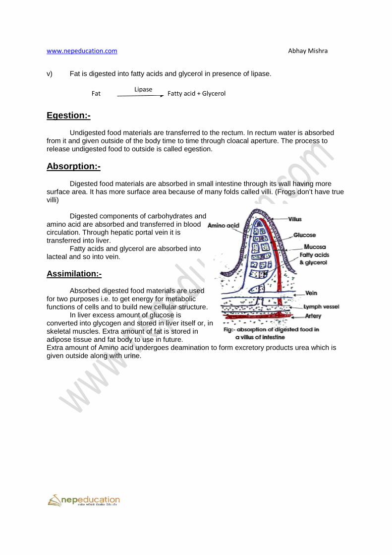
Egestion:- Undigested food materials are transferred to the rectum. In rectum water is absorbed from it and given outside of the body time to time through cloacal aperture. The process to release undigested food to outside is called egestion. Absorption:- Digested food materials are absorbed in small intestine through its wall having more surface area. It has more surface area because of many folds called villi. (Frogs don’t have true villi) Digested components of carbohydrates and amino acid are absorbed and transferred in blood circulation. Through hepatic portal vein it is transferred into liver. Fatty acids and glycerol are absorbed into lacteal and so into vein. Assimilation:-
Absorbed digested food materials are used for two purposes i.e. to get energy for metabolic functions of cells and to build new cellular structure. In liver excess amount of glucose is converted into glycogen and stored in liver itself or, in skeletal muscles. Extra amount of fat is stored in adipose tissue and fat body to use in future.Extra amount of Amino acid undergoes deamination given outside along with urine.
Fat Fatty acid + GlycerolLipase
Abhay Mishra
fatty acids and glycerol in presence of lipase.
Undigested food materials are transferred to the rectum. In rectum water is absorbed outside of the body time to time through cloacal aperture. The process to
release undigested food to outside is called egestion.
Digested food materials are absorbed in small intestine through its wall having more surface area because of many folds called villi. (Frogs don’t have true
Digested components of carbohydrates and and transferred in blood
circulation. Through hepatic portal vein it is
and glycerol are absorbed into
Absorbed digested food materials are used for two purposes i.e. to get energy for metabolic functions of cells and to build new cellular structure.
glucose is converted into glycogen and stored in liver itself or, in skeletal muscles. Extra amount of fat is stored in adipose tissue and fat body to use in future. Extra amount of Amino acid undergoes deamination to form excretory products urea which is
Fat Fatty acid + Glycerol
Abhay Mishra
Undigested food materials are transferred to the rectum. In rectum water is absorbed outside of the body time to time through cloacal aperture. The process to
Digested food materials are absorbed in small intestine through its wall having more surface area because of many folds called villi. (Frogs don’t have true
to form excretory products urea which is