diffusion measurement in phantoms and tissues using slim localization
TRANSCRIPT
JOURNAL OF MAGNETIC RESONANCE 129, 161–164 (1997)ARTICLE NO. MN971257
Diffusion Measurement in Phantoms and TissuesUsing SLIM Localization
Yihong Yang,1 Su Xu,1 M. Joan Dawson, and Paul C. Lauterbur
Biomedical Magnetic Resonance Laboratory, University of Illinois at Urbana–Champaign, Urbana, Illinois 60801
Received February 17, 1997; revised August 12, 1997
A new approach to efficient localized diffusion measurements scopic image reconstruction. The accuracy of SLIM, includ-has been developed and evaluated on phantoms and isolated tis- ing SLIM studies of inhomogeneous compartments, has beensues. The combination of a diffusion-sensitive pulse sequence with verified using simulations and experiments on phantomsSLIM (spectral localization by imaging) makes efficient and accu- (15, 16) , as well as in 31P spectroscopy studies of humanrate localized water and metabolite diffusion measurements possi-
brain (17) and excised uterus (18) . 1H SLIM has been com-ble with a substantial improvement in spatial or time resolution
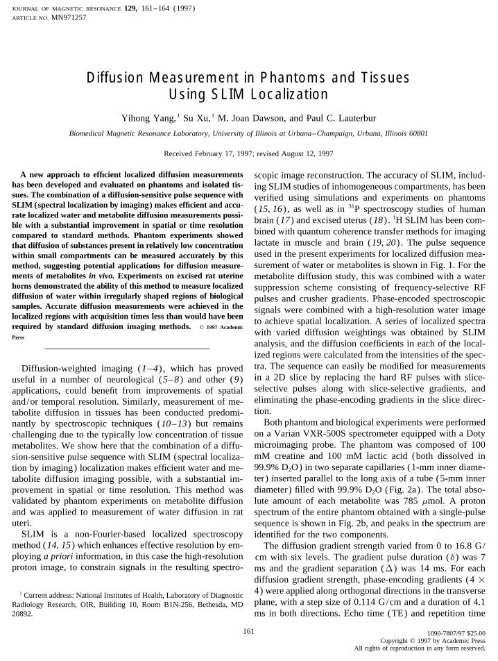
bined with quantum coherence transfer methods for imagingcompared to standard methods. Phantom experiments showedlactate in muscle and brain (19, 20) . The pulse sequencethat diffusion of substances present in relatively low concentrationused in the present experiments for localized diffusion mea-within small compartments can be measured accurately by thissurement of water or metabolites is shown in Fig. 1. For themethod, suggesting potential applications for diffusion measure-
ments of metabolites in vivo. Experiments on excised rat uterine metabolite diffusion study, this was combined with a waterhorns demonstrated the ability of this method to measure localized suppression scheme consisting of frequency-selective RFdiffusion of water within irregularly shaped regions of biological pulses and crusher gradients. Phase-encoded spectroscopicsamples. Accurate diffusion measurements were achieved in the signals were combined with a high-resolution water imagelocalized regions with acquisition times less than would have been to achieve spatial localization. A series of localized spectrarequired by standard diffusion imaging methods. q 1997 Academic
with varied diffusion weightings was obtained by SLIMPress
analysis, and the diffusion coefficients in each of the local-ized regions were calculated from the intensities of the spec-tra. The sequence can easily be modified for measurementsDiffusion-weighted imaging (1–4) , which has provedin a 2D slice by replacing the hard RF pulses with slice-useful in a number of neurological (5–8) and other (9)selective pulses along with slice-selective gradients, andapplications, could benefit from improvements of spatialeliminating the phase-encoding gradients in the slice direc-and/or temporal resolution. Similarly, measurement of me-tion.tabolite diffusion in tissues has been conducted predomi-
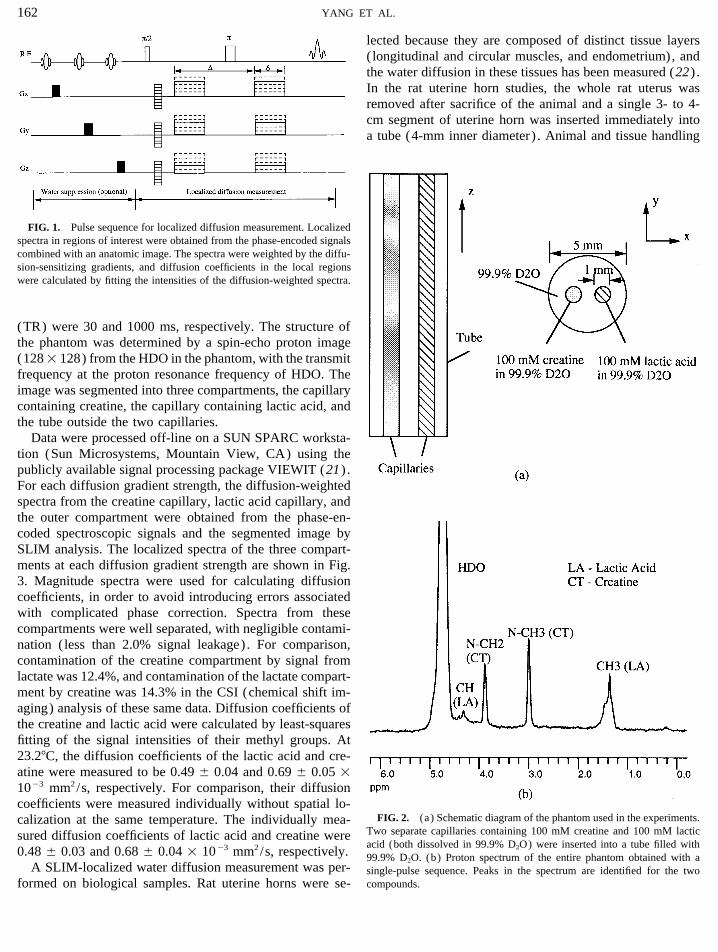
Both phantom and biological experiments were performednantly by spectroscopic techniques (10–13) but remainson a Varian VXR-500S spectrometer equipped with a Dotychallenging due to the typically low concentration of tissuemicroimaging probe. The phantom was composed of 100metabolites. We show here that the combination of a diffu-mM creatine and 100 mM lactic acid (both dissolved insion-sensitive pulse sequence with SLIM (spectral localiza-99.9% D2O) in two separate capillaries (1-mm inner diame-tion by imaging) localization makes efficient water and me-ter) inserted parallel to the long axis of a tube (5-mm innertabolite diffusion imaging possible, with a substantial im-diameter) filled with 99.9% D2O (Fig. 2a) . The total abso-provement in spatial or time resolution. This method waslute amount of each metabolite was 785 mmol. A protonvalidated by phantom experiments on metabolite diffusion
and was applied to measurement of water diffusion in rat spectrum of the entire phantom obtained with a single-pulseuteri. sequence is shown in Fig. 2b, and peaks in the spectrum are
SLIM is a non-Fourier-based localized spectroscopy identified for the two components.method (14, 15) which enhances effective resolution by em- The diffusion gradient strength varied from 0 to 16.8 G/ploying a priori information, in this case the high-resolution cm with six levels. The gradient pulse duration (d) was 7proton image, to constrain signals in the resulting spectro- ms and the gradient separation (D) was 14 ms. For each
diffusion gradient strength, phase-encoding gradients (4 14) were applied along orthogonal directions in the transverse1 Current address: National Institutes of Health, Laboratory of Diagnosticplane, with a step size of 0.114 G/cm and a duration of 4.1Radiology Research, OIR, Building 10, Room B1N-256, Bethesda, MD
20892. ms in both directions. Echo time (TE) and repetition time
161 1090-7807/97 $25.00Copyright q 1997 by Academic Press
All rights of reproduction in any form reserved.
AID JMR 1257 / 6j25$$$101 12-03-97 14:31:49 maga
162 YANG ET AL.
lected because they are composed of distinct tissue layers( longitudinal and circular muscles, and endometrium), andthe water diffusion in these tissues has been measured (22) .In the rat uterine horn studies, the whole rat uterus wasremoved after sacrifice of the animal and a single 3- to 4-cm segment of uterine horn was inserted immediately intoa tube (4-mm inner diameter) . Animal and tissue handling
FIG. 1. Pulse sequence for localized diffusion measurement. Localizedspectra in regions of interest were obtained from the phase-encoded signalscombined with an anatomic image. The spectra were weighted by the diffu-sion-sensitizing gradients, and diffusion coefficients in the local regionswere calculated by fitting the intensities of the diffusion-weighted spectra.
(TR) were 30 and 1000 ms, respectively. The structure ofthe phantom was determined by a spin-echo proton image(1281 128) from the HDO in the phantom, with the transmitfrequency at the proton resonance frequency of HDO. Theimage was segmented into three compartments, the capillarycontaining creatine, the capillary containing lactic acid, andthe tube outside the two capillaries.
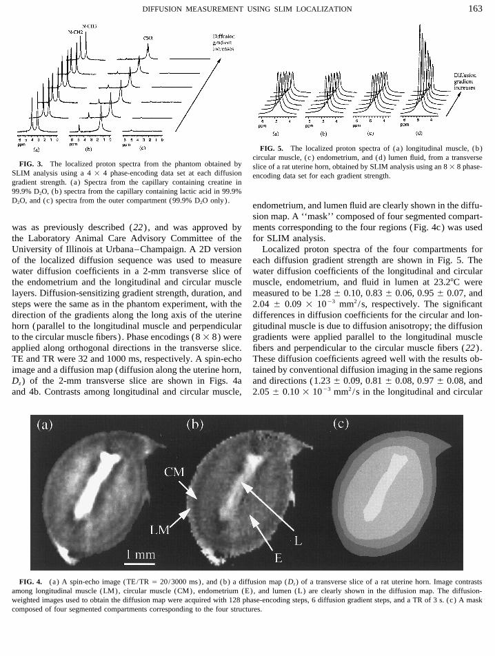
Data were processed off-line on a SUN SPARC worksta-tion (Sun Microsystems, Mountain View, CA) using thepublicly available signal processing package VIEWIT (21) .For each diffusion gradient strength, the diffusion-weightedspectra from the creatine capillary, lactic acid capillary, andthe outer compartment were obtained from the phase-en-coded spectroscopic signals and the segmented image bySLIM analysis. The localized spectra of the three compart-ments at each diffusion gradient strength are shown in Fig.3. Magnitude spectra were used for calculating diffusioncoefficients, in order to avoid introducing errors associatedwith complicated phase correction. Spectra from thesecompartments were well separated, with negligible contami-nation (less than 2.0% signal leakage). For comparison,contamination of the creatine compartment by signal fromlactate was 12.4%, and contamination of the lactate compart-ment by creatine was 14.3% in the CSI (chemical shift im-aging) analysis of these same data. Diffusion coefficients ofthe creatine and lactic acid were calculated by least-squaresfitting of the signal intensities of their methyl groups. At23.27C, the diffusion coefficients of the lactic acid and cre-atine were measured to be 0.49 { 0.04 and 0.69 { 0.05 11003 mm2/s, respectively. For comparison, their diffusioncoefficients were measured individually without spatial lo-
FIG. 2. (a) Schematic diagram of the phantom used in the experiments.calization at the same temperature. The individually mea-Two separate capillaries containing 100 mM creatine and 100 mM lacticsured diffusion coefficients of lactic acid and creatine wereacid (both dissolved in 99.9% D2O) were inserted into a tube filled with
0.48 { 0.03 and 0.68 { 0.04 1 1003 mm2/s, respectively. 99.9% D2O. (b) Proton spectrum of the entire phantom obtained with aA SLIM-localized water diffusion measurement was per- single-pulse sequence. Peaks in the spectrum are identified for the two
compounds.formed on biological samples. Rat uterine horns were se-
AID JMR 1257 / 6j25$$$102 12-03-97 14:31:49 maga
163DIFFUSION MEASUREMENT USING SLIM LOCALIZATION
FIG. 5. The localized proton spectra of (a) longitudinal muscle, (b)circular muscle, (c) endometrium, and (d) lumen fluid, from a transverse
FIG. 3. The localized proton spectra from the phantom obtained by slice of a rat uterine horn, obtained by SLIM analysis using an 8 1 8 phase-SLIM analysis using a 4 1 4 phase-encoding data set at each diffusion encoding data set for each gradient strength.gradient strength. (a) Spectra from the capillary containing creatine in99.9% D2O, (b) spectra from the capillary containing lactic acid in 99.9%D2O, and (c) spectra from the outer compartment (99.9% D2O only).
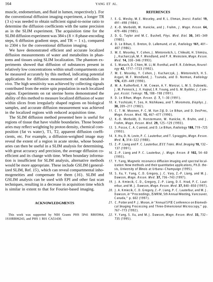
endometrium, and lumen fluid are clearly shown in the diffu-sion map. A ‘‘mask’’ composed of four segmented compart-ments corresponding to the four regions (Fig. 4c) was usedwas as previously described (22) , and was approved by
the Laboratory Animal Care Advisory Committee of the for SLIM analysis.Localized proton spectra of the four compartments forUniversity of Illinois at Urbana–Champaign. A 2D version
of the localized diffusion sequence was used to measure each diffusion gradient strength are shown in Fig. 5. Thewater diffusion coefficients of the longitudinal and circularwater diffusion coefficients in a 2-mm transverse slice of
the endometrium and the longitudinal and circular muscle muscle, endometrium, and fluid in lumen at 23.27C weremeasured to be 1.28 { 0.10, 0.83 { 0.06, 0.95 { 0.07, andlayers. Diffusion-sensitizing gradient strength, duration, and
steps were the same as in the phantom experiment, with the 2.04 { 0.09 1 1003 mm2/s, respectively. The significantdifferences in diffusion coefficients for the circular and lon-direction of the gradients along the long axis of the uterine
horn (parallel to the longitudinal muscle and perpendicular gitudinal muscle is due to diffusion anisotropy; the diffusiongradients were applied parallel to the longitudinal muscleto the circular muscle fibers) . Phase encodings (8 1 8) were
applied along orthogonal directions in the transverse slice. fibers and perpendicular to the circular muscle fibers (22) .These diffusion coefficients agreed well with the results ob-TE and TR were 32 and 1000 ms, respectively. A spin-echo
image and a diffusion map (diffusion along the uterine horn, tained by conventional diffusion imaging in the same regionsand directions (1.23 { 0.09, 0.81 { 0.08, 0.97 { 0.08, andDz) of the 2-mm transverse slice are shown in Figs. 4a
and 4b. Contrasts among longitudinal and circular muscle, 2.05 { 0.10 1 1003 mm2/s in the longitudinal and circular
FIG. 4. (a) A spin-echo image (TE/TR Å 20/3000 ms), and (b) a diffusion map (Dz) of a transverse slice of a rat uterine horn. Image contrastsamong longitudinal muscle (LM), circular muscle (CM), endometrium (E), and lumen (L) are clearly shown in the diffusion map. The diffusion-weighted images used to obtain the diffusion map were acquired with 128 phase-encoding steps, 6 diffusion gradient steps, and a TR of 3 s. (c) A maskcomposed of four segmented compartments corresponding to the four structures.
AID JMR 1257 / 6j25$$$102 12-03-97 14:31:49 maga
164 YANG ET AL.
muscle, endometrium, and fluid in lumen, respectively) . For REFERENCESthe conventional diffusion imaging experiment, a longer TR
1. E. G. Wesby, M. E. Moseley, and R. L. Ehman, Invest. Radiol. 19,(3 s) was needed to obtain sufficient signal-to-noise ratio to 491–498 (1984).determine the diffusion coefficients with the same precision 2. K.-D. Merboldt, W. Hanicke, and J. Frahm, J. Magn. Reson. 64,
479–486 (1985).as in the SLIM experiment. The acquisition time for the3. D. G. Taylor and M. C. Bushell, Phys. Med. Biol. 30, 345–349SLIM diffusion experiment was 384 s (81 8 phase-encoding
(1985).steps, 6 diffusion gradient steps, and TR Å 1 s) , compared4. D. Le Bihan, E. Breton, D. Lallemand, et al., Radiology 161, 401–to 2304 s for the conventional diffusion imaging.
407 (1986).We have demonstrated efficient and accurate localized
5. M. E. Moseley, Y. Cohen, J. Mintorovitch, L. Chileuitt, H. Shimizu,diffusion measurements of water and metabolites in phan- J., Kucharczyk, M. F. Wendland, and P. R. Weinstein, Magn. Reson.toms and tissues using SLIM localization. The phantom ex- Med. 14, 330–346 (1993).
6. S. Warach, D. Chien, W. Li, M. Ronthal, and R. R. Edelman, Neurol-periments showed that diffusion of substances present inogy 42, 1717–1723 (1992).relatively low concentrations within small compartments can
7. M. E. Moseley, Y. Cohen, J. Kucharczyk, J. Mintorovitch, H. S.be measured accurately by this method, indicating potentialAsgari, M. F. Wendland, J. Tsuruda, and D. Norman, Radiologyapplications for diffusion measurement of metabolites in 176, 439–449 (1990).
vivo. The high sensitivity was achieved by collecting signals 8. M. A. Rutherford, F. M. Cowan, A. Y. Manzur, L. M. S. Dubowitz,contributed from the entire spin population in each localized J. M. Pennock, J. V. Hajnal, I. R. Young, and G. M. Bydder, J. Com-
put. Assist. Tomogr. 15, 188–198 (1991).region. Experiments on rat uterine horns demonstrated the9. D. Le Bihan, Magn. Reson. Q. 7, 1–30 (1991).ability of this method to measure localized diffusion of water
10. K. Yoshizaki, Y. Seo, H. Nishikawa, and T. Moromoto, Biophys. J.within slices from irregularly shaped regions on biological38, 209–211 (1982).samples, and accurate diffusion measurement was achieved
11. C. T. W. Moonen, P. C. M. Van Zijl, D. Le Bihan, and D. DesPres,in the localized regions with reduced acquisition time.Magn. Reson. Med. 13, 467–477 (1990).
The SLIM diffusion method presented here is useful for 12. K.-D. Merboldt, D. Horstermann, W. Hanicke, H. Bruhn, and J.regions of tissue that have visible boundaries. Those bound- Frahm, Magn. Reson. Med. 29, 125–129 (1993).aries may reflect weighting by proton density, chemical com- 13. S. Posse, C. A. Cuenod, and D. Le Bihan, Radiology 188, 719–725
(1993).position (fat vs water) , T1, T2, apparent diffusion coeffi-14. X. Hu, D. N. Levin, P. C. Lauterbur, and T. Spraggins, Magn. Reson.cients, etc. For example, a diffusion-weighted image may
Med. 8, 314–322 (1988).reveal the extent of a region in acute stroke, whose bound-15. Z.-P. Liang and P. C. Lauterbur, IEEE Trans. Med. Imaging 10, 132–aries can then be useful in a SLIM analysis for determining,
137 (1991).with great accuracy and precision, the average diffusion co- 16. Z.-P. Liang and P. C. Lauterbur, J. Magn. Reson. B 102, 54–60efficient and its change with time. When boundary informa- (1993).tion is insufficient for SLIM analysis, alternative methods 17. Y. Yang, Magnetic resonance diffusion imaging and spectral local-
ization: New methods and their quantitative applications, Ph.D. the-would be more appropriate. These include GSLIM (general-sis, University of Illinois at Urbana–Champaign (1995).ized SLIM, Ref. 15) , which can reveal compartmental inho-
18. S. Xu, Y. Yang, C. D. Gregory, J. C. Vary, Z.-P. Liang, and M. J.mogeneities and compensate for them (16) . SLIM andDawson, Magn. Reson. Med. 37, 736–743 (1997).
GSLIM analysis can be used with EPI and other fast scan19. J. A. Kmiecik, C. D., Gregory, Z.-P. Liang, D. E. Hrad, P. C. Laut-
techniques, resulting in a decrease in acquisition time which erbur, and M. J. Dawson, Magn. Reson. Med. 37, 840–850 (1997).is similar in extent to that for Fourier-based imaging. 20. J. A. Kmiecik, C. D. Gregory, Z.-P. Liang, P. C. Lauterbur, and M. J.
Dawson, in ‘‘Proceedings, ISMRM, 5th Annual Meeting, Vancouver,Canada,’’ p. 602 (1997).
21. C. Potter and P. J. Moran, in ‘‘Annual SPIE Conference on Biomedi-ACKNOWLEDGMENTScal Imaging Processing and Three-Dimensional Microscopy,’’ pp.767–773 (1992).
This work was supported by NIH Grants PHS 5P41 RR05964, 22. Y. Yang, S. Xu, and M. J. Dawson, Magn. Reson. Med. 33, 732–735 (1995).1S10RR06243, and PHS 1 R01 CA51430.
AID JMR 1257 / 6j25$$$102 12-03-97 14:31:49 maga