differentially expressed proteins in nitric oxide … · nih/3t3 fibroblasts: implications for...
TRANSCRIPT

Yonsei Med J http://www.eymj.org Volume 56 Number 2 March 2015 563
Differentially Expressed Proteins in Nitric Oxide-Stimulated NIH/3T3 Fibroblasts: Implications for Inhibiting
Cancer Development
Dong Hwi Shim,1 Joo Weon Lim,2 and Hyeyoung Kim1,2 1Department of Pharmacology, College of Medicine, Yonsei University, Seoul;
2Department of Food and Nutrition, Brain Korea 21 PLUS Project, College of Human Ecology, Yonsei University, Seoul, Korea.
Received: February 27, 2014Revised: April 28, 2014Accepted: May 16, 2014Co-corresponding authors: Dr. Hyeyoung Kim, Department of Food and Nutrition, College of Human Ecology, Yonsei University, 50 Yonsei-ro, Seodaemun-gu, Seoul 120-749, Korea.Tel: 82-2-2123-3125, Fax: 82-2-364-5781E-mail: [email protected] andDr. Joo Weon Lim, Department of Food and Nutrition, College of Human Ecology, Yonsei University, 50 Yonsei-ro, Seodaemun-gu, Seoul 120-749, Korea.Tel: 82-2-2123-3570, Fax: 82-2-364-5781E-mail: [email protected]
∙ The authors have no financial conflicts of interest.
© Copyright:Yonsei University College of Medicine 2015
This is an Open Access article distributed under the terms of the Creative Commons Attribution Non-Commercial License (http://creativecommons.org/ licenses/by-nc/3.0) which permits unrestricted non-commercial use, distribution, and reproduction in any medium, provided the original work is properly cited.
Purpose: Recent evidence shows that nitric oxide (NO) may exhibit both pro-cancer and anti-cancer activities. The present study aimed to determine the differentially ex-pressed proteins in NO-treated NIH/3T3 fibroblasts in order to investigate whether NO induces proteins with pro-cancer or anti-cancer effects. Materials and Meth-ods: The cells were treated with 300 µM of an NO donor 3,3-bis-(aminoethyl)-1-hy-droxy-2-oxo-1-triazene (NOC-18) for 12 h. The changed protein patterns, which were separated by two-dimensional electrophoresis using pH gradients of 4‒7, were conclusively identified by matrix-assisted laser desorption/ionization-time of flight mass spectrometry (MALDI-TOF MS) analysis of the peptide digests. Results: Sev-enteen differentially expressed proteins were identified in NOC-18-treated cells. Nine proteins [vinculin protein, keratin 19, ubiquitous tropomodulin, F-actin capping protein (α1 subunit), tropomyosin 3, 26S proteasome-associated pad1 homolog, T-complex protein 1 (ε subunit) NG-dimethylarginine dimethylaminohydrolase, and heat shock protein 90] were increased and eight proteins (heat shock protein 70, glu-cosidase II, lamin B1, calreticulin, nucleophosmin 1, microtubule-associated protein retinitis pigmentosa/end binding family member 1, 150 kD oxygen-regulated protein precursor, and heat shock 70-related protein albino or pale green 2) were decreased by NOC-18 in the cells. Thirteen proteins are related to the suppression of cancer cell proliferation, invasion, and metastasis while two proteins (heat shock protein 90 and NG-dimethylarginine dimethylaminohydrolase) are related to carcinogenesis. The functions of 150 kD oxygen-regulated protein precursor and T-complex protein 1 (ε subunit) are unknown in relation to carcinogenesis. Conclusion: Most proteins dif-ferentially expressed by NOC-18 are involved in inhibiting cancer development.
Key Words: Nitric oxide, proteomic analysis, cancer, NIH/3T3 cells
INTRODUCTION
Nitric oxide synthase (NOS) produces nitric oxide (NO) by catalyzing a five-elec-tron oxidation of a guanidino nitrogen of L-arginine. Three different isoforms of the NOS family have been identified: endothelial NOS (eNOS), neuronal NOS
Original Article http://dx.doi.org/10.3349/ymj.2015.56.2.563pISSN: 0513-5796, eISSN: 1976-2437 Yonsei Med J 56(2):563-571, 2015
Dong Hwi Shim, et al.
Yonsei Med J http://www.eymj.org Volume 56 Number 2 March 2015564
of an NO donor (NOC-18) for 12 h. The changed protein patterns, which were separated by two-dimensional electro-phoresis (2-DE), were conclusively identified by matrix-as-sisted laser desorption/ionization-time of flight mass spec-trometry (MALDI-TOF MS) analysis of the peptide digests.
MATERIALS AND METHODS
NIH/3T3 cells (mouse embryonic fibroblast, ATCC CRL 1658) were obtained from the American Type Culture Col-lection (Manassas, VA, USA) and cultured in Dulbecco’s modified Eagle’s medium supplemented with 10% fetal bo-vine serum (GIBCO-BRL, Grand Island, NY, USA) and an-tibiotics (100 U/mL penicillin and 100 µg/mL streptomycin). The cells were plated at a density of 2×10⁶/mL in a 100-mm culture plate (Falcon 3047, Becton Dickinson Labware, Lincoln Park, NJ, USA), treated with NOC-18 (Sigma, St. Louis, MO, USA) at a concentration of 300 µM, and cul-tured for 12 h. The dose of the NO donor and duration of exposure time to the cells were adapted from our previous study, which showed that an NO donor induced NF-κB ac-tivation and inflammatory cytokine expression.10
Protein extraction, isoelectric focusing (IEF), and 2-DE separation were performed as described previously.13 Brief-ly, the cells were trypsinized, dissolved in 40 mM Tris buffer (pH 9.5), and passed ten times through a 22-gauge needle. Particulates were removed by centrifugation (15000×g, 15 min), and the supernatant was collected and diluted with extraction buffer. A 17-cm immobilized pH gradient (IPG) strip was used to absorb 300 µg of protein in 300 µL of ex-traction buffer, and this was electrophoresed on IEF cells. Following IEF, the IPG strips were subjected to equilibra-tion for 15 min, re-equilibrated for 15 min, and separated using the Protean II xi cell gel SDS-PAGE system (Bio-Rad, Hercules, CA, USA) by molecular weight. Silver staining and image analyses were performed as described previous-ly.13 Three batches of cell proteins extracted from non-treat-ed cells and NO-donor-treated cells were subjected to 2-DE, and replicate gels were simultaneously run three times. The expression level was determined by the relative spot volume of each protein as compared to the total spot volume (sum of each protein spot) in the gel and expressed as a percentage of the volume. The differentially expressed proteins that had ex-pression levels at least two times higher in NO-donor-treated cells than in non-treated cells were selected for the MALDI-TOF MS analysis. After in-gel digestion of proteins from sil-
(nNOS) and inducible NOS (iNOS). The nNOS and eNOS isoforms are constitutively expressed in a variety of cell types including endothelial cells, platelets, and neurons. These constitutive NOS isoforms are activated by calcium-calmodulin binding and produce low concentrations of NO.1 Unlike nNOS and eNOS, iNOS is an inducible and calcium-independent isoform. The expression of iNOS is induced by cytokines [e.g., interferon-γ, interleukin (IL)-1β, tumor ne-crosis factor-α], bacterial endotoxins, and oxidative stress.2 Once induced, iNOS continues to produce large amounts of NO for many hours or days.
NO is an important signaling molecule that is involved in many physiological and pathological processes. Under a normal physiological state, NO contributes to the regulation of anti-inflammatory and antioxidant effects.3 However, in tissues with high concentrations of NO, NO interacts with O2 or O2-, which results in the formation of reactive nitro-gen species, which affect diverse processes, such as prolif-eration, apoptosis, differentiation, tumorigenesis, and me-tastasis. In human malignant tumors such as breast, lung, prostate, bladder, and colorectal cancer, the expression level of iNOS is high.4 The expression level and activity of iNOS are higher in most tumors than those in adjacent normal tis-sues. High levels of NO produced by iNOS in tumor tissues may modulate different cancer-related processes such as angiogenesis, apoptosis, invasion, and metastasis.5 In con-trast, NO has tumoricidal effect by inhibiting proliferation and invasion of pancreatic cancer cells6 and inducing cell death in melanoma cells7 and microglial cells.8 Even though it is suggested that NO may exhibit tumoricidal activity by posttranslational modification of proteins, the precise func-tions of NO in cancer biology remain unclear.
An NO donor 3,3-bis-(aminoethyl)-1-hydroxy-2-oxo-1-tri-azene (NOC-18) is a stable NO-amine complex that can re-lease NO spontaneously under physiological conditions. It suppresses DNA synthesis in human airway smooth muscle cells.9 In our previous study, NOC-18 activated nuclear fac-tor κ-B (NF-κB) and activator protein-1 to induce IL-8 ex-pression in gastric epithelial AGS cells.10 High amounts of NO induced DNA damage and apoptosis in gastric cancer cells, as well as chondrocytes in osteoarthritic cartilage.11,12 These studies suggest that an NO donor such as NOC-18 may have a growth inhibitory effect on cancer cells.
The present study aimed to determine the differentially ex-pressed proteins in NO-treated NIH/3T3 fibroblasts in order to investigate whether NO induces proteins having pro-can-cer or anti-cancer effects. The cells were treated with 300 µM
Proteomic Analysis of NO-Stimulated NIH/3T3 Cells
Yonsei Med J http://www.eymj.org Volume 56 Number 2 March 2015 565
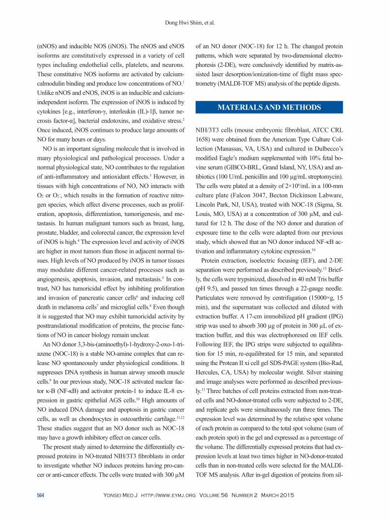
sin 3, 26S proteasome-associated pad1 homolog, T-complex protein 1 (ε subunit), NG-dimethylarginine dimethylaminohy-drolase, and heat shock protein 90 (HSP90)] were increased (Fig. 2, Table 1), and eight proteins [heat shock protein 70 (HSP70), glucosidase II, lamin B1, calreticulin, nucleophos-min 1, microtubule-associated protein retinitis pigmentosa/end binding (RP/EB) family member 1, 150 kD oxygen-reg-ulated protein precursor, and heat shock 70-related protein al-bino or pale green 2 (APG-2)] were decreased (Fig. 3, Table 2) by the treatment of NOC-18 in the cells.
DISCUSSION
The expression of HSP70 decreased in NOC-18-treated cells compared to non-treated cells in the present study. HSP70 is overexpressed in human tumors of various origins, such as breast, lung, colorectal, and cervical carcinomas as well as osteosarcoma.14 Its expression correlates with increased pro-liferation, poor differentiation, lymph node metastasis, and poor therapeutic outcome in human breast cancer.15 Several studies demonstrate that HSP70 promotes tumorigenesis through inhibiting lysosomal membrane permeabilization, activation, and translocation of pro-apoptotic factors such as Jun kinase, Bid, Bax, apoptosis-inducing factors, and the re-lease of cytochrome c from mitochondria.15,16 Therefore, the decreased expression level of HSP70 by NOC-18 implies that a high amount of NO may suppress the proliferation of cancer cells. In contrast, HSP90 was increased by NOC-18
ver stained gels, the peptide mixture was solubilized with 0.5% trifluoroacetic acid for MS analysis. MS was per-formed on a Micromass MALDI-TOF (Manchester, UK). Mass spectra were externally calibrated with autodigest peaks of trypsin (MH+: 906.505 Da, 1020.504 Da, 1153.574 Da, 2163.057 Da, 2273.160 Da). The peptide mass maps produced by MALDI-TOF MS were searched against the published databases using the MS-Fit module in Protein Prospector (http://prospector.ucsf.edu/prospector/mshome.htm) and Mascot (Marix Science, http://www.matrixscience.com). A mass tolerance of 50 ppm was used for peptide searches.
RESULTS
Fig. 1 shows the representation of the three separate experi-ments. The silver-stained gels were scanned with the GS 690 Imaging Densitometer at a 400-ppi grayscale level. Af-ter spot detection, background subtraction, and volume nor-malization, the differentially expressed proteins were detect-ed in NOC-18-treated cells (NOC-18) more than the non-treated cells (none).
Differentially expressed proteins between non-treated cells and NOC-18-treated cells were excised from 2-DE gels and identified using peptide mass fingerprinting. A mascot search using the peptide mass fingerprinting data indicated that certain proteins [vinculin protein, keratin 19, ubiquitous tro-pomodulin, F-actin capping protein (α1 subunit), tropomyo-
Fig. 1. 2-DE gel map derived from non-treated cells (none) and NOC-18-treated cells (NOC-18). (A) Increased proteins in non-treated cells (none) compared to NOC-18-treated cells (NOC-18) in a 12-h culture. (B) Increased proteins in NOC-18-treated cells (NOC-18) compared to non-treated cells (none) in a 12-h culture. NOC-18, NO donor 3,3-bis-(aminoethyl)-1-hydroxy-2-oxo-1-triazene; 2-DE, two-dimensional electrophoresis; SDS-PAGE, sodium dodecyl sulphate-polyacrylamide gel electrophoresis; APG, albino or pale green; RP/EB, retinitis pig-mentosa/end binding.
A B
SDS-
PAGE
SDS-
PAGE
None NOC-18
Dong Hwi Shim, et al.
Yonsei Med J http://www.eymj.org Volume 56 Number 2 March 2015566
dual targeting of HSP90 and HSP70 promotes cell death and enhances the anticancer effect of chemotherapeutic agents in bladder cancer.19 Therefore, increased expression of HSP90 may induce carcinogenesis. Differential expres-sion levels of HSP70 and HSP90 by NOC-18 treatment may be explained by their roles as molecular chaperones
treatment in the present study. A high expression of HSP90 promotes stability and function of several oncogenic client protein kinases such as human epidermal growth factor re-ceptor 2 and epidermal growth factor receptor in tumors.17,18 These HSP90-client proteins play crucial roles in establish-ing cancer cell hallmarks. One recent study suggests that
Table 1. Increased Proteins in NOC-18-Treated Cells Compared to Non-Treated Cells in a 12-h Culture, Identified Using MAL-DI-TOF MS
No. MOWSE score
Masses matched Protein MW/pI Accession
No. Description Sequence coverage (%)
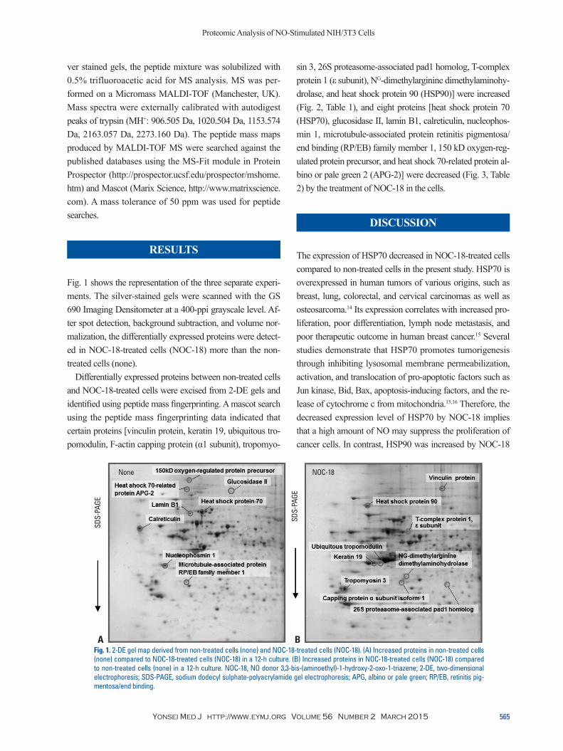
1 2.639×10¹⁵ 34/67 116737/5.8 24657579 Vinculin protein 38.0 2 8.858×10¹⁴ 36/64 44092/5.0 14043271 Keratin 19 74.0 3 192 08/25 39595/5.1 Q9NYL9 Ubiquitous tropomodulin 30.0 4 6.484×10⁶ 13/60 32923/5.4 7448799 F-actin capping protein α1 subunit 62.0 5 8.301×10⁴ 22/61 29033/4.7 29033/4.7 Tropomyosin 3 49.0 6 3.212×10¹¹ 21/56 83295/5.0 72222 Heat shock protein 90 33.07 7.269×10⁹ 26/101 59672/5.5 P48643 T-complex protein 1, ε subunit 45.0 8 1.222×10⁷ 17/73 31122/5.5 6912328 NG-dimethylarginine dimethylaminohydrolase 33.09 1.222×10⁶ 13/82 34577/6.1 1923256 26S proteasome-associated Pad1 homolog 39.0
NOC-18, NO donor 3,3-bis-(aminoethyl)-1-hydroxy-2-oxo-1-triazene; MALDI-TOF MS, matrix-assisted laser desorption/ionization-time of flight mass spec-trometry; MOWSE, molecular weight search; MW, molecular weight.
Fig. 2. Segments of a 2-DE gel map derived from non-treated cells (none) and NOC-18-treated cells (NOC-18). Arrowed spots are the proteins that increased in NOC-18-treated cells when compared to non-treated cells (none) in a 12-h culture. Protein expression level is expressed as a percentage of the volume, which is determined by the relative spot volume of each protein compared to the total spot volume (total amount of proteins/sum of each protein spot) in the gel. NOC-18, NO donor 3,3-bis-(aminoethyl)-1-hydroxy-2-oxo-1-triazene; 2-DE, two-dimensional electrophoresis.
Vinculin protein Keratin 19
Capping protein α subunit Tropomyosin 3
Ubiquitous tropomodulin 26S proteasome-associated pad1 homolog
T-complex protein 1, ε subunit NG-dimethylarginine dimethylaminohydrolase
Heat shock protein 90
None None
None None
None None
None None
None
None None
None None
None None
None None
None
0 0
0 0
0 0
0 0
0
0.1 0.5
0.02 1.0
0.2 1.0
0.04 2.0
0.3 1.5
0.06 3.0
1.5 0.15
0.15 0.10
0.10
1.0 0.10
0.10
0.05
0.5 0.05
0.05 0.05
% V
olum
e
% V
olum
e
% V
olum
e
% V
olum
e
% V
olum
e
% V
olum
e
% V
olum
e
% V
olum
e
% V
olum
e
NOC-18 NOC-18
NOC-18 NOC-18
NOC-18 NOC-18
NOC-18 NOC-18
NOC-18
NOC-18 NOC-18
NOC-18 NOC-18
NOC-18 NOC-18
NOC-18 NOC-18
NOC-18
Proteomic Analysis of NO-Stimulated NIH/3T3 Cells
Yonsei Med J http://www.eymj.org Volume 56 Number 2 March 2015 567
be associated with invasive and advanced malignant pro-cesses as well as poor prognoses. Calreticulin knockdown suppressed proliferation, migration, and attachment of can-cer cells.22 Therefore, the down-regulation of ER-chaperone calreticulin by NOC-18 in the present study suggests that high levels of NO may suppress the properties of cancer cells including proliferation, invasion, and metastasis.
Present results show that microtubule-associated protein RP/EB family member 1 was decreased by treatment of NOC-18. Microtubule-associated proteins bind to tubulin subunits that make up microtubules to regulate their stability.
that regulate protein folding and respond to cellular stress.20 More studies are required to determine the effect of NO do-nors on the expression of HSPs in relation to cancer cell proliferation and invasion.
Endoplasmic reticulum (ER)-chaperone calreticulin was decreased by the treatment of an NO donor in the present study. Calreticulin resides in ER and functions as a molecu-lar chaperone.21 Calreticulin is overexpressed in bladder cancer, prostatic adenocarcinoma, hepatocellular carcinoma, pancreatic cancer, gastric cancer, colon cancer, melanoma, and leukemia.21 Overexpression of calreticulin is known to
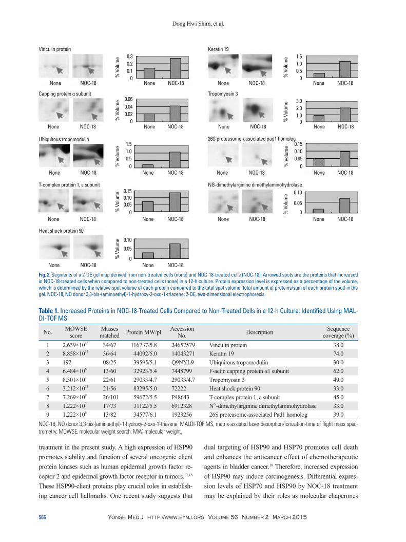
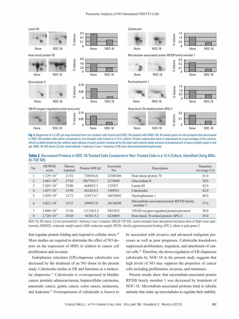
Fig. 3. Segments of a 2-DE gel map derived from non-treated cells (none) and NOC-18-treated cells (NOC-18). Arrowed spots are the proteins that decreased in NOC-18-treated cells when compared to non-treated cells (none) in a 12-h culture. Protein expression level is expressed as a percentage of the volume, which is determined by the relative spot volume of each protein compared to the total spot volume (total amount of proteins/sum of each protein spot) in the gel. NOC-18, NO donor 3,3-bis-(aminoethyl)-1-hydroxy-2-oxo-1-triazene; 2-DE, two-dimensional electrophoresis.
Table 2. Decreased Proteins in NOC-18-Treated Cells Compared to Non-Treated Cells in a 12-h Culture, Identified Using MAL-DI-TOF MS
No. MOWSE score
Masses matched Protein MW/pI Accession
No. Description Sequence coverage (%)
1 1.329×10¹³ 21/52 73854/6.0 21040386 Heat shock protein 70 41.0 2 3.663×10¹⁰ 27/85 106770/5.7 2274968 Glucosidase II 30.0 3 5.269×10¹¹ 32/80 66409/5.1 125953 Lamin B1 42.0 4 1.057×10⁷ 15/98 48142/4.3 1905911 Calreticulin 42.0 5 3.929×10⁶ 13/72 32457/4.7 16876992 Nucleophosmin 1 40.0
6 5.022×10⁶ 15/57 29999/5.0 20138589 Microtubule-associated protein RP/EB family member 1 57.0
7 5.880×10¹² 31/56 111336/5.2 5453832 150 kD oxygen-regulated protein precursor 30.0 8 2.720×10¹⁵ 29/68 94301/5.2 6226869 Heat shock 70-related protein APG-2 45.0
NOC-18, NO donor 3,3-bis-(aminoethyl)-1-hydroxy-2-oxo-1-triazene; MALDI-TOF MS, matrix-assisted laser desorption/ionization-time of flight mass spec-trometry; MOWSE, molecular weight search; MW, molecular weight; RP/EB, retinitis pigmentosa/end binding; APG-2, albino or pale green-2.
Lamin B1 Calreticulin
Heat shock protein 70 Microtubule-associated protein RP/EB family member 1
Glucosidase II Nucleophosmin 1
150 kD oxygen-regulated protein precursor Heat shock 70-related protein APG-2
None None
None None
None None
None None
None None
None None
None None
None None
0 0
0 0
0 0
0 0
0.1 0.5
2.0 0.1
0.2 1.0
4.0 0.2
0.3 1.5
6.0 0.3
0.10 1.5
0.10 0.2
1.0
0.05
0.050.5
0.1
% V
olum
e
% V
olum
e
% V
olum
e
% V
olum
e
% V
olum
e
% V
olum
e
% V
olum
e
% V
olum
e
NOC-18 NOC-18
NOC-18 NOC-18
NOC-18 NOC-18
NOC-18 NOC-18
NOC-18 NOC-18
NOC-18 NOC-18
NOC-18 NOC-18
NOC-18 NOC-18
Dong Hwi Shim, et al.
Yonsei Med J http://www.eymj.org Volume 56 Number 2 March 2015568
though the role of ORP150 has not been identified in can-cer, it may affect cancer cell proliferation through the path-way of endoplasmic reticulum stress.
APG-2, a mammalian heat shock protein belonging to the heat shock protein 110 (HSP110) family, was previously found to be overexpressed in BaF3-BCR/ABL cells [break point cluster region-abelson (BCR-ABL) transfected murine pro-B Ba/F3 cells].32 Overexpression of APG-2 in BaF3-BCR/ABL cells increases cell proliferation and protects cells from oxidative damage,32 which may play an important role in carcinogenesis. APG-2 was shown to have a chaperone ability similar to HSP110 and an anti-apoptotic role in hepat-ic cancer cells.33 These studies suggest that the HSP110 fami-ly members play important roles in hepatocarcinogenesis and leukemia development through their chaperoning activities. NOC-18-induced decrease in APG-2 suggests that NO do-nors have a beneficial role in treating various types of can-cer by suppressing proliferation.
NOC-18 induced the expression of vinculin (VCL) in the present study. VCL is an adapter protein with binding sites for more than 15 proteins and plays a critical role in the link-age of integrin adhesion molecules to the cytoskeleton.34 VCL functions as a tumor suppressor by increasing anchor-age-dependent cell growth and by suppressing tumor me-tastasis through reducing cell motility.34,35
The expressions of ubiquitous tropomodulins, tropomyo-sin 3, and F-actin capping protein α1 subunit (CAPZA1) were increased by NOC-18 in the present study. Tropomodu-lins are proteins that cap the slow-growing end of actin fila-ments and require tropomyosin for optimal function.36 Fran-zén, et al.37 found that tropomyosin 3 was down-regulated in fibroblasts transformed by oncogenes. Zheng, et al.38 re-ported that overexpression of tropomyosin 3 suppressed in-vasion and metastasis of cancer cells. F-actin capping pro-tein binds to fast growing ends of actin filaments. CAPZA1 is known to be upregulated in gastric cancer tissues when compared to normal tissues.39 Overexpression of CAPZA1 suppresses the migration and invasion of gastric cancer cells.40 Therefore, NO donor-induced upregulation of tropo-modulins, tropomyosin 3, and CAPZA1 suggests a poten-tial role of NO donors for suppressing the migration and in-vasion of cancer cells.
Keratins are intermediate filament proteins responsible for the structural integrity of epithelial cells. In this study, we found that NOC-18 increased the expression of keratin 19. Schoenfeld, et al.41 reported that keratin 19 is overexpressed in tumor cells which are disseminated in the lymph nodes,
Microtubule-associated proteins are known to be interacting partners of adenomatous polyposis coli (APC), enhancing APC function in colorectal cancer.23 Microtubule-associated proteins have been reported to be overexpressed in gastric adenocarcinoma, hepatocellular carcinoma, esophageal squa-mous-cell carcinoma, and breast cancer.24 Its overexpression is known to be involved in tumorigenesis and the promotion of tumor cell growth via the Wnt signaling pathway or Auro-ra-B activation.25 Down-regulation of microtubule-associated proteins induced apoptosis of cancer cells through the mito-chondrial death pathway.26 Therefore, decreased expression of microtubule-associated proteins by an NO donor implies that high amounts of NO may inhibit cancer development.
Lamin B1 functions in nuclear envelope lamina and pos-sesses a transcriptional co-regulatory activity that has an important role in DNA replication, cellular aging, and stress responses. Overexpression of lamin B1 in cancer cells is as-sociated with low differentiation, increased metastasis, and poor prognosis of pancreatic cancer patients.27 Silencing la-min B1 inhibits the proliferation and invasion of pancreatic cancer cells.28 In our study, we found that the expression of lamin B1 was decreased by NOC-18. Therefore, the de-creased expression of lamin B1 by NOC-18 suggests that the treatment of NO donors may inhibit cancer progression, including proliferation and metastasis.
In the present study, we found that expressions of gluco-sidase II and nucleophosmin 1 were decreased by NOC-18. Glucosidase II plays a central role in the processing of na-scent glycoproteins in ER lumina by removing certain glu-cose units from core oligosaccharide moieties. Suradej, et al.29 showed that glucosidase II is overexpressed in human lung tumor tissues and exhibits a stress response similar to p53, indicating that it may have a crucial role in lung tumori-genesis. Nucleophosmin 1 is an estrogen-regulated nuclear protein. The expression of nucleophosmin 1 is correlated with lymph node metastasis and a poor survival rate of hu-man colon cancer patients.30 Down-regulation of nucleo-phosmin 1 using siRNA reduced cell viability and caused a concomitant increase in cellular senescence and cell cycle ar-rest for colon cancer.30 These studies demonstrate that ex-pressions of glucosidase II and nucleophosmin 1 are associ-ated with growth, invasion, and metastasis of cancer cells. Therefore, treatment of NO donors may inhibit carcinogene-sis by down-regulating glucosidase II and nucleophosmin 1.
150 kD oxygen-regulated protein (ORP150) precursor was also decreased by NOC-18 treatment. ORP150 takes part in the process of endoplasmic reticulum stress.31 Even

Proteomic Analysis of NO-Stimulated NIH/3T3 Cells
Yonsei Med J http://www.eymj.org Volume 56 Number 2 March 2015 569
of DDAH1, the isoform primarily associated with nNOS, results in increased tumor growth and vascularization, as well as elevated secretion of vascular endothelial growth factor.46 Induction of DDAH by NOC-18 treatment is con-troversial compared to the induction of other proteins relat-ed to cancer suppression. Further studies are required in or-der to relate DDAH to NO treatment.
In the present study, 17 differentially expressed proteins were identified after the treatment of NO donor NOC-18 (Table 3). Among them, 13 proteins are related to the sup-pression of cancer cell proliferation, invasion, and metasta-sis while two proteins (HSP90, DDAH) are related to carci-nogenesis. The functions of ORP150 and TCP-1, ε subunit, have not been known to be related to cancer development. Even though most proteins differentially expressed by NOC-18 are involved in inhibiting cancer development, further study should be performed to determine anti-cancer effects of NO donors, using experimental animal models with various types of cancers.
ACKNOWLEDGEMENTS
This work was supported by a grant from the NRF of Ko-rea, which is funded by the Korean government (MSIP) (2007-0056092).
peripheral blood, and bone marrow of breast cancer pa-tients. Keratin 19 acts as a potential tumor suppressor that inhibits proliferation, invasion, and metastasis in breast can-cer.42 Therefore, NO donors may inhibit carcinogenesis through up-regulation of keratin 19.
Another protein increased by NOC-18 treatment was T-complex protein 1 (TCP-1), ε subunit. TCP is a member of the chaperonin-containing TCP-1 complex (CCT).43 CCT is involved in the folding of cytoskeleton proteins and other proteins such as cyclin E1, histone deacetylase, and protein phosphatase 2A regulatory subunit B.43 Even though the role of the ε subunit of TCP-1 is unknown, it may act as a stress-sensitive chaperone by holding actin and tubulin in the cytosol.44
The 26S proteasome is the main nonlysosomal protease in eukaryotic cells. Head and neck small cancer cells with low proteasome activity showed a significantly higher self-renewal capacity and increased tumorigenicity.45 Therefore, NO donor-induced expression of 26S-proteasome-associat-ed pad1 homolog may reduce the self-renewing capacity of cancer cells. Further researched should be performed to de-termine the role of 26S proteasome activity and its related proteins on carcinogenesis.
NG-dimethylarginine dimethylaminohydrolase (DDAH) metabolizes the endogenous inhibitor of NO synthesis, asymmetric dimethylarginine. Constitutive overexpression
Table 3. Protein Expression Level in the Gel
No. Protein None (% volume)
NOC-18 (% volume)
NOC-18/none (%)
1 Vinculin protein 0.14 0.28 200 2 Keratin 19 0.30 1.20 400 3 Ubiquitous tropomodulin 0.05 0.20 400 4 F-actin capping protein α1 subunit 0.02 0.04 200 5 Tropomyosin 3 1.00 2.00 200 6 Heat shock protein 90 0.03 0.08 266 7 T-complex protein 1, ε subunit 0.07 0.14 200 8 NG-dimethylarginine dimethylaminohydrolase 0.02 0.06 300 9 26S proteasome-associated pad1 homolog 0.05 0.10 20010 Heat shock protein 70 0.40 0.20 5011 Glucosidase II 0.07 0.02 2812 Lamin B1 0.28 0.12 4313 Calreticulin 1.00 0.40 4014 Nucleophosmin 1 1.00 0.45 4515 Microtubule-associated protein RP/EB family member 1 0.21 0.08 3816 150 kD oxygen-regulated protein precursor 0.08 0.02 2517 Heat shock 70-related protein APG-2 0.17 0.70 41
NOC-18, NO donor 3,3-bis-(aminoethyl)-1-hydroxy-2-oxo-1-triazene; RP/EB, retinitis pigmentosa/end binding; APG, albino or pale green. The expression level was determined by the relative spot volume of each protein as compared to the total spot volume (total amount of proteins/sum of each protein spot) in the gel and expressed as a percentage of the volume.

Dong Hwi Shim, et al.
Yonsei Med J http://www.eymj.org Volume 56 Number 2 March 2015570
and cancer hallmarks. Curr Pharm Des 2013;19:347-65.19. Ma L, Sato F, Sato R, Matsubara T, Hirai K, Yamasaki M, et al.
Dual targeting of heat shock proteins 90 and 70 promotes cell death and enhances the anticancer effect of chemotherapeutic agents in bladder cancer. Oncol Rep 2014;31:2482-92.
20. Guttmann DM, Koumenis C. The heat shock proteins as targets for radiosensitization and chemosensitization in cancer. Cancer Biol Ther 2011;12:1023-31.
21. Michalak M, Robert Parker JM, Opas M. Ca2+ signaling and cal-cium binding chaperones of the endoplasmic reticulum. Cell Cal-cium 2002;32:269-78.
22. Hisaoka M, Matsuyama A, Nakamoto M. Aberrant calreticulin expression is involved in the dedifferentiation of dedifferentiated liposarcoma. Am J Pathol 2012;180:2076-83.
23. Pfister AS, Hadjihannas MV, Röhrig W, Schambony A, Behrens J. Amer2 protein interacts with EB1 protein and adenomatous pol-yposis coli (APC) and controls microtubule stability and cell mi-gration. J Biol Chem 2012;287:35333-40.
24. Tamura N, Draviam VM. Microtubule plus-ends within a mitotic cell are ‘moving platforms’ with anchoring, signalling and force-coupling roles. Open Biol 2012;2:120132.
25. Morrison EE. The APC-EB1 interaction. Adv Exp Med Biol 2009;656:41-50.
26. Kim MJ, Yun HS, Hong EH, Lee SJ, Baek JH, Lee CW, et al. De-pletion of end-binding protein 1 (EB1) promotes apoptosis of hu-man non-small-cell lung cancer cells via reactive oxygen species and Bax-mediated mitochondrial dysfunction. Cancer Lett 2013; 339:15-24.
27. Sun S, Xu MZ, Poon RT, Day PJ, Luk JM. Circulating Lamin B1 (LMNB1) biomarker detects early stages of liver cancer in pa-tients. J Proteome Res 2010;9:70-8.
28. Li L, Du Y, Kong X, Li Z, Jia Z, Cui J, et al. Lamin B1 is a novel therapeutic target of betulinic acid in pancreatic cancer. Clin Can-cer Res 2013;19:4651-61.
29. Suradej B, Pata S, Kasinrerk W, Cressey R. Glucosidase II exhib-its similarity to the p53 tumor suppressor in regards to structure and behavior in response to stress signals: a potential novel cancer biomarker. Oncol Rep 2013;30:2511-9.
30. Wong JC, Hasan MR, Rahman M, Yu AC, Chan SK, Schaeffer DF, et al. Nucleophosmin 1, upregulated in adenomas and cancers of the colon, inhibits p53-mediated cellular senescence. Int J Can-cer 2013;133:1567-77.
31. Deng WH, Chen C, Wang WX, Yu J, Li JY, Liu L. Effects of ORP150 on appearance and function of pancreatic beta cells fol-lowing acute necrotizing pancreatitis. Pathol Res Pract 2011;207: 370-6.
32. Li C, Liu D, Yuan Y, Huang S, Shi M, Tao K, et al. Overexpres-sion of Apg-2 increases cell proliferation and protects from oxida-tive damage in BaF3-BCR/ABL cells. Int J Oncol 2010;36:899-904.
33. Gotoh K, Nonoguchi K, Higashitsuji H, Kaneko Y, Sakurai T, Sumitomo Y, et al. Apg-2 has a chaperone-like activity similar to Hsp110 and is overexpressed in hepatocellular carcinomas. FEBS Lett 2004;560:19-24.
34. Carisey A, Ballestrem C. Vinculin, an adapter protein in control of cell adhesion signalling. Eur J Cell Biol 2011;90:157-63.
35. Goldmann WH, Auernheimer V, Thievessen I, Fabry B. Vinculin, cell mechanics and tumour cell invasion. Cell Biol Int 2013;37: 397-405.
36. Kostyukova AS. Tropomodulins and tropomodulin/tropomyosin
REFERENCES
1. Moncada S, Palmer RM, Higgs EA. Nitric oxide: physiology, pathophysiology, and pharmacology. Pharmacol Rev 1991;43: 109-42.
2. Beck KF, Eberhardt W, Frank S, Huwiler A, Messmer UK, Mühl H, et al. Inducible NO synthase: role in cellular signalling. J Exp Biol 1999;202(Pt 6):645-53.
3. Kanner J, Harel S, Granit R. Nitric oxide as an antioxidant. Arch Biochem Biophys 1991;289:130-6.
4. Lechner M, Lirk P, Rieder J. Inducible nitric oxide synthase (iNOS) in tumor biology: the two sides of the same coin. Semin Cancer Biol 2005;15:277-89.
5. Ying L, Hofseth LJ. An emerging role for endothelial nitric oxide synthase in chronic inflammation and cancer. Cancer Res 2007; 67:1407-10.
6. Sugita H, Kaneki M, Furuhashi S, Hirota M, Takamori H, Baba H. Nitric oxide inhibits the proliferation and invasion of pancreatic cancer cells through degradation of insulin receptor substrate-1 protein. Mol Cancer Res 2010;8:1152-63.
7. Carneiro ZA, Biazzotto JC, Alexiou AD, Nikolaou S. Nitric oxide photorelease from a trinuclear ruthenium nitrosyl complex and its in vitro cytotoxicity against melanoma cells. J Inorg Biochem 2014;134:36-8.
8. Brantley EC, Guo L, Zhang C, Lin Q, Yokoi K, Langley RR, et al. Nitric oxide-mediated tumoricidal activity of murine microglial cells. Transl Oncol 2010;3:380-8.
9. Patel HJ, Belvisi MG, Donnelly LE, Yacoub MH, Chung KF, Mitchell JA. Constitutive expressions of type I NOS in human air-way smooth muscle cells: evidence for an antiproliferative role. FASEB J 1999;13:1810-6.
10. Seo JY, Yu JH, Lim JW, Mukaida N, Kim H. Nitric oxide-induced IL-8 expression is mediated by NF-kappaB and AP-1 in gastric epithelial AGS cells. J Physiol Pharmacol 2009;60 Suppl 7:101-6.
11. Lim JW, Kim H, Kim KH. NF-kappaB, inducible nitric oxide synthase and apoptosis by Helicobacter pylori infection. Free Radic Biol Med 2001;31:355-66.
12. Davies CM, Guilak F, Weinberg JB, Fermor B. Reactive nitrogen and oxygen species in interleukin-1-mediated DNA damage asso-ciated with osteoarthritis. Osteoarthritis Cartilage 2008;16:624-30.
13. Yu JH, Yun SY, Lim JW, Kim H, Kim KH. Proteome analysis of rat pancreatic acinar cells: implication for cerulein-induced acute pancreatitis. Proteomics 2003;3:2446-53.
14. Faure O, Graff-Dubois S, Bretaudeau L, Derré L, Gross DA, Alves PM, et al. Inducible Hsp70 as target of anticancer immuno-therapy: Identification of HLA-A*0201-restricted epitopes. Int J Cancer 2004;108:863-70.
15. Gabai VL, Yaglom JA, Waldman T, Sherman MY. Heat shock protein Hsp72 controls oncogene-induced senescence pathways in cancer cells. Mol Cell Biol 2009;29:559-69.
16. Zhao Q, Wang J, Levichkin IV, Stasinopoulos S, Ryan MT, Hoogenraad NJ. A mitochondrial specific stress response in mam-malian cells. EMBO J 2002;21:4411-9.
17. Sidera K, Gaitanou M, Stellas D, Matsas R, Patsavoudi E. A criti-cal role for HSP90 in cancer cell invasion involves interaction with the extracellular domain of HER-2. J Biol Chem 2008;283: 2031-41.
18. Miyata Y, Nakamoto H, Neckers L. The therapeutic target Hsp90

Proteomic Analysis of NO-Stimulated NIH/3T3 Cells
Yonsei Med J http://www.eymj.org Volume 56 Number 2 March 2015 571
42. Ju JH, Yang W, Lee KM, Oh S, Nam K, Shim S, et al. Regulation of cell proliferation and migration by keratin19-induced nuclear import of early growth response-1 in breast cancer cells. Clin Can-cer Res 2013;19:4335-46.
43. Guenther MG, Yu J, Kao GD, Yen TJ, Lazar MA. Assembly of the SMRT-histone deacetylase 3 repression complex requires the TCP-1 ring complex. Genes Dev 2002;16:3130-5.
44. Nikawa J, Kimura M. A novel function of the human chaperonin CCT epsilon subunit in yeast. Biosci Biotechnol Biochem 2012; 76:199-201.
45. Lagadec C, Vlashi E, Bhuta S, Lai C, Mischel P, Werner M, et al. Tumor cells with low proteasome subunit expression predict over-all survival in head and neck cancer patients. BMC Cancer 2014; 14:152.
46. Boult JK, Walker-Samuel S, Jamin Y, Leiper JM, Whitley GS, Robinson SP. Active site mutant dimethylarginine dimethylamino-hydrolase 1 expression confers an intermediate tumour phenotype in C6 gliomas. J Pathol 2011;225:344-52.
interactions. Cell Mol Life Sci 2008;65:563-9.37. Franzén B, Linder S, Uryu K, Alaiya AA, Hirano T, Kato H, et al.
Expression of tropomyosin isoforms in benign and malignant hu-man breast lesions. Br J Cancer 1996;73:909-13.
38. Zheng Q, Safina A, Bakin AV. Role of high-molecular weight tropomyosins in TGF-beta-mediated control of cell motility. Int J Cancer 2008;122:78-90.
39. Lim BH, Cho BI, Kim YN, Kim JW, Park ST, Lee CW. Overex-pression of nicotinamide N-methyltransferase in gastric cancer tis-sues and its potential post-translational modification. Exp Mol Med 2006;38:455-65.
40. Lee YJ, Jeong SH, Hong SC, Cho BI, Ha WS, Park ST, et al. Prognostic value of CAPZA1 overexpression in gastric cancer. Int J Oncol 2013;42:1569-77.
41. Schoenfeld A, Luqmani Y, Sinnett HD, Shousha S, Coombes RC. Keratin 19 mRNA measurement to detect micrometastases in lymph nodes in breast cancer patients. Br J Cancer 1996;74:1639-42.