difference in the source of anti-aqp4-igg and anti-mog-igg
TRANSCRIPT
Neurology Publish Ahead of PrintDOI 101212WNL0000000000012175
Difference in the Source of Anti-AQP4-IgG and Anti-MOG-IgG Antibodies in CSF in Patients With
Neuromyelitis Optica Spectrum Disorder
This is an open access article distributed under the terms of the Creative Commons
Attribution-NonCommercial-NoDerivatives License 40 (CC BY-NC-ND) which permits downloading and
sharing the work provided it is properly cited The work cannot be changed in any way or used
commercially without permission from the journal
Neurologyreg Published Ahead of Print articles have been peer reviewed and accepted for publication This
manuscript will be published in its final form after copyediting page composition and review of proofs
Errors that could affect the content may be corrected during these processes
Copyright copy 2021 The Author(s) Published by Wolters Kluwer Health Inc on behalf of the American Academy of Neurology
Published Ahead of Print on May 12 2021 as 101212WNL0000000000012175
Tetsuya Akaishi MDPhD 12 Toshiyuki Takahashi MDPhD 13 Tatsuro Misu MDPhD 1 Kimihiko Kaneko MDPhD 1
Yoshiki Takai MDPhD 1 Shuhei Nishiyama MDPhD 1 Ryo Ogawa MDPhD 1 Juichi Fujimori MDPhD 4 Tadashi Ishii
MDPhD 2 Masashi Aoki MDPhD 1 Kazuo Fujihara MDPhD 5 Ichiro Nakashima MDPhD 4
1 Department of Neurology Tohoku University Graduate School of Medicine Sendai Japan
2 Department of Education and Support for Regional Medicine Tohoku University Hospital Sendai Japan
3 Department of Neurology National Hospital Organization Yonezawa National Hospital Yonezawa Japan
4 Department of Neurology Tohoku Medical and Pharmaceutical University Sendai Japan
5 Department of Multiple Sclerosis Therapeutics Fukushima Medical University Fukushima Japan
Correspondence to T Akaishi
Email t-akaishimedtohokuacjp
Number of characters in title 141
Abstract word count 250
Word count of main text 4485
References 49
Copyright copy 2021 The Author(s) Published by Wolters Kluwer Health Inc on behalf of the American Academy of Neurology
Figures 4
Tables 2
Supplementary Materials Relevant datasets have been deposited to Dryad httpsdoiorg105061dryadwm37pvmmn
Statistical analysis
Tetsuya Akaishi and Toshiyuki Takahashi completed the statistical analysis
Disclosure
T Akaishi T Takahashi T Misu K Kaneko Y Takai S Nishiyama R Ogawa J Fujimori T Ishii and M Aoki report no
disclosures K Fujihara received speaker honoraria and travel funding from Bayer Biogen Japan Eisai Mitsubishi Tanabe
Novartis Astellas Takeda Asahi Kasei Medical Daiichi Sankyo and Nihon Pharmaceutical and received research support
from Bayer Biogen Asahi Kasei Medical The Chemo-Sero-Therapeutic Research Institute Teva Mitsubishi Tanabe
Pharma Teijin Chugai Ono Nihon Pharmaceutical and Genzyme I Nakashima received speaker honoraria and travel
funding from Mitsubishi Tanabe Pharma Biogen Japan and Novartis Pharmaceuticals and received research support from
LSI Medience Corporation
Study Funding
This study is not industry-sponsored
This work was supported by MHLW Program Grant Number 20FC1030 and JSPS KAKENHI Grant Number 20K07892
Copyright copy 2021 The Author(s) Published by Wolters Kluwer Health Inc on behalf of the American Academy of Neurology
ABSTRACT
Objective
To elucidate the differences in the source and in the level of intrathecal synthesis between anti-aquaporin-4 antibodies
(AQP4-IgG) and anti-myelin oligodendrocyte glycoprotein antibodies (MOG-IgG)
Methods
Thirty-eight patients with MOG-IgG-associated disease and 36 with AQP4-IgG-positive neuromyelitis optica spectrum
disorders (NMOSD) were studied for the antibody titers in the sera and cerebrospinal fluids (CSF) simultaneously collected
in the acute attacks The quotients between CSF and serum levels of albumin total IgG and each disease-specific antibody
were calculated Intrathecal production level in each disease-specific antibody was evaluated by calculating antibody index
from these quotients
Results
Eleven of the 38 patients with MOG-IgG were positive for the antibody only in the CSF while no patient with AQP4-IgG
showed CSF-restricted AQP4-IgG Blood-brain barrier compromise as shown by raised albumin quotients was seen in
750 of MOG-IgG-positive cases and 438 of AQP4-IgG-positive cases Moreover MOG-IgG quotients were more than
10 times higher than AQP4-IgG quotients (effect size r = 0659 p lt 00001) Elevated antibody index (gt40) was confirmed
in 12 of 21 with MOG-IgG whereas it was seen only in one of 16 with AQP4-IgG (φ = 0528 p lt 00001) The CSF
MOG-IgG titers (rho = +0519 p = 0001) and antibody indexes for MOG-IgG (rho = +0472 p = 0036) correlated with the
CSF cell counts but not with clinical disability
Conclusions
Intrathecal production of MOG-IgG may occur more frequently than that of AQP4-IgG This finding implies the different
properties of B-cell trafficking and antibody production between MOG-IgG-associated disease and AQP4-IgG-positive
Copyright copy 2021 The Author(s) Published by Wolters Kluwer Health Inc on behalf of the American Academy of Neurology
NMOSD
Search Terms
1 anti-aquaporin-4 antibodies
2 intrathecal synthesis
3 anti-myelin oligodendrocyte glycoprotein antibody
4 neuromyelitis optica spectrum disorders
5 production site
Glossary
AI = antibody index AQP4-IgG = anti-aquaporin-4 immunoglobulin G CSF = cerebrospinal fluid MOG-IgG =
anti-myelin oligodendrocyte glycoprotein immunoglobulin G MS = multiple sclerosis NMOSD = neuromyelitis optica
spectrum disorder OCB = oligoclonal bands = albumin quotient = immunoglobulin G quotient
Copyright copy 2021 The Author(s) Published by Wolters Kluwer Health Inc on behalf of the American Academy of Neurology
Introduction
Neuromyelitis optica spectrum disorder (NMOSD) is a demyelinating neurological condition in the CNS1 2 In contrast to
multiple sclerosis (MS) which usually lacks disease-specific antibodies NMOSD is characterized by the presence of
specific antibodies such as anti-aquaporin-4 immunoglobulin G (AQP4-IgG) and anti-myelin oligodendrocyte glycoprotein
immunoglobulin G (MOG-IgG)3-5 Patients with MOG-IgG-associated disease and those with AQP4-IgG-positive NMOSD
both typically present with recurrent neurological episodes represented by optic neuritis (ON) and acute myelitis4 6
Although these two conditions are likely to present similar clinical manifestations in the acute phase of attacks the resulting
neurological sequelae are generally thought to be worse in AQP4-IgG-positive NMOSD7-10 suggesting that these two
disorders should be considered as independent disease entities with different approaches for relapse prevention11-13 Based
on the suggested differences in the clinical spectrum and eventual neurological prognoses between patients with MOG-IgG
and those with AQP4-IgG neurological conditions related to MOG-IgG have been considered separately from other
conditions of NMOSD and regarded as an independent disease entity called the MOG-IgG-associated disease
(MOGAD)14-17 Thereafter the clinical need and rationale to discriminate these two conditions have been vigorously
discussed18 19 Differences in properties of B-cell trafficking and antibody production site between these diseases is
among the topics that are still controversial In this study to evaluate the prevalence and clinical impact of intrathecal
production of these disease-specific antibodies we measured the titers of these antibodies in time-matched paired serum and
CSF samples obtained in the acute phase of attacks
Copyright copy 2021 The Author(s) Published by Wolters Kluwer Health Inc on behalf of the American Academy of Neurology
Methods
Patients and Disease Groups based on Specific Antibody
We initially recruited patients in our facility with acute neurological episodes in whom time-matched paired serum and CSF
MOG-IgG and AQP4-IgG titers were simultaneously evaluated during the acute phase of the neurological episodes The
enrollment period for the patients treated in our facility (Tohoku University) was between 2006 and 2020 To increase the
sample size data of the patients treated in other facilities in Japan between 2019 and 2020 were additionally collected
Based on the results of the MOG-IgG and AQP4-IgG titrations for their serum and CSF samples patients were divided into
the following four disease groups MOG-IgG-associated disease AQP4-IgG-positive NMOSD MS without these antibodies
and other conditions (eg acute disseminated encephalomyelitis neuro-Behccedilets disease neuro-Sweet disease cerebral
infarction idiopathic ON and tumors) Patients with MOG-IgG in either serum or CSF were categorized as
MOG-IgG-associated disease and those with AQP4-IgG in either serum or CSF were categorized as AQP4-IgG-positive
NMOSD Patients in the first three disease groups were enrolled in this study Patients for whom time-matched paired serum
and CSF samples were unavailable were not recruited in this study
Variables from Time-Matched Paired Serum and CSF Samples
In each of the three enrolled disease groups CSF cell count (mononuclearpolymorphonuclear) CSF protein level presence
of CSF-restricted oligoclonal bands (OCB) and further CSF derivatives from time-matched paired serum and CSF samples
were collected as follows albumin quotient ( CSFserum albumin ratio) and IgG quotient (13 CSFserum total
IgG ratio) IgG index (ie a calculated value to estimate intrathecal total-IgG synthesis) and antibody index (AI) for each
specific antibody
Copyright copy 2021 The Author(s) Published by Wolters Kluwer Health Inc on behalf of the American Academy of Neurology
Titration of each specific antibody (IgG-spec MOG-IgG or AQP4-IgG) was performed using a live cell-based
assay (CBA) as described in our previous reports20 21 Screening of the serum samples to estimate seropositivity was
performed at dilutions of 116 for AQP4-IgG and 1128 for MOG-IgG then the antibody titers were calculated
semi-quantitatively using consecutive two-fold end-point dilutions Screening for CSF samples was performed without
diluting the samples and positive samples were further studied for antibody titers using the aforementioned
semi-quantitative serial dilution method If MOG-IgG was positive only in the CSF we additionally tested the serum of the
patients at a dilution of 116 to exclude the presence of serum MOG-IgG with low titers between 116 and 164
Using the time-matched paired serum and CSF samples in the acute phase the following derivatives were
comprehensively calculated based on the equations described below 13 13 IgG index ()
AI and corrected AI
= $amp ( amp )+ [$] $amp ( amp 01$ [$]
13 = ( amp )+ [$] ( amp 01$ [$]
13 = 2030 04565 771 amp )+ [8]2030 04565 771 amp 01$ [8]
amp9 = 13
The upper reference limit of is age-dependent which is usually calculated using the following equation22
( ) = 4 + (3 15frasl )10B
The values of above this upper reference limit indicate blood-CSF barrier compromise
Copyright copy 2021 The Author(s) Published by Wolters Kluwer Health Inc on behalf of the American Academy of Neurology
A two-dimensional scatter plot with and 13 is called a Reibergram which is useful for visually
estimating the presence of intrathecal IgG synthesis and barrier dysfunction in the central nervous system23 The range
between the upper () and lower hyperbolic discrimination line on the Reibergram includes 99 (plusmn 3 standard deviation)
of previously investigated patients with miscellaneous conditions without intrathecal IgG synthesis23 24 The upper line
() is defined by the following equation for each value of
() = 093FG + 6 ∙ 10J minus 17 ∙ 10B
A value above this line on the Reibergram indicates the presence of intrathecal IgG synthesis25 26 This is useful for
estimating the intrathecal synthesis for the total IgG level After estimating the intrathecal synthesis for each specific
antibody the value of the AI calculated by the following equation can be referenced
= 1313
The normal range of the AI is 06ndash13 Values of the AI ge 15 indicate intrathecal disease-specific IgG (ie MOG-IgG
AQP4-IgG) synthesis27 As the above equation shows the value of the AI is comprised of four independent parameters (ie
IgG-spec level in the CSF serum IgG-spec level total IgG level in the CSF and total serum IgG level) thus the value of
the AI is relative and it can increase after immunosuppressive treatments27 The sensitivity of intrathecally synthesized
specific antibodies can be increased by applying the concept of the corrected AI which discriminates two cases as follows
)M1157 = 1313
(6M1 13 lt )
Copyright copy 2021 The Author(s) Published by Wolters Kluwer Health Inc on behalf of the American Academy of Neurology
)M1157 = 13
(6M1 13 gt )
In this study corrected AI was used as the AI-values for MOG-IgG and AQP4-IgG If antibody titers instead of antibody
concentration are used to calculate AI as in the present study the cut-off AI value of 4 is recommended for judging the
presence of intrathecal antibody synthesis23 28 Thus the prevalence of patients with each specific antibody with AI gt 4 was
also evaluated
Statistical Analysis
Distributions of quantitative variables were described as median (interquartile range ie 25ndash75 percentiles) Categorical
variables were described as number and the prevalence [] in each disease group Comparisons of quantitative variables
between the two disease groups (ie MOG-IgG-positive and AQP4-IgG-positive groups) were performed using the
Studentrsquos t-test for variables with normal distributions and Mann-Whitney U test for variables with non-normal distributions
Comparisons of categorical variables between two groups were performed using the Fisherrsquos exact test Correlations
between two quantitative variables were evaluated using Spearmanrsquos rank correlation coefficient (rho) Correlations
between binary variables and continuous variables were evaluated using point-biserial correlation coefficients (1) Test of
no correlation was performed on the correlation coefficients (rho 1) to judge the significance of correlations For each
statistical comparison effect size with either of the following values was reported Phi (φ calculated as PQG Rfrasl ) or r
(calculated as S radicRfrasl ) To calculate the odds ratio (OR) with its 95 confidence interval (CI) for the prevalence of
intrathecal synthesis among MOG-IgG-positive cases patients with AQP4-IgG were used as the reference group
Statistical testing in this study was done at a two-tailed α level of 005 and significance threshold correction
based on the Bonferroni method was adopted as appropriate to adjust the statistical significance in multiple statistical
Copyright copy 2021 The Author(s) Published by Wolters Kluwer Health Inc on behalf of the American Academy of Neurology
comparisons For the main outcomes (ie quotient of each specific antibody and AI for each specific antibody) a two-tailed
α level of 005 was used as the threshold of statistical significance whereas a two-tailed α level of 0005 was adopted in
other comparisons Statistical analyses were performed using IBM SPSS Statistics 220 (IBM Corp Armonk New York
USA)
Standard Protocol Approvals Registrations and Patient Consents
This study was approved by the Institutional Review Board of Tohoku University Graduate School of Medicine Written
informed consent was obtained from all enrolled patients
Data availability
All relevant data underlying the findings described in this study have been deposited to Dryad
(httpsdoiorg105061dryadwm37pvmmn)
Results
Patients
A total of 241 patients with acute neurological episodes based on objective evidence of CNS lesions for whom
time-matched paired serum and CSF samples were available were initially recruited in this study Patients whose serum and
CSF samples were obtained at different times with a time interval of 1 d or more (37 patients with MOG-IgG 44 patients
with AQP4-IgG and 31 patients with MS) were not included
Based on the results of the titration tests for MOG-IgG and AQP4-IgG with the time-matched paired samples the
Copyright copy 2021 The Author(s) Published by Wolters Kluwer Health Inc on behalf of the American Academy of Neurology
initially recruited 241 patients were further divided into the following four disease groups 38 patients with
MOG-IgG-associated disease 36 patients with AQP4-IgG-positive NMOSD 83 patients with MS and 84 patients with
other conditions Patients in the first three disease groups were enrolled in this study Eleven of the 38 MOG-IgG-positive
patients (289) were seronegative with only CSF-restricted MOG-IgG In these 11 patients with CSF-restricted MOG-IgG
four were with isolated ON two were with acute myelitis three were with cerebral lesions and the remaining two were
with mixed distributions (ie ON myelitis and cerebral lesions) The remaining 27 patients were MOG-IgG seropositive
with (n=24) or without (n=3) MOG-IgG in the CSF All the 36 AQP4-IgG-positive patients were seropositive with (n=32) or
without (n=4) AQP4-IgG in the CSF
The paired serum and CSF samples in the acute phase of attacks before starting acute treatments were available
for all patients with MOG-IgG but four of the 38 patients (two with CSF-restricted MOG-IgG two with serum MOG-IgG)
had already undergone relapse prevention treatment (low-dose oral prednisolone) before sample collection Furthermore the
paired samples in the acute phase of attacks were available for all patients with AQP4-IgG but one of the 36 patients was
enrolled just after the initiation of high-dose intravenous steroid therapy as acute treatment Four of the 36 patients with
AQP4-IgG had already undergone relapse prevention treatment (three with low-dose oral prednisolone and one with oral
prednisolone and methotrexate) before sample collection Twenty-eight of the 38 MOG-IgG-positive patients were analyzed
at onset whereas the remaining 10 were analyzed during relapses Twenty-five of the 36 AQP4-IgG-positive patients were
analyzed at onset whereas the other 11 during relapses
Demographics and Laboratory Data
The demographics and measured laboratory data in each disease group are listed in the upper third part of Table 1 Female
rate was 747 in MS group 605 in MOG-IgG-associated disease group and 917 in AQP4-IgG-positive NMOSD
Copyright copy 2021 The Author(s) Published by Wolters Kluwer Health Inc on behalf of the American Academy of Neurology
group The age distribution was significantly higher in AQP4-IgG-positive NMOSD group than those in the other two
disease groups Among 21 of the 38 MOG-IgG-positive patients (553) and 16 of the 36 AQP4-IgG-positive patients
(444) 13 were obtained at the same time as the titration for each specific antibody enabling the calculation of
AI Further the was obtained for 20 of the 38 MOG-IgG-positive patients and 16 of the 36 AQP4-IgG-positive
patients The IgG index was obtained for 23 of the 38 MOG-IgG-positive patients and 28 of the 36 AQP4-IgG-positive
patients The level of CSF cell counts was slightly higher in cases with MOG-IgG or AQP4-IgG than that in patients with
MS Pleocytosis with CSF white blood cell count gt 5 microL was observed in 2235 (629) patients with MOG-IgG and in
2135 (600) patients with AQP4-IgG The protein level in the CSF was also higher in cases with MOG-IgG (Cohenrsquos d =
0776 Studentrsquos t-test p = 00004) or AQP4-IgG (Cohenrsquos d = 1121 p lt 00001) than that in cases with MS The blood-
and CSF-derived quotients are listed in the middle part of Table 1 Both and 13 were lowest in patients with
MS while the derived IgG index was highest in these patients The distribution of 13 in cases with MOG-IgG
(UV13) was significantly higher than that in cases with AQP4-IgG (WXY13) To visually confirm this difference a
scatter plot with simultaneous serum and CSF titers for each specific IgG (ie MOG-IgG and AQP4-IgG) is shown in
Figure 1 Eleven of the 38 patients with MOG-IgG showed positive results for the antibody in the CSF alone and negative
results for that in the serum None of the patients with serum AQP4-IgG showed positive results for the antibody in the CSF
alone The prevalence of OCB was not different between patients with CSF-restricted MOG-IgG and other MOG-IgG
seropositive patients (49 [444] vs 818 [444] φ = 0000 Fisherrsquos exact test p = 1000) The number of cases with
respective clinical phenotypes of MOGAD in OCB-positive and -negative groups were as follows 4 with ON 2 with acute
myelitis and 6 with brain involvement in 12 OCB-positive cases and 7 with ON 3 with acute myelitis and 5 with brain
involvement in 15 OCB-negative ones
Copyright copy 2021 The Author(s) Published by Wolters Kluwer Health Inc on behalf of the American Academy of Neurology
Intrathecal IgG Synthesis and Barrier Dysfunction by Disease Group
Next and 13 for each patient were plotted on the Reibergram for all the disease groups as shown in Figure
2A Most patients with MOG-IgG or AQP4-IgG were distributed below the hyperbolic curve of () which was
different from the distribution of the patients with MS Raised value above the age-specific reference range (ie
suggestive of blood-CSF barrier compromise) was confirmed in 15 of 20 MOG-IgG-positive patients whereas raised
was confirmed in seven of the 16 AQP4-IgG-positive patients (750 vs 438 φ = 0319 Fisherrsquos exact test p=00874)
The prevalence of raised in patients with MOG-IgG was significantly higher than that of 191 in patients with MS
(φ = 0503 p lt 00001) whereas the difference between AQP4-IgG-positive patients and patients with MS narrowly failed
to reach the statistical significance (φ = 0227 p = 00517) If we use the cut off value of 060 as the upper reference limit of
IgG index nine of the 23 MOG-IgG-positive patients and 15 of 28 AQP4-IgG-positive patients showed elevated IgG index
(391 vs 536 φ = 0144 p = 04004) The prevalence of increased IgG index in patients with MOG-IgG was
significantly lower than that in 5478 (692) patients with MS (φ = 0261 p = 00136) but not in those with AQP4-IgG (φ
= 0145 chi-square test p = 01359)
Meanwhile as shown in the previous section 13 for MOG-IgG and AQP4-IgG showed a completely
different distribution Based on this scatter plots with and 13 by disease group (ie MOG-IgG-associated
AQP4-IgG-positive) were also plotted on the Reibergram as shown in Figure 2B
Intrathecal Synthesis of MOG-IgG and AQP4-IgG
With the achieved values of 13 and 13 the AI was calculated for each of the MOG-IgG-positive and
AQP4-IgG-positive groups Although the distribution of and 13 in the patients with MOG-IgG did not
surpass the line (suggesting the absence of intrathecal IgG synthesis) the quotient of the specific MOG-IgG showed
Copyright copy 2021 The Author(s) Published by Wolters Kluwer Health Inc on behalf of the American Academy of Neurology
totally different results Grouped scatter plots for the IgG index 13 and the AI in each disease group are shown in
Figure 3 Both the AI values (effect size r = 0659 Mann-Whitney U test p lt 00001) and the rate of cases with an AI ge 15
(φ = 0728 Fisherrsquos exact test p lt 00001) were significantly higher in the MOG-IgG-positive patients than those in the
AQP4-IgG-positive patients The calculated unadjusted OR on taking AI value ge 15 among MOG-IgG-positive patients
compared to AQP4-IgG-positive patients as reference was 4200 (95 CI 615ndash28668) The AI value was still significant
even after excluding the five cases with CSF-restricted MOG-IgG (effect size r = 0603 p = 00006) When an AI value gt 4
was adopted for judging the presence of intrathecal synthesis the prevalence of cases with AI gt 4 was still significantly
higher in MOG-IgG-positive patients (571 vs 63 φ = 0528 p = 00016) with the calculated OR of 2000 (95 CI
221ndash18069)
Association between AI for MOG-IgG and Clinical Manifestation
To identify the background factor leading to the production of an elevated AI in patients with MOG-IgG we checked the
relationship between the corrected AI values (ie level of intrathecal synthesis for each specific antibody) and other
variables (eg clinical manifestation and laboratory data) The Spearmanrsquos correlation coefficient (rho) for each pair of the
variables in each disease group is listed in Table 2 CSF level of MOG-IgG significantly correlated with the CSF cell count
(rho = +0519 p = 00014) Meanwhile the correlation between CSF level of AQP4-IgG and the CSF cell count was weaker
(rho = +0311 p = 00691) The clinical manifestation of encephalitis was not associated with an elevated AI for MOG-IgG
(1 = +0047 p = 07813) In detail the median (IQR) of the AI for MOG-IgG among those with encephalitis was 46
(IQR 27ndashge 100 n = 9) and that among those without encephalitis was 43 (IQR 32ndash392 n = 12) suggesting the same
level of intrathecal MOG-IgG synthesis irrespective of the clinical manifestation of encephalitis The median and IQR of the
AI for MOG-IgG among those with ON was 732 (IQR 43ndashge 100 n = 7) which was slightly higher than the 39 (IQR
Copyright copy 2021 The Author(s) Published by Wolters Kluwer Health Inc on behalf of the American Academy of Neurology
27ndash250 n = 14) among those without ON but did not reach statistical significance (effect size r = 0361 Mann-Whitney U
test p = 00985)
After excluding cases with relapse prevention and with CSF-restricted MOG-IgG
Lastly because the evaluated cohort included some patients with relapse prevention treatment at the CSF study comparison
of AI values between MOG-IgG-positive and AQP4-IgG-positive cases was additionally performed after excluding those
treated with relapse prevention or with acute treatments at the lumbar punctures Furthermore to exclude the bias from the
heterogeneity of the cohort with MOG-IgG seropositive cases and others with CSF-restricted MOG-IgG we further
excluded the MOG-IgG seronegative cases with CSF-restricted MOG-IgG Consequently AI-values from 15 MOG-IgG
seropositive cases and those from 12 AQP4-IgG seropositive cases were eligible for further analyses Scatter plots of
13 and 13 among these serologically homogeneous cohorts are shown in Figure 4 The serum titers of
each disease-specific antibody were 1128 or higher in all patients except for AQP4-IgG in one patient (serum AQP4-IgG
titer 164 CSF AQP4-IgG negative) The distribution of AI value was still significantly higher in MOG-IgG seropositive
cases than in AQP4-IgG seropositive cases (effect size r = 0653 Mann-Whitney U test p = 00007) The rate of AI gt 4 was
also still significantly higher in MOG-IgG seropositive cases than in AQP4-IgG seropositive cases (615 vs 012 φ = 0478
Fisherrsquos exact test p = 00200)
Discussion
In the present study we evaluated simultaneously acquired serum and CSF samples to compare the concurrent
serum and CSF titers for each specific antibody (ie MOG-IgG and AQP4-IgG) Thereafter we evaluated the correlations
Copyright copy 2021 The Author(s) Published by Wolters Kluwer Health Inc on behalf of the American Academy of Neurology
between the levels of intrathecal production in these disease-specific antibodies and patient demographics or clinical data
The observed general CSF parameters in the present cohorts with MOG-IgG or AQP4-IgG (eg CSF white blood cell count
CSF protein) were similar to those reported in previous studies from other countries29 As an exception the prevalence of
CSF-restricted OCB in MOG-IgG-positive patients in the present cohort (444) was higher than that previously reported
(5-20)29-31 This may be partly attributed to the relatively high rate of MOG-IgG-positive patients with cerebral of
brainstem lesions in the present cohort30 In our cohort there were 77 other patients with MOGAD who were excluded from
this study due to unavailability of paired serum and CSF samples in the acute phase of attacks Among them 57 had ON 9
had acute myelitis and 20 had brain involvement which is different from the proportion of the clinical phenotypes of the
patients with MOGAD enrolled in the present study This may have contributed to a relatively high OCB-positive rate in the
patients with MOGAD included in this study The calculated value of the IgG index a marker of intrathecal total-IgG
synthesis that is elevated in most patients with MS32 33 was not elevated in either of the patients with MOG-IgG or
AQP4-IgG Meanwhile the prevalence of raised suggesting disrupted blood-CSF barrier function was significantly
higher in MOG-IgG-positive patients than that in patients with MS These facts clearly imply that patients with MOG-IgG
and those with AQP4-IgG have distinct properties in blood-brainblood-CSF barrier permeability and intrathecal total-IgG
production from patients with MS compatible with the general conception that the clinical manifestations in
MOG-IgG-associated disease largely overlap with those in AQP4-IgG-positive NMOSD rather than with those in MS2 3
Meanwhile the results of the present study demonstrated that the levels of intrathecal production for each disease-specific
antibody were largely different between MOG-IgG and AQP4-IgG Both calculated 13 (ie UV13
WXY13) and the AI suggested that most of the MOG-IgG in the CSF is intrathecally produced by the CSF plasmablasts
migrated from the peripheral blood whereas most of AQP4-IgG in the CSF is extrathecally produced and passively
transferred from the blood into the CSF
Copyright copy 2021 The Author(s) Published by Wolters Kluwer Health Inc on behalf of the American Academy of Neurology
The pathogenicity of disease-specific antibodies (MOG-IgG AQP4-IgG) in the CNS has been generally thought
to be exerted after they are passively transferred from the peripheral blood into the CNS or CSF through an open
blood-brain barrier or blood-CSF barrier34 One of the reasons for this general conception is the absence of an elevated IgG
index and the reduced prevalence of OCB in patients with MOG-IgG or with AQP4-IgG compared to those in patients with
MS35 36 Although AQP4-IgG in the CSF has been shown to be largely derived from tissue-resident (for example bone
marrow) or circulating peripheral AQP4-IgG-producing plasmablasts28 some of these peripheral AQP4-IgG-producing
plasmablasts migrates into the CNS or the intrathecal space and are involved in intrathecal AQP4-IgG production34 37 In
fact AQP4-specific CSF plasmablast was cloned from the CSF lymphocytes of the AQP4-IgG-positive patients38
Meanwhile intrathecal MOG-IgG production is still controversial An elevated rate of intrathecal MOG-IgG production was
reported to be present in some of the patients diagnosed with MS based on the titers measured using the ELISA method39
Later Jarius et al reported that most of the MOG-IgG-positive patients do not show intrathecal MOG-IgG production based
on the titers measured using CBA indicating a predominant peripheral origin of CSF MOG-IgG40 More recently several
case series with acute neurological episodes accompanied by CSF-restricted MOG-IgG have been reported41 42 Thereafter
whether the patients with CSF-restricted MOG-IgG should be managed separately from other MOG-IgG seropositive
patients has been a recent topic of discussion In this study the measured CSF MOG-IgG titer was about ten- to
hundred-times higher than that estimated when assuming that all MOG-IgG in the CSF are passively transferred from the
blood without intrathecal synthesis Furthermore the CSF titer and calculated AI for MOG-IgG significantly correlated with
the CSF white blood cell count supporting the hypothesis that these CSF white blood cells intrathecally produce MOG-IgG
that comprises most of the CSF MOG-IgG Notably five of the nine patients with CSF-restricted MOG-IgG who were
studied for OCB were negative for the presence of OCB This fact implies that the intrathecal synthesis of MOG-IgG cannot
always be detectable by the elevated IgG index or OCB positivity Such intrathecal MOG-IgG synthesis can be safely
Copyright copy 2021 The Author(s) Published by Wolters Kluwer Health Inc on behalf of the American Academy of Neurology
evaluated only by calculating UV13 and the AI in each patient A possible explanation for the observed difference in
the origin sites of MOG-IgG and AQP4-IgG in the CSF may be the different distributions of molecular expression between
MOG and AQP4 proteins The molecular expression of MOG is generally limited to the nervous system43 44 whereas that
of AQP4 ranges broadly across the human body not limited to the nervous system45 46 The expression of MOG protein
limited to the nervous system would result in the antigen-specific B-cell differentiation and maturation into
MOG-IgG-producing plasmablasts within the CNS or CSF This theory is further supported by the observed correlation
between the calculated MOG-AI and CSF white blood cell count To be noted this study does not deny the presence and
role of intrathecal AQP4-IgG production As described above AQP4-IgG-producing plasmablasts in the CSF have been
already identified and cloned in a few previous studies34 37 38 Another previous study demonstrated correlations between
the CSF AQP4-IgG titer and CSF levels of some pro-inflammatory cytokines such as interleukin (IL)-1β and IL-647
Furthermore this study is not enough to conclude the clinical impact of intrathecal disease-specific IgG production because
the subsequent clinical course and neurological disturbance level in the chronic phase after acute treatments were evaluated
in less than half of the enrolled patients As seen in Table 2 neither MOG-AI nor AQP4-AI showed a positive correlation
with the concurrent neurological disability level in the CSF study a finding that was compatible with a previous study that
reported that the estimated intrathecal MOG-IgG production level did not correlate with clinical disability or radiographic
outcome measures39 Further studies are warranted to evaluate the clinical impact of intrathecal MOG-IgG and AQP4-IgG
production on the clinical severity or prognosis of patients with these antibodies
This study has several limitations First all enrolled patients were of Asian ethnicity and these results are not
generalized due to the study being restricted to Asians Second the titers for MOG-IgG and AQP4-IgG were measured
semi-quantitatively with two-fold end-point dilutions Consequently the measured titers may have some extent of errors
with two-fold at most and the calculated 13 may have errors with four-fold at most However because this
Copyright copy 2021 The Author(s) Published by Wolters Kluwer Health Inc on behalf of the American Academy of Neurology
possible bias is applied to all measured cases the conclusions are not significantly affected by such errors derived from the
measurement system Moreover we applied the AI cut-off value at 4 not 15 in this study that used titers not antibody
concentrations Third possible influence of the antigen sink effect by MOG and AQP4 proteins on cellular membrane to the
measured titers for each disease-specific antibody was not considered in this study It has been known that therapeutic
monoclonal antibodies can be deprived and eliminated faster in low dose by unbound targets on the cellular surface
especially when the target antigens are internalizing receptors48 49 While the CSF AQP4-IgG titer may have decreased due
to this sink model the model was unlikely to have resulted in the elevated MOG-AI level Consequently the overall results
of this study were not significantly biased by the possible antigen sink effect however the exact decrement in CSF
AQP4-IgG titer by the effect remains unknown Another limitation could be the small sample size as is evident from the
wide CI for all unadjusted OR Lastly the sample from most of the evaluated patients were collected at the first clinical
episode The observed findings might be different if we measure the same data among the patients with relapses at the
second or later clinical episodes
In conclusion among patients with NMOSD with either AQP4-IgG or MOG-IgG a larger proportion of
MOG-IgG than AQP4-IgG is intrathecally produced in the CSF Such differences in proportions of intrathecal origin
between the antibodies in the CSF may reflect the different properties of B-cell trafficking and antibody production between
MOG-IgG-associated disease and AQP4-IgG-positive NMOSD
Appendix 1- Authors
Name Location Contribution
Tetsuya Akaishi Department of Neurology Tohoku
University Japan
Drafting the manuscript study concept and
design analysis and interpretation of data
acquisition of data statistical analysis
Copyright copy 2021 The Author(s) Published by Wolters Kluwer Health Inc on behalf of the American Academy of Neurology
Toshiyuki Takahashi Department of Neurology Tohoku
University Japan
Study concept and design analysis and
interpretation of data acquisition of data
statistical analysis revising the manuscript
Tatsuro Misu Department of Neurology Tohoku
University Japan
Analysis and interpretation of data
acquisition of data statistical analysis
revising the manuscript study supervision
Kimihiko Kaneko Department of Neurology Tohoku
University Japan
Analysis and interpretation of data
acquisition of data statistical analysis
revising the manuscript
Yoshiki Takai Department of Neurology Tohoku
University Japan
Analysis and interpretation of data
acquisition of data statistical analysis
revising the manuscript
Shuhei Nishiyama Department of Neurology Tohoku
University Japan
Analysis and interpretation of data
acquisition of data statistical analysis
revising the manuscript
Ryo Ogawa Department of Neurology Tohoku
University Japan
Analysis and interpretation of data
acquisition of data statistical analysis
revising the manuscript
Juichi Fujimori Department of Neurology Tohoku
Medical and Pharmaceutical University
Japan
Analysis and interpretation of data
acquisition of data statistical analysis
revising the manuscript
Tadashi Ishii Department of Education and Support for
Regional Medicine Tohoku University
Hospital Japan
Analysis and interpretation of data
statistical analysis revising the manuscript
Masashi Aoki Department of Neurology Tohoku
University Japan
Revising the manuscript study concept
and design interpretation of data
acquisition of data study supervision
Kazuo Fujihara Department of Multiple Sclerosis
Therapeutics Fukushima Medical
University Japan
Revising the manuscript study concept
and design interpretation of data
acquisition of data study supervision
Ichiro Nakashima Department of Neurology Tohoku
Medical and Pharmaceutical University
Japan
Revising the manuscript study concept
and design interpretation of data
acquisition of data study supervision
REFERENCES
1 Pittock SJ Lucchinetti CF Neuromyelitis optica and the evolving spectrum of autoimmune aquaporin-4
Copyright copy 2021 The Author(s) Published by Wolters Kluwer Health Inc on behalf of the American Academy of Neurology
channelopathies a decade later Ann N Y Acad Sci 2016136620-39
2 Hor JY Asgari N Nakashima I et al Epidemiology of Neuromyelitis Optica Spectrum Disorder and Its
Prevalence and Incidence Worldwide Front Neurol 202011501
3 Wingerchuk DM Banwell B Bennett JL et al International consensus diagnostic criteria for
neuromyelitis optica spectrum disorders Neurology 201585177-189
4 Jarius S Paul F Aktas O et al MOG encephalomyelitis international recommendations on diagnosis
and antibody testing Journal of neuroinflammation 201815134
5 Lennon VA Kryzer TJ Pittock SJ Verkman AS Hinson SR IgG marker of optic-spinal multiple
sclerosis binds to the aquaporin-4 water channel J Exp Med 2005202473-477
6 Pandit L Mustafa S Nakashima I Takahashi T Kaneko K MOG-IgG-associated disease has a
stereotypical clinical course asymptomatic visual impairment and good treatment response Mult Scler J Exp
Transl Clin 201842055217318787829
7 Akaishi T Nakashima I Takeshita T et al Different etiologies and prognoses of optic neuritis in
demyelinating diseases J Neuroimmunol 2016299152-157
8 Mariano R Messina S Kumar K Kuker W Leite MI Palace J Comparison of Clinical Outcomes of
Transverse Myelitis Among Adults With Myelin Oligodendrocyte Glycoprotein Antibody vs Aquaporin-4 Antibody
Disease JAMA Netw Open 20192e1912732
9 Song H Zhou H Yang M et al Different Characteristics of Aquaporin-4 and Myelin Oligodendrocyte
Glycoprotein Antibody-Seropositive Male Optic Neuritis in China J Ophthalmol 201920194015075
10 Liu H Zhou H Wang J et al The prevalence and prognostic value of myelin oligodendrocyte
glycoprotein antibody in adult optic neuritis J Neurol Sci 2019396225-231
11 Kitley J Waters P Woodhall M et al Neuromyelitis optica spectrum disorders with aquaporin-4 and
myelin-oligodendrocyte glycoprotein antibodies a comparative study JAMA Neurol 201471276-283
12 Houmlftberger R Sepulveda M Armangue T et al Antibodies to MOG and AQP4 in adults with
neuromyelitis optica and suspected limited forms of the disease Multiple sclerosis (Houndmills Basingstoke
England) 201521866-874
13 Borisow N Mori M Kuwabara S Scheel M Paul F Diagnosis and Treatment of NMO Spectrum
Disorder and MOG-Encephalomyelitis Front Neurol 20189888
14 Juryńczyk M Jacob A Fujihara K Palace J Myelin oligodendrocyte glycoprotein (MOG)
antibody-associated disease practical considerations Pract Neurol 201919187-195
15 Kim H Lee EJ Kim S et al Serum biomarkers in myelin oligodendrocyte glycoprotein
antibody-associated disease Neurol Neuroimmunol Neuroinflamm 20207
16 Tajfirouz DA Bhatti MT Chen JJ Clinical Characteristics and Treatment of MOG-IgG-Associated Optic
Neuritis Curr Neurol Neurosci Rep 201919100
17 Parrotta E Kister I The Expanding Clinical Spectrum of Myelin Oligodendrocyte Glycoprotein (MOG)
Antibody Associated Disease in Children and Adults Front Neurol 202011960
18 Sato DK Callegaro D Lana-Peixoto MA et al Distinction between MOG antibody-positive and AQP4
antibody-positive NMO spectrum disorders Neurology 201482474-481
19 Cobo-Calvo A Sepuacutelveda M Rollot F et al Evaluation of treatment response in adults with relapsing
MOG-Ab-associated disease Journal of neuroinflammation 201916134
20 Takahashi T Fujihara K Nakashima I et al Establishment of a new sensitive assay for anti-human
aquaporin-4 antibody in neuromyelitis optica Tohoku J Exp Med 2006210307-313
Copyright copy 2021 The Author(s) Published by Wolters Kluwer Health Inc on behalf of the American Academy of Neurology
21 Takahashi T Fujihara K Nakashima I et al Anti-aquaporin-4 antibody is involved in the pathogenesis
of NMO a study on antibody titre Brain a journal of neurology 20071301235-1243
22 Reiber H Cerebrospinal fluid--physiology analysis and interpretation of protein patterns for diagnosis
of neurological diseases Multiple sclerosis (Houndmills Basingstoke England) 1998499-107
23 Reiber H Lange P Quantification of virus-specific antibodies in cerebrospinal fluid and serum sensitive
and specific detection of antibody synthesis in brain Clin Chem 1991371153-1160
24 Reiber H Otto M Trendelenburg C Wormek A Reporting cerebrospinal fluid data knowledge base and
interpretation software Clin Chem Lab Med 200139324-332
25 Jarius S Eichhorn P Wildemann B Wick M Usefulness of antibody index assessment in cerebrospinal
fluid from patients negative for total-IgG oligoclonal bands Fluids Barriers CNS 2012914
26 Padilla-Docal B Dorta-Contreras AJ Bu-Coifiu-Fanego R Rodriacuteguez-Rey A Gutieacuterrez-Hernaacutendez JC
de Paula-Almeida SO Reibergram of intrathecal synthesis of C4 in patients with eosinophilic meningitis caused
by Angiostrongylus cantonensis Am J Trop Med Hyg 2010821094-1098
27 Reiber H Knowledge-base for interpretation of cerebrospinal fluid data patterns Essentials in
neurology and psychiatry Arq Neuropsiquiatr 201674501-512
28 Jarius S Franciotta D Paul F et al Cerebrospinal fluid antibodies to aquaporin-4 in neuromyelitis
optica and related disorders frequency origin and diagnostic relevance Journal of neuroinflammation 2010752
29 Jurynczyk M Messina S Woodhall MR et al Clinical presentation and prognosis in MOG-antibody
disease a UK study Brain a journal of neurology 20171403128-3138
30 Jarius S Pellkofer H Siebert N et al Cerebrospinal fluid findings in patients with myelin
oligodendrocyte glycoprotein (MOG) antibodies Part 1 Results from 163 lumbar punctures in 100 adult patients
Journal of neuroinflammation 202017261
31 Mariotto S Ferrari S Monaco S et al Clinical spectrum and IgG subclass analysis of anti-myelin
oligodendrocyte glycoprotein antibody-associated syndromes a multicenter study Journal of neurology
20172642420-2430
32 Bonnan M Intrathecal IgG synthesis a resistant and valuable target for future multiple sclerosis
treatments Mult Scler Int 20152015296184
33 Akaishi T Takahashi T Fujihara K et al Impact of intrathecal IgG synthesis on neurological disability
in patients with multiple sclerosis Multiple sclerosis and related disorders 202045102382
34 Kowarik MC Astling D Gasperi C et al CNS Aquaporin-4-specific B cells connect with multiple B-cell
compartments in neuromyelitis optica spectrum disorder Annals of clinical and translational neurology
20174369-380
35 Houmlftberger R Guo Y Flanagan EP et al The pathology of central nervous system inflammatory
demyelinating disease accompanying myelin oligodendrocyte glycoprotein autoantibody Acta Neuropathol
2020139875-892
36 Ciotti JR Eby NS Wu GF Naismith RT Chahin S Cross AH Clinical and laboratory features
distinguishing MOG antibody disease from multiple sclerosis and AQP4 antibody-positive neuromyelitis optica
Multiple sclerosis and related disorders 202045102399
37 Chihara N Aranami T Oki S et al Plasmablasts as migratory IgG-producing cells in the pathogenesis
of neuromyelitis optica PloS one 20138e83036
38 Bennett JL Lam C Kalluri SR et al Intrathecal pathogenic anti-aquaporin-4 antibodies in early
neuromyelitis optica Annals of neurology 200966617-629
Copyright copy 2021 The Author(s) Published by Wolters Kluwer Health Inc on behalf of the American Academy of Neurology
39 Klawiter EC Piccio L Lyons JA Mikesell R OConnor KC Cross AH Elevated intrathecal myelin
oligodendrocyte glycoprotein antibodies in multiple sclerosis Archives of neurology 2010671102-1108
40 Jarius S Ruprecht K Kleiter I et al MOG-IgG in NMO and related disorders a multicenter study of 50
patients Part 1 Frequency syndrome specificity influence of disease activity long-term course association with
AQP4-IgG and origin Journal of neuroinflammation 201613279
41 Mariotto S Gajofatto A Batzu L et al Relevance of antibodies to myelin oligodendrocyte glycoprotein in
CSF of seronegative cases Neurology 201993e1867-e1872
42 Aoe S Kume K Takata T et al Clinical significance of assaying anti-MOG antibody in cerebrospinal
fluid in MOG-antibody-associated diseases A case report Multiple sclerosis and related disorders
201928165-166
43 Peschl P Bradl M Houmlftberger R Berger T Reindl M Myelin Oligodendrocyte Glycoprotein Deciphering
a Target in Inflammatory Demyelinating Diseases Front Immunol 20178529
44 Hacohen Y Absoud M Deiva K et al Myelin oligodendrocyte glycoprotein antibodies are associated with
a non-MS course in children Neurol Neuroimmunol Neuroinflamm 20152e81
45 Brown D Katsura T Kawashima M Verkman AS Sabolic I Cellular distribution of the aquaporins a
family of water channel proteins Histochem Cell Biol 19951041-9
46 Mobasheri A Marples D Young IS Floyd RV Moskaluk CA Frigeri A Distribution of the AQP4 water
channel in normal human tissues protein and tissue microarrays reveal expression in several new anatomical
locations including the prostate gland and seminal vesicles Channels (Austin) 2007129-38
47 Sato DK Callegaro D de Haidar Jorge FM et al Cerebrospinal fluid aquaporin-4 antibody levels in
neuromyelitis optica attacks Annals of neurology 201476305-309
48 Keizer RJ Huitema AD Schellens JH Beijnen JH Clinical pharmacokinetics of therapeutic monoclonal
antibodies Clinical pharmacokinetics 201049493-507
49 Wang B Lau YY Liang M et al Mechanistic modeling of antigen sink effect for mavrilimumab following
intravenous administration in patients with rheumatoid arthritis Journal of clinical pharmacology
2012521150-1161
Copyright copy 2021 The Author(s) Published by Wolters Kluwer Health Inc on behalf of the American Academy of Neurology
Table 1 Demographics and laboratory data for different disease groups
MS MOG-IgG (+) AQP4-IgG (+) Effect size p-value
n 83 38 36 - -
Women n () 63 (759) 23 (605) 33 (917) 0363 (φ) 00024
Age at CSF study 30 (25 ndash 39) years 23 (8 ndash 44) years 52 (46 ndash 63) years 0552 (r) lt 00001
Optic neuritis at CSF study n () 1368 (191) 1538 (395) 1033 (303) 0096 (φ) 0420
Acute myelitis at CSF study n
()
4368 (632) 938 (237) 2233 (667) 0432 (φ) 00003
Brain lesions at CSF study n () 4368 (632) 1738 (447) 533 (152) 0319 (φ) 00099
Times of previous neurological
episodes before CSF study dagger
0 (0ndash0) 0 (0ndash0) 0 (0ndash1) 0083 (r) 0475
CSF study results
CSF cell count [microL] 3 (1ndash7) 15 (4ndash39) 8 (3ndash40) 0029 (r) 0810
Mononuclear cells [microL] 3 (1ndash7) 14 (4ndash36) 7 (3ndash32) 0049 (r) 0683
PMNs [microL] 0 (0ndash0) 0 (0ndash2) 1 (0ndash2) 0092 (r) 0447
CSF protein [mgdL] 29 (23ndash37) 43 (29ndash53) 48 (33ndash68) 0179 (r) 0138
[times 10B] 45 (35ndash57) 58 (41ndash79) 62 (39ndash97) 0016 (r) 0924
higher than age-specific
reference range n ()
1368 (191) 1520 (750) 716 (438) 0319 (φ) 0087
13[times 10B] 32 (23ndash43) 39 (24ndash46) 43 (20ndash69) 0030 (r) 0854
IgG index 070 (057ndash089) 058 (051ndash063) 060 (050ndash071) 0030 (r) 0854
Copyright copy 2021 The Author(s) Published by Wolters Kluwer Health Inc on behalf of the American Academy of Neurology
CSF-OCB n () 5980 (738) 1227 (444) 313 (231) 0207 (φ) 0298
Serum IgG-spec titer [1X] - 128 (0ndash1024) 8192 (1024ndash
32768)
0581(r) lt 00001
CSF IgG-spec titer [1X] - 8 (4ndash16) 16 (4ndash32) 0124 (r) 0286
13 - 0063 (0010ndashge1) 00020 (00010ndash
00039)
0663 (r) lt 00001
Intrathecal MOG-IgG and AQP4-IgG syntheses
AI - 46 (27ndash732) 065 (015ndash103) 0659 (r) lt 00001
Corrected AI - 46 (32ndash563) 065 (021ndash103) 0659 (r) lt 00001
AI ge 15 n () - 1821 (857) 216 (125) 0728 (φ) lt 00001
Corrected AI ge 15 n () - 1821 (857) 216 (125) 0728 (φ) lt 00001
AI gt 4 n () - 1221 (571) 116 (63) 0528 (φ) 00016
Corrected AI gt 4 n () - 1221 (571) 116 (63) 0528 (φ) 00016
The shown effect sizes and p-values are for the comparisons between MOG-IgG-positive and AQP4-IgG-positive cases
Data from patients with MS were listed only for reference and not included in the statistical comparisons Effect sizes were
described with Phi (φ) for Fisherrsquos exact test or r (=S radicRfrasl ) for Mann-Whitney U test
Abbreviations AI antibody index AQP4-IgG anti-aquaporin-4 antibodies CSF cerebrospinal fluid IgG immunoglobulin
G IgG-spec each specific antibody (ie MOG-IgG or AQP4-IgG) MS multiple sclerosis MOG-IgG anti-myelin
oligodendrocyte glycoprotein antibody OCB oligoclonal bands PMNs polymorphonuclear leukocytes CSFserum
albumin quotient 13 CSFserum IgG quotient 13 CSFserum quotient of the titers for each specific
antibody
Copyright copy 2021 The Author(s) Published by Wolters Kluwer Health Inc on behalf of the American Academy of Neurology
median and interquartile range (25ndash75 percentile) followed by Mann-Whitney U test other comparisons of the
prevalence are performed by Fisherrsquos exact test
Copyright copy 2021 The Author(s) Published by Wolters Kluwer Health Inc on behalf of the American Academy of Neurology
Table 2 Correlation coefficients between the AI and other variables
MOG-IgG (+) AQP4-IgG (+)
CSF MOG-IgG titer (n
= 38)
Corrected MOG-AI
(n = 21)
CSF AQP4-IgG titer
(n = 36)
Corrected AQP4-AI (n
= 16)
Men +0079 (p = 064) -0045 (p = 085) -0061 (p = 073) -0123 (p = 065)
Age at CSF study dagger -0081 (p = 063) -0060 (p = 080) +0109 (p = 053) -0041 (p = 088)
Disease duration at CSF
study [years] dagger
-0202 (p = 022) +0249 (p = 028) -0373 (p = 0033) +0413 (p = 013)
Number of attacks
before CSF study dagger
+0018 (p =092) +0341 (p = 013) -0289 (p = 0088) +0424 (p = 010)
Moderate-to-severe
cases Dagger
-0209 (p = 027) +0008 (p = 098) +0251 (p = 023) -0320 (p = 027)
Type of clinical manifestation at CSF study
Brain involvement +0047 (p = 078) +0222 (p = 033) -0147 (p = 0414) -0151 (p = 061)-
Optic neuritis +0008 (p = 096) +0351 (p = 012) +0169 (p = 035) +0525 (p = 0054)
Acute myelitis -0040 (p = 081) -0029 (p = 090) +0179 (p = 032) +0190 (p = 051)
Laboratory data dagger
White blood cell count +0216 (p = 030) +0194 (p = 046) +0430 (p = 0041) -0051 (p = 086)
CSF cell count (total) +0519 (p = 0001) +0472 (p = 0036) +0311 (p = 0069) -0499 (p = 0049)
(mononuclear cells) +0489 (p = 0003) +0474 (p = 0035) +0316 (p = 0069) -0502 (p = 0047)
(PMNs) +0490 (p = 0003) +0341 (p = 014) +0282 (p = 011) -0271 (p = 031)
Copyright copy 2021 The Author(s) Published by Wolters Kluwer Health Inc on behalf of the American Academy of Neurology
Serum total protein level -0001 (p = 100) +0157 (p = 056) -0193 (p = 044) +0603 (p = 0050)
CSF protein level +0183 (p = 030) -0114 (p = 063) +0401 (p = 0017) -0429 (p = 0097)
The shown p-values aside from each correlation coefficient are the results of the test of no correlation P-values less than 0005
were regarded to be statistically significant by adopting the Bonferroni correction for multiple statistical comparisons
Abbreviations AI antibody index AQP4-IgG anti-aquaporin-4 antibodies CSF cerebrospinal fluid MOG-IgG anti-myelin
oligodendrocyte glycoprotein antibody PMNs polymorphonuclear leukocytes
The shown correlation coefficients are point-biserial correlation coefficients (1) dagger the shown correlation coefficients are
Spearmanrsquos rank correlation coefficients (rho) Dagger moderate-to-severe clinical severity was judged by the expanded disability status
scale score (cut off gt 30) at CSF study in 30 MOG-IgG-positive patients and 25 AQP4-IgG-positive patients
Copyright copy 2021 The Author(s) Published by Wolters Kluwer Health Inc on behalf of the American Academy of Neurology
Figure legends
Figure 1 Simultaneous titers of specific antibodies (MOG-IgG AQP4-IgG) in the serum and in the CSF
MOG-IgG showed higher quotients (ie ratio of CSFserum levels) than AQP4-IgG irrespective of the serum titer levels
This shows that a larger proportion of specific antibody in the CSF originates intrathecally in MOG-IgG than that in
AQP4-IgG
Abbreviations AQP4-IgG anti-aquaporin-4 antibodies CSF cerebrospinal fluid MOG-IgG anti-myelin oligodendrocyte
glycoprotein antibody
Copyright copy 2021 The Author(s) Published by Wolters Kluwer Health Inc on behalf of the American Academy of Neurology
Figure 2 Scatter plots for each patient on the Reibergram according to disease type
(A) Scatter plots with and 13 on the Reibergram Plots in patients with MOG-IgG or AQP4-IgG distribute
below and along the [ line whereas many of them were with raised (B) Scatter plots with and 13
on the Reibergram Plots in patients with MOG-IgG distribute above the [ line and not along the line
Abbreviations AQP4-IgG anti-aquaporin-4 antibodies IgG immunoglobulin G MS multiple sclerosis MOG-IgG
anti-myelin oligodendrocyte glycoprotein antibody albumin quotient 13 IgG quotient 13 quotient
for each specific antibody (ie UV13 WXY13) () upper line of the distribution of patients without
intrathecal IgG synthesis on the Reibergram
Copyright copy 2021 The Author(s) Published by Wolters Kluwer Health Inc on behalf of the American Academy of Neurology
Figure 3 Distributions of IgG index ]^_ and antibody index by disease type
Grouped scatter plots by disease groups for IgG index (A) 13 (B) and antibody index (C) are shown IgG index
reflects the intrathecal total-IgG production whereas 13 and antibody index represent the intrathecal production of
each specific antibody
Abbreviations AQP4-IgG anti-aquaporin-4 antibodies CSF cerebrospinal fluid IgG immunoglobulin G MS multiple
sclerosis MOG-IgG anti-myelin oligodendrocyte glycoprotein antibody 13 quotient for each specific antibody
(ie UV13 WXY13)
Copyright copy 2021 The Author(s) Published by Wolters Kluwer Health Inc on behalf of the American Academy of Neurology
Figure 4 Distributions of AI among untreated seropositive patients
Scatter plots of AI after excluding patients with relapse prevention therapy or with CSF-restricted MOG-IgG are shown
Both the distribution of AI value and the prevalence of AI-value ge 15 or gt 4 were still significantly higher in the MOG-IgG
seropositive patients than those in the AQP4-IgG seropositive patients The diagonal solid lines represent the AI values of
15 and 40
Abbreviations AI antibody index AQP4-IgG anti-aquaporin-4 antibodies IgG immunoglobulin G MOG-IgG
anti-myelin oligodendrocyte glycoprotein antibody 13 quotient for each specific antibody (ie UV13
WXY13) upper line of the distribution of patients without intrathecal IgG synthesis on the Reibergram
Copyright copy 2021 The Author(s) Published by Wolters Kluwer Health Inc on behalf of the American Academy of Neurology
DOI 101212WNL0000000000012175 published online May 12 2021Neurology
Tetsuya Akaishi Toshiyuki Takahashi Tatsuro Misu et al Patients With Neuromyelitis Optica Spectrum Disorder
Difference in the Source of Anti-AQP4-IgG and Anti-MOG-IgG Antibodies in CSF in
This information is current as of May 12 2021
ServicesUpdated Information amp
ullhttpnneurologyorgcontentearly20210512WNL0000000000012175fincluding high resolution figures can be found at
Citations
ullotherarticleshttpnneurologyorgcontentearly20210512WNL0000000000012175fThis article has been cited by 1 HighWire-hosted articles
Subspecialty Collections
httpnneurologyorgcgicollectionoptic_neuritisOptic neuritis see Neuro-ophthalmologyOptic Nerve
httpnneurologyorgcgicollectionmultiple_sclerosisMultiple sclerosis
httpnneurologyorgcgicollectionall_demyelinating_disease_cnsAll Demyelinating disease (CNS)
httpnneurologyorgcgicollectionacute_disseminated_encephalomyelitisAcute disseminated encephalomyelitiscollection(s) This article along with others on similar topics appears in the following
Permissions amp Licensing
httpwwwneurologyorgaboutabout_the_journalpermissionsentirety can be found online atInformation about reproducing this article in parts (figurestables) or in its
Reprints
httpnneurologyorgsubscribersadvertiseInformation about ordering reprints can be found online
0028-3878 Online ISSN 1526-632XKluwer Health Inc on behalf of the American Academy of Neurology All rights reserved Print ISSNis now a weekly with 48 issues per year Copyright Copyright copy 2021 The Author(s) Published by Wolters
reg is the official journal of the American Academy of Neurology Published continuously since 1951 itNeurology
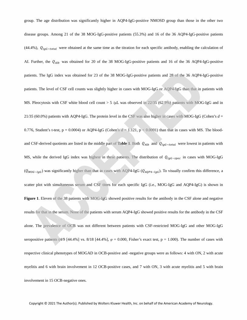
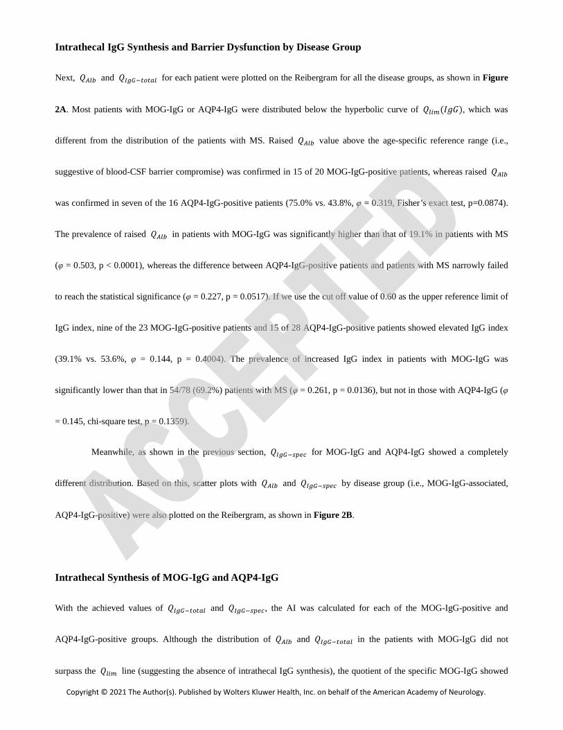
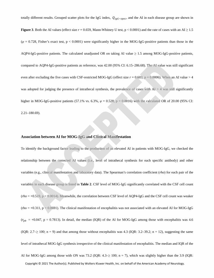
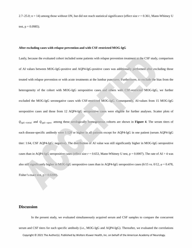
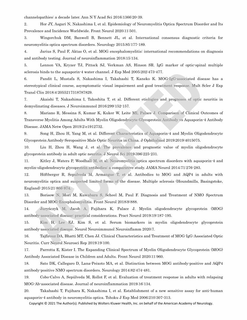
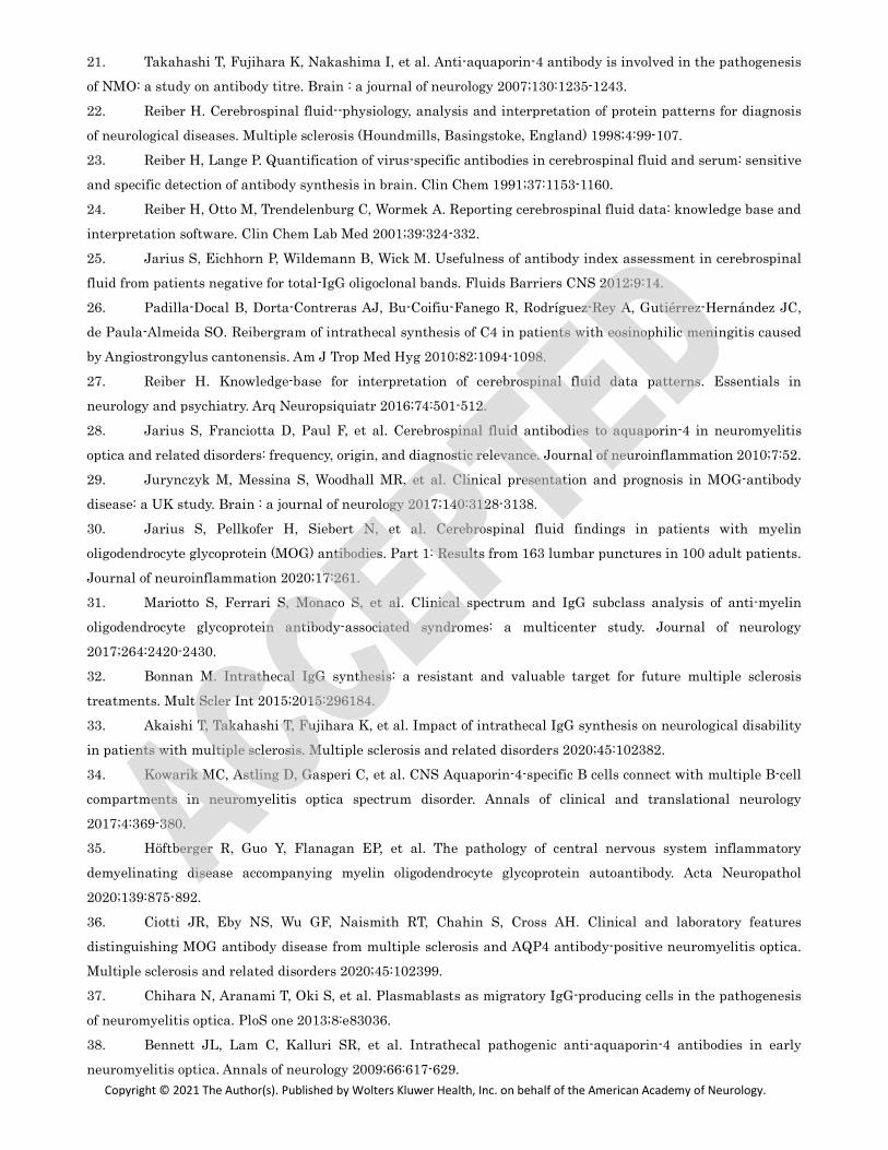
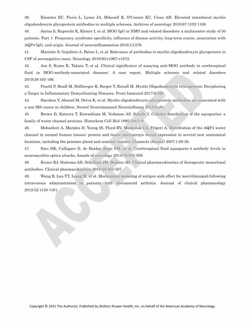
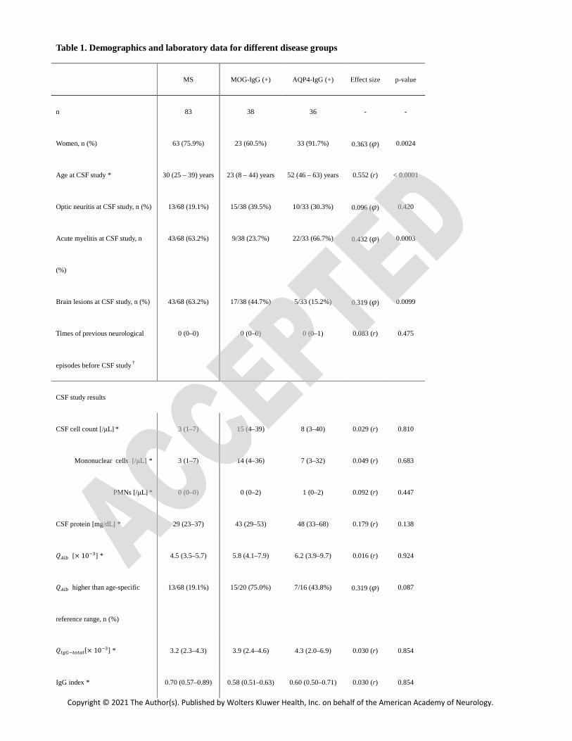
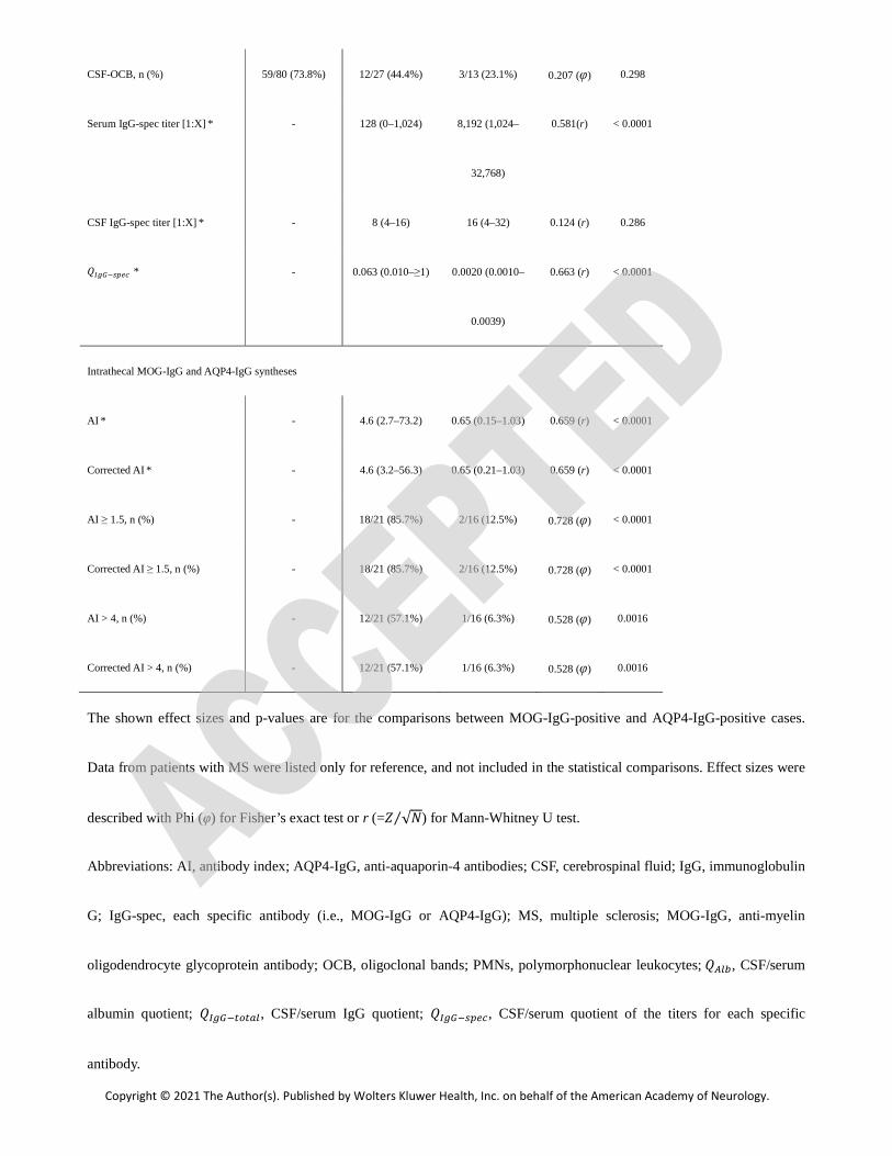
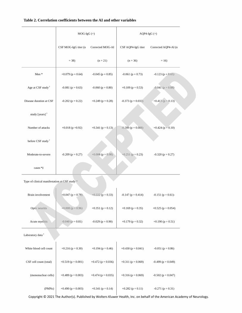
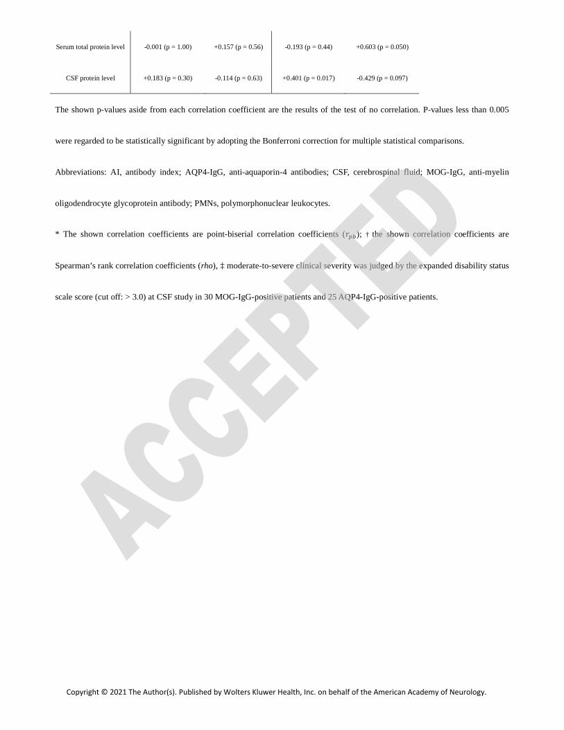
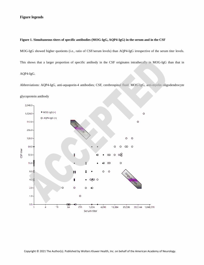
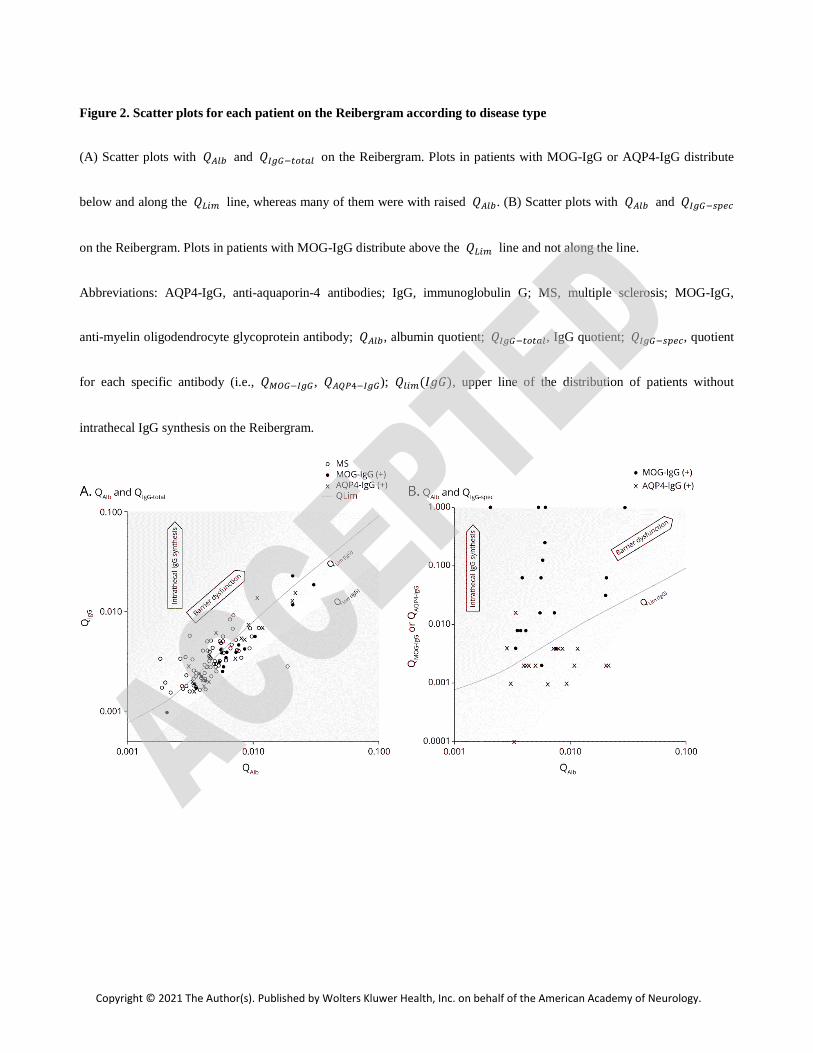
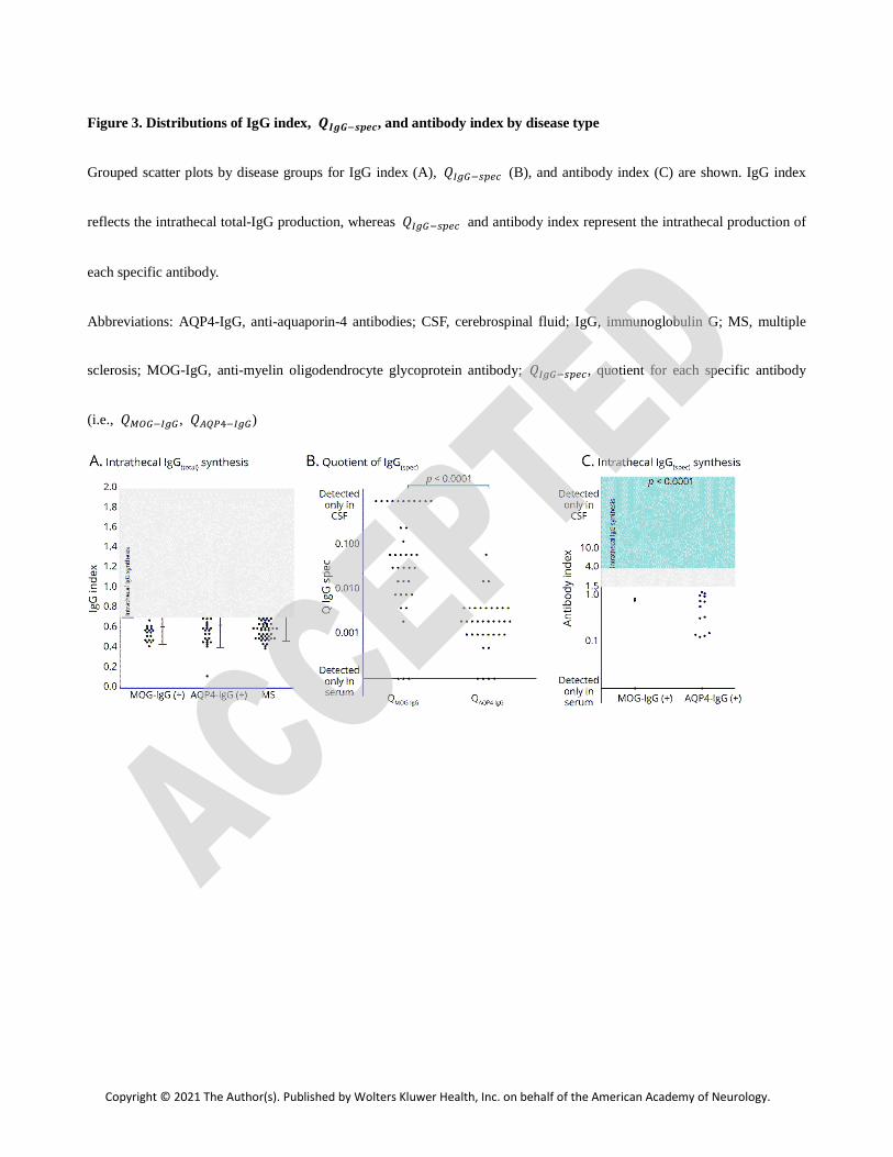
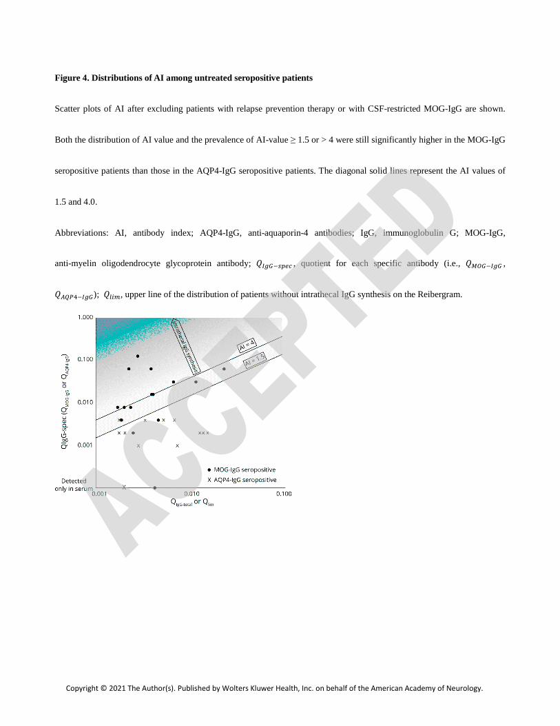