dietary omega-3 fatty acid deficiency and high fructose
TRANSCRIPT

Nutrients 2013, 5, 2901-2923; doi:10.3390/nu5082901
nutrients ISSN 2072-6643
www.mdpi.com/journal/nutrients
Review
Dietary Omega-3 Fatty Acid Deficiency and High Fructose
Intake in the Development of Metabolic Syndrome Brain,
Metabolic Abnormalities, and Non-Alcoholic Fatty Liver Disease
Artemis P. Simopoulos
The Center for Genetics, Nutrition and Health, 2001 S Street, NW, Suite 530, Washington, DC 20009,
USA; E-Mail: [email protected]; Tel.: +1-202-462-5062; Fax: +1-202-462-5241.
Received: 8 June 2013; in revised form: 24 July 2013 / Accepted: 24 July 2013 /
Published: 26 July 2013
Abstract: Western diets are characterized by both dietary omega-3 fatty acid deficiency
and increased fructose intake. The latter found in high amounts in added sugars such as
sucrose and high fructose corn syrup (HFCS). Both a low intake of omega-3 fatty acids or
a high fructose intake contribute to metabolic syndrome, liver steatosis or non-alcoholic
fatty liver disease (NAFLD), promote brain insulin resistance, and increase the
vulnerability to cognitive dysfunction. Insulin resistance is the core perturbation of
metabolic syndrome. Multiple cognitive domains are affected by metabolic syndrome in
adults and in obese adolescents, with volume losses in the hippocampus and frontal lobe,
affecting executive function. Fish oil supplementation maintains proper insulin signaling in
the brain, ameliorates NAFLD and decreases the risk to metabolic syndrome suggesting
that adequate levels of omega-3 fatty acids in the diet can cope with the metabolic
challenges imposed by high fructose intake in Western diets which is of major public
health importance. This review presents the current status of the mechanisms involved in
the development of the metabolic syndrome, brain insulin resistance, and NAFLD a most
promising area of research in Nutrition for the prevention of these conditions, chronic
diseases, and improvement of Public Health.
Keywords: metabolic syndrome and insulin resistance; dietary omega-3 fatty acid
deficiency; high fructose intake; non-alcoholic fatty liver disease; brain insulin resistance;
hippocampus; learning; memory; appetite; inflammation; endocannabinoids;
neurodegenerative diseases
OPEN ACCESS

Nutrients 2013, 5 2902
1. Introduction
Over the past century, major changes have taken place in the food composition of Western diets, in
terms of essential fatty acids and sugar intake, particularly fructose [1–6]. Today’s Western diets are
characterized by increases in total fat, especially in saturated fat and omega-6 fatty acids and decreases
in omega-3 fatty acids in comparison to the fatty acid intake during evolution for which our genes
were programmed to respond [1,3,6,7]. Omega-3 deficiency contributes to insulin resistance and the
metabolic syndrome [1,8,9] brain metabolic abnormalities [10–13], liver steatosis or non-alcoholic
fatty liver disease (NAFLD) [14]. An increase in fructose intake contributes to similar metabolic
effects. The largest increase in the intake of fructose occurred with the introduction of high fructose
corn syrup (HFCS) in soft drinks and processed foods about 30 years ago [2,5,6]. These dietary
changes: the omega-3 fatty acid deficiency and the excessive fructose intake occurred as a result of
agribusiness, modern agriculture and food processing, and not because there was any scientific
evidence that required decreasing omega-3 fatty acid intake while increasing omega-6 fatty acids and
fructose intake. With the decrease in omega-3 fatty acid intake there has been an absolute and relative
increase in omega-6 fatty acids leading to an increase in the omega-6/omega-3 ratio from 1–2/1 to
about 16/1 in terms of both 18, 20 and 22 carbon atoms (linoleic acid (LA), arachidonic acid (AA),
alpha-linolenic acid (ALA), eicosapentaenoic acid (EPA), and dochosahexaenoic acid (DHA)) and
LA/ALA and AA/EPA + DHA ratios [15]. Omega-3 fatty acids have been studied extensively since
1985 and have been shown to play an important role in growth and development and in health and
disease [1,3].
In epidemiological studies, animal experiments and clinical intervention studies the consumption of
HFCS, or sugar sweetened beverages has been linked to the presence of unfavorable lipid levels,
high triglycerides, high small dense LDL and low HDL [4], cardiovascular disease [5], type 2
diabetes [6,16,17], insulin resistance [18,19], the metabolic syndrome and liver steatosis [14]. More
recently it has been shown that moderate amounts of fructose and sucrose significantly alter hepatic
insulin sensitivity and lipid metabolism compared with similar amounts of glucose [20–23]. Recent
studies reveal the broad effects of the metabolic syndrome on mental health disorders, cognitive
function, mood changes and depression [10–12]. Furthermore, diabetic and obese individuals have
increased vulnerability to mental health [13]. For these reasons the effects of metabolic syndrome on
brain are being extensively investigated. In this paper I review the effects of the interaction of omega-3
fatty acid deficiency and high fructose intake in the development of metabolic syndrome, brain
metabolic abnormalities, cognitive function, mental health, and liver steatosis or NAFLD. Fortunately,
these adverse effects may be prevented or ameliorated with dietary repletion of omega-3 fatty acids.
2. The Metabolic Syndrome
The Metabolic Syndrome first made its appearance as ―Syndrome X‖ described by Reaven in his
Banting lecture in 1988 [24]. Reaven described ―Syndrome X‖ as consisting of resistance to
insulin-stimulated glucose uptake, glucose intolerance, hyperinsulinemia, increased very-low-density
lipoprotein triglyceride, and decreased high-density lipoprotein cholesterol and hypertension. Reaven
considered insulin resistance to be of primary importance in coronary artery disease, hypertension and
Nutrients 2013, 5 2903
type 2 diabetes. Since then an explosion of publications has described the relationships among insulin
resistance, hyperinsulinemia, type 2 diabetes, obesity, hypertension, and coronary artery disease [25].
Insulin resistance is a metabolic state in which insulin in physiological concentrations fails to produce
a normal biologic response, and is the core perturbation in the metabolic syndrome. Obesity is a classic
state of insulin resistance is present in the majority of patients with impaired glucose tolerance, in
patients with type 2 diabetes, and in about 25% of non-obese healthy individuals with normal glucose
tolerance [24]. Although many definitions of the metabolic syndrome have been developed by various
national and international organizations, they all agree on the essential components (central obesity,
insulin resistance/glucose intolerance, dyslipidemia and hypertension). Estimates from the U.S. show
that the prevalence of the metabolic syndrome among adults ranges between 20% and 22%, and the
prevalence of NAFLD is about 20% [26,27]. More recent estimates in the U.S. show that the
prevalence of the metabolic syndrome among adults ranges from 34.3% to 38.5% depending on the
criteria used to define abdominal obesity [28].
Metabolic syndrome as a risk factor for neurological disorders has been a focus of research since
the observations that at the molecular level metabolic syndrome is accompanied by dysregulation in
the expression of cytokines and chemokines and alterations in the levels of leptin [10]. Such changes
modulate immune response and inflammation that lead to alterations in the hypothalamic
―bodyweight/appetite/satiety set point‖ [10]. The involvement of the brain in the pathogenesis of
metabolic syndrome is associated with neurochemical changes in stroke, depression and Alzheimer’s
disease [13]. Furthermore, the metabolic syndrome has been associated with cognitive function and
metabolic brain abnormalities [11]. In a literature search on metabolic syndome brain and cognition,
Yates et al. [11] included studies investigating individual components of metabolic syndrome.
Multiple cognitive domains were affected by metabolic syndrome in adults. In adolescents the majority
of findings were in executive functioning [11]. Brain literature in adults implicated metabolic
syndrome in ischemic stroke, white matter alterations, and altered brain metabolism. In adolescents,
individual metabolic syndrome factors were associated with volume losses in the hippocampus and
frontal lobes [11]. Potential factors include impaired vascular reactivity, neuroinflammation, oxidative
stress, and abnormal lipid metabolism [10,11]. The deficiency of omega-3 fatty acids, impacts
negatively on cognitive performance and brain structure, as a result of insulin resistance-associated
cerebrovascular reactivity [10–12].
Insulin receptors are expressed in the brain and the physiological roles for insulin in the CNS are
being delineated. Epidemiological evidence suggests an increased risk of developing dementia,
including Alzheimer’s disease, in persons with obesity, type 2 diabetes, but also in those with poor
insulin sensitivity without diabetes, suggesting a mechanistic link between adiposity, insulin sensitivity
and dementia [12]. In their review, Williamson et al. express their concerns on the difficulties in
quantifying insulin sensitivity in the brain and emphasize the need to develop technology for this
purpose, so that insulin sensitivity’s role in the ―new age‖ dementia that is related to today’s diet and
lifestyle is better defined [12].
De La Monte [13] in his recent review on the role of peripheral insulin resistance in cognitive
impairment and Alzheimer’s disease concludes that systemic insulin resistance diseases linked to
obesity, type 2 diabetes and non-alcoholic steatohepatitis promote neurodegeneration. Insulin-resistance
dysregulates lipid metabolism which promotes ceramide accumulation with attendant inflammation

Nutrients 2013, 5 2904
and endoplasmic reticulum stress. De La Monte states that ―toxic ceramides generated in extra-CNS
tissues, e.g., liver, get released into peripheral blood, and subsequently transit across the blood brain
barrier into the brain where they induce brain insulin resistance, inflammation and cell death‖.
3. Dietary Omega-3 Fatty Acid Deficiency
In 1991, Storlien et al. studied the influence of dietary fat composition on development of insulin
resistance in rats [8]. They showed that impaired insulin action in skeletal muscle is related to
triglyceride accumulation, suggesting intracellular glucose-fatty acid cycle involvement, and that
long-chain omega-3 fatty acids in phospholipid of skeletal muscle may be important for efficient
insulin action. In rats made insulin resistant with a high fat diet, the resistance could be prevented by
the inclusion of omega-3 fatty acids in the diet, but only under circumstances in which the EPA and
DHA became incorporated in the phospholipid component of the muscle cells. Subsequently,
Borkman et al. [9] performed studies in human subjects on the relationship between insulin sensitivity
and the fatty acid composition of skeletal muscle membrane phospholipids, and showed that decreased
insulin sensitivity is associated with decreased concentrations of long-chain polyunsaturated fatty acids
(PUFA) in skeletal-muscle phospholipids, raising the possibility, that changes in the fatty acid
composition of muscle cell membrane modulates the action of insulin in human subjects as was shown
earlier in rats [8]. Skeletal muscle is the principle site of insulin mediated glucose disposal and the
fatty acid composition of membranes influences the action of insulin within the skeletal muscle,
whereas the fasting serum insulin concentration was positively correlated with the percentages of LA
indicating that high or increased LA is associated with decreased insulin sensitivity [9]. The results of
this study, in conjunction with the studies in cell systems and animals, suggest that variations in insulin
sensitivity are related to differences in the membrane content of long-chain PUFA within skeletal
muscle phospholipids. Therefore, abnormalities in the fatty-acid composition of membranes may be
involved in the pathogenesis of a cluster of disorders linked to insulin resistance and hyperinsulinemia,
including obesity, hypertension, type 2 diabetes mellitus, and coronary artery disease, suggesting that
diet may influence their development [1,3]. Furthermore, these conditions are known to have genetic
determinants for hypertension, type 2 diabetes, some forms of hyperlipidemia, obesity, and insulin
resistance (Figure 1) [29] and have a common abnormality in smooth muscle response and insulin
resistance [30,31]. Figure 2 is a hypothetical scheme of how a decrease in EPA and DHA in muscle
cell membrane phospholipids from (1) a decrease in dietary intake per se, (2) increased dietary intake
of trans fatty acids, (3) increased intake of LA, or (4) genetic variants in delta-6 and delta-5
desaturases may lead to a decrease in EPA and DHA muscle cell membrane phospholipids [31]. Such
a decrease contributes to insulin resistance and hyperinsulinemia, with the subsequent development of
obesity, hypertension, type 2 diabetes, and coronary artery disease (including asymptomatic
atherosclerosis and microvascular angina) [31].

Nutrients 2013, 5 2905
Figure 1. Network of interrelated functions, connected by physiological mechanisms,
controlled by heredity and influenced by environmental factors [29]. (Reproduced
with permission).
Figure 2. The effects of dietary eicosapentaenoic acid (EPA) + dochosahexaenoic acid
(DHA) on the composition of the EPA + DHA in skeletal muscle membrane phospholipids
and their relationship to insulin resistance/hyperinsulinemia and chronic diseases (obesity,
T2DM, hypertension, coronary artery disease). (Modified from Reference [31]).

Nutrients 2013, 5 2906
A decrease in brain and retina DHA, resulting from nutritional deficiency or aging is associated
with reduced cognitive ability, increased emotional behavior and decreased visual capacity [32].
Epidemiological and clinical data link omega-3 fatty acid deficiency with mood disorders [33].
The endocannabinoid system includes the two endogenous ligands 2-arachidonoylglycerol (2-AG)
and N-arachidonoylethanolamine (anandamide or AEA) and two cannabinoid receptors (CB1 and
CB2). Central CB1 receptor activation by increased endocannabinoid levels or exogenous agonists
induces hyperphagia along with increased lipogenesis and peripheral adiposity [34,35]. CB1 receptors
are predominantly expressed in the brain, but also in the peripheral tissues of the gastrointestinal tract,
adrenal glands, liver, adipose tissue, and skeletal muscles [36]. In the liver, CB1 activation increases de
novo lipogenesis through stimulation of cytosolic fatty acid synthase activity leading to fatty liver and
obesity [35]. Pharmacological blockade of the CB1 receptor is effective in treating obesity and related
metabolic derangements. However serious psychiatric side effects, including depression and suicidal
tendencies [37], caused marketplace withdrawal of Rimonabant, a selective CB1 antagonist [38].
Endocannabinoids are endogenous lipid mediators made from essential fatty acids available only from
dietary sources. The two best characterized endocannabinoids, 2-AG and AEA, are both metabolic
derivatives of a single fatty acid precursor AA in phospholipids. Endocannabinoids are formed
enzymatically on demand from the pool of AA plasma phospholipids formed from LA or ingested as
AA. In a study on mice Alvheim et al. [39] showed that dietary linoleic acid increased tissue AA
which led to increased 2-AG + 1-AG and AEA resulting in the development of diet induced obesity by
increasing food intake, feed efficiency and adiposity in mice. Reducing LA in the diet and adding
EPA + DHA reduced the AA phospholipid pool, normalized endocannabinoid tone and reversed the
obesity. Obesity is associated with neurological morbidity, including major depression [40], but the
underlying pathophysiological mechanisms remain poorly understood. Lafourcade et al. [41] used a
specific diet to mimic lifelong omega-6/omega-3 imbalance of essential fatty acids in mice and showed
that reducing omega-3 levels markedly diminished the synaptic and behavioral functions of the
cannabinoid CB1R. Omega-3 deficiency profoundly affected emotional behaviors in the rodents and
support the clinical and epidemiological associations between omega-6/omega-3 imbalance and mood
disorders [33,41]. The authors concluded ―by linking diet to altered synaptic functions of the CB1R in
relevant brain areas, our data provide the first synaptic substrate for the impairment of emotional
behavior, including depression, associated with the low levels of n-3 PUFAs that are frequently
observed in Western diets‖ [41].
4. High Fructose Intake
There has been a shift in the types of nutrients consumed in the American diet. The consumption of
fructose has increased mostly because of an increased consumption of soft drinks and many other
beverages that are high in fructose and because of the consumption of foods such as breakfast cereals,
baked goods, condiments, and prepared desserts all sweetened with sucrose and high-fructose corn
syrup (HFCS) [2]. HFCS is produced by the enzymatic isomerization of dextrose to fructose [2]. The
commercial use of HFCS began to increase in the 1970s and by 1985, HFCS accounted for ~35% of
the total amount of sweeteners by dry weight in the food supply [2]. Although HFCS can contain up to
90% fructose, most of the HFCS used in beverages contains ~55% fructose [2,4]. Young males,

Nutrients 2013, 5 2907
15–18 years of age, reported the highest intake of fructose intakes with the 90th percentile intake from
all sources of about 100 grams per day [2]. The use of crystalline fructose has been expanded to the
general food supply. The use of glucose syrup also increased whereas the contribution of other
sweeteners, supplied as honey, molasses and maple syrup remained constant at 1% [2].
Over the past 35 years concerns have been expressed about the adverse effects of excessive
consumption of sugar (Table 1) (Figure 3). From 2001 to 2004 the usual intake of added sugar in the
American diet was 22.2 teaspoons per day, which is 335 calories per day [2].
Figure 3. The effects of dietary Fructose and its relationship to insulin
resistance/hyperinsulinemia and chronic diseases (liver steatosis, obesity, T2DM,
hypertension, coronary artery disease).
In 2003 the WHO report on Diet and Chronic Diseases recommended that added sugar should not
constitute more than 10% of energy intake [42]. Dietary sugars contribute to the development of
obesity [2]. A large number of calories are obtained from beverages, processed food, and deserts made
with sucrose or high fructose corn syrup, which are absorbed as a mixture of glucose and fructose.
Apart from weight gain from excess sweet calories there are a number of metabolic consequences
harmful to health. The best documented adverse effects known for decades is the dyslipidemia that
develops with the ingestion of large amounts of sugar even when substituted calorie for calorie for
fat [43]. An important mechanism is the de-novo lipogenesis (DNL) synthesis of the saturated fat
palmitate from glucose, fructose or both. There is a marked increase in DNL after excess carbohydrate
calories [44] leading to high triglycerides, small dense LDL and low HDL that accelerate
atherosclerosis. The recent study by Sevastianova et al. [45] expands the lipogenic effects of dietary
sugar beyond dyslipidemia to include fatty liver. A 2% increase in body weight and similar increases

Nutrients 2013, 5 2908
in subcutaneous and visceral adipose tissue were measured by proton magnetic resonance
spectroscopy. The results showed for the first time a link between excess dietary sugar and the
accumulation of liver fat by DNL a pathway uniquely stimulated by dietary sugar. There was also an
increase in fasting insulin level.
In the U.S., hepatic steatosis affects a large fraction of obese adults and children, may progress to
non-alcoholic steatohepatitis, cirrhosis, and liver failure. The implication that a persistent excess of
calories as dietary sugar can cause or exacerbate fatty liver by DNL lends additional support to public
health recommendations to limit intake of sugars [2,42]. The increased liver fat resulting from
increased DNL and the imbalance between triglyceride synthesis and secretion may increase oxidative
stress, inflammation and insulin resistance (Figure 3) [4]. In 2009, the American Heart Association
Nutrition Committee recommended that women and men consume no more than 100 and 150 kcal of
added sugar per day, respectively [2]. In 2011, Stanhope et al. [46] compared the effects of consuming
glucose, fructose, or high-fructose corn syrup (HFCS) at 25% of energy requirements on risk factors
for cardiovascular disease, and concluded that ―consumption of HFCS sweetened beverages for 2 weeks
at 25% of energy increased risk factors for cardiovascular disease comparably with fructose and more
than glucose in young adults‖ [46]. This well executed study contradicts the conclusions from recent
reviews that sugar intakes as high as 25%–50% of energy, have no adverse longtime effects with
respect to components of the metabolic syndrome [47] and that fructose consumption up to
140 g/day does not result in biologically relevant increase of fasting or postprandial triglycerides in
healthy, normal-weight [48] or overweight or obese [49] humans. These reviews [47–49] unfortunately
influenced the decision of the Committee of the 2010 dietary guidelines for Americans in which
maximal intake levels of 25% or less of total energy from added sugars is suggested instead of the 10%
level of the recommendation of the WHO/FAO report [42]. Stanhope et al. [50] clearly showed the
twenty-four hour triglyceride area under the curve increased compared with baseline during fructose
consumption and HFCS but not with glucose. Fasting LDL and apoB concentrations were increased
during consumption of fructose and HFCS but not with glucose indicating that the adverse effects
associated with chronic consumption of sugar-sweetened beverages results from the specific effects of
fructose [39] rather than from circulating glucose and insulin excursions [22,51–54]. In 2009,
Stanhope et al. had shown that consumption of fructose-sweetened beverages at 25% of energy
increased visceral adipose tissue and de novo lipogenesis, produced dyslipidemia, and decreased
glucose tolerance/insulin sensitivity in older, overweight/obese men and women, whereas
glucose-sweetened beverages did not [4]. Similarly Silbernagel et al. [55] reported that consumption of
fructose or glucose (150 g/day) for 4 weeks lowered insulin sensitivity and increased HOMA-IR in
subjects of similar age (31 ± 9 years) and BMI = (25.9 ± 22).
In terms of their composition commonly ingested sugars, sucrose and HFCS consist of 50%–55%
fructose [2]. In most studies, the fructose and HFCS at 25% of energy but not glucose lead to
significant increases in postprandial TG, and fasting and postprandial concentrations of LDL, apoB,
and the apoB to apoAI ratio all established risk factors of CVD. At present, U.S. food labels contain
information on total sugars per serving but do not distinguish between sugars that are naturally present
in foods and added sugars. Thus, it is impossible for consumers to determine the amount of added
sugars in foods or beverages or some drugs and cough syrups. However, a few sources of information
do exist on added sugar content. The Department of Agriculture in the U.S. published a database for
Nutrients 2013, 5 2909
the added sugar content of selected foods in 2006 [56]. In addition, several voluntary food-labeling
systems are in place or are being developed, some of which include criteria for limiting added sugars.
These include the Smart Choices Program [57], the Hannaford’s grocery chain Guiding Stars
program [58], the Overall Nutrient Quality Index [59], and the Nutrient Rich Foods Coalition nutrient
density score [60].
It is ironic indeed, that originally fructose was proposed as the ideal sweetener for persons with
Type 2 Diabetes because it does not raise blood sugar and is unable to stimulate insulin secretion.
Fructose is now implicated in the epidemics of obesity and diabetes type 2, insulin resistance,
hypertension, visceral adiposity, and the metabolic syndrome [2,4]. The effects of fructose on fasting
triglycerides may be higher in women, especially post menopausal women than in men [2], in
sedentary overweight people [61], in those with the metabolic syndrome [62] and in those eating low
fiber diets [62].
Mechanisms by which fructose increases fasting and postprandial triglyceride levels are: increased
de novo lipogenesis in the liver [4,62,63], increased hepatic triglycerides synthesis, and secretion of
very-low-density lipoproteins [64]; reduced lipoprotein lipase activity at the adipocyte, which
decreases the rate of peripheral triglyceride clearance [64]. A high consumption of sugar-sweetened
beverages and foods is associated with evidence of increased inflammation and oxidative
stress [65,66]. Others have shown that fructose may slow the basal metabolic rate in mice [67].
Fructose is the only sugar that raises uric acid concentrations [68]. Fructose, compared with glucose, is
preferentially metabolized to lipid in the liver [4]. Fructose consumption induces insulin resistance,
impaired glucose tolerance, hyperinsulinemia, hypertriglyceridemia, and hypertension in animal
models [4] (Table 1).
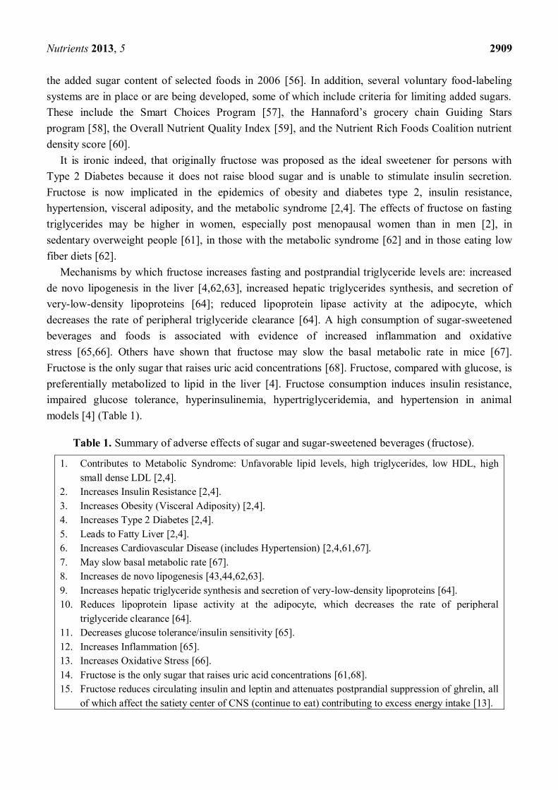
Table 1. Summary of adverse effects of sugar and sugar-sweetened beverages (fructose).
1. Contributes to Metabolic Syndrome: Unfavorable lipid levels, high triglycerides, low HDL, high
small dense LDL [2,4].
2. Increases Insulin Resistance [2,4].
3. Increases Obesity (Visceral Adiposity) [2,4].
4. Increases Type 2 Diabetes [2,4].
5. Leads to Fatty Liver [2,4].
6. Increases Cardiovascular Disease (includes Hypertension) [2,4,61,67].
7. May slow basal metabolic rate [67].
8. Increases de novo lipogenesis [43,44,62,63].
9. Increases hepatic triglyceride synthesis and secretion of very-low-density lipoproteins [64].
10. Reduces lipoprotein lipase activity at the adipocyte, which decreases the rate of peripheral
triglyceride clearance [64].
11. Decreases glucose tolerance/insulin sensitivity [65].
12. Increases Inflammation [65].
13. Increases Oxidative Stress [66].
14. Fructose is the only sugar that raises uric acid concentrations [61,68].
15. Fructose reduces circulating insulin and leptin and attenuates postprandial suppression of ghrelin, all
of which affect the satiety center of CNS (continue to eat) contributing to excess energy intake [13].

Nutrients 2013, 5 2910
Vartanian et al. carried out a meta-analysis reviewing 88 cross-sectional and prospective studies
evaluating the relationship between soft drink intake and nutrition on health outcomes [69]. Higher
intake of soft drinks was associated with greater energy intake, higher body weight, lower intake of
other nutrients and worse health indices. Further analyses from a larger trial confirmed these findings,
specifically greater weight loss as sugar-sweetened beverage intake decreased [70].
5. Dietary Omega-3 Deficiency, High Fructose Intake, Insulin Resistance, and the Brain
The Omega-3 fatty acids, EPA and DHA have been shown to be essential for visual function and
cerebral maturation of the infant and to play an important role in improving mental health, learning
and memory, neurogenerative diseases, depression and schizophrenia [71]. Sucrose infusion directly
into the nucleus accumbens alters dopamine and opioid neurotransmission, increasing food intake in
rats [72]. Both sweet and high fat foods mobilize opioids and dopamine within the nucleus accumbens,
establishing hard-wired pathways for craving in these areas [73,74]. Chronic hyperinsulinemia may
also contribute to increased caloric intake by preventing dopamine clearance from the nucleus
accumbens, fostering pleasure derived from food in situations in which energy states are replete,
contributing to excess energy intake [75]. Teff et al. [76] showed that dietary fructose reduces
circulating insulin and leptin and attenuates postprandial suppression of ghrelin all of which affect the
satiety center in the Central Nervous System (CNS). Page et al. [77] studied the effects of fructose vs.
glucose on regional cerebral blood flow in brain regions involved with appetite and reward pathways
in healthy volunteers and found that the animal studies on the brain effects of fructose on appetite
promotion are relevant to the humans. The major new finding is that hypothalamic brain signal
generated in response to fructose ingestion was statistically different from the response following
glucose ingestion [77]. Thus, after glucose consumption there is an increased sensation of fullness and
satiety but not after fructose consumption suggesting that when the human brain is exposed to fructose,
neuro-biological pathways involved in appetite regulation are modulated thereby promoting food intake.
Due to the central role of brain in the metabolic control of many diseases, the role of brain
endoplasmic reticulum (ER) stress in metabolic disease has come into focus recently. Expanding on
the previous knowledge that brain ER stress underlies neurodegenerative diseases [78] a number of
studies have causally linked brain ER stress to the development of metabolic syndrome and related
disorders such as overeating, obesity, leptin resistance, insulin resistance, beta-cell dysfunction
and hypertension [79–82] under conditions of overnutrition [79,83] and related inflammatory
insults [82]. The CNS, particularly hypothalamus, is the central regulator of energy and body weight
balance [84–99].
Insulin receptors are expressed in the brain and the physiological roles of insulin in the CNS are
starting to be delineated [12]. Disrupted neuronal insulin action may underlie the link between diabetes
and neurodegenerative disorders [100]. Longitudinal studies have identified a higher risk of dementia
or cognitive decline associated with type 2 diabetes and also in insulin resistance without
diabetes [100]. Obesity has a major influence on the development of insulin resistance, and is
considered the underlying cause of the current epidemic of type 2 diabetes mellitus (T2DM) [2].
At this time it is not clear what molecular lesions in liver, muscle, and adipose tissue of patients is
responsible for the development of insulin resistance and ultimately T2DM, or if every tissue develops

Nutrients 2013, 5 2911
insulin resistance by the same route. The molecular pathology of insulin resistance remains
controversial. It could be related to the development of a post-receptor deficit, reducing the insulin
―sensing‖ or signaling capacity of individual cells [101]. This, in turn, results in a requirement for
higher levels of insulin to stimulate glucose uptake into muscle, to reduce glucose production in liver
and to correctly regulate adipose tissue. The analysis of intracellular signaling pathways in specific
tissue may prove to be a means to establish the insulin sensitivity of each tissue. This is most important
in the more unusual insulin target tissues such as the brain, where tissue insulin sensitivity currently
cannot be assessed by HOMA or clamp techniques.
Agraval and Gomez-Pinilla [102] stimulated by the recent research revealing the broad effects of
metabolic syndrome on mental health, cognitive function, and mood, via studies which showed that
diabetic and obese individuals have increase vulnerability to mental health disorders, investigated the
effect of DHA to counteract the effects of metabolic syndrome in the CNS in adult male
Sprague-Dawley rats. Abundant consumption of fructose is an important contribution to the metabolic
syndrome [103]. Studies have shown that rats fed on a high fructose diet develop hepatic oxidative
damage and altered lipid metabolism as a result of the burden of fructose metabolism [104]. Agraval
and Gomez-Pinilla [102] randomly assigned six animals on an omega-3 fatty acid diet (0.5% flaxseed
oil (ALA) and 1.2% DHA) or omega-3 deficient diet with or without fructose solution 15% as drinking
water for 6 weeks. The total fat content in both diets was 10 g per 100 g diet. There were no
differences among the groups on body weight, calorie intake, and food and water consumption. The
deficiency of omega-3 fatty acids resulted in a significant increase in latency time with the Barnes
maze test, indicating memory impairment, which was further enhanced by fructose intake. The effects
of fructose on memory in the omega-3 deficient rats was ameliorated by the omega-3 diet indicating
that dietary omega-3 fatty acid deficiency influences the vulnerability for fructose induced changes. In
the omega-3 deficient animal there was an increase in triglycerides that was further increased by
fructose intake. Fructose also increased the glucose and insulin levels in the omega-3 deficient animal.
The presence of omega-3 fatty acids in the diet reduced the fructose increase in insulin and triglyceride
levels. There was a correlation between serum triglycerides and insulin resistance index and between
triglycerides and fructose induced memory deficits suggesting that memory performance may rely on
levels of insulin resistance index. The deficient omega-3 fatty acid diet and fructose led to a decrease
in pTyr1R levels in the hippocampus which was reversed by the omega-3 diet. There was a negative
correlation between insulin resistance levels and pTyr1R suggesting that the increased insulin
resistance in the body may disrupt insulin receptor signaling in the brain. The Akt phosphorylation was
found to be decreased with omega-3 fatty acid deficiency which was exacerbated by fructose intake
but was alleviated by Akt phosphorylation in the presence of the omega-3 fatty acid diet. The omega-3
fatty acid deficient diet led to decreased phosphorylation of LKB1 whereas the omega-3 fatty acid diet
increased the phosphorlyation of LKB1. There was also a positive correlation between phosphorylated
LKB1 and DHA and a negative correlation with AA pointing to a concomitant alteration of LKB1
after diet treatment and the altered lipid composition in brain (Table 2).

Nutrients 2013, 5 2912
Table 2. Summary of adverse effects of omega-3 deficient diets and high fructose,
corrected by the omega-3 diet [102].
Factor Omega-3
Deficient Diet Fructose Omega-3 Diet
Increase in latency time in the
Barnes Maze Test indicating
memory impairment
↑ Potentiates this effect Ameliorates
Triglycerides ↑ Potentiates this effect Ameliorates
Insulin Resistance ↑ Potentiates this effect Corrects
pTyrIR levels in Hippocampus ↓ Decreases this effect Reversed
Akt phosphorylation ↓ Exacerbates this effect Alleviates
Phosphorylation in KLβ1 ↓ ____ Increases
Phosphorylation of CREB ↓ Exacerbates this effect
Counterregulated the fructose
induced alteration in synaptic
plasticity via CREB Synapsin 1
Synaptophysin
Hippocampus and Frontal
lobe volume ↓ ____ ____
Sir2 ↓ ↓ Normalizes
Liver Steatosis ↑ ↑ Reverses
Omega-3 deficiency showed decreased phosphorylation of CREB which was further exacerbated by
fructose treatment. The presence of omega-3 fatty acids counter regulated the fructose induced
alteration in synaptic plasticity via CREB. Similar results were obtained with Synapsin I and
Synaptophysin. Omega-3 fatty acid deficient diet makes the brain more vulnerable to fructose induced
free radical attack. Clinical studies indicate that a balanced ratio of omega-6/omega-3 is important to
maintain health and normal development [1]. The data show that omega-3 deficiency with or without
fructose promoted a decrease in the phosphorylation of LKB1, indicating that activation of LKB1 may
rely on levels of omega-3 fatty acid in the brain. The changes in LKB1 phosphorylation varied in
direct proportion to DHA level and inverse proportion to AA level, which suggests that a decline in the
ratio of AA/DHA contributes to energy homeostasis (Table 2).
6. Dietary Omega-3 Deficiency and High Fructose Intake in the Development of Non-Alcoholic
Fatty Liver Disease (NAFLD)
As shown earlier in this paper high fructose intake leads to obesity, insulin resistance and in
comparison to glucose, is preferentially metabolized to lipids in the liver increasing triglyceride
synthesis while decreasing their secretion leading to NAFLD (Table 1). Numerous in-vitro and in-vivo
studies have demonstrated that omega-3 fatty acids are able to coordinate both the upregulation of lipid
oxidation by binding and activating peroxisome proliferator activated receptor α (PPARα) [105,106],
and the downregulation of lipid synthesis-suppressing lipogenesis by inhibiting sterol regulatory
element binding protein-1c (SREBP-lc) gene expression and for activation by proteolysis [106–108].
Several clinical studies have reported the beneficial effects of EPA and DHA supplementation on
triglyceridemia [109] blood pressure [110] inflammation [111] and insulin sensitivity [30,31]. A lower

Nutrients 2013, 5 2913
intake of omega-3 fatty acids was suggested to be associated with NAFLD [112,113]. Experiments in
rats and mice that were omega-3 deficient for two generations displayed several features of the
metabolic syndrome including hepatic steatosis [114,115]. Pachikian et al. [116] investigated in mice
the effect of omega-3 depletion for 3 months on hepatic lipid composition and metabolism using
molecular integrative and physiological approaches in-vitro and in-vivo. They observed a stimulation
of the hepatic lipogenic pathway most likely induced by the increased expression and activity of
SREBP-1c. Specifically this study showed (1) decreased omega-3 fatty acids in the phospholipid
fractions and changes (increases) in hepatic endocannabinoid content and AA; (2) omega-3 fatty acid
depletion decreased fatty acid oxidation and promoted hepatic lipid synthesis and storage;
(3) microarray analysis confirmed a metabolic shift in favor of fatty acid and cholesterol synthesis at
the expense of fatty acid oxidation in the livers of omega-3 fatty acid depleted mice; (4) SREBP-1c is
involved (higher expression, activation) in the metabolic alterations occurring in the livers of omega-3
fatty acid depleted mice; (5) mice depleted of omega-3 fatty acids displayed hepatic insulin resistance
as shown by the higher hepatic glucose production upon insulin stimulation when compared with
control mice (by euglycemic hyperinsulinemic clamp); (6) omega-3 fatty acid depletion did not induce
hepatic endoplasmic reticulum (ER) stress; (7) increased liver X receptor (LXR) activity occurred in
the livers of omega-3 fatty acid depleted mice. Insulin is considered to be the classical driver of
SREBP-1c activation which largely explains carbohydrate induced lipogenesis [117]. Because there
were no changes in insulin levels, omega-3 fatty acid depletion promoted insulin resistance by an
insulin independent pathway. This study demonstrated that the metabolic characteristics in this model
of omega-3 fatty acid depletion are opposite to the ones occurring with omega-3 fatty acid
supplementation [118]. The consumption of a diet containing low levels of omega-3 fatty acids for
3 months was sufficient to induce hepatic omega-3 fatty acid depletion in phospholipids, steatosis and
insulin resistance. Decreased fatty acid oxidation and increased triglyceride and cholesterol synthesis
both contributed to lipid accumulation. Because the activation of SREBP-1c related pathways occurred
in a hepatic insulin resistant state and independently of ER stress, it is consistent with increased liver X
receptor (LXR) activity, and a higher endocannabinoid ligand level (2-AG). The SREBP-1c results in
human biopsies of NAFLD patients are characterized by an increased expression of SREBP-1c and
decreased expression of PPARα in patients with omega-3 depletion [119]. In an elegant study,
Gonzalez-Periz et al. [14] investigated the effect of omega-3 fatty acid supplementation in the ob/ob
mice, an obesity model of insulin resistance and fatty liver disease. They showed that dietary intake of
omega-3 fatty acids had insulin-sensitizing actions in adipose tissue and lower and improved insulin
tolerance in obese-mice. Omega-3 fatty acids upregulated the genes involved in insulin sensitivity
(PPARα), glucose transport (GLUT-2/GLUT-4) and insulin receptor signaling (IRS-1/IRS-2).
Furthermore omega-3 fatty acids increased adiponectin, and induced AMPK phosphorylation, a
fuel-sensing enzyme and a gatekeeper of the energy balance. At the same time hepatic steatosis was
alleviated by omega-3 fatty acids. Lipidomic analysis showed that omega-3 fatty acids inhibited the
formation of omega-6 derived eicosanoids, while induced the formation of omega-3 derived resolvins
and protectins from EPA and DHA respectively. Resolvin E1 and protectin D1 mimicked the
insulin-sensitizing and antisteatotic effects of omega-3 fatty acids, and induced adiponectin expression
to a similar extent as that of rosiglitazone- an antidiabetic drug. This study clearly showed the

Nutrients 2013, 5 2914
beneficial effects of omega-3 fatty acids and their lipid autacoids (resolvins + protectins) in preventing
obesity induced insulin resistance and hepatic steatosis.
7. Conclusions, Health Implications and Recommendations
Long term consumption of western diet which is high in saturated fat, omega-6 fatty acids and sugar
especially fructose, while low or deficient in omega-3 fatty acids contributes to the development of
metabolic syndrome including metabolic syndrome of the brain and NAFLD. The metabolic syndrome
is a complex entity consisting of a constellation of metabolic risk factors including central (or
vascular) obesity, insulin resistance/impaired glucose tolerance, dyslipidemia (hypertriglyceridemia
and low HDL-C) and hypertension associated with an atherogenic, procoagulant and inflammatory
state. The concept of the metabolic syndrome had been mainly associated with the ―body‖. Recent
studies, however, have provided data that expand the traditional concept to include the effects of
metabolic syndrome in the brain. New evidence supports the harmful impact of the metabolic
syndrome on the brain, impacting synaptic plasticity and cognitive function. Deficiency of dietary
omega-3 fatty acids increases vulnerability to impaired cognitive functions, and intake of high fructose
diet exacerbates this condition. In terms of public health, it is encouraging that the unhealthy effects of
sugars and especially fructose leading to insulin resistance, metabolic syndrome, brain metabolic
abnormalities, and NAFLD can be counteracted or ameliorated by omega-3 fatty acids. Thus, the right
combination of foods is crucial for brain health as well as for overall health. In animal experiments, the
omega-3 supplementation was indeed essential for normalizing the phosphorylation of CREB and
Synapsin I and Synaptophysin even in the presence of fructose, suggesting that omega-3 fatty acids
can restore the cognitive function under challenging conditions by normalizing the action of insulin
resistance on synaptic plasticity.
Epidemiological studies, clinical trials, animal experiments and cell studies indicate the need for
change by the Agricultural food producers and processing industries. We must avoid omega-3 fatty
acid deficient diets and excess fructose intake if we are going to decrease the risk of obesity, insulin
resistance, diabetes, atherosclerosis, hypertension, depression and deficits in learning, executive
function, memory and neurodegenerative diseases. Inflammation is an important consequence of the
metabolic syndrome [120]. An inflammatory state in brain regulatory centers, such as hypothalamus,
disrupts its metabolic function that leads to neural and neuroendocrine regulation of a number of
physiological processes such as energy balance, glucose metabolism, insulin resistance, cardiovascular
disease, and obesity. Although current understanding of the central inflammatory mechanisms of
metabolic syndrome and related diseases are in an early stage, this area of research is very
promising [121]. Omega-3 fatty acid deficient and high fructose diets are the result of agribusiness and
modern agriculture that led to the increased production of omega-6 rich vegetable oils, an absolute and
relative deficiency of omega-3 fatty acids and to the expansion of processing of food ingredients that
led to increases in high fructose intake for the first time in human’s evolutionary history.
Recent studies with human volunteers indicate that it is essential to begin modifications of the
current Western diet as follows:
1. Increase the intake of omega-3 fatty acids ALA, EPA, and DHA, and decrease LA and
AA [122]. This can be accomplished by changing the oils in the Western diet and substituting

Nutrients 2013, 5 2915
the high omega-6 oils (corn oil, sunflower, safflower, soybean) with low omega-6 fatty acids
and oils high in omega-3’s in order to balance the omega-6/omega-3 ratio such as (1) Olive oil
which is low in omega-6 fatty acids (LA 6%–12%), (2) Canola oil which has a ratio of
omega-6/omega-3 of 2:1. (3) Chia and Perilla oil which contain 55%–60% omega-3 fatty acids.
Industry has recognized the need to change the oils by decreasing the high omega-6 content
through genetic manipulation, i.e., by developing high monounsaturated sunflower oil.
Furthermore through genetic engineering of soybeans, by increasing Stearidonic acid, industry
has increased the omega-3 content of soybean oil which has been shown in animal and human
studies to be more effective than its precursor, α-linolenic acid, to be metabolized to EPA, thus
enriching membrane phospholipids with EPA. Hence, stearidonic acid can serve as a
―pro-eicosapentaenoic acid‖ [123,124]. It is necessary that food labels state the levels of
omega-6 and omega-3 fatty acids separately instead of simply reporting them as PUFA.
Furthermore studies and all journal publications should distinguish the concentration of
omega-6 and omega-3 fatty acids instead of simply PUFA. The omega-6 and omega-3 fatty
acids are physiologically and metabolically distinct and have opposing properties. Therefore,
their balance is important for health.
2. Decrease the amount of added sugar to less than 10% of energy intake, and remove high
fructose corn syrup from sweetened beverages, snacks, cookies and other forms of processed
foods. Again, industry has recognized the detrimental effects to health, resulting from high
fructose intake and is searching for natural ingredients low in sugars (and fructose) to substitute
for HFCS. Furthermore, the levels of glucose and fructose should be stated in all food
labels [125–128]. At present, U.S. food labels contain information on total sugars per serving
but do not distinguish between sugars that are naturally present in foods and added sugars.
Thus, it is impossible for consumers to determine the amount of added sugars in foods or
beverages, or some drugs and cough syrups.
References
1. Simopoulos, A.P. The importance of the omega-6/omega-3 Fatty Acid ratio in cardiovascular
disease and other chronic diseases. Exp. Biol. Med. (Maywood) 2008, 233, 674–688.
2. Johnson, R.K.; Appel, L.J.; Brands, M.; Howard, B.V.; Lefevre, M.; Lustig, R.H.; Sacks, F.;
Steffen, L.M.; Wylie-Rosett, J. American Heart Association Nutrition Committee of the Council
on Nutrition, Physical Activity, and Metabolism and the Council on Epidemiology and
Prevention. Dietary sugars intake and cardiovascular health: A scientific statement from the
American Heart Association. Circulation. 2009, 120, 1011–1020.
3. Simopoulos, A.P. Omega-3 fatty acids in health and disease and in growth and development. Am.
J. Clin. Nutr. 1991, 54, 438–463.
4. Stanhope, K.L.; Schwarz, J.M.; Keim, N.L.; Griffen, S.C.; Bremer, A.A.; Graham, J.L.;
Hatcher, B.; Cox, C.L.; Dyachenko, A.; Zhang, W.; et al. Consuming fructose-sweetened, not
glucose-sweetened, beverages increases visceral adiposity and lipids and decreases insulin
sensitivity in overweight/obese humans. J. Clin. Investig. 2009, 119, 1322–1334.

Nutrients 2013, 5 2916
5. Fung, T.T.; Malik, V.; Rexrode, K.M.; Manson, J.E.; Willett, W.C.; Hu, F.B. Sweetened
beverage consumption and risk of coronary heart disease in women. Am. J. Clin. Nutr. 2009, 89,
1037–1042.
6. Montonen, J.; Järvinen, R.; Knekt, P.; Heliövaara, M.; Reunanen, A. Consumption of sweetened
beverages and intakes of fructose and glucose predict type 2 diabetes occurrence. J. Nutr. 2007,
137, 1447–1454.
7. Blasbalg, T.L.; Hibbeln, J.R.; Ramsden, C.E.; Majchrzak, S.F.; Rawlings, R.R. Changes in
consumption of omega-3 and omega-6 fatty acids in the United States during the 20th century.
Am. J. Clin. Nutr. 2011, 93, 950–962.
8. Storlien, L.H.; Jenkins, A.B.; Chisholm, D.J.; Pascoe, W.S.; Khouri, S.; Kraegen, E.W. Influence
of dietary fat composition on development of insulin resistance in rats. Relationship to muscle
triglyceride and omega-3 fatty acids in muscle phospholipid. Diabetes 1991, 40, 280–289.
9. Borkman, M.; Storlien, L.H.; Pan, D.A.; Jenkins, A.B.; Chisholm, D.J.; Campbell, L.V. The
relation between insulin sensitivity and the fatty-acid composition of skeletal-muscle
phospholipids. N. Engl. J. Med. 1993, 328, 238–244.
10. Farooqui, A.A.; Farooqui, T.; Panza, F.; Frisardi, V. Metabolic syndrome as a risk factor for
neurological disorders. Cell Mol. Life Sci. 2012, 69, 741–762.
11. Yates, K.F.; Sweat, V.; Yau, P.L.; Turchiano, M.M.; Convit, A. Impact of metabolic syndrome
on cognition and brain: A selected review of the literature. Arterioscler. Thromb. Vasc. Biol.
2012, 32, 2060–2067.
12. Williamson, R.; McNeilly, A.; Sutherland, C. Insulin resistance in the brain: An old-age or
new-age problem? Biochem. Pharmacol. 2012, 84, 737–745.
13. De La Monte, S.M. Metabolic derangements mediate cognitive impairment and Alzheimer’s
disease: Role of peripheral insulin-resistance diseases. Panminerva Med. 2012, 54, 171–178.
14. González-Périz, A.; Horrillo, R.; Ferré, N.; Gronert, K.; Dong, B.; Morán-Salvador, E.; Titos, E.;
Martínez-Clemente, M.; López-Parra, M.; Arroyo, V.; et al. Obesity-induced insulin resistance
and hepatic steatosis are alleviated by omega-3 fatty acids: A role for resolvins and protectins.
FASEB J. 2009, 23, 1946–1957.
15. Simopoulos, A.P. Evolutionary Aspects of Diet and Essential Fatty Acids. In Fatty Acids and
Lipids—New Findings; Hamazaki, T., Okuyama, H., Eds.; Kagar: Basel, Switzerland, 2001;
pp. 18–27.
16. Palmer, J.R.; Boggs, D.A.; Krishnan, S.; Hu, F.B.; Singer, M.; Rosenberg, L. Sugar sweetened
beverages and incidence of type 2 diabetes mellitus in African American women. Arch. Intern.
Med. 2008, 168, 1487–1492.
17. Schulze, M.B.; Manson, J.E.; Ludwig, D.S.; Colditz, G.A.; Stampfer, M.J.; Willett, W.C.;
Hu, F.B. Sugar-sweetened beverages, weight gain, and incidence of type 2 diabetes in young and
middle-aged women. JAMA 2004, 292, 927–934.
18. Bremer, A.A.; Auinger, P.; Byrd, R.S. Sugar-sweetened beverage intake trends in U.S.
adolescents and their association with insulin resistance-related parameters. J. Nutr. Metab. 2009,
doi:10.1155/2010/196476.

Nutrients 2013, 5 2917
19. Yoshida, M.; McKeown, N.M.; Rogers, G.; Meigs, J.B.; Saltzman, E.; D’Agostino, R.;
Jacques, P.F. Surrogate markers of insulin resistance are associated with consumption of
sugar-sweetened drinks and fruit juice in middle and older-aged adults. J. Nutr. 2007, 137,
2121–2127.
20. Aeberli, I.; Hochuli, M.; Gerber, P.A.; Sze, L.; Murer, S.B.; Tappy, L.; Spinas, G.A.; Berneis, K.
Moderate amounts of fructose consumption impair insulin sensitivity in healthy young men: A
randomized controlled trial. Diabetes Care 2013, 36, 150–156.
21. Tappy, L.; Lê, K.A. Metabolic effects of fructose and the worldwide increase in obesity. Physiol.
Rev. 2010, 90, 23–46.
22. Tappy, L.; Mittendorfer, B. Fructose toxicity: Is the science ready for public health actions?
Curr. Opin. Clin. Nutr. Metab. Care 2012, 15, 357–361.
23. Lecoultre, V.; Egli, L.; Carrel, G.; Theytaz, F.; Kreis, R.; Schneiter, P.; Boss, A.; Zwygart, K.;
Lê, K.A.; Bortolotti, M.; et al. Effects of fructose and glucose overfeeding on hepatic insulin
sensitivity and intrahepatic lipids in healthy humans. Obesity 2013, 21, 782–785.
24. Reaven, G.M. Banting lecture 1988: Role of insulin resistance in human disease.
Diabetes 1988, 37, 1595–1607.
25. Foster, D.W. Insulin resistance—A secret killer? N. Engl. J. Med. 1989, 320, 733–734.
26. Angulo, P. Nonalcoholic fatty liver disease. N. Engl. J. Med. 2002, 346, 1221–1231.
27. Sanyal, A.J. American Gastroenterological Association. AGA technical review on nonalcoholic
fatty liver disease. Gastroenterology 2002, 123, 1705–1725.
28. Ford, E.S.; Li, C.; Zhao, G. Prevalence and correlates of metabolic syndrome based on a
harmonious definition among adults in the US. J. Diabetes 2010, 2, 180–193.
29. Ferrannini, E. Metabolic abnormalities of hypertension. A lesson in complexity. Hypertension
1991, 18, 636–639.
30. Simopoulos, A.P. Fatty acid composition of skeletal-muscle membrane phospholipids, insulin
resistance and obesity. Nutr. Today 1994, 29, 12–16.
31. Simopoulos, A.P. Is insulin resistance influenced by dietary linoleic acid and trans fatty acids?
Free Radic. Biol. Med. 1994, 17, 367–372.
32. Lauritzen, L.; Hansen, H.S.; Jorgensen, M.H.; Michaelsen, K.F. The essentiality of long chain
n-3 fatty acids in relation to development and function of the brain and retina. Prog. Lipid Res.
2001, 40, 1–94.
33. Parker, G.; Gibson, N.A.; Brotchie, H.; Heruc, G.; Rees, A.M.; Hadzi-Pavlovic, D. Omega-3
fatty acids and mood disorders. Am. J. Psychiatry 2006, 163, 969–978.
34. Di Marzo, V.; Goparaju, S.K.; Wang, L.; Liu, J.; Bátkai, S.; Járai, Z.; Fezza, F.; Miura, G.I.;
Palmiter, R.D.; Sugiura, T.; et al. Leptin-regulated endocannabinoids are involved in maintaining
food intake. Nature 2001, 410, 822–825.
35. Osei-Hyiaman, D.; DePetrillo, M.; Pacher, P.; Liu, J.; Radaeva, S.; Bátkai, S.; Harvey-White, J.;
Mackie, K.; Offertáler, L.; Wang, L.; et al. Endocannabinoid activation at hepatic CB1 receptors
stimulates fatty acid synthesis and contributes to diet-induced obesity. J. Clin. Investig. 2005,
115, 1298–1305.

Nutrients 2013, 5 2918
36. Pagotto, U.; Marsicano, G.; Cota, D.; Lutz, B.; Pasquali, R. The emerging role of the
endocannabinoid system in endocrine regulation and energy balance. Endocr. Rev. 2006, 27,
73–100.
37. Christensen, R.; Kristensen, P.K.; Bartels, E.M.; Bliddal, H.; Astrup, A. Efficacy and safety of
the weight-loss drug rimonabant: A meta-analysis of randomised trials. Lancet 2007, 370,
1706–1713.
38. Samat, A.; Tomlinson, B.; Taheri, S.; Thomas, G.N. Rimonabant for the treatment of obesity.
Recent Pat. Cardiovasc. Drug Discov. 2008, 3, 187–193.
39. Alvheim, A.R.; Malde, M.K.; Osei-Hyiaman, D.; Lin, Y.H.; Pawlosky, R.J.; Madsen, L.;
Kristiansen, K.; Frøyland, L.; Hibbeln, J.R. Dietary linoleic acid elevates endogenous 2-AG and
anandamide and induces obesity. Obesity 2012, 20, 1984–1994.
40. Evans, D.L.; Charney, D.S.; Lewis, L.; Golden, R.N.; Gorman, J.M.; Krishnan, K.R.;
Nemeroff, C.B.; Bremner, J.D.; Carney, R.M.; Coyne, J.C.; et al. Mood disorders in the
medically ill: Scientific review and recommendations. Biol. Psychiatry 2005, 58, 175–189.
41. Lafourcade, M.; Larrieu, T.; Mato, S.; Duffaud, A.; Sepers, M.; Matias, I.;
de Smedt-Peyrusse, V.; Labrousse, V.F.; Bretillon, L.; Matute, C.; et al. Nutritional omega-3
deficiency abolishes endocannabinoid-mediated neuronal functions. Nat. Neurosci. 2011, 14,
345–350.
42. World Health Organization. Diet, Nutrition and the Prevention of Chronic Diseases. Report of
the Joint WHO/FAO Expert Consultation; WHO Technical Report Series No. 916; WHO:
Geneva, Switzerland, 2003.
43. Parks, E.J.; Hellerstein, M.K. Carbohydrate-induced hypertriacylglycerolemia: Historical
perspective and review of biological mechanisms. Am. J. Clin. Nutr. 2000, 71, 412–433.
44. Schwarz, J.M.; Neese, R.A.; Turner, S.; Dare, D.; Hellerstein, M.K. Short-term alterations in
carbohydrate energy intake in humans. Striking effects on hepatic glucose production, de novo
lipogenesis, lipolysis, and wholebody fuel selection. J. Clin. Investig. 1995, 96, 2735–2743.
45. Sevastianova, K.; Santos, A.; Kotronen, A.; Hakkarainen, A.; Makkonen, J.; Silander, K.;
Peltonen, M.; Romeo, S.; Lundbom, J.; Lundbom, N.; et al. Effect of short-term carbohydrate
overfeeding and long-term weight loss on liver fat in overweight humans. Am. J. Clin. Nutr.
2012, 96, 727–734.
46. Stanhope, K.L.; Bremer, A.A.; Medici, V.; Nakajima, K.; Ito, Y.; Nakano, T.; Chen, G.;
Fong, T.H.; Lee, V.; Menorca, R.I.; et al. Consumption of fructose and high fructose corn syrup
increase postprandial triglycerides, LDL-cholesterol, and apolipoprotein-B in young men and
women. J. Clin. Endocrinol. Metab. 2011, 96, E1596–E1605.
47. Ruxton, C.H.; Gardner, E.J.; McNulty, H.M. Is sugar consumption detrimental to health? A
review of the evidence 1995–2006. Crit. Rev. Food Sci. Nutr. 2010, 50, 1–19.
48. Dolan, L.C.; Potter, S.M.; Burdock, G.A. Evidence-based review on the effect of normal dietary
consumption of fructose on development of hyperlipidemia and obesity in healthy, normal
weight individuals. Crit. Rev. Food Sci. Nutr. 2010, 50, 53–84.
49. Dolan, L.C.; Potter, S.M.; Burdock, G.A. Evidence-based review on the effect of normal dietary
consumption of fructose on blood lipids and body weight of overweight and obese individuals.
Crit. Rev. Food Sci. Nutr. 2010, 50, 889–918.

Nutrients 2013, 5 2919
50. Stanhope, K.L.; Griffen, S.C.; Bremer, A.A.; Vink, R.G.; Schaefer, E.J.; Nakajima, K.;
Schwarz, J.M.; Beysen, C.; Berglund, L.; Keim, N.L.; et al. Metabolic responses to prolonged
consumption of glucose- and fructose-sweetened beverages are not associated with postprandial
or 24-h glucose and insulin excursions. Am. J. Clin. Nutr. 2011, 94, 112–119.
51. Ding, E.L.; Malik, V.S. Convergence of obesity and high glycemic diet on compounding
diabetes and cardiovascular risks in modernizing China: An emerging public health dilemma.
Glob. Health 2008, 4, 4.
52. Hu, F.B.; Malik, V.S. Sugar-sweetened beverages and risk of obesity and type 2 diabetes:
Epidemiologic evidence. Physiol. Behav. 2010, 100, 47–54.
53. Schernhammer, E.S.; Hu, F.B.; Giovannucci, E.; Michaud, D.S.; Colditz, G.A.; Stampfer, M.J.;
Fuchs, C.S. Sugar-sweetened soft drink consumption and risk of pancreatic cancer in two
prospective cohorts. Cancer Epidemiol. Biomarkers Prev. 2005, 14, 2098–2105.
54. Lê, K.A.; Faeh, D.; Stettler, R.; Ith, M.; Kreis, R.; Vermathen, P.; Boesch, C.; Ravussin, E.;
Tappy, L. A 4-wk high-fructose diet alters lipid metabolism without affecting insulin sensitivity
or ectopic lipids in healthy humans. Am. J. Clin. Nutr. 2006, 84, 1374–1379.
55. Silbernagel, G.; Machann, J.; Unmuth, S.; Schick, F.; Stefan, N.; Häring, H.U.; Fritsche, A.
Effects of 4-week very-high-fructose/glucose diets on insulin sensitivity, visceral fat and
intrahepatic lipids: An exploratory trial. Br. J. Nutr. 2011, 106, 79–86.
56. Nutrient Data Laboratory, Beltsville Human Nutrition Research Center, Agricultural Research
Service, US Department of Agriculture. USDA database for the added sugars content of selected
foods. February 2006. Available online: http://www.nal.usda.gov/fnic/foodcamp/Data/add_sug/
addsug01.pdf (accessed on 15 February 2013).
57. Smart Choices Program: Guiding Food Choices. Smart Choices Program Web site. Available
online: http://smartchoicesprogram.com (accessed on 15 February 2013).
58. Guiding Stars Program. Hannaford Brothers Co Web site. Available online:
http://www.hannaford.com/Contents/Healthy_Living/Guiding_Stars/index.shtml (accessed on 15
February 2013).
59. NuVal Nutritional Scoring System. Available online: http://www.nuval.com (accessed on 15
February 2013).
60. Drewnowski, A. Concept of a nutritious food: Toward a nutrient density score. Am. J. Clin. Nutr.
2005, 82, 721–732.
61. Nguyen, S.; Choi, H.K.; Lustig, R.H.; Hsu, C.Y. Sugar-sweetened beverages, serum uric acid,
and blood pressure in adolescents. J. Pediatr. 2009, 154, 807–813.
62. Fried, S.K.; Rao, S.P. Sugars, hypertriglyceridemia, and cardiovascular disease. Am. J. Clin.
Nutr. 2003, 78, 873S–880S.
63. Parks, E.J.; Skokan, L.E.; Timlin, M.T.; Dingfelder, C.S. Dietary sugars stimulate fatty acid
synthesis in adults. J. Nutr. 2008, 138, 1039–1046.
64. Chong, M.F.; Fielding, B.A.; Frayn, K.N. Mechanisms for the acute effect of fructose on
postprandial lipemia. Am. J. Clin. Nutr. 2007, 85, 1511–1520.
65. Ceriello, A.; Bortolotti, N.; Crescentini, A.; Motz, E.; Lizzio, S.; Russo, A.; Ezsol, Z.;
Tonutti, L.; Taboga, C. Antioxidant defences are reduced during the oral glucose tolerance test in
normal and non-insulin-dependent diabetic subjects. Eur. J. Clin. Investig. 1998, 28, 329–333.

Nutrients 2013, 5 2920
66. Ma, S.W.; Tomlinson, B.; Benzie, I.F. A study of the effect of oral glucose loading on plasma
oxidant:antioxidant balance in normal subjects. Eur. J. Nutr. 2005, 44, 250–254.
67. Jürgens, H.; Haass, W.; Castañeda, T.R.; Schürmann, A.; Koebnick, C.; Dombrowski, F.;
Otto, B.; Nawrocki, A.R.; Scherer, P.E.; Spranger, J.; et al. Consuming fructose-sweetened
beverages increases body adiposity in mice. Obes. Res. 2005, 13, 1146–1156.
68. Stavric, B.; Johnson, W.J.; Clayman, S.; Gadd, R.E.; Chartrand, A. Effect of fructose
administration on serum urate levels in the uricase inhibited rat. Experientia 1976, 32, 373–374.
69. Vartanian, L.R.; Schwartz, M.B.; Brownell, K.D. Effects of soft drink consumption on nutrition
and health: A systematic review and meta-analysis. Am. J. Public Health 2007, 97, 667–675.
70. Chen, L.; Appel, L.J.; Loria, C.; Lin, P.H.; Champagne, C.M.; Elmer, P.J.; Ard, J.D.;
Mitchell, D.; Batch, B.C.; Svetkey, L.P.; et al. Reduction in consumption of sugar-sweetened
beverages is associated with weight loss: The PREMIER trial. Am. J. Clin. Nutr. 2009, 89,
1299–1306.
71. Simopoulos, A.P.; Bazan, N.G. Omega-3 Fatty Acids, the Brain and Retina. World Review of
Nutrition and Dietetics; Karger: Basel, Switzerland, 2009; Volume 99.
72. Spangler, R.; Wittkowski, K.M.; Goddard, N.L.; Avena, N.M.; Hoebel, B.G.; Leibowitz, S.F.
Opiate-like effects of sugar on gene expression in reward areas of the rat brain. Brain Res. Mol.
Brain Res. 2004, 124, 134–142.
73. Kelley, A.E.; Bakshi, V.P.; Haber, S.N.; Steininger, T.L.; Will, M.J.; Zhang, M. Opioid
modulation of taste hedonics within the ventral striatum. Physiol. Behav. 2002, 76, 365–377.
74. Pelchat, M.L.; Johnson, A.; Chan, R.; Valdez, J.; Ragland, J.D. Images of desire: Food-craving
activation during fMRI. Neuroimage 2004, 23, 1486–1493.
75. Anderzhanova, E.; Covasa, M.; Hajnal, A. Altered basal and stimulated accumbens dopamine
release in obese OLETF rats as a function of age and diabetic status. Am. J. Physiol. Regul.
Integr. Comp. Physiol. 2007, 293, R603–R611.
76. Teff, K.L.; Elliott, S.S.; Tschöp, M.; Kieffer, T.J.; Rader, D.; Heiman, M.; Townsend, R.R.;
Keim, N.L.; D’Alessio, D.; Havel, P.J. Dietary fructose reduces circulating insulin and leptin,
attenuates postprandial suppression of ghrelin, and increases triglycerides in women. J. Clin.
Endocrinol. Metab. 2004, 89, 2963–2972.
77. Page, K.A. Effects of fructose vs glucose on regional cerebral blood flow in brain regions
involved with appetite and reward pathways. JAMA 2013, 309, 63–70.
78. Tabas, I.; Ron, D. Integrating the mechanisms of apoptosis induced by endoplasmic reticulum
stress. Nat. Cell Biol. 2011, 13, 184–190.
79. Zhang, X.; Zhang, G.; Zhang, H.; Karin, M.; Bai, H.; Cai, D. Hypothalamic IKKbeta/NF-kappaB
and ER stress link overnutrition to energy imbalance and obesity. Cell 2008, 135, 61–73.
80. Purkayastha, S.; Zhang, H.; Zhang, G.; Ahmed, Z.; Wang, Y.; Cai, D. Neural dysregulation of
peripheral insulin action and blood pressure by brain endoplasmic reticulum stress. Proc. Natl.
Acad. Sci. USA 2011, 108, 2939–2944.
81. Won, J.C.; Jang, P.G.; Namkoong, C.; Koh, E.H.; Kim, S.K.; Park, J.Y.; Lee, K.U.; Kim, M.S.
Central administration of an endoplasmic reticulum stress inducer inhibits the anorexigenic
effects of leptin and insulin. Obesity 2009, 17, 1861–1865.

Nutrients 2013, 5 2921
82. Denis, R.G.; Arruda, A.P.; Romanatto, T.; Milanski, M.; Coope, A.; Solon, C.; Razolli, D.S.;
Velloso, L.A. TNF-alpha transiently induces endoplasmic reticulum stress and an incomplete
unfolded protein response in the hypothalamus. Neuroscience 2010, 170, 1035–1044.
83. Ozcan, L.; Ergin, A.S.; Lu, A.; Chung, J.; Sarkar, S.; Nie, D.; Myers, M.G., Jr.; Ozcan, U.
Endoplasmic reticulum stress plays a central role in development of leptin resistance.
Cell Metab. 2009, 9, 35–51.
84. Myers, M.G.; Cowley, M.A.; Munzberg, H. Mechanisms of leptin action and leptin resistance.
Annu. Rev. Physiol. 2008, 70, 537–556.
85. Murphy, K.G.; Bloom, S.R. Gut hormones and the regulation of energy homeostasis. Nature
2006, 444, 854–859.
86. Sandoval, D.; Cota, D.; Seeley, R.J. The integrative role of CNS fuel-sensing mechanisms in
energy balance and glucose regulation. Annu. Rev. Physiol. 2008, 70, 513–535.
87. Coll, A.P.; Farooqi, I.S.; O’Rahilly, S. The hormonal control of food intake. Cell 2007, 129,
251–262.
88. Flier, J.S. Neuroscience. Regulating energy balance: The substrate strikes back. Science 2006,
312, 861–864.
89. Friedman, J.M. Modern science versus the stigma of obesity. Nat. Med. 2004, 10, 563–569.
90. Morton, G.J.; Cummings, D.E.; Baskin, D.G.; Barsh, G.S.; Schwartz, M.W. Central nervous
system control of food intake and body weight. Nature 2006, 443, 289–295.
91. Cone, R.D. Anatomy and regulation of the central melanocortin system. Nat. Neurosci. 2005, 8,
571–578.
92. Elmquist, J.K.; Coppari, R.; Balthasar, N.; Ichinose, M.; Lowell, B.B. Identifying hypothalamic
pathways controlling food intake, body weight, and glucose homeostasis. J. Comp. Neurol. 2005,
493, 63–71.
93. Kahn, B.B.; Alquier, T.; Carling, D.; Hardie, D.G. AMP-activated protein kinase: Ancient energy
gauge provides clues to modern understanding of metabolism. Cell Metab. 2005, 1, 15–25.
94. Pissios, P.; Bradley, R.L.; Maratos-Flier, E. Expanding the scales: The multiple roles of MCH in
regulating energy balance and other biological functions. Endocr. Rev. 2006, 27, 606–620.
95. Belgardt, B.F.; Bruning, J.C. CNS leptin and insulin action in the control of energy homeostasis.
Ann. N. Y. Acad. Sci. 2010, 1212, 97–113.
96. Virtue, S.; Vidal-Puig, A. Nothing Iffy about HIF in the Hypothalamus. PLoS Biol. 2011,
9, e1001116.
97. Meister, B. Neurotransmitters in key neurons of the hypothalamus that regulate feeding behavior
and body weight. Physiol. Behav. 2007, 92, 263–271.
98. Yi, C.X.; Habegger, K.M.; Chowen, J.A.; Stern, J.; Tschop, M.H. A role for astrocytes in the
central control of metabolism. Neuroendocrinology 2011, 93, 143–149.
99. Lam, T.K.; Schwartz, G.J.; Rossetti, L. Hypothalamic sensing of fatty acids. Nat. Neurosci.
2005, 8, 579–584.
100. Cole, A.R.; Astell, A.; Green, C.; Sutherland, C. Molecular connexions between dementia and
diabetes. Neurosci. Biobehav. Rev. 2007, 31, 1046–1063.
101. Shulman, G.I. Cellular mechanism of insulin resistance. J. Clin. Investig. 2000, 106, 171–176.

Nutrients 2013, 5 2922
102. Agrawal, R.; Gomez-Pinilla, F. ―Metabolic syndrome‖ in the brain: Deficiency in omega-3 fatty
acid exacerbates dysfunctions in insulin receptor signalling and cognition. J. Physiol. 2012, 590,
2485–2499.
103. Gerrits, P.M.; Tsalikian, E. Diabetes and fructose metabolism. Am. J. Clin. Nutr. 1993, 58,
796S–799S.
104. Kelley, G.L.; Allan, G.; Azhar, S. High dietary fructose induces a hepatic stress response
resulting in cholesterol and lipid dysregulation. Endocrinology. 2004, 145, 548–555.
105. Pawar, A.; Jump, D.B. Unsaturated fatty acid regulation of peroxisome proliferator-activated
receptor alpha activity in rat primary hepatocytes. J. Biol. Chem. 2003, 278, 35931–35939.
106. Schmitz, G.; Ecker, J. The opposing effects of n-3 and n-6 fatty acids. Prog. Lipid Res. 2008, 47,
147–155.
107. Sekiya, M.; Yahagi, N.; Matsuzaka, T.; Najima, Y.; Nakakuki, M.; Nagai, R.; Ishibashi, S.;
Osuga, J.; Yamada, N.; Shimano, H. Polyunsaturated fatty acids ameliorate hepatic steatosis in
obese mice by SREBP-1 suppression. Hepatology 2003, 38, 1529–1539.
108. Xu, J.; Cho, H.; O’Malley, S.; Park, J.H.; Clarke, S.D. Dietary polyunsaturated fats regulate rat
liver sterol regulatory element binding proteins-1 and -2 in three distinct stages and by different
mechanisms. J. Nutr. 2002, 132, 3333–3339.
109. Davidson, M.H. Mechanisms for the hypotriglyceridemic effect of marine omega-3 fatty acids.
Am. J. Cardiol. 2006, 98, 27i–33i.
110. Appel, L.J.; Miller, E.R., III; Seidler, A.J.; Whelton, P.K. Does supplementation of diet with
―fish oil‖ reduce blood pressure? A meta-analysis of controlled clinical trials. Arch. Intern. Med.
1993, 153, 1429–1438.
111. Simopoulos, A.P. Omega-3 fatty acids in inflammation an autoimmune diseases. J. Am. Coll.
Nutr. 2002, 21, 495–505.
112. Zelber-Sagi, S.; Nitzan-Kaluski, D.; Goldsmith, R.; Webb, M.; Blendis, L.; Halpern, Z.; Oren, R.
Long term nutritional intake and the risk for non-alcoholic fatty liver disease (NAFLD): A
population based study. J. Hepatol. 2007, 47, 711–717.
113. Shapiro, H.; Tehilla, M.; Attal-Singer, J.; Bruck, R.; Luzzatti, R.; Singer, P. The therapeutic
potential of long-chain omega-3 fatty acids in nonalcoholic fatty liver disease. Clin. Nutr. 2011,
30, 6–19.
114. Louchami, K.; Zhang, Y.; Oguzhan, B.; Delporte, C.; Portois, L.; Carpentier, Y.A.; Genten, F.;
Danguy, A.; Malaisse, W.J.; Sener, A. Rapid changes in liver lipid composition and pancreatic
islet K+ handling and secretory behaviour provoked by the intravenous administration of a
medium-chain triglyceride: Fish oil emulsion to long-chain polyunsaturated omega3 fatty acid
depleted rats. Int. J. Mol. Med. 2006, 18, 1047–1055.
115. Pachikian, B.D.; Neyrinck, A.M.; Cani, P.D.; Portois, L.; Deldicque, L.; de Backer, F.C.;
Bindels, L.B.; Sohet, F.M.; Malaisse, W.J.; Francaux, M.; et al. Hepatic steatosis in n-3 fatty acid
depleted mice: Focus on metabolic alterations related to tissue fatty acid composition.
BMC Physiol. 2008, 8, 21.

Nutrients 2013, 5 2923
116. Pachikian, B.D.; Essaghir, A.; Demoulin, J.B.; Neyrinck, A.M.; Catry, E.; de Backer, F.C.;
Dejeans, N.; Dewulf, E.M.; Sohet, F.M.; Portois, L.; et al. Hepatic n-3 polyunsaturated fatty acid
depletion promotes steatosis and insulin resistance in mice: Genomic analysis of cellular targets.
PLoS One 2011, 6, e23365.
117. Ferre, P.; Foufelle, F. SREBP-1c transcription factor and lipid homeostasis: Clinical perspective.
Horm. Res. 2007, 68, 72–82.
118. Jump, D.B. N-3 polyunsaturated fatty acid regulation of hepatic gene transcription. Curr. Opin.
Lipidol. 2008, 19, 242–247.
119. Pettinelli, P.; del Pozo, T.; Araya, J.; Rodrigo, R.; Araya, A.V.; Smok, G.; Csendes, A.;
Gutierrez, L.; Rojas, J.; Korn, O.; et al. Enhancement in liver SREBP-1c/PPAR-alpha ratio and
steatosis in obese patients: Correlations with insulin resistance and n-3 long-chain
polyunsaturated fatty acid depletion. Biochim. Biophys. Acta 2009, 1792, 1080–1086.
120. Yaqoob, P.; Calder, P.C. Fatty acids and immune function: New insights into mechanisms.
Br. J. Nutr. 2007, 98 (Suppl 1.), 41–45.
121. Cai, D.; Liu, T. Inflammatory cause of metabolic syndrome via brain stress and NF-κB. Aging
2012, 4, 98–115.
122. Simopoulos, A.P. Nutrigenetics/nutrigenomics. Ann. Rev. Public Health 2010, 31, 53–68.
123. Harris, W.S. Stearidonic acid as a ―pro-eicosapentaenoic acid‖. Curr. Opin. Lipidol. 2012, 23,
30–34.
124. Harris, W.S. Stearidonic acid-enhanced soybean oil: A plant-based source of (n-3) fatty acids for
foods. J. Nutr. 2012, 142, 600S–604S.
125. Simopoulos, A.P. Healthy Agriculture, Healthy Nutrition, Healthy People. World Review of
Nutrition and Dietetics; Karger: Basel, Switzerland, 2011; Volume 102.
126. Simopoulos, A.P.; Faergeman, O.; Bourne, P.G. Action plan for a healthy agriculture, healthy
nutrition, healthy people. J. Nutrigenet. Nutrigenomics 2011, 4, 65–68.
127. Simopoulos, A.P.; Bourne, P.G.; Faergeman, O. Bellagio report on healthy agriculture, healthy
nutrition, healthy people. Nutrients 2013, 5, 411–423.
128. Yon, M.A.; Mauger, S.L.; Pickavance, L.C. Relationships between dietary macronutrients and
adult neurogenesis in the regulation of energy metabolism. Br. J. Nutr. 2013, 109, 1573–1589.
© 2013 by the authors; licensee MDPI, Basel, Switzerland. This article is an open access article
distributed under the terms and conditions of the Creative Commons Attribution license
(http://creativecommons.org/licenses/by/3.0/).