differential expression of p2x receptors on neurons from ...s copies/cv1293.pdf · differential...
TRANSCRIPT

Neuropharmacology 48 (2005) 766e777
www.elsevier.com/locate/neuropharm
Differential expression of P2X receptors on neuronsfrom different parasympathetic ganglia
Bei Ma, Huai-Zhen Ruan, Geoffrey Burnstock, Philip M. Dunn*
Autonomic Neuroscience Institute, Royal Free and University College Medical School, Rowland Hill Street,
London NW3 2PF, UK
Received 12 July 2004; received in revised form 29 November 2004; accepted 17 December 2004
Abstract
Whole-cell patch clamp recording and immunohistochemistry were used to investigate the expression of P2X receptors on rat
parasympathetic ganglion neurons of the otic, sphenopalatine, submandibular, intracardiac and paratracheal ganglia. Neurons fromall five ganglia responded to ATP with a rapidly activating, sustained inward current. Neurons of intracardiac and paratrachealganglia were insensitive to ab-meATP, while all neurons in the otic and some neurons of sphenopalatine and submandibular ganglia
responded. Lowering pH potentiated ATP responses in neurons from all five ganglia. Co-application of Zn2C potentiated ATPresponses in intracardiac, paratracheal and submandibular ganglion neurons. Immunohistochemistry revealed strong and specificstaining for the P2X2 subunit in all five ganglia and strong P2X3 staining in otic, sphenopalatine and submandibular ganglia. Inconclusion, there is heterogeneity in P2X receptor expression in different parasympathetic ganglia of the rat, but the predominant
receptor subtypes involved appear to be homomeric P2X2 and heteromeric P2X2/3.� 2005 Elsevier Ltd. All rights reserved.
Keywords: P2X receptor; Otic; Submandibular; Sphenopalatine; Intracardiac; Paratracheal; Parasympathetic ganglion
1. Introduction
P2X receptors are ATP-gated ion channels found onmany types of non-excitable and excitable cells includingperipheral and central neurons (see Burnstock, 1997 forreview). To date, seven P2X receptor subunit genes(P2X1eP2X7) and several splice variants have beencloned (for review, see North, 2002). When heterolo-gously expressed, these subunits form functional homo-meric and heteromeric channels (see North, 2002; Brownet al., 2002) with different but overlapping propertieswith regard to agonist and antagonist selectivity,desensitization and single channel characteristics. Over
* Corresponding author. Tel.: C44 207 8302948; fax: C44 207
8302949.
E-mail address: [email protected] (P.M. Dunn).
0028-3908/$ - see front matter � 2005 Elsevier Ltd. All rights reserved.
doi:10.1016/j.neuropharm.2004.12.021
the last 5 years much effort has been devoted tounderstanding the identity of native P2X receptors (seeSchwiebert, 2003).
Previous studies indicate that in the rat, sensoryneurons are sensitive to ab-methylene ATP (ab-meATP)due to the presence of the P2X3 subunit, whilesympathetic neurons lack this subunit and do notrespond to ab-meATP (see Dunn et al., 2001). However,neurons from guinea-pig superior cervical ganglion(Reekie and Burnstock, 1994) and guinea-pig coeliacganglion (Khakh et al., 1995) respond to ab-meATPand the receptors present on guinea-pig coeliac ganglionneurons resemble the heteromeric P2X2/3 receptor foundon rat nodose ganglion neurons (Khakh et al., 1995;Lewis et al., 1995). These findings suggest the existenceof inter-species and inter-ganglion variation.
Some studies have examined the actions of ATP onparasympathetic ganglia, but because of their small size

767B. Ma et al. / Neuropharmacology 48 (2005) 766e777
and diffuse nature, they have so far received much lessattention than sympathetic and sensory ganglia. Guinea-pig intracardiac neurons respond to ATP analogueswith an agonist potency order of 2-methylthio ATPRATPO ab-meATP, which was taken to indicatethe involvement of a P2Y-like receptor (Allen andBurnstock, 1990). In rat submandibular ganglion neu-rons, ATP evoked an inward current, and acidificationto pH 6.2 increased the response amplitude twofold,whereas alkalization to 8.2 and 9.2 markedly reducedcurrent amplitude (Liu and Adams, 2001). Cell dialysiswith anti-P2X2 and/or anti-P2X4 but not anti-P2X1
antibodies attenuated the ATP-evoked current. Thesedata might indicate the presence of homomeric and orheteromeric P2X2 and P2X4 receptor subtypes (Liu andAdams, 2001; Smith et al., 2001). Neurons of theparasympathetic ganglia share the same embryologicalorigins as those of the sympathetic and sensory ganglia.However, each parasympathetic ganglion innervatesa specific target organ. It is therefore of interest todetermine what types of P2X receptors they express, andwhether there is significant inter-ganglionic variation.Furthermore, exploitation of the potential therapeutictargets of purinergic signalling systems (Burnstock,2002) will require the widest possible understanding ofthe variety of P2 receptors, and their roles in differenttissues.
In this study, we have for the first time, compareddirectly the P2X receptors expressed in five differentparasympathetic ganglia from a single species, namelythe rat. The ganglia studied: otic, sphenopalatine,submandibular, paratracheal and intracardiac provideinnervation to a variety of different tissues. The oticganglion provides motor innervation to cranial bloodvessels, the lacrimal and parotid glands. The sub-mandibular ganglion innervates the submandibularand sublingual salivary glands; while the sphenopalatineganglion, supplies the lacrimal gland and the cranialblood vessels. The paratracheal and intracardiac gan-glia, provide motor innervation to airway and heart,respectively.
We have used whole-cell patch clamp recording fromdissociated neurons to study the pharmacologicalproperties of the P2X receptors. In addition, we usedimmunohistochemistry on both sections of ganglia andganglion cell cultures to investigate the distribution ofP2X receptor subunits.
2. Materials and methods
2.1. Cell culture
Sixteen to 18-day-old rats, weighing 30e40 g, werekilled by inhalation of a rising concentration of CO2
and death was confirmed by cardiac haemorrhage, in
accordance with British Home Office and local ethicscommittee regulations. Otic (Suzuki and Hardebo,1991), submandibular (Lichtman, 1977) and sphenopa-latine (Spencer, 1990) ganglia were rapidly dissected outand placed in Leibovitz L-15 medium (Life Technolo-gies, Paisley, UK). The ganglia were then desheathedand each ganglion was cut into 2e4 pieces. Forpreparation of intracardiac neurons, hearts were ex-cised, the atria were separated and the medial regioncontaining intracardiac ganglia was isolated and cut intopieces (Fieber and Adams, 1991). For preparation ofparatracheal neurons, the trachea was removed and thesuperficial membranous layer containing paratrachealganglia was rapidly isolated (Aibara et al., 1992;Ishibashi et al., 2001). The ganglia or tissues containingganglia were incubated in 4 ml Ca2C- and Mg2C-freeHanks’ balanced salt solution with 10 mM Hepes buffer(pH 7.4) (HBSS; Life Technologies) containing1.5 mgml�1 collagenase (Class II, Worthington Bio-chemical Corporation, UK) and 6 mgml�1 bovineserum albumin (Sigma, Poole, UK) at 37 �C for45 min. The ganglia were then incubated in 4 ml HBSScontaining 1 mgml�1 trypsin (Sigma) at 37 �C for15 min. The solution was replaced with 1 ml growthmedium comprising L-15 medium supplemented with10% bovine serum, 50 ng ml�1 nerve growth factor,2 mgml�1 NaHCO3, 5.5 mgml�1 glucose, 200 i.u. ml�1
penicillin and 200 mg ml�1 streptomycin. The gangliawere dissociated into single cells by gentle trituration.The cell suspension was diluted to 8 ml, and centrifugedat 160 g for 5 min. The pellet was resuspended in 0.8 mlgrowth medium and plated onto 35 mm Petri dishescoated with 10 mg ml�1 laminin (Sigma). Cells were main-tained at 37 �C in a humidified atmosphere contain-ing 5% CO2, and used on the following day.
2.2. Electrophysiological recording
Whole-cell voltage-clamp recording was carried outat room temperature (20e22 �C) using an Axopatch200B amplifier (Axon Instruments, Foster City, CA,USA) with membrane potential held at �60 mV. Datawere acquired using pCLAMP software (Version 8,Axon Instruments). Signals were filtered at 2 kHz(�3 dB frequency, Bessel filter, 80 dB per decade), thendigitized at 10e50 kHz (Digidata 1320A interface, AxonInstruments) and stored on the hard disk of a PC forviewing and analysis. Traces were acquired usingClampfit (pCLAMP software) and plotted using Origin 7(Microcal, Northampton, MA, USA).
2.3. Solutions and drugs
External solution contained (mM): NaCl 154, KCl4.7, MgCl2 1.2, CaCl2 2.5, Hepes 10 and glucose 5.6; thepH was adjusted to 7.4 using NaOH. Recording

768 B. Ma et al. / Neuropharmacology 48 (2005) 766e777
electrodes (resistance 2e4 MU) were filled with internalsolution which contained (mM): KCl 120, Hepes 10,tripotassium citrate 10 and EGTA 10; the pH wasadjusted to 7.2 using KOH. In some experimentsa similar solution was used in which KC was replacedby CsC. No difference in response was observed betweenthe two internal solutions. All responses were normal-ized to that evoked by 100 mM ATP in the same cell,unless otherwise stated.
ATP, ab-meATP and ivermectin were obtained fromSigma Chemical Co. (Poole, UK). Solutions of ATP andother drugs were prepared using deionized water andstored frozen, except for ivermectin, which was dis-solved in dimethylsulphoxide to 1 mM. All drugs werethen diluted in extracellular bathing solution to the finalconcentration. They were applied rapidly througha manifold comprising seven capillaries made of fusedsilica coated with polyimide, with 250 mm internaldiameter (SGE, Milton Keynes, UK), connected toa single outlet made of the same tubing, which wasplaced about 200 mm from the cell. Solutions weredelivered by gravity flow from independent reservoirs.One barrel was used to apply drug-free solution toenable rapid termination of drug application. Agonistswere applied for 4 s at 2 min intervals, a time sufficientfor responses to be reproducible.
2.4. Immunohistochemistry
2.4.1. SectionsThe rats were killed as described above. The otic,
sphenopalatine, submandibular ganglia and the dorsalsurfaces of the atria including intracardiac ganglia weredissected out. They were fixed in 4% formaldehyde (in0.1 M phosphate buffer) containing 0.03% picric acid(pH 7.4) for 120 min, then rapidly frozen by immersionin isopentane at �70 �C for 2 min. Sections (10 mm)were cut using a cryostat, thaw-mounted on gelatin-coated poly-L-lysine-coated slides and air-dried at roomtemperature. Antibodies against C-terminal of the ratP2X1e6 receptors obtained from Roche Palo Alto, USA(see Oglesby et al., 1999 for details) were used with anindirect three layer immunofluorescent method. Briefly,the sections were incubated overnight with the primaryantibodies diluted to 3 mg/ml with 10% normal horseserum in phosphate buffered saline (PBS) containing0.05% Merthiolate and 0.2% Triton X-100. Subse-quently the sections were incubated with biotinylateddonkey anti-rabbit IgG (Jackson Immunoresearch, PA,USA) diluted 1:500 in 1% normal horse serum in PBScontaining 0.05% Merthiolate for 1 h, followed byincubation in Streptavidin-FITC (or Streptavidin-TexasRed) diluted 1:200 in PBS containing 0.05% Merthio-late for 1 h. All incubations were held at roomtemperature and separated by three 5-min washes inPBS. Slides were mounted with Citifluor (Citifluor,
London, UK) and examined with fluorescence micros-copy. Control experiments were performed both byusing an excess of the appropriate homologue peptideantigen to absorb the primary antibodies and byomission of the primary antibody to confirm thespecificity of the immunoreaction.
2.4.2. Cultured neuronsParatracheal, sphenopalatine, submandibular, intra-
cardiac and paratracheal ganglion neurons were disso-ciated as above, plated in chamber slides and maintainedin culture for 2e3 days. They were fixed in 4%formaldehyde (in 0.1 M phosphate buffer) containing0.03% picric acid (pH 7.4) for 120 min, then washedwith distilled water three times. Immunohistochemistrywas then carried out as described above.
2.5. Data analysis
Data are expressed as meanG SEM. Statisticalcomparisons were made using t-test for unpairedsamples and analysis of variance (ANOVA; Tukey’sMultiple Comparison Test), as appropriate, using Excel(Microsoft, USA), or Origin 7 (Microcal). Statisticalsignificance was taken as P! 0.05. Concentrationeresponse data were fitted with the Hill equation:YZA/[1C (K/X )nH], where A is the maximum effect,K is the EC50 and nH is the Hill coefficient. Thecombined data from the given number of cells werefitted, and the results are presented as valuesG SEM,determined by the fitting routine.
3. Results
3.1. Electrophysiology
3.1.1. AgonistsFast application of ATP (100 mM) to isolated rat otic,
sphenopalatine, submandibular, intracardiac and para-tracheal ganglion neurons produced a rapidly activating,sustained inward current (Fig. 1). No rapidly desensitiz-ing currents were detected in any of these ganglionneurons. The proportion of cells that responded to ATPvaried from 64% in intracardiac to 98% in the oticganglion (see Table 1). The amplitudes of the responsesto 100 mM ATP were similar in otic, sphenopalatine,submandibular and paratracheal ganglia, with meanamplitudes of 0.49G 0.13 nA, 0.23G 0.05 nA, 0.38G0.06 nA, 0.38G 0.19 nA (nO 15 cells for each ganglion).However, responses in intracardiac ganglion neuronswere significantly smaller than in the other ganglionneurons (P! 0.05) with a mean value of 0.02G 0.004nA (nZ 16).
When ab-meATP was tested as an agonist, themajority of neurons from the otic ganglion responded

769B. Ma et al. / Neuropharmacology 48 (2005) 766e777
with a rapidly activating, sustained inward current(Fig. 1A). However, there were many neurons insubmandibular and sphenopalatine ganglia that re-sponded to ATP but not to ab-meATP (Fig. 1D, Eand Table 1). In contrast, in paratracheal and in-tracardiac ganglion neurons, ab-meATP failed to evokeany significant response at concentrations up to 100 mMin any cell tested (Fig. 1B, C).
We investigated the concentration-dependence forthe activation of the P2X receptors on these neurons byATP and ab-meATP. In sphenopalatine and subman-dibular ganglia, there are clearly two populations ofneurons, expressing different types of P2X receptor (oneactivated by ab-meATP, and one not). We thereforeanalysed these two populations of neurons separately.
submandibular
0.1 nA
2 s
D
sphenopalatine
E
2 s0.2 nA
αβ-me ATP 100 µM
A
ATP 100 µM
C
paratracheal
B
intracardiac
otic
2 s0.3 nA
65%
2 s
0.2 nA
18%
2 s
0.5 nA
41%
59%
αβ-me ATP 100 µMATP 100 µM
Fig. 1. Traces of currents evoked by ATP (100 mM) and ab-meATP
(100 mM) in rat otic (A), intracardiac (B), paratracheal (C), sub-
mandibular (D) and sphenopalatine (E) ganglion neurons. While
almost all otic ganglion neurons responded to both ATP and ab-
meATP, cardiac and paratracheal ganglia responded only to ATP. In
submandibular and sphenopalatine ganglia, some neurons responded
to both agonists, while others failed to respond to ab-meATP.
Concentrationeresponse curves for ATP on neuronsfrom all five ganglia are shown in Fig. 2A, B. Onparatracheal ganglion neurons, the concentrationeresponse curve gave an EC50 value of 158 mM. Insphenopalatine ganglion and submandibular ganglia,the ab-meATP-insensitive neurons had a similar sensi-tivity to ATP as those in the paratracheal ganglion(Fig. 2 and Table 1). In contrast, neurons whichresponded to ab-meATP were more sensitive to ATPwith EC50 values similar to the 19 mM observed for oticganglion neurons (see Table 1). Furthermore, concen-trationeresponse curves obtained in ab-meATP-sensi-tive neurons from these ganglia were shallower thanthose in cells which did not respond to ab-meATP.Surprisingly, the concentrationeresponse curve for ATPon intracardiac neurons was also shallow, with a Hillcoefficient of 0.6, although these cells did not respond toab-meATP. Concentrationeresponse curves for ab-meATP were obtained in neurons from otic, sphenopa-latine and submandibular ganglia, and had EC50 valuesranging from 47 mM to 137 mM and all had Hillcoefficients R1 (Fig. 2C and Table 1).
In those otic, submandibular and sphenopalatineganglion neurons which responded to ab-meATP, theresponse to ATP was greater than that to the sameconcentration of ab-meATP. Thus the response toab-meATP 100 mM was 49G 0.2%, 33G 1.3% and66G 2.0% of that evoked by ATP 100 mM in the samecell in otic, submandibular and sphenopalatine ganglia,respectively. This difference may have been due to thedifferent affinity of the receptors for the two agonists.Alternatively, if these neurons express a mixture of ab-meATP-sensitive and -insensitive P2X receptors, ATPwould be able to activate all the receptors and thusgive a greater maximum response. To investigate thispossibility, we carried out cross-desensitization experi-ments on otic ganglion neurons. When ab-meATP100 mMwas applied for 2 min, the response desensitized.
Table 1
Summary of the properties of responses to ATP and ab-meATP in different parasympathetic ganglion neurons
Ganglion ATP ab-meATP
%a EC50 (mM) nH % Of control %a EC50 (mM) nH % Of control
Zn2C b pHc Zn2Cb pHc
Otic 96 19G 5.1 0.8 89G 5 162G 30 96 72G 3.3 1.2 146G 16 142G 2.5
Sphenopalatined 65 37G 11 1.0 95G 5 112G 4 52 47G 1.1 1.4 108G 10 172G 27
18 134G 7 1.8 244G 76 185G 7 NR
Submandibulard 59 43G 29 0.7 121G 15 137G 10 59 137G 57 1.0 131G 29 211G 23
41 122G 62 0.9 260G 45 478G 55 NR
Intracardiac 64 222G 27 0.6 252G 75 369G 86 NR
Paratracheal 100 158G 72 1.3 1196G 75 220G 38 NR
nHZHill coefficient; NRZ no response.a Percentage of neurons responding to the agonist.b Change in response amplitude in the presence of 10 mM Zn2C.c Change in response amplitude on lowering pH to 6.8.d For sphenopalatine and submandibular ganglia, neurons were subdivided into those which responded to ab-meATP and those which did not.

770 B. Ma et al. / Neuropharmacology 48 (2005) 766e777
The time course for the decay in the ab-meATP-inducedcurrent fitted well to the sum of two exponentials, withthe time constants of 6.6G 0.2 s and 37.8G 3.5 s,respectively (nZ 6). After a 1-min application of
0.1 1 10 100 1000
0.0
0.5
1.0
C
OTGSPG (1)SPG (2)ICG
B
0.1 1 10 100 1000
0.0
0.5
1.0PTGSMG (1)SMG (2)
Rel
ativ
e cu
rrent
Rel
ativ
e cu
rrent
Rel
ativ
e cu
rrent
A
0.1 1 10 100 1000
0.0
0.5
1.0
ATP (µM)
ATP (µM)
SPGOTGSMG
αβ-me ATP (µM)
Fig. 2. Concentration-dependence of responses to ATP and ab-meATP
in parasympathetic ganglion neurons. (A) Concentrationeresponsecurves to ATP in paratracheal neurons (PTG, 6), submandibular
neurons which responded to ab-meATP (SMG(1), ,) and sub-
mandibular neurons with no response to ab-meATP (SMG(2),-). (B)
Concentrationeresponse curves for ATP in otic ganglion neurons
(OTG, +), intracardiac neurons (ICG, ;), sphenopalatine neurons
responding to ab-meATP (SPG(1), B) and sphenopalatine
neurons with no response to ab-meATP (SPG(2), C). (C) Concen-
trationeresponse curves for ab-meATP in sphenopalatine (SPG, B),
otic (OTG, +) and submandibular (SMG, ,) ganglion neurons. Each
point represents the meanG SEM from 3 to 6 neurons. Responses
were normalized with respect to that obtained with 100 mM ATP on
the same cell, and fitted with the Hill equation. The data have then
been rescaled to the fitted maximum.
ab-meATP, the response to ab-meATP declined to4.1G 0.3% of the peak, while the response to 100 mMATP was reduced to 7.4G 0.6% of control (Fig. 3).Following a 4-min recovery period, the response to ATPregained its control amplitude. The similar extent of thedesensitization is in marked contrast to the situation inguinea-pig sympathetic ganglion neurons (Zhong et al.,2000) and indicates that, at least in the otic ganglion, fewab-meATP-insensitive receptors are present. When ATPwas used as the desensitizing agonist, there was againa comparable reduction in the response to both ATPand ab-meATP (Fig. 3DeF).
3.1.2. Allosteric modulatorsIn a previous study on mouse otic ganglion neurons,
we demonstrated the expression of two populations ofP2X receptors, namely P2X2 homomers and P2X2/3
heteromers (Ma et al., 2004). Because of the limitedselectivity of the currently available P2X receptoragonists and antagonists, in this study we have usedthe allosteric modulators HC, Zn2C and ivermectin toinvestigate the types of P2X receptor expressed on ratparasympathetic ganglion neurons.
3.1.2.1. Low pH. Responses at most P2X receptors areinhibited by acidification. The exception to this arereceptors containing the P2X2 subunit, where reducingthe pH produces a potentiation of the response byincreasing the affinity for ATP (King et al., 1997; Stoop
CB
ATP100 µM
ATP100 µM
αβ-me ATP100 µMαβ-me ATP
100 µM
αβ-me ATP100 µM
ATP100 µM
A
D E
F
αβ-me ATP100 µM
10 S
0.2 nA
ATP100 µM
Fig. 3. Cross-desensitization of P2X receptors on rat otic ganglion
neurons. Traces of the membrane current showing the response to
ATP (100 mM) recorded from one cell before (A), after a prolonged
application of ab-meATP (100 mM; B) and after 4 min recovery. (C)
Desensitization produced a comparable decrease in the amplitude of
the response to both ATP and ab-meATP. Panels DeF show the effect
of desensitization with 100 mM ATP on the response to ab-meATP.
Again the response to both agonists is reduced by a similar proportion.
771B. Ma et al. / Neuropharmacology 48 (2005) 766e777
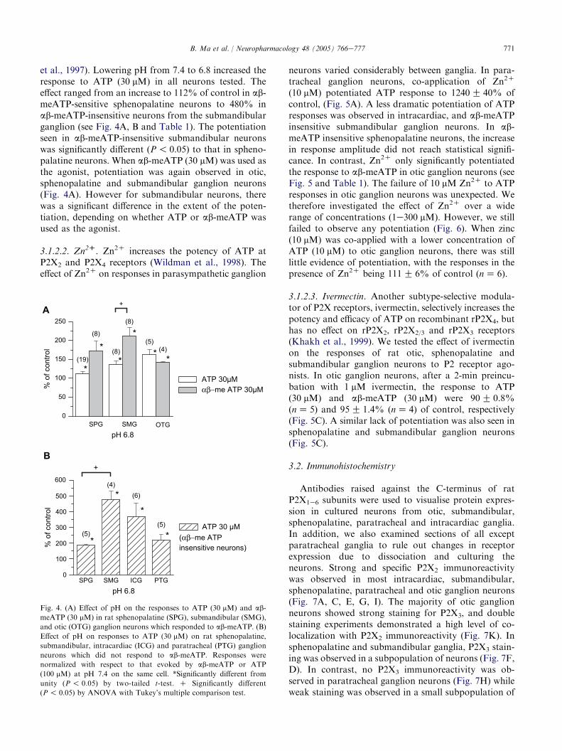
et al., 1997). Lowering pH from 7.4 to 6.8 increased theresponse to ATP (30 mM) in all neurons tested. Theeffect ranged from an increase to 112% of control in ab-meATP-sensitive sphenopalatine neurons to 480% inab-meATP-insensitive neurons from the submandibularganglion (see Fig. 4A, B and Table 1). The potentiationseen in ab-meATP-insensitive submandibular neuronswas significantly different (P! 0.05) to that in spheno-palatine neurons. When ab-meATP (30 mM) was used asthe agonist, potentiation was again observed in otic,sphenopalatine and submandibular ganglion neurons(Fig. 4A). However for submandibular neurons, therewas a significant difference in the extent of the poten-tiation, depending on whether ATP or ab-meATP wasused as the agonist.
3.1.2.2. Zn2D. Zn2C increases the potency of ATP atP2X2 and P2X4 receptors (Wildman et al., 1998). Theeffect of Zn2C on responses in parasympathetic ganglion
0
100
200
300
400
500
600
+
*
**
*
(4)(6)
(5)(5)
ATP 30 µM(αβ−me ATPinsensitive neurons)%
of c
ontro
l
pH 6.8SPG SMG ICG PTG
+
0
50
100
150
200
250
**
*
** (4)
(5)
(8)
ATP 30µMαβ−me ATP 30µM%
of c
ontro
l
pH 6.8SPG SMG OTG
A
B
(19)
(8)
(8)
*
Fig. 4. (A) Effect of pH on the responses to ATP (30 mM) and ab-
meATP (30 mM) in rat sphenopalatine (SPG), submandibular (SMG),
and otic (OTG) ganglion neurons which responded to ab-meATP. (B)
Effect of pH on responses to ATP (30 mM) on rat sphenopalatine,
submandibular, intracardiac (ICG) and paratracheal (PTG) ganglion
neurons which did not respond to ab-meATP. Responses were
normalized with respect to that evoked by ab-meATP or ATP
(100 mM) at pH 7.4 on the same cell. *Significantly different from
unity (P ! 0.05) by two-tailed t-test. C Significantly different
(P! 0.05) by ANOVA with Tukey’s multiple comparison test.
neurons varied considerably between ganglia. In para-tracheal ganglion neurons, co-application of Zn2C
(10 mM) potentiated ATP response to 1240G 40% ofcontrol, (Fig. 5A). A less dramatic potentiation of ATPresponses was observed in intracardiac, and ab-meATPinsensitive submandibular ganglion neurons. In ab-meATP insensitive sphenopalatine neurons, the increasein response amplitude did not reach statistical signifi-cance. In contrast, Zn2C only significantly potentiatedthe response to ab-meATP in otic ganglion neurons (seeFig. 5 and Table 1). The failure of 10 mM Zn2C to ATPresponses in otic ganglion neurons was unexpected. Wetherefore investigated the effect of Zn2C over a widerange of concentrations (1e300 mM). However, we stillfailed to observe any potentiation (Fig. 6). When zinc(10 mM) was co-applied with a lower concentration ofATP (10 mM) to otic ganglion neurons, there was stilllittle evidence of potentiation, with the responses in thepresence of Zn2C being 111G 6% of control (nZ 6).
3.1.2.3. Ivermectin. Another subtype-selective modula-tor of P2X receptors, ivermectin, selectively increases thepotency and efficacy of ATP on recombinant rP2X4, buthas no effect on rP2X2, rP2X2/3 and rP2X3 receptors(Khakh et al., 1999). We tested the effect of ivermectinon the responses of rat otic, sphenopalatine andsubmandibular ganglion neurons to P2 receptor ago-nists. In otic ganglion neurons, after a 2-min preincu-bation with 1 mM ivermectin, the response to ATP(30 mM) and ab-meATP (30 mM) were 90G 0.8%(nZ 5) and 95G 1.4% (nZ 4) of control, respectively(Fig. 5C). A similar lack of potentiation was also seen insphenopalatine and submandibular ganglion neurons(Fig. 5C).
3.2. Immunohistochemistry
Antibodies raised against the C-terminus of ratP2X1e6 subunits were used to visualise protein expres-sion in cultured neurons from otic, submandibular,sphenopalatine, paratracheal and intracardiac ganglia.In addition, we also examined sections of all exceptparatracheal ganglia to rule out changes in receptorexpression due to dissociation and culturing theneurons. Strong and specific P2X2 immunoreactivitywas observed in most intracardiac, submandibular,sphenopalatine, paratracheal and otic ganglion neurons(Fig. 7A, C, E, G, I). The majority of otic ganglionneurons showed strong staining for P2X3, and doublestaining experiments demonstrated a high level of co-localization with P2X2 immunoreactivity (Fig. 7K). Insphenopalatine and submandibular ganglia, P2X3 stain-ing was observed in a subpopulation of neurons (Fig. 7F,D). In contrast, no P2X3 immunoreactivity was ob-served in paratracheal ganglion neurons (Fig. 7H) whileweak staining was observed in a small subpopulation of

772 B. Ma et al. / Neuropharmacology 48 (2005) 766e777
intracardiac ganglion neurons (Fig. 7B). Some, P2X4
and P2X6 immunoreactivity was observed in all aboveganglion neurons while P2X5 immunoreactivity was notdetected (Table 2). The results of immunoreactivity in
0
50
100
150
200
ATP 30µMαβ−me ATP 30µM
(6)
(5)
(7)(7)
(7)(20)
% o
f con
trol
Zn2+ (10µM)
ATP 30µMαβ−me ATP 30µM
SPG SMG OTG
0
200
4001000
1200
1400
*
*
*
(5)
(6)(4)ATP 30µM
(αβ−meATP in sensitiveneurons)%
of c
ontro
l
Zn2+ (10µM)SPG SMG ICG PTG
A
B
(5)
+
*
0
20
40
60
80
100
120
(4)(5)(3)(4)(4)(4)
OTGSMG
% o
f con
trol
Ivermectin (1µM)SPG
C
Fig. 5. (A) Effect of Zn2C on the response to ATP (30 mM) on rat
sphenopalatine (SPG), submandibular (SMG), intracardiac (ICG) and
paratracheal (PTG) ganglion neurons which did not respond to ab-
meATP. (B) Effect of Zn2C on the responses to ATP (30 mM) and ab-
meATP (30 mM) on rat sphenopalatine, submandibular, and otic
ganglion neurons with a response to ab-meATP. Responses were
normalized with respect to those evoked by ab-meATP or ATP
(30 mM) in the same cell in the absence of Zn2C. (C) Averaged peak
amplitudes of the current induced by ATP (30 mM) and ab-meATP
(30 mM) in the presence of 1 mM ivermectin in rat sphenopalatine,
submandibular and otic ganglion neurons. Responses were normalized
with respect to those evoked by ATP (30 mM) or ab-meATP (30 mM)
in the same cell, in the absence of ivermectin. The numbers of neurons
tested are shown in parentheses. *Significantly different from unity
(P ! 0.05) by two-tailed t-test. C Significantly different (P ! 0.05) by
ANOVA with Tukey’s multiple comparison test.
cultured neurons of otic, sphenopalatine, submandibu-lar and intracardiac ganglia were similar with those inganglion sections (Table 3, Fig. 8). The main differencewas that although moderate P2X4 immunoreactivity wasobserved in sections, the only strong P2X4 stainingpresent in cultures could be seen in glial cells rather thanneurons. These results suggest that some of the strongerP2X4 immunoreactivity detected in sections of gangliawas in fact in non-neuronal cells.
4. Discussion
In this study, we have used immunohistochemistryand pharmacological experiments to investigate the P2Xreceptors present in five different parasympatheticganglia of the rat. Based on the sensitivity of neuronsto ab-meATP, the ganglia can be divided into threegroups. Neurons in the paratracheal and intracardiac
ATP30 µM
+ Zn 2+
1µM+ Zn2+
10 µM
2 s
B
+ Zn2+
100 µM
0.05 nA
A
0.0
0.2
0.4
0.6
0.8
1.0
1.2
30010010
Rel
ativ
e cu
rrent
Zn2+(µM)
ATP 30 µM
1
Fig. 6. (A) Traces showing the response to ATP (30 mM) in otic
ganglion neurons, when co-applied with different concentrations of
Zn2C. (B) Averaged peak amplitudes of the current induced by ATP
(30 mM) in the presence of increasing concentrations of Zn2C in rat
otic ganglion neurons. Responses were normalized with respect to
those evoked by ATP (30 mM) on the same cell, in the absence of Zn2C
(nZ 4).

Fig. 7. Immunostaining for P2X2 and P2X3 subunits in neurons from different parasympathetic ganglia. P2X2 (A) and P2X3 (B) immunoreactivity in
sections of an intracardiac ganglion. P2X2 (C) and P2X3 (D) immunoreactivity in sections of a submandibular ganglion. P2X2 (E) and P2X3 (F)
immunoreactivity in cultured neurons from sphenopalatine ganglia. P2X2 (G) and P2X3 (H) immunoreactivity in cultured neurons from paratracheal
ganglia. Double labelling for P2X2 and P2X3 in a single section of otic ganglion are shown in I and J. The overlay of the images (J) shows a high
degree of co-localization (yellow). Calibration barZ 50 mm.

774 B. Ma et al. / Neuropharmacology 48 (2005) 766e777
ganglia are virtually devoid of any response to ab-meATP. In contrast, all neurons in the otic ganglionrespond to this agonist, while sphenopalatine andsubmandibular ganglia contain a mixture of ab-meATP-sensitive and insensitive neurons. For technicalreasons, we used juvenile animals in this study. If thereare developmental changes in P2X receptor subunitexpression in these neurons, as occurs elsewhere in thenervous system (see Dunn et al., 2001; Cheung andBurnstock, 2002), a different expression profile may bepresent in adult rats.
4.1. Nature of the receptors
The ganglia we investigated all stained strongly forthe P2X2 subunit, suggesting that it is heavily involvedin the receptors we were studying. From published dataon recombinant receptors, only those containing theP2X1, P2X3, or P2X5 subunits respond to ab-meATP(see North, 2002; Wildman et al., 2002). The lack of anydetectable P2X1 or P2X5 immunostaining would argueagainst the involvement of these subunits. In contrast,immunoreactivity for P2X3 was present in otic, sub-mandibular and sphenopalatine ganglia, which allcontain neurons sensitive to ab-meATP, while intracar-diac and paratracheal ganglion neurons which did notstain for P2X3 failed to respond to this agonist. Theabsence of any rapidly desensitizing responses, evenwhen low concentrations of agonist were used, wouldargue against the presence of homomeric P2X3 receptors
Table 2
Summary of the P2X receptor subunit immunoreactivity in sections of
parasympathetic ganglia
Ganglion Antibody
P2X1 P2X2 P2X3 P2X4 P2X5 P2X6
Otic � CCC CCC C � CC
Submandibular � CCC CC C � CSphenopalatine � CCC CC C � C
Intracardiac � CC G C � C
CCC, very strong signal; CC, strong signal; C, moderate signal;
G , just detectable; �, undetectable.
Table 3
Summary of the P2X receptor subunit immunoreactivity in cultured
neurons of parasympathetic ganglia
Ganglion Antibody
P2X1 P2X2 P2X3 P2X4 P2X5 P2X6
Otic � CCC CCC G � CC
Submandibular � CCC CC G � CSphenopalatine � CCC CC G � C
Intracardiac � CC � G � C
Paratracheal � CC � G � C
CCC, very strong signal; CC, strong signal; C, moderate signal;
G , just detectable; �, undetectable.
on otic, submandibular and sphenopalatine ganglionneurons, and would suggest that the responses to ab-meATP were mediated by heteromeric P2X2/3 receptors(see Lewis et al., 1995).
The data we have obtained are in general consistentwith the notion that parasympathetic ganglion neuronscontain either homomeric P2X2 and/or heteromericP2X2/3 receptors. Thus, responses to ATP and ab-meATP were potentiated by lowering the pH, which isa characteristic of P2X2 containing receptors (see North,2002). However, responses in cells sensitive to ab-meATP were not potentiated as much, which would beconsistent with data for the P2X2/3 heteromer (Liu et al.,2001). Furthermore, the potency of ATP was alwaysgreater in cells which responded to ab-meATP, which isagain in keeping with the presence of P2X2/3 receptors inthese neurons. The mechanisms for regulating theassembly of P2X receptors are still unclear. However,the lack of any rapidly desensitizing homomeric P2X3
receptors might occur if these neurons always express anexcess of P2X2 receptor subunits.
The effects of zinc were, however, less consistent withthe general hypothesis. Zinc is an allosteric modulator,potentiating the response to ATP at receptors contain-ing P2X2 and P2X4 subunits, by increasing the affinityfor the agonist (Wildman et al., 1998; Miller et al., 1998;Xiong et al., 1999). Potentiation of responses in thoseneurons which failed to respond to ab-meATP inparatracheal, intracardiac, and submandibular ganglia,would agree with the presence of the P2X2 subunit inthese neurons, but the much more dramatic effect onparatracheal ganglion neurons might suggest that thereceptors present in these ganglia are not identical. Whatis harder to account for is the failure of 10 mM Zn2C topotentiate ATP responses in the ab-meATP-sensitiveneurons in otic, sphenopalatine and submandibularganglias, yet it did potentiate responses to ab-meATPin otic ganglion neurons. The extent of potentiation byZn2C will depend on the concentration of agonist usedrelative to its EC50. The agonist concentration (30 mM)used in our experiments with Zn2C was between theEC10 and EC60 values (for paratracheal and oticneurons, respectively) so this alone cannot account forour results. Other possible explanations include theinvolvement of additional subunits, or splice variants inthe P2X receptors in these ganglia. Although thepresence of splice variants of the P2X2 subunit has beendescribed in sympathetic neurons (Schadlich et al.,2001), this is associated with changes in the kinetics ofdesensitization, rather than changes in pharmacologicalprofile. Further studies will be required to test thesehypotheses.
In a previous study of submandibular ganglionneurons, it was suggested that the main P2X receptorinvolved is the P2X4 subunit (Liu and Adams, 2001).Although we observed some immunoreactivity for the

775B. Ma et al. / Neuropharmacology 48 (2005) 766e777
Fig. 8. Immunostaining for P2X1e6 subunits in sections of rat sphenopalatine ganglion. While strong staining for P2X2 (B) and P2X3 (C) subunits
was observed, there was no detectable staining for P2X1 (A) and P2X5 (E). Low levels of staining for P2X4 (D) and P2X6 (F) were present in some
cells. Calibration barZ 50 mm.
P2X4 subunit in both sections and cultured cells, themajority of this staining appeared to be in non-neuronalcells. Furthermore, we did not observe any potentiationby ivermectin, an allosteric modulator of P2X4-contain-ing receptors (Khakh et al., 1999), on submandibular,
otic or sphenopalatine ganglion neurons. Thus ourresults provide little support for the involvement ofP2X4 subunits. However, the differences in the EC50 forATP between neurons from different ganglia may befurther evidence of heterogeneity.

776 B. Ma et al. / Neuropharmacology 48 (2005) 766e777
4.2. Comparison with sympathetic andsensory ganglia
Parasympathetic ganglion neurons, like those of thesympathetic and most sensory ganglia are derived fromthe neural crest. While sensory neurons express P2X2
and P2X3 subunits in varying proportions, giving rise toeither homomeric P2X3 receptors or heteromeric P2X2/3
receptors, sympathetic ganglion neurons in the ratappear to express almost exclusively P2X2 receptors(see Dunn et al., 2001). In contrast, chromaffin cells ofthe rat adrenal medulla, which are also of neural crestorigin, are devoid of P2X receptors (Liu et al., 1999). Wehave now shown that there is considerable heterogeneityin the expression of P2X receptors in rat parasympa-thetic ganglia. While neurons in some ganglia such asintracardiac and paratracheal express only P2X2 recep-tors, these otic ganglion neurons express predominantlyheteromeric P2X2/3 receptors. In other ganglia such asthe sphenopalatine and submandibular, neurons expresseither P2X2 or P2X2/3 receptors. The basis of thisheterogeneity remains to be determined, but possiblemechanisms include the regulation of subunit expressionby either target tissues, or pre-ganglionic axons.
4.3. Inter-species variation
In the mouse otic ganglion, all neurons respond toATP, but less than 50% respond to ab-meATP (Maet al., 2004). In contrast, we have observed that allneurons in rat otic ganglion respond to both agonists.Furthermore, in mouse otic ganglion neurons, responsesto ATP and ab-meATP are potentiated both bylowering the extracellular pH, and by the co-applicationof zinc. Immunohistochemistry and gene deletionexperiments suggest that in the mouse otic ganglion,the receptors present are P2X2 and P2X2/3 (Ma et al.,2004). The underlying basis for the novel pharmacolog-ical properties we have observed in rat otic ganglionneurons remains to be determined, but the differenceswe have observed between rat and mouse otic ganglionneurons highlight the need for caution in extrapolatingfrom knockout studies carried out in the mouse.
4.4. Physiological role
ATP is stored with acetylcholine in synaptic vesiclesof cholinergic nerve terminals and is co-released withacetylcholine from motor nerve terminals (Silinsky,1975) and sympathetic pre-ganglionic fibres (Vizi et al.,1997). ATP is almost certainly co-released with acetyl-choline from pre-ganglionic nerve terminals in para-sympathetic ganglia, thus P2X receptors on theseneurons are likely to be activated during synaptictransmission. The fast excitatory postsynaptic potentialsin some otic and cardiac ganglion neurons are attenu-
ated but not abolished by high concentrations ofmecamylamine or tubocurarine (Callister and Sah,1997; Seabrook et al., 1990), and it is tempting tospeculate that the residual component is mediated byATP. A second possibility is that these receptorsrespond to ATP released from surrounding glial cellsas part of a glial cell-neuron signalling system. A thirdpossibility is that these receptors are involved in theregulation of transmitter release at the nerve terminal.Such a role has recently been demonstrated for P2Xreceptors on the sympathetic nerve terminals in the vasdeferens (Queiroz et al., 2003).
5. Conclusion
We have shown that neurons in different para-sympathetic ganglia of the rat express P2X receptorswith different properties. Based on immunohistochem-istry and preliminary pharmacological characterization,homomeric P2X2 and heteromeric P2X2/3 receptorsappear to be involved. However, differences in agonistpotency, and the modulatory effect of Zn2C suggest thatthe receptors expressed in different ganglia are notidentical. Better pharmacological tools are required fora more detailed characterization of the P2X receptorsubtypes present in these ganglia.
Acknowledgements
This work was supported by The Wellcome Trust.We are grateful to Chrystalla Orphanedes for editorialassistance.
References
Aibara, K., Ebihara, S., Akaike, N., 1992. Voltage-dependent ionic
currents in dissociated paratracheal ganglion cells of the rat. J.
Physiol. 457, 591e610.Allen, T.G., Burnstock, G., 1990. The actions of adenosine 5#-
triphosphate on guinea-pig intracardiac neurones in culture. Br. J.
Pharmacol. 100, 269e276.
Brown, S.G., Townsend-Nicholson, A., Jacobson,K.A., Burnstock,G.,
King, B.F., 2002. Heteromultimeric P2X1/2 receptors show a novel
sensitivity to extracellular pH. J. Pharmacol. Exp. Ther. 300,
673e680.Burnstock, G., 1997. The past, present and future of purine nucleotides
as signalling molecules. Neuropharmacology 36, 1127e1139.
Burnstock, G., 2002. Potential therapeutic targets in the rapidly
expanding field of purinergic signalling. Clin. Med. 2, 45e53.Callister, R.J., Sah, P., 1997. The removal of acetylcholine by diffusion
at nicotinic synapses in the rat otic ganglion. J. Physiol. 505, 165e
175.
Cheung, K.K., Burnstock, G., 2002. Localization of P2X3 receptors
and coexpression with P2X2 receptors during rat embryonic
neurogenesis. J. Comp. Neurol. 443, 368e382.
Dunn, P.M., Zhong, Y., Burnstock, G., 2001. P2X receptors in
peripheral neurones. Prog. Neurobiol. 65, 107e134.

777B. Ma et al. / Neuropharmacology 48 (2005) 766e777
Fieber, L.A., Adams, D.J., 1991. Adenosine triphosphate-evoked
currents in cultured neurones dissociated from rat parasympathetic
cardiac ganglia. J. Physiol. 434, 239e256.
Ishibashi, H., Mochidome, T., Okai, J., Ichiki, H., Shimada, H.,
Takahama, K., 2001. Activation of potassium conductance by
ophiopogonin-D in acutely dissociated rat paratracheal neurones.
Br. J. Pharmacol. 132, 461e466.
Khakh, B.S., Humphrey, P.P.A., Surprenant, A., 1995. Electrophys-
iological properties of P2X-purinoceptors in rat superior cervical,
nodose and guinea-pig coeliac neurones. J. Physiol. 484, 385e395.
Khakh, B.S., Proctor, W.R., Dunwiddie, T.V., Labarca, C.,
Lester, H.A., 1999. Allosteric control of gating and kinetics at
P2X4 receptor channels. J. Neurosci. 19, 7289e7299.
King, B.F., Wildman, S.S., Ziganshina, L.E., Pintor, J., Burnstock, G.,
1997. Effects of extracellular pH on agonism and antagonism at
a recombinant P2X2 receptor. Br. J. Pharmacol. 121, 1445e1453.Lewis, C., Neidhart, S., Holy, C., North, R.A., Buell, G.,
Surprenant, A., 1995. Coexpression of P2X2 and P2X3 receptor
subunits can account for ATP-gated currents in sensory neurons.
Nature 377, 432e435.
Lichtman, J.W., 1977. The reorganization of synaptic connections in
the rat submandibular ganglion. J. Physiol. 273, 155e175.
Liu, D.M., Adams, D.J., 2001. Ionic selectivity of native ATP-
activated (P2X) receptor channels in dissociated neurones from rat
parasympathetic ganglia. J. Physiol. 534, 423e435.
Liu, M., Dunn, P.M., King, B.F., Burnstock, G., 1999. Rat chromaffin
cells lack P2X receptors while those of the guinea-pig express
a receptor with novel pharmacology. Br. J. Pharmacol. 128, 61e68.
Liu, M., King, B.F., Dunn, P.M., Rong, W., Townsend-Nicholson, A.,
Burnstock, G., 2001. Coexpression of P2X(3) and P2X(2) receptor
subunits in varying amounts generates heterogeneous populations
of P2X receptors that evoke a spectrum of agonist responses
comparable to that seen in sensory neurons. J. Pharmacol. Exp.
Ther. 296, 1043e1050.Ma, B., Ruan, H.-Z., Cockayne, D., Ford, A.P.D.W., Burnstock, G.,
Dunn, P.M., 2004. Identification of P2X receptors in cultured
mouse and rat parasympathetic otic ganglion neurones including
P2X knockout studies. Neuropharmacology 46, 1039e1048.Miller, K.J., Michel, A.D., Chessell, I.P., Humphrey, P.P., 1998.
Cibacron blue allosterically modulates the rat P2X4 receptor.
Neuropharmacology 37, 1579e1586.
North, R.A., 2002. Molecular physiology of P2X receptors. Physiol.
Rev. 82, 1013e1067.
Oglesby, I.B., Lachnit, W.G., Burnstock, G., Ford, A.P.D.W., 1999.
Subunit specificity of polyclonal antisera to the carboxy terminal
region of P2X receptors P2X1 through P2X7. Drug Dev. Res. 47,
189e195.
Queiroz, G., Talaia, C., Goncalves, J., 2003. ATP modulates
noradrenaline release by activation of inhibitory P2Y receptors
and facilitatory P2X receptors in the rat vas deferens. J. Pharmacol.
Exp. Ther. 307, 809e815.
Reekie, F.M., Burnstock, G., 1994. Some effects of purines on
neurones of guinea-pig superior cervical ganglia. Gen. Pharmacol.
25, 143e148.
Schadlich, H., Wirkner, K., Franke, H., Bauer, S., Grosche, J.,
Burnstock, G., Reichenbach, A., Illes, P., Allgaier, C., 2001.
P2X(2), P2X(2-2) and P2X(5) receptor subunit expression and
function in rat thoracolumbar sympathetic neurons. J. Neurochem.
79, 997e1003.
Schwiebert, E.M., 2003. Purinergic receptors and signalling. In:
Current topics in membranes, vol. 54. Academic Press, San Diego.
Seabrook, G.R., Fieber, L.A., Adams, D.J., 1990. Neurotransmission
in neonatal rat cardiac ganglion in situ. Am. J. Physiol. 259, H997e
H1005.
Silinsky, E.M., 1975. On the association between transmitter secretion
and the release of adenine nucleotides from mammalian motor
nerve terminals. J. Physiol. 247, 145e162.
Smith, A.B., Hansen, M.A., Liu, D.-M., Adams, D.J., 2001. Pre- and
postsynaptic actions of ATP on neurotransmission in rat sub-
mandibular ganglia. Neuroscience 107, 283e291.
Spencer, S.E., 1990. CNS projections to the pterygopalatine para-
sympathetic preganglionic neurons in the rat: a retrograde trans-
neuronal viral cell body labelling study. Brain Res. 534, 149e169.
Stoop, R., Surprenant, A., North, R.A., 1997. Different sensitivities to
pH of ATP-induced currents at four cloned P2X receptors. J.
Neurophysiol. 78, 1837e1840.Suzuki, N., Hardebo, J.E., 1991. The pathway of parasympathetic
fibers to cerebral vessels from the otic ganglion in the rat. J. Auton.
Nerv. Syst. 36, 39e46.Vizi, E.S., Liang, S.D., Sperlagh, B., Kittel, A., Juranyi, Z., 1997.
Studies on the release and extracellular metabolism of endogenous
ATP in rat superior cervical ganglion: support for neurotransmitter
role of ATP. Neuroscience 79, 893e903.Wildman, S.S., Brown, S.G., Rahman, M., Noel, C.A., Churchill, L.,
Burnstock, G., Unwin, R.J., King, B.F., 2002. Sensitization by
extracellular Ca(2C) of rat P2X(5) receptor and its pharmacolog-
ical properties compared with rat P2X(1). Mol. Pharmacol. 62,
957e966.
Wildman, S.S., King, B.F., Burnstock, G., 1998. Zn2C modulation of
ATP-responses at recombinant P2X2 receptors and its dependence
on extracellular pH. Br. J. Pharmacol. 123, 1214e1220.Xiong, K., Peoples, R.W., Montgomery, J.P., Chiang, Y.,
Stewart, R.R., Weight, F.F., Li, C., 1999. Differential modulation
by copper and zinc of P2X2 and P2X4 receptor function. J.
Neurophysiol. 81, 2088e2094.
Zhong, Y., Dunn, P.M., Burnstock, G., 2000. Guinea-pig sympathetic
neurons express varying proportions of two distinct P2X receptors.
J. Physiol. 523, 391e402.