dichloroacetate (dca) and cancer: an overview towards
TRANSCRIPT
Review ArticleDichloroacetate (DCA) and Cancer: An Overview towardsClinical Applications
Tiziana Tataranni 1 and Claudia Piccoli 1,2
1Laboratory of Pre-Clinical and Translational Research, IRCCS-CROB, Referral Cancer Center of Basilicata, Rionero in Vulture (Pz),85028, Italy2Department of Clinical and Experimental Medicine, University of Foggia, Foggia 71121, Italy
Correspondence should be addressed to Tiziana Tataranni; [email protected]
Received 24 July 2019; Revised 12 September 2019; Accepted 11 October 2019; Published 14 November 2019
Guest Editor: Kanhaiya Singh
Copyright © 2019 Tiziana Tataranni and Claudia Piccoli. This is an open access article distributed under the Creative CommonsAttribution License, which permits unrestricted use, distribution, and reproduction in any medium, provided the original workis properly cited.
An extensive body of literature describes anticancer property of dichloroacetate (DCA), but its effective clinical administration incancer therapy is still limited to clinical trials. The occurrence of side effects such as neurotoxicity as well as the suspicion of DCAcarcinogenicity still restricts the clinical use of DCA. However, in the last years, the number of reports supporting DCAemployment against cancer increased also because of the great interest in targeting metabolism of tumour cells. Dissecting DCAmechanism of action helped to understand the bases of its selective efficacy against cancer cells. A successful coadministration ofDCA with conventional chemotherapy, radiotherapy, other drugs, or natural compounds has been tested in several cancermodels. New drug delivery systems and multiaction compounds containing DCA and other drugs seem to amelioratebioavailability and appear more efficient thanks to a synergistic action of multiple agents. The spread of reports supporting theefficiency of DCA in cancer therapy has prompted additional studies that let to find other potential molecular targets of DCA.Interestingly, DCA could significantly affect cancer stem cell fraction and contribute to cancer eradication. Collectively, thesefindings provide a strong rationale towards novel clinical translational studies of DCA in cancer therapy.
1. Introduction
Cancer is one of the leading causes of death worldwide.Despite the significant progression in diagnostic and thera-peutic approaches, its eradication still represents a challenge.Too many factors are responsible for therapy failure orrelapse, so there is an urgent need to find new approachesto treat it. Apart from the typical well-known properties fea-turing malignant cells, including abnormal proliferation,deregulation of apoptosis, and cell cycle [1, 2], cancer cellsalso display a peculiar metabolic machine that offers a furtherpromising approach for cancer therapy [3–5]. Our group hadalready suggested the importance of a metabolic characteri-zation of cancer cells to predict the efficacy of a metabolictreatment [6]. Drugs able to affect cancer metabolism arealready under consideration, showing encouraging resultsin terms of efficacy and tolerability [7]. In the last decade,the small molecule DCA, already used to treat acute and
chronic lactic acidosis, inborn errors of mitochondrialmetabolism, and diabetes [8], has been largely purposed asan anticancer drug. DCA is a 150Da water-soluble acidmolecule, analog of acetic acid in which two of the threehydrogen atoms of the methyl group have been replaced bychlorine atoms (Figure 1(a)) [9]. DCA administration indoses ranging from 50 to 200mg/Kg/die is associated to adecrease of tumour mass volume, proliferation rate, andmetastasis dissemination in several preclinical models [10].Our group had already observed an inverse correlationbetween DCA ability to kill cancer cells and their mito-chondrial respiratory capacity in oral cell carcinomas[11]. Moreover, we recently described DCA ability to affectmitochondrial function and retarding cancer progression ina pancreatic cancer model [12]. To date, consistent data fromclinical trials and case reports describing DCA administra-tion in cancer patients are available [13–16], but, despitethe growing body of literature sustaining the efficacy of
HindawiOxidative Medicine and Cellular LongevityVolume 2019, Article ID 8201079, 14 pageshttps://doi.org/10.1155/2019/8201079
DCA against cancer, it is not under clinical use yet. Thisreview is aimed at summarizing the very recent reports sug-gesting the employment of DCA in cancer therapy, in combi-nation with chemotherapy agents, radiotherapy, and otherchemical or natural compounds showing anticancer proper-ties. Moreover, we described data about new purposedpharmacological formulations of DCA able to avoid sideeffects and ameliorate drug bioavailability and efficacy, fur-ther encouraging its possible clinical employment. Finally,we reviewed latest findings suggesting other potentialmechanisms of action of DCA, including new data about itsaptitude to affect cancer stem cell fraction.
2. DCA and Cancer: Mechanism of Action
The potential efficacy of DCA in cancer therapy comes frommetabolic properties of cancer cells, typically characterizedby increased glycolytic activity and reduced mitochondrialoxidation, regardless of oxygen availability, the well-knownWarburg effect [17]. The excessive glycolysis and the result-ing lactate overproduction provoke a state of metabolicacidosis in tumour microenvironment [18]. Glycolysis-derived lactate is taken up by surrounding cells to supporttumour growth and inhibits apoptotic cell death mechanisms[19, 20]. Several enzymes involved in glycolysis regulate apo-ptosis, and their overexpression in cancer cells contributes toapoptosis suppression [21]. In this setting, salts of DCAselectively target cancer cells shifting their metabolism fromglycolysis to oxidative phosphorylation by inhibition of pyru-vate dehydrogenase kinase (PDK), the inhibitor of pyruvatedehydrogenase (PDH) [10]. PDH activation fosters mito-chondrial oxidation of pyruvate and disrupts the metabolicadvantage of cancer cells. Mitochondrial DNA mutations,often occurring in tumorigenesis and resulting in respiratorychain dysfunction [22, 23], make malignant cells unable tosustain cellular energy demand. Furthermore, reducing lac-tate production, DCA counteracts the acidosis state oftumour microenvironment, contributing to the inhibitionof tumour growth and dissemination [24]. The delivery of
pyruvate into mitochondria causes organelles remodellingresulting in an increased efflux of cytochrome c and otherapoptotic-inducing factors and upregulation of ROS levelswith a consequent reduction of cancer cell viability [9](Figure 1(b)).
3. Side Effects and Limitations toDCA Employment
Clinical employment of DCA is available in both oral andparenteral formulations, and doses range from 10 to50mg/Kg/die [25]. No evidence of severe hematologic,hepatic, renal, or cardiac toxicity confirms DCA safety [26].Common gastrointestinal side effects often occur in a per-centage of patients treated with DCA [15]. The best-knownlimitation to DCA administration, observed both in preclin-ical and in clinical studies, is peripheral neuropathy [27]. Theselectivity of DCA-induced damage for the nervous systemmay be due to the lack of well-equipped machinery able tohandle a more sustained oxidative phosphorylation in cellsproducing ATP mostly via glycolysis [28]. The resultingmitochondrial overload compromises the antioxidant sys-tems’ efficiency, unable to face the excessive amount ofROS. In this setting, the contemporary administration ofantioxidants should represent a further strategy to minimizeDCA-induced neuropathy [27]. The expression and theactivity of glutathione transferase zeta1 (GSTZ1), the firstenzyme responsible for DCA clearance, may influence theentity of damage. Nonsynonymous functional single-nucleotide polymorphisms (SNPs) in human GSTZ1 genegive rise to different haplotypes that are responsible for a dif-ferent DCA kinetic and dynamics. A clear associationbetween GSTZ1 haplotype and DCA clearance has beendemonstrated. On this basis, a personalized DCA dosage,not only based on body weight, may minimize or preventadverse effects in patients chronically treated with this drug[29]. The occurrence of neuropathy is associated to DCAchronic oral administration and is a reversible effect, limitedto the time of treatment [30]. The intravenous route reduces,
OH
Cl
Cl
O
(a)
Cancer cells Cancer cells deathCancer cells
Lactate
Tum
orM
icro
envi
ronm
ent
Lactate
Pyruvate
GlycolysisPDK
DCA
PDH
OxidativePhosphorylation
Apoptosis restorationCytochrome c
Glucose
(b)
Figure 1: (a) Chemical structure of DCA. (b) Mechanism of action of DCA: PDK: pyruvate dehydrogenase kinase; PDH: pyruvatedehydrogenase. Black dotted lines, biochemical processes inhibited by DCA; Red arrows, metabolic pathways activated by DCA.
2 Oxidative Medicine and Cellular Longevity
therefore, the potential for neurotoxicity and let the achieve-ment of higher drug concentrations bypass the digestivesystem [13].
Since DCA is among water disinfection by-productsfound in low concentrations in drinking water, its potentialcarcinogenicity is under evaluation. Studies performed inmouse models associate DCA early-life exposure to anincreased incidence of hepatocellular tumours [31]. It is con-ceivable that persistent changes in cell metabolism inducedby DCA may produce epigenetic effects. Long-term induc-tion of PDH and other oxidative pathways related to glucosemetabolism could contribute to increase reactive oxygen spe-cies and mitochondrial stress [27]. However, no evidence ofcarcinogenetic effect is reported in clinical studies, whenDCA is administered in cancer therapy.
4. Synergistic Effect of DCA andChemotherapeutic Agents
Combining different drugs is a well-accepted strategy to pro-duce a synergistic beneficial effect in cancer therapy, reducingdrug dosage, minimizing toxicity risks, and overcoming drugresistance. Coadministration of DCA and traditional chemo-therapeutic agents has been purposed and tested in severalcancer models (Table 1). DCA treatment seems to improvethe efficacy of chemotherapy by inducing biochemical andmetabolic alterations, resulting in significant changes of can-cer cells’ energetic balance. A study performed in non-small-cell lung cancer (NSCLC) showed both in vitro and in vivothat coadministration of DCA with paclitaxel increased theefficiency of cell death through autophagy inhibition [32].An effective combination of DCA and doxorubicin (DOX)was tested in HepG2 cells, demonstrating the ability ofDCA to disrupt cellular antioxidant defences, thus favouringoxidative damage in turn triggered by DOX treatment [33].There is a strong association between PDK overexpressionand chemoresistance; thus, it is conceivable that PDK inhibi-tion might help to resensitize cancer cells to drugs. PDK2 iso-form overexpression was associated to paclitaxel resistance inNSCLC. Interestingly, DCA combination with paclitaxel wasmore effective in killing resistant cells than either paclitaxelor DCA treatment alone [34]. Similarly to NSCLC, an inter-esting in vivo study performed in advanced bladder cancershowed an increased expression of PDK4 isoform in highgrade compared to lower-grade cancers and cotreatment ofDCA and cisplatin dramatically reduced tumour volumesas compared to either DCA or cisplatin alone [35]. A recentstudy confirmed the ability of DCA to revert PDK4-relatedchemoresistance also in human hepatocellular carcinoma(HCC) [36].
5. Synergistic Effect of DCA and Other PotentialAnticancer Drugs
A consistent body of literature suggests positive effects ofDCA coadministration with compounds currently employedto treat other diseases but showing anticancer properties inseveral cancer models (Table 2). Contemporary administra-tion of DCA and the antibiotic salinomycin, recently redis-
covered for its cytotoxic properties as a potential anticancerdrug, has been tested in colorectal cancer cell lines. Theirtreatment seems to exert a synergistic cytotoxic effect byinhibiting the expression of proteins related to multidrugresistance [37]. Cancer cells lacking metabolic enzymesinvolved in arginine metabolism may result to sensitivity toarginase treatment. Interestingly, a combined administrationof recombinant arginase and DCA produces antiproliferativeeffects in triple-negative breast cancer, due to the activationof p53 and the induction of cell cycle arrest [38]. COX2inhibitors, primarily used as anti-inflammatory drugs, havebeen recently suggested as antitumor drugs because of theirantiproliferative activity. An intriguing study performed incervical cancer cells showed the inability of DCA to kill cer-vical cancer cells overexpressing COX2 and demonstratedthat COX2 inhibition by celecoxib makes cervical cancer cellsmore sensitive to DCA both in vitro and in vivo experiments[39]. Since DCA fosters oxidative phosphorylation bydecreasing glycolytic activity, the combination of DCA withother drugs enhancing a state of glucose dependence maybe a promising strategy. Such an approach has been testedin head and neck cancer in which the administration of pro-pranolol, a nonselective beta-blocker able to affect tumourcells’ mitochondrial metabolism, produced glycolytic depen-dence and energetic stress, making cells more vulnerable toDCA treatment [40]. Similar results were obtained in mela-noma cells in which the administration of retinoic acid recep-tor β (RARβ) inhibitors confer sensitization to DCA [41]. Apositive effect of DCA coadministration with metformin, ahypoglycaemic drug widely used to treat diabetes wasdemonstrated in a preclinical model of glioma [42] as wellas in a low metastatic variant of Lewis lung carcinoma(LLC) [43]. Jiang and colleagues investigated the effects ofphenformin, a metformin analog, and DCA in glioblastoma,demonstrating that contemporary inhibition of complex Iand PDK by phenformin and DCA, respectively, decreasedself-renewal and viability of glioma stem cells (GSCs), thussuggesting their possible employment to affect cancer stemcell fraction [44].
6. Combined Use of DCA andNatural Compounds
The clinical employment of natural compounds represents apromising novel approach to treat several diseases [45]. Anincreasing body of literature supports the detection, amongnatural compounds, of biologically active substances isolatedby plants, mushrooms, and bacteria or marine organism thatshow beneficial effects for human health [46–48]. Theassumption of natural compounds or their derivatives seemsto represent an encouraging approach to prevent cancerinitiation or recurrence, and it is generally called chemo-prevention [49]. Moreover, natural substances producebeneficial effects in cancer therapy when coadministeredwith other drugs, showing their ability to overcome drugresistance, to increase anticancer potential, and to reducedrug doses and toxicity [50, 51]. Interestingly, the coad-ministration of DCA and natural compounds has beenrecently purposed. A study investigated the combined
3Oxidative Medicine and Cellular Longevity
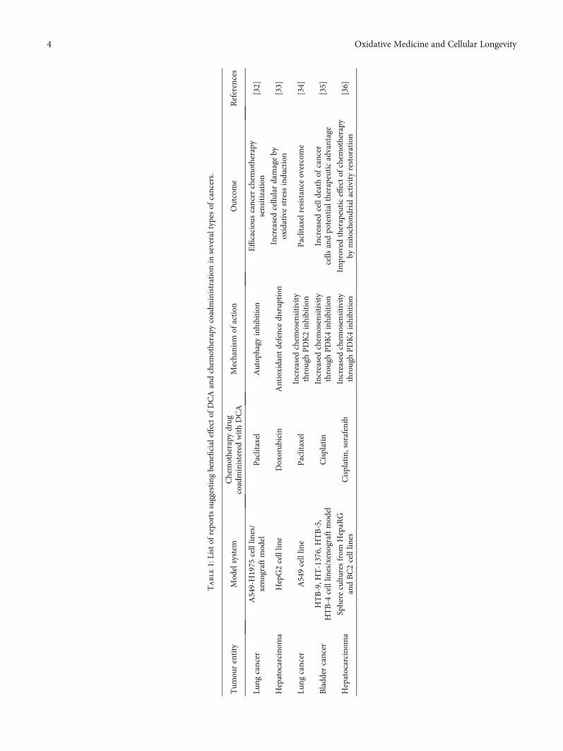
Table1:Listof
repo
rtssuggesting
beneficialeffectof
DCAandchem
otherapy
coadministrationin
severaltypes
ofcancers.
Tum
ourentity
Mod
elsystem
Chemotherapy
drug
coadministeredwithDCA
Mechanism
ofaction
Outcome
References
Lung
cancer
A549-H1975
celllin
es/
xeno
graftmod
elPaclitaxel
Autop
hagy
inhibition
Efficaciou
scancer
chem
otherapy
sensitization
[32]
Hepatocarcino
ma
HepG2celllin
eDoxorub
icin
Antioxidant
defencedisrup
tion
Increasedcellu
lardamageby
oxidativestressindu
ction
[33]
Lung
cancer
A549celllin
ePaclitaxel
Increasedchem
osensitivity
throughPDK2inhibition
Paclitaxelresistance
overcome
[34]
Bladd
ercancer
HTB-9,H
T-1376,HTB-5,
HTB-4
celllin
es/xenograftmod
elCisplatin
Increasedchem
osensitivity
throughPDK4inhibition
Increasedcelldeathof
cancer
cells
andpo
tentialtherapeuticadvantage
[35]
Hepatocarcino
ma
Sphere
cultu
resfrom
HepaR
GandBC2celllin
esCisplatin,sorafenib
Increasedchem
osensitivity
throughPDK4inhibition
Improved
therapeuticeffectof
chem
otherapy
bymitocho
ndrialactivity
restoration
[36]
4 Oxidative Medicine and Cellular Longevity
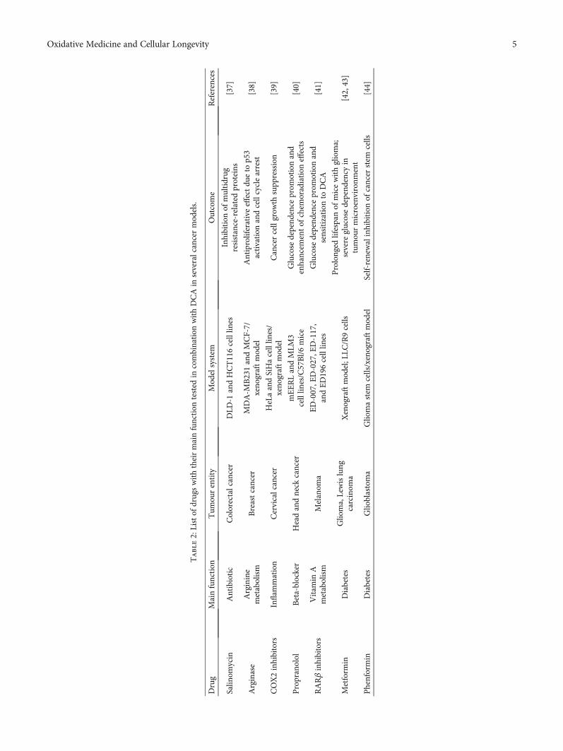
Table2:Listof
drugswiththeirmainfunction
tested
incombination
withDCAin
severalcancermod
els.
Drug
Mainfunction
Tum
ourentity
Mod
elsystem
Outcome
References
Salin
omycin
Antibiotic
Colorectalcancer
DLD
-1andHCT116celllin
esInhibition
ofmultidrug
resistance-related
proteins
[37]
Arginase
Arginine
metabolism
Breastcancer
MDA-M
B231andMCF-7/
xeno
graftmod
elAntiproliferativeeffectdu
eto
p53
activation
andcellcyclearrest
[38]
COX2inhibitors
Inflam
mation
Cervicalcancer
HeLaandSiHacelllin
es/
xeno
graftmod
elCancercellgrow
thsupp
ression
[39]
Propranolol
Beta-blocker
Headandneck
cancer
mEERLandMLM
3celllin
es/C57Bl/6mice
Glucose
depend
ence
prom
otionand
enhancem
entof
chem
oradiation
effects
[40]
RARβinhibitors
Vitam
inA
metabolism
Melanom
aED-007,E
D-027,E
D-117,
andED196celllin
esGlucose
depend
ence
prom
otionand
sensitizationto
DCA
[41]
Metform
inDiabetes
Glio
ma,Lewislung
carcinom
aXenograftmod
el;L
LC/R9cells
Prolonged
lifespanof
micewithglioma;
severe
glucosedepend
ency
intumou
rmicroenvironm
ent
[42,43]
Phenformin
Diabetes
Glio
blastoma
Glio
mastem
cells/xenograftmod
elSelf-renewalinhibition
ofcancer
stem
cells
[44]
5Oxidative Medicine and Cellular Longevity
effect of DCA with essential oil-blended curcumin, a com-pound with beneficial properties both in prevention andtreatment of cancer [52], demonstrating an anticancerpotential against HCC [53]. In particular, the combinationof both compounds synergistically reduced cell survival,promoting cell apoptosis and inducing intracellular ROSgeneration. Betulin, a natural compound isolated frombirch bark, is already known for its antiproliferative andcytotoxic effects against several cancer cell lines [54–56].An in vitro investigation of the antitumor activity of betu-lin derivatives in NSCLC confirmed its ability to inhibitin vivo and in vitro growth of lung cancer cells, blockingG2/M phase of the cell cycle and inducing caspase activa-tion and DNA fragmentation. Interestingly, betulin deriva-tive Bi-L-RhamBet was able to perturb mitochondrialelectron transport chain (ETC), inducing ROS production.Given the property of DCA to increase the total oxidationof glucose in mitochondria via the Krebs cycle and ETC,the authors combined Bi-L-RhamBet with DCA, demon-strating its significant potentiated cytotoxicity [57].
7. DCA and Radiosensitization
Radiotherapy represents a further strategy to treat cancer andprovides a local approach by the administration of high-energy rays [58]. The main effect of radiation is the inductionof ROS with a consequent DNA damage, chromosomalinstability, and cell death by apoptosis [59]. However, severaltumours show or develop radioresistance that is responsiblefor radiotherapy failure and high risk of tumour recurrenceormetastasis [60]. Several factorsmay be responsible of radio-resistance [61]. Among these, hypoxia, a common conditionof tumour microenvironment characterized by low oxygenlevels and reduced ROS species generation, can block the effi-cacy of ionizing radiations [62]. Increasing tumour oxygena-tion so to favour a considerable amount of ROS [63] ordirectly induce ROS production may therefore represent astrategy to increase radiosensitization [64, 65]. In this setting,DCA administration, known to induce ROS production[11, 66], could represent a strategy to overcome tumourradioresistance. Moreover, metabolic alterations featuringcancer development are known to affect radiosensitivity[67, 68]. Therefore, targeting cancer metabolic intermedi-ates may represent a strategy to improve a selective cancerresponse to irradiation [69]. The efficacy of DCA toincrease radiation sensitivity has been already demon-strated both in glioblastoma cells [70] and in oesophagealsquamous cell carcinoma [71]. More recently, it wasdemonstrated that DCA increases radiosensitivity in a cel-lular model of medulloblastoma, a fatal brain tumour inchildren, inducing alterations of ROS metabolism andmitochondrial function and suppressing DNA repaircapacity [72]. Since the role of immunotherapy in the res-toration of the immune defences against tumour progres-sion and metastasis is arousing great attention in the lastyears [73], Gupta and Dwarakanath provided a state ofthe art of the possible effects of glycolytic inhibitors,including DCA, on tumour radiosensitization, focusingtheir attention on the interplay between metabolic modi-
fiers and immune modulation in the radiosensitizationprocesses [74]. Interestingly, they reported the ability ofDCA to promote immune stimulation through the inhibi-tion of lactate accumulation, further sustaining its utiliza-tion as adjuvant of radiotherapy.
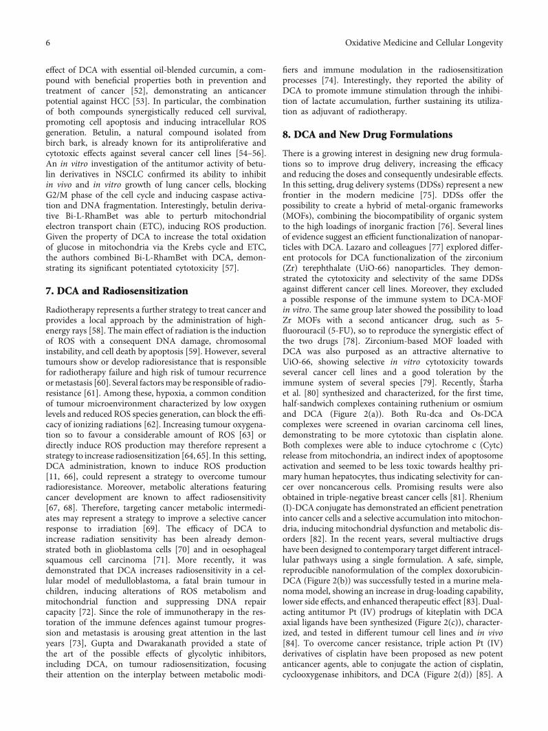
8. DCA and New Drug Formulations
There is a growing interest in designing new drug formula-tions so to improve drug delivery, increasing the efficacyand reducing the doses and consequently undesirable effects.In this setting, drug delivery systems (DDSs) represent a newfrontier in the modern medicine [75]. DDSs offer thepossibility to create a hybrid of metal-organic frameworks(MOFs), combining the biocompatibility of organic systemto the high loadings of inorganic fraction [76]. Several linesof evidence suggest an efficient functionalization of nanopar-ticles with DCA. Lazaro and colleagues [77] explored differ-ent protocols for DCA functionalization of the zirconium(Zr) terephthalate (UiO-66) nanoparticles. They demon-strated the cytotoxicity and selectivity of the same DDSsagainst different cancer cell lines. Moreover, they excludeda possible response of the immune system to DCA-MOFin vitro. The same group later showed the possibility to loadZr MOFs with a second anticancer drug, such as 5-fluorouracil (5-FU), so to reproduce the synergistic effect ofthe two drugs [78]. Zirconium-based MOF loaded withDCA was also purposed as an attractive alternative toUiO-66, showing selective in vitro cytotoxicity towardsseveral cancer cell lines and a good toleration by theimmune system of several species [79]. Recently, Štarhaet al. [80] synthesized and characterized, for the first time,half-sandwich complexes containing ruthenium or osmiumand DCA (Figure 2(a)). Both Ru-dca and Os-DCAcomplexes were screened in ovarian carcinoma cell lines,demonstrating to be more cytotoxic than cisplatin alone.Both complexes were able to induce cytochrome c (Cytc)release from mitochondria, an indirect index of apoptosomeactivation and seemed to be less toxic towards healthy pri-mary human hepatocytes, thus indicating selectivity for can-cer over noncancerous cells. Promising results were alsoobtained in triple-negative breast cancer cells [81]. Rhenium(I)-DCA conjugate has demonstrated an efficient penetrationinto cancer cells and a selective accumulation into mitochon-dria, inducing mitochondrial dysfunction and metabolic dis-orders [82]. In the recent years, several multiactive drugshave been designed to contemporary target different intracel-lular pathways using a single formulation. A safe, simple,reproducible nanoformulation of the complex doxorubicin-DCA (Figure 2(b)) was successfully tested in a murine mela-noma model, showing an increase in drug-loading capability,lower side effects, and enhanced therapeutic effect [83]. Dual-acting antitumor Pt (IV) prodrugs of kiteplatin with DCAaxial ligands have been synthesized (Figure 2(c)), character-ized, and tested in different tumour cell lines and in vivo[84]. To overcome cancer resistance, triple action Pt (IV)derivatives of cisplatin have been proposed as new potentanticancer agents, able to conjugate the action of cisplatin,cyclooxygenase inhibitors, and DCA (Figure 2(d)) [85]. A
6 Oxidative Medicine and Cellular Longevity
novel complex containing DCA, Platinum, and Biotin (DPB)has been successfully tested, exhibiting multifacet antitumorproperties (Figure 2(e)). Authors demonstrated the abilityof such a prodrug to affect energy metabolism, to promoteapoptosis, and to interact with DNA. The high selectivity ofbiotin for cancer cells minimizes the detrimental effects onnormal cells and improves the curative effect on tumours[86]. Features and experimental evidence of the main classesof compounds are summarized in Table 3.
9. Other Proposed Mechanisms ofAction of DCA
The metabolic shift from glycolysis to glucose oxidation dueto the inhibition of PDK and the consequent activation ofPDH is the best-known and well-accepted molecular effectof DCA administration. The consequent biochemical alter-ations, including ROS increase and mitochondrial membranepotential variation, may be responsible for proliferationarrest and cancer cell death, thus explaining DCA beneficialpotential in cancer treatment [9]. However, the molecularintermediates activated after DCA administration are stillunknown. It is conceivable that such a small molecule mightdirectly or indirectly affect other cellular and molecular tar-gets (Figure 3), displaying other mechanisms of action, soto explain its efficacy also in cellular models where it doesnot produce the expected metabolic shift [12]. A proteomicapproach applied to cells of lung cancer demonstrated theability of DCA to increase the concentration of every TCA
intermediate while it did not affect glucose uptake or the gly-colytic process from glucose to pyruvate [87]. In the attemptto shed light to DCA mode of action, Dubuis and colleaguesused a metabolomics-based approach on several ovarian can-cer cell lines treated with DCA and found a common markeddepletion of intracellular pantothenate, a CoA precursor, aswell as a concomitant increase of CoA, thus suggestingDCA ability to increase CoA de novo biosynthesis. Since highconcentrations of CoA resulted to be toxic for cells, this met-abolic effect could be responsible of cancer cell toxicity medi-ated by DCA [88]. A very recent work by El Sayed et al.introduced a novel evidence-based hypothesis, suggestingthat DCA efficiency against cancer may derive from its abilityto antagonize acetate [89], known to be an energetic substratefor glioblastoma and brain metastases, able to enhance DNA,RNA, and protein synthesis and posttranslational modifica-tions, thus favouring cell proliferation and cancer progres-sion. Moreover, high acetate levels are associated toanticancer drug resistance [90]. It has been shown thatDCA is able to revert metabolic alterations induced by ace-tate by restoring physiological serum levels of lactate and freefatty acid and potassium and phosphorus concentration.According to the authors, thanks to a structural similarityto acetate, DCA could inhibit metabolic effects driven by ace-tate, responsible for cancer cell growth and chemoresistance[89]. Another possible additional effect of DCA could bepH modulation. pH level modulation is known to affect pro-liferation and apoptosis processes [91] as well as chemother-apy sensitivity [92]. DCA treatment may both increase and
+ +
Os
Os-DCA Ru-DCA
O
O O
OCl
Cl Cl
ClN
N N
N
Ru
(a)
DOX-DCA
O O
O O
O
O OOH
HN
Cl
Cl
OH
OH
OHHO
(b)
O
O
O
O
Cl
ClCl
Cl ClNH2
NH2
Cl
Pt
(c)
CAD CID
O
O
OO
O
O O
O
O
O
H3N H3NH3NH3N
Cl ClCl
Pt PtCl Cl Cl
ClCl
(d)
DPB
O
O
O
O
O
Cl
Cl
Cl
Pt
Cl
NH3NH3NHHN
H H
S
(e)
Figure 2: New drug formulations containing DCA. (a) Schematic representation of Os-DCA and Ru-DCA complexes [81]. (b) Doxorubicin(DOX)-DCA complex [83]. (c) Dual action Pt prodrugs of kiteplatin and DCA [84]. (d) Examples of triple action Pt(IV) derivatives of cisplatincontaining DCA (red), derivatives of cisplatin (black), and COX inhibitors (green) [85]. (e) Chemical structure of DPB containing DCA (red),biotin (blue), and Platinum (Pt) complex (black) [86].
7Oxidative Medicine and Cellular Longevity
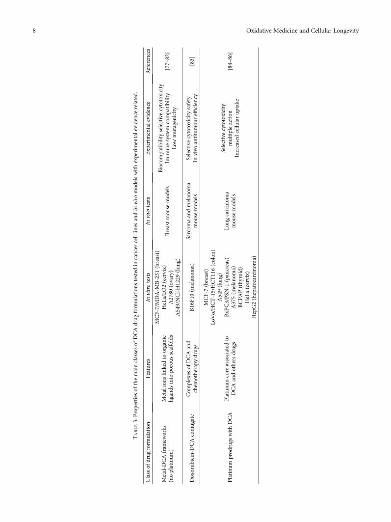
Table3:Propertiesof
themainclassesof
DCAdrug
form
ulations
tested
incancer
celllin
esandin
vivo
mod
elswithexperimentalevidencerelated.
Classof
drug
form
ulation
Features
Invitrotests
Invivo
tests
Experim
entalevidence
References
Metal-D
CAfram
eworks
(noplatinum
)Metalions
linkedto
organic
ligands
into
porous
scaffolds
MCF-7/MDA-M
B-231
(breast)
HeLa/LO
2(cervix)
A2780
(ovary)
A549/NCl-H1229
(lun
g)
Breastmou
semod
els
Biocompatibilityselectivecytotoxicity
Immun
esystem
compatibility
Lowmutagenicity
[77–82]
Doxorub
icin-D
CAconjugate
Com
plexes
ofDCAand
chem
otherapy
drugs
B16F10(m
elanom
a)Sarcom
aandmelanom
amou
semod
els
Selectivecytotoxicity
safety
Invivo
antitumou
reffi
ciency
[83]
Platinu
mprod
rugs
withDCA
Platinu
mcore
associated
toDCAandothersdrugs
MCF-7(breast)
LoVo/HCT-15/HCT116(colon
)A549(lun
g)BxP
C3/PSN
-1(pancreas)
A375(m
elanom
a)BCPAP(thyroid)
HeLa(cervix)
HepG2(hepatocarcino
ma)
Lung
carcinom
amou
semod
els
Selectivecytotoxicity
multipleaction
Increasedcellu
larup
take
[84–86]
8 Oxidative Medicine and Cellular Longevity
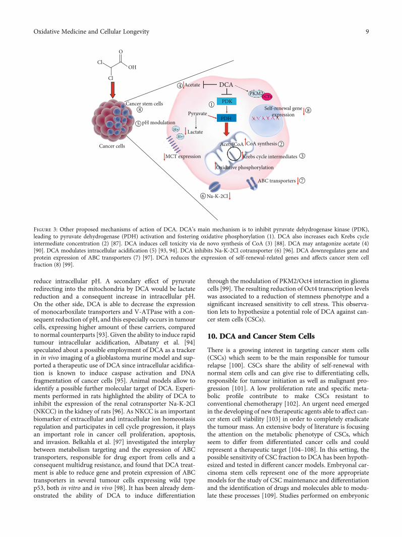
reduce intracellular pH. A secondary effect of pyruvateredirecting into the mitochondria by DCA would be lactatereduction and a consequent increase in intracellular pH.On the other side, DCA is able to decrease the expressionof monocarboxilate transporters and V-ATPase with a con-sequent reduction of pH, and this especially occurs in tumourcells, expressing higher amount of these carriers, comparedto normal counterparts [93]. Given the ability to induce rapidtumour intracellular acidification, Albatany et al. [94]speculated about a possible employment of DCA as a trackerin in vivo imaging of a glioblastoma murine model and sup-ported a therapeutic use of DCA since intracellular acidifica-tion is known to induce caspase activation and DNAfragmentation of cancer cells [95]. Animal models allow toidentify a possible further molecular target of DCA. Experi-ments performed in rats highlighted the ability of DCA toinhibit the expression of the renal cotransporter Na-K-2Cl(NKCC) in the kidney of rats [96]. As NKCC is an importantbiomarker of extracellular and intracellular ion homeostasisregulation and participates in cell cycle progression, it playsan important role in cancer cell proliferation, apoptosis,and invasion. Belkahla et al. [97] investigated the interplaybetween metabolism targeting and the expression of ABCtransporters, responsible for drug export from cells and aconsequent multidrug resistance, and found that DCA treat-ment is able to reduce gene and protein expression of ABCtransporters in several tumour cells expressing wild typep53, both in vitro and in vivo [98]. It has been already dem-onstrated the ability of DCA to induce differentiation
through the modulation of PKM2/Oct4 interaction in gliomacells [99]. The resulting reduction of Oct4 transcription levelswas associated to a reduction of stemness phenotype and asignificant increased sensitivity to cell stress. This observa-tion lets to hypothesize a potential role of DCA against can-cer stem cells (CSCs).
10. DCA and Cancer Stem Cells
There is a growing interest in targeting cancer stem cells(CSCs) which seem to be the main responsible for tumourrelapse [100]. CSCs share the ability of self-renewal withnormal stem cells and can give rise to differentiating cells,responsible for tumour initiation as well as malignant pro-gression [101]. A low proliferation rate and specific meta-bolic profile contribute to make CSCs resistant toconventional chemotherapy [102]. An urgent need emergedin the developing of new therapeutic agents able to affect can-cer stem cell viability [103] in order to completely eradicatethe tumour mass. An extensive body of literature is focusingthe attention on the metabolic phenotype of CSCs, whichseem to differ from differentiated cancer cells and couldrepresent a therapeutic target [104–108]. In this setting, thepossible sensitivity of CSC fraction to DCA has been hypoth-esized and tested in different cancer models. Embryonal car-cinoma stem cells represent one of the more appropriatemodels for the study of CSC maintenance and differentiationand the identification of drugs and molecules able to modu-late these processes [109]. Studies performed on embryonic
Cancer cells
8
5
Cancer stem cells
pH modulation
AcetatePKM2
Self-renewal geneexpression
CoA synthesisAcetylCoA
Krebs cycle intermediates
Oxidative phosphorylation
ABC transporters
Na-K-2Cl
MCT expression
Lactate
PyruvatePDH
PDKOCT4
H+
H+
DCACl
ClOH
O
1
2
3
7
8
4
6
Figure 3: Other proposed mechanisms of action of DCA. DCA’s main mechanism is to inhibit pyruvate dehydrogenase kinase (PDK),leading to pyruvate dehydrogenase (PDH) activation and fostering oxidative phosphorylation (1). DCA also increases each Krebs cycleintermediate concentration (2) [87]. DCA induces cell toxicity via de novo synthesis of CoA (3) [88]. DCA may antagonize acetate (4)[90]. DCA modulates intracellular acidification (5) [93, 94]. DCA inhibits Na-K-2Cl cotransporter (6) [96]. DCA downregulates gene andprotein expression of ABC transporters (7) [97]. DCA reduces the expression of self-renewal-related genes and affects cancer stem cellfraction (8) [99].
9Oxidative Medicine and Cellular Longevity
stem cells (ESCs) constitute preliminary important proofssupporting a possible efficacy of DCA [110]. Interestingly,DCA treatment of ESCs promotes loss of pluripotency andshifts towards a more active oxidative metabolism, accompa-nied by a significant decrease in HIF1a and p53 expression[111]. Vega-Naredo et al. [112] described the importance ofmitochondrial metabolism in directing stemness and differ-entiation in such a model. They characterized the metabolicprofile of stem cell fraction and guessed the less susceptibilityof stem phenotype to mitochondrial-directed therapies.Forcing CSCs towards an oxidative metabolism by DCAtreatment enabled departure from stemness to differentia-tion. Several reports support the existence of CSCs in glioma[113, 114], and the efficiency of DCA to hit CSCs has beenextensively evaluated in such a cancer type, so difficult totreat with conventional therapies and characterized by lowrates of survival. Already in 2010, Michelakis and colleagueshad suggested, both in vitro and in vivo, DCA ability toinduce apoptosis of cancer stem cell fraction [26]. A ratmodel of glioma, recapitulating several features of humanglioblastoma, confirmed the efficacy of DCA to potentiateapoptosis of glioma CSCs, characterized by a significant gly-colytic pathway overstimulation, compared to normal stemcells [115]. Also, Jiang et al. investigated the effect of DCAon the small population of glioma stem cells (GSCs) isolatedfrom glioblastoma, demonstrating a reduction of self-renewalproperties and an increase in cell death percentage [44].Moreover, an in vivo test on mice bearing DCA-treatedGSC-derived xenografts showed a significant increase inoverall survival. DCA treatment was also tested in melanomastem cell fraction, and the derived bioenergetics modulationwas able to counteract protumorigenic action of a c-Metinhibitor [116]. A very recent work performed on humanhepatocellular carcinoma identified PDK4 overexpressionin spheres originated from cancer cells, featuring a definedstem-like phenotype. Interestingly, DCA treatment was ableto reduce cell viability both of cancer-differentiated cellsand cancer stem cells and reversed chemoresistance to con-ventional therapy [36]. Our group has recently experiencedthe ability of DCA to reduce the expression of cancer stemcell markers CD24/CD44/EPCAM in a pancreatic cancer cellline as well as to compromise spheroid formation and viabil-ity [12], further corroborating data obtained in other cancermodels. Together with chemoresistance, also radioresistancerepresents a limit to an efficient cancer treatment, and CSCsseem to be responsible for such refractoriness [117]. Sun et al.demonstrated the ability of DCA to increase radiosensitivityof medulloblastoma cells by affecting stem-like clones, reduc-ing the expression percentage of CD133-positive cells andreducing sphere formation [72]. Moreover, in the same cellu-lar model, they showed an altered mechanism of DNA repairinduced by DCA able to explain the increased effectiveness ofradiotherapy.
11. Conclusions
Targeting cancer cell metabolism represents a new pharma-cological approach to treat cancer. DCA ability to shiftmetabolism from glycolysis to oxidative phosphorylation
has increased the interest towards this drug already knownfor its anticancer properties. The evidence accumulated inthe last years confirms the capability of DCA to overcomechemo, radioresistance in several cancer types and lets tohypothesize additional cellular targets able to explain its skillto kill cancer cells. There is a need to design further clinicalstudies now limited to poor-prognosis patients withadvanced, recurrent neoplasms, already refractory to otherconventional therapies. Its potential efficacy against cancerstem cells as well as the development of new drug formula-tions takes us closer to reach an effective clinical employmentof DCA.
Conflicts of Interest
The authors declare no conflict of interest.
Acknowledgments
This work was supported by Current Research Funds, ItalianMinistry of Health, to IRCCS-CROB, Rionero in Vulture,Potenza, Italy.
References
[1] T. G. Lee, E. H. Jeong, I. J. Min, S. Y. Kim, H. R. Kim, andC. H. Kim, “Altered expression of cellular proliferation,apoptosis and the cell cycle-related genes in lung cancer cellswith acquired resistance to Egfr tyrosine kinase inhibitors,”Oncology Letters, vol. 14, no. 2, pp. 2191–2197, 2017.
[2] J. Chen, “The cell-cycle arrest and apoptotic functions of P53in tumor initiation and progression,” Cold Spring HarborPerspectives in Medicine, vol. 6, no. 3, p. a026104, 2016.
[3] C. Thakur and F. Chen, “Connections between metabolismand epigenetics in cancers,” Seminars in Cancer Biology,vol. 57, pp. 52–58, 2019.
[4] S. Subramaniam, V. Jeet, J. A. Clements, J. H. Gunter, andJ. Batra, “Emergence of micrornas as key players in cancer cellmetabolism,” Clinical Chemistry, vol. 65, no. 9, pp. 1090–1101, 2019.
[5] D. Williams and B. Fingleton, “Non-canonical roles for met-abolic enzymes and intermediates in malignant progressionand metastasis,” Clinical & Experimental Metastasis, vol. 36,no. 3, pp. 211–224, 2019.
[6] T. Tataranni, F. Agriesti, V. Ruggieri et al., “Rewiring carbo-hydrate catabolism differentially affects survival of pancreaticcancer cell lines with diverse metabolic profiles,” Oncotarget,vol. 8, no. 25, pp. 41265–41281, 2017.
[7] A. Luengo, D. Y. Gui, and M. G. Vander Heiden, “Targetingmetabolism for cancer therapy,” Cell Chemical Biology,vol. 24, no. 9, pp. 1161–1180, 2017.
[8] M. O. James, S. C. Jahn, G. Zhong, M. G. Smeltz, Z. Hu, andP. W. Stacpoole, “Therapeutic applications of dichloroacetateand the role of glutathione transferase zeta-1,” Pharmacology& Therapeutics, vol. 170, pp. 166–180, 2017.
[9] E. D. Michelakis, L. Webster, and J. R. Mackey, “Dichloroace-tate (Dca) as a potential metabolic-targeting therapy for can-cer,” British Journal of Cancer, vol. 99, no. 7, pp. 989–994,2008.
10 Oxidative Medicine and Cellular Longevity
[10] S. Kankotia and P. W. Stacpoole, “Dichloroacetate andcancer: new home for an orphan drug?,” Biochimica etBiophysica Acta, vol. 1846, no. 2, pp. 617–629, 2014.
[11] V. Ruggieri, F. Agriesti, R. Scrima et al., “Dichloroacetate, aselective mitochondria-targeting drug for oral squamous cellcarcinoma: a metabolic perspective of treatment,” Oncotar-get, vol. 6, no. 2, pp. 1217–1230, 2015.
[12] T. Tataranni, F. Agriesti, C. Pacelli et al., “Dichloroacetateaffects mitochondrial function and stemness-associatedproperties in pancreatic cancer cell lines,” Cells, vol. 8, no. 5,p. 478, 2019.
[13] A. Khan, D. Marier, E. Marsden, D. Andrews, and I. Eliaz, “Anovel form of dichloroacetate therapy for patients withadvanced cancer: a report of 3 cases,” Alternative Therapiesin Health and Medicine, vol. 20, Supplement 2, pp. 21–28,2014.
[14] E. M. Dunbar, B. S. Coats, A. L. Shroads et al., “Phase 1 trial ofdichloroacetate (Dca) in adults with recurrent malignantbrain tumors,” Investigational New Drugs, vol. 32, no. 3,pp. 452–464, 2014.
[15] Q. S.-C. Chu, R. Sangha, J. Spratlin et al., “A phase I open-labeled, single-arm, dose-escalation, study of dichloroacetate(DCA) in patients with advanced solid tumors,” Investiga-tional New Drugs, vol. 33, no. 3, pp. 603–610, 2015.
[16] A. Khan, D. Andrews, and A. C. Blackburn, “Long-term sta-bilization of stage 4 colon cancer using sodium dichloroace-tate therapy,” World Journal of Clinical Cases, vol. 4, no. 10,pp. 336–343, 2016.
[17] G. Sutendra and E. D. Michelakis, “Pyruvate dehydrogenasekinase as a novel therapeutic target in oncology,” Frontiersin Oncology, vol. 3, p. 38, 2013.
[18] S. R. Pillai, M. Damaghi, Y. Marunaka, E. P. Spugnini, S. Fais,and R. J. Gillies, “Causes, consequences, and therapy oftumors acidosis,” Cancer Metastasis Reviews, vol. 38, no. 1-2, pp. 205–222, 2019.
[19] R. J. DeBerardinis, J. J. Lum, G. Hatzivassiliou, and C. B.Thompson, “The biology of cancer: metabolic reprogram-ming fuels cell growth and proliferation,” Cell Metabolism,vol. 7, no. 1, pp. 11–20, 2008.
[20] N. Zamzami and G. Kroemer, “The mitochondrion in apo-ptosis: how Pandora’s box opens,” Nature Reviews MolecularCell Biology, vol. 2, no. 1, pp. 67–71, 2001.
[21] J. W. Kim and C. V. Dang, “Multifaceted roles of glycolyticenzymes,” Trends in Biochemical Sciences, vol. 30, no. 3,pp. 142–150, 2005.
[22] P.A.Gammage andC. Frezza, “MitochondrialDNA: the over-looked oncogenome?,”BMCBiology, vol. 17, no. 1, p. 53, 2019.
[23] L. H. Stockwin, S. X. Yu, S. Borgel et al., “Sodium dichloroa-cetate selectively targets cells with defects in the mitochon-drial ETC,” International Journal of Cancer, vol. 127, no. 11,pp. 2510–2519, 2010.
[24] P. W. Stacpoole, N. V. Nagaraja, and A. D. Hutson, “Efficacyof dichloroacetate as a lactate-lowering drug,” Journal of Clin-ical Pharmacology, vol. 43, no. 7, pp. 683–691, 2003.
[25] P. W. Stacpoole, “Therapeutic targeting of the pyruvate dehy-drogenase complex/pyruvate dehydrogenase kinase(PDC/PDK) axis in cancer,” JNCI: Journal of the NationalCancer Institute, vol. 109, no. 11, 2017.
[26] E. D. Michelakis, G. Sutendra, P. Dromparis et al., “Metabolicmodulation of glioblastoma with dichloroacetate,” ScienceTranslational Medicine, vol. 2, no. 31, article 31ra34, 2010.
[27] P. W. Stacpoole, C. J. Martyniuk, M. O. James, and N. A. Cal-cutt, “Dichloroacetate-induced peripheral neuropathy,”International Review of Neurobiology, vol. 145, pp. 211–238,2019.
[28] N. Felitsyn, P. W. Stacpoole, and L. Notterpek, “Dichloroace-tate causes reversible demyelination in vitro: potential mech-anism for its neuropathic effect,” Journal of Neurochemistry,vol. 100, no. 2, pp. 429–436, 2007.
[29] T. Langaee, R. Wagner, L. P. Horne et al., “Personalized dos-ing of dichloroacetate using Gstz1 clinical genotyping assay,”Genetic Testing and Molecular Biomarkers, vol. 22, no. 4,pp. 266–269, 2018.
[30] D. Brandsma, T. P. Dorlo, J. H. Haanen, J. H. Beijnen, andW. Boogerd, “Severe encephalopathy and polyneuropathyinduced by dichloroacetate,” Journal of Neurology, vol. 257,no. 12, pp. 2099-2100, 2010.
[31] United States Environmental Protection Agency, EPA, Toxi-cological Review of Dichloroacetic Acid, CAS 79-43-6, 2003.
[32] X. Lu, D. Zhou, B. Hou et al., “Dichloroacetate enhances theantitumor efficacy of chemotherapeutic agents via inhibitingautophagy in non-small-cell lung cancer,” Cancer Manage-ment and Research, vol. 10, pp. 1231–1241, 2018.
[33] A. Korga, M. Ostrowska, M. Iwan, M. Herbet, and J. Dudka,“Inhibition of glycolysis disrupts cellular antioxidant defenseand sensitizes Hepg2 cells to doxorubicin treatment,” FEBSOpen Bio, vol. 9, no. 5, pp. 959–972, 2019.
[34] H. Sun, A. Zhu, X. Zhou, and F. Wang, “Suppression of pyru-vate dehydrogenase kinase-2 re-sensitizes paclitaxel-resistanthuman lung cancer cells to paclitaxel,” Oncotarget, vol. 8,no. 32, pp. 52642–52650, 2017.
[35] B. L. Woolbright, D. Choudhary, A. Mikhalyuk et al., “Therole of pyruvate dehydrogenase kinase-4 (PDK4) in bladdercancer and chemoresistance,” Molecular Cancer Therapeu-tics, vol. 17, no. 9, pp. 2004–2012, 2018.
[36] K. Fekir, H. Dubois-Pot-Schneider, R. Désert et al., “Retrodif-ferentiation of human tumor hepatocytes to stem cells leadsto metabolic reprogramming and chemoresistance,” CancerResearch, vol. 79, no. 8, pp. 1869–1883, 2019.
[37] A. Skeberdytė, I. Sarapinienė, J. Aleksander-Krasko,V. Stankevičius, K. Sužiedėlis, and S. Jarmalaitė, “Dichloroa-cetate and salinomycin exert a synergistic cytotoxic effect incolorectal cancer cell lines,” Scientific Reports, vol. 8, no. 1,p. 17744, 2018.
[38] A. Verma, Y. M. Lam, Y. C. Leung et al., “Combined use ofarginase and dichloroacetate exhibits anti‐proliferative effectsin triple negative breast cancer cells,” The Journal of Phar-macy and Pharmacology, vol. 71, no. 3, pp. 306–315, 2019.
[39] B. Li, X. Li, H. Xiong et al., “Inhibition of COX2 enhances thechemosensitivity of dichloroacetate in cervical cancer cells,”Oncotarget, vol. 8, no. 31, pp. 51748–51757, 2017.
[40] C. Lucido, W. Miskimins, and P. Vermeer, “Propranolol pro-motes glucose dependence and synergizes with dichloroace-tate for anti-cancer activity in HNSCC,” Cancers, vol. 10,no. 12, p. 476, 2018.
[41] C. Abildgaard, C. Dahl, A. Abdul-Al, A. Christensen, andP. Guldberg, “Inhibition of retinoic acid receptor Β signalingconfers glycolytic dependence and sensitization to dichloroa-cetate in melanoma cells,” Oncotarget, vol. 8, no. 48,pp. 84210–84223, 2017.
[42] I. V. Prokhorova, O. N. Pyaskovskaya, D. L. Kolesnik, andG. I. Solyanik, “Influence of metformin, sodium
11Oxidative Medicine and Cellular Longevity
dichloroacetate and their combination on the hematologicaland biochemical blood parameters of rats with gliomas C6,”Experimental Oncology, vol. 40, no. 3, pp. 205–210, 2018.
[43] D. L. Kolesnik, O. N. Pyaskovskaya, Y. R. Yakshibaeva, andG. I. Solyanik, “Time-dependent cytotoxicity of dichloroace-tate and metformin against Lewis lung carcinoma,” Experi-mental Oncology, vol. 41, no. 1, pp. 14–19, 2019.
[44] W. Jiang, S. Finniss, S. Cazacu et al., “Repurposing phenfor-min for the targeting of glioma stem cells and the treatmentof glioblastoma,” Oncotarget, vol. 7, no. 35, pp. 56456–56470, 2016.
[45] B. Waltenberger, A. Mocan, K. Šmejkal, E. Heiss,A. Atanasov, and A. G. Atanasov, “Natural products to coun-teract the epidemic of cardiovascular and metabolic disor-ders,” Molecules, vol. 21, no. 6, p. 807, 2016.
[46] M. Zadorozhna, T. Tataranni, and D. Mangieri, “Piperine:role in prevention and progression of cancer,” MolecularBiology Reports, vol. 46, no. 5, pp. 5617–5629, 2019.
[47] G. Della Sala, F. Agriesti, C. Mazzoccoli, T. Tataranni,V. Costantino, and C. Piccoli, “Clogging the ubiquitin-proteasome machinery with marine natural products: lastdecade update,” Marine Drugs, vol. 16, no. 12, p. 467, 2018.
[48] A. G. Atanasov, B. Waltenberger, E. M. Pferschy-Wenziget al., “Discovery and resupply of pharmacologically activeplant-derived natural products: a review,” BiotechnologyAdvances, vol. 33, no. 8, pp. 1582–1614, 2015.
[49] M. B. Sporn and N. Suh, “Chemoprevention: an essentialapproach to controlling cancer,” Nature Reviews Cancer,vol. 2, no. 7, pp. 537–543, 2002.
[50] C. K. Singh, J. George, and N. Ahmad, “Resveratrol‐basedcombinatorial strategies for cancer management,” Annals ofthe New York Academy of Sciences, vol. 1290, pp. 113–121,2013.
[51] S. Redondo-Blanco, J. Fernández, I. Gutiérrez-del-Río, C. J.Villar, and F. Lombó, “New insights toward colorectal cancerchemotherapy using natural bioactive compounds,” Frontiersin Pharmacology, vol. 8, p. 109, 2017.
[52] B. B. Aggarwal, A. Kumar, and A. C. Bharti, “Anticancerpotential of curcumin: preclinical and clinical studies,” Anti-cancer Research, vol. 23, no. 1A, pp. 363–398, 2003.
[53] P. C. Kan, Y. J. Chang, C. S. Chien, C. Y. Su, and H. W. Fang,“Coupling dichloroacetate treatment with curcumin signifi-cantly enhances anticancer potential,” Anticancer Research,vol. 38, no. 11, pp. 6253–6261, 2018.
[54] K. Hata, K. Hori, H. Ogasawara, and S. Takahashi, “Anti-leukemia activities of Lup-28-Al-20(29)-En-3-one, a lupanetriterpene,” Toxicology Letters, vol. 143, no. 1, pp. 1–7,2003.
[55] C. A. Dehelean, S. Feflea, J. Molnár, I. Zupko, and C. Soica,“Betulin as an antitumor agent tested in vitro on A431, Helaand MCF7, and as an angiogenic inhibitor in vivo in the camassay,” Natural Product Communications, vol. 7, no. 8,pp. 981–985, 2012.
[56] M. Drag, P. Surowiak, M. Drag-Zalesinska, M. Dietel,H. Lage, and J. Oleksyszyn, “Comparision of the cytotoxiceffects of birch bark extract, betulin and betulinic acidtowards human gastric carcinoma and pancreatic carcinomadrug-sensitive and drug-resistant cell lines,” Molecules,vol. 14, no. 4, pp. 1639–1651, 2009.
[57] M. Mihoub, A. Pichette, B. Sylla, C. Gauthier, and J. Legault,“Bidesmosidic betulin saponin bearing L-rhamnopyranoside
moieties induces apoptosis and inhibition of lung cancer cellsgrowth in vitro and in vivo,” PLoS One, vol. 13, no. 3, articlee0193386, 2018.
[58] H. Wang, H. Jiang, M. Van De Gucht, and M. De Ridder,“Hypoxic radioresistance: can ROS be the key to overcomeit?,” Cancers, vol. 11, no. 1, p. 112, 2019.
[59] J. P. Pouget, S. Frelon, J. L. Ravanat, I. Testard, F. Odin, andJ. Cadet, “Formation of modified DNA bases in cells exposedeither to gamma radiation or to high-let particles,” RadiationResearch, vol. 157, no. 5, pp. 589–595, 2002.
[60] K. Rycaj and D. G. Tang, “Cancer stem cells and radioresis-tance,” International Journal of Radiation Biology, vol. 90,no. 8, pp. 615–621, 2014.
[61] L. Tang, F. Wei, Y. Wu et al., “Role of metabolism in cancercell radioresistance and radiosensitization methods,” Journalof Experimental & Clinical Cancer Research, vol. 37, no. 1,p. 87, 2018.
[62] G. Xie, Y. Liu, Q. Yao et al., “Hypoxia-induced angiotensin IIby the lactate-chymase-dependent mechanism mediatesradioresistance of hypoxic tumor cells,” Scientific Reports,vol. 7, no. 1, article 42396, 2017.
[63] J. Overgaard, “Hypoxic radiosensitization: adored andignored,” Journal of Clinical Oncology, vol. 25, no. 26,pp. 4066–4074, 2007.
[64] Y. Zhang and S. G. Martin, “Redox proteins and radiother-apy,” Clinical Oncology, vol. 26, no. 5, pp. 289–300, 2014.
[65] H. Jiang, H. Wang, and M. De Ridder, “Targeting antioxidantenzymes as a radiosensitizing strategy,” Cancer Letters,vol. 438, pp. 154–164, 2018.
[66] M. R. Niewisch, Z. Kuçi, H. Wolburg et al., “Influence ofdichloroacetate (DCA) on lactate production and oxygenconsumption in neuroblastoma cells: is DCA a suitable drugfor neuroblastoma therapy?,” Cellular Physiology and Bio-chemistry, vol. 29, no. 3-4, pp. 373–380, 2012.
[67] S. P. Pitroda, B. T. Wakim, R. F. Sood et al., “Stat1-dependentexpression of energy metabolic pathways links tumourgrowth and radioresistance to the Warburg effect,” BMCMedicine, vol. 7, no. 1, p. 68, 2009.
[68] T. Shimura, N. Noma, Y. Sano et al., “Akt-mediatedenhanced aerobic glycolysis causes acquired radioresistanceby human tumor cells,” Radiotherapy and Oncology,vol. 112, no. 2, pp. 302–307, 2014.
[69] V. Bol, A. Bol, C. Bouzin et al., “Reprogramming of tumormetabolism by targeting mitochondria improves tumorresponse to irradiation,” Acta Oncologica, vol. 54, no. 2,pp. 266–274, 2015.
[70] H. Shen, E. Hau, S. Joshi, P. J. Dilda, and K. L. McDonald,“Sensitization of glioblastoma cells to irradiation by modulat-ing the glucose metabolism,”Molecular Cancer Therapeutics,vol. 14, no. 8, pp. 1794–1804, 2015.
[71] G. Dong, Q. Chen, F. Jiang et al., “Diisopropylamine dichlor-oacetate enhances radiosensitization in esophageal squamouscell carcinoma by increasing mitochondria-derived reactiveoxygen species levels,” Oncotarget, vol. 7, no. 42, pp. 68170–68178, 2016.
[72] L. Sun, T. Moritake, K. Ito et al., “Metabolic analysis of radio-resistant medulloblastoma stem-like clones and potentialtherapeutic targets,” PLoS One, vol. 12, no. 4, articlee0176162, 2017.
[73] L. Zitvogel, L. Apetoh, F. Ghiringhelli, F. André, A. Tesniere,and G. Kroemer, “The anticancer immune response:
12 Oxidative Medicine and Cellular Longevity
indispensable for therapeutic success?,” The Journal ofClinical Investigation, vol. 118, no. 6, pp. 1991–2001, 2008.
[74] S. Gupta and B. Dwarakanath, “Modulation of Immuno-biome during radio-sensitization of tumors by glycolyticinhibitors,” Current Medicinal Chemistry, vol. 25, 2018.
[75] D. Peer, J. M. Karp, S. Hong, O. C. Farokhzad, R. Margalit,and R. Langer, “Nanocarriers as an emerging platform forcancer therapy,” Nature Nanotechnology, vol. 2, no. 12,pp. 751–760, 2007.
[76] R. C. Huxford, J. Della Rocca, and W. Lin, “Metal-organicframeworks as potential drug carriers,” Current Opinion inChemical Biology, vol. 14, no. 2, pp. 262–268, 2010.
[77] I. A. Lázaro, S. A. Lázaro, and R. S. Forgan, “Enhancing anti-cancer cytotoxicity through bimodal drug delivery fromultrasmall Zr MOF nanoparticles,” Chemical Communica-tions, vol. 54, no. 22, pp. 2792–2795, 2018.
[78] I. Abánades Lázaro, S. Haddad, J. M. Rodrigo-Muñoz et al.,“Surface-functionalization of Zr-fumarate MOF for selectivecytotoxicity and immune system compatibility in nanoscaledrug delivery,” ACS Applied Materials & Interfaces, vol. 10,no. 37, pp. 31146–31157, 2018.
[79] I. Abánades Lázaro, S. Haddad, J. M. Rodrigo-Muñoz et al.,“Mechanistic investigation into the selective anticancer cyto-toxicity and immune system response of surface-functional-ized, dichloroacetate-loaded, UiO-66 nanoparticles,” ACSApplied Materials & Interfaces, vol. 10, no. 6, pp. 5255–5268, 2018.
[80] P. Štarha, Z. Trávníček, J. Vančo, and Z. Dvořák, “Half-sand-wich Ru(II) and Os(II) bathophenanthroline complexescontaining a releasable dichloroacetato ligand,” Molecules,vol. 23, no. 2, p. 420, 2018.
[81] J. Pracharova, V. Novohradsky, H. Kostrhunova et al., “Half-sandwich Os(ii) and Ru(ii) bathophenanthroline complexes:anticancer drug candidates with unusual potency and a cellu-lar activity profile in highly invasive triple-negative breastcancer cells,” Dalton Transactions, vol. 47, no. 35,pp. 12197–12208, 2018.
[82] J. Yang, Q. Cao, H. Zhang et al., “Targeted reversal and phos-phorescence lifetime imaging of cancer cell metabolism _via_a theranostic rhenium(I)-DCA conjugate,” Biomaterials,vol. 176, pp. 94–105, 2018.
[83] C. Yang, T. Wu, Y. Qin et al., “A facile doxorubicin-dichloroacetate conjugate nanomedicine with high drugloading for safe drug delivery,” International Journal ofNanomedicine, vol. 13, pp. 1281–1293, 2018.
[84] S. Savino, V. Gandin, J. D. Hoeschele, C. Marzano, G. Natile,and N. Margiotta, “Dual-acting antitumor Pt(IV) prodrugs ofkiteplatin with dichloroacetate axial ligands,” Dalton Trans-actions, vol. 47, no. 21, pp. 7144–7158, 2018.
[85] E. Petruzzella, R. Sirota, I. Solazzo, V. Gandin, and D. Gibson,“Triple action Pt(iv) derivatives of cisplatin: a new class ofpotent anticancer agents that overcome resistance,” ChemicalScience, vol. 9, no. 18, pp. 4299–4307, 2018.
[86] S. Jin, Y. Guo, D. Song et al., “Targeting energy metabolismby a platinum(IV) prodrug as an alternative pathway for can-cer suppression,” Inorganic Chemistry, vol. 58, no. 9,pp. 6507–6516, 2019.
[87] W. Zhang, X. Hu, W. Zhou, and K. Y. Tam, “liquidchromatography-tandem mass spectrometry methodrevealed that lung cancer cells exhibited distinct metaboliteprofiles upon the treatment with different pyruvate dehydro-
genase kinase inhibitors,” Journal of Proteome Research,vol. 17, no. 9, pp. 3012–3021, 2018.
[88] S. Dubuis, K. Ortmayr, and M. Zampieri, “A framework forlarge-scale metabolome drug profiling links coenzyme ametabolism to the toxicity of anti-cancer drug dichloroace-tate,” Communications Biology, vol. 1, no. 1, p. 101, 2018.
[89] S. M. El Sayed, H. Baghdadi, N. S. Ahmed et al., “Dichloroa-cetate is an antimetabolite that antagonizes acetate anddeprives cancer cells from its benefits: a novel evidence-based medical hypothesis,” Medical Hypotheses, vol. 122,pp. 206–209, 2019.
[90] D. M. Jaworski, A. M. Namboodiri, and J. R. Moffett, “Acetateas a metabolic and epigenetic modifier of cancer therapy,”Journal of Cellular Biochemistry, vol. 117, no. 3, pp. 574–588, 2016.
[91] B. A. Webb, M. Chimenti, M. P. Jacobson, and D. L. Barber,“Dysregulated pH: a perfect storm for cancer progression,”Nature Reviews Cancer, vol. 11, no. 9, pp. 671–677, 2011.
[92] D. Neri and C. T. Supuran, “Interfering with pH regulation intumours as a therapeutic strategy,” Nature Reviews Drug Dis-covery, vol. 10, no. 10, pp. 767–777, 2011.
[93] A. Kumar, S. Kant, and S. M. Singh, “Antitumor and chemo-sensitizing action of dichloroacetate implicates modulation oftumor microenvironment: a role of reorganized glucosemetabolism, cell survival regulation and macrophage differ-entiation,” Toxicology and Applied Pharmacology, vol. 273,no. 1, pp. 196–208, 2013.
[94] M. Albatany, A. Li, S. Meakin, and R. Bartha, “Dichloroace-tate induced intracellular acidification in glioblastoma:in vivo detection using AACID-CEST MRI at 9.4 Tesla,”Journal of Neuro-Oncology, vol. 136, no. 2, pp. 255–262, 2018.
[95] H. J. Park, J. C. Lyons, T. Ohtsubo, and C. W. Song, “Acidicenvironment causes apoptosis by increasing caspase activity,”British Journal of Cancer, vol. 80, no. 12, pp. 1892–1897,1999.
[96] J. Stanevičiūtė, M. Juknevičienė, J. Palubinskienė et al.,“Sodium dichloroacetate pharmacological effect as relatedto Na-K-2Cl cotransporter inhibition in rats,” Dose Response,vol. 16, no. 4, article 155932581881152, 2018.
[97] S. Belkahla, A. U. Haq Khan, D. Gitenay et al., “Changes inmetabolism affect expression of ABC transporters throughERK5 and depending on p53 status,” Oncotarget, vol. 9,no. 1, pp. 1114–1129, 2018.
[98] J. A. Bush and G. Li, “Cancer chemoresistance: the relation-ship between P53 and multidrug transporters,” InternationalJournal of Cancer, vol. 98, no. 3, pp. 323–330, 2002.
[99] M. Morfouace, L. Lalier, L. Oliver et al., “Control of gliomacell death and differentiation by PKM2-Oct4 interaction,”Cell Death & Disease, vol. 5, no. 1, pp. e1036–e1036, 2014.
[100] A. Turdo, V. Veschi, M. Gaggianesi et al., “Meeting the chal-lenge of targeting cancer stem cells,” Frontiers in Cell andDevelopment Biology, vol. 7, p. 16, 2019.
[101] P. Zhu and Z. Fan, “Cancer stem cells and tumorigenesis,”Biophysics Reports, vol. 4, no. 4, pp. 178–188, 2018.
[102] S. Prasad, S. Ramachandran, N. Gupta, I. Kaushik, and S. K.Srivastava, “Cancer cells stemness: a doorstep to targetedtherapy,” Biochimica et Biophysica Acta - Molecular Basis ofDisease, p. 165424, 2019.
[103] M. Yang, P. Liu, and P. Huang, “Cancer stem cells, metabo-lism, and therapeutic significance,” Tumour Biology, vol. 37,no. 5, pp. 5735–5742, 2016.
13Oxidative Medicine and Cellular Longevity
[104] P. Sancho, D. Barneda, and C. Heeschen, “Hallmarks of can-cer stem cell metabolism,” British Journal of Cancer, vol. 114,no. 12, pp. 1305–1312, 2016.
[105] F. Sotgia, M. Fiorillo, and M. P. Lisanti, “Hallmarks of thecancer cell of origin: comparisons with “energetic” cancerstem cells (e-CSCs),” Aging, vol. 11, no. 3, pp. 1065–1068,2019.
[106] S. Skvortsov, I. I. Skvortsova, D. G. Tang, and A. Dubrovska,“Concise review: prostate cancer stem cells: current under-standing,” Stem Cells, vol. 36, no. 10, pp. 1457–1474, 2018.
[107] S. Bordel, “Constraint based modeling of metabolism allowsfinding metabolic cancer hallmarks and identifying personal-ized therapeutic windows,” Oncotarget, vol. 9, no. 28,pp. 19716–19729, 2018.
[108] Y. Y. Wang, J. Chen, X. M. Liu, R. Zhao, and H. Zhe, “Nrf2-mediated metabolic reprogramming in cancer,” OxidativeMedicine and Cellular Longevity, vol. 2018, Article ID9304091, 7 pages, 2018.
[109] M. W. McBurney, “P19 embryonal carcinoma cells,” TheInternational Journal of Developmental Biology, vol. 37,no. 1, pp. 135–140, 1993.
[110] R. Loureiro, S. Magalhães-Novais, K. A. Mesquita et al., “Mel-atonin antiproliferative effects require active mitochondrialfunction in embryonal carcinoma cells,” Oncotarget, vol. 6,no. 19, pp. 17081–17096, 2015.
[111] A. S. Rodrigues, M. Correia, A. Gomes et al., “Dichloroace-tate, the pyruvate dehydrogenase complex and the modula-tion of mESC pluripotency,” PLoS One, vol. 10, no. 7, articlee0131663, 2015.
[112] I. Vega-Naredo, R. Loureiro, K. A. Mesquita et al., “Mito-chondrial metabolism directs stemness and differentiationin P19 embryonal carcinoma stem cells,” Cell Death and Dif-ferentiation, vol. 21, no. 10, pp. 1560–1574, 2014.
[113] S. K. Singh, C. Hawkins, I. D. Clarke et al., “Identification ofhuman brain tumour initiating cells,” Nature, vol. 432,no. 7015, pp. 396–401, 2004.
[114] X. Yuan, J. Curtin, Y. Xiong et al., “Isolation of cancer stemcells from adult glioblastoma multiforme,” Oncogene,vol. 23, no. 58, pp. 9392–9400, 2004.
[115] M. Morfouace, L. Lalier, M. Bahut et al., “Comparison ofspheroids formed by rat glioma stem cells and neural stemcells reveals differences in glucose metabolism and promisingtherapeutic applications,” The Journal of Biological Chemis-try, vol. 287, no. 40, pp. 33664–33674, 2012.
[116] L. Kucerova, L. Demkova, S. Skolekova, R. Bohovic, andM. Matuskova, “Tyrosine kinase inhibitor SU11274 increasedtumorigenicity and enriched for melanoma-initiating cells bybioenergetic modulation,” BMC Cancer, vol. 16, no. 1, p. 308,2016.
[117] Z. Zhao, K. Zhang, Z. Wang et al., “A comprehensive reviewof available omics data resources and molecular profiling forprecision glioma studies,” Biomedical Reports, vol. 10, no. 1,pp. 3–9, 2019.
14 Oxidative Medicine and Cellular Longevity
Stem Cells International
Hindawiwww.hindawi.com Volume 2018
Hindawiwww.hindawi.com Volume 2018
MEDIATORSINFLAMMATION
of
EndocrinologyInternational Journal of
Hindawiwww.hindawi.com Volume 2018
Hindawiwww.hindawi.com Volume 2018
Disease Markers
Hindawiwww.hindawi.com Volume 2018
BioMed Research International
OncologyJournal of
Hindawiwww.hindawi.com Volume 2013
Hindawiwww.hindawi.com Volume 2018
Oxidative Medicine and Cellular Longevity
Hindawiwww.hindawi.com Volume 2018
PPAR Research
Hindawi Publishing Corporation http://www.hindawi.com Volume 2013Hindawiwww.hindawi.com
The Scientific World Journal
Volume 2018
Immunology ResearchHindawiwww.hindawi.com Volume 2018
Journal of
ObesityJournal of
Hindawiwww.hindawi.com Volume 2018
Hindawiwww.hindawi.com Volume 2018
Computational and Mathematical Methods in Medicine
Hindawiwww.hindawi.com Volume 2018
Behavioural Neurology
OphthalmologyJournal of
Hindawiwww.hindawi.com Volume 2018
Diabetes ResearchJournal of
Hindawiwww.hindawi.com Volume 2018
Hindawiwww.hindawi.com Volume 2018
Research and TreatmentAIDS
Hindawiwww.hindawi.com Volume 2018
Gastroenterology Research and Practice
Hindawiwww.hindawi.com Volume 2018
Parkinson’s Disease
Evidence-Based Complementary andAlternative Medicine
Volume 2018Hindawiwww.hindawi.com
Submit your manuscripts atwww.hindawi.com