diced ear cartilage with perichondrial attachment in rhinoplasty.pdf
DESCRIPTION
CartilageTRANSCRIPT

http://aes.sagepub.com/Aesthetic Surgery Journal
http://aes.sagepub.com/content/32/7/825The online version of this article can be found at:
DOI: 10.1177/1090820X12455635
2012 32: 825Aesthetic Surgery Journaland S. Jaber Mousavi
Farhad Hafezi, Hamed Bateni, Bijan Naghibzadeh, Amir Hossein Nouhi, Abolhasan Emami, S. Javad Fatemi, Mirsepehr PedramDiced Ear Cartilage With Perichondrial Attachment in Rhinoplasty : A New Concept
Published by:
http://www.sagepublications.com
On behalf of:
American Society for Aesthetic Plastic Surgery
can be found at:Aesthetic Surgery JournalAdditional services and information for
http://aes.sagepub.com/cgi/alertsEmail Alerts:
http://aes.sagepub.com/subscriptionsSubscriptions:
http://www.sagepub.com/journalsReprints.navReprints:
http://www.sagepub.com/journalsPermissions.navPermissions:
What is This?
- Aug 31, 2012Version of Record >>
at HINARI - Parent on November 16, 2012aes.sagepub.comDownloaded from

Rhinoplasty
Aesthetic Surgery Journal32(7) 825 –832© 2012 The American Society for Aesthetic Plastic Surgery, Inc.Reprints and permission: http://www .sagepub.com/journalsPermissions.navDOI: 10.1177/1090820X12455635www.aestheticsurgeryjournal.com
In recent years, there has been a trend toward nasal skele-ton reshaping, instead of reduction and excision. Jacques Joseph (1865-1934) performed his first cosmetic rhinoplasty on May 11, 1898. The reduction and excision approach, initiated at the beginning of the 20th century, was used as the standard technique for rhinoplasty for more than 60 years. Use of the spreader graft, tip graft, and radix augmen-tation was suggested by Jack Sheen1 and Mark Constantian.2 The development of open-approach rhinoplasty with demand for a strong columella and alar strut was recom-mended by Gunter and Friedman.3 Tebbetts4,5 popularized tip sutures to replace the aggressive excision and reduction of lower lateral cartilage. These new approaches encour-aged rhinoplasty surgeons to use cartilage as a supportive structural graft. Although the results of rhinoplasty have improved over the past 30 years, the shortage of available cartilage is a dilemma, especially in secondary cases.
The nasal septum is always the first and best choice for cartilage, but the amount of this valuable resource is often
Diced Ear Cartilage With Perichondrial Attachment in Rhinoplasty: A New Concept
Farhad Hafezi, MD, FACS; Hamed Bateni, MD; Bijan Naghibzadeh, MD, FACS; Amir Hossein Nouhi, MD; Abolhasan Emami, MD; S. Javad Fatemi, MD; Mirsepehr Pedram, DVM; and S. Jaber Mousavi, MD
AbstractBackground: Diced cartilage is a valuable material that has recently been added to the graft options in rhinoplasty. Shaping, fixation, and resorption are the main concerns with this material. Perichondrially attached diced conchal cartilage may be a new possibility to solve some of these problems.Objectives: The authors evaluate the outcome of perichondrially attached diced cartilage in a rabbit model and compare the results with injectable cartilage grafting.Methods: Ear cartilage was removed from 1 auricle in each of the 16 rabbits included in this study; samples were divided in 2 pieces. After precise weighing, both segments were diced. The perichondrium was left attached to 1 of the pieces. Both segments were inserted in 2 separate pockets in the dorsum of the animal. After a 3-month period, both samples were removed and measured for growth/resorption.Results: At the beginning of this study, the difference in weight between groups was statistically insignificant (P = .213), but 3 months after insertion, significant growth was observed in the perichondrial group (P = .019).Conclusions: The vascularization and significant growth in weight of the perichondrially attached diced cartilage samples are evidence of the viability of this material. The structural integrity and solid framework afforded by this option suggest that the material should be used more frequently in nasoskeletal augmentation.
Keywordsrhinoplasty, diced cartilage, perichondrial attachment, nasal reconstruction
Accepted for publication March 9, 2012.
Dr Hafezi is Professor of Plastic Surgery, Dr Emami is Associate Professor of Plastic Surgery, and Dr Fatemi is Associate Professor of Plastic Surgery, Tehran University of Medical Sciences, Tehran, Iran. Dr Mousavi is an epidemiologist in the Burn Research Center, and Dr Pedram is a veterinary surgeon at Tehran University of Medical Sciences, St Fatima Hospital, Tehran, Iran. Dr Bateni is an attending plastic surgeon at Mehr Hospital, Tehran, Iran. Dr Naghibzadeh is Professor of ENT Surgery, Shahid Beheshty University of Medical Sciences, Loghman Hakim Hospital, Tehran, Iran. Dr Nouhi is a privately employed pathologist in Tehran, Iran.
Corresponding Author:Dr Farhad Hafezi, Professor of Plastic Surgery, No. 41, Zaferanieh Street, Zaferanieh, Tehran, Iran E-mail: [email protected]
INTE
RNAT
IONAL CONTRIBUTION
Scan this code with your smartphone to see the operative video. Need help? Visit www.aestheticsurgery.com.
at HINARI - Parent on November 16, 2012aes.sagepub.comDownloaded from

826 Aesthetic Surgery Journal 32(7)
limited due to either previous resections or the danger of dorsum collapse posed by overresection. This is why rhi-noplasty surgeons are always looking for alternative sources, such as alloplastic material or heterotopic autolo-gous cartilage from the concha or rib. Alloplastic material has a high chance of extrusion. The concha is composed of delicate elastic and convoluted cartilage, so it will not provide the optimal shape and support. Rib cartilage blocks have problems related to warping and visibility, and some surgeons may hesitate to use this valuable resource.
In this report, we discuss diced cartilage as a solution to solve some of these problems. Of note, there are certain disadvantages to wrapping diced cartilage with Surgicel (Ethicon, Inc, Somerville, New Jersey) or fascia: resorption and the need for another incision to harvest fascia, respec-tively. Free diced cartilage, although promising as a useful tool in the hands of the experienced rhinoplasty surgeon, involves issues related to the scattering of small cartilage pieces in the larger pocket and the difficulty of sculpting the material and then maintaining the desired shape. The ideal diced cartilage graft is one in which the small pieces are attached together with some connective tissue. This allows the material to be molded and provides the option of suturing and fixing it to the desired position. The con-chal cartilage has a distinct perichondrium, which is resistant to the sharp edge of the dicing blade and keeps these small pieces together without tearing or the loss of structural integrity. This combination of perichondrium and diced cartilage gives the surgeon the ability to shape the graft on the bench by rolling it on itself, which “sutures” the cartilage pieces by fashioning a tube or by placing the graft as a lining under the thin skin of the nose to cover the underlying irregularities. In this article, we investigate the characteristics, viability, and longevity of this new tool in a rabbit model.
MethOds
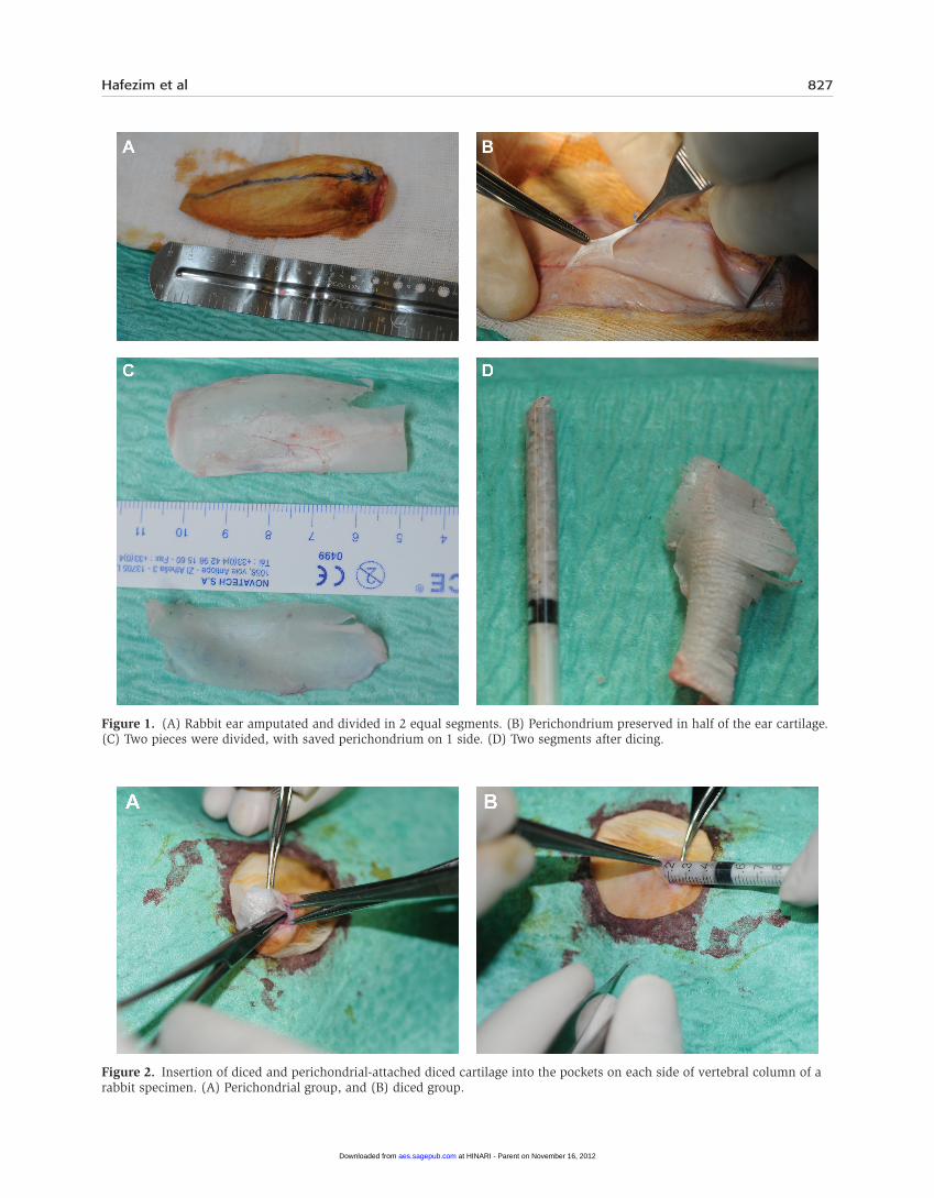
Sixteen white male New Zealand rabbits 12 to 14 weeks of age and weighing 1500 to 2000 g were selected for this study. Under general anesthesia induced by an intramus-cular injection of ketamine 50 mg/kg and xylazine 5 mg/kg, 1 auricle from each rabbit was amputated by a veteri-nary surgeon and divided into 2 pieces longitudinally (Figure 1A-C). One piece (half of 1 ear cartilage) was denuded of all soft tissues; the perichondrium was pre-served on the other half, so the small pieces were kept attached together as a single sheet. Both samples were weighed accurately in sterile conditions using a precise laboratory scale (.01-g accuracy). The samples without attached perichondrium were diced to 0.5-mm pieces (Figure 1D).
Two 1-cm transverse incisions were made on either side of the spinal column of each rabbit. A sample of
pure diced cartilage was injected in the subcutaneous pocket on 1 side through a 1-mL tuberculin syringe. The diced cartilage attached to the perichondrium was fash-ioned into a tube shape and inserted in a subcutaneous pocket on the opposite side of the vertebral column (Figure 2A,B). During the operation, 0.1 mg/kg alatro-floxacin was administered subcutaneously to all rabbits. A co-amoxiclav suspension was administered in the rab-bits’ drinking water for 3 days postoperatively as a pro-phylactic antibiotic. Animals were preserved in their cages for 3 months. Two rabbits died during this period and were excluded from the study. The animals were kept in their standard cages at 22 to 24°C with 12 inter-mittent hours of light and darkness. They had free access to water and food. After 3 months,6,7 all animals were sacrificed by means of an intraperitoneal injection of a high dose of sodium thiopental. Through generous longitudinal incisions on both sides of the spinal col-umn, both samples were positioned under direct visu-alization. Two samples were integrated as a clump (which made complete removal easier from the recipi-ent bed) and weighed accurately (Figure 3A,B). The pre- and postoperative weights of the specimens were compared. A paired t test was used for the pre- and postoperative comparison of results. To compare the end result between groups, an independent t test was applied.
The authors used the same technique described here in a patient study. Although no data from that trial are pre-sented in this report, the results 6 months after harvesting, dicing, and placement of the graft in 1 case is shown in Figures 4 through 6 and in a video, which can be seen at www.aestheticsurgeryjournal.com. You may also scan the code on the first page of this article to be taken directly to the video on www.YouTube.com.
Results
At the beginning of the study, the mean weight of the pure-diced group was 0.63 ± 0.18 g. The mean weight in the attached perichondrium group was 0.71 ± 0.14 g (Table 1). This difference is most likely due to the weight of the attached perichondrium, although it should be men-tioned that during the dicing, weighing, and transferring procedures, some tiny pieces of cartilage may have been lost, which may have added bias to the result. Three months after the cartilage was inserted, the mean weight changed to 0.60 ± 0.13 g in the pure-diced group and to 0.75 ± 0.18 g in the group with perichondrium (Figure 7). A paired t test was applied to analyze the results. Before insertion of the cartilage, the difference in weight between the groups was statistically insignificant (P = .213). Postoperatively (3 months later), this difference became significant (P = .019).
at HINARI - Parent on November 16, 2012aes.sagepub.comDownloaded from
Hafezim et al 827
Figure 1. (A) Rabbit ear amputated and divided in 2 equal segments. (B) Perichondrium preserved in half of the ear cartilage. (C) Two pieces were divided, with saved perichondrium on 1 side. (D) Two segments after dicing.
Figure 2. Insertion of diced and perichondrial-attached diced cartilage into the pockets on each side of vertebral column of a rabbit specimen. (A) Perichondrial group, and (B) diced group.
at HINARI - Parent on November 16, 2012aes.sagepub.comDownloaded from

828 Aesthetic Surgery Journal 32(7)
Figure 3. Specimens were removed after 90 days and weighed. Cartilage grafts in both groups were transformed into solid masses. (A) Perichondrial group, and (B) diced group.
Figure 4. The same harvesting technique is shown in a human case. (A) Exposure of the conchal cartilage with its attached perichondrium. (B) The harvested concha is shown with its attached perichondrium. (C) Segment A was used for dorsal augmentation and segment B as a tip graft.
at HINARI - Parent on November 16, 2012aes.sagepub.comDownloaded from
Hafezim et al 829
disCussiOnA review of the MEDLINE database revealed that the first published paper on dicing autogenous cartilage was pub-lished more than 64 years ago,8 although Daniel reported on its application in rhinoplasty approximately 70 years ago.9,10 Erol11 popularized this technique by wrapping diced cartilage in Surgicel in 2000. Further investigation proved that this material may produce foreign body reac-tions, which reduce graft viability and resorption.6,12-15 In contrast, bare cartilage is not resorbed and may proliferate significantly.16
The next step in this technique was the application of temporal fascia.7,17-20 The pieces of diced cartilage integrated into a single cartilage mass,15 but there were shortcomings—namely, the need for a second incision over the temporal area. Many patients refused to undergo such an incision. Recently, several authors have demon-strated the increased resorption of wrapped diced cartilage as compared with bare diced grafts.21,22 Histological analy-sis has demonstrated that the chondrocyte regeneration of an AlloDerm (LifeCell, Inc, Branchburg, New Jersey)–treated group was significantly superior to that of a fascia-treated group.12 In 2009 at the American Society for Aesthetic Plastic Surgery (ASAPS) meeting in Las Vegas, Nevada, Erol described syringe injection of diced cartilage without any wrapping. He achieved good outcomes with no resorption. This approach eliminates the wrapping material, which acts as a barrier to the blood supply. The only shortcoming of this technique is the lack of structural integrity and the possibility of small cartilage pieces dis-persing in a larger pocket. To prevent this problem, we decided to preserve the perichondrium when dicing the conchal cartilage. In harvesting the concha, the donor site was hidden, and no visible scar was produced. Conchal cartilage is elastic, is difficult to crush, and will not pro-duce strong structural support as in septal or rib cartilage grafts.
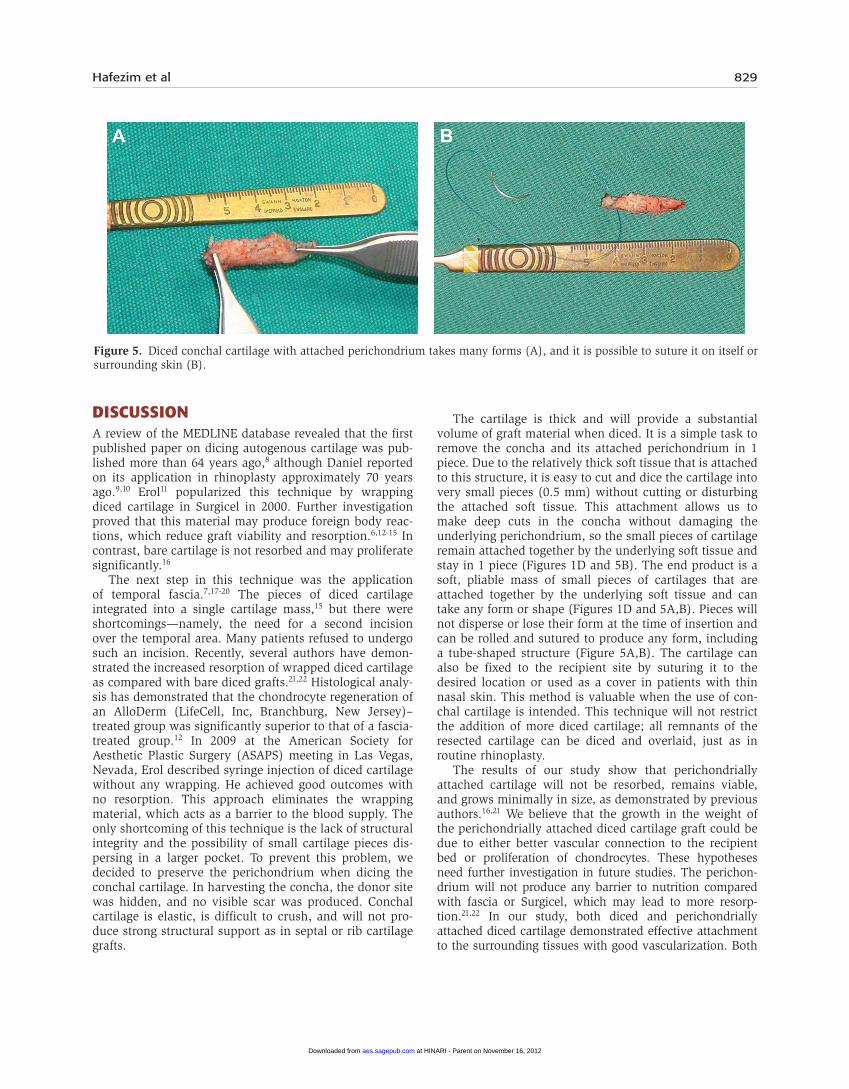
The cartilage is thick and will provide a substantial volume of graft material when diced. It is a simple task to remove the concha and its attached perichondrium in 1 piece. Due to the relatively thick soft tissue that is attached to this structure, it is easy to cut and dice the cartilage into very small pieces (0.5 mm) without cutting or disturbing the attached soft tissue. This attachment allows us to make deep cuts in the concha without damaging the underlying perichondrium, so the small pieces of cartilage remain attached together by the underlying soft tissue and stay in 1 piece (Figures 1D and 5B). The end product is a soft, pliable mass of small pieces of cartilages that are attached together by the underlying soft tissue and can take any form or shape (Figures 1D and 5A,B). Pieces will not disperse or lose their form at the time of insertion and can be rolled and sutured to produce any form, including a tube-shaped structure (Figure 5A,B). The cartilage can also be fixed to the recipient site by suturing it to the desired location or used as a cover in patients with thin nasal skin. This method is valuable when the use of con-chal cartilage is intended. This technique will not restrict the addition of more diced cartilage; all remnants of the resected cartilage can be diced and overlaid, just as in routine rhinoplasty.
The results of our study show that perichondrially attached cartilage will not be resorbed, remains viable, and grows minimally in size, as demonstrated by previous authors.16,21 We believe that the growth in the weight of the perichondrially attached diced cartilage graft could be due to either better vascular connection to the recipient bed or proliferation of chondrocytes. These hypotheses need further investigation in future studies. The perichon-drium will not produce any barrier to nutrition compared with fascia or Surgicel, which may lead to more resorp-tion.21,22 In our study, both diced and perichondrially attached diced cartilage demonstrated effective attachment to the surrounding tissues with good vascularization. Both
Figure 5. Diced conchal cartilage with attached perichondrium takes many forms (A), and it is possible to suture it on itself or surrounding skin (B).
at HINARI - Parent on November 16, 2012aes.sagepub.comDownloaded from
830 Aesthetic Surgery Journal 32(7)
types of cartilage were integrated to solid masses (Figure 3A,B).
Our findings in this rabbit model suggest that cartilage grafts with attached perichondrium may survive better than diced-cartilage grafts. Further investigations of septal and rib cartilage will be necessary to prove the benefits of perichondrium in humans. The perichondrium of the con-cha is thicker in human beings than in a thin rabbit ear, so dicing is easier.
COnClusiOns
In an attempt to address the possible shortcomings of work-ing with diced cartilage, we preserved the attachment of the perichondrium to the diced ear cartilage. Three months after the insertion of grafts in 14 rabbits, the viability, growth in weight, and solid and rigid framework of the cartilage in the perichondrially attached group were evident. Perichondrially attached, diced conchal cartilage provides the possibility of
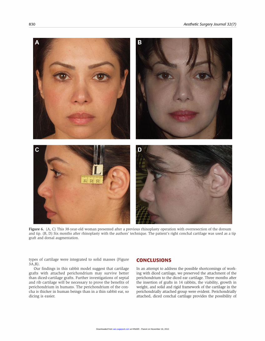
Figure 6. (A, C) This 38-year-old woman presented after a previous rhinoplasty operation with overresection of the dorsum and tip. (B, D) Six months after rhinoplasty with the authors’ technique. The patient’s right conchal cartilage was used as a tip graft and dorsal augmentation.
at HINARI - Parent on November 16, 2012aes.sagepub.comDownloaded from

Hafezim et al 831
shaping and fixing this combination to the nasal skeleton. It can be used either as a cover in patients with thin nasal skin or as a dorsal augmentation graft. When the conchal cartilage is intended for structural augmentation in rhinoplasty, this
technique may convert the weak and irregularly shaped con-chal cartilage to a more useful tool.
disclosures
The authors declared no potential conflicts of interest with respect to the research, authorship, and publication of this article.
Funding
The funding for this paper was provided by an educational grant from Tehran University of Medical Sciences, which was used for equipment and the animal laboratory. The authors received no stipend or payment.
ReFeRenCes
1. Sheen JH. Spreader graft: a method of reconstructing the roof of the middle nasal vault following rhinoplasty. Plast Reconstr Surg. 1984;73(2):230-239.
2. Constantian MB. Four common anatomic variants that predispose to unfavorable rhinoplasty results: a study based on 150 consecutive secondary rhinoplasties. Plast Reconstr Surg. 2000;105(1):316-331.
3. Gunter JP, Friedman RM. Lateral crural strut graft: tech-nique and clinical applications in rhinoplasty. Plast Reconstr Surg. 1997;99(4):943-952.
4. Tebbetts JB. Shaping and positioning the nasal tip with-out structural disruption: a new, systematic approach. Plast Reconstr Surg. 1994;94:61-77.
5. Tebbetts JB. Secondary tip modification: shaping and position-ing the nasal tip using nondestructive techniques. In: Teb-betts JB, ed. Primary Rhinoplasty: A New Approach to the Logic and the Techniques. St Louis, MO: Mosby, 1998.
6. Kazikdas KC, Ergur B, Tugyan K, Guneli E, Kaya D, Sahan M. Viability of crushed and diced cartilage grafts wrapped in oxi-dized regenerated cellulose and esterified hyaluronic acid: an experimental study. Laryngoscope. 2007;117(10):1728-1734.
7. Coskun BU, Seven H, Yigit O, et al. Comparison of diced cartilage graft wrapped in surgicell and diced cartilage graft wrapped in fascia: an experimental study. Laryngo-scope. 2005;115(4):668-671.
8. Gordon SD, Waeern RF. Autogenous diced cartilage transplants to bone; an experimental study. Ann Surg. 1947;125(2):237-240.
9. Burian F. The Plastic Surgery Atlas. New York: Macmillan; 1968.
10. Denecke HJ, Meyer R. Plastic Surgery of the Head and Neck. New York: Springer-Verlag; 1967.
11. Erol OO. The Turkish delight: a pliable graft for rhino-plasty. Plast Reconstr Surg. 2000;105(6):2229-2241.
12. Kim HK, Chu LS, Kim JW, et al. The viability of diced cartilage grafts wrapped in autogenous fascia and Allo-Derm® in a rabbit model. J Plast Reconstr Aesthetic Surg. 2011;64(8):e193-e200.
13. Daniel RK. Diced cartilage grafts in rhinoplasty surgery: current techniques and applications. Plast Reconstr Surg. 2008;122(6):1883-1891.
Table 1. Weight of Cartilages at the Beginning and End of the Study
Rabbit No.
Cartilage Without Perichondrium,
Before Insertion, mg
Cartilage Without Perichondrium, After Removal,
mg
Cartilage With Perichondrium,
Before Insertion, mg
Cartilage With Perichondrium, After Removal,
mg
1 340 450 590 650
2 700 700 870 107
3 630 610 690 620
4 890 830 850 780
5 800 770 830 860
6 680 600 710 650
7 510 530 610 700
8 560 540 560 610
9 370 420 610 670
10 460 420 510 550
11 760 610 840 810
12 470 520 510 590
13 820 730 830 840
14 830 730 910 171
Figure 7. Linear schema showing the difference in weight between the 2 graft groups at the beginning and 3 months after the insertion of cartilage. There was a significant increase in the weight of the perichondrium-attached group.
at HINARI - Parent on November 16, 2012aes.sagepub.comDownloaded from

832 Aesthetic Surgery Journal 32(7)
14. Calvert JW, Brenner K, DaCosta-Iyer M, Evans GR, Daniel RK. Histological analysis of human diced cartilage grafts. Plast Reconstr Surg. 2006;118(1):230-236.
15. Daniel RK, Calvert JW. Diced cartilage grafts in rhi-noplasty surgery. Plast Reconstr Surg. 2004;113(7): 2156-2171.
16. Yilmaz S, Erçöçen AR, Can Z, Yenidünya S, Edali N, Yor-muk E. Viability of diced, rushed cartilage grafts and the effects of Surgicel (oxidized regenerated cellulose) on cartilage grafts. Plast Reconstr Surg. 2001;108(4):1054-1060; discussion 1061-1062.
17. Brenner KA, McConnell MP, Evans GR, Calvert JW. Sur-vival of diced cartilage grafts: an experimental study. Plast Reconstr Surg. 2006;117(1):105-115.
18. Gerbault O, Aiach G. Diced cartilage wrapped in deep temporal aponeurosis (DC-F): A new technique in
augmentation rhinoplasty [in French]. Ann Chir Plast Esthet. 2009;54(5):477-485.
19. Calvert J, Brenner K. Autogenous dorsal reconstruc-tion: maximizing the utility of diced cartilage and fascia. Semin Plast Surg. 2008;22(2):110-119.
20. Kelly MH, Bulstrode NW, Waterhouse N. Versatility of diced cartilage-fascia grafts in dorsal nasal augmentation. Plast Reconstr Surg. 2007;120(6):1654-1659; discussion 1654-1659.
21. Fatemi MJ, Hasani ME, Rahimian S, Bateni H, Pedram M, Mousavi SJ. Survival of block and fascial-wrapped diced cartilage grafts: an experimental study in rabbits [pub-lished online July 11, 2011]. Ann Plast Surg.
22. Bullocks JM, Echo A, Guerra G, Stal S, Yuksel EA. Novel autologous scaffold for diced-cartilage grafts in dorsal augmentation rhinoplasty. Aesthetic Plast Surg. 2011;35(4):569-579.
at HINARI - Parent on November 16, 2012aes.sagepub.comDownloaded from