diagnosis of white spot disease in shrimp - acfs disease_in shrimp... · the presumptive laboratory...
TRANSCRIPT
THAI AGRICULTURAL STANDARD
TAS 10451-2007
DIAGNOSIS OF WHITE SPOT DISEASE IN SHRIMP
National Bureau of Agricultural Commodity and Food Standards
Ministry of Agriculture and Cooperatives
ICS 11.220 ISBN 978-974-403-475-5

UNOFFICAL TRANSLATION
THAI AGRICULTURAL STANDARD
TAS 10451-2007
DIAGNOSIS OF WHITE SPOT DISEASE IN SHRIMP
National Bureau of Agricultural Commodity and Food Standards
Ministry of Agriculture and Cooperatives
50 Phaholyothin Road, Ladyao, Chatuchak, Bangkok 10900 Telephone (662) 561 2277 www.acfs.go.th
Published in the Royal Gazette Vol.124 Special Section 78D,
dated 29 June B.E.2550 (2007)

(2)
Ad hoc Sub-Committee on the Elaboration of Standard for Diagnosis of Aquatic Animal Disease
1. Director General of the Department of Fisheries Chairperson
Mr. Sitdhi Boonyaratpalin Mr. Jaranthada Karnasuta (alternate)
2. Representative of the Department of Fisheries Member Mr. Somkiat Kanchanakhan
3. Representative of the Department of Livestock Development Member Mrs. Montakan Vongpakorn
4. Representative of the Faculty of Science, Member Mahidol University Prof. Timothy William Flegel Mrs. Wansika Kiatpathomchai (alternate)
5. Representative of the Faculty of Fisheries Member Kasetsart University Mr. Nontawit Areechon Mr. Prapansak Srisapoome (alternate)
6. Representative of the Office of Commodity and System Standards Member National Bureau of Agriculture Commodity and Food Standards Miss Metanee Sukontarug
7. Representative of the Faculty of Veterinary Science Member Chulalongkorn University Mr. Jirasak Tangtrongpiros
8. Representative of the Faculty of Veterinary Science Member Kasetsart University Mrs. Pareeya Udomkusonsri
9. Representative of the Faculty of Veterinary Science Member Khon Kaen University Mr. Komkrich Pimpukdee Mr. Prapansak Chaveerach (alternate)
10. Representative of the Thai Association of Veterinary Laboratory Member Diagnosticians
Assoc. Prof. Janenuj Wongtavatchai
11. Experts Member Mrs. Supranee Chinabut Miss Jiraporn Kasornchandra Mr. Visanu Boonyawiwat

(3)
12. Representative of National Bureau of Agricultural Commodity and Food Standards Miss Darunee Tuntasuwan Member and secretary
13. Representative of National Bureau of Agricultural Commodity and Food Standards Mr. Somkiat Sripisuth Member and assistant secretary
(4)
The Thai Agricultural Standard (TAS) on Diagnosis of White Spot Disease in Shrimp establishes the provisions on certified free status of white spot disease in shrimp and the provisions will be used as a manual for laboratories using the rapid staining method, histopathological method and nested polymerase chain reaction (nested-PCR) method to support the production system and ensure the quality of shrimp and its products for domestic and export markets.
The standard is based on the information of the following documents:
1. Bondad-Reantaso MG, McGladdery SE, East I. and Subasinghe RP 2001. Asia Diagnostic Guide to Aquatic Animal Diseases. FAO Fisheries Technical Paper No. 402, Supplement 2. FAO. Rome, Italy.
2. OIE 2003.White spot disease., Manual of Diagnostic Tests for Aquatic Animals. Chapter 4.1.2. World Organisation for Animal Health. Paris, France.
Remark: The standard title has been amended from “Thai Agricultural Commodity and Food Standard (TACFS)” to “Thai Agricultural Standard (TAS)” in accordance with the enforcement of The Agricultural Standards Act B.E. 2551 (2008).
NOTIFICATION OF THE NATIONAL COMMITTEE ON AGRICULTURAL COMMODITY AND FOOD STANDARDS
SUBJECT: THAI AGRICULTURAL COMMODITY AND FOOD STANDARD: DIAGNOSIS OF WHITE SPOT DISEASE IN SHRIMP
B.E.2550 (2007)
The resolution of the 1/2550 session of the National Committee on Agricultural
Commodity and Food Standards dated on 2 May B.E.2550 (2007) endorsed the Thai Agricultural Commodity and Food Standard entitled Diagnosis of White Spot Disease in Shrimp to improve the quality, facilitate trade and protect consumers’ health.
By virtue of the Cabinet Resolution on Appointment and Authorization of the National Committee on Agricultural Commodity and Food Standards dated 3 April B.E.2550 (2007), the notification on Thai Agricultural Commodity and Food Standard entitled Diagnosis of White Spot Disease in Shrimp is hereby issued as a voluntary standard. The details of which are attached herewith.
Notified on 29 May B.E.2550 (2007)
Professor Thira Sutabutra
Minister of the Ministry of Agriculture and Cooperatives Chairperson of the National Committee on Agricultural Commodity and Food Standards

TAS 10451-2007
THAI AGRICULTURAL STANDARD ON
DIAGNOSIS OF WHITE SPOT DISEASE IN SHRIMP
1 SCOPE The Thai Agricultural Standard establishes details for the diagnosis of white spot disease in the laboratory using the rapid staining method, histopathological method, and nested polymerase chain reaction method.
2 DEFINITIONS
For the purpose of this standard:
2.1 Shrimp mean animals in the genus Penaeus.
2.2 White spot disease or WSD means a disease occurring in shrimp caused by white spot syndrome virus (WSSV), characterized by lesions that appear as white spots under the shrimp’s shell in the area of the cephalothorax and base of the tail, sometimes their body colour may turn red; and can cause death.
2.3 Diagnosis means test or inspection to analyse and determine the presence of a disease.
2.4 Larva means newly hatched laval shrimp that will undergo 3 stages of metamorphosis of nauplius, zoea and mysis within approximately 13 days.
2.5 Post larva or PL means shrimp that have the same appendages as adult shrimp, are about 5 mm long, and will grow from mysis stage to juvenile stage in about 25 days or more. The convention is to designate post larva shrimp with the abbreviation “PL” followed by a number that means the number of days since they passed from mysis to post larva stage. For example, “PL21” means shrimp that have been at the post larva stage for 21 days.
2.6 Juvenile means shrimp that are 2-3 cm long and are the same as adults but have not yet reached reproductive maturity.
2.7 Adult means fully mature shrimp that can reproduce.
2.8 Haemolymph means liquid containing heme that circulates in the haemocoels or spaces in the body of an invertebrate animal, which plays the same role as the blood and lymph of a vertebrate animal.
2.9 Carrier means an animal that carries a disease-causative agent but does not appear to be diseased.
2.10 Presumptive test means a fast and convenient laboratory procedure to test for a disease, such as rapid staining or histopathological methods.
TAS 10451-2007 2
2.11 Confirmation test means a laboratory procedure to confirm the results of a diagnosis, which is accepted to be highly specific and sensitive.
3 DIAGNOSIS After receiving the results of a diagnostic technique for white spot disease in the laboratory through the rapid staining method, histopathological method, nested polymerase chain reaction (nested-PCR) method, In-situ hybridization, Western blot analysis, and dot blot analysis, the veterinarian or expert should also consider the history of epidemiology, pathogenesis and clinical signs of the shrimp (Appendix A) to ensure the effectiveness of disease treatment or prevention. Each diagnosis method has a different level of sensitivity and specificity (Appendix B).
The presumptive laboratory tests to diagnose white spot disease recommended in this standard are the rapid staining method and the histopathological method. If the results are negative, the diagnosis should be confirmed by using the nested polymerase chain reaction method, which is highly sensitive and specific. The choice of which diagnosis test be used depends on the testing objectives and on the type of sample. For instance, when certifying that broodstock shrimp are disease free before breeding or when certifying that post larval shrimp are disease free before stocking into the culture ponds or in case where a disease is suspected but the shrimp show no clinical sign, the nested polymerase chain reaction test shall be used. In the case of shrimp that are obviously diseased or shrimp that are being raised in culture ponds, the histopathological or the rapid staining method can be used.
3.1 SAMPLING
For the number of samples to be taken, refer to Table C1 (Appendix C). 3.2 RAPID STAINING METHOD The principle of this procedure is to detect evidence of the disease in the shrimp tissue by staining the tissue with hematoxylin and eosin (H&E) for a short time and observing it under a light microscope. 3.2.1 Collection and storage of samples
This method for diagnosis of white spot disease can only be used on large-sized post larva shrimp (older than PL30). It cannot be used on post larva or shrimp that do not exhibit any clinical signs. The most suitable sample is gill tissue or tissue beneath carapace that shows signs of the disease. The gill sample shall be no more than 0.5 cm long. 3.2.2 Procedures
(1) Put 0.5 ml of HCl Davidson’s fixative (see Appendix D, item D.1.2) in a 1.5 ml plastic tube. Cut off a piece of gill from the sample shrimp and immerse it in the fixative for 1 h, or alternatively use a sample taken as in item 3.3.1.
(2) Pour off the HCl Davidson’s fixative and cover the top of the tube with gauze then rinse in a gentle stream of tap water for 15 min (be careful not to let the sample flow out of the tube).
TAS 10451-2007
3
(3) Pour off the water and add 3 drops of hematoxylin (covering the sample) and immerse for 10 min.
(4) Pour off the stain and rinse in a gentle stream of tap water for 15 min.
(5) Pour off the water and add 3 drops of eosin (covering the sample) and immerse for 2 min.
(6) Pour off the stain and add 0.5 ml of 50% ethanol and immerse for 1 min, then pour off the 50% ethanol.
(7) Add 0.5 ml of 70% ethanol and immerse for 1 min, then pour off the 70% ethanol.
(8) Add 0.5 ml of 90% ethanol and immerse for 1 min, then pour off the 90% ethanol; repeat.
(9) Add 0.5 ml of absolute ethanol and immerse for 1 min, then pour off the absolute ethanol; repeat.
(10) Add 0.5 ml of xylene and immerse for 1 min, then pour off the xylene; again add 0.5 ml xylene but do not pour off and keep the sample hydrated in xylene.
(11) Using forceps, break the gill into small pieces and place on a microscope slide. Use forceps to break the gill into primary gill lamellae and separate individual pieces so that the secondary gill lamellae can be easily inspected.
(12) Add one drop of permount and cover the slide with a cover slip. Leave for 3 min- 5 min, and then observe under a light microscope at 400x.
3.2.3 Interpretation
Purplish-red inclusions can be observed in the hypertrophied nuclei of the subcuticular epithelial cells (Figures E3 and E4).
Note: The duration of time for staining with eosin can be reduced if the sample size is smaller. 3.3 HISTOPATHOLOGICAL METHOD The principle of this method is to detect evidence of the disease in the shrimp tissue by staining fixed tissue with hematoxylin and eosin (H&E) and observing it under a light microscope. 3.3.1 Collection and storage of samples
(1) Take live shrimp and immerse them in chilled Davidson’s fixative (Appendix D, item D.1.1) of a volume that is approximately ten times the volume of the shrimp as follows:
(1.1) If the shrimp are nauplius to PL20 stage, they may be kept whole in Davidson’s fixative.
(1.2) If the shrimp are PL21 stage to ≤ 3 g, they may be kept whole in Davidson’s fixative but an incision shall be made lengthwise along the carapace to allow the fixative to reach the hepatopancreas.
(1.3) If the shrimp are ≥ 3 g, Davidson’s fixative shall be injected into the shrimp’s mouth, under the back of the carapace, into the hepatopancreas and the abdomen from the third to the last pair of walking legs (periopods) as well
TAS 10451-2007 4
as all over the dorsal and ventral portions of the cephalothorax, using from 1 ml -10 ml of Davidson’s fixative per shrimp, depending on shrimp size. The shell shall be incised lengthwise from the sixth abdominal segment to the cephalothorax.
(1.4) If the shrimp are over 12 g, Davidson’s fixative shall be injected thoroughly into the cephalothorax and the ventral side of the body from cephalothorax to tail, after which the shrimp shall be cut in half in cross section between the cephalothorax and abdomen.
(2) The samples shall be immersed in Davidson’s fixative for 24 h - 48 h, depending on the shrimp size, then transferred to 70% ethanol to extend the storage life.
3.3.2 Procedures
Samples stored as in 3.3.1 above shall be treated as follows:
(1) Dehydrate by immersing in 95% ethanol and 100% ethanol in sequence.
(2) Clear ethanol by immersing in xylene.
(3) Impregnate the samples with paraffin.
(4) Embed the samples in paraffin.
(5) Sectioning the paraffin-embedded samples and place on microscope slides.
(6) Stain with H&E.
(6.1) Liquefy the paraffin in the samples by heating the slides to 60°C for 30 min, then immerse in xylene.
(6.2) Rehydrate the sample by immersing the slide in 100% ethanol and 95% ethanol in sequence.
(6.3) Rinse the slide in running water for 5 min.
(6.4) Immerse the slide in Mayer’s hematoxylin for 5 min -7 min.
(6.5) Rinse the slide in running water for 15 s - 30 s.
(6.6) Immerse the slide in eosin for 30 s - 60 s.
(6.7) Dehydrate the slide by immersing in 95% ethanol and 100% ethanol in sequence.
(6.8) Clear ethanol by immersing in xylene.
(6.9) Add 1 drop permount and cover with a cover slip.
(6.10) Observe under a light microscope.
3.3.3 Interpretation
The main histopathological features of WSSV infected shrimp are the finding of the cells and tissues originating from the ectoderm and mesoderm. More importantly, these cells have shown hypertrophied nuclei that stain red in the early stages of infection and dark blue in the later stages. Sometimes the chromatin is pressed against the nuclear membrane (Figure E 3).
TAS 10451-2007
5
3.4 NESTED POLYMERASE CHAIN REACTION METHOD The principle of this method is to multiply the amount of WSSV DNA exponentially up to a detectable level by using 2 primers.
The following procedures are adapted from the Manual of Diagnostic Tests for Aquatic Animals by OIE1 (2003) using nested PCR.
3.4.1 Collection and storage of samples
(1) Put live shrimp along with the water in which they were raised in a plastic bag or other suitable container. Samples from different ponds or tanks shall be kept separately. Clearly label the samples and transport them live to the laboratory.
(2) If it is not possible to take live samples, put the dead samples in a plastic bag, seal it securely and pack it on ice, then deliver it to the laboratory within 24 h. If it is not possible to send the samples to the laboratory within such time frame, freeze the specimens whole with dry ice or in a freezer that is -20°C or lower and send them to the laboratory as soon as possible.
(3) If it is not possible to obtain live or frozen samples, keep the samples cool in chilled 90-95% ethanol of about 10 times the volume of the sample, and test within 3 days. If it is necessary to store the specimen for a longer time, then it may be fixed in fixative (Appendix D, item D.1.2.). For juvenile stage shrimp or older, collect a sample of the gills or the pleopods. If the shrimp are smaller than 3 g, they shall be fixed whole but tested within 7 days.
(4) If the sample to be tested is haemolymph, it shall be taken from a haemocoel in the abdomen under the first pair of swimming legs (the ventral sinus) or the third to last pair of walking legs (the cardiac sinus) (Figure E2) or from the heart using a needle and syringe that is preloaded with fixative (Appendix D, item D.1.3) of a volume equal to the volume of the sample. The sample shall be frozen and delivered to the laboratory within 6 h - 8 h. If it is not possible to send the sample to the laboratory within such time frame, freeze the sample with dry ice or in a freezer that is -20°C or lower, pack the frozen sample in a cooler with ice and deliver to the laboratory within 24 h.
(5) If the haemolymph sample shall be stored for a longer period, it shall be immersed in lysis solution (Appendix D, item D.2.1) at the ratio of 1:2 (sample: solution). Shrimp organs such as pleopods, eye stalks, gills, or whole shrimp of stage younger than PL16 can also be masticated in storage lysis buffer (Appendix D, item D.2.2), which will allow the sample to be stored at room temperature for over 1 year.
3.4.2 Procedures
(1) Extraction of target DNA from the sample
(1.1) Sample preparation, depending on the type of specimen, as follows:
(1.1.1) For juvenile to pre-adult shrimp, collect the number of samples specified in Appendix C, Table C1, finely grind and keep 100 mg - 200 mg of ground shrimp for use in the next step.
1The Office International des Epizooties (OIE) or World Organisation for Animal Health is an international organisation consisting of representatives of governments that was established by international agreement in January 1924 to coordinate efforts to control epidzootics in livestock.

TAS 10451-2007 6
(1.1.2) For shrimp younger than PL15, use the whole shrimp.
(1.1.3) For shrimp aged PL16 - PL20, use the whole shrimp but remove the eyes because the eyes contain substances that will interfere with the nested PCR
(1.1.4) For haemolymph, collect the sample as stated in section (4) of 3.4.1.
(1.2) Place 1.5 ml of the sample in a microcentrifuge tube and add 600 µl of lysis solution (Appendix D, item D.2.1). Finely grind the mixture and incubate at 65°C for 1 h.
Note: In the case of haemolymph samples stored in lysis solution as in 3.4.1 (5), only take 800 µl of the sample and proceed directly to step (1.3).
(1.3) Add 5 mol NaCl solution for a final concentration of 0.7 mol NaCl.
(1.4) Gradually add 10% N-cetyl-N,N,N-trimethylammonium bromide (CTAB) in 0.7 mol NaCl at the ratio of 1:10 of the specimen and mix thoroughly.
(1.5) Incubate at 65°C for 10 min then place at room temperature.
(1.6) Add 24:1 chloroform:isoamyl alcohol of a volume equal to the volume of the specimen in 3.4.1(5) and gently mix.
(1.7) Centrifuge at 13,000 g for 5 min; pipette off the supernatant and transfer to a
new microcentrifuge tube.
(1.8) Add phenol of a volume equal to the specimen and agitate gently to combine.
(1.9) Centrifuge at 13,000 g for 5 minutes; pipette the supernatant and transfer to a
new microcentrifuge tube. For greater purity, the phenol extraction step may be repeated once or twice.
(1.10) Pipette the supernatant and transfer to a new microcentrifuge tube. Add 24:1 chloroform:isoamyl alcohol of a volume equal to 2 times the volume of the specimen and gently mix. Centrifuge at 13,000 g for 5 min.
(1.11) Pipette the supernatant and transfer to a new microcentrifuge. Pellet the DNA with 95% ethanol, volume 2 times that of the specimen and chill at -20° C for 30 min or -80° C for 15 min.
(1.12) Centrifuge at 13,000 g for 30 min. Pour off the ethanol and rinse the DNA
pellet with 70% ethanol then leave until the ethanol evaporates entirely.
(1.13) Dissolve the pellet with 100 µl distilled water and incubate at 65° C for 15 min (the distilled water shall be sterilized and distilled at least twice)
(1.14) Store the resulting DNA solution at -20° C until use. (2) First stage of target DNA multiplication
(2.1) Prepare 100 µl PCR cocktail, consisting of Tris/HCl 10 mmol pH 8.8, KCl 50 mmol, MgCl2 1.5 mmol, Triton X-100 0.1%, 200 µmol of each dNTP, 100 pmol of primers 146 F1 and 146 R1 (Table 1) and DNA polymerase 2 units.

TAS 10451-2007
7
Table 1. Nucleotide sequence of primers 146 F1 and 146 R1
Primer Sequence 146 F1 5’-ACT-ACT-AAC-TTC-AGC-CTA-TCT-AG-3’ 146 R1 5’-TAA-TGC-GGG-TGT-AAT-GTT-CTT-ACG-A-3’
(2.2) Add 1µl of the DNA sample (containing 0.1 µg - 0.3 µg DNA) to a tube containing the PCR cocktail. For every diagnosis there shall be a positive control, negative control and blank (non-template control).
(2.3) Insert the sample into the PCR machine and set the program for 1 cycle at 94°C for 4 min, 55°C for 1 min and 72°C for 2 min, followed by 39 cycles at 94°C for 1 min, 55°C for 1 min and 72° C for 2 min, and lastly followed by 1 cycle at 72° C for 5 min.
(3) Second stage of target DNA multiplication
(3.1) Pipette 10 µl of the PCR product obtained from (2.3) and transfer to a tube containing 90 µl of PCR cocktail with the same components as in (2.1), except that the primers are primers 146 F2 and 146 R2 (Table 2).
Table 2. Nucleotide sequence of primers 146 F2 and 146 R2
Primer Sequence 146 F2 5’-GTA-ACT-GCC-CCT-TCC-ATC-TCC-A-3’ 146 R2 5’-TAC-GGC-AGC-TGC-TGC-ACC-TTG-T-3’
(3.2) Proceed as in (2.3)
(4) Separate the DNA using agarose gel electrophoresis as follows:
(4.1) Add 2 µl of 6X loading dye to the PCR product and place 10 µl in a well of agarose gel, concentration of 1-1.5%.
(4.2) Apply electric current to separate the DNA in TBE solution (Tris, boric acid, EDTA).
(4.3) Stain with ethidium bromide (0.5 µg/ml) or other dye.
(4.4) Use a 100 bp DNA ladder as the marker. 3.4.3 Interpretation
- 941 bp (base pair) DNA band means a positive reading compared to the positive control. A 1,447 bp band may also be observed.
- No 941 bp DNA band means a negative reading compared to the negative control.
- This procedure is sensitive enough to detect PCR product when there are 20 or more copies of the target DNA in the sample.
Note: Other PCR methods may be used for diagnosis if they are at least as sensitive and specific as this one and have been published in a scientific journal.

TAS 10451-2007 8
APPENDIX A
EPIDEMIOLOGY, PATHOGENESIS AND CLINICAL SIGNS OF WHITE SPOT DISEASE
A1. EPIDEMIOLOGY White spot disease is a disease listed in the register of the Office International des Epizooties (OIE) and named in a 2005 ministerial regulation under the Thai government’s 1956 Animal Epidemic Act. It is caused by WSSV, which is a 305 kbp dsDNA virus in the family Nimaviridae, genus Whispovirus. The virus is cylindrical to oval measuring 80x120x250 nm-380 nm with an envelope. White spot disease has been reported in black tiger shrimp (Penaeus monodon), Pacific white shrimp (P. vannamei), P. japonicus, P. chinensis, P. indicus, P. merguiensis, P. setiferus and P. sylirostris. It has also been reported in several kinds of crab. Experiments show that WSSV can be fatal to P. articus, P. duodarum and P. setiferus. The first record of white spot disease dated back in 1993, when it was reported on a P. japonicus farm in Japan. After that the disease spread to several Asian countries including China, South Korea, India and the Philippines; and also to the American continent, where it was identified by different names. In Thailand the first reported case was in Trang Province in 1994, where it was called systemic ectodermal and mesodermal baculovirus (SEMBV).
A2. PATHOGENESIS
WSSV destroys tissues that originate from the ectoderm and mesoderm such as the cuticular epithelium, digestive tract lining, gills, heart, nerve tissue, muscles, and haemolymph generating tissue, causing hypertrophied nuclei until the nucleus fills almost the entire cell, or what is known as a Cowdry type A inclusion. White spot disease can be transmitted vertically (trans-ovum), horizontally by consumption of infected tissue (e.g. cannibalism), and by water borne route. It can also be spread indirectly via carriers such as shrimp, crabs, or 40 other kinds of crustaceans. The exact mechanism of contagion has not been elucidated but it has been shown that healthy shrimp kept together with diseased shrimp can contract the disease within 36 h - 48 h via a mechanical vector. A3. CLINICAL SIGNS
Shrimp with white spot disease display different clinical signs depending on their age and on farm management factors. The severity of the infection also depends on stress-causing factors such as unsuitable water quality. Diseased shrimp will accumulate at the surface of the water or near the edges of the pond. If the carapace is removed white spots are easily observed. After the clinical signs are observed, swift death is expected. However, white spots on the shrimp’s shell could come from other causes such as high alkalinity, bacterial infection, or a change in the environment. White spot disease is commonly found in juvenile shrimp, with 3 main phases:
TAS 10451-2007
9
A.3.1 Acute phase
Infected shrimp will die quickly in 1-5 days after the first clinical signs exhibit. The cumulative mortality is 80-100%. White spots of diameter 0.5 mm - 2 mm are observed in the shell (Figure E.1). The shrimp may be pink to reddish. They do not eat; swim slowly; usually swim near the water surface or at the edges of the pond.
A.3.2 Sub-acute phase
Infected shrimp will die gradually in an unspecified time limit, depending on the water quality and farm management. White spots may or may not be observed. The shrimp eat less; move more slowly; and the cumulative mortality rate by harvest time is 30-80%.
A.3.3 Chronic phase
Infected shrimp may or may not have white spots and do not die.
TAS 10451-2007 10
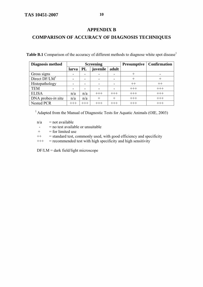
APPENDIX B
COMPARISON OF ACCURACY OF DIAGNOSIS TECHNIQUES
Table B.1 Comparison of the accuracy of different methods to diagnose white spot disease1
Screening Diagnosis method larva PL juvenile adult
Presumptive Confirmation
Gross signs - - - - + - Direct DF/LM1 - - - - + + Histopathology - - - - ++ ++ TEM - - - - +++ +++ ELISA n/a n/a +++ +++ +++ +++ DNA probes-in situ n/a n/a + + +++ +++ Nested PCR +++ +++ +++ +++ +++ +++
1 Adapted from the Manual of Diagnostic Tests for Aquatic Animals (OIE, 2003)
n/a = not available
- = no test available or unsuitable + = for limited use ++ = standard test, commonly used, with good efficiency and specificity
+++ = recommended test with high specificity and high sensitivity
DF/LM = dark field/light microscope

TAS 10451-2007
11
APPENDIX C
SAMPLING PLAN
Table C1. Sampling guide1
Number of samples per
estimated disease prevalence Lot sizes
(number of shrimp)1% 2% 5% 10%
50 50 50 35 20 100 96 75 45 23 250 194 110 50 25 500 225 130 55 26
1,000 258 140 55 27 1,500 271 140 55 27 2,000 277 145 60 27 4,000 288 145 60 27 10,000 299 145 60 27
≥ 100,000 299 150 60 30
1 Adapted from the Manual of Diagnostic Tests for Aquatic Animals (OIE, 2003) Note: The sample size depends on the estimated prevalence of the disease and the lot size, or the total number of shrimp in the pond. 1) For PL shrimp the sample size shall be based on the estimated disease prevalence of 1% or 2%. Live shrimp shall be taken from 5 different spots around the pond. For testing broodstock shrimp, all of the shrimp shall be tested or the number of samples shall be based on the estimated prevalence of 5%. For juvenile shrimp, the estimated prevalence may be 2-10%. 2) Interpretation: for example, if the estimate prevalence is 2%, - Positive diagnosis means 2% or more of the samples are infected
- Negative diagnosis means less than 2% of the samples are infected or none of them are infected.
TAS 10451-2007 12
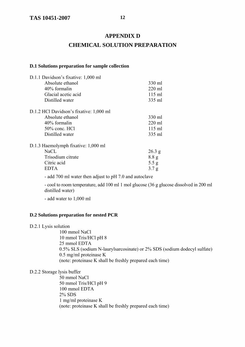
APPENDIX D
CHEMICAL SOLUTION PREPARATION
D.1 Solutions preparation for sample collection D.1.1 Davidson’s fixative: 1,000 ml Absolute ethanol 330 ml 40% formalin 220 ml Glacial acetic acid 115 ml Distilled water 335 ml D.1.2 HCl Davidson’s fixative: 1,000 ml Absolute ethanol 330 ml 40% formalin 220 ml 50% conc. HCl 115 ml Distilled water 335 ml D.1.3 Haemolymph fixative: 1,000 ml NaCL 26.3 g Trisodium citrate 8.8 g Citric acid 5.5 g EDTA 3.7 g
- add 700 ml water then adjust to pH 7.0 and autoclave
- cool to room temperature, add 100 ml 1 mol glucose (36 g glucose dissolved in 200 ml distilled water)
- add water to 1,000 ml
D.2 Solutions preparation for nested PCR D.2.1 Lysis solution 100 mmol NaCl 10 mmol Tris/HCl pH 8 25 mmol EDTA 0.5% SLS (sodium N-laurylsarcosinate) or 2% SDS (sodium dodecyl sulfate) 0.5 mg/ml proteinase K (note: proteinase K shall be freshly prepared each time) D.2.2 Storage lysis buffer 50 mmol NaCl 50 mmol Tris/HCl pH 9 100 mmol EDTA 2% SDS 1 mg/ml proteinase K (note: proteinase K shall be freshly prepared each time)

TAS 10451-2007
13
APPENDIX E
WHITE SPOT DISEASE DIAGNOSIS ILLUSTRATIONS (a) (b) Figure E.1 Clinical signs of white spot disease in black tiger shrimp. The shrimp body is
reddish with white spots on the cephalothorax (a) and body (b). (by courtesy of Dr. Jiraporn Kasornchandra)
Figure E.2 Method for collecting haemolymph from the ventral sinus or the cardiac sinus (arrow).
(by courtesy of Assoc. Prof. Janenuj Wongtavatchai, DVM).

TAS 10451-2007 14
Figure E.3 Histopathological characteristics of white spot disease: normal nucleus (white
arrows); infected cells will have hypertrophied nuclei, which are stained red in the first stage, called Cowdry type A inclusions (red arrow); in the later stages they stain dark blue (black arrows) (H&E stain). (by courtesy of Prof. Timothy W. Flegel)
Figure E.4 Histopathological characteristics of white spot disease in gill tissue after rapid
staining: in the early stages the nuclei of infected cells become hypertrophied and stain red; this is called a Cowdry type A inclusion; in the later stages the nuclei are basophilic and stain dark blue. (by courtesy of Prof. Timothy W. Flegel)
TAS 10451-2007
15
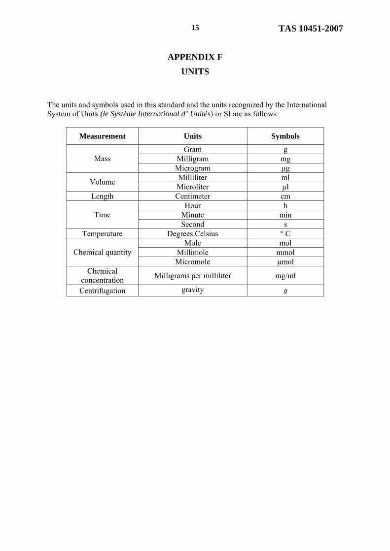
APPENDIX F
UNITS
The units and symbols used in this standard and the units recognized by the International System of Units (le Système International d’ Unités) or SI are as follows:
Measurement Units Symbols
Gram g Milligram mg Mass Microgram µg Milliliter ml
Volume Microliter µl
Length Centimeter cm Hour h
Minute min Time Second s
Temperature Degrees Celsius ° C Mole mol
Millimole mmol Chemical quantity Micromole µmol
Chemical concentration
Milligrams per milliliter mg/ml
Centrifugation gravity g