diagnosis and management of pulmonary embolism a · pdf filepulmonary embolism a focus on...
TRANSCRIPT

Diagnosis and Management of Pulmonary Embolism
A Focus on Catheter Directed Thrombolysis and Pulmonary Embolism Response Team
Saurabh Malhotra, MD MPH FACC FASNCAssistant Professor of Medicine (Cardiology) and Nuclear Medicine
Jacobs School of Medicine and Biomedical Sciences

DISCLOSURES
Research Support
• Becker Fund for Heart Research, Community Foundation of Greater Buffalo
• Intersocietal Accreditation Commission (IAC)
Application Reviewer• Intersocietal Accreditation Commission (IAC)

53 y/o female with HTN presents to ED with worsening dyspnea and chest pain
Diaphoretic and severely short of breath
HR 120s, SBP 100s
Hypoxemic correcting with O2 via NC
Elevated Tn and BNP
Clinical Case

Chest CTA Shows Large Thrombus Burden

Right Ventricular Dysfunction on Echocardiography

6
Undefiled, Hyperdynamic Left Ventricle

– Anticoagulation alone
– IV thrombolysis
– Catheter directed treatment
– Surgical thromboembolectomy
7
Clinical CasePatient with large pulmonary embolism with tachycardia, hypotension and RV dysfunction.

Pulmonary EmbolismAnnual incidence
• United States: 69 per 100,000/year • Over 600,000 cases annually• 1-2 PE episodes per 1000 people, up to 10 per 1000 in
the elderly population
Venous thromboembolism• PE commonly originates from lower limb deep vein
thrombosis (DVT) • 79% of patients presenting with PE have evidence of
DVT • PE occurs in up to 50% of patients with proximal DVT
1. Silverstein et al. Arch intern Med 19982. Wood et al. Chest 20023. Tapson. N Engl J Med 20084. Geering et al. CMAJ 2012
5. Chunilal et al. JAMA 20036. Siccama et al. Ageing Res Rev 2011

Pulmonary EmbolismA Silent and Fatal Epidemic
• PE causes or contributes to 15% of all hospital deaths1,2
• More people die each year from PE than highway fatalities, breast cancer and AIDS combined3
Cause of Death # of deaths/yrPE4,5 Up to 200,000Highway fatalities6 42,116Breast Cancer7 40,200AIDS8 14,499
1.Kasper et al. J Am Coll Cardiol. 1997;30:1165-11712.According to http://www.sirweb.org/patients/deep-vein-thrombosis/3.Goldhaber. Deep-vein thrombosis: Advancing awareness to protect patient lives. American Public Health Association White
Paper. 2003.4.Anderson et al. Arch Intern Med. 1991;151:933-938.5.Silverstein et al. Arch Internal Med. 1998;158:585-593.6.National Highway and Traffic Safety Association. Fatality Analysis Reporting System (FARS) Web-Based Encyclopedia.
Accessed January 31, 2002.7.American Cancer Society. Breast cancer facts and figures, 2001-2002. Accessed January 31, 2002.8.Centers for Disease Control Report. HIV/AIDS Surveillance Report 2001. Volume 13, Number 2.

Acute and Long Term Management of PE
Konstantinides, JACC, 2016

Massive PE [High risk]5% PE population
58% mortality @ 3 monthsMinor PE [Low risk]55% PE population
Good prognosisLow mortality rate
Submassive PE [Moderate / Intermediate risk]
40% PE population21% mortality @ 3 months
Goldhaber et al, Lancet, 1999
More than 1/3rd of the Pulmonary Embolism are Submassive: ICOPER

Patient risk stratification (per AHA Scientific Statement 2011)Massive PE Submassive PE Minor/Nonmassive PE
High risk Moderate/intermediate risk Low risk
– Sustained hypotension (systolic BP <90 mmHg for 15 min)
– Inotropic support– Pulselessness– Persistent profound bradycardia (HR
<40 bpm with signs or symptoms of shock)
– Systemically normotensive (systolic BP 90 mmHg)
– RV dysfunction– Myocardial necrosis
– Systemically normotensive (systolic BP 90 mmHg)
– No RV dysfunction– No myocardial necrosis
RV dysfunction– RV/LV ratio > 0.9 or RV systolic dysfunction
on echo– RV/LV ratio > 0.9 on CT– Elevation of BNP (>90 pg/mL)– Elevation of NTpro-BNP (>500 pg/mL)– ECG changes:
– new complete or incomplete RBBB– anteroseptal ST elevation or depression– anteroseptal T-wave inversion
Evidence of Right Ventricular Dysfunction Needed for Submassive PE
Jaff et al, Circulation, 2011
Quiroz, Circulation, 2004

13Am J Respir Crit Care Med. 2005; 172: 1041-6. J Thrombosis Thrombolysis. 2016; 41: 608-12.
Pulmonary Embolism Severity Index (PESI)

Risk Stratification in PE
Adapted from ESC 2014 Guidelines: Diagnosis and Management of PE

Contemporary Therapy for PE
ANTICOAGULATION (AC) – HEPARIN • AC therapy prevents further clot growth• LMWH as effective as UFH in reducing recurrent PE• LMWH carries reduced bleeding risk compared to UFH
STANDARD OF CARE: usually UFH or LMWH, followed by oral warfarin
• However, AC therapy relies on endogenous t-PA to dissolve occluding clot• a process that typically occurs over several weeks or months• endogenous fibrinolysis may often be incomplete at the end
Simonneau et al. NEJM, 1997Buller et al. NEJM, 2003 Meyer et al. Thromb Heamost 1995Arcasoy et al. Clin Chest Med 2003

Heparin + LyticHeparin‐only
Greater Clinical Improvement with LyticsCompared to Heparin Alone for Submassive PE
Kline et al., Chest, 2009
N=144(39)
N=18(0)

Advanced Treatment of Pulmonary Embolism
Reperfusion Therapies Hemodynamic Support

Surgical Thromboembolectomy

Right ventricular dysfunction (RVD) is a predictor of poor clinical outcomes:
1. Mortality2. Adverse events3. VTE recurrence
19
Intermediate Risk PE Needs Aggressive Therapy

Right Ventricular Dilatation Predicts Hospital Mortality
Fremont et al., Chest, 2008
- 950 patients from ICOPER.- All had RV/LV ratios on echocardiography.
Mortality rate: - 1.9% if RV/LV ratio < 0.9- 6.6% if RV/LV ratio ≥ 0.9

Hypokinetic Right Ventricle Predicts Short-term Mortality
Goldhaber et al., Chest, 2008
- 2454 patients from ICOPER (52 hospitals in 7 countries).- All-cause mortality:
- 11% in first 2 weeks- 17% at 3 months Mortality rate at 3 months:
21% with hypokinesis
15% with no hypokinesis

Persistence of Right Ventricular DysfunctionPredicts Mortality
• PE patients with RVD unresolved exhibit 2x increased incidence of mortality compared to those with RVD resolved at discharge
− Retrospective analysis of 301 patients with first episode PE with mean f/u at 3.1 years
− Mortality rate at f/u: Persistent RVD: 24% (10% PE related deaths) Resolved RVD: 13% (0 PE related deaths)
Grifoni et al., Arch Int Med, 2006

Persistence of Right Ventricular DysfunctionPredicts Recurrent VTE
Incidence of VTE at 4 years:
0.4% with persistent RVD
0.05% with no RVD
PE patients with RVD unresolved exhibit 8x increased incidence of recurrent VTE compared to those with RVD resolved at discharge
Grifoni et al., Arch Int Med, 2006

Thrombolysis in Acute Pulmonary Embolism
REDUCE THROMBUS BURDEN (not achievable by AC alone)
• Reverse RV afterload / failure; prevention of hemodynamic collapse• Improve pulmonary reperfusion/capillary blood flow / gas exchange • Restore systemic arterial perfusion pressure• Decrease the risk of developing chronic pulmonary hypertension
Piazza and Goldhaber, Vascular Medicine, 2010

100 mg t-PA infused over 2 hours Indicated for management of acute massive PE
in adults: For the lysis of acute pulmonary emboli, defined as
obstruction of blood flow to a lobe or multiple segments of the lungs.
For the lysis of pulmonary emboli accompanied by unstable hemodynamics, e.g., failure to maintain blood pressure without supportive measures.
IV Thrombolysis with t-PA

Metaanalysis of randomized clinical trials for PE comparing thrombolytic therapy with heparin
Total of 11 trials, 748 patients included
Data from trials that included massive PE:
26
Wan et al., Circulation, 2004
Lower Risk of Recurrent PE or Death with Thrombolysis Compared with Heparin

PEITHO Trial (n=1005)Primary Objective:– Investigate clinical benefits (efficacy)
of thrombolysis with tenecteplaseover placebo in normotensive patients with acute intermediate‐risk PE (both treatment arms receive standard heparin anticoagulation)
Secondary Objective:– To assess the safety of tenecteplase in
patients with intermediate‐risk PE27
Comparison of Thrombolysis with Heparin in Intermediate-risk PE
Meyer et al., NEJM, 2014

28
IV Thrombolysis Reduces the Risk of Acute Hemodynamic Compromise (< 7 days)
Meyer et al., NEJM, 2014

29
Benefit from Thrombolysis was offset bya Greater Incidence of Acute Major Bleeds
Meyer et al., NEJM, 2014
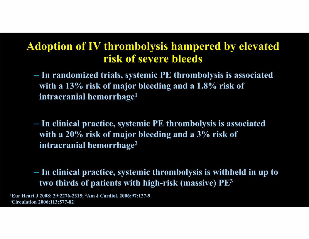
– In randomized trials, systemic PE thrombolysis is associated with a 13% risk of major bleeding and a 1.8% risk of intracranial hemorrhage1
– In clinical practice, systemic PE thrombolysis is associated with a 20% risk of major bleeding and a 3% risk of intracranial hemorrhage2
– In clinical practice, systemic thrombolysis is withheld in up to two thirds of patients with high-risk (massive) PE3
1Eur Heart J 2008: 29:2276-2315; 2Am J Cardiol. 2006;97:127-93Circulation 2006;113:577-82
30
Adoption of IV thrombolysis hampered by elevated risk of severe bleeds

“Safe Dose” IV Thrombolytics for Moderate PE
MOPETT Trial• Single center prospective trial of 120
patients• tPA dose: 50mg or 0.5 mg/kg if body
weight < 50 kg• RV dysfunction was not a pre-requisite
for inclusion• Primary endpoint: PHTN at 28 months
• Lytic: 16%• Heparin: 57%
• Bleeding: 0%• No difference in death or recurrent PE
Sharifi et al. AJC, 2013

Catheter Directed Thrombolysis (CDT)
Placement in the left and right pulmonary arteries for the treatment of bilateral PE

EkoSonic Endovascular System for CDT
Infusion Catheter
Ultrasonic Core
Central Coolant Lumen
Therapy Optimization Sensor
Drug Lumen
Guidewire or MSD (0.035” diameter)
Features 5.4 Fr catheter 106 and 135 cm working length 6, 12, 18, 24, 30, 40 and 50 cm treatment zones

EkoSonic Endovascular System for CDTMechanism of Action
How ultrasonic energy unlocks the clot Ultrasonic energy causes fibrin strands to
thin, exposing plasminogen receptor sites
Thrombus permeability and lytic penetration are dramatically increased
Ultrasound pressure waves force lytic agent deep into the clot and keep it there
WITH ULTRASOUNDENERGY
WITHOUT ULTRASOUNDENERGY
ULTRASOUND ENERGY& THROMBOLYTIC
Braatan et al. Thrmob Haemost 1997Francis et al. Ultrasound in Medicine and Biology, 1995Soltani et al. Physics in Medicine and Biology, 2008

RCT of EKOS vs Heparin in Intermediate Risk PEThe ULTIMA Trial
Kucher et al. Circulation, 2014
Primary Objective: Determine whether fixed low-dose catheter-directed ultrasound accelerated thrombolysis is superior to heparin alone in reversal of RV dilatation in submassive / intermediate risk PE

Greater Reduction in RV Dilatation with EKOS with tPA and Heparin
Kucher et al. Circulation, 2014

Greater Improvement in RV Systolic Functionfrom EKOS with t-PA and Heparin
Kucher et al. Circulation, 2014

Similar Safety Outcomes from EKOS with t-PA and Heparinvs. Heparin-alone
Kucher et al. Circulation, 2014

CDT with EKOS is Safe and EfficaciousSEATTLE II Study
Piazza et al., JACC Interventions, 2015

CDT with EKOS is Safe and EfficaciousSEATTLE II Study
Piazza et al., JACC Interventions, 2015

CDT with EKOS is Safe and EfficaciousSEATTLE II Study
N %
Total enrollment 150* 100%Massive / Submassive PE 31 / 119 21% / 79%History of previous DVT 30 20%History of previous PE 15 10%Concomitant use of antiplateletagents
51 34%
Unilateral / Bilateral PE 20 / 130 13% / 87%Total rtPA dose 23.7 ± 2.9 mg
Piazza et al., JACC Interventions, 2015

CDT with EKOS is Safe and EfficaciousSEATTLE II Study
Piazza et al., JACC Interventions, 2015

Reduced Risk of Intracranial Hemorrhage with CDT

Low Incidence of Complications with CDT
Engelberger and Kucher. Eur Heart J. 2014

Minimal Risk of Adverse Events from EKOS Registry• Single-center retrospective observational study • 60 consecutive patients with either massive or submassive PE• No intracranial hemorrhage, one intra-abdominal hemorrhage leading
to hypovolemic shock and death, and one puncture site hematoma
Treatment detailsBilateral PEUnilateral PE
N=53 (88%)N=7 (12%)
Massive PESubmassive PE
N=12 (20%)N=48 (80%)
Thrombus clearance:- Complete (>90%)- Near complete (50-90%)- Partial (<50%)
N=33 (57%)N=24 (41%)
N=1 (2%)Total rtPA doseTotal infusion time
35.1±1.1 mg19.6±6.0 hrs
OutcomesSurvival to dischargeICU stay (median)Hospital stay (median)
N=57 (95%)1 day9 days
90-day survival:- Overall- Submassive PE- Massive PE
N=56 (93%)N=47 (98%)N=9 (75%)
Adverse events:- Major bleeding- Minor bleeding- Cardiopulmonary arrest- Acute renal injury- Recurrent PE
N=1 (1.7%)N=1 (1.7%)N=1 (1.7%)N=1 (1.7%)N=0 (0%)
Kennedy et al. J Vasc Interv Radiol 2013

Better Outcomes but Higher Cost with CDTNationwide Comparison of CDT with Systemic Lysis
Large observational comparative analysis Lysis: 1521 patients CDT: 352 patients
Primary endpoint: in-hospital mortality
Secondary endpoint: in-hospital mortality and ICH
Patel et al. CCI 2015

– RV dysfunction in PE patients predicts poor outcomes:–Mortality–Adverse events–VTE recurrence
– Anticoagulant therapy does not actively resolve the existing thrombus
– IV thrombolysis is not used broadly:–Clinical data show improvement in hemodynamics, –but it carries an elevated risk of severe bleeding, including ICH
47
Summary

–CDT with EKOS for the treatment of massive/submassive pulmonary embolism–Loosens the fibrin structure –Increases drug penetration into the fibrin matrix–Ultimately reduces drug dose, treatment time and risk of complications
–Goals of CDT:–Restoration of hemodynamics as evidenced by a reduced RV/LV ratio and
decreased PA pressure–Resolution of pulmonary artery obstruction –Favorable outcomes with low dose thrombolysis (20-24 mg tPA)–No reports of intracranial hemorrhage in published clinical studies
48
Summary

53 y/o female with HTN presents to ED with worsening dyspnea and chest pain
Diaphoretic and severely short of breath
HR 120s, SBP 100s
Hypoxemic correcting with O2 via NC
Elevated Tn and BNP
Clinical Case

PA pressure:80/48 (56)
Pulmonary Angiogram

Pulmonary Angiogram

Echocardiograms14 hours after CDT

EchocardiogramsAt discharge: day 4

Echocardiograms6 weeks post discharge

–59 y/o female with ovarian cancer currently undergoing treatment at RPCI presents to the ED with shortness of breath
–Tachycardic, elevated D-dimer and cardiac biomarkers
–CTA: saddle pulmonary embolism
55
Clinical Case 2

Clinical Case 2What to do?

Clinical Case 2Surgical Thromboembolectomy

Buffalo General Medical Center/Gates Vascular InstitutePERT Program
• PERT: Collaborative multi-disciplinary team to assist primary providers in the evaluation and management of patients with intermediate and high risk pulmonary embolism
• National PERT Consortium:• Advance the science of PE care by performing research• Develop advanced treatment protocols• Educating clinicians and community members
- Interventional cardiologists
- Imaging cardiologists
- Pulmonary and Critical Care
- Fellows
- Sonographers

Buffalo General Medical Center/Gates Vascular InstitutePERT Algorithm

60
Buffalo General Medical Center/Gates Vascular InstitutePERT Algorithm

61
Buffalo General Medical Center/Gates Vascular InstitutePERT Algorithm