diagnosis and management of acute pancreatitis
TRANSCRIPT
DIAGNOSIS AND MANAGEMENT
OF
ACUTE PANCREATITIS
By
MARY HOERNER
A manuscript submitted in partial fulfillment of The requirements for the degree of
MASTER OF NURSING
WASHINGTON STATE UNIVERSITY College ofNursing
Intercollegiate Center for Nursing Education
June 1999
To the Faculty ofWashington State University:
The members ofthe committee appointed to examine the ICNE research
requirements and manuscript ofMary J. Hoerner find it satisfactory and recommend that
it be accepted.

2
TABLE OF CONTENTS
~CICNO~EI)<J~NTS 2
ABSTRACT 3
LIST OF TABLES 4
LIST OF FI<JURES 5
MANUSCRIPT 6
TABLES 16
REFERENCE LIST 23

3
ACKNOWLEDGMENTS
At the beginning, the destiny of the journey was unknown. At the conclusion, the
path was well traveled, but the destiny is still unknown. My endeavor to become a Nurse
Practitioner has finally ended, but my travels into the future are just beginning. This was
not a singular venture but was rather the joint effort of multiple people. The commitment
could never have been completed without the support, assistance and love from my
husband Ron and my daughter Nicole. The knowledge and expertise from Dr. Lorna
Schumann, whose steps I hope to follow. And the fun, laughs and encouragement from
Kim Tucker, who talked me into taking this long, but fulfilling journey.
4
ABSTRACT
Much controversy exists regarding treatment for the obscure pathogenesis of
acute pancreatitis. Many patients will recover spontaneously regardless of the cause or
the treatment, while others experience a fulminent course with multi-organ failure and
resultant death. The merits ofvarious tests have been explored and reviewed looking at
sensitivity, specificity and cost, as related to the clinical setting. Patient care providers
will be increasingly called upon to treat and appropriately refer patients with pancreatitis
and its complications. Understanding treatment options and keeping current with
evolving guidelines is imperative for the management of these patients.
5
LIST OF TABLES�
Table 1: Causes of acute pancreatitis......� 14�
Table 2: Ranson's 11 early objective signs of acute pancreatitis............� 15�
Table 3: Specifics of laboratory and diagnostic studies for acute pancreatitis 16�
Table 4: Complications associated with acute pancreatitis..................... 18�
6
LIST OF FIGURES
Figure 1: Pathophysiological mechanisms of acute pancreatitis. . . . . . . . . .. . . . . 18�
Figure 2: Management for acute pancreatitis treatment......................... 19�
7
Introduction
Acute pancreatitis follows a precarious pathway, from a mild and self-limiting
episode, to complete multi-organ failure with fatality. This sudden onset disease process
stems from a multitude of causes and has an enigmatic pathogenesis. Most patients with
acute pancreatitis recover rapidly and completely, regardless of the cause or the
treatment, but in a small percentage of patients the disease takes a fulminate course. Of
these patients, severe attacks may result in shock, respiratory failure, renal failure and
death (Marshall, 1993 pp.1185). Treatment is dependent upon the cause of the symptoms,
but in most cases is largely supportive in nature. "Despite recent advances in diagnosis
and treatment, acute pancreatitis continues to be a serious illness with an overall mortality
of5-10%" (Banks, 1997, p.377).
Acute pancreatitis is a common disorder and seems to be on the rise with certain
disease processes such as acquired immunodeficiency syndrome (AIDS). In the United
States, gallstones account for about 45% of the cases ofobstructive pancreatitis. Alcohol
is the second leading cause of pancreatitis, accounting for approximately 35% of the
cases (Steinberg and Tenner, 1994). The frequency of these two causes varies,
dependent upon the patient population and the clinicians referral population. Many other
causes of acute pancreatitis have been identified as well, but only account for
approximately 10% of the populations causes (See table 1 for causes)
Acute pancreatitis is best defined as an acute inflammatory process of the
pancreas that may also involve peripancreatic tissues and/or remote organ systems
(Banks, 1997). Although there are many theories on the pathogenesis of pancreatitis, it is

8
well known that pancreatitis stems from an auto-digestive process within the pancreas
itself
The pancreas has been described as a fish shaped organ that lies behind the
stomach with important endocrine and exocrine roles. The exocrine pancreas is
constructed with lobules that consist of acinar cells, all ofwhich secrete digestive
enzymes into a system of microscopic ducts. These ducts are terminal branches of larger
ducts that drain into the main pancreatic duct. In most people, the main pancreatic duct
(the duct ofWirsung) transports the pancreatic juices, and eventually joins the common
bile duct from the liver and the gallbladder, to release the enzymes where the common
channel enters the duodenum. The pancreas is responsible for secreting amylolytic
enzymes (amylase), lipolytic enzymes (lipase, phospholipase ~ and cholesterol
esterace), and proteolytic enzymes (trypsin, chymotrypsin). These enzymes assist in the
breakdown of starch, fats, and proteins (Harrisons, 1998). Under normal circumstances,
these enzymes are secreted in their inactive form and become activated once they enter
the intestine (Porth, 1994, p.868). The autodigestion theory suggests that these enzymes
are activated within the pancreas rather than within the intestinal lumen (Harrisons,
1998). Figure 1 presents the pathophysiological mechanisms of acute pancreatitis.
The activation of trypsin to trypsinogen within the pancreas, is thought to be the
initiating event in acute pancreatitis. Why trypsin is activated remains unanswered.
Endogenously secreted trypsin inhibitor ordinarily prevents trypsin from being activated
inside of the pancreas. "Although trypsin is a proteolytic enzyme in it's own right, it has
been found responsible for the activation of many other enzymes and bioactive
substances that appear to cause most of the pancreatic and systemic abnormalities that
9
characterize acute pancreatitis" (Marshall, 1993, p.1188). Trypsin is also known to have
effects on the cardiovascular and coagulation systems. Trypsin converts the peptide
kallikrein to bradykinin, which causes vasodilation, increased vascular permeability, and
leukocyte accumulation. Elastase, once activated, dissolves the elastic fibers of blood
vessels, causing necrosis and erosion and potential for hemorrhage to occur. This may
also lead to thrombosis and hypoperfusion, potentially causing ischemia. Early
activation of lipase may cause pancreatic parenchymal necrosis (Marshall, 1993).
"The damage to pancreatic cells and blood vessels that these enzymes
and substances cause can result in tissue hypoxia and further cellular
necrosis, resulting in a vicious circle in which more pancreatic enzymes
are released and more pancreatic injury occurs" (Marshall. 1993,
pp.1188).
Regardless of the cause, the sum effect of the early activation of these enzymes is
auto-digestion of the pancreas leading to pancreatic parenchymal destruction.
Clinical Presentation
Diagnosis of acute pancreatitis is made on the basis of the clinical presentation
combined with the results of lab and radiographic findings (Lillemoe & Yea, 1998). The
presentation can range from a mild, difficult to diagnose state, to a morbid condition
associated with hypovolemia, sepsis, shock, metabolic changes, and death.
Abdominal pain, localized to the epigastrium, is a common finding of acute
pancreatitis. The degree of pain is dependent upon the amount of pancreatic
involvement. It is not unusual for this pain to radiate to various parts of the body, spine,
flank, left shoulder, but primarily the back. History may help to reveal the obvious
10
causes of onset, such as the use of alcohol. For most, onset of acute pancreatitis presents
as gradually increasing epigastric pain, reaching maximal intensity within 30-60 minutes
which may persist for hours to days without relief This abdominal pain is frequently
described as unbearable, worsens in the supine position, and palpation of the abdomen
elicits guarding (Banks, 1997).
Nausea and vomiting to the point of persistent retching, is a common sequela of
acute pancreatitis. Other symptoms may be present, but are not specific for this disease
process. Fever greater than 101 degrees, tachycardia, and abdominal distention resulting
from a paralytic ileus may occur. In patients with severe pancreatitis, hypotension,
hypovolemia, and hypoperfusion may be present (Lillemoe &Yea, 1998). If
hemorrhagic pancreatitis occurs, a bluish discoloration in the left flank area (Grey
Turner's Sign) or in the periumbilical region (Cullen's Sign) may be noted. These two
signs would indicate increased severity of acute pancreatitis, and increases the mortality
significantly. Although unusual, jaundice might be present indicating common bile duct
obstruction, or some form ofgallstone associated pancreatitis (Lillemoe&Yea, 1998).
The majority of patients with acute pancreatitis require conservative therapy.
Ranson's Criteria, the most widely used multiple clinical criteria system, is used to help
assess and identify those patients with severe signs and symptoms so that more
aggressive treatment and surveillance can prevent complications (Marshall, 1993).
To help classify uncomplicated versus complicated pancreatitis, Ranson uses 11
early objective signs to classify the severity of pancreatitis. (See table 2)

11
Studies have shown that there is a direct relationship between the number of
prognostic signs and the risk of death and/or complication requiring more than one week
of an ICU setting for care (Ranson, 1995).
Laboratory and Diagnostic Testing
Mild cases of pancreatitis represent a diagnostic challenge as the clinical
symptoms may be non-specific and lab values may only be mildly elevated. In patients
with moderate to severe pancreatitis, diagnosis may be straightforward and easy to
determine by the clinical history, elevated lab values of amylase or lipase, and by a CT
scan of the pancreas.
Serum amylase levels have remained central to diagnosis, because they are readily
available, quick and simple to perform, and have a relatively low cost. Levels greater
than 65 lUlL should make one suspicious of pancreatic injury. Levels greater than 130
lUlL make the diagnosis most likely, and greater than three times normal is diagnostic
(Harrison, 1998). Persistent elevation of amylase levels may be indicative of pancreatic
necrosis, pseudocyst or abscess formation (Lillemoe & Yeo, 1998).
Serum lipase levels tend to stay elevated longer than serum amylase levels and are
useful in patients who are not examined until several days after the onset of symptoms.
Serum lipase levels will parallel amylase levels and the value of doing both tests will
enhance diagnostic abilities (Harrison, 1998).
Leukocytosis, hyperglycemia, hypocalcemia, hypertryglyceridemia, are other
common abnormal lab results that may be noted, but are not specific for the diagnosis of
12
pancreatitis. However, these tests may help diagnose or narrow down the cause of
pancreatitis.
Advanced imaging techniques, especially the use ofCT scanning has dramatically
improved the ease of diagnosing acute pancreatitis. The pancreas has notoriously been
the hidden organ for most radiographic studies, but CT scanning has provided a means of
visualizing the pancreas to help note pancreatic inflammation (Agarwal, Pitchumoni,
Sivaprasad, 1990). Sequential, contrast-enhanced CT, allows detection of pancreatic
necrosis, which becomes important for assessing the severity of the disease (Marshall,
1993). A slight enlargement of the gland may demonstrate mild pancreatitis. Moderate
to severe cases of pancreatitis will show evidence of pancreatic necrosis and
peripancreatic inflammatory changes, as well as fluid collection (Marshall, 1993).
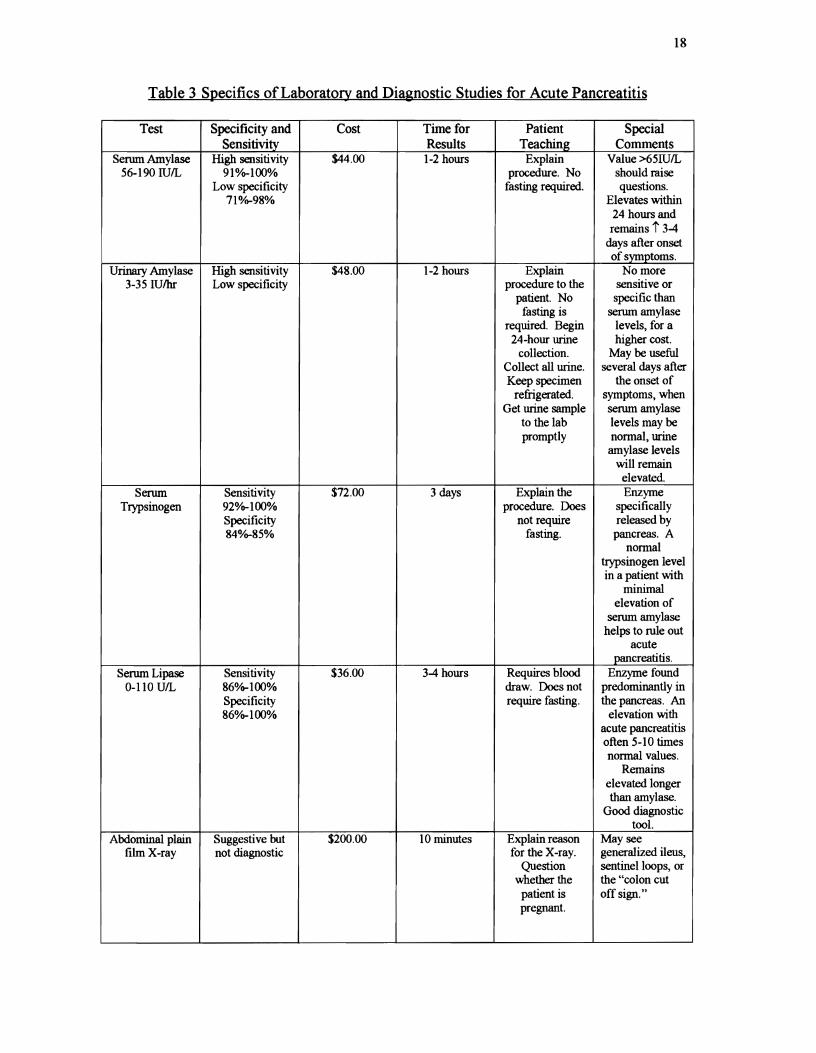
Table 3 lists the specifics of the most commonly ordered tests used in the
diagnosis of acute pancreatitis. Routine lab studies may be helpful but are not diagnostic
in themselves. A thorough clinical history and physical, amylase and lipase levels, and
CT scanning provide the best diagnostic tools for the diagnosis of acute pancreatitis.
(See table 3 for specifics of laboratory and diagnostic studies for acute pancreatitis.)

13
Treatment:
There are four primary therapeutic objectives to consider when treating acute
pancreatitis. These are:
1.) To limit the severity of pancreatic inflammation.
2.) To carefully observe and prevent complications by interruption of their pathogenesis.
3.) To support the patient and treat complications aggressively as they arise.
4.) To identify the cause of pancreatitis and eliminate the causative factor, if treatable
(Ranson, 1995; Marshall, 1993).
Ranson's 11 early objective signs provide valuable information regarding the
severity of the disease process. Ranson's criteria provides data on admission criteria,
and 48 hours post-admission to detect systemic changes, and to provide early warning
that an episode may become severe (Banks, 1997).
Treatment for acute pancreatitis is supportive. Figure 2 presents the decision
making algorithm for the management of acute pancreatitis. If mild pancreatitis is
diagnosed, as defined by the absence of organ dysfunction, elimination of oral intake,
intravenous hydration, and analgesia may be all that is called for. In most cases, the
disease will subside spontaneously within 3-7 days after treatment is initiated
(Harrisons, 1997). Oral intake is discontinued until there is almost complete resolution
of abdominal pain. Nasogastric suction is often instituted to "rest" the pancreas by
decreasing duodenal enzyme release, but studies have shown that NG tubes do not
decrease pain nor shorten hospital stays (Steinberg et aI, 1994). Nasogastric tubes are
appropriate for vomiting patients, or those who may present with an ileus.

14
Pharmacological management has been suggested to help shorten the course of
acute pancreatitis. The use ofanticholinergic drugs have been recommended for the
suppression ofgastric and pancreatic secretions. H2 blockers are used to decrease
gastric acid secretion. Glucagon and Somatostatin are believed to cause hormonal
suppression of pancreatic secretions. Prophylactic antibiotics are used for prevention of
infectious complications. Studies have shown that pharmacological management is
controversial and the therapeutic benefits are questionable at best (Marshall, 1993).
Severe pancreatitis, as defined by the presence of organ failure, requires close
observation and follow-up in a specialized unit. Danger signals on presentation, such as
hypotension, oliguria, hypoxemia, or evidence of third space losses guide the provider's
decision regarding admission to ICU (Merck, 1997). Endoscopic retrograde
cholangiopancreatography (ERCP) with sphincterotomy is recommended with patients
who have gallstone induced pancreatitis, within 2-3 days after the onset. A
computerized tomography (CT) scan may be performed, if a surgical condition can not
be excluded. A contrast ermanced CT is useful for distinguishing between interstitial
and necrotizing pancreatitis and may help distinguish between different options for
treatment (Banks, 1997).
If the patient is diagnosed with having severe necrotizing pancreatitis, as defined
as "one or more areas of non-viable pancreatic parenchyma," guided percutaneous
aspiration should be performed. This procedure will help to differentiate between
infected versus sterile necrosis. Sterile necrosis can continue to be treated medically, but
infected necrosis may require surgical debridement after 4-6 weeks (Banks, 1997).
15
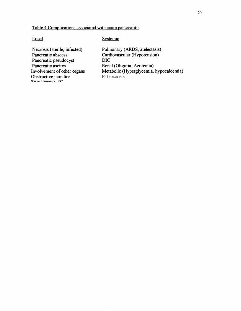
There are many local and systemic complications that can occur as sequela of acute
pancreatitis. Table 4 lists some possible complications. (See table 4)
Summary Many aspects of acute pancreatitis treatment have been controversial, but with the
refinement of contrast enhanced computed tomography, advances have been made
towards quicker diagnosis and treatment ofacute pancreatitis. The management of this
disease process continues to be supportive, with continual observation for complications,
and determination of cause so those future occurrences may be prevented
(Marshall, 1993).
16
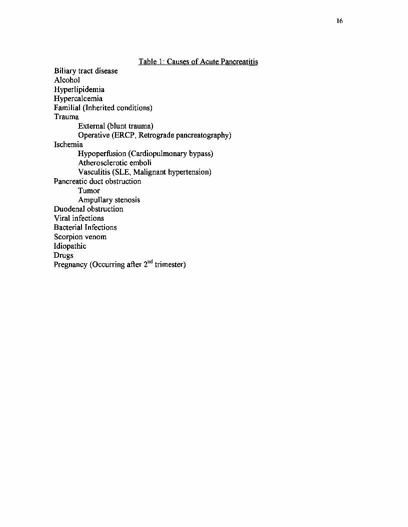
Table 1: Causes of Acute Pancreatitis Biliary tract disease Alcohol Hyperlipidemia Hypercalcemia Familial (Inherited conditions) Trauma
External (blunt trauma) Operative (ERCP, Retrograde pancreatography)
Ischemia Hypoperfusion (Cardiopulmonary bypass) Atherosclerotic emboli Vasculitis (SLE, Malignant hypertension)
Pancreatic duct obstruction Tumor Ampullary stenosis
Duodenal obstruction Viral infections Bacterial Infections Scorpion venom Idiopathic Drugs Pregnancy (Occurring after 2nd trimester)

17
Table 2 Ranson's 11 Early Objective Signs of Acute Pancreatitis At admission or diagnosis
Age >55yr WBC count>16,000/mm3 Glucose >200mg/dl Lactic Dehydrogenase >350 lUlL SOOT >250u/L
During initial 48 hours of hospitalization Hematocrit decrease >10% BUN rise >5mg/dl Serum Ca++ <8.0mg/dl Arterial oxygen pressure <60mm Hg Base deficit> 4 mEq/L Estimated fluid sequestration >6 Liters
2 or fewer signs: 1% mortality 3 signs: severe pancreatitis 3-4 signs: 15% mortality 5-6 signs: 40% mortality More than 6 signs: 100% mortality
18
Table 3 Specifics ofLaboratory and Diagnostic Studies for Acute Pancreatitis
Test Specificity and Cost Time for Patient Special Sensitivity Results Teaching Comments
Serum Amylase High sensitivity $44.00 1-2 hours Explain Value >65IUIL 56-190 lUlL 910/0-100% procedure. No should mise
Low specificity fasting required. questions. 710/0-98% Elevates within
24 hours and remains t 3-4
days after onset of symptoms.
Urinary Amylase High sensitivity $48.00 1-2 hours Explain No more 3-35 IU/hr Low specificity procedure to the sensitive or
patient. No specific than fasting is serum amylase
required. Begin levels, for a 24-hour urine higher cost.
collection. May be useful Collect all urine. several days after Keep specimen the onset of
refrigerated. symptoms, when Get urine sample serum amylase
to the lab levels may be promptly normal, urine
amylase levels will remain
elevated.
Serum Sensitivity $72.00 3 days Explain the Enzyme Trypsinogen 920/0-100% procedure. Does specifically
Specificity not require released by 840/0-85% fasting. pancreas. A
normal trypsinogen level in a patient with
minimal elevation of
serum amylase helps to rule out
acute pancreatitis.
Serum Lipase Sensitivity $36.00 3-4 hours Requires blood Enzyme found 0-110 UIL 860/0-100% draw. Does not predominantly in
Specificity require fasting. the pancreas. An 860/0-100% elevation with
acute pancreatitis often 5-10 times normal values.
Remains elevated longer than amylase.
Good diagnostic tool.
Abdominal plain Suggestive but $200.00 10 minutes Explain reason May see film X-ray not diagnostic for the X-ray. generalized ileus,
Question sentinel loops, or whether the the "colon cut
patient is off sign." pregnant.

19
UltrasoWld of the Low sensitivity 5350.00 30 minutes Helps to confmn pancreas 620/0-95% pancreatitis
High specificity associated with 98% gallstones. Can
provide infonnation on
edema, inflanunation,
pseudocyst, and mass lesions.
Contrast Sensitivity 85% 5600.00+ 30-60 minutes Explain Very diagnostic enhancedCT Specificity 10OOA» procedure. for determining
scan Patient must lie the severity of still. Ask about pancreatitis and iodine allergies. the extent of
Patient should be pancreatic NPO 4-8 hours necrosis that may
pre-test. be present. Most Encourage sensitive non-
patient to drink invasive imaging fluids post-test. method.
ERCP 5450.00 1-2 hours Explain the Risk associated Endoscopic depending on procedure. with perfonning Retrograde ease Patient must be theERCP Cholangio- NPO. Do not eat includes the
pancreatography or drink Wltil gag possible reflex has exacerbation of returned. an already
inflamed pancreas. Helps
diagnose gallstones.
MRI Highly specific 51100.00 1-2 hours Explain the Provides Magnetic but CT scan just procedure to the infonnation
Resonance as good patient. Assure similar to a CT Imaging there is no metal scan but is much
being worn. more costly and Patient should offers no real
remain advantage. motionless as
possible. MRCP 51100.00+ 1-2 hours Relatively new
Magnetic diagnostic Resonance ability. Cholangio
pancreatography
Sources: Harrison's, 1998, Agarwal, 1989, Greene, 1996,� Cost: Obtained from Kennewick General Hospital, Kennewick, Washington and are only the cost for the test. They do not include� miscellaneous charges (physician, contrast,etc ... ) Timelines differ from facility to facility.�
20
Table 4 Complications associated with acute pancreatitis
Necrosis (sterile, infected) Pancreatic abscess Pancreatic pseudocyst Pancreatic ascites Involvement of other organs Obstructive jaundice Source: Hamson's, 1997
Systemic�
Pulmonary (ARDS, atelectasis)� Cardiovascular (Hypotension)� DIC� Renal (Oliguria, Azotemia)� Metabolic (Hyperglycemia, hypocalcemia)� Fat necrosis�

21
Figure 1 Pathophysiological Mechanisms of Acute Pancreatitis
Causative factors
I Pancreatic Acinar Cell Damage I
Trypsin Activation
Vasodialation Dissolves elastic t Vascular penneability fibers of blood Leukocyte accwnulation vfI!S:S:fI!ls:
Edema Edema Vascular damage Inflammation Vascular Hemorrhage
Damage
Source: Arch Intern Med/Vol. 153, May 24, 1993, Marshall, John M.D.

22�
Figure 2 Management for Acute Pancreatitis Treatment
History-previous episodes, ETOH� abuse�
Abrupt onset of deep epigastric pain� with radiation to back�
NN, diaphoresis, weakness� Abdominal distention�
Leukocytosis, tamylase, tlipase�
Assess severity using Ranson's 11� prognostic signs�
Mild� Absence of� Perform ultrasound if Severe
suspicious of gallstone Presence oforgan� dysfunction� induced pancreatitis organ
dysfunction
Supportive therapy:� NPO� Supportive Therapy as for mild, plus:
Analgesia (Demerol) Contrast enhanced cr scan,�
IV fluid and electrolyte replacement� daily CMP panel, with a daily chest
NG tube if indicated X-rav�
Q 24 hr amylase, lipase, cac�
Necrotizing Necrotizing with clinical without clinical improvement improvement
Yes No Continue medical Guided therapy. Anticipate percutaneous
potential aspiration complications
~iet when abdominal Contrast enhanced tlPain decreased and CTscan for 'mylase, lipase levels further :;ontinue to decrease management
Determine organism Treat medically consider surgery
( Source: Harrison's, 1997)
23
Reference List
Agarwal, N., Pitchumoni,C.S. & Sivaprasad, A.V. (1990) Evaluating Test for
Acute Pancreatitis. The American Journal of Gastroenterology, Vol. 85, 356-365.
Banks, P. (1997) Practice Guidelines in Acute Pancreatitis. The American
Journal of Gastroenterology, 92(3) 377-386.
Duncan, F., Amorosino, C. (1985) The treatment of acute pancreatitis: A
continuing challenge. The New England Journal ofMedicine , Vo1.312, 436-438.
Lillemoe, K., Yeo, C., (1998) Management of the complications of pancreatitis,
Current Problems in Surgery, 35,(1 ) 3-98.
Marshall, J. (1993) Acute Pancreatitis: A Review with and Emphasis on New
Developments. Arch Internal Medicine, 153, 1185-1198.
Noone, J. (1995) Acute Pancreatitis: An Orem Approach to Nursing Assessment
and Care. Critical Care Nurse, 27-37.
Pagana, K.D., Pagana, T.J., (1998) Mosby's Manual ofDiagnostic and
Laboratory Test, St.Louis: Mosby.
Porth, C. (1994) Pathophysiology Concepts of Altered Health Status, 4th Edition,
J. B. _Lippincott Co.
Ranson, J. H.C., (1995) The Current Management ofAcute Pancreatitis,
Advances in Surgery, 28, 93-111.
Smith, A. (1991) When the Pancreas Self Destructs, American Journal of
Nursing, September, 38-52.

24
Smolen, D. (1997) Nursing Care of Clients with Biliary Tract and Exocrine
Pancreatic Disorders in Black,J., Matassairin-Jacobs, E. Medical Surgical Nursing (5th
Edition), 1907-1933.
Steinberg, W., Tenner,S. (1994) Acute Pancreatitis, The New England Journal of
Medicine, 330, 1198-1210.
Toskes, P.P., Greenberger, N.J., (1998) Approach to the Patient With Pancreatic
Disease. In A. Fauci, E. Braunwald, K.Isselbacher, J. Wilson, J. Martin, D. Kasper, S
Hauser, D. Lango, Harrisons Principle of Intemal Medicine, (14th Edition), 1741-1748.
NewYork:McGraw-Hill.